Prophylactic Anticonvulsants in Intracerebral Hemorrhage
22
MACKEY 1 Prophylactic Anticonvulsants in Intracerebral Hemorrhage Jason Mackey, MD MS 1,2 Ashley D. Blatsioris, MPA 1 Elizabeth A.S. Moser, MS 3 Ravan J.L. Carter, BS 2 Chandan Saha, PhD 3 Alec Stevenson, BS 1 Abigail L. Hulin, BSN 1 Darren P. O’Neill, MD 4 Aaron A. Cohen-Gadol, MD 5 Thomas J. Leipzig, MD 5 Linda S. Williams, MD 1,2,6 1 Department of Neurology, Indiana University School of Medicine, Indianapolis, IN 2 Regenstrief Institute, Indianapolis, IN 3 Department of Biostatistics, Indiana University School of Medicine, Indianapolis, IN 4 Department of Radiology, Indiana University School of Medicine, Indianapolis, IN 5 Department of Neurosurgery, Indiana University School of Medicine, Indianapolis, IN 6 Richard L. Roudebush VA Medical Center Corresponding author: Jason Mackey, MD MS 355 West 16 th St, Suite 3200 ___________________________________________________________________ This is the author's manuscript of the article published in final edited form as: Mackey, J., Blatsioris, A. D., Moser, E. A., Carter, R. J., Saha, C., Stevenson, A., ... & Williams, L. S. (2017). Prophylactic Anticonvulsants in Intracerebral Hemorrhage. Neurocritical care. https://doi.org/10.1007/s12028-017-0385-8
Transcript of Prophylactic Anticonvulsants in Intracerebral Hemorrhage

MACKEY 1
Prophylactic Anticonvulsants in Intracerebral Hemorrhage
Jason Mackey, MD MS1,2
Ashley D. Blatsioris, MPA1
Elizabeth A.S. Moser, MS3
Ravan J.L. Carter, BS2
Chandan Saha, PhD3
Alec Stevenson, BS1
Abigail L. Hulin, BSN1
Darren P. O’Neill, MD4
Aaron A. Cohen-Gadol, MD5
Thomas J. Leipzig, MD5
Linda S. Williams, MD1,2,6
1Department of Neurology, Indiana University School of Medicine, Indianapolis, IN
2Regenstrief Institute, Indianapolis, IN
3Department of Biostatistics, Indiana University School of Medicine, Indianapolis, IN
4Department of Radiology, Indiana University School of Medicine, Indianapolis, IN
5Department of Neurosurgery, Indiana University School of Medicine, Indianapolis, IN
6Richard L. Roudebush VA Medical Center
Corresponding author:
Jason Mackey, MD MS
355 West 16th St, Suite 3200
___________________________________________________________________
This is the author's manuscript of the article published in final edited form as:
Mackey, J., Blatsioris, A. D., Moser, E. A., Carter, R. J., Saha, C., Stevenson, A., ... & Williams, L. S. (2017). Prophylactic Anticonvulsants in Intracerebral Hemorrhage. Neurocritical care. https://doi.org/10.1007/s12028-017-0385-8

MACKEY 2
Indianapolis, IN 46202
317-962-5913 (phone)
317-962-2141 (fax)
Cover title: Anticonvulsants in ICH
Number of words: 2544
Table 1: Univariate logistic regression for prophylactic anticonvulsant (PA) administration
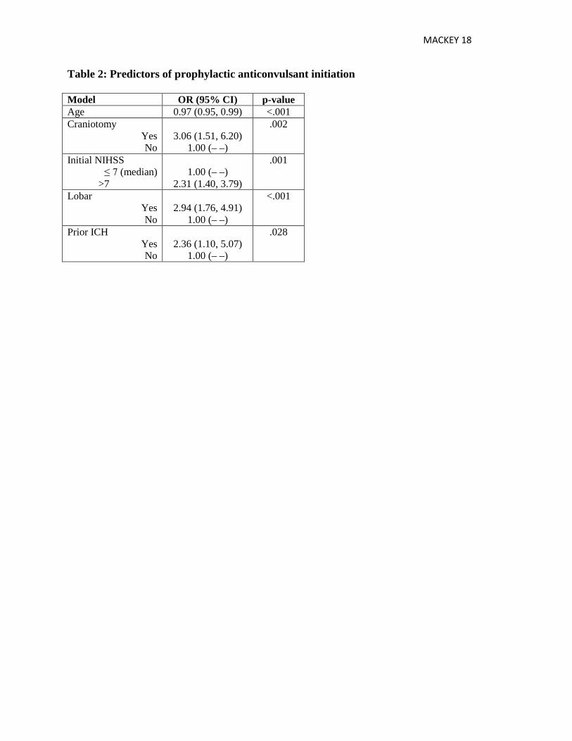
Table 2: Predictors of prophylactic anticonvulsant initiation
Table 3: Propensity-matched anticonvulsant prophylaxis vs. no prophylaxis
Table 4: Univariate logistic regression for poor functional outcomes (mRS 4-6) using propensity-
matched cohort (N=186)
Table 5: Predictors of poor outcome (mRS 4-6) at hospital discharge
Search terms: intracerebral hemorrhage, outcomes, anticonvulsants, health services, guideline
adherence

MACKEY 3
Abstract:
Background and Purpose: Prophylactic anticonvulsants are routinely prescribed in the acute
setting for ICH patients, but some studies have reported an association with worse outcomes. We
sought to characterize the prevalence and predictors of prophylactic anticonvulsant
administration after ICH as well as guideline adherence. We also sought to determine if
prophylactic anticonvulsants were independently associated with poor outcome.
Methods: We performed a retrospective study of primary ICH in our two academic centers. We
used a propensity matching approach to make treated and non-treated groups comparable. We
conducted multiple logistic regression analysis to identify independent predictors of prophylactic
anticonvulsant initiation and its association with poor outcome as measured by modified Rankin
score.
Results: We identified 610 patients with primary ICH, of whom 98 were started on prophylactic
anticonvulsants. Levetiracetam (97%) was most commonly prescribed. Age (OR: 0.97, 95% CI:
0.95-0.99, p < .001), lobar location (OR: 2.94, 95% CI: 1.76-4.91, p < .001), higher initial
NIHSS score (OR: 2.31, 95% CI: 1.40-3.79, p = .001), craniotomy (OR: 3.06, 95% CI: 1.51-
6.20, p = .002) and prior ICH (OR: 2.36, 95% CI: 1.10-5.07, p = .028) were independently
associated with prophylactic anticonvulsant initiation. Prophylactic anticonvulsant use was not
associated with worse functional outcome (mRS 4-6) at hospital discharge or with increased
case-fatality. There was no difference in prescribing patterns after 2010 guideline publication.
Discussion: Levetiracetam was routinely prescribed following ICH and was not associated with
worse outcomes. Future investigations should examine the effect of prophylactic levetiracetam
on cost and neuropsychological outcomes as well as the role of continuous EEG in identifying
subclinical seizures.

MACKEY 4
Introduction:
Intracerebral hemorrhage (ICH) has high morbidity and mortality1 and treatment remains largely
supportive. Seizures are a common complication in the acute setting2 and prophylactic treatment
with anticonvulsants is common,3 though the guidelines have recommended that patients without
seizures should not receive prophylactic anticonvulsants.4, 5 Whether prophylactic
anticonvulsants are associated with poor outcome in ICH remains unclear.3, 6-9 We therefore
sought to identify factors associated with prophylactic anticonvulsant initiation and to determine
whether prophylactic anticonvulsants were independently associated with poor clinical outcome.
We also sought to evaluate whether prophylactic anticonvulsant prescribing patterns changed
after guideline publication in 2010.
Methods:
This study was approved by the Indiana University Institutional Review Board, the Indiana
Network for Patient Care (INPC) board of directors, and Wishard Memorial Hospital.
Cohort assembly
We evaluated all patients ≥18 years old with primary ICH presenting to two academic centers via
a query of the INPC database (http://www.ihie.org). The INPC is a health information exchange
serving multiple hospital systems in Central Indiana.10 For inclusion in the study the index ICH
had to occur between January 1, 2009 and December 31, 2011; we additionally queried the
database until February 29, 2012 to identify patients with an index ICH during the study period
but who were subsequently discharged in the following two months. We used discharge ICD-9
codes of 431 and 432.9 to identify potential cases; these codes have >85% sensitivity for the
identification of patients with ICH.11 A vascular neurologist (J.M.) reviewed the medical record

MACKEY 5
and imaging scans of all potential cases to ensure proper case characterization. Patients with
evidence of traumatic ICH or an aneurysm, encephalitis, or brain tumor as a cause of the
hemorrhage were excluded. Patients with hemorrhagic transformation of an ischemic infarct or
hemorrhage due to venous sinus thrombosis, carotid endarterectomy, or thrombolytic
administration for ischemic stroke were also excluded.
Clinical data abstraction
Under the close supervision of a vascular neurologist, data abstractors ascertained via
standardized chart review demographic data, vascular risk factors, and processes of care. All
available referring hospital and transfer data were reviewed. If a formal NIH stroke scale
(NIHSS) score was not reported at presentation we used a validated method for estimation.12 The
neurologist reviewed the initial imaging scan from the academic center for each patient as well
as all available imaging scans from the referring hospital. Hematoma volume was calculated with
the ABC/2 method.13
Clinical outcome measures included modified Rankin score (mRS) at discharge. Date, time, and
cause of death were recorded for patients who died during the hospitalization. Discharge
disposition was also recorded. We determined vital status via present-day chart review and
obituary query. We then performed a National Death Index (http://www.cdc.gov/nchs/ndi.htm)
query for the vital status of all patients for whom we still could not account. All clinical data
were recorded in REDCap.14
Prophylactic anticonvulsant abstraction

MACKEY 6
For the prophylactic anticonvulsant analysis, we excluded patients with a history of seizure,
those with witnessed or suspected seizures, and those with baseline mRS of 4 or 5. We identified
time and location for first prophylactic anticonvulsant use and abstracted all medications and
doses for the duration of the hospitalization. For each day we calculated the daily dose of the
prophylactic anticonvulsants using the World Health Organization defined daily dose (DDD)
classification for levetiracetam (1500mg), phenytoin (300mg), and fosphenytoin (450mg),
(http://www.whocc.no/ddd/definition_and_general_considera/), as well as the number of dose
days, the average daily dose, and the cumulative dose for each patient. For example, if a patient
received levetiracetam 500mg BID for a total of 3 days the mean daily dose would be 0.67
(1000/1500) and the cumulative dose would be 2 (0.67*3). We also reviewed all available
documentation to determine whether the patient was discharged on the prophylactic
anticonvulsant. We further reviewed the entirety of the available medical record and abstracted
the last known prophylactic anticonvulsant administration.
Statistical Methods
Our two dichotomous primary outcomes were whether a patient had a prophylactic
anticonvulsant administered and whether a patient had worse functional outcome at hospital
discharge as measured by mRS of 4-6. We assembled the prophylactic anticonvulsant cohort for
the first primary outcome and the functional outcome cohort for the second primary outcome as
described below. To analyze the functional outcome data, we assessed how comparable the
treatment and corresponding matched control groups were at baseline. Chi-square, Fisher’s
exact, Student’s t, or Wilcoxon rank sum tests were used for this comparison. We considered
several covariates as listed in Table 1 to identify factors associated with each of the two primary

MACKEY 7
outcomes and used univariate and multiple logistic regression analyses. These variables included
general patient characteristics, variables significant in previous studies, and variables which
treating physicians may have considered as predisposing patients to higher seizure risk. We
assessed the association at univariate level and the covariates found to be significant at a p-value
of <0.20 were included in a stepwise multiple logistic regression model. Statistical analyses were
performed with SAS version 9.4 (SAS institute, Cary NC).
Prophylactic anticonvulsant analysis cohort assembly (total n=506)
Of the 610 patients in the overall cohort, 41 (6.7%) were excluded because of a previous history
of seizures and 45 (7.4%) had a witnessed or suspected seizure associated with the index ICH
prior to anticonvulsant initiation. An additional 18 patients were excluded from this analysis
because the baseline mRS was 4 (n=16) or 5 (n=2). The final cohort therefore included 506
patients, with 98 who were administered a prophylactic anticonvulsant and 408 who were not
administered a prophylactic anticonvulsant.
Functional outcome analyses cohort assembly (total n=186)
We then constructed a control group of patients (a group of patients not treated with prophylactic
anticonvulsants) who would be as comparable to the treated group of patients as possible. We
used the propensity score based matching approach and matched each treated patient to a control
patient if the difference in propensity score was within a pre-defined standard propensity score
caliper. Using calipers of width equal to 0.2 of the pooled standard deviation of the logit of the
propensity score removes about 99% of the bias due to the measured confounders.15 For each
treated patient we selected a control patient if the absolute difference of the propensity score on

MACKEY 8
the logit scale was within 0.2 of the pooled standard deviation of the logit of the propensity
score. The matching was done without replacement. We identified 93 control patients as a match
to 93 treated patients. We could not identify a suitable match for 5 of the treated patients.
Results:
We identified 506 patients with primary ICH from 2009 to 2011, of whom 98 (19.4%) were
given a prophylactic anticonvulsant, and 408 (80.6%) who were not given a prophylactic
anticonvulsant. Of the 98 given a prophylactic anticonvulsant, 45 (45.9%) presented to a
referring hospital initially. The mean age was 61.5, 50 (51.0%) were women, and 33 (33.7%)
were black. Mean ICH volume was 28.5mL and 52 (53.1%) had intraventricular extension.
Overall 22 (22.5%) patients died in the hospital and 40 (40.8%) died in the first year following
ICH.
Of the 408 not given a prophylactic anticonvulsant, 272 (66.7%) presented to a referring hospital
initially. The mean age was 67.2, 184 (45.1%) were women, and 100 (24.5%) were black. Mean
ICH volume was 18.8mL and 191 (46.8%) had intraventricular extension. Overall 79 (19.4%)
patients died in the hospital and 153 (37.5%) died in the first year following ICH.
Prophylactic anticonvulsant analysis
Levetiracetam alone was prescribed in 95 of 98 (97%) cases; one patient was prescribed both
levetiracetam and phenytoin, one was prescribed phenytoin alone, and one was prescribed
phenytoin and a single dose of fosphenytoin. Initiation of prophylactic anticonvulsants occurred

MACKEY 9
in the ICU (61, 62.2%), academic center ED (26, 26.5%), on the hospital floor (5, 5.1%), in the
operating room (4, 4.1%), and at the outside hospital (2, 2%).
The univariate analysis assessing association of factors with initiation of prophylactic
anticonvulsant is shown in Table 1. Younger age, lower baseline mRS, lower GCS, higher
NIHSS score, greater ICH volume, supratentorial ICH, lobar location, and craniotomy were
associated with prophylactic anticonvulsant use. The multiple logistic regression analysis is
shown in Table 2. Younger age, craniotomy, prior ICH, higher NIHSS score, and lobar location
were independently associated with prophylactic anticonvulsant initiation.
Duration and intensity subanalysis
For the 98 patients prescribed prophylactic anticonvulsants, the mean and median duration of
treatment in the hospital was 11.7 days and 6.5 days, respectively. The mean daily dose and
median daily dose were 0.6125 and 0.6132, respectively. The median cumulative dose was 4.0
(1.7, 9.0).
Functional outcomes analyses
After using the propensity score based matching approach, the treated and control groups were
found to be very similar in demographic characteristics and clinical outcomes as shown in Table
3. Univariate and multiple logistic regression analyses results for association with worse mRS of
4-6 are shown in Tables 4 and 5, respectively. Prophylactic anticonvulsant initiation was not
associated with worse functional outcome of mRS either in unadjusted or adjusted analyses for
other significant predictors of mRS. Higher NIHSS score, greater ICH volume, intraventricular

MACKEY 10
extension, and worse baseline mRS were independently associated with worse functional
outcome of mRS at discharge.
Prophylactic anticonvulsants were also not associated with higher inpatient case-fatality or with
case-fatality at one year in univariate analysis (data not shown).
Prophylactic anticonvulsants at discharge and afterward
Of the 98 patients started on prophylactic anticonvulsants, 2 (2%) had a subsequent seizure
during the admission and 74 of the 96 remaining (77.1%) survived to discharge. Of the 42
(56.8%) patients discharged from the hospital on a prophylactic anticonvulsant, 13 (31%) were
still on an anticonvulsant at 3 months and 6 (14.3%) were still on an anticonvulsant at 1 year
following index ICH.
Guideline implementation
We also dichotomized the study time period into before and after online 2010 guideline
publication (online July 22, 2010)4 to assess the effect of the guideline on anticonvulsant
prescribing patterns. Of 284 patients admitted prior to online ICH guideline publication, 55
(19.4%) were given prophylactic anticonvulsants compared with 43 of 222 (19.4%) after.
Discussion:
We found that levetiracetam was routinely prescribed in our ICH population and that there was
no association with worse outcomes at hospital discharge or at one year. From a resource

MACKEY 11
utilization standpoint, prophylactic anticonvulsants were very commonly continued through
hospital discharge and, in some cases, months or even years afterward. We also found no
significant change in prescribing habits after a new guideline recommended against prophylaxis
in 2010.
Several studies in recent years have evaluated the prevalence and predictors of anticonvulsant
prophylaxis in ICH as well as a potential association with poor outcome. Prevalence of
prophylaxis has generally ranged from 20-40%.7, 8, 16, 17 In one study investigators evaluated 295
subjects from the placebo arm of the CHANT trial and found that prophylactic anticonvulsants
were independently associated with a very poor outcome (mRS of 5 or 6.)3 The most commonly
prescribed anticonvulsant was phenytoin. Another large study, also predominantly with
phenytoin, found that prophylactic anticonvulsants were associated with reduced 90-day
mortality and improved 90-day functional outcome, but these associations disappeared when the
analysis was restricted to patients surviving beyond five days in an effort to diminish
confounding by indication.7
More recent studies have evaluated levetiracetam in ICH patients. A prospective study of 98
patients, of whom 40 received prophylactic anticonvulsants, found that phenytoin was associated
with poor outcome (mRS 4-6) at 3 months but that levetiracetam was not. This study also
evaluated duration and intensity of therapy and reported a median duration of about 1 week.
Most patients receiving levetiracetam were prescribed 500mg BID.6 Other studies comparing
levetiracetam and phenytoin have found that levetiracetam was associated with improved
cognitive outcomes at discharge and fewer seizures18 as well as improved long-term outcomes.19

MACKEY 12
A large study using a portion of the ERICH cohort found that prophylactic levetiracetam was not
independently associated with poor outcome. After adjustment for multiple factors associated
with poor outcome, prophylactic levetiracetam was not associated with worse functional
outcome at 3 months.8
Our study confirms these findings and extends them by including a rigorous propensity score
matching analysis to our outcome models. Levetiracetam is a newer anticonvulsant whose
precise mechanism of action is unclear. Levetiracetam has fewer side effects and drug
interactions than phenytoin.20 A recent multicenter study found that levetiracetam use increased
between 2007 and 2012 with a corresponding decrease in phenytoin use,17 which may reflect
changes in prescribing behavior based on a study suggesting potential harm from phenytoin.6
That we did not identify an association with levetiracetam and adverse outcomes is unsurprising
but reassuring nonetheless.
Strengths of this study include a large, well-characterized cohort, extensive review of referring
hospital data, and a pre- and post-guideline publication timeframe, as well as the rigorous
methodology noted above. There are several limitations to this work. This study is retrospective
in nature with the well-known inherent limitations. Prophylactic anticonvulsant initiation was not
randomized and was left to the discretion of the treating physician, though we attempted to adjust
for that using propensity matching. There may also be other factors, such as individual physician
prescribing habits, that play a role in prophylactic anticonvulsant initiation for which we cannot
account in this study. Finally, because we did not systematically evaluate patients with
continuous EEG misclassification bias is possible.

MACKEY 13
In this large retrospective study we found that prophylactic levetiracetam was commonly
prescribed in our ICH population and that it was not associated with poor functional outcomes at
hospital discharge or with one-year case-fatality. Future investigations should examine the effect
of levetiracetam on cost and whether continuous EEG monitoring adds to decision-making about
anticonvulsants in patients with ICH. Study of the impact of prolonged levetiracetam on quality
of life and neuropsychological outcomes in ICH patients is also warranted as longer exposure
could be deleterious. Because there are few specific treatments for ICH, more health services
research, including guideline adherence research, in ICH is needed as well. Finally, only a
randomized controlled trial will be able to answer definitively whether ICH patients benefit from
prophylactic anticonvulsants.
Acknowledgements
N/A
Sources of Funding
This work was supported by awards from the IU Health Values Fund (IUH VFR365), the IU
CTSI PDT (ICTSI NIH/NCRR RR025761), the IUH/IUSM Strategic Research Initiative, and an
IU CTSI KL2 award (NIH, UL1TR001108, Shekhar PI).
Conflict of Interest/Disclosures

MACKEY 14
Dr. Mackey is funded by Research Grant; Significant; IUH-VFR-365, IUH/IUSM Strategic
Research Initiative, and CTSI PDT. NIH LRP recipient. Indiana University CTSI KL2 award
recipient.
A.D. Blatsioris is funded by Research Grant; Significant; IUH-VFR-365, IUH/IUSM Strategic
Research Initiative.
E.A.S. Moser is funded by Research Grant; Significant; IUH-VFR-365, IUH/IUSM Strategic
Research Initiative.
R.J.L. Carter is funded by Research Grant; Significant; IUH-VFR-365, IUH/IUSM Strategic
Research Initiative.
C. Saha is funded by Research Grant; Significant; IUH-VFR-365, IUH/IUSM Strategic Research
Initiative.
A. Stevenson is funded by Research Grant; Significant; IUH-VFR-365, IUH/IUSM Strategic
Research Initiative.
A.L. Hulin is funded by Research Grant; Significant; IUH-VFR-365, IUH/IUSM Strategic
Research Initiative.
Dr. O’Neill reports no disclosures.

MACKEY 15
Dr. Cohen-Gadol reports no disclosures.
Dr. Leipzig reports no disclosures.
Dr. Williams reports no disclosures.
References:
1. van Asch CJ, Luitse MJ, Rinkel GJ, van der Tweel I, Algra A, Klijn CJ. Incidence, case fatality, and functional outcome of intracerebral haemorrhage over time, according to age, sex, and ethnic origin: A systematic review and meta-analysis. Lancet Neurol. 2010;9:167-176
2. Szaflarski JP, Rackley AY, Kleindorfer DO, Khoury J, Woo D, Miller R, et al. Incidence of seizures in the acute phase of stroke: A population-based study. Epilepsia. 2008;49:974-981
3. Messé SR, Sansing LH, Cucchiara BL, Herman ST, Lyden PD, Kasner SE, et al. Prophylactic antiepileptic drug use is associated with poor outcome following ich. Neurocrit Care. 2009;11:38-44
4. Morgenstern LB, Hemphill JC, Anderson C, Becker K, Broderick JP, Connolly ES, et al. Guidelines for the management of spontaneous intracerebral hemorrhage. Stroke. 2010;41:2108-2129
5. Hemphill JC, Greenberg SM, Anderson CS, Becker K, Bendok BR, Cushman M, et al. Guidelines for the management of spontaneous intracerebral hemorrhage: A guideline for healthcare professionals from the american heart association/american stroke association. Stroke. 2015
6. Naidech AM, Garg RK, Liebling S, Levasseur K, Macken MP, Schuele SU, et al. Anticonvulsant use and outcomes after intracerebral hemorrhage. Stroke. 2009;40:3810-3815
7. Battey TW, Falcone GJ, Ayres AM, Schwab K, Viswanathan A, McNamara KA, et al. Confounding by indication in retrospective studies of intracerebral hemorrhage: Antiepileptic treatment and mortality. Neurocrit Care. 2012;17:361-366
8. Sheth KN, Martini SR, Moomaw CJ, Koch S, Elkind MS, Sung G, et al. Prophylactic antiepileptic drug use and outcome in the ethnic/racial variations of intracerebral hemorrhage study. Stroke. 2015:3532-3535
9. Gilmore EJ, Maciel CB, Hirsch LJ, Sheth KN. Review of the utility of prophylactic anticonvulsant use in critically ill patients with intracerebral hemorrhage. Stroke. 2016;47:2666-2672
10. McDonald CJ, Overhage JM, Barnes M, Schadow G, Blevins L, Dexter PR, et al. The indiana network for patient care: A working local health information infrastructure. Health Affairs. 2005;24:1214-1220
11. Alwell K, Khoury J, Moomaw C, Kleindorfer D, Woo D, Flaherty M, et al. Icd-9 codes positive predictive value for stroke subtypes in a population-based epidemiology study Stroke. 2009;40:e183

MACKEY 16
12. Williams LS, Yilmaz EY, Lopez-Yunez AM. Retrospective assessment of initial stroke severity with the nih stroke scale. Stroke. 2000;31:858-862
13. Kothari RU, Brott T, Broderick JP, Barsan WG, Sauerbeck LR, Zuccarello M, et al. The abcs of measuring intracerebral hemorrhage volumes. Stroke. 1996;27:1304-1305
14. Harris PA, Taylor R, Thielke R, Payne J, Gonzalez N, Conde JG. Research electronic data capture (redcap)--a metadata-driven methodology and workflow process for providing translational research informatics support. J Biomed Inform. 2009;42:377-381
15. Rosenbaum PR, Rubin DB. Constructing a control group using multivariate matched sampling methods that incorporate the propensity score. The American Statistician. 1985;39:33-38
16. Reddig RT, Nixdorf KE, Jensen MB. The prophylactic use of an antiepileptic drug in intracerebral hemorrhage. Clin Neurol Neurosurg. 2011;113:895-897
17. Naidech AM, Beaumont J, Jahromi B, Prabhakaran S, Kho A, Holl JL. Evolving use of seizure medications after intracerebral hemorrhage: A multicenter study. Neurology. 2017;88:52-56
18. Taylor S, Heinrichs RJ, Janzen JM, Ehtisham A. Levetiracetam is associated with improved cognitive outcome for patients with intracranial hemorrhage. Neurocrit Care. 2011;15:80-84
19. Szaflarski JP, Sangha KS, Lindsell CJ, Shutter LA. Prospective, randomized, single-blinded comparative trial of intravenous levetiracetam versus phenytoin for seizure prophylaxis. Neurocrit Care. 2010;12:165-172
20. Brophy GM, Human T, Shutter L. Emergency neurological life support: Pharmacotherapy. Neurocrit Care. 2015;23 Suppl 2:S48-68
Table 1: Univariate logistic regression for prophylactic anticonvulsant (PA) administration Not Prescribed PA
(N = 408) Prescribed PA
(N = 98) Unadjusted OR
(95% CI) for predicting PA
p-value
N (%) N (%) Age - - 0.97 (0.96, 0.99) <.001 Sex
Female Male
184 (78.6%) 224 (82.4%)
50 (21.4%) 48 (17.6%)
1.27 (0.82, 1.97)
1.00 (– –)
.292
Race Black
Non-Black
100 (75.2%) 308 (82.6%)
33 (24.8%) 65 (17.4%)
1.56 (0.97, 2.52)
1.00 (– –)
.065
Baseline mRS 0-1 2-3
287 (78.2%) 121 (87.1%)
80 (21.8%) 18 (12.9%)
1.87 (1.08, 3.26)
1.00 (– –)
.026
GCS - - 0.93 (0.88, 0.98) .007 Initial NIHSS
≤ 7 (median) > 7
223 (86.1%) 185 (74.9%)
36 (13.9%) 62 (25.1%)
1.00 (– –)
2.08 (1.32, 3.27)
.002
ICH volume (mL) Q1 (0-2.3)
Q2 (2.4-10.1) Q3 (10.2-27.0)
Q4 (27.1-187.5)
117 (90.0%) 106 (86.2%) 99 (78.6%) 84 (67.2%)
13 (10.0%) 17 (13.8%) 27 (21.4%) 41 (32.8%)
1.00 (– –)
1.44 (0.67, 3.11) 2.45 (1.20, 5.01) 4.39 (2.22, 8.71)
<.001
Subarachnoid extension Yes
40 (75.5%)
13 (24.5%)
1.41 (0.72, 2.75)
.317

MACKEY 17
Not Prescribed PA (N = 408)
Prescribed PA (N = 98)
Unadjusted OR (95% CI) for predicting PA
p-value
N (%) N (%) No 368 (81.2%) 85 (18.8%) 1.00 (– –)
Intraventricular extension Yes No
191 (78.6%) 217 (82.5%)
52 (21.4%) 46 (17.5%)
1.28 (0.83, 2.00)
1.00 (– –)
.267
Supratentorial Yes No
340 (78.7%) 62 (92.5%)
92 (21.3%) 5 (7.5%)
3.35 (1.31, 8.58)
1.00 (– –)
.012
Lobar Yes No
137 (72.9%) 265 (85.2%)
51 (27.1%) 46 (14.8%)
2.15 (1.37, 3.36)
1.00 (– –)
<.001
Initial SBP, mmHg Q1 (86-155)
Q2 (156-178) Q3 (179-210) Q4 (211-282)
104 (83.9%) 107 (82.9%) 93 (76.2%) 95 (79.2%)
20 (16.1%) 22 (17.1%) 29 (23.8%) 25 (20.8%)
1.00 (– –)
1.07 (0.55, 2.08) 1.62 (0.86, 3.06) 1.37 (0.71, 2.62)
.403
Initial DBP, mmHg Q1 (36-81) Q2 (82-98)
Q3 (99-113) Q4 (114-183)
105 (84.7%) 105 (78.4%) 96 (81.4%) 92 (78.0%)
19 (15.3%) 29 (21.6%) 22 (18.6%) 26 (22.0%)
1.00 (– –)
1.53 (0.81, 2.89) 1.27 (0.65, 2.48) 1.56 (0.81, 3.00)
.514
Charlson 0-1 >1
268 (80.7%) 140 (80.5%)
64 (19.3%) 34 (19.5%)
1.00 (– –)
1.02 (0.64, 1.62)
.943
Craniotomy Yes No
23 (53.5%)
384 (83.1%)
20 (46.5%) 78 (16.9%)
4.28 (2.24, 8.17)
1.00 (– –)
<.001
Prior ICH Yes No
28 (70.0%)
379 (81.5%)
12 (30.0%) 86 (18.5%)
1.89 (0.92, 3.86)
1.00 (– –)
.081
Prior ischemic stroke Yes No
78 (87.6%)
329 (79.1%)
11 (12.4%) 87 (20.9%)
1.00 (– –)
1.88 (0.96, 3.68)
.068
MACKEY 18
Table 2: Predictors of prophylactic anticonvulsant initiation Model OR (95% CI) p-value Age 0.97 (0.95, 0.99) <.001 Craniotomy
Yes No
3.06 (1.51, 6.20)
1.00 (– –)
.002
Initial NIHSS ≤ 7 (median)
>7
1.00 (– –)
2.31 (1.40, 3.79)
.001
Lobar Yes No
2.94 (1.76, 4.91)
1.00 (– –)
<.001
Prior ICH Yes No
2.36 (1.10, 5.07)
1.00 (– –)
.028

MACKEY 19
Table 3: Propensity-matched anticonvulsant prophylaxis vs. no prophylaxis
Prophylactic anticonvulsants
N=93
No prophylaxis N=93
p-value
Age, mean (SD) 62.3 ± 13.5 62.0 ± 14.4 .908 Female 47 (50.5%) 50 (53.8%) .660 Black 32 (34.4%) 28 (30.1%) .530 Baseline mRS, median (IQR) 1 (0, 1) 1 (0, 1) .842 GCS, median (IQR) 13 (9, 15) 14 (9, 15) .729 Initial NIHSS, median (IQR) 12 (4, 19) 9 (3, 26) .634 ICH volume (mL), median (IQR) 21.9 (7.5, 39.4) 17.5 (5.0, 44.7) .778 Subarachnoid extension 13 (14.0%) 15 (16.1%) .682 Intraventricular extension 50 (53.8%) 54 (58.1%) .555 Supratentorial 87 (93.5%) 83 (89.2%) .296 Lobar 46 (49.5%) 45 (48.4%) .883 Initial SBP, mmHg (SD) 187.7 ± 37.5 185.8 ± 42.8 .748 Initial DBP, mmHg (SD) 104.2 ± 27.0 103.3 ± 28.2 .833 Charlson, median (IQR) 1 (0, 2) 1 (0, 2) .985 Craniotomy 15 (16.1%) 16 (17.2%) .844 Prior ICH 11 (11.8%) 15 (16.1%) .398 Prior ischemic stroke 11 (11.8%) 14 (15.1%) .519

MACKEY 20
Table 4: Univariate logistic regression for poor functional outcomes (mRS 4-6) using propensity-matched cohort (N=186) mRS 0-3
(N = 53) mRS 4-6 (N = 133)
Unadjusted OR (95% CI) for predicting
mRS 4-6
p-value
N (%) N (%) Age
Q1 (22-53) Q2 (54-62) Q3 (63-71) Q4 (72-95)
19 (38.8%) 16 (30.8%) 11 (28.2%) 7 (15.2%)
30 (61.2%) 36 (69.2%) 28 (71.8%) 39 (84.8%)
1.00 (– –)
1.43 (0.63, 3.25) 1.61 (0.65, 3.98) 3.53 (1.31, 9.48)
.097
Sex Female
Male
26 (26.8%) 27 (30.3%)
71 (73.2%) 62 (69.7%)
1.19 (0.63, 2.25)
1.00 (– –)
.594
Race Black
Non-Black
14 (23.3%) 39 (31.0%)
46 (76.7%) 87 (69.0%)
1.47 (0.73, 2.99)
1.00 (– –)
.284
Baseline mRS 0-1 2-3
47 (32.2%) 6 (15.0%)
99 (67.8%) 34 (85.0%)
1.00 (– –)
2.69 (1.06, 6.85)
.038
GCS, median (IQR) - 0.65 (0.54, 0.79) <.001 Initial NIHSS
≤ 11 (median) > 11
48 (49.5%)
5 (5.6%)
49 (50.5%) 84 (94.4%)
1.00 (– –)
16.46 (6.14, 44.11)
<.001
ICH volume (mL) Q1 (0-6.0)
Q2 (6.1-18.6) Q3 (18.7-43.3)
Q4 (43.4-130.6)
23 (46.9%) 16 (36.4%) 12 (25.5%)
2 (4.3%)
26 (53.1%) 28 (63.6%) 35 (74.5%) 44 (95.7%)
1.00 (– –)
1.55 (0.67, 3.56) 2.58 (1.09, 6.12)
19.46 (4.24, 89.35)
<.001
Subarachnoid extension Yes No
2 (7.1%)
51 (32.3%)
26 (92.9%)
107 (67.7%)
6.20 (1.42, 27.12)
1.00 (– –)
.016
Intraventricular extension Yes No
17 (16.3%) 36 (43.9%)
87 (83.7%) 46 (56.1%)
4.01 (2.03, 7.89)
1.00 (– –)
<.001
Supratentorial Yes No
48 (28.2%) 5 (31.2%)
122 (71.8%) 11 (68.8%)
1.15 (0.38, 3.50)
1.00 (– –)
.799
Lobar Yes No
30 (33.0%) 23 (24.2%)
61 (67.0%) 72 (75.8%)
1.00 (– –)
1.54 (0.81, 2.92)
.187
Initial SBP, mmHg Q1 (107-156)
Q2 (157-182.5) Q3 (182.6-211)
Q4 (212-282)
15 (31.2%) 17 (33.3%) 9 (21.4%) 12 (26.7%)
33 (68.8%) 34 (66.7%) 33 (78.6%) 33 (73.3%)
1.00 (– –)
0.91 (0.39, 2.11) 1.67 (0.64, 4.34) 1.25 (0.51, 3.07)
.605

MACKEY 21
mRS 0-3 (N = 53)
mRS 4-6 (N = 133)
Unadjusted OR (95% CI) for predicting
mRS 4-6
p-value
N (%) N (%) Initial DBP, mmHg
Q1 (47-86) Q2 (87-100)
Q3 (101-112) Q4 (113-183)
12 (25.5%) 18 (34.6%) 12 (29.3%) 11 (23.9%)
35 (74.5%) 34 (65.4%) 29 (70.7%) 35 (76.1%)
1.00 (– –)
0.65 (0.27, 1.55) 0.83 (0.32, 2.12) 1.09 (0.43, 2.80)
.652
Charlson 0-1 >1
37 (29.8%) 16 (25.8%)
87 (70.2%) 46 (74.2%)
1.00 (– –)
1.22 (0.62, 2.43)
.566
Craniotomy Yes No
8 (25.8%) 45 (29.0%)
23 (74.2%)
110 (71.0%)
1.18 (0.49, 2.82)
1.00 (– –)
.717
Prior ICH Yes No
8 (33.8%) 45 (28.1%)
18 (69.2%)
115 (71.9%)
1.00 (– –)
1.14 (0.46, 2.80)
.782
Prior ischemic stroke Yes No
4 (16.0%) 49 (30.4%)
21 (84.0%)
112 (69.6%)
2.30 (0.75, 7.04)
1.00 (– –)
.146
Prophylactic anticonvulsant Yes No
24 (25.8%) 29 (31.2%)
69 (74.2%) 64 (68.8%)
1.30 (0.69, 2.47)
1.00 (– –)
.417

MACKEY 22
Table 5: Predictors of poor outcome (mRS 4-6) at hospital discharge Model OR (95% CI) p-value Prophylactic anticonvulsant
Yes No
1.41 (0.61, 3.29)
1.00 (– –)
.424
Initial NIHSS ≤ 11 (median)
>11
1.00 (– –)
13.95 (4.80, 40.50)
<.001
ICH volume (mL) Q1 (0-6.0)
Q2 (6.1-18.6) Q3 (18.7-43.3)
Q4 (43.4-130.6)
1.00 (– –)
2.02 (0.72, 5.66) 2.24 (0.73, 6.84)
19.28 (3.58, 103.71)
.007
Intraventricular extension Yes No
3.33 (1.41, 7.88)
1.00 (– –)
.006
Baseline mRS 0-1 2-3
1.00 (– –)
5.05 (1.53, 16.66)
.008