Properties of Zinc Oxide Nanoparticles and Their Activity ...
13
NANO REVIEW Open Access Properties of Zinc Oxide Nanoparticles and Their Activity Against Microbes Khwaja Salahuddin Siddiqi 1 , Aziz ur Rahman 2 , Tajuddin 2 and Azamal Husen 3* Abstract Zinc oxide is an essential ingredient of many enzymes, sun screens, and ointments for pain and itch relief. Its microcrystals are very efficient light absorbers in the UVA and UVB region of spectra due to wide bandgap. Impact of zinc oxide on biological functions depends on its morphology, particle size, exposure time, concentration, pH, and biocompatibility. They are more effective against microorganisms such as Bacillus subtilis, Bacillus megaterium, Staphylococcus aureus, Sarcina lutea, Escherichia coli, Pseudomonas aeruginosa, Klebsiella pneumonia, Pseudomonas vulgaris, Candida albicans, and Aspergillus niger. Mechanism of action has been ascribed to the activation of zinc oxide nanoparticles by light, which penetrate the bacterial cell wall via diffusion. It has been confirmed from SEM and TEM images of the bacterial cells that zinc oxide nanoparticles disintegrate the cell membrane and accumulate in the cytoplasm where they interact with biomolecules causing cell apoptosis leading to cell death. Keywords: Zinc oxide nanoparticles, Microorganisms, Antimicrobial, Toxicity, Mechanism, Biodistribution Background Nanotechnology deals with the manufacture and appli- cation of materials with size of up to 100 nm. They are widely used in a number of processes that include ma- terial science, agriculture, food industry, cosmetic, med- ical, and diagnostic applications [1–10]. Nanosize inorganic compounds have shown remarkable antibac- terial activity at very low concentration due to their high surface area to volume ratio and unique chemical and physical features [11]. In addition, these particles are also more stable at high temperature and pressure [12]. Some of them are recognized as nontoxic and even contain mineral elements which are vital for human body [13]. It has been reported that the most antibacterial inorganic materials are metallic nanoparticles and metal oxide nanoparticles such as silver, gold, copper, titanium oxide, and zinc oxide [14, 15]. Zinc is an essential trace element for human system without which many enzymes such as carbonic anhy- drase, carboxypeptidase, and alcohol dehydrogenase be- come inactive, while the other two members, cadmium and mercury belonging to the same group of elements having the same electronic configuration, are toxic. It is essential for eukaryotes because it modulates many physiological functions [16, 17]. Bamboo salt, containing zinc, is used as herbal medicine for the treatment of in- flammation by regulating caspase-1 activity. Zinc oxide nanoparticles have been shown to reduce mRNA expres- sion of inflammatory cytokines by inhibiting the activa- tion of NF-kB (nuclear factor kappa B cells) [18]. Globally, bacterial infections are recognized as serious health issue. New bacterial mutation, antibiotic resistance, outbreaks of pathogenic strains, etc. are increasing, and thus, development of more efficient antibacterial agents is demand of the time. Zinc oxide is known for its antibac- terial properties from the time immemorial [19]. It had been in use during the regime of Pharaohs, and historical records show that zinc oxide was used in many ointments for the treatment of injuries and boils even in 2000 BC [20]. It is still used in sun screen lotion, as a supplement, photoconductive material, LED, transparent transistors, solar cells, memory devices [21, 22], cosmetics [23, 24], and catalysis [25]. Although considerable amount of ZnO is produced every year, very small quantity is used as medicine [26]. The US Food and Drug Administration has recognized (21 CFR 182.8991) zinc oxide as safe [27]. It is characterized by photocatalytic and photooxidizing prop- erties against biochemicals [28]. * Correspondence: [email protected] 3 Department of Biology, College of Natural and Computational Sciences, University of Gondar, P.O. Box #196, Gondar, Ethiopia Full list of author information is available at the end of the article © The Author(s). 2018 Open Access This article is distributed under the terms of the Creative Commons Attribution 4.0 International License (http://creativecommons.org/licenses/by/4.0/), which permits unrestricted use, distribution, and reproduction in any medium, provided you give appropriate credit to the original author(s) and the source, provide a link to the Creative Commons license, and indicate if changes were made. Siddiqi et al. Nanoscale Research Letters (2018) 13:141 https://doi.org/10.1186/s11671-018-2532-3
Transcript of Properties of Zinc Oxide Nanoparticles and Their Activity ...

Siddiqi et al. Nanoscale Research Letters (2018) 13:141 https://doi.org/10.1186/s11671-018-2532-3
NANO REVIEW Open Access
Properties of Zinc Oxide Nanoparticles andTheir Activity Against Microbes
Khwaja Salahuddin Siddiqi1, Aziz ur Rahman2, Tajuddin2 and Azamal Husen3*Abstract
Zinc oxide is an essential ingredient of many enzymes, sun screens, and ointments for pain and itch relief. Itsmicrocrystals are very efficient light absorbers in the UVA and UVB region of spectra due to wide bandgap. Impactof zinc oxide on biological functions depends on its morphology, particle size, exposure time, concentration, pH,and biocompatibility. They are more effective against microorganisms such as Bacillus subtilis, Bacillus megaterium,Staphylococcus aureus, Sarcina lutea, Escherichia coli, Pseudomonas aeruginosa, Klebsiella pneumonia, Pseudomonasvulgaris, Candida albicans, and Aspergillus niger. Mechanism of action has been ascribed to the activation of zincoxide nanoparticles by light, which penetrate the bacterial cell wall via diffusion. It has been confirmed from SEMand TEM images of the bacterial cells that zinc oxide nanoparticles disintegrate the cell membrane and accumulatein the cytoplasm where they interact with biomolecules causing cell apoptosis leading to cell death.
Keywords: Zinc oxide nanoparticles, Microorganisms, Antimicrobial, Toxicity, Mechanism, Biodistribution
BackgroundNanotechnology deals with the manufacture and appli-cation of materials with size of up to 100 nm. They arewidely used in a number of processes that include ma-terial science, agriculture, food industry, cosmetic, med-ical, and diagnostic applications [1–10]. Nanosizeinorganic compounds have shown remarkable antibac-terial activity at very low concentration due to their highsurface area to volume ratio and unique chemical andphysical features [11]. In addition, these particles are alsomore stable at high temperature and pressure [12]. Someof them are recognized as nontoxic and even containmineral elements which are vital for human body [13]. Ithas been reported that the most antibacterial inorganicmaterials are metallic nanoparticles and metal oxidenanoparticles such as silver, gold, copper, titanium oxide,and zinc oxide [14, 15].Zinc is an essential trace element for human system
without which many enzymes such as carbonic anhy-drase, carboxypeptidase, and alcohol dehydrogenase be-come inactive, while the other two members, cadmiumand mercury belonging to the same group of elements
* Correspondence: [email protected] of Biology, College of Natural and Computational Sciences,University of Gondar, P.O. Box #196, Gondar, EthiopiaFull list of author information is available at the end of the article
© The Author(s). 2018 Open Access This articleInternational License (http://creativecommons.oreproduction in any medium, provided you givthe Creative Commons license, and indicate if
having the same electronic configuration, are toxic. It isessential for eukaryotes because it modulates manyphysiological functions [16, 17]. Bamboo salt, containingzinc, is used as herbal medicine for the treatment of in-flammation by regulating caspase-1 activity. Zinc oxidenanoparticles have been shown to reduce mRNA expres-sion of inflammatory cytokines by inhibiting the activa-tion of NF-kB (nuclear factor kappa B cells) [18].Globally, bacterial infections are recognized as serious
health issue. New bacterial mutation, antibiotic resistance,outbreaks of pathogenic strains, etc. are increasing, andthus, development of more efficient antibacterial agents isdemand of the time. Zinc oxide is known for its antibac-terial properties from the time immemorial [19]. It hadbeen in use during the regime of Pharaohs, and historicalrecords show that zinc oxide was used in many ointmentsfor the treatment of injuries and boils even in 2000 BC[20]. It is still used in sun screen lotion, as a supplement,photoconductive material, LED, transparent transistors,solar cells, memory devices [21, 22], cosmetics [23, 24],and catalysis [25]. Although considerable amount of ZnOis produced every year, very small quantity is used asmedicine [26]. The US Food and Drug Administration hasrecognized (21 CFR 182.8991) zinc oxide as safe [27]. It ischaracterized by photocatalytic and photooxidizing prop-erties against biochemicals [28].
is distributed under the terms of the Creative Commons Attribution 4.0rg/licenses/by/4.0/), which permits unrestricted use, distribution, ande appropriate credit to the original author(s) and the source, provide a link tochanges were made.

Siddiqi et al. Nanoscale Research Letters (2018) 13:141 Page 2 of 13
Zinc oxide has been classified by EU hazard classifica-tion as N; R50-53 (ecotoxic). Compounds of zinc are eco-toxic for mammals and plants in traces [29, 30]. Humanbody contains about 2–3 g of zinc, and the daily require-ment is 10–15 mg [29, 31]. No report has demonstratedcarcinogenicity, genotoxicity, and reproduction toxicity inhumans [29, 32]. However, zinc powder inhaled oringested may produce a condition called zinc fever, whichis followed by chill, fever, cough, etc.Morphology of zinc oxide nanoparticles depends on
the process of synthesis. They may be nanorods, nano-plates [33–35], nanospheres [36], nanoboxes [35], hex-agonal, tripods [37], tetrapods [38], nanowires,nanotubes, nanorings [39–41], nanocages, and nano-flowers [42, 43]. Zinc oxide nanoparticles are more ac-tive against gram-positive bacteria relative to other NPsof the same group of elements. Ready to eat food ismore prone to infection by Salmonella, Staphylococcusaureus, and E. coli which pose a great challenge to foodsafety and quality. The antimicrobial compounds are in-corporated in the packed food to prevent them fromdamage. Antimicrobial packaging contains a nontoxicmaterial which inhibits or slows down the growth of mi-crobes present in food or packaging material [44]. Anantimicrobial substance for human consumption mustpossess the following properties.
a) It should be nontoxic.b) It should not react with food or container.c) It should be of good taste or tasteless.d) It should not have disagreeable smell.
Zinc oxide nanoparticle is one such inorganic metaloxide which fulfills all the above requirements, andhence, it can safely be used as medicine, preservative inpackaging, and an antimicrobial agent [45, 46]. It easilydiffuses into the food material, kill the microbes, andprevent human being from falling ill. In accordance withthe regulations 1935/2004/EC and 450/2009/EC of theEuropean Union, active packaging is defined as activematerial in contact with food with ability to change thecomposition of the food or the atmosphere around it[47]. Therefore, it is commonly used as preservative andincorporated in polymeric packaging material to preventfood material from damage by microbes [48]. Zinc oxidenanoparticles have been used as an antibacterial sub-stance against Salmonella typhi and S. aureus in vitro.Of all the metal oxide nanoparticles studied thus far,zinc oxide nanoparticles exhibited the highest toxicityagainst microorganisms [49]. It has also been demon-strated from SEM and TEM images that zinc oxidenanoparticles first damage the bacterial cell wall, thenpenetrate, and finally accumulate in the cell membrane.They interfere with metabolic functions of the microbes
causing their death. All the characteristics of the zincoxide nanoparticles depend on their particle size, shape,concentration, and exposure time to the bacterial cell.Further, biodistribution studies of zinc oxide nanoparti-cles have also been examined. For instance, Wang et al.[50] have investigated the effect of long-term exposureof zinc oxide nanoparticle on biodistribution and zincmetabolism in mice over 3 to 35 weeks. Their resultsshowed minimum toxicity to mice when they were ex-posed to 50 and 500 mg/kg zinc oxide nanoparticle indiet. At higher dose of 5000 mg/kg, zinc oxide nanopar-ticle decreased body weight but increased the weight ofthe pancreas, brain, and lung. Also, it increased theserum glutamic-pyruvic transaminase activity andmRNA expression of zinc metabolism-related genessuch as metallothionein. Biodistribution studies showedthe accumulation of sufficient quantity of zinc in theliver, pancreas, kidney, and bones. Absorption and distri-bution of zinc oxide nanoparticle/zinc oxide microparti-cles are largely dependent on the particle size. Li et al.[51] have studied biodistribution of zinc oxide nanopar-ticles fed orally or through intraperitoneal injection to6 weeks old mice. No obvious adverse effect was de-tected in zinc oxide nanoparticles orally treated mice in14 days study. However, intraperitoneal injection of 2.5 g/kg body weight given to mice showed accumulationof zinc in the heart, liver, spleen, lung, kidney, and testes.Nearly ninefold increase in zinc oxide nanoparticle inthe liver was observed after 72 h. Zinc oxide nanoparti-cles have been shown to have better efficiency in liver,spleen, and kidney biodistribution than in orally fedmice. Since zinc oxide nanoparticles are innocuous inlow concentrations, they stimulate certain enzymes inman and plants and suppress diseases. Singh et al. [52]have also been recently reviewed the biosynthesis of zincoxide nanoparticle, their uptake, translocation, and bio-transformation in plant system.In this review, we have attempted to consolidate all
the information regarding zinc oxide nanoparticles asantibacterial agent. The mechanism of interaction ofzinc oxide nanoparticles against a variety of microbeshas also been discussed in detail.
Antimicrobial Activity of Zinc Oxide NanoparticlesIt is universally known that zinc oxide nanoparticles areantibacterial and inhibit the growth of microorganismsby permeating into the cell membrane. The oxidativestress damages lipids, carbohydrates, proteins, and DNA[53]. Lipid peroxidation is obviously the most crucialthat leads to alteration in cell membrane which eventu-ally disrupt vital cellular functions [54]. It has been sup-ported by oxidative stress mechanism involving zincoxide nanoparticle in Escherichia coli [55]. However, forbulk zinc oxide suspension, external generation of H2O2

Siddiqi et al. Nanoscale Research Letters (2018) 13:141 Page 3 of 13
has been suggested to describe the anti-bacterial proper-ties [56]. Also, the toxicity of nanoparticles, releasingtoxic ions, has been considered. Since zinc oxide isamphoteric in nature, it reacts with both acids and alka-lis giving Zn2+ ions.
The free Zn2+ ions immediately bind with thebiomolecules such as proteins and carbohydrates, and allvital functions of bacteria cease to continue. The toxicity ofzinc oxide, zinc nanoparticles, and ZnSO4·7H2O has beentested (Table 1) against Vibrio fischeri. It was found thatZnSO4·7H2O is six times more toxic than zinc oxidenanoparticles and zinc oxide. The nanoparticles are actuallydispersed in the solvent, not dissolved, and therefore, theycannot release Zn2+ ions. The bioavailability of Zn2+ ions isnot always 100% and may invariably change withphysiological pH, redox potential, and the anions associatedwith it such as Cl− or SO4
2−.Solubility of zinc oxide (1.6–5.0 mg/L) in aqueous
medium is higher than that of zinc oxide nanoparticles (0.3–3.6 mg/L) in the same medium [57] which is toxic toalgae and crustaceans. Both nano-zinc oxide and bulk zincoxide are 40–80-fold less toxic than ZnSO4 against V.fischeri. The higher antibacterial activity of ZnSO4 is dir-ectly proportional to its solubility releasing Zn2+ ions,which has higher mobility and greater affinity [58] towardbiomolecules in the bacterial cell due to positive charge onthe Zn2+ and negative charge on the biomolecules.
Since zinc oxide and its nanoparticles have limitedsolubility, they are less toxic to the microbes than highly
Table 1 The toxicity (30-min EC50, EC20 and NOEC, and MIC) of metbacteria Vibrio fischeri [59]
Chemical Toxicity to Vibrio fischeri, EC50, EC20, NOEC, and M
EC50 ± SD EC20 ±
ZnO 1.8 ± 0.1 (1.4 ± 0.08) 1.0 ± 0
Nano-ZnO 1.9 ± 0.2 (1.5 ± 0.16) 0.9 ± 0
ZnSO4·7H2O 1.1 ± 0.25 (0.25 ± 0.06) 0.8 ± 0
CuO 3811 ± 1012 (3049 ± 819) 903 ±
Nano-CuO 79 ± 27 (63 ± 22) 24 ± 5
CuSO4 1.6 ± 0.29 (0.64 ± 0.12) 0.9 ± 0
TiO2 > 20,000 > 20,0
Nano-TiO2 > 20,000 > 20,0
soluble ZnSO4·7H2O. However, it is not essential formetal oxide nanoparticles to enter the bacterial cell tocause toxicity [59]. Contact between nanoparticles andthe cell wall is sufficient to cause toxicity. If it is correct,then large amounts of metal nanoparticles are requiredso that the bacterial cells are completely enveloped andshielded from its environment leaving no chance for nu-trition to be absorbed to continue life process. Sincenanoparticles and metal ions are smaller than the bacter-ial cells, it is more likely that they disrupt the cell mem-brane and inhibit their growth.A number of nanosized metal oxides such as ZnO,
CuO, Al2O3, La2O3, Fe2O3, SnO2, and TiO2 have beenshown to exhibit the highest toxicity against E. coli [49].Zinc oxide nanoparticles are externally used for thetreatment of mild bacterial infections, but the zinc ion isan essential trace element for some viruses and humanbeings which increase enzymatic activity of viral inte-grase [45, 60, 61]. It has also been supported by an in-crease in the infectious pancreatic necrosis virus by 69.6% when treated with 10 mg/L of Zn [46]. It may be dueto greater solubility of Zn ions relative to ZnO alone.The SEM and TEM images have shown that zinc oxidenanoparticles damage the bacterial cell wall [55, 62] andincrease permeability followed by their accumulation inE. coli preventing their multiplication [63].In the recent past, antibacterial activity of zinc oxide
nanoparticle has been investigated against four knowngram-positive and gram-negative bacteria, namelyStaphylococcus aureus, E. coli, Salmonella typhimurium,and Klebsiella pneumoniae. It was observed that thegrowth-inhibiting dose of the zinc oxide nanoparticleswas 15 μg/ml, although in the case of K. pneumoniae, itwas as low as 5 μg/ml [63, 64]. It has been noticed thatwith increasing concentration of nanoparticles, growthinhibition of microbes increases. When they were incu-bated over a period of 4–5 h with a maximum concen-tration of zinc oxide nanoparticles of 45 μg/ml, thegrowth was strongly inhibited. It is expected that if the
al oxide aqueous suspensions CuSO4 and ZnSO4·7H2O to
IC (mg l− 1)
SD NOEC MIC
.4 (0.8 ± 0.3) 1.0 (0.8) 200 (160)
.4 (0.7 ± 0.3) 0.75 (0.6) 100 (80)
.3 (0.2 ± 0.1) 0.5 (0.11) 10 (2.0)
457 (722 ± 366) 313 (250) 20,000 (16,000)
(19 ± 4) 16 (12) 200 (160)
.3 (0.36 ± 0.12) 0.63 (0.25) 2.5 (1.0)
00 > 20,000 > 20,000
00 > 20,000 > 20,000

Siddiqi et al. Nanoscale Research Letters (2018) 13:141 Page 4 of 13
incubation time is increased, the growth inhibitionwould also increase without much alteration in themechanism of action [63].It has been reported that the metal oxide nanoparticles
first damage the bacterial cell membrane and then perme-ate into it [64]. It has also been proposed that the releaseof H2O2 may be an alternative to anti-bacterial activity[65]. This proposal, however, requires experimental proofbecause the mere presence of zinc oxide nanoparticle isnot enough to produce H2O2. Zinc nanoparticles or zincoxide nanoparticles of extremely low concentration can-not cause toxicity in human system. Daily intake of zincvia food is needed to carry out the regular metabolic func-tions. Zinc oxide is known to protect the stomach and in-testinal tract from damage by E. coli [65]. The pH in thestomach varies between 2 to 5, and hence, zinc oxide inthe stomach can react with acid to produce Zn2+ ions.They can help in activating the enzyme carboxy peptidase,carbonic anhydrase, and alcohol dehydrogenase whichhelp in the digestion of carbohydrate and alcohol.Premanathan et al. [66] have reported the toxicity of zincoxide nanoparticles against prokaryotic and eukaryoticcells. The MIC of zinc oxide nanoparticles against E. coli,Pseudomonas aeruginosa, and S. aureus were found to be500 and 125 μg/ml, respectively. Two mechanisms ofaction have been proposed for the toxicity of zinc oxidenanoparticles, namely (1) generation of ROS and (2)induction of apoptosis. Metal oxide nanoparticles induceROS production and put the cells under oxidative stresscausing damage to cellular components, i.e., lipids,proteins, and DNA [67–69]. Zinc oxide nanoparticles,therefore, induce toxicity through apoptosis. They arerelatively more toxic to cancer cells than normal cells,although they cannot distinguish between them.Recently, Pati et al. [70] have shown that zinc oxide
nanoparticles disrupt bacterial cell membrane integrity,reduce cell surface hydrophobicity, and downregulatethe transcription of oxidative stress-resistance genes inbacteria. They enhance intracellular bacterial killing byinducing ROS production. These nanoparticles disruptbiofilm formation and inhibit hemolysis by hemolysintoxin produced by pathogens. Intradermal administra-tion of zinc oxide nanoparticles was found to signifi-cantly reduce the skin infection and inflammation inmice and also improved infected skin architecture.
Solubility and Concentration-Dependent Activity of ZincOxide NanoparticleNanoparticles have also been used as a carrier to delivertherapeutic agents to treat bacterial infection [1, 9].Since zinc oxide nanoparticles up to a concentration of100 μg/ml are harmless to normal body cells, they canbe used as an alternative to antibiotics. It was found that90% bacterial colonies perished after exposing them to a
dose of 500–1000 μg/ml of zinc oxide nanoparticles onlyfor 6 h. Even the drug-resistant S. aureus, Mycobacter-ium smegmatis, and Mycobacterium bovis when treatedwith zinc oxide nanoparticles in combination with a lowdose of anti-tuberculosis drug, rifampicin (0.7 μg/ml), asignificant reduction in their growth was observed.These pathogens were completely destroyed when incu-bated for 24 h with 1000 μg/ml of zinc oxide nanoparti-cles. It is, therefore, concluded that if the same dose isrepeated, the patient with such infective diseases may becompletely cured. It was also noted that the size of zincoxide nanoparticles ranging between 50 and 500 nmhave identical effect on bacterial growth inhibition.Cytotoxicity of zinc oxide has been studied by many re-
searchers in a variety of microbes and plant systems [71–74]. Toxicity of zinc oxide nanoparticles is concentrationand solubility dependent. It has been shown that max-imum exposure concentration of zinc oxide (125 mg/l)suspension released 6.8 mg/l of Zn2+ ions. Toxicity is acombined effect of zinc oxide nanoparticles and Zn2+ ionsreleased in the aqueous medium. However, minimal effectof metal ions was detected which suggests that thebacterial growth inhibition is mainly due to interaction ofzinc oxide nanoparticles with microorganisms. Thecytotoxic effect of a particular metal oxide nanoparticle isspecies sensitive which is reflected by the growth inhibitionzone for several bacteria [75].It has been suggested that growth inhibition of bacter-
ial cells occurs mainly by Zn2+ ions which are producedby extracellular dissolution of zinc oxide nanoparticles[76]. Cho et al. [77] have concluded from their studieson rats that zinc oxide nanoparticles remain intact ataround neutral or biological pH but rapidly dissolveunder acidic conditions (pH 4.5) in the lysosome of themicrobes leading to their death. This is true because inacidic condition, zinc oxide dissolves and Zn2+ ions areproduced, which bind to the biomolecules inside thebacterial cell inhibiting their growth.
The zinc oxide nanoparticles have been shown to be cyto-toxic to different primary immune-competent cells. Thetranscriptomics analysis showed that nanoparticles had acommon gene signature with upregulation of metallothio-nein genes ascribed to the dissolution of the nanoparticles[78]. However, it could not be ascertained if the absorbedzinc was Zn2+ or zinc oxide or both, although smaller sizedzinc oxide nanoparticles have greater concentration in theblood than larger ones (19 and > 100 nm). The efficiency ofzinc oxide nanoparticles depends mainly on the medium ofreaction to form Zn2+ and their penetration into the cell.
Siddiqi et al. Nanoscale Research Letters (2018) 13:141 Page 5 of 13
Chiang et al. [79] have reported that dissociation ofzinc oxide nanoparticles results in destruction of cellularZn homeostasis. The characteristic properties of nano-particles and their impact on biological functions are en-tirely different from those of the bulk material [80].Aggregation of nanoparticles influences cytotoxicity ofmacrophages, and their concentration helps in modula-tion of nanoparticle aggregation. Low concentration ofzinc oxide nanoparticles is ineffective, but at higher con-centration (100 μg/ml), they exhibited cytotoxicity whichvaries from one pathogen to another.The inadvertent use of zinc oxide nanoparticles may
sometime adversely affect the living system. Theirapoptosis and genotoxic potential in human liver cellsand cellular toxicity has been studied. It was foundthat a decrease in liver cell viability occurs when theyare exposed to 14–20 μg/ml of zinc oxide nanoparti-cles for 12 h. It also induced DNA damage by oxida-tive stress. Sawai et al. [56] have demonstrated thatROS generation is directly proportional to the con-centration of zinc oxide powder. ROS triggered a de-crease in mitochondria membrane potential leading toapoptosis [81]. Cellular uptake of nanoparticles is notmandatory for cytotoxicity to occur.
Size-Dependent Antibacterial Activity of Zinc OxideNanoparticlesIn a study, Azam et al. [82] have reported that the anti-microbial activity against both gram-negative (E. coli andP. aeruginosa) and gram-positive (S. and Bacillus subti-lis) bacteria increased with increase in surface-to-volumeratio due to a decrease in particle size of zinc oxidenanoparticles. Moreover, in this investigation, zinc oxidenanoparticles have shown maximum (25 mm) bacterialgrowth inhibition against B. subtilis (Fig. 1).It has been reported that the smaller size of zinc oxide
nanoparticles exhibits greater antibacterial activity thanmicroscale particles [83]. For instance, Au55 nanoparticlesof 1.4-nm size have been demonstrated to interact withthe major grooves of DNA which accounts for its toxicity[84]. Although contradictory results have been reported,many workers showed positive effect of zinc oxidenanoparticles on bacterial cells. However, Brayner et al.
a b
Fig. 1 Antibacterial activity and/or zone of inhibition produced by zinc oxistrains namely a Escherichia coli, b Staphylococcus aureus, c Pseudomonas a
[63] from TEM images have shown that zinc oxidenanoparticle of 10–14 nm were internalized (whenexposed to microbes) and damaged the bacterial cellmembrane. It is also essential that the zinc/zinc oxidenanoparticles must not be toxic to human being sincethey are toxic to T cells above 5 mM [85] and toneuroblastoma cells above 1.2 mM [86]. Nair et al. [87]have exclusively explored the size effect of zinc oxidenanoparticles on bacterial and human cell toxicity. Theyhave studied the influence of zinc oxide nanoparticles onboth gram-positive and gram-negative bacteria and osteo-blast cancer cell lines (MG-63).It is known that antibacterial activity of zinc oxide
nanoparticle is inversely proportional to their size anddirectly proportional to their concentration [88]. It hasalso been noticed that it does not require UV light foractivation; it functions under normal or even diffusedsunlight. Cytotoxic activity perhaps involves both theproduction of ROS and accumulation of nanoparticles inthe cytoplasm or on the outer cell membrane. However,the production of H2O2 and its involvement in the acti-vation of nanoparticles cannot be ignored. Raghupathiet al. [88] have synthesized zinc oxide nanoparticlesfrom different zinc salts and observed that nanoparticlesobtained from Zn(NO3)2 were smallest in size (12 nm)and largest in surface area (90.4). Authors have shownthat the growth inhibition of S. aureus at a concentra-tion of 6 mM of zinc oxide nanoparticles is sizedependent. It has also been indicated from the viable celldetermination during the exposure of bacterial cells tozinc oxide nanoparticles that the number of cells recov-ered decreased significantly with decrease in size of zincoxide nanoparticles. Jones et al. [89] have shown thatzinc oxide nanoparticles of 8-nm diameter inhibited thegrowth of S. aureus, E. coli, and B. subtilis. Zinc oxidenanoparticles ranging between 12 and 307 nm were se-lected and confirmed the relationship between antibac-terial activity and their size. Their toxicity to microbeshas been ascribed to the formation of Zn2+ ions fromzinc oxide when it is suspended in water and also tosome extent to a slight change in pH. Since Zn2+ ionsare scarcely released from zinc oxide nanoparticles, theantibacterial activity is mainly owing to smaller zinc
c d
de nanoparticles against gram-positive and gram-negative bacterialeruginosa, and d Bacillus subtilis [82]

Siddiqi et al. Nanoscale Research Letters (2018) 13:141 Page 6 of 13
oxide nanoparticles. When the size is 12 nm, it inhibitsthe growth of S. aureus, but when the size exceeds100 nm, the inhibitory effect is minimal [89].
Shape, Composition, and Cytotoxicity of Zinc OxideNanoparticlesZinc oxide nanoparticles have shown cytotoxicity inconcentration-dependent manner and type of cells ex-posed due to different sensitivity [90, 91]. Sahu et al.[90] have highlighted the difference of cytotoxicity be-tween particle size and different sensitivity of cells to-ward the particles of the same composition. In anotherrecent study, Ng et al. [91] examined the concentration-dependent cytotoxicity in human lung MRC5 cells. Au-thors have reported the uptake and internalization ofzinc oxide nanoparticles into the human lung MRC5cells by using TEM investigation. These particles werenoticed in the cytoplasm of the cells in the form of elec-tron dense clusters, which are further observed to beenclosed by vesicles, while zinc oxide nanoparticles werenot found in untreated control cells. Papavlassopouloset al. [92] have synthesized zinc oxide nanoparticle tetra-pods by entirely a novel route known as “Flame trans-port synthesis approach”. Tetrapods have differentmorphology compared to the conventionally synthesizedzinc oxide nanoparticles. Their interaction with mam-malian fibroblast cells in vitro has indicated that theirtoxicity is significantly lower than those of the sphericalzinc oxide nanoparticles. Tetrapods exhibited hexagonalwurtzite crystal structure with alternating Zn2+ and O2−
ions with three-dimensional geometry. They block theentry of viruses into living cells which is further en-hanced by precisely illuminating them with UV radi-ation. Since zinc oxide tetrapods have oxygen vacanciesin their structure, the Herpes simplex viruses are at-tached via heparan sulfate and denied entry into bodycells. Thus, they prevent HSV-1 and HSV-2 infectionin vitro. Zinc oxide tetrapods may therefore be used asprophylactic agent against these viral infections. Thecytotoxicity of zinc oxide nanoparticles also depends onthe proliferation rate of mammalian cells [66, 93]. Thesurface reactivity and toxicity may also be varied by con-trolling the oxygen vacancy in zinc oxide tetrapods.When they are exposed to UV light, the oxygen vacancyin tetrapods is readily increased. Alternatively, the oxy-gen vacancy can be decreased by heating them inoxygen-rich environment. Thus, it is the unique prop-erty of zinc oxide tetrapods that can be changed at willwhich consequently alter their antimicrobial efficiency.Animal studies have indicated an increase in pulmon-
ary inflammation, oxidative stress, etc. on respiratory ex-posure to nanoparticles [94]. Yang et al. [95] haveinvestigated the cytotoxicity, genotoxicity, and oxidativestress of zinc oxide nanoparticles on primary mouse
embryo fibroblast cells. It was observed that zinc oxidenanoparticles induced significantly greater cytotoxicitythan that induced by carbon and SiO2 nanoparticles. Itwas further confirmed by measuring glutathione deple-tion, malondialdehyde production, superoxide dismutaseinhibition, and ROS generation. The potential cytotoxiceffects of different nanoparticles have been attributed totheir shape.
Polymer-Coated NanoparticlesMany bacterial infections are transmitted by contactwith door knobs, key boards, water taps, bath tubs, andtelephones; therefore, it is essential to develop and coatsuch surfaces with inexpensive advanced antibacterialsubstances so that their growth is inhibited. It is import-ant to use such concentrations of antibacterial sub-stances that they may kill the pathogens but spare thehuman beings. It may happen only if they are coatedwith a biocompatible hydrophilic polymer of low cost.Schwartz et al. [96] have reported the preparation of anovel antimicrobial composite material hydrogel by mix-ing a biocompatible poly (N-isopropylacrylamide) withzinc oxide nanoparticles. The SEM image of the com-posite film showed uniform distribution of zinc oxidenanoparticles. It exhibited antibacterial activity against E.coli at a very low zinc oxide concentration (1.33 mM).Also, the coating was found to be nontoxic towardmammalian cell line (N1H/3T3) for a period of 1 week.Zinc oxide/hydrogel nanocomposite may safely be usedas biomedical coating to prevent people from contract-ing bacterial infections.Although zinc oxide nanoparticles are stable, they have
been further stabilized by coating them with differentpolymers such as polyvinyl pyrolidone (PVP), polyvinyl al-cohol (PVA), poly (α, γ, L-glutamic acid) (PGA), polyethyl-ene glycol (PEG), chitosan, and dextran [97, 98]. Theantibacterial activity of engineered zinc oxide nanoparti-cles was examined against gram-negative and gram-positive pathogens, namely E. coli and S. aureus andcompared with commercial zinc oxide powder. Thepolymer-coated spherical zinc oxide nanoparticles showedmaximum bacterial cell destruction compared to bulk zincoxide powder [99]. Since nanoparticles coated with poly-mers are less toxic due to their low solubility and sus-tained release, their cytotoxicity can be controlled bycoating them with a suitable polymer.
Effect of Particle Size and Shape of Polymer-CoatedNanoparticles on Antibacterial ActivityE. coli and S. aureus exposed to different concentrationsof poly ethylene glycol (PEG)-coated zinc oxide nanopar-ticles (1–7 mM) of varying size (401 nm–1.2 μm)showed that the antimicrobial activity increases withdecreasing size and increasing concentration of

Siddiqi et al. Nanoscale Research Letters (2018) 13:141 Page 7 of 13
nanoparticles. However, the effective concentration in allthese cases was above 5 mM. There occurs a drasticchange in cell morphology of E. coli surface which canbe seen from the SEM images of bacteria before andafter their exposure to zinc oxide nanoparticles [84]. Ithas been nicely demonstrated by Nair et al. [87] thatPEG-capped zinc oxide particles and zinc oxide nano-rods are toxic to human osteoblast cancer cells (MG-63)at concentration above 100 μM. The PEG starch-coatednanorods/nanoparticles do not damage the healthy cells.
In Vivo and In Vitro Antimicrobial Activity for WoundDressingOf all natural and synthetic wound dressing materials,the chitosan hydrogel microporous bandages laced withzinc oxide nanoparticles developed by Kumar et al. [100]are highly effective in treating burns, wounds, and dia-betic foot ulcers. The nanoparticles of approximately70–120 nm are dispersed on the surface of the bandage.The degradation products of chitosan were identified asD-glucosamine and glycosamine glycan. They are non-toxic to the cells because they are already present in ourbody for the healing of injury. The wound generally con-tains P. aeruginosa, S. intermedicus, and S. hyicus whichwere also identified from the swab of mice wound andsuccessfully treated with chitosan zinc oxide bandage inabout 3 weeks [100].
Effect of Doping on Toxicity of Zinc Oxide NanoparticlesDoping of zinc oxide nanoparticles with iron reducesthe toxicity. The concentration of Zn2+ and zincoxide nanoparticles is also an important factor fortoxicity. The concentration that reduced 50% viabilityin microbial cells exposed to nano- and microsizezinc oxide is very close to the concentration of Zn2+
that induced 50% reduction in viability in Zn2+-treated cells [101, 102].Coating of zinc oxide nanoparticles with mercaptopro-
pyl trimethoxysilane or SiO2 reduces their cytotoxicity[103]. On the contrary, Gilbert et al. [104] showed thatin BEAS-2B cells, uptake of zinc oxide nanoparticles isthe main mechanism of zinc accumulation. Also, theyhave suggested that zinc oxide nanoparticles dissolvecompletely generating Zn2+ ions which are bonded tobiomolecules of the target cells. However, the toxicity ofzinc oxide nanoparticles depends on the uptake andtheir subsequent interaction with target cells.
Interaction Mechanism of Zinc OxideNanoparticlesNanoparticles may be toxic to some microorganisms, butthey may be essential nutrients to some of them [55, 105].Nanotoxicity is essentially related to the microbial cellmembrane damage leading to the entry of nanoparticles
into the cytoplasm and their accumulation [55]. The im-pact of nanoparticles on the growth of bacteria and viruseslargely depends on particle size, shape, concentration, ag-glomeration, colloidal formulation, and pH of the media[106–108]. The mechanism of antimicrobial activity ofzinc oxide nanoparticles has been depicted in Fig. 2.Zinc oxide nanoparticles are generally less toxic than
silver nanoparticles in a broad range of concentrations(20 to 100 mg/l) with average particle size of 480 nm[55, 62, 63]. Metal oxide nanoparticles damage the cellmembrane and DNA [63, 109–111] of microbes via dif-fusion. However, the production of ROS throughphotocatalysis causing bacterial cell death cannot be ig-nored [112]. UV-Vis spectrum of zinc oxide nanoparti-cle suspension in aqueous medium exhibits peaksbetween 370 and 385 nm [113]. It has been shown thatit produces ROS (hydroxyl radicals, superoxides, andhydrogen peroxide) in the presence of moisture whichostensibly react with bacterial cell material such as pro-tein, lipids, and DNA, eventually causing apoptosis. Xieet al. [114] have examined the influence of zinc oxidenanoparticles on Campylobacter jejuni cell morphologyusing SEM images (Fig. 3). After a 12-h treatment (0.5 mg/ml), C. jejuni was found to be extremely sensitiveand cells transformed from spiral shape to coccoidforms. SEM studies showed the ascendency of coccoidforms in the treated cells and display the formation ofirregular cell surfaces and cell wall blebs (Fig. 3a).Moreover, these coccoid cells remained intact and pos-sessed sheathed polar flagella. However, SEM image ofthe untreated cells clearly showed spiral shapes (Fig.3b). In general, it has been demonstrated from SEMand TEM images of bacterial cells treated with zincoxide nanoparticles that they get ruptured and, in manycases, the nanoparticles damage the cell wall forcingtheir entry into it [114, 115].Zinc oxide nanoparticles have high impact on the cell
surface and may be activated when exposed to UV-Vislight to generate ROS (H2O2) which permeate into thecell body while the negatively charged ROS species suchas O2
2− remain on the cell surface and affect theirintegrity [116, 117]. Anti-bacterial activity of zinc oxidenanoparticles against many other bacteria has also beenreported [1, 5, 114, 115]. It has been shown from TEMimages that the nanoparticles have high impact on thecell surface (Fig. 4).Sinha et al. [118] have also shown the influence of zinc
oxide nanoparticles and silver nanoparticles on thegrowth, membrane structure, and their accumulation incytoplasm of (a) mesophiles: Enterobacter sp. (gram nega-tive) and B. subtilis (gram positive) and (b) halophiles:halophilic bacterium sp. (gram positive) and Marinobactersp. (gram negative). Nanotoxicity of zinc oxide nanoparti-cles against halophilic gram-negative Marinobacter species

Fig. 2 Mechanisms of zinc oxide nanoparticle antimicrobial activity
Siddiqi et al. Nanoscale Research Letters (2018) 13:141 Page 8 of 13
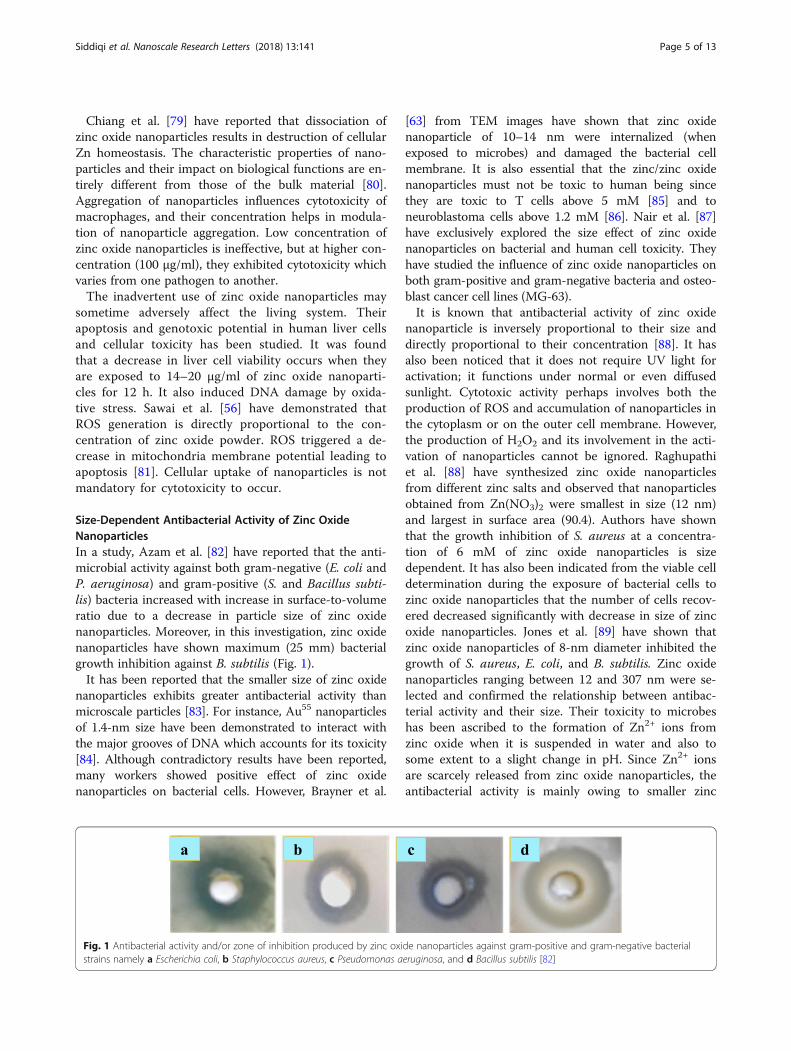
and gram-positive halophilic bacterial species showed 80%growth inhibition. It was demonstrated that zinc oxidenanoparticles below 5 mM concentration are ineffectiveagainst bacteria. The bulk zinc oxide also did not affectthe growth rate and viable counts, although they showedsubstantial decrease in these parameters. Enterobacterspecies showed dramatic alterations in cell morphologyand reduction in size when treated with zinc oxide.TEM images shown by Akbar and Anal [115] re-
vealed the disrupted cell membrane and accumulationof zinc oxide nanoparticles in the cytoplasm (Fig. 4)which was further confirmed by FTIR, XRD, andSEM. It has been suggested that Zn2+ ions areattached to the biomolecules in the bacterial cell viaelectrostatic forces. They are actually coordinatedwith the protein molecules through the lone pair ofelectrons on the nitrogen atom of protein part.Although there is significant impact of zinc oxidenanoparticles on both the aquatic and terrestrialmicroorganisms and human system, it is yet to beestablished whether it is due to nanoparticles alone oris a combined effect of the zinc oxide nanoparticlesand Zn2+ ions [55, 106, 109, 119]. Antibacterialinfluence of metal oxide nanoparticles includes its
a
Fig. 3 SEM images of Campylobacter jejuni. a Untreated cells from the sammid-log phase of growth were treated with 0.5 mg/ml of zinc oxide nanop
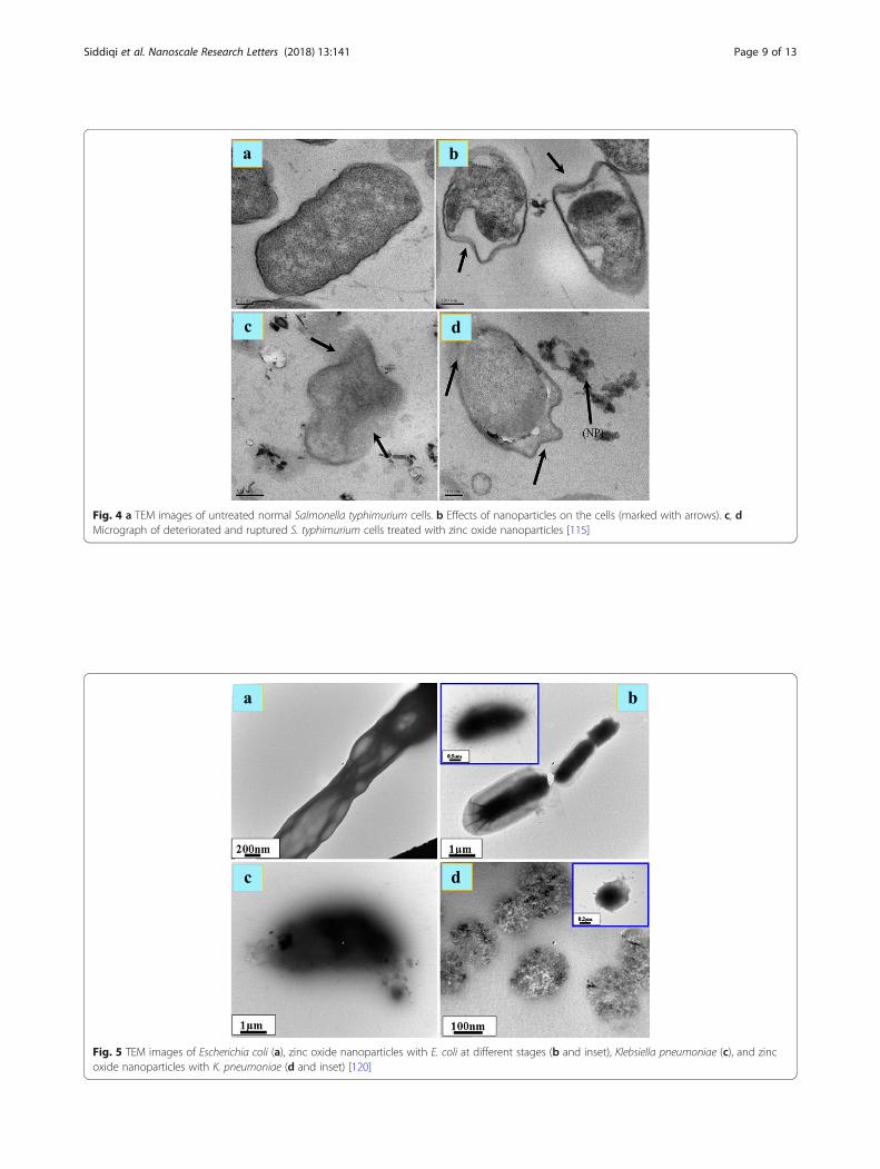
diffusion into the bacterial cell, followed by release ofmetal ions and DNA damage leading to cell death[63, 109–111]. The generation of ROS throughphotocatalysis is also a reason of antibacterial activity[62, 112]. Wahab et al. [120] have shown that whenzinc oxide nanoparticles are ingested, their surfacearea is increased followed by increased absorptionand interaction with both the pathogens and theenzymes. Zinc oxide nanoparticles can therefore beused in preventing the biological system frominfections. It is clear from TEM images (Fig. 5a, b) ofE. coli incubated for 18 h with MIC of zinc oxidenanoparticles that they had adhered to the bacterialcell wall. The outer cell membrane was rupturedleading to cell lysis. In some cases, the cell cleavageof the microbes has not been noticed, but the zincoxide nanoparticles can yet be seen entering the innercell wall (Fig. 5c, d). As a consequence of it, theintracellular material leaks out leading to cell death,regardless of the thickness of bacterial cell wall.Mechanism of interaction of zinc oxide nanoparticles
with bacterial cells has been outlined below [120]. Zincoxide absorbs UV-Vis light from the sun and splits theelements of water.
b
e growth conditions were used as a control. b C. jejuni cells in thearticles for 12 h under microaerobic conditions [114]
a b
c d
Fig. 4 a TEM images of untreated normal Salmonella typhimurium cells. b Effects of nanoparticles on the cells (marked with arrows). c, dMicrograph of deteriorated and ruptured S. typhimurium cells treated with zinc oxide nanoparticles [115]
a b
c d
Fig. 5 TEM images of Escherichia coli (a), zinc oxide nanoparticles with E. coli at different stages (b and inset), Klebsiella pneumoniae (c), and zincoxide nanoparticles with K. pneumoniae (d and inset) [120]
Siddiqi et al. Nanoscale Research Letters (2018) 13:141 Page 9 of 13

Siddiqi et al. Nanoscale Research Letters (2018) 13:141 Page 10 of 13
Dissolved oxygen molecules are transformed intosuperoxide, O2
−, which in turn reacts with H+ togenerate HO2 radical and after collision with electronsproduces hydrogen peroxide anion, HO2
−. Theysubsequently react with H+ ions to produce H2O2.
It has been suggested that negatively charged hydroxylradicals and superoxide ions cannot penetrate into thecell membrane. The free radicals are so reactive thatthey cannot stay in free and, therefore, they can eitherform a molecule or react with a counter ion to give an-other molecule. However, it is true that zinc oxide canabsorb sun light and help in cleaving water moleculeswhich may combine in many ways to give oxygen.Mechanism of oxygen production in the presence of zincoxide nanoparticles still needs experimental evidence.
Zinc oxide at a dose of 5 μg/ml has been found to behighly effective for all the microorganisms which can betaken as minimum inhibitory dose.
ConclusionsZinc is an indispensable inorganic element universally usedin medicine, biology, and industry. Its daily intake in anadult is 8–15 mg/day, of which approximately 5–6 mg/dayis lost through urine and sweat. Also, it is an essential con-stituent of bones, teeth, enzymes, and many functional pro-teins. Zinc metal is an essential trace element for man,animal, plant, and bacterial growth while zinc oxide nano-particles are toxic to many fungi, viruses, and bacteria.People with inherent genetic deficiency of soluble zinc-binding protein suffer from acrodermatitis enteropathica, agenetic disease indicated by python like rough and scalyskin. Although conflicting reports have been received aboutnanoparticles due to their inadvertent use and disposal,some metal oxide nanoparticles are useful to men, animals,and plants. The essential nutrients become harmful whenthey are taken in excess. Mutagenic potential of zinc oxidehas not been thoroughly studied in bacteria even thoughDNA-damaging potential has been reported. It is true that
zinc oxide nanoparticles are activated by absorption of UVlight without disturbing the other rays. If zinc oxide nano-particles produce ROS, they can damage the skin and can-not be used as sun screen. Antibacterial activity may becatalyzed by sunlight, but hopefully, it can prevent the for-mation of ROS. Zinc oxide nanoparticles and zinc nanopar-ticles coated with soluble polymeric material may be usedfor treating wounds, ulcers, and many microbial infectionsbesides being used as drug carrier in cancer therapy. It hasgreat potential as a safe antibacterial drug which may re-place antibiotics in future. Application of zinc oxide nano-particles in different areas of science, medicine, andtechnology suggests that it is an indispensable substancewhich is equally important to man and animals. However,longtime exposure with higher concentration may be harm-ful to living system.
AcknowledgementsThe authors are thankful to publishers for the permission to adopt the tableand figures in this review.
Authors’ contributionsAH, KSS, AR, and T gathered the research data. AH and KSS analyzed thesedata and wrote this review paper. All the authors read and approved thefinal manuscript.
Competing interestsThe authors declare that they have no competing interests.
Publisher’s NoteSpringer Nature remains neutral with regard to jurisdictional claims inpublished maps and institutional affiliations.
Author details1Department of Chemistry, Aligarh Muslim University, Aligarh, Uttar Pradesh202002, India. 2Department of Saidla (Unani Pharmacy), Aligarh MuslimUniversity, Aligarh, Uttar Pradesh 202002, India. 3Department of Biology,College of Natural and Computational Sciences, University of Gondar, P.O.Box #196, Gondar, Ethiopia.
Received: 4 October 2017 Accepted: 16 April 2018
References1. Husen A, Siddiqi KS (2014) Phytosynthesis of nanoparticles: concept,
controversy and application. Nano Res Lett 9:2292. Husen A, Siddiqi KS (2014) Plants and microbes assisted selenium
nanoparticles: characterization and application. J Nanobiotechnol 12:283. Husen A, Siddiqi KS (2014) Carbon and fullerene nanomaterials in plant
system. J Nanobiotechnol 12:164. Siddiqi KS, Husen A (2016) Fabrication of metal nanoparticles from fungi
and metal salts: scope and application. Nano Res Lett 11:985. Siddiqi KS, Husen A (2016) Fabrication of metal and metal oxide
nanoparticles by algae and their toxic effects. Nano Res Lett 11:3636. Siddiqi KS, Husen A (2016) Engineered gold nanoparticles and plant
adaptation potential. Nano Res Lett 11:4007. Siddiqi KS, Husen A (2016) Green synthesis, characterization and uses of
palladium/platinum nanoparticles. Nano Res Lett 11:4828. Siddiqi KS, Rahman A, Tajuddin, Husen A (2016) Biogenic fabrication of iron/
iron oxide nanoparticles and their application. Nano Res Lett 11:4989. Siddiqi KS, Husen A (2017) Recent advances in plant-mediated engineered
gold nanoparticles and their application in biological system. J TraceElements Med Biol 40:10–23
10. Siddiqi KS, Husen A, Rao RAK (2018) A review on biosynthesis of silvernanoparticles and their biocidal properties. J Nanobiotechnol 16:14

Siddiqi et al. Nanoscale Research Letters (2018) 13:141 Page 11 of 13
11. Rai M, Yadav A, Gade A (2009) Silver nanoparticles as a new generation ofantimicrobials. Biotechnol Adv 27:76–83
12. Sawai J (2003) Quantitative evaluation of antibacterial activities of metallicoxide powders (ZnO, MgO and CaO) by conductimetric assay. J MicrobiolMethods 54:177–182
13. Roselli M, Finamore A, Garaguso I, Britti MS, Mengheri E (2003) Zinc oxideprotects cultured enterocytes from the damage induced by Escherichia coli.J Nutr 133:4077–4082
14. Husen A (2017) Gold nanoparticles from plant system: Synthesis,characterization and their application, In: Nanoscience and Plant–Soil SystemsVol.–48 (Eds. Ghorbanpourn M, Manika K, Varma A) Springer InternationalPublishing AG, Gewerbestrasse 11, 6330 Cham, Switzerland, pp.455–479
15. Chaudhry Q, Scotter M, Blackburn J, Ross B, Boxall A, Castle L, Aitken R,Watkins R (2008) Applications and implications of nanotechnologies for thefood sector. Food Add Cont: Part A 25:241–258
16. Jansen J, Karges W, Rink L (2009) Zinc and diabetes—clinical links andmolecular mechanisms. J Nutr Biochem 20:399–417
17. Maremanda KP, Khan S, Jena G (2014) Zinc protects cyclophosphamide-induced testicular damage in rat: involvement of metallothionein, tesminand Nrf2. Biochem Biophys Res Commun 445:591–596
18. Kim MH, Seo JH, Kim HM, Jeong HJ (2014) Zinc oxide nanoparticles, a novelcandidate for the treatment of allergic inflammatory diseases. Eur JPharmacol 738:31–39
19. Frederickson CJ, Koh JY, Bush AI (2005) The neurobiology of zinc in healthand disease. Na Rev Neurosci 6:449–462
20. Halioua B, Ziskind B (2005) Medicine in the days of the pharaohs. Press ofHarvard University Press, Belknap
21. Ozgur U, Ya IA, Liu C, Teke A, Reshchikov MA, Doğan S, Avrutin V, Cho SJ,Morkoç H (2005) A comprehensive review of ZnO materials and devices. JAppl Phys 98:041301
22. Klingshirn C ZnO: from basics towards applications. Phys Status Solidi 244:3027–3073
23. De Graaf TP, Galley E, Butcher KE (1999) Use of an antimicrobial agent.European patent, p EP1079799
24. Brahms J, Mattai J, Jacoby R, Chopra S, Guenin E (2005) Dry deodorantcontaininga sesquiterpene alcohol and zinc oxide. U.S. Patent 20050191257 A1
25. Speight JG (2002) Chemical and process design handbook. McGraw Hill, Inc., New York
26. Brown HE (1976) Zinc oxide: properties and applications. International LeadZinc Research Organization, New York
27. Lopes de Romana D, Brown KH, Guinard JX (2002) Sensory trial to assessthe acceptability of zinc fortificants added to iron-fortified wheat products. JFood Sci 67:461–465
28. Szabo T, Nemeth J, Dekany I (2003) Zinc oxide nanoparticles incorporatedin ultrathin layer silicate films and their photocatalytic properties. Coll Surf A230:23–35
29. Auer G, Griebler WD, Jahn B (2005) Industrial inorganic pigments, 3rd edn.Wiley-VCH Verlag GmbH & Co. KGaA, Weinheim
30. Heideman G, Noordermeer JWM, Datta RN, Noordermeer WM, van Baarle B(2006) Various ways to reduce zinc oxide levels in S-SBR rubber compounds.Macromol Symp 245-246:657–667
31. Patnaik P (2003) Handbook of inorganic chemicals. McGraw Hill, New York32. Araujo-Lima CF, Nunes RJM, Carpes RM, Aiub FAF, Felzenszwalb I (2017)
Pharmacokinetic and toxicological evaluation of a zinc gluconate-basedchemical sterilant using in vitro and in silico approaches. BioMed Res Inter2017:5746768
33. Wang TX, Lou TJ (2008) Solvothermal synthesis and photoluminescenceproperties of ZnO nanorods and nanorod assemblies from ZnO2nanoparticles. Mater Lett 62:2329–2331
34. Jang JS, Yu CJ, Choi SH, Ji SM, Kim ES, Lee JS (2008) Topotactic synthesis ofmesoporous ZnS and ZnO nanoplates and their photocatalytic activity. JCatal 254:144–155
35. Mahmud S, Johar M, Abdullah PGA, Chong J, Mohamad AK (2006)Nanostructure of ZnO fabricated via french process and its correlation toelectrical properties of semiconducting varistors. Synth React Inorg Met OrgChem Nano-Met Chem 36:155–159
36. Kakiuchi K, Hosono E, Kimura T, Imai H, Fujihara S (2006) Fabrication ofmesoporous ZnO nanosheets from precursor templates grown in aqueoussolutions. J Sol-Gel Sci Technol 39:63–72
37. Mahmud S, Abdullah MJ (2006) Nanotripods of zinc oxide, IEEE Conf.Emerging Technol.—Nanoelectron pp. 442–446
38. Shen L, Zhang H, Guo S (2009) Control on the morphologies of tetrapodZnO nanocrystals. Mater Chem Phys 114:580–583
39. Ding Y, Wang ZL (2009) Structures of planar defects in ZnO nanobelts andnanowires. Micron 40:335–342
40. Wang ZL (2004) Nanostructures of zinc oxide. Mater Tod 7:26–3341. Wang ZL (2004) Zinc oxide nanostructures: growth, properties and
applications. J Phys Condens Mat 16:R829–R85842. Moezzi A, Cortie M, McDonagh A (2011) Aqueous pathways for the
formation of zinc oxide nanoparticles. Dalton Trans 40:4871–487843. Xie J, Li P, Li Y, Wang Y, Wei Y (2009) Morphology control of ZnO particles via
aqueous solution route at low temperature. Mater Chem Phys 114:943–94744. Soares NFF, Silva CAS, Santiago-Silva P, Espitia PJP, Gonçalves MPJC, Lopez
MJG, Miltz J, Cerqueira MA, Vicente AA, Teixeira J, da Silva WA, Botrel DA(2009) Active and intelligent packaging for milk and milk products. In:Coimbra JSR, Teixeira JA (eds) Engineering aspects of milk and dairyproducts. CRC Press Taylor & Francis Group pp, New York, pp 175–199
45. Baum MK, Shor-Posner G, Campa A (2000) Zinc status in humanimmunodeficiency virus infection. J Nutr 130:1421S–1423S
46. Hiller JM, Perlmutter A (1971) Effect of zinc on viral-host interactions in arainbow trout cell line, RTG-2. Water Res 5:703–710
47. Restuccia D, Spizzirri UG, Parisi OI, Giuseppe Cirillo G, Iemma F, Puoci F, Vinci G,Picci N (2010) New EU regulation aspects and global market of active andintelligent packaging for food industry applications. Food Control 21:1425–1435
48. Espitia PJP, Soares NFF, Coimbra JSR, Andrade NJ, Cruz RS, Medeiros EAA(2015) Zinc oxide nanoparticles: synthesis, antimicrobial activity and foodpackaging application. Food Bioprocess Technol 5:1447–1464
49. Hu X, Cook S, Wang P, Hwang HM (2009) In vitro evaluation of cytotoxicityof engineered metal oxide nanoparticles. Sci Total Environ 407:3070–3072
50. Wang C, Lu J, Zhou L, Li J, Xu J, Li W, Zhang L, Zhong X, Wang T (2016)Effects of long-term exposure to zinc oxide nanoparticles on development,zinc metabolism and biodistribution of minerals (Zn, Fe, Cu, Mn) in mice.PLoS One 11:e0164434
51. Li CH, Shen CC, Cheng YW, Huang SH, Wu CC, Kao CC, Liao JW, Kang JJ(2012) Organ biodistribution, clearance, and genotoxicity of orallyadministered zinc oxide nanoparticles in mice. Nanotoxicology 6:746–756
52. Singh A, Singh NB, Afzal S, Singh T, Hussain I (2017) Zinc oxidenanoparticles: a review of their biological synthesis, antimicrobial activity,uptake, translocation and biotransformation in plants. J Mater Sci. https://doi.org/10.1007/s10853-017-1544-1
53. Kelly SA, Havrilla CM, Brady TC, Abramo KH, Levin ED (1998) Oxidative stressin toxicology: established mammalian and emerging piscine model systems.Environ Health Perspect 106:375–384
54. Rikans LE, Hornbrook KR (1997) Lipid peroxidation, antioxidant protectionand aging. Biochim Biophys Acta 1362:116–127
55. Zhang L, Jiang Y, Ding Y, Povey M, York D (2007) Investigation into theantibacterial behaviour of suspensions of ZnO nanoparticles (ZnOnanofluids). J Nanopart Res 9:479–489
56. Sawai J, Shoji S, Igarashi H, Hashimoto A, Kokugan T, Shimizu M, Kojima H(1998) Hydrogen peroxide as an antibacterial factor in zinc oxide powderslurry. J Ferment Bioeng 86:521–522
57. Lin D, Xing B (2007) Phytotoxicity of nanoparticles: inhibition of seedgermination and root growth. Environ Pollut 150:243–250
58. Kahru A, Ivask A, Kasemets K, Pollumaa L, Kurvet I, François M, DubourguierHC (2005) Bio-tests and biosensors in ecotoxicological risk assessment offield soils polluted with zinc, lead and cadmium. Environ Toxicol Chem 24:2973–2982
59. Heinlaan M, Ivask A, Blinova I, Dubourguier HC, Kahru A (2008) Toxicity ofnanosized and bulk ZnO, CuO and TiO2 to bacteria Vibrio fischeri andcrustaceans Daphnia magna and Thamnocephalus platyurus. Chemosphere71:1308–1316
60. Elster C, Fourest E, Baudin F, Larsen K, Cusack S, Ruigrok RW (1994) A smallpercentage of influenza virus M1 protein contains zinc but zinc does notinfluence in vitro M1 RNA interaction. Gen J Virol 75:37–42
61. Lee SP, Xiao J, Knutson JR, Lewis MS, Han MK (1997) Zn2+ promotes theself-association of human immunodeficiency virus type-1 integrase in vitro.Biochemistry 36:173–180
62. Adams LK, Lyon DY, Alvarez PJJ (2006) Comparative eco-toxicity ofnanoscale TiO2, SiO2, and ZnO water suspensions. Water Res 40:3527–3532
63. Brayner R, Ferrari-Iliou R, Brivois N, Djediat S, Benedetti MF, Fiévet F (2006)Toxicological impact studies based on Escherichia coli bacteria in ultrafineZnO nanoparticles colloidal medium. Nano Lett 6:866–870

Siddiqi et al. Nanoscale Research Letters (2018) 13:141 Page 12 of 13
64. Stoimenov PK, Klinger RL, Marchin GL, Klabunde KJ (2002) Metal oxidenanoparticles as bactericidal agents. Langmuir 18:6679–6686
65. Yamamoto O, Komatsu M, Sawai J, Nakagawa ZE (2004) Effect of latticeconstant of zinc oxide on antibacterial characteristics. J Mater Sci MaterMed 15:847–851
66. Premanathan M, Karthikeyan K, Jeyasubramanian K, Manivannan G (2011)Selective toxicity of ZnO nanoparticles toward Gram-positive bacteria andcancer cells by apoptosis through lipid peroxidation. Nanomedicine 7:184–192
67. Lovric J, Cho SJ, Winnik FM, Maysinger D (2005) Unmodified cadmiumtelluride quantum dots induce reactive oxygen species formation leading tomultiple organelle damage and cell death. Chem Biol 12:1227–1234
68. Xia T, Kovochich M, Brant J, Hotze M, Sempf J, Oberley T, Sioutas C, Yeh JI,Wiesner MR, Nel AE (2006) Comparison of the abilities of ambient andmanufactured nanoparticles to induce cellular toxicity according to anoxidative stress paradigm. Nano Lett 6:1794–1807
69. Long TC, Saleh N, Tilton RD, Lowry GV, Veronesi B (2006) Titaniumdioxide (P25) produces reactive oxygen species in immortalized brainmicroglia (BV2): implications for nanoparticle neurotoxicity. Environ SciTechnol 40:4346–4352
70. Pati R, Mehta RK, Mohanty S, Padhi A, Sengupta M, Vaseeharan B, GoswamiC, Sonawane A (2014) Topical application of zinc oxide nanoparticlesreduces bacterial skin infection in mice and exhibits antibacterial activity byinducing oxidative stress response and cell membrane disintegration inmacrophages. Nanomedicine 10:1195–1208
71. Siddiqi KS, Husen A (2017) Plant response to engineered metal oxidenanoparticles. Nano Res Lett 12:92
72. Liu Y, He L, Mustapha A, Li H, Hu ZQ, Lin M (2009) Antibacterial activities ofzinc oxide nanoparticles against Escherichia coli O157:H7. J Appl Microbiol107:1193–1201
73. Dutta RK, Sharma PK, Bhargave R, Kumar N, Pandey AC (2010) Differentialsusceptibility of Escherichia coli cells toward transition metal-doped andmatrix-embedded ZnO nanoparticles. J Phys Chem B 114:5594–5599
74. Banoee M, Seif S, Nazari ZE, Jafari-Fesharaki P, Shahverdi HR, Moballegh A,Moghaddam KM, Shahverdi AR (2010) ZnO nanoparticles enhancedantibacterial activity of ciprofloxacin against Staphylococcus aureus andEscherichia coli. J Biomed Mater Res B 93B:557–561
75. Baek YW, An YJ (2011) Microbial toxicity of metal oxide nanoparticles (CuO,NiO, ZnO, and Sb2O3) to Escherichia coli, Bacillus subtilis, and Streptococcusaureus. Sci Total Environ 409:1603–1608
76. Karlsson HL, Toprak MS, Fadeel B (2014) Toxicity of metal and metal oxidenanoparticle. In: Nordberg GF, Fowler BA, Nordberg M (eds) Handbook onthe toxicology of metals, 4th edn. Academic Press pp, London, pp 75–112
77. Cho WS, Duffin R, Howie SE, Scotton CJ, Wallace WA, Macnee W, Bradley M,Megson IL, Donaldson K (2011) Progressive severe lung injury by zinc oxidenanoparticles; the role of Zn2+ dissolution inside lysosomes. Part FibreToxicol 8:27
78. Tuomela S, Autio R, Buerki-Thurnherr T, Arslan O, Kunzmann A, Andersson-Willman B, Wick P, Mathur S, Scheynius A, Krug HF, Fadeel B, Lahesmaa R (2013)Gene expression profiling of immune-competent human cells exposed toengineered zinc oxide or titanium dioxide nanoparticles. PLoS One 8:e68415
79. Chiang HM, Xia Q, Zou X, Wang C, Wang S, Miller BJ, Howard PC, Yin JJ,Beland FA, Yu H, Fu PP (2012) Nanoscale ZnO induces cytotoxicity and DNAdamage in human cell lines and rat primary neuronal cells. J NanosciNanotechnol 12:2126–2135
80. Seabra AB, Haddad P, Duran N (2013) Biogenic synthsis of nanostructurediron compound: applications and perspectives. IET Nanobiotechnol 7:90–99
81. Sharma V, Anderson D, Dhawan A (2012) Zinc oxide nanoparticles induceoxidative DNA damage and ROS-triggered mitochondria mediatedapoptosis in human liver cells (HepG2). Apoptosis 17:852–870
82. Azam A, Ahmed AS, Oves M, Khan MS, Habib SS, Memic A (2012) Antimicrobialactivity of metal oxide nanoparticles against Gram-positive and Gram-negativebacteria: a comparative study. Int J Nanomedicine 7:6003–6009
83. Yamamoto O (2013) Influence of particle size on the antibacterial activity ofzinc oxide. Int J Inorg Mater 3:643–646
84. Tsoli M, Kuhn H, Brandau W, Esche H, Schmid G (2005) Cellular uptake andtoxicity of Au55 clusters. Small 1:841–844
85. Reddy KM, Feris K, Bell J, Wingett DG, Hanley C, Punnoose A (2007)Selective toxicity of zinc oxide nanoparticles to prokaryotic and eukaryoticsystems. Appl Phys Lett 90:2139021–2139023
86. Jeng HA, Swanson J (2006) Toxicity of metal oxide nanoparticles inmammalian cells. J Enviorn Sci Health 41:2699–2711
87. Nair S, Sasidharan A, Divya Rani VV, Menon D, Nair S, Manzoor K, RainaS (2009) Role of size scale of ZnO nanoparticles and microparticles ontoxicity toward bacteria and osteoblast cancer cells. J Mater Sci MaterMed 20:S235–S241
88. Raghupathi KR, Koodali RT, Manna AC (2011) Size-dependent bacterialgrowth inhibition and mechanism of antibacterial activity of zinc oxidenanoparticles. Langmuir 27:4020–4028
89. Jones N, Ray B, Koodali RT, Manna AC (2008) Antibacterial activity of ZnOnanoparticles suspensions on a broad spectrum of microorganisms. FEMSMicrobiol Lett 279:71–76
90. Sahu D, Kannan GM, Tailang M, Vijayaraghavan R (2016) In vitro cytotoxicityof nanoparticles: a comparison between particle size and cell type. JNanosci 2016:4023852
91. Ng CT, Yong LQ, Hande MP, Ong CN, Yu LE, Bay BH, Baeg GH (2017) Zincoxide nanoparticles exhibit cytotoxicity and genotoxicity through oxidativestress responses in human lung fibroblasts and Drosophila melanogaster. IntJ Nanomedicine 12:1621–1637
92. Papavlassopoulos H, Mishra YK, Kaps S, Paulowicz I, Abdelaziz R, Elbahri M,Maser E, Adelung R, Röhl C (2014) Toxicity of functional nano-micro zincoxide tetrapods: impact of cell culture conditions, cellular age and materialproperties. PLoS One 9:e84983
93. Taccola L, Raffa V, Riggio C, Vittorio O, Iorio MC, Vanacore R, Pietrabissa A,Cuschieri A (2011) Zinc oxide nanoparticles as selective killers ofproliferating cells. Int J Nanomedicine 6:1129–1140
94. Zhou YM, Zhong CY, Kennedy IM, Leppert VJ, Pinkerton KE (2003)Oxidative stress and NFkappaB activation in the lungs of rats: asynergistic interaction between soot and iron particles. Toxicol ApplPharmacol 190:157–169
95. Yang H, Liu C, Yang D, Zhang H, Xi Z (2009) Comparative study ofcytotoxicity, oxidative stress and genotoxicity induced by four typicalnanomaterials: the role of particle size, shape and composition. J ApplToxicol 29:69–78
96. Schwartz VB, Thétiot F, Ritz S, Pütz S, Choritz L, Lappas A, Förch R,Landfester K, Jonas U (2012) Antibacterial surface coatings from zinc oxidenanoparticles embedded in poly(N-isopropylacrylamide) hydrogel surfacelayers. Adv Funct Mater 22:2376–2386
97. Stankovic A, Dimitrijevic S, Uskokovic D (2013) Influence of size scaleand morphology on antibacterial properties of ZnO powdershydrothermally synthesized using different surface stabilizing agents.Colloids Surf B 102:21–28
98. Laurent S, Forge D, Port M, Roch A, Robic C, Elst LV, Muller RN (2008)Magnetic iron oxide nanoparticles: synthesis, stabilization, vectorization,physicochemical characterizations, and biological applications. ChemRev 108:2064–2110
99. Yamamoto O, Hotta M, Sawai J, Sawai J, Sasamoto T, Kojima H (1998)Influence of powder characteristic of ZnO on antibacterial activity: effect ofspecific surface area. J Ceram Soc Jpn 106:1007–1011
100. Kumar PTS, Lakshmanan VK, Anilkumar TV, Ramya C, Reshmi P, UnnikrishnanAG, Nair SV, Jayakumar R (2012) Flexible and microporous chitosanhydrogel/nano ZnO composite bandages for wound dressing: in vitro andin vivo evaluation. ACS Appl Mater Interfaces 4:2618–2629
101. George S, Pokhrel S, Xia T, Gilbert B, Ji Z, Schowalter M, Rosenauer A,Damoiseaux R, Bradley KA, Mädler L, Nel AE (2010) Use of a rapidcytotoxicity screening approach to engineer a safer zinc oxide nanoparticlethrough iron doping. ACS Nano 4:15–12
102. Song W, Zhang J, Guo J, Zhang J, Ding F, Li L, Sun Z (2010) Role of thedissolved zinc ion and reactive oxygen species in cytotoxicity of ZnOnanoparticles. Toxicol Lett 199:389–339
103. Buerki-Thurnherr T, Xiao L, Diener L, Arslan O, Hirsch C, Maeder-Althaus X,Grieder K, Wampfler B, Mathur S, Wick P, Krug HF (2013) In vitro mechanisticstudy towards a better understanding of ZnO nanoparticle toxicity.Nanotoxicology 7:402–416
104. Gilbert B, Fakra SC, Xia T, Pokhrel S, Mädler L, Nel AE (2012) The fate of ZnOnanoparticles administered to human bronchial epithelial cells. ACS Nano 6:4921–4930
105. Raffi M, Hussain F, Bhatti TM, Akhter JI, Hameed A, Hasan MM (2008)Antibacterial characterization of silver nanoparticles against E. Coli ATCC-15224. J Mater Sci Technol 24:2192–2196
106. Choi OK, Hu ZQ (2008) Size dependent and reactive oxygen species relatednanosilver toxicity to nitrifying bacteria. Environ Sci Technol42:4583–4588

Siddiqi et al. Nanoscale Research Letters (2018) 13:141 Page 13 of 13
107. Lok CN, Ho CM, Chen R, He QY, Yu WY, Sun H, Tam PK, Chiu JF, Che CM(2005) Proteomic analysis of the mode of antibacterial action of silvernanoparticles. Proteome Res 5:916–924
108. Pal S, Tak YK, Song JM (2007) Does the antibacterial activity of silvernanoparticles depend on the shape of the nanoparticle? A study of thegram-negative bacterium Escherichia coli. Appl Environ Microbiol 73:1712–1720
109. Sondi I, Salopek-Sondi B (2004) Silver nanoparticles as antimicrobial agent: acase study on E. coli as a model for Gram-negative bacteria. J Colloid InterfSci 275:177–182
110. Elechiguerra J, Burt J, Morones J, Camacho-Bragado A, Gao X, Lara HH,Yacaman MJ (2005) Interaction of silver nanoparticles with HIV-1. JNanobiotechnol 3:6
111. Huang Z, Zheng X, Yan D, Yin G, Liao X, Kang Y, Yao Y, Huang D, Hao B(2008) Toxicological effect of ZnO nanoparticles based on bacteria.Langmuir 24:4140–4144
112. Zan L, Fa W, Peng T, Gong ZK (2007) Photocatalysis effect of nanometerTiO2 and TiO2-coated ceramic plate on hepatitis B virus. J PhotochemPhotobiol B 86:165–169
113. Bajpai KS, Chand N, Chaurasia V (2012) Nano zinc oxide-loaded calciumalginate films with potential antibacterial properties. Food BioprocessTechnol 5:1871–1881
114. Xie Y, He Y, Irwin LP, Jin T, Shi X (2011) Antibacterial activity andmechanism of action of zinc oxide nanoparticles against Campylobacterjejuni. Appl Environ Microbiol 77:2325–2331
115. Akbar A, Anal AK (2014) Zinc oxide nanoparticles loaded active packaging, achallenge study against Salmonella typhimurium and Staphylococcus aureusin ready-to-eat poultry meat. Food Control 38:88–95
116. Akhtar MJ, Ahamed M, Kumar S, Majeed Khan MA, Ahmad J, Alrokayan SA(2012) Zinc oxide nanoparticles selectively induce apoptosis in humancancer cells through reactive oxygen species. Int J Nanomedicine 7:845–857
117. Leung YH, Xu X, Ma APY, Liu F, Ng AMC, Shen Z, Gethings LA, Guo MY,Djurišić AB, Lee PKH, Lee HK, Chan WK, Leung FCC (2016) Toxicity of ZnOand TiO2 to Escherichia coli cells. Sci Rep 6:35243
118. Sinha R, Karan R, Sinha A, Khare SK (2011) Interaction and nanotoxic effectof ZnO and Ag nanoparticles on mesophilic and halophilic bacterial cells.Bioresour Technol 102:1516–1520
119. Jones N, Ray B, Ranjit KT, Manna AC (2008) Antibacterial activity of ZnOnanoparticle suspensions on a broad spectrum of microorganisms. FEMSMicrobiol Lett 279:71–76
120. Wahab R, Mishra A, Yun SI, Kim YS, Shin HS (2010) Antibacterial activity ofZnO nanoparticles prepared via non-hydrolytic solution route. ApplMicrobiol Biotechnol 87:1917–1925