Prokaryotic and Eukaryotic Cells. Prokaryotic vs. Eukaryotic No true nucleus No organelles Complex...
32
Prokaryotic and Eukaryotic Cells
-
Upload
todd-sneary -
Category
Documents
-
view
246 -
download
3
Transcript of Prokaryotic and Eukaryotic Cells. Prokaryotic vs. Eukaryotic No true nucleus No organelles Complex...
Prokaryotic and Eukaryotic Cells
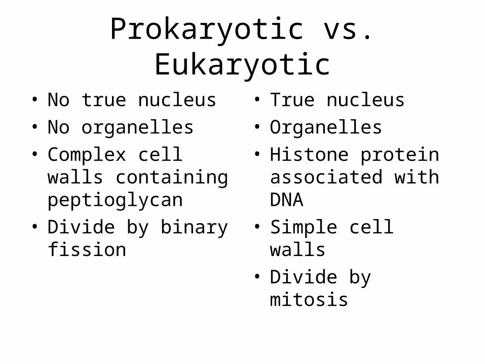
Prokaryotic vs. Eukaryotic
• No true nucleus• No organelles• Complex cell walls
containing peptioglycan
• Divide by binary fission
• True nucleus• Organelles• Histone protein
associated with DNA• Simple cell walls• Divide by mitosis
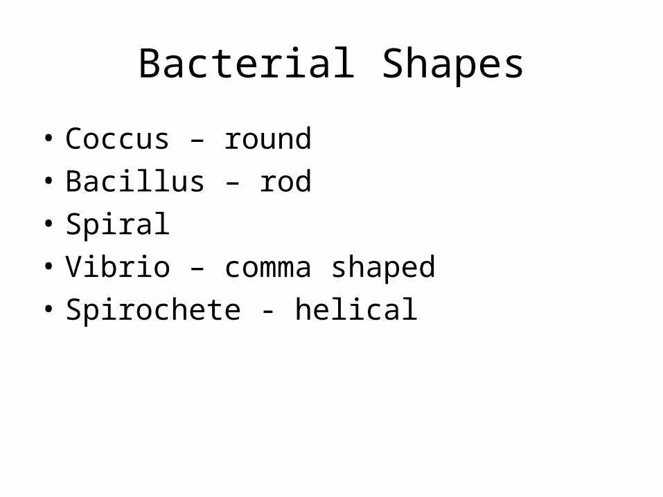
Bacterial Shapes
• Coccus – round
• Bacillus – rod
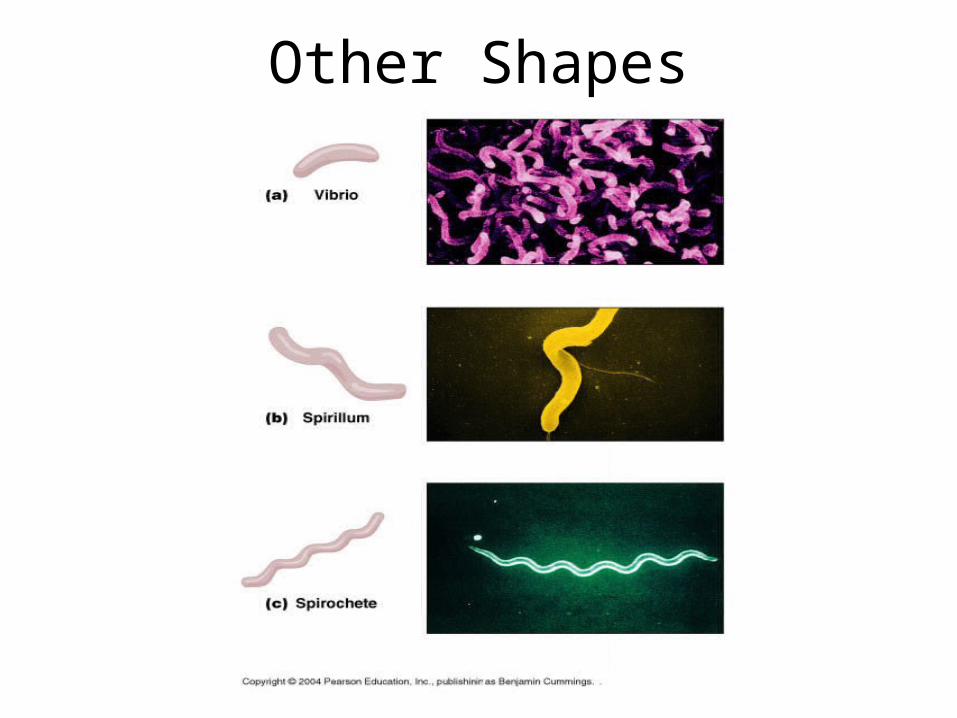
• Spiral
• Vibrio – comma shaped
• Spirochete - helical
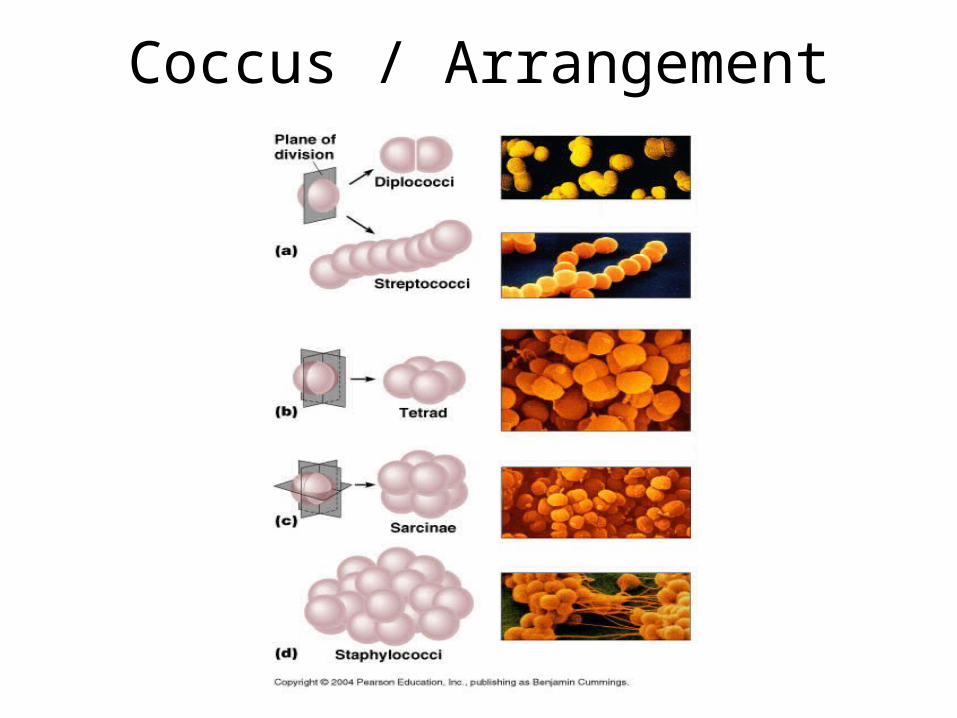
Coccus / Arrangement
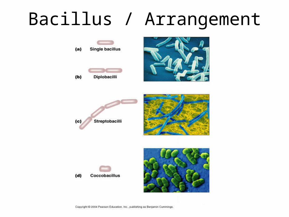
Bacillus / Arrangement
Other Shapes
Bacterial Shapes
• Most bacteria exhibit only one shape, they are said to be MONOMORPHIC
• Some bacteria can exhibit many shapes, they are said to be PLEOMORPHIC
• Members of genus Corynebacterium are pleomorphic
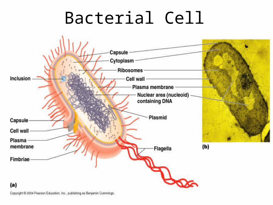
Bacterial Cell
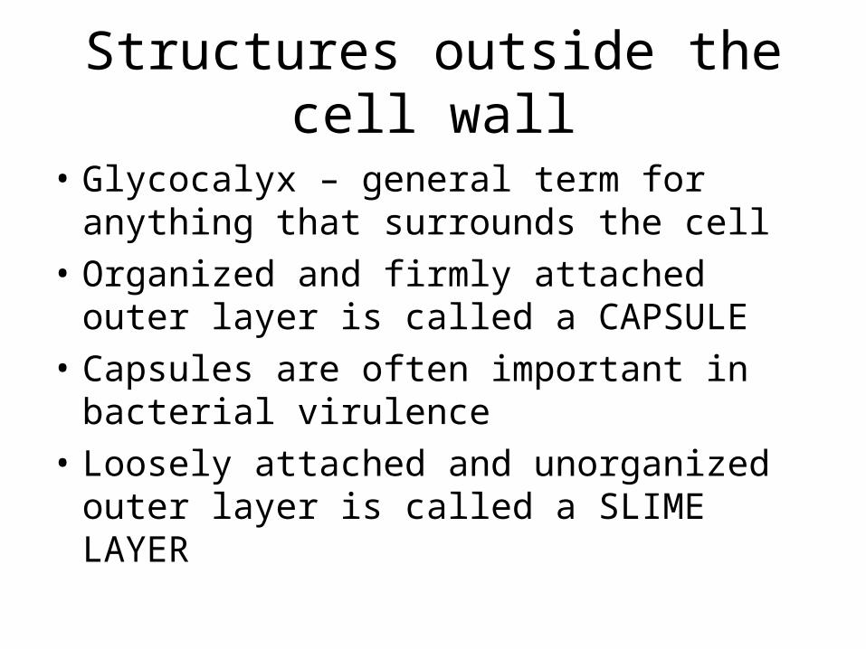
Structures outside the cell wall
• Glycocalyx – general term for anything that surrounds the cell
• Organized and firmly attached outer layer is called a CAPSULE
• Capsules are often important in bacterial virulence
• Loosely attached and unorganized outer layer is called a SLIME LAYER
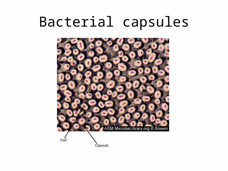
Bacterial capsules
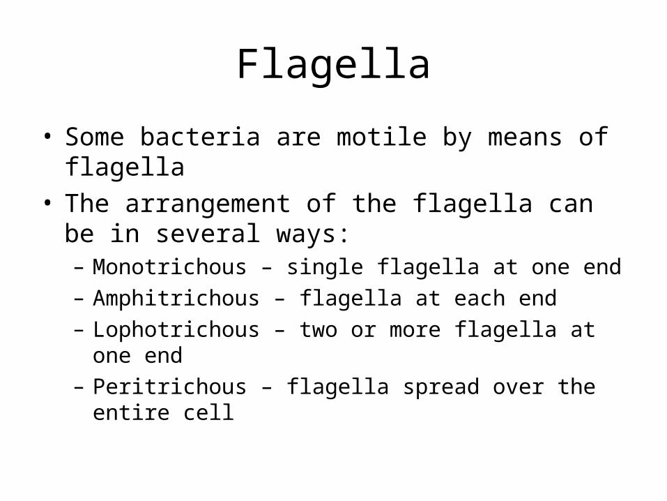
Flagella
• Some bacteria are motile by means of flagella• The arrangement of the flagella can be in
several ways:– Monotrichous – single flagella at one end– Amphitrichous – flagella at each end– Lophotrichous – two or more flagella at one end– Peritrichous – flagella spread over the entire cell
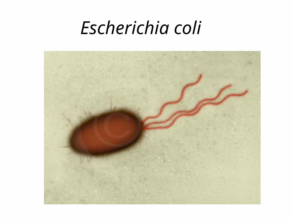
Escherichia coli
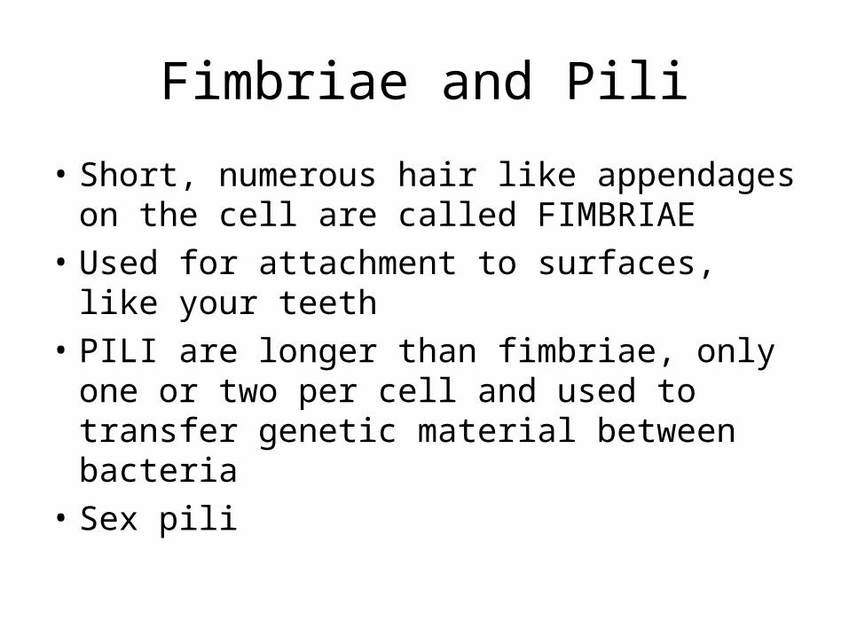
Fimbriae and Pili
• Short, numerous hair like appendages on the cell are called FIMBRIAE
• Used for attachment to surfaces, like your teeth
• PILI are longer than fimbriae, only one or two per cell and used to transfer genetic material between bacteria
• Sex pili
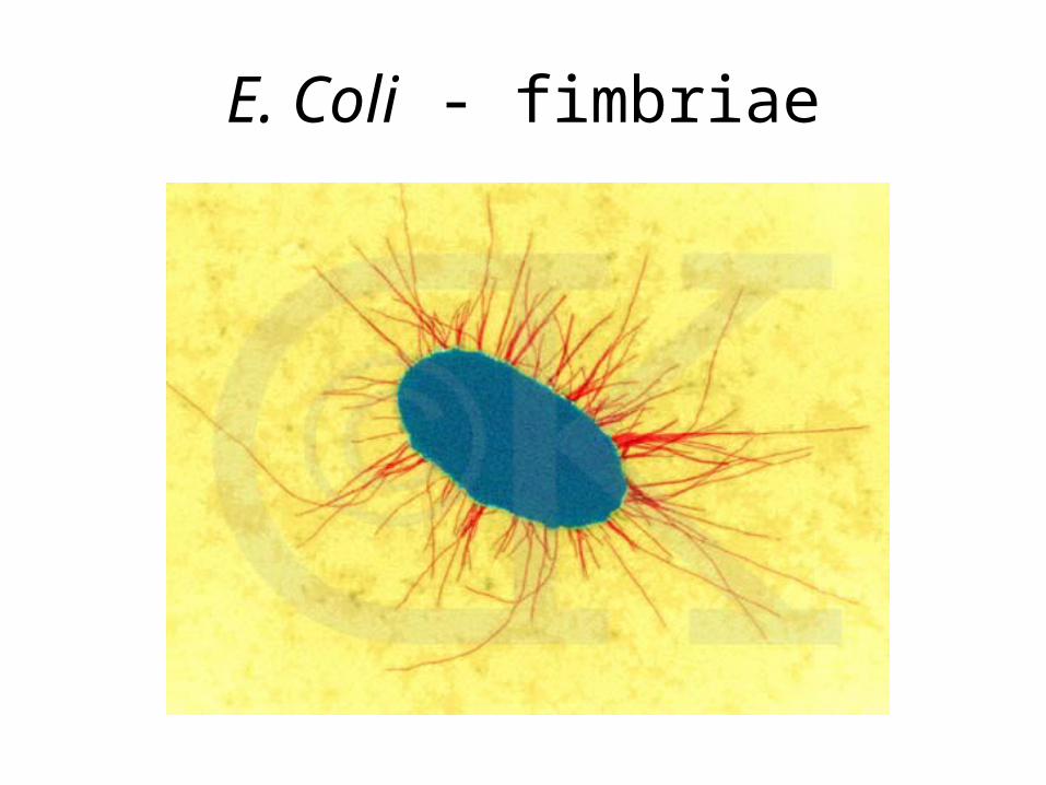
E. Coli - fimbriae
Bacterial Cell Wall
• All bacteria, except for one genus, are surrounded by a rigid cell wall
• Cell wall is composed of a complex macromolecule called PEPTIDOGLYCAN
• Function of the cell wall is to– Maintain the shape of the cell– Prevent the cell from rupturing in high water
pressure– Anchor point for flagella
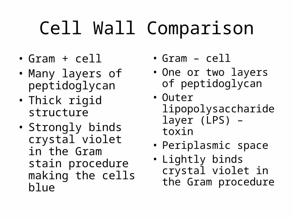
Cell Wall Comparison
• Gram + cell• Many layers of
peptidoglycan• Thick rigid structure• Strongly binds crystal
violet in the Gram stain procedure making the cells blue
• Gram – cell• One or two layers of
peptidoglycan• Outer
lipopolysaccharide layer (LPS) – toxin
• Periplasmic space• Lightly binds crystal
violet in the Gram procedure
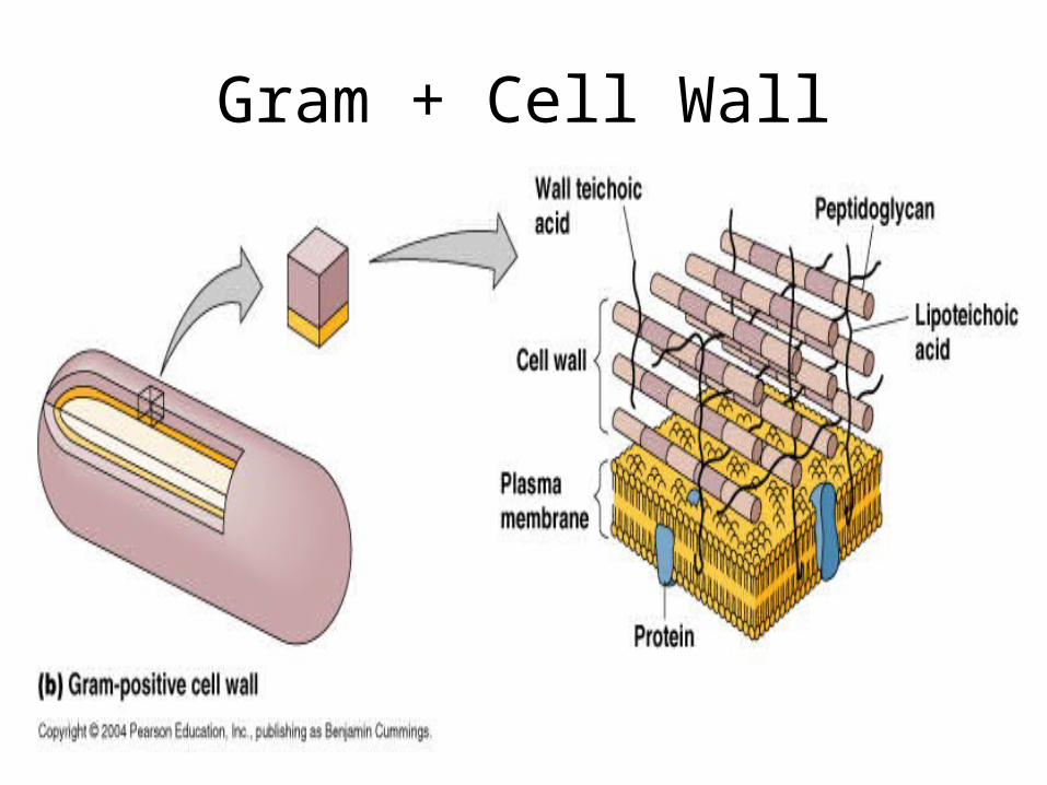
Gram + Cell Wall
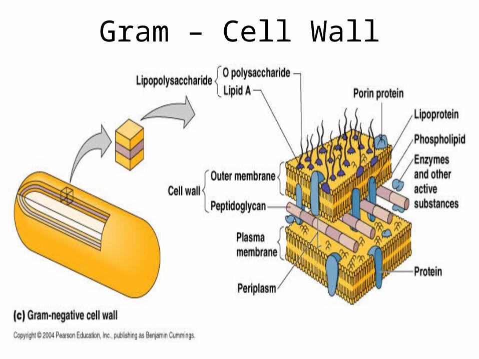
Gram – Cell Wall
Can a bacteria survive without a cell wall?
• Yes, but only special conditions
• The enzyme lysozyme can break down the protein of the cell wall
• Gram + cell in an isotonic environment + lysozyme = protoplast
• Gram – cell in an isotonic environment + lysozyme = spheroplast
Acid-fast cells
• Cell wall is about 60% peptidoglycan
• Cell wall contains MYCOLIC ACID
• Waxy lipid substance
• Cells generally stain as Gram +
• Strongly bind carbol fuchsin in the acid-fast staining procedure
• Cells stain a bright purple red color in the acid-fast procedure
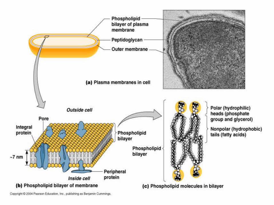
Plasma Membrane
• Phospholipid bilayer with associated proteins
• Selective permeability
• Nutrient breakdown
• Energy production
Nuclear area
• Bacterial cells contain ONE chromosome• Circular in structure• Just DNA, no associated proteins• Some cells contain PLASMIDS• Plasmids are separate from the main
chromosome• Also circular in structure, but smaller• Contain only a few genes• Often carry genes for antibiotic resistance
Ribosomes
• Protein synthesis
• Prokaryotic ribosomes are 70S in size
• Eukaryotic ribosomes are 80S in size
• Difference in size is important in antibiotic activity
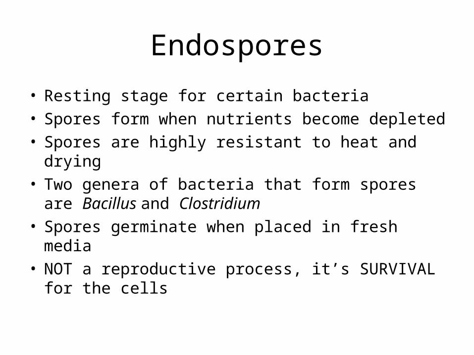
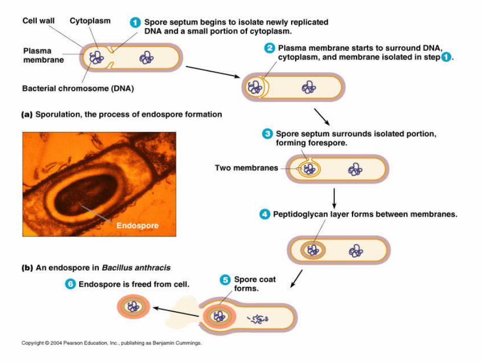
Endospores
• Resting stage for certain bacteria• Spores form when nutrients become depleted• Spores are highly resistant to heat and drying• Two genera of bacteria that form spores are
Bacillus and Clostridium• Spores germinate when placed in fresh media• NOT a reproductive process, it’s SURVIVAL for
the cells
Movement Across Membranes
• Simple diffusion – passive
• Facilitated diffusion – passive
• Osmosis – passive
• Active transport – requires the cell use energy
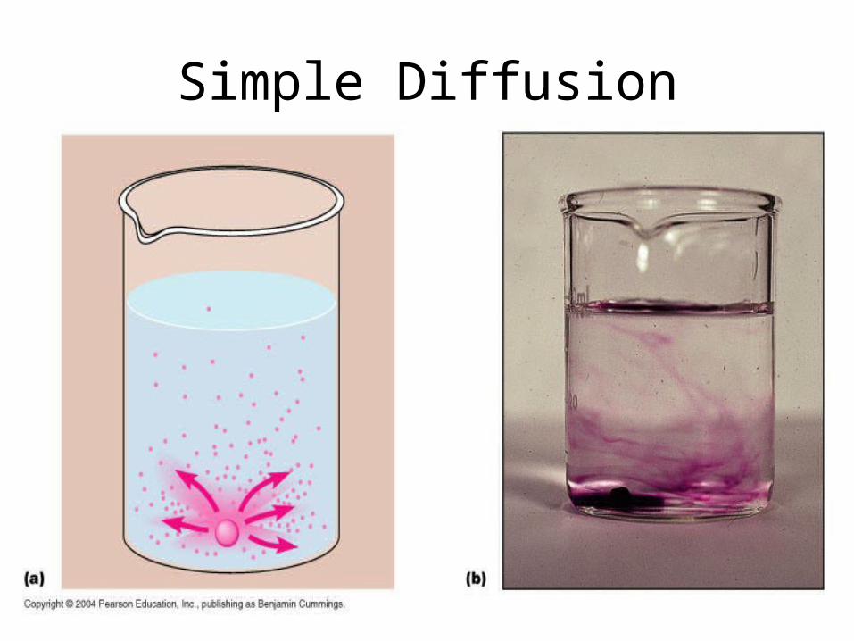
Simple Diffusion
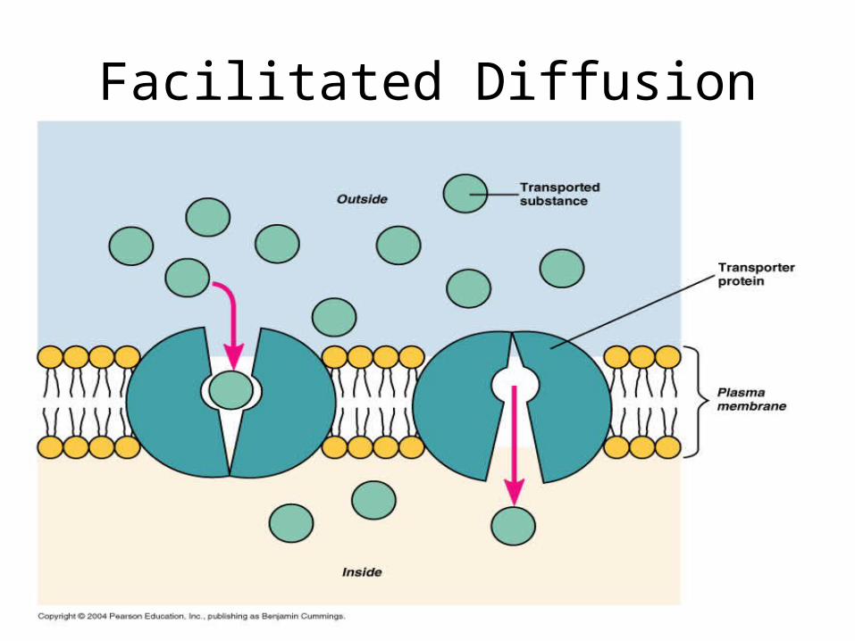
Facilitated Diffusion
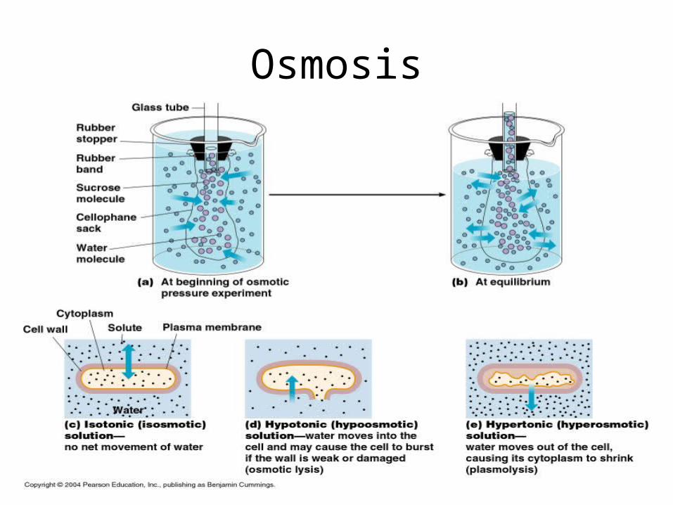
Osmosis
Active Transport
• Cell uses energy in the form of ATP
• Nutrients are concentrated inside the cell against the concentration gradient
• Transporters in the cell membrane are responsible for this active process
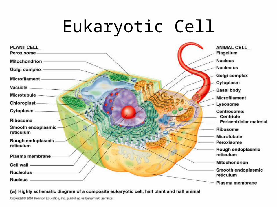
Eukaryotic Cell