Project Report - AYUSH RESEARCH PORTALayushportal.nic.in/EMR/PRECLINICAL_FINAL_REPORT-1.pdf ·...
25
Project Report “Cissus Quadrangularis Linn. (Asthisamharaka) as a regulator of bone progenitor cells: Molecular and functional basis for osteogenic regenerative medicine” Principal Investigator Dr. Kumar MR Bhat Institute Department of Anatomy, Kasturba Medical College Manipal University, Manipal-576104 Supported by Department of AYUSH, Ministry of Health & Family Welfare, Govt. of India, New Delhi
Transcript of Project Report - AYUSH RESEARCH PORTALayushportal.nic.in/EMR/PRECLINICAL_FINAL_REPORT-1.pdf ·...

Project Report
“Cissus Quadrangularis Linn. (Asthisamharaka) as a regulator of
bone progenitor cells: Molecular and functional basis for osteogenic
regenerative medicine”
Principal Investigator
Dr. Kumar MR Bhat
Institute
Department of Anatomy, Kasturba Medical College
Manipal University, Manipal-576104
Supported by
Department of AYUSH, Ministry of Health & Family
Welfare, Govt. of India, New Delhi

Page 1 of 24
Final report
1. Title of the project:
Cissus Quadrangularis Linn. (Asthisamharaka) as a regulator of bone progenitor cells: Cellular and
functional basis for osteogenic regenerative medicine
2. PI –name and address
Dr. Kumar MR Bhat, Associate Professor, Dept. of Anatomy, Kasturba Medical College, Manipal
University, Manipal – 576104, India
3. Co-I name and address
Dr. K Satyamoorthy, Director, Manipal Life Sciences Center, Manipal University, Manipal – 576104,
India
4. Other scientific staff engaged in the study
Ms. Priyadarshini Pai (first 8 months)& Ms. Chinchu (last 4 months) as JRF
5. Non-scientific staff engaged in this study
Ms. Rekha Nayak, Technician, Manipal Life Sciences Center, Manipal University, Manipal
6. Implementing institution and other collaborating institutions
Kasturba Medical College, Manipal University, Manipal
7. Date of commencement – 9/10/10
8. Duration - One year
9. Date of completion - After request for extension of deadline to submit the final report -
15th December 2011
10. Objectives as approved
1. Effect of Cissus quadrangularis (CQ) and Emblica officinalis (EO) on
o Proliferation of MSCs
o Differentiation of MSCs into osteoblast
o Biological activity of osteoblast
o Differentiation of osteoclast cells
o Biological activity of osteoclast
2. Osteoprotective mechanisms of action of these plant extracts singly or in combination, on
osteoporosis and also as a preventive measure for osteoporosis using osteoporotic rat model.

Page 2 of 24
11. Deviation made from the original objectives if any, while implementing the project and
reasons thereof.
Experiments related to osteoclast isolation and differentiation, evaluation of CQ and EO on their
proliferation, differentiation and biological activities did not give any conclusive results even after
four repeated attempts. This may be due to technical error and needs further evaluation.
12. Experimental work giving full details of experimental set up methods adopted, data collected,
supported by the necessary tables, charts, diagrams and photographs
a) Isolation and culturing of Rat bone marrow mesenchymal stem cells (BMSCs);
Rat bone marrow was collected after the ethical committee clearance, under aseptic precaution, from
the bones of the lower limb. Using either Ficoll method or RBC lysis method, bone marrow
mesenchymal stem cells (BMSCs) were obtained and cultured in DMEM with 15% FBS, 100 U/ml
penicillin, and 100 µg/ml streptomycin. The culture was allowed to grow for 4 days of initial culture.
On 5th
day the floating cells were removed and then the cells were allowed to grow and when they
reach 80% confluence, these cells were either subcultured or cryopreserved for further experiments.
For osteoclast cells – after 3 hr of the bone marrow cell culture, the floating cells were collected and
sub cultured.
b) Differentiation of BMSCs into osteoblast
Once the cells are confluent, the stem cell media in which MSCs are growing then be replaced with
osteogenic medium (Dulbelco's Modified Eagle Medium with 10% fetal bovine serum (Gibco), 1 µM
dexamethasone (Sigma), 10 mM µ-glycerol phosphate (Sigma), 0.05 mM ascorbic acid (Sigma), and
100 U/ml penicillin, 100 µg/ml streptomycin (Gibco). The medium was replaced every 3 days and
MSCs were differentiated into osteoblasts after 3 weeks.
c) Differentiation of Bone Marrow haemopoitic stem cells into osteoclasts
After 3 hour initial cell culture, the floating cells were collected and subcultured in DMEM
supplemented with 10% fetal bovine serum (Gibco), 100 U/ml penicillin, 100 µg/ml streptomycin
(Gibco) and 10-8
M vitamin D3 and 10-6
M dexamethasone. The osteoclast media was changed every
3-4 day. After culturing for about 10 days, cells were subjected to TRAP (Tartrate-Resistant Acid
Phosphatase) staining. Here, we also test the effect of CQ and EO extract on osteoclast differentiation
and its role in altering the biological activity of osteoclast.
Page 3 of 24
d) Plant Extraction:
The dried and powered aerial parts of Cissus quadrangularis Linn was thoroughly extracted with 95%
ethanol. The ethanol extract was drained out and concentrated under reduced pressure; the produced
black residue was suspended with distilled water and the suspension was successively extracted with
petroleum ether (b.p. 60–90), benzene, chloroform, acetone, diethyl ether and ethyl acetate. The
organic solvent was evaporated and the residue was weighed. The activity assay was conducted for
each of these extracts by co-culture with osteoblasts cells in vitro. The dark brown residue of
petroleum ether extract was then dissolved in distilled water containing 0.1% DMSO to get the final
concentration of 1µg/µl. After filtering this solution using syringe filter, a homogenous mixture was
then used for our experiments.
Aqueous extract EO was prepared by dissolving the dried powder in distilled water and decoction
was prepared. Decoction was then filtered after 48hours, and to the filtrate 2 to 3 drops of
chloroform was added, finally the filtrate was evaporated to dryness. The dried powder was then
dissolved in sterile water with appropriate concentration to prepare the stock solution. The diluted
stock solutions were used for experiments.
e) Treatment with plant extract for in vitro assay:
a) Control b) Treated with CQ c) Treated with EO d) Treated with CQ+ EO
The undifferentiated (mesenchymal stem cells) cells were treated with different doses (CQ -
100,200,300µg/ml and EO- 25, 50, 100 µg/ml of media) of the plant extract for 1-15 days. These
doses were fixed after the acute toxicity studies using MTT assay.
f) Staining methods
f1.Alkaline phosphatase staining - Above said 4 group of cells were grown in 24-well plates, were
washed with PBS twice, fixed with 4% paraformaldehyde and air dried. The fixed cells were treated
with ALP substrate and counterstained with Methyl Green.
f2. Von Kossa staining (to determine the extent of bone mineralization): The media was aspirated
from the cultures (4 group) grown in 24 well plates and frozen at -80°C for 10-15 minutes. Then
these cells were rinsed with distilled water and dried completely. Cells were fixed in 95% ethanol and
treated with 5% silver nitrate (AgNO3) and was incubated in a cool and dark place for 10-20 minutes.
These cells were then treated with 0.5% hydroquinone (aqueous) and exposed to UV for 5 minutes.

Page 4 of 24
After rinsing with distilled water, 5% sodium thiosulphate was added to these cells and swirled for
1min. Then the cells were counter stain with methyl green stain for 1min.
g) Cell count - After trypsinising the cells, the live cells in the suspension were counted using 0.04%
of trypan blue and hemocytometer.
h) MTT assay - Ten thousand MSCs were plated in 96 well plates with stem cell media. After cells
have attached to the plate, cells are then treated as follows a) only with stem cell media b) stem cell
media + different doses of plant extract. After incubation for 24, 48 & 72 hrs MTT stock solution
was added to each well and was incubated for 4 hours at 37°C. Then, the formazan crystals were
dissolved either in DMSO in lysis solution (1:24 ratio of HCl: isopropanol) and incubated for 1hr at
room temperature. The absorbance of the colored solution was measured using ELISA plate Reader
at wavelength 570nm. Readings were analyzed appropriate computer software program.
II. In vivo study
a. Animals - All the studies were conducted with proper approval from the Institutional Animal
Ethical Committee, Kasturba Medical College, Manipal, according to prescribed guidelines of
Committee for the Purpose of Control and Supervision of Experiments on Animals (CPCSEA),
Government of India. Virgin female Wistar rats weighing approximately 225 g were housed in the
Central Research animal research facility of Manipal University. The rats were housed in sanitized
polypropylene cages containing sterile paddy husk as bedding. The animals were maintained under
controlled conditions of temperature (23 ± 2°C), humidity (50 ± 5%) and a 12 hour light–dark cycle.
All animals were allowed free access to distilled water and fed on a commercial diet.
b. Osteoporotic rat model - Overiectomised female rat are known to develop osteoporosis due to
lack of Oestrogen. Ovaries were removed from 21 day old female mice using aseptic surgical
methods. The onset of osteoporosis was confirmed by histopathological study of the decalcified
bones looking for osteoporotic changes in the bone formation.
c. CQ, EO treatment for the osteoporosis rat model - Once osteoporosis is confirmed, rats were
treated with different doses of plant extracts for different duration. Here, we also tested the
preventive role of CQ on osteoporosis.
To identify the reference dose of CQ and EO, the healthy Wister rats of either sex weighing 150-
200g maintained under standard laboratory conditions were used for acute toxicity test according to

Page 5 of 24
OECD guidelines 425( OECD guidelines 2000). The animals were treated with single dose of 100 to
1000mg/kg body weight of petroleum ether extract of CQ and 50-500mg/kg body weight of aqueous
extract of EO (3 animals per group). Animals were kept overnight fasting prior to treatment. After
administration of extract, food was withheld for further 3-4 hrs. Then these animals were observed
individually at least once during first 30 min after dosing, periodically during first 24 hrs. (with
special attention during the first 4 hrs.) and daily thereafter for a period of 14 days. Observation were
done daily for change in skin and fur, eyes and mucus membrane (nasal) respiratory rate, circulatory
signs ( heart rate and blood pressure), autonomic effects ( salivation, lacrimation , perspiration,
piloerection, urinary incontinence and defecation) and central nervous system (ptosis, drowsiness,
gait, tremors, and convulsions) changes. Based on the changes observed the reference dose for CQ
was fixed as 500mg (per kg body weight/day) and for EO 200mg (per kg body weight/day).
Experimental animals were divided randomly into five groups of six animals each. Group 1 was sham
operated and served as basal control. All the other groups were ovariectomized and received
treatment for 3 months starting from the fifteenth day of ovariectomy. Group 2 received vehicle and
served as ovariectomized control. Group 3 was orally administered Raloxifen (5.4 mg/kg body
weight/day) (Dr. Reddy’s Laboratory, Hyderabad, India). Groups 4 was treated with 500mg (per kg
body weight/day) of CQ, group 5 with 200mg (per kg body weight/day) of EO and group 6 with both
reference doses of CQ and EO was treated orally for 30 days. At the end of the treatment blood
samples from all the groups were withdrawn by tail vein method to assess biochemical parameters.
The animals were then sacrificed using sodium pentathionate and bones were isolated for
histopathological studies.
d. Prenatal exposure to CQ - After confirmation of pregnancy, the pregnant rats were treated with
the reference dose of plant extracts for 5-16 days of gestation period. Female offspring were collected
at the end of gestation and are subjected to ovariectomy at the 8th week of age. Onset of osteoporosis
was confirmed by histological studies and effect of prenatal exposure to CQ was assessed.
e. Histopathological evaluation –
The right femur was collected from all groups of animals were dissected free of soft tissue, and fixed
with PLP fixative (2% paraformaldehyde containing 0.075 M lysine and 0.01 M sodium periodate
solution, pH 7.4, stored at 4°C) for 24 hr at 4°C and decalcified in EDTA-G solution (14.5 g EDTA,
1.25 g NaOH, and 15 ml glycerol were dissolved in distilled water and the pH was adjusted to pH
7.3) then bone tissues will be dehydrated in a graded series of alcohols and embedded in paraffin

Page 6 of 24
wax. The 5micron thick sections were stained with H &E, ALP, Van Kosa and Trichrome staining
for Collagen fibres. Sections were also evaluated for the trabacular pattern, thickness and collagen
architecture and bone density. The bone thickness was measured using Image J software.
f. Biochemical parameters
f1. Serum calcium - The test was carried out by using diagnostic reagent kit (Span diagnostic Ltd.,
Surat, India) for the in vitro determination of serum calcium. The calcium present in the serum was
precipitated with naphthyl hydroxamic acid (calcium reagent). The precipitate will then be dissolved
in EDTA reagent and calcium from this solution was complexed with color reagent to give a colored
complex that was measured colorimetrically.
f2. Serum alkaline phosphatase (ALP) - In vitro determination of ALP by Kind and King’s method
was carried out by using diagnostic reagent kit (Span diagnostic Ltd.). ALP from serum converts
phenyl phosphate to inorganic phosphate and phenol at pH 10. Phenol so formed reacts in alkaline
medium with 4-aminoantipyrine in presence of oxidizing agent potassium-ferricyanide and forms an
orange red colored complex, which can be measured colorimetrically. The color intensity is
proportional to enzyme activity.
f3. Tartrate resistant acid phosphatase (TRAP) - In vitro determination of TRAP by King’s
method was carried out by using diagnostic reagent kits (Span Diagnostic Ltd.). Acid phosphatase
from serum converts phenyl phosphate to inorganic phosphate and phenol at pH 4.8. Phenol so
formed reacts in alkaline medium with aminoantipyrine in presence of oxidizing agent potassium-
ferricyanide and forms an orange red colored complex, which can be measured colorimetrically. The
color intensity is proportional to enzyme activity. Since tartrate inhibits the prostratic fraction of the
enzyme, the difference in acid phosphatase activity without and with tartrate represents the activity of
prostatic fraction.
g. Statistical analysis: All groups were compared using oneway ANOVA and performed
Bonferroni’s post test to compare with in the groups.

Page 7 of 24
13. Detailed analyses of results indicating contributions made towards increasing the state of knowledge in
the subject
I. Effect of prenatal exposure of CQ on induced osteoporosis in postnatal life
Figure 2. Total weight of the dried femur was significantly high in the pups born to mother treated
with CQ (CQ) in comparison with that of normal control (NC)group (*p<0.05). Further, sham
operation (SHAM) did not alter the weight of the femur. However, when osteoporosis was induced in
the pups born to the untreated mother (OVX), the weight of the femur was significantly lesser than
15
$
#
β
Figure1. Pups born to CQ administered mother (CQ) had longer femur when compared to control
(NC)(*p<0.05) indicating the positive effect of CQ on fetal bone growth. However, when osteoporosis
was induced in the pups born to the untreated mother (OVX), the length of the femur was slightly
higher than that of normal control and sham operated pups due to initial compensatory mechanism(
$P<0.05). Further, induction of osteoporosis in pups born to the CQ administered mother(CQ+ OVX)
did not show any decrease in the length of the femur. Instead, they had significantly longer femur
resulting due to additive effect of initial compensatory mechanism and beneficial role of prenatal
exposure to CQ (SHAM vs. CQ+OVX= #P<0.05 and OVX vs. CQ+OVX= βP< 0.05).

Page 8 of 24
that of normal and sham operated pups ($p< 0.05). Interestingly, when osteoporosis was induced in
pups born to the CQ treated mother (CQ+OVX), the weight of the femur was almost near to normal
and better than that of pups born to untreated mother (β p< 0.05). These results indicate that the
prenatal exposure to CQ significantly facilitate the peak bone mass formation in the fetal life thus
preventing the deterioration of bones when these animals were subjected to osteoporosis later in life.
Figure 3. An average thickness of the trabecular bone in the epiphysial end of the femur was
nearly 60µm in pups born to untreated mother (NC) and was significantly increased to nearly
100µm in pups born to CQ treated mother (*p< 0.05, CQ). However, no change in the thickness
of the trabecular bone was found in sham operated pups (SHAM) and remained equal to that of
normal group. After the induction of osteoporosis, pups born to untreated mother showed
significantly decreased trabecular bones (OVX) when compared to that of sham operated pups ($
p< 0.05). However, such osteoporosis induced destruction of trabecular bone was found to be
less in the pups born to the CQ treated mother (CQ+OVX).

Page 9 of 24
Figure 4. An average thickness of the cortical bone in the shaft of the femur was nearly 500µm
in pups born to untreated mother (NC) and was significantly increased by nearly 100µm in pups
born to CQ treated mother (CQ, *p< 0.05). However, no change in the thickness of the cortical
bone was found in sham operated pups (SHAM) when compared to that of normal group. After
ovariectomy, pups born to untreated mother (OVX) showed significantly decreased cortical
thickness when compared to that of sham operated pups ($ p< 0.05). Further, the cortical bone
loss was significantly decreased in the pups born to the mother treated with CQ (CQ+OVX).
Therefore, these results indicate that, the prenatal exposure to CQ increases the
bone length, weight and thickness of the cortical and trabecular bones. When the
pups born to CQ treated mother were induced osteoporosis in their adult life, the
extent of deterioration of bone is significantly decreased when compared with pups
born to the untreated mother. This may be because of favorable induction of CQ on
optimization of fetal bone growth during osteogenesis period to attain the peak
bone mass. This optimization and attaining the peak bone mass during the fetal life
slow down the deterioration of bone due to osteoporosis induced in adult life.
Therefore, the active ingredients of CQ may cross through the placental barrier
and has direct effect on osteogenesis in the fetus. Further evaluation is needed to
explore the possible bio-active ingredients of CQ that are crossing the placental
barrier and the molecular and cellular mechanisms of osteoprotective actions of
these ingredients. Further, the role of CQ on optimization of fetal bone growth
induction during osteogenesis period needs to be evaluated.

Page 10 of 24
II. Effect of CQ and EO in prevention of osteoporotic deterioration of the
bone in adult osteoporotic model
Figure 5: Representative microphotographs of histological sections of decalcified shaft of
the femur in the different experimental groups showing the cortical bone (Indicated by
arrow) and medullary cavity (M). Paraffin embedded sections of 5-micron thickness were
stained with haematoxylin and eosin. Cortical bone destruction was evident in the
ovariectomized (OVX) animals compared to the sham control (SHAM) group. However, when
ovariectomized rats were treated with the reference dose of raloxifene (OVX+RAL), CQ (OVX
+ CQ), EO (OVX+EO) and CQ + EO (OVX+CQ+EO) prevented the cortical bone destruction
compared to non-treated ovariectomized (OVX) group. Data presented in graph is Mean ± SEM.
The significant difference versus OVX group are indicated as ***P< 0.001.
Figure 7: Representative microphotograph of histological sections of lower end of the
femur in different experimental groups showing the trabecular bone (T). Paraffin embedded

Page 11 of 24
sections of 5-micronthickness were stained with haematoxylin and eosin and thickness of
trabaculae were measured. Significant reduction in trabecular bone thickness was found in
ovariectomized (OVX) group when compared with sham control (SHAM) group. Further, when
ovariectomized rats were treated with the reference doses of raloxifene(OVX+RAL), Cissus
quadrangularis (OVX + CQ), Emblica officinalis (OVX+EO) and Cissus quadrangularis &
Emblica afficinalis together (OVX+CQ+EO), the destruction of the trabecular bone was
minimum and the average thickness of trabecular bones were almost similar to that of control
group. Data presented in graph as Mean ± SEM. The significant difference versus OVX group
are indicated as ***P< 0.001.
These results indicate that the administration of CQ and EO individually on to
osteoporosed adult animals has significant preventive effect on bone destruction.
CQ may act as bone protector due to its high content phytosteroids which may
mimic the action of oestrogen directly influencing the osteoblast activity. Further,
the presence of high level of calcium and vitamin C &D may also have an additive
effect on preventing the degenerative changes due to osteoporosis by facilitating
the non-cellular organic and inorganic substance deposition in the bone matrix.
EO, on the other hand, rich in Vitamin C may help in reinforcing the bone with the
addition of new collagen fibers and ground substance by influencing the fibroblast
activities in the bone lamella thus slowing the bone deterioration during
osteoporosis. When CQ and EO were administered together to the animals with
osteoporosis, the additive effect on neo-osteogenesis was observed with increase in
the bone mass and fibrous component.

Page 12 of 24
III. Effect of CQ and EO on growth plate of the bones
Figure 9: Representative photomicrographs of histological sections of lower epiphysis of
the femur in different experimental groups showing the growth plate (arrow). Paraffin
embedded sections of decalcified bones at 5-microns thickness were stained with haematoxylin
and eosin. Thin growth plate with less ground and fibrous substance in the matrix and incomplete
trabecular formation at the elongation zone was observed in the ovariectomized (OVX) group of
animals compared to the sham control (SHAM) group. However, when ovariectomized rats were
treated with the reference doses of raloxifene (OVX+RAL), CQ (OVX + CQ), EO (OVX+EO)
and CQ+EO together (OVX+CQ+EO) growth plates were found to be more thicker than that of
OVX group and showed improved trabecular bone formation at the growing end of the bones.
Further, administration of EO has increased the hydroxyprolin /chondroitin sulphate / collagen in
the growth plate which is evident by the deep blue stained matrix in the growth plate cartilage.
Therefore, these findings suggest that the CQ not only protects the trabecular bone
and cortical bones during osteoporosis but also influences the growth plates at the
metaphysical ends of the bones to increase the length of the bone. Administration
of EO alone or with CQ influences the content of the cartilaginous matrix by

Page 13 of 24
increasing the ground substances and collagen fibers. Therefore, the combination
of CQ and EO can influence on better and stronger bone growth at the growing
ends of the bone.
IV. Effect of CQ and EO on collagen content of the trabecular and cortical
bone
Figure 10: Masson’s trichrome stained representative photomicrographs of the lower end
of the femur in different experimental groups showing the collagen content (blue) in the
trabecular bone. Decreased trabecular bone formation and their collagen content were observed
in the ovariectomized (OVX) animals in comparison to the sham control (SHAM) group. After
induction of osteoporosis, treatment with raloxifene (OVX+RAL) increased the formation of
trabecular bone but did not alter the collagen content. However, when CQ (OVX + CQ) was
administered, thicker trabaculae with more collagen fibers were observed. Whereas, Emblica
officinalis (OVX+EO) only altered the collagen content in the trabecular bones without
increasing the trabecular bone formation. Further, when CQ and EO administered together, we
observed thicker trabecular bone formation with more collagen content in them.
OVX+CQ
OVX +RAL OVX
OVX+EO OVX+CQ+EO
SHAM

Page 14 of 24
Figure 10: Masson’s trichrome stained photomicrographs of the cortical bone of shaft of
the femur in different experimental groups showing the collagen content (blue).Decreased
thickness of the cortical bone and their collagen content was observed in the ovariectomized
(OVX) animals in comparison to the sham control (SHAM) group. After induction of
osteoporosis, treatment with raloxifene (OVX+RAL) and CQ (OVX + CQ) could increase the
formation of cortical bone with increased collagen content in them. Whereas, Emblica officinalis
(OVX+EO) only altered the collagen content in the cortical bone without increasing the new
cortical bone formation. Further, when CQ and EO administered together, we observed thicker
cortical bone with more collagen content in it.
Therefore, both CQ and EO individually can alter the collagen content in the bone.
However, the EO has significant influence on collagen content in the bone. Further
investigations are needed to evaluate the influence of CQ and EO on fibroblast
activity.

Page 15 of 24
V. Effect of CQ and EO on Serum Calcium, ALP, TRAP and Hydroxyprolin
content
Figure 11. Mean serum calcium concentration in different experimental groups. Serum
calcium is significantly increased in osteoporosed rats (OVX) compared to SHAM group due to
increase in the bone resorption. However, when such osteoporotic animals were fed with
reference dose of Raloxifine (OVX+RAL), CQ (OVX+CQ), EO (OVX+EO) and both CQ & EO,
Serum calcium level was brought back to near normal level. Data represented as Mean ± SEM.
The significant difference versus OVX group are indicated as ***P< 0.001.Therefore, treating
with CQ or EO singly or in combination can prevent the active resorption in the osteoporosis rat
model thus decreases the free calcium in the blood serum.
Figure 12.Mean serum alkaline phosphatase concentration in different experimental
groups. Serum alkaline phosphatase was significantly increased in osteoporosed rats (OVX)
compared to SHAM group as an indication of temporary compensatory mechanism to overcome
Page 16 of 24
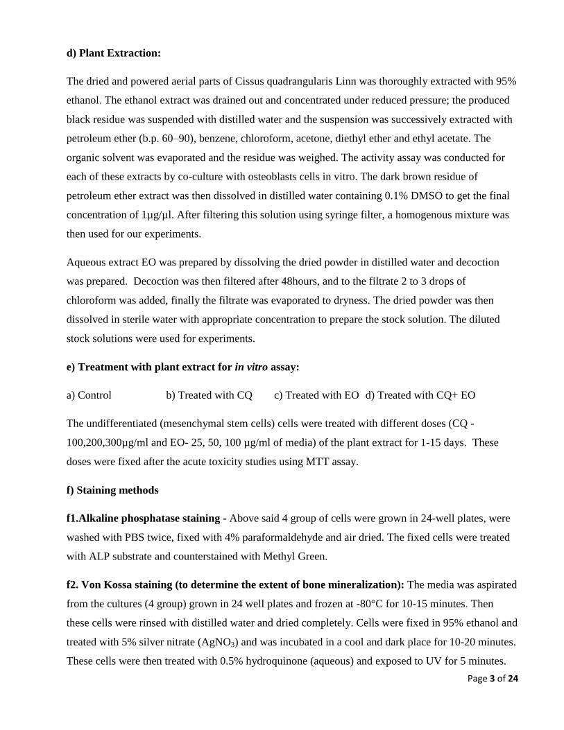
the osteoporotic changes. However, serum ALP concentration was further increased in
osteoporotic animals treated with reference doses of Raloxifine (OVX+RAL), CQ (OVX+CQ),
EO (OVX+EO) and both CQ & EO (OVX+CQ+EO) compared to that of OVX group. Further,
the Raloxifine and CQ+ EO treated groups showed high concentration of serum ALP level
indicating the increased activity of the osteoblasts in these animals. Data presented as Mean ±
SEM. The significant difference versus OVX group are indicated as ***P< 0.001and *P< 0.05.
These results indicates that, administration of CQ, EO or RAL to the osteoporotic
animals increases the activity of the existing osteoblast and/or facilitates the
differentiation of stem cells into new osteoblasts resulting in increased serum ALP.
Figure 13. Mean serum tartate resistant acid phosphatase (TRAP) concentration in
different experimental groups. Serum TRAP was significantly increased in osteoporosed rats
(OVX) compared to SHAM group indicating the active resorption of bone. However, the serum
TRAP concentration was decreased in Raloxifine treated (OVX+RAL), CQ treated (OVX+CQ),
EO treated (OVX+EO) and both CQ & EO treated (OVX+CQ+EO) groups compared to OVX
group. Data presented as Mean ± SEM. The significant difference versus OVX group are
indicated as ***P< 0.001. However, decrease in the TRAP concentration in these groups was not
significantly differing from that of control group.
Therefore, the active ingredients of CQ or EO can also influence negatively on the
osteoclast activity in osteoporotic condition thus slowing the bone degenerative
changes. However, further validation is warranted.

Page 17 of 24
Figure 14. Mean serum hydroxyprolin concentration in different experimental groups.
Serum hydroxyprolin was significantly increased in osteoporosed rats (OVX) compared to
SHAM group due to active destruction of collagen content of the bone. Serum hydroxyprolin
concentration was decreased in Raloxifine treated (OVX+RAL), CQ treated (OVX+CQ), EO
treated (OVX+EO) and both CQ & EO treated (OVX+CQ+EO) osteoporotic rat groups
compared to OVX group and almost similar to that of SHAM control. Data presented as Mean ±
SEM. The significant difference versus OVX group are indicated as ***P< 0.001.
Even though the serum hydroxyprolin levels are not direct indicator of bone
metabolism in normal condition, in osteoporosis condition, it is an indicator of
bone metabolism. As seen in the result, the serum hydroxyprolin content in CQ, EO
and RAL administered group is near to that of control group and significantly
lesser than that of osteoporotic animal group indicating the lesser bone destruction
in these animals.

Page 18 of 24
VI. Effect of CQ and EO on proliferation of bone marrow MSCs
Figure 15. Results of MTT assay when bone marrow mesenchymal stem cells (BMSC) are
grown in different concentration of CQ for 24, 48 and 72 hours. Cells grown in basal media
with 100µg and 200µg/ml of CQ showed higher proliferation rate (3 to 4 fold) compared to that
of cells with vehicle control indicating the stimulatory influence on BMSCs. Due to large
internal variations these data was not statistically significant. Further cells, with 300µg/ml of
CQ showed nearly 2 fold growth in comparison with that of control group.
Figure 16. Results of MTT assay when BMSC were grown in different concentration of EO
for 24, 48 and 72 hours. Results indicate that EO in all different concentration (25, 50, 75
µg/ml) showed higher proliferation rate compared to cells grown in vehicle control alone.
Therefore, EO can alter BMSCs proliferation rate. However, due to large internal variations the
statistical analysis did not show any significance.

Page 19 of 24
Figure 17. Results of MTT assay when BMSCs grown in different concentration of CQ and
EO together for 24, 48 and 72 hours. Cells grown in different combination/concentration of
EO and CQ together showed marked increase in the proliferation rate at 48 and 72 hr of
exposure. Even though noticeable influence of EO and CQ on cell proliferation is seen, due to
large internal variations the statistical analysis did not show any significance differences.
Therefore, the petroleum ether extract of CQ and aqueous extract of EO has
stimulatory influence on proliferation of BMSCs when added to cell culture media
at different concentrations and with different combinations. This may be the one of
the reasons for the osteoportective properties of CQ and EO by increasing the
availability of BMSCs for differentiation into new bone forming cells – osteoblasts
and thus increasing the formation of new bone and preventing the osteoporotic
changes in the bone. However, further cellular and molecular evaluation is
required to explore the role of CQ and EO on BMSCs.
Page 20 of 24
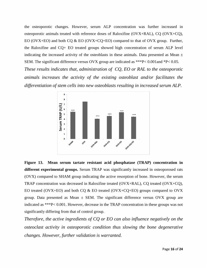
VII. Effect of CQ and EO on osteoblast
Figure 18. Alkaline phosphatase staining of MSCs. Rat bone marrow mesenchymal stem cells
grown in basal media (A) treated with 300ug/ml of CQ (B) showed moderate level of
differentiation into osteoblast which is evident by weakly expressing ALP after treating with CQ
for 17 days. Further, MSCs grown in osteogenic media with 200µg/ml of CQ (C) and 300µg/ml
of CQ (D) showed much stronger ALP staining with 15 days of treatment indicating significant
level of differentiation into osteoblast. Normally, BMSCs takes nearly 21 days to differentiate
into osteoblast in the osteogenic media. However, cells grown in either basal media or in
osteogenic media treated with 100µg/ml of CQ, EO alone and other combination of CQ and EO
did not show any differentiation indications.
This result indicates that CQ alone can induce the differentiation of BMSCs grown
in basal media (without any additional stimulator) into osteoblasts. Further, CQ
can facilitate the early differentiation of osteoblast when BMSCs grown in
osteogenic media which additionally contains other stimulants of differentiation.
Further, the exact molecular mechanisms involved in CQ mediated differentiation
of BMSCs into osteoblast and their activity needs to be evaluated.
VIII. Effect of CQ and EO on osteoclast
Administration of CQ and EO singly or in combinations did not show any differences in
differentiation at the end of 20 days of treatment with these plant extracts. All cells in all the
groups were stained for TRAP (representative picture is given below). This is may be due to
technical error which we could not rectify even after 4 attempts. Therefore, further evaluation is
required. However, the increased serum TRAP level in the osteoporotic condition was brought
down near to normal level when osteoporotic animals were treated with reference dose of RAL,

Page 21 of 24
CQ and EO. This indicates the negative influence of these drugs/plant extracts on osteoclast
activity.
Figure 19. Representative photograph of TRAP stained Osteoclast cells.
IX. Effect of CQ and EO on osteoblast activity (extracellular mineralization)
Figure 20. Rat bone marrow mesenchymal stem cells grown in osteogenic media (A) with
addition of CQ at 100 (B), 200 (C) and 300µg/ml (C) showed deposition(black) of minerals in
their extracellular matrix when stained at the end of 15 days of treatment. However, most
significant deposition was found only in the cells grown in osteogenic media with 300µg/ml of
CQ (D). Normally, nearly 28 days are required to differentiate the mesenchymal stem cells in to
functional osteoblast. The addition of CQ in to osteogenic media with or without EO shown to
be additively facilitating the functional ability of osteoblast cells by increasing the extracellular
secretions in which calcium was deposited. No such changes were observed in cells treated with
CQ or EO in basal media and cells treated with EO alone. These preliminary results indicate that,
the CQ not only influences the proliferation and differentiation of BMSCs but also influences
Page 22 of 24
the biological activities of differentiated osteoblast. However, the results are not satisfactory and
needs further evaluation.
14. Conclusions summarizing the achievements and indication of the scope for future work
These preliminary studies show that treating the osteoporotic animals with reference doses of CQ
can ameliorate the osteoporotic degenerative changes by increasing the formation of both
trabecular and cortical bones. CQ can also influence the growing end of the bones by facilitating
the proliferation of growth plate cartilage and differentiation of cartilage in to tabacular bone at
differentiation zone of growth plate. These changes are likely due to the stimulatory effect of
active ingredients of CQ on the proliferation of bone marrow mesenchymal stem cells (BMSCs)
and differentiation of these cells into active osteoblast cells. As seen in the result, addition of CQ
to growth medium can stimulate differentiation of BMSCs into active osteoblast cells. The
Mesenchymal stem cells grown in osteogenic condition media with different does of CQ showed
mineral deposition in their extracellular matrix indicating the increased biological activity of the
differentiated osteoblast cells. Further, CQ may also negatively influence the osteoclast cells
differentiation and their activities thus slowing the bone resorption in osteoporotic condition.
However, the role of CQ on osteoclast activity could not be established with the current sets of
experiments and needs further investigations. CQ is known to contain Phytosterol, flavonoids
and triterpenoids, vitamin C, quercitin, quercitrin, β-sitosterol, lupeol and freidalin. Therefore,
any of these phytosterol, flavonoids along with rich Vitamin C and D can elucidate such
osteoprotective effect. However, further evaluation is required to identify the active compounds
and their osteoprotective mechanisms. In addition, the offspring born to mother treated with CQ
showed less intense degenerative changes when they are subjected to osteoporosis in their adult
life. Therefore, the results indicate that the active ingredients of the CQ can cross the placental
barrier and has positive effect on optimizing the bone growth in fetal life. Therefore, prenatal
exposure to CQ can facilitate to attain the peak bone mass during osteogenesis thus preventing
the degenerative changes resulting from osteoporosis in their adult life. However, further
research is needed to identify such active ingredients in these plants which crosses the placental
barriers and are responsible for osteoprotective action. On the other hand, the EO can alone
increase the proliferation of mesenchymal stem cells and influences the collagen content in the
bone. Therefore, EO may not directly differentiate the stem cells into osteoblast but helps in
Page 23 of 24
reinforcing the exiting bones by increasing the collagen contents. Further, the role of EO on
fibroblast activity needs to be evaluated. When cells administered together with CQ to the
osteoporosed rat, additive effect on both bone mass and its collagen contents were observed.
Therefore, the combination of CQ and EO can be the best treatment strategies for preventing the
osteoporotic changes thus providing the additional quality life span. Osteoporosed animals
treated with both CQ and EO singly or in combination showed healthier growth plate with
proliferative and differentiation zones indicating the beneficial effect of these two plant extracts
on osteogenesis at the growing end of the bone which results in elongation of the bone.
Additionally, administering the EO resulted in increase in ground substance and fibrous
component in the growth plate thus forming the healthier hyaline cartilage with active growth
and differentiation.
The exact mechanism of actions of CQ and EO on osteoblast, osteoclast and fibroblast cells,
signaling cascade involved in CQ and EO mediated osteoprotection and finding the bioactive
ingredients of these two medicinal plants are needs to be further evaluated.
15. Procurement / usage of equipment - NONE
16. Manuscript for publication (300 words for possible publication in council’s bulletin)
Repair of bone defects secondary to trauma, osteoporosis, osteomyelitis and fracture nonunion,
poses a significant problem for many clinicians. Several strategies have been employed to
increase the bone healing process in various clinical conditions. As a part of our continuing
screening of biologically active natural anti-osteoporotic agents, we have evaluated the efficacy
of petroleum ether extract of Cissus quadrangularis Linn. (CQ) and Emblica offcinalis (EO) on
the osteomodulation. These plants and their medicinal properties have been described in ancient
medicinal system such as Ayurveda. Extract of CQ reported to contain phytoestrogenic steroids,
ascorbic acid, carotene, calcium, and anabolic steroids and shown to heal the bone fractures in
several in vivo studies. Fruits of EO are known to contain vitamin C and antioxidants. Therefore,
the present study is aimed to evaluate osteoprotective effect of CQ and EO by assessing the
structural and biochemical alteration using an osteoporotic rat model.
Our results show that the prenatal exposure to CQ can prevent the degenerative changes
secondary to osteoporosis in their adult life. Therefore, active ingredients of the CQ may cross

Page 24 of 24