Project Proposal - NUI Group Community Code
12
Crystal structure of a wild-type Cre recombinase– loxP synapse reveals a novel spacer conformation suggesting an alternative mechanism for DNA cleavage activation Eric Ennifar, Joachim E. W. Meyer, Frank Buchholz 1 , A. Francis Stewart 2 and Dietrich Suck* Structural and Computational Biology Programme, EMBL, Meyerhofstrasse 1, D-69117 Heidelberg, Germany, 1 Max Planck Institute of Molecular Cell Biology and Genetics and 2 Biotec, Technische Universita ¨t Dresden, c/o MPI-CBG, Pfotenhauerstrasse 108, D-01307 Dresden, Germany Received March 26, 2003; Revised June 16, 2003; Accepted July 24, 2003 PDB accession nos 1nzb, 1ouq ABSTRACT Escherichia coli phage P1 Cre recombinase cata- lyzes the site-specific recombination of DNA con- taining loxP sites. We report here two crystal structures of a wild-type Cre recombinase–loxP syn- aptic complex corresponding to two distinct reac- tion states: an initial pre-cleavage complex, trapped using a phosphorothioate modification at the cleav- able scissile bond that prevents the recombination reaction, and a 3¢-phosphotyrosine protein–DNA intermediate resulting from the first strand cleavage. In contrast to previously determined Cre complexes, both structures contain a full tetrameric complex in the asymmetric unit, unequivocally showing that the anti-parallel arrangement of the loxP sites is an intrinsic property of the Cre–loxP recombination synapse. The conformation of the spacer is different to the one observed for the symmetrized loxS site: a kink next to the scissile phosphate in the top strand of the pre-cleavage complex leads to unstacking of the TpG step and a widening of the minor groove. This side of the spacer is interacting with a ‘cleav- age-competent’ Cre subunit, suggesting that the first cleavage occurs at the ApT step in the top strand. This is further confirmed by the structure of the 3¢-phosphotyrosine intermediate, where the DNA is cleaved in the top strands and covalently linked to the ‘cleavage-competent’ subunits. The cleavage is followed by a movement of the C-terminal part containing the attacking Y324 and the helix N interacting with the ‘non-cleaving’ subunit. This rearrangement could be responsible for the inter- conversion of Cre subunits. Our results also sug- gest that the Cre-induced kink next to the scissile phosphodiester activates the DNA for cleavage at this position and facilitates strand transfer. INTRODUCTION The Cre–loxP recombination system from phage P1 has been the target of extensive biochemical, biophysical and in vivo studies (reviewed in 1). Cre, a member of the integrase family of recombinases, requires no accessory proteins and mediates all steps in site-specific recombination from synapse forma- tion, DNA cleavage, strand exchange, religation, to Holliday junction (HJ) isomerization and resolution (Fig. 1A). It is a very robust enzyme that functions well in a variety of organisms, including bacteria, yeast, plants, insects and mammals. The applied use of Cre allows sophisticated genome engineering, including conditional gene deletions (2), inducible chromosomal translocations (3,4) and other advanced genome manipulations (reviewed in 5,6). Mechan- istic studies in a number of laboratories and in particular crystallographic studies by the groups of Van Duyne and Baldwin have shed light on how the enzyme interacts with DNA, as well as the formation of the 3¢-phosphotyrosine and the HJ intermediates (7–11). These studies have provided architectural details of these intermediates and have been the basis for a model assuming a subtle, protein-mediated isomerization of the HJ intermediate not involving any branch migration or helical restacking. In the crystal structure of a Cre–loxS synaptic complex containing the inactive R173K or Y324F mutants bound to a symmetrized version of the naturally occurring loxP site (Fig. 1B), an asymmetric bend in the spacer or cross-over region of the DNA was observed involving an atypical large negative roll and positive tilt opening up the major groove (9). This sharp bend is located at the opposite end of the spacer, 5 bp away from the scissile phosphate activated for cleavage (Fig. 1C, bottom). The authors argue, that bending in the left half of the cross-over region stimulates cleavage in the right *To whom correspondence should be addressed. Tel: +49 6221 387307; Fax: +49 6221 387306; Email: [email protected] Present address: Joachim E. W. Meyer, Lion Bioscience AG, Waldhoferweg 98, 69123 Heidelberg, Germany The authors wish it to be known that, in their opinion, the first two authors should be regarded as joint First Authors Nucleic Acids Research, 2003, Vol. 31, No. 18 5449–5460 DOI: 10.1093/nar/gkg732 Nucleic Acids Research, Vol. 31 No. 18 ª Oxford University Press 2003; all rights reserved Downloaded from https://academic.oup.com/nar/article-abstract/31/18/5449/2376017 by guest on 15 April 2019
Transcript of Project Proposal - NUI Group Community Code

Crystal structure of a wild-type Cre recombinase±loxP synapse reveals a novel spacer conformationsuggesting an alternative mechanism for DNAcleavage activationEric Ennifar, Joachim E. W. Meyer, Frank Buchholz1, A. Francis Stewart2 and Dietrich Suck*
Structural and Computational Biology Programme, EMBL, Meyerhofstrasse 1, D-69117 Heidelberg, Germany,1Max Planck Institute of Molecular Cell Biology and Genetics and 2Biotec, Technische UniversitaÈ t Dresden,c/o MPI-CBG, Pfotenhauerstrasse 108, D-01307 Dresden, Germany
Received March 26, 2003; Revised June 16, 2003; Accepted July 24, 2003 PDB accession nos 1nzb, 1ouq
ABSTRACT
Escherichia coli phage P1 Cre recombinase cata-lyzes the site-speci®c recombination of DNA con-taining loxP sites. We report here two crystalstructures of a wild-type Cre recombinase±loxP syn-aptic complex corresponding to two distinct reac-tion states: an initial pre-cleavage complex, trappedusing a phosphorothioate modi®cation at the cleav-able scissile bond that prevents the recombinationreaction, and a 3¢-phosphotyrosine protein±DNAintermediate resulting from the ®rst strand cleavage.In contrast to previously determined Cre complexes,both structures contain a full tetrameric complex inthe asymmetric unit, unequivocally showing that theanti-parallel arrangement of the loxP sites is anintrinsic property of the Cre±loxP recombinationsynapse. The conformation of the spacer is differentto the one observed for the symmetrized loxS site: akink next to the scissile phosphate in the top strandof the pre-cleavage complex leads to unstacking ofthe TpG step and a widening of the minor groove.This side of the spacer is interacting with a `cleav-age-competent' Cre subunit, suggesting that the®rst cleavage occurs at the ApT step in the topstrand. This is further con®rmed by the structure ofthe 3¢-phosphotyrosine intermediate, where the DNAis cleaved in the top strands and covalently linkedto the `cleavage-competent' subunits. The cleavageis followed by a movement of the C-terminal partcontaining the attacking Y324 and the helix Ninteracting with the `non-cleaving' subunit. Thisrearrangement could be responsible for the inter-conversion of Cre subunits. Our results also sug-gest that the Cre-induced kink next to the scissile
phosphodiester activates the DNA for cleavage atthis position and facilitates strand transfer.
INTRODUCTION
The Cre±loxP recombination system from phage P1 has beenthe target of extensive biochemical, biophysical and in vivostudies (reviewed in 1). Cre, a member of the integrase familyof recombinases, requires no accessory proteins and mediatesall steps in site-speci®c recombination from synapse forma-tion, DNA cleavage, strand exchange, religation, to Hollidayjunction (HJ) isomerization and resolution (Fig. 1A). It is avery robust enzyme that functions well in a variety oforganisms, including bacteria, yeast, plants, insects andmammals. The applied use of Cre allows sophisticatedgenome engineering, including conditional gene deletions(2), inducible chromosomal translocations (3,4) and otheradvanced genome manipulations (reviewed in 5,6). Mechan-istic studies in a number of laboratories and in particularcrystallographic studies by the groups of Van Duyne andBaldwin have shed light on how the enzyme interacts withDNA, as well as the formation of the 3¢-phosphotyrosine andthe HJ intermediates (7±11). These studies have providedarchitectural details of these intermediates and have been thebasis for a model assuming a subtle, protein-mediatedisomerization of the HJ intermediate not involving any branchmigration or helical restacking.
In the crystal structure of a Cre±loxS synaptic complexcontaining the inactive R173K or Y324F mutants bound to asymmetrized version of the naturally occurring loxP site(Fig. 1B), an asymmetric bend in the spacer or cross-overregion of the DNA was observed involving an atypical largenegative roll and positive tilt opening up the major groove (9).This sharp bend is located at the opposite end of the spacer,5 bp away from the scissile phosphate activated for cleavage(Fig. 1C, bottom). The authors argue, that bending in the lefthalf of the cross-over region stimulates cleavage in the right
*To whom correspondence should be addressed. Tel: +49 6221 387307; Fax: +49 6221 387306; Email: [email protected] address:Joachim E. W. Meyer, Lion Bioscience AG, Waldhoferweg 98, 69123 Heidelberg, Germany
The authors wish it to be known that, in their opinion, the ®rst two authors should be regarded as joint First Authors
Nucleic Acids Research, 2003, Vol. 31, No. 18 5449±5460DOI: 10.1093/nar/gkg732
Nucleic Acids Research, Vol. 31 No. 18 ã Oxford University Press 2003; all rights reserved
Dow
nloaded from https://academ
ic.oup.com/nar/article-abstract/31/18/5449/2376017 by guest on 15 April 2019

half and vice versa. Thus, the bending direction woulddetermine, which of the two strands of the spacer will becleaved ®rst, and thereby provide a structural explanation forthe de®ned order of strand exchange observed for Cre (12±14).However, the crystal structures of Cre±DNA complexescontaining modi®ed lox sites with symmetrized spacer regionsdo not allow direct conclusions concerning the role of thespacer sequence in determining the architecture of thesynaptic complex and thereby the outcome of the recombina-tion process. Mutation studies suggest, that the base identity atcertain positions of the spacer plays an important role in thisprocess (15).
In a recent study by Baldwin and coworkers, the order ofstrand exchange was determined from an analysis of the strandcomposition of the HJ intermediate (11). Their results showthat Cre cleaves and exchanges the top strands (also referred toas `upper' strands) ®rst. In the crystal structure of a Cre±loxPHJ complex reported in the same paper, the `cleavage-competent' Cre subunit (also called `cleaving' or `activated'subunit) is bound to the left arm of the junction. Differences inprotein contacts to the scissile A and G bases on the left andright hand side of the spacer, respectively, and the orientationof the 198±208 loop region are being proposed as a physicalbasis for the cleavage order preference by Cre. However, incontrast with this study, a recent re-investigation of the orderof strand exchange (16) supports earlier results (12),suggesting that the bottom strand was exchanged ®rst.Clearly, the understanding of such discrepancies needs furtherinvestigation.
All crystal structures reported so far of pre-synaptic,synaptic or HJ complexes of Cre are isomorphous, with acrystallographic dyad relating the two Cre-bound DNAduplexes (or opposing strands in the HJ complex) and therebyimposing the 2-fold symmetric, anti-parallel arrangement ofthe duplexes. We report here two crystal structures of a wild-type Cre±loxP synaptic complex containing the full 200 kDatetrameric complex in the asymmetric unit, (i.e. without anysymmetry imposed by the crystal lattice). One corresponds toa pre-cleavage synaptic complex obtained using a loxP sitecontaining a phosphorothioate modi®cation at the scissilebond that prevents the recombination reaction. The secondstructure, obtained with an unmodi®ed loxP site is relevant tothe synaptic complex after the ®rst cleavage, forming a 3¢-phosphotyrosine protein±DNA intermediate. The overall
architecture of both complexes is similar to that observed inthe loxA and loxS complexes, displaying quasi 2-foldsymmetry with an anti-parallel orientation of the two
Figure 1. (A) Schematic representation of Cre±loxP-mediated recombin-ation. Cre molecules are represented by ellipses (green for non-cleavingsubunits and orange for active subunits) and labeled A, B, A¢ and B¢. Redand black spheres indicate cleavage sites. This ®gure is adapted fromGopaul et al. (8). (B) Sequences of the loxP (used in this study) and thesymmetrized loxS (used by Van Duyne's group) DNA. The 13 bp invertedrepeat sequences are indicated with black arrows. The thymidine positionsubstituted by a 5-iodo-deoxyuridine is highlighted. The spacer region isboxed and arrowheads depict cleavage sites. Residues noted in lower casewere added at the 3¢ and 5¢ ends for crystallization of the complex. (C) Inthe Cre±loxP synaptic complex, the 8 bp spacer region is kinkedimmediately next to the top strand cleavage site (top). After the ®rstcleavage, a 3¢-phosphotyrosine covalent intermediate is formed (middle).Some rearrangements in the spacer region redistribute the kink over severalresidues in the cleaved DNA but not all base pairs are still formed. Unlikein the present structures, in the Cre±loxS synaptic complex, the kink is 5 bpaway from the cleavage site (bottom).
5450 Nucleic Acids Research, 2003, Vol. 31, No. 18
Dow
nloaded from https://academ
ic.oup.com/nar/article-abstract/31/18/5449/2376017 by guest on 15 April 2019

Cre-bound loxP duplexes (see Fig. 3A). Therefore, the pseudosquare planar arrangement of the four Cre subunits and DNAarms observed previously is indeed an intrinsic property of theCre±loxP synapse. A striking feature of the pre-cleavagecomplex, which clearly distinguishes it from the Cre±loxSstructure, is a pronounced kink at the TG/AC step on the leftside of the spacer next to the scissile phosphate at the ApT step(Fig. 1C, top). In addition, our 3¢-phosphotyrosine inter-mediate complex, very similar to the Cre±loxA structure,unambiguously shows that the scissile ApT step in the topstrand is bound to the Cre subunit displaying an `active' or`cleavage-competent' conformation (Fig. 1C, middle). Ourstructures therefore suggest that a Cre-induced kink in thewild-type loxP site activates the neighboring scissile phos-phate for cleavage. Implications of the Cre-induced spacerconformation and differences between DNA contacts of activeand inactive Cre subunits for the mechanism of recombinationwill be discussed.
MATERIALS AND METHODS
Protein and DNA puri®cation
Cre protein was expressed in Escherichia coli from a pGem-derived vector, puri®ed by chromatography on phospho-cellulose P11, Mono S (Pharmacia) and gel ®ltration(Superdex 200, Pharmacia). As a result of cloning, theexpressed protein contained four additional N-terminal resi-dues compared with the SWISSPROT sequence RECR_BPP1:Phe-Gln-Val-Pro. Since the N-terminal tail does not appear inthe electron density map, we use the numbering of thepublished sequence.
DNA oligomers containing the loxP site were chemicallysynthesized. 37mer oligomers (Fig. 1) without any modi®ca-tions (oligos 37-5 for `top' strand and 37-9 for `bottom'strand), iodinated (oligo 37-5i, with a subsitution of a deoxy-thymidine into a 5-iodo-deoxyuridine) or containing a non-bridging phosphorothioate modi®cation (mixed R and Sdiastereomers) introduced either at the ApT (oligo 37-2) orat the GpC (oligo 37-6) steps were used for crystallizationexperiments. Oligos were dissolved in 100 mM HEPES/NaOHpH 7.6, 50 mM MgCl2, mixed at equimolar ratios andannealed by reducing the temperature from 90 to 25°C in athermocycler at ±0.5°C min±1.
Crystallization and data collection
Crystals were obtained at 20°C with the hanging drop method.Drops (2 ml) contained 0.13 mM Cre, 0.15 mM annealed oligo,50 mM NaCl, 0.5 mM EDTA, 5% (w/v) glycerol, 50 mMHEPES/NaOH pH 7.6, 75 mM MgCl2 and 12±15% (w/v) PEG2000 monomethyl ether. For micro-seeding, which stronglyincreased reproducibility, ~0.3 ml of a solution containingcrystal seeds was added to the drops. Diffraction qualitycrystals are obtained after 8±12 h only. Crystals werestabilized in a solution containing 20% (w/v) PEG 2000MME, 100 mM HEPES/NaOH pH 7.6, 100 mM MgCl2 and20% (w/v) glycerol. Prior to data collection, crystals were¯ash frozen in liquid ethane. Data were processed with CCP4(17) and HKL (18) packages.
Structure solution and re®nement
The structure was solved by molecular replacement with theprogram AMoRe (19) using coordinates 1CRX from theprotein data bank. A mercury derivative was collected from acrystal soaked for 2 days in a stabilizing solution plus 2 mMethyl-mercury thiosalicylate (EMTS). Experimental phases upto 4.5 AÊ resolution were obtained from this dataset with theprogram SHARP (20) and were used during initial steps ofre®nement, assuming a 70% solvent content.
Crystallographic re®nement was performed with the CNSpackage (21). Anomalous difference maps were calculated inthe 20±3.2 AÊ resolution range to unambiguously assign theposition of both iodine (f" = 2.9 electrons at l = 0.93 AÊ ) atomspresent in the asymmetric unit, revealing two peaks at 8.6 and7.9 s above the mean level (these peaks were not present in astructure obtained with a native DNA). As observed pre-viously with a brominated RNA exposed to intense X-raydoses (22), a fast radiolytic cleavage resulting in de-iodinationof the uridine occurs during the data collection. In the presentcase this reaction takes place even more rapidly as the C-Ibond is more radiation sensitive than the C-Br bond and due tothe extremely long exposure time for data collection (54 s perdegree on beamline ID14-2 at the ESRF, Grenoble). As aconsequence of this radiolysis, the free iodine does not diffusein solvent channels, but is rather trapped at the protein±DNAinterface, ~5 AÊ from the C5 of the uridine (SupplementaryMaterial Fig. S1). Sequence assignment and orientation of theloxP site was further con®rmed by using simulated annealingcomposite omit maps, which show additional density corres-ponding to N2 of guanines compared with adenines and C5 ofthymines compared with cytidines (see Fig. 3B). Severalsigni®cant peaks appearing in electron density maps wereattributed either to water molecules or to magnesium ionsdepending on the relative charge of the surrounding ligands,their distance, the peak height and the geometry according to(23). To check the identity of cations, attempts at soakingcrystal into stabilizing solution containing zinc or manganesewere done but systematically lead to crystal cracks.
Coordinates and structure factors for both structures havebeen deposited in the protein data bank (PDB entries 1nzb and1ouq).
Recombination assays
For recombination assays, 48mer oligomers were used (5¢-CGA TCC GAT AAC TTC GTA TAA TGT ATG CTA TACGAA GTT ATC TCC GAC-3¢ for `top' strand and 5¢-GTCGGA GAT AAC TTC GTA TAG CAT ACA TTA TAC GAAGTT ATC GGA TCG-3¢ for `bottom' strand). Recombinationwas performed in the presence of an excess of 37meroligomer, either without any modi®cation (oligo 37-5/9),with a phosphorothioate on the top strand at the ApT step(oligo 37-2/9), on the bottom strand at the GpC step (oligo 37-5/6), or on both strands (oligo 37-2/6). The reaction buffercontained 20 mM HEPES/NaOH pH 7.6, 2 mM MgCl2, 70 mMNaCl, 5% glycerol, 2 mM of 48mer oligo, 36 mM of 37meroligo and 37 mM of Cre protein. Reactions were carried out at39°C for 1 h and stopped by addition of proteinase K digestion(1 mg ml±1 ®nal concentration for 30 min at 37°C or overnightincubation at room temperature) and addition of SDS to 1.25%just before loading samples on the gel. Recombination
Nucleic Acids Research, 2003, Vol. 31, No. 18 5451
Dow
nloaded from https://academ
ic.oup.com/nar/article-abstract/31/18/5449/2376017 by guest on 15 April 2019

products were analyzed on a pre-heated denaturing 15%polyacrylamide gel (8 M urea, 45 mM Tris±borate pH 8.3,2 mM EDTA) and visualized by ethidium bromide staining.
RESULTS AND DISCUSSION
The Cre-induced recombination is blocked byphosphorothioate modi®cation of the loxP cleavage sites
Many enzymes that catalyze phosphodiester bond hydrolysishave a signi®cantly reduced activity on DNA containingphosphorothioate linkages obtained by the substitution of anon-bridging phosphate oxygen by a sulfur (24). It was shownsome 15 years ago that the Rp-phosphorothioate modi®cationof the DNA backbone inhibits the cleavage induced by lambdaintegrase and can be used to determine the order of strandexchange in the recombination process (25,26). As Cre alsobelongs to the tyrosine recombinase family of site-speci®cintegrases, we decided to use this phosphorothioate strategy toprevent Cre cleavage. As oligonucleotides with a stereo-speci®c phosphorothioate modi®cation are not availablecommercially, we used a mixed R and S racemic mixture ofa phosphorothioated-modi®ed loxP site, either on the top (atApT step) or the bottom (GpC step) strand, to check for thishypothesis and to block the ®rst or the second strand Cre-induced cleavage and therefore the recombination process.
Recombination assays performed on loxP sites modi®edwith the introduction of a phosphorothioate at the scissilebonds showed that such DNAs are strongly resistant to proteincleavage (Fig. 2). Despite the use of a racemic mixture ofmodi®ed DNA, no cleavage was detectable in solution in thetimescale of the experiment. At a ®rst sight, one would expect
a different impact on the DNA cleavage for S- and R-phosphorothioates, resulting in a partial cleavage inhibitiononly. Our results show however that both phosphate oxygenatoms seem to be essential for the cleavage, which is inagreement with the active site model of the integrase familyproposed by Van Duyne (1). In this model, both oxygen atomsare interacting with essential residues (R173, R292 and W315)and are required for the reaction, as also observed in ourstructures (see Figs 6 and S3).
Unexpectedly however, both top and bottom strand modi-®cations seem to have the same effect, blocking any DNAcleavage (Fig. 2). Based on the Cre recombination mechanism(Fig. 1A), one would have rather expected either no cleavagefor the modi®cation that prevents the ®rst strand cleavage(the complex remaining a pre-cleavage complex), or 50% ofcleavage for the modi®cation blocking the second strandcleavage, as only one strand is exchanged in the HJintermediate. As discussed below, this result is in agreementwith the observation of the same uncleaved Cre±loxP substrate
Figure 2. In vitro recombination experiments performed on native loxP sitesor modi®ed by the introduction of a phosphorothioate at the protein cleavagesite. Lanes marked `48' contain the 48mer loxP oligomer. Lanes containingthe recombinase are indicated with `Cre'. The 37mer loxP oligomer is pre-sent in lanes marked with `5/9' (unmodi®ed DNA), `2/9' (phosphorothioateon the top strand), `5/6' (phosphorothioate on the bottom strand) and `2/6'(phosphorothioate on top and bottom strands). Due to the presence of over-hangs on the 37mer loxP site used for crystallization experiments, two dif-ferent sizes of recombinant DNA are obtained with unmodi®ed loxP site,corresponding to a 42mer and a 43mer oligomers. No recombinant DNAwas detectable in our conditions when a phosphorothioate was present eitheron the top or the bottom strand.
Figure 3. (A) View of the synaptic tetramer as observed in the asymmetricunit of the present structure. The two DNA duplexes are in black and red.The four Cre proteins subunits are in dark green, light green (non-cleavingsubunits), orange and red (cleaving subunits). (B) View of the 3Fobs±2Fcalc
electron density map (contoured at 1.4 s level) around A-T and G-C basepairs showing the overall quality of the electron density map. Note the addi-tional density observed for N2 of guanines compared with adenines and C5of thymines compared with cytidines.
5452 Nucleic Acids Research, 2003, Vol. 31, No. 18
Dow
nloaded from https://academ
ic.oup.com/nar/article-abstract/31/18/5449/2376017 by guest on 15 April 2019

complex in crystals containing either top or bottom strandmodi®cation of the DNA. A possible explanation for the lackof observation of any cleavage on the top strand when thebottom strand is modi®ed may be that the ®rst strand cleavageoccurs, but quickly reverses to the substrate complex after re-ligation as it cannot proceed to the second strand cleavage dueto the phosphorothioate modi®cation. In the same way, wecannot exclude that when the top strand is modi®ed, Creshortcuts the recombination cycle and cleaves and re-ligatesthe bottom strand at the GpC step.
The tetrameric Cre synapse is a dimer of dimers withanti-parallel orientation of the loxP duplexes
The orthorhombic crystals contain a full ~200 kDa tetramericcomplex in the asymmetric unit, with one Cre subunit boundto each of the inverted repeats of the loxP site duplexes(Fig. 3A). Despite the relatively limited resolution of ourstructures (2.8 and 2.9 AÊ resolution, respectively), the anti-parallel orientation of the synapsed loxP sites was clear fromthe interpretation of electron density maps (Fig. 3B).However, in order to eliminate any ambiguity due to thepalindromic sequence with respect to the succession ofpyrimidines and purines, we used a modi®ed DNA containinga thymidine to 5¢-iodo-deoxyuridine substitution in theinverted repeat region in only one of the strands.Identi®cation of the iodine by anomalous difference maps(see Materials and Methods) con®rmed the anti-parallelorientation of the spacer. The quaternary arrangement of thesubunits and DNA arms in the synapse is quasi 2-foldsymmetric, i.e. subunits A/B and A¢/B¢ in Figure 3A arerelated by a local dyad with an overall r.m.s.d. for the Ca-atoms (21±341) of ~0.4 AÊ . Distinctly larger deviations(~2.6 AÊ ) are found for subunits not related by the local dyad(Fig. S2). Differences in the latter pairs are particularlysigni®cant in the C-terminal region and the loop regions 198±208 and 314±318 affecting the positions of lysine 201, aresidue reported to be catalytically important (9), the activesite tyrosine 324 and tryptophane 315. In addition, there issome change in the relative orientation of the N- andC-terminal domains corresponding to a rotation of ~10°.
The synapsed loxP sites are oriented anti-parallel with thefour Cre-bound arms forming a HJ-like, pseudo square-planar array. A similar overall architecture was found inthe Cre±loxA and loxS as well as the Cre±loxP HJcomplexes. However, in the latter structures, an exact 2-foldsymmetry is imposed by the crystal lattice. Our structurecontaining a wild-type loxP site clearly shows, that themolecular 2-fold symmetry with an anti-parallel orientation ofthe DNA duplexes is an intrinsic property of the Cre±loxPrecombination synapse.
Identi®cation of two different Cre±loxP structures:a pre-cleavage complex and a 3¢-phosphotyrosinecovalent intermediate
An uncleaved complex could unambiguously be identi®edfrom several data sets obtained with a DNA containing aphosphorothioate modi®cation either at top or bottom strandscissile bonds. Indeed, although they interfere with differentcleavage steps, both modi®cations lead to the same crystalstructure. As our recombination experiments clearly showedthat the introduction of a phosphorothioate at scissile bonds
prevents recombination, and given the short timescalerequired for crystallization (see Materials and Methods), weassumed that these structures correspond to the pre-cleavagesynaptic complex, and not to a recombination product. Sincethe best diffracting crystal was obtained for a modi®cation onthe bottom strand of the DNA, such a crystal was used forre®nement in Table 1. It shows an extremely well-de®nedelectron density map for the protein as well as for the DNA,including the complete spacer region (Fig. 4A). However, dueto the relatively limited resolution and the use of a racemicmixture of R- and S-phosphorothioates, it was not possible toobserve the sulfur either on Fobs±Fcalc difference density mapsor on anomalous difference maps.
The 3¢-phosphotyrosine covalent intermediate was identi-®ed during the re®nement of several data sets obtained fromcrystals containing unmodi®ed DNA. These crystals display asigni®cant lack of isomorphism compared with previous datasets (see Table 1). Fourier difference density maps, combinedwith simulated annealing composite omit maps revealed amajor movement (~3 AÊ ) of the C-terminal part of the activatedCre subunit, which contains the attacking tyrosine. Inparticular, an extremely short distance between the Tyr324hydroxyl group and the cleavable phosphate at the ApT step ofthe top strands was observed (<2 AÊ ), correlated with a strongnegative density peak in the difference density map for thesugar-phosphate backbone at this position. In addition,His289, a residue belonging to the active site of the proteinthat could act as a proton donor for the Tyr324 leaving group(7), is directly contacting the scissile phosphate as observedpreviously in the Cre±loxA covalent intermediate, but not inCre±loxP, Cre±loxS or Cre±HJ intermediate complexes (seebelow). Finally, a superposition of this structure with allavailable Cre±DNA complexes shows that this structure only®ts with the Cre±loxA suicide substrate (see below) alsocontaining a 3¢-phosphotyrosine covalent intermediate (7). Asthe isomerization of the complex is not possible given theDNA-driven crystal packing, and as the activated Cre subunitis oriented in the same way than in the pre-cleavage complex,we assumed that this structure is relevant for the ®rst cleavagestep, and not the second one. Noticeably, no uncleavedcomplex was detectable on electron density maps, even using
Table 1. Data collection and re®nement summary
Synaptic complexa,b Covalentintermediatea
Space group P212121 P212121
Unit cell (AÊ ) A = 107.8, b = 161.1,c = 195.7
a = 108.9, b = 164.2,c = 194.7
X-ray source ESRF ID29 SLS PXMax. resolution (AÊ ) 2.8 2.9Completenessc (%) 97.4 (94.6) 96.2 (98.2)Rsym
c (%) 7.3 (31.8) 6.7 (33.0)R factor (%) 23.0 23.0Rfree factor (%) 25.2 26.2r.m.s.d. bonds length (AÊ ) 0.007 0.020r.m.s.d. bonds angles 1.2° 1.6°Estimated coordinate error (AÊ ) 0.41 0.45
aStructure obtained with an iodinated loxP DNA.bPhosphorothioate modi®cation of the loxP DNA at the cleavage site on thebottom strand.cValues in parenthesis correspond to percentage in outermost shell.
Nucleic Acids Research, 2003, Vol. 31, No. 18 5453
Dow
nloaded from https://academ
ic.oup.com/nar/article-abstract/31/18/5449/2376017 by guest on 15 April 2019

low contour levels. We were however not able to detect thiscovalent intermediate by gel electrophoretic mobility assaysusing dissolved crystals, probably due to the reversibility ofthe reaction in solution.
Interestingly, using the same wild-type Cre and a similarloxP DNA substrate, Baldwin and coworkers obtained a Cre±HJ intermediate structure (11). This different result might beattributed to the presence in our crystallization conditions ofdivalent cations (75 mM MgCl2) that promote the recombina-tion process, but are not essential (27). Another importantdifference in crystallization conditions concerns the pH used:5.0±5.5 in the Cre±HJ intermediate instead of 7.6 in thepresent structure. This difference in pH can affect the
protonation of some important amino acids, and in particularof the His289 located in the active site. In agreement with thishypothesis, the crystallization conditions used for the Cre±HJintermediate prevent cleavage and a 10-min pre-incubationwas necessary for the reaction to take place (11).
DNA bending and conformation of the spacer in theCre±loxP synapse: comparisons with Cre±loxA andCre±loxS complexes
A global superposition with the Cre±loxA and Cre±loxScomplexes highlights the overall similarity of the quaternaryarrangement (Fig. 4B). Superimposing the A and B subunits ofthe Cre±loxP structure with the corresponding subunits in the
Figure 4. (A) Stereoview of the 3Fobs±2Fcalc electron density map (contoured at 1.4 s level) around the active site of the `cleavage-competent' Cre subunitand the kink region of the loxP site. A metal ion (possibly a magnesium) is indicated by an orange sphere. (B) DNA backbone superposition of the wild-typeloxP sequence as observed in the present structure (in blue) and the symmetrized loxS sequence (PDBID 4CRX; the second duplex was generated by applying2-fold symmetry). While the overall bending is the same, marked differences are found in the spacer region resulting from different locations of kinks.(C) Stereoview of the 3Fobs±2Fcalc electron density map (contoured at 1.4 s level) showing a magnesium-mediated protein±DNA contact.
5454 Nucleic Acids Research, 2003, Vol. 31, No. 18
Dow
nloaded from https://academ
ic.oup.com/nar/article-abstract/31/18/5449/2376017 by guest on 15 April 2019
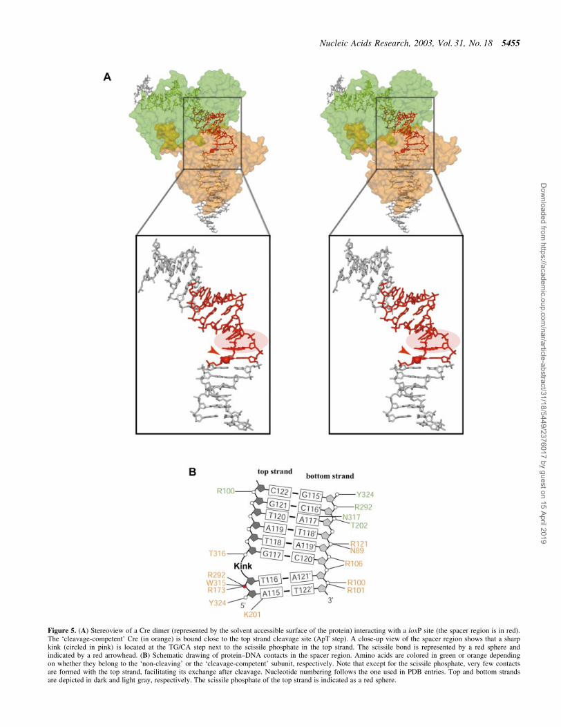
Figure 5. (A) Stereoview of a Cre dimer (represented by the solvent accessible surface of the protein) interacting with a loxP site (the spacer region is in red).The `cleavage-competent' Cre (in orange) is bound close to the top strand cleavage site (ApT step). A close-up view of the spacer region shows that a sharpkink (circled in pink) is located at the TG/CA step next to the scissile phosphate in the top strand. The scissile bond is represented by a red sphere andindicated by a red arrowhead. (B) Schematic drawing of protein±DNA contacts in the spacer region. Amino acids are colored in green or orange dependingon whether they belong to the `non-cleaving' or the `cleavage-competent' subunit, respectively. Note that except for the scissile phosphate, very few contactsare formed with the top strand, facilitating its exchange after cleavage. Nucleotide numbering follows the one used in PDB entries. Top and bottom strandsare depicted in dark and light gray, respectively. The scissile phosphate of the top strand is indicated as a red sphere.
Nucleic Acids Research, 2003, Vol. 31, No. 18 5455
Dow
nloaded from https://academ
ic.oup.com/nar/article-abstract/31/18/5449/2376017 by guest on 15 April 2019
loxA and loxS complexes using the Ca-atoms of residues 20±323 results in an r.m.s.d. of 1.9 and 1.8 AÊ , respectively. Theinterfaces between subunits are also mostly conserved.Likewise, the speci®c interaction of the Cre subunits withthe 13 bp inverted repeats is essentially the same in thesestructures, with the N-terminal domain contacting the DNA inthe major groove on one side, the C-terminal domain inadjacent major and minor grooves on the opposite side. But incontrast to previously reported Cre complexes, several metalion-mediated protein±DNA contacts are observed in theseregions (Fig. 4C), most likely due to the presence of largeamounts of magnesium in our crystallization conditions. Likein the loxA and loxS complexes, direct protein contacts to thebases in the spacer region are rare. A common feature is alsothe distinctly different protein±phosphate backbone contactpattern for the top and bottom strands (see Discussion below).
The overall bending of the DNA duplexes in the Cre±loxPsynapse is rather similar to the one in Cre±loxA and Cre±loxScomplexes, with an angle of ~100° between the left and righthand arms (Fig. 4B). However, this bend is achieved by adistinctly different conformation of the spacer regions. In thewild-type Cre±loxP pre-cleavage complex, a pronounced kink
at the TG/CA step next to the scissile phosphate (Figs 4A and5) contributes roughly half to the overall bending. A positiveroll of ~45° leads to an unstacking of bases at this step and amassive widening of the minor groove (Figs 4A and 5A). Incontrast, bending in the symmetrized loxS synapse is primarilydue to a kink 5 bp away at the other end of the spacer (Fig. 1C,bottom). The kink observed in the loxS complex, which isassociated with a large negative roll and positive tilt, anopening of the major and compression of the minor groove, isatypical and not normally found in protein±DNA complexes.The striking differences between the symmetric loxS and thewild-type loxP spacer conformations have at least in part to beattributed to the different nucleotide sequences within thespacer regions. As a consequence of the different spacerconformations we do not see the unfavorable close approachof two phosphates in the center of the spacer observed in theCre±loxS complex. It was speculated by the authors that theenergy associated with a stereochemically strained spacerconformation promotes the subsequent strand exchange step(9). On the basis of the loxP complex, we would instead argue,that a Cre-induced kink activates the neighboring scissilephosphate for cleavage and facilitates dissociation of thecleaved strand during strand exchange. In this context it isinteresting to note that divalent cations, although not essential,do promote the Cre-catalyzed recombination considerably(27). Interestingly, it was proposed that solvated divalentcations promote and stabilize unstacked conformations byinteractions with the nucleobase p electrons (28). Stabilizationof a kinked DNA structure together with an overallelectrostatic shielding effect, may therefore explain theobserved acceleration of Cre-catalyzed recombination in thepresence of divalent cations.
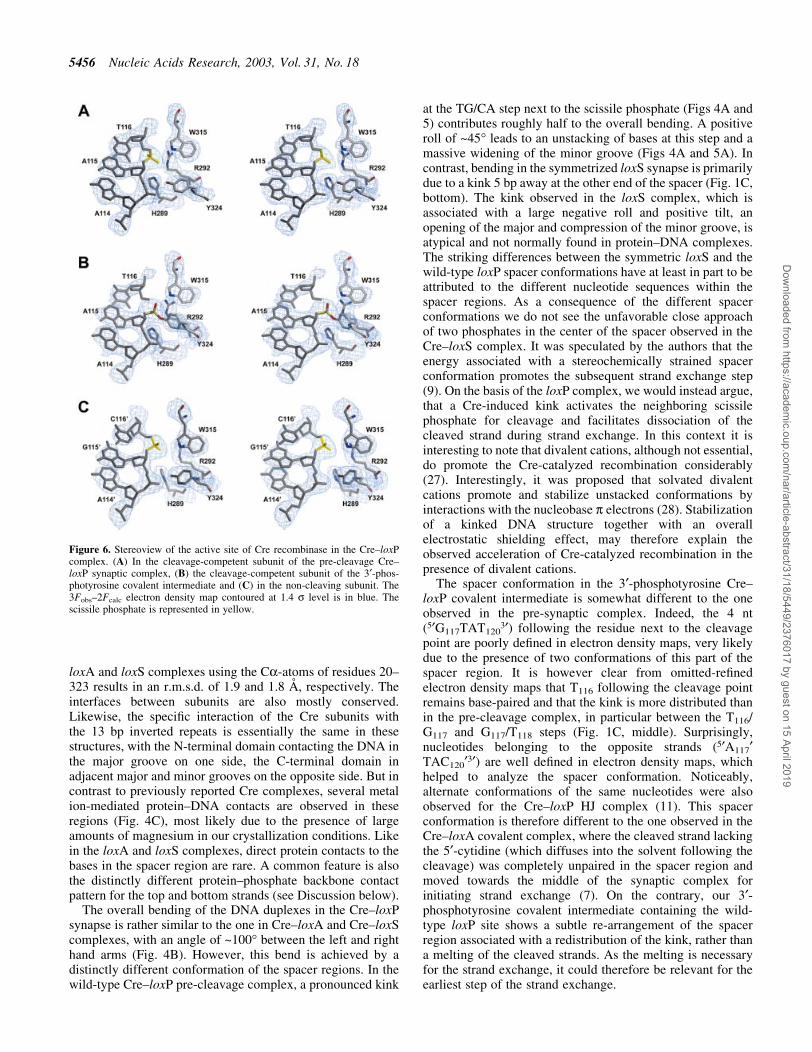
The spacer conformation in the 3¢-phosphotyrosine Cre±loxP covalent intermediate is somewhat different to the oneobserved in the pre-synaptic complex. Indeed, the 4 nt(5¢G117TAT120
3¢) following the residue next to the cleavagepoint are poorly de®ned in electron density maps, very likelydue to the presence of two conformations of this part of thespacer region. It is however clear from omitted-re®nedelectron density maps that T116 following the cleavage pointremains base-paired and that the kink is more distributed thanin the pre-cleavage complex, in particular between the T116/G117 and G117/T118 steps (Fig. 1C, middle). Surprisingly,nucleotides belonging to the opposite strands (5¢A117¢TAC120¢3¢) are well de®ned in electron density maps, whichhelped to analyze the spacer conformation. Noticeably,alternate conformations of the same nucleotides were alsoobserved for the Cre±loxP HJ complex (11). This spacerconformation is therefore different to the one observed in theCre±loxA covalent complex, where the cleaved strand lackingthe 5¢-cytidine (which diffuses into the solvent following thecleavage) was completely unpaired in the spacer region andmoved towards the middle of the synaptic complex forinitiating strand exchange (7). On the contrary, our 3¢-phosphotyrosine covalent intermediate containing the wild-type loxP site shows a subtle re-arrangement of the spacerregion associated with a redistribution of the kink, rather thana melting of the cleaved strands. As the melting is necessaryfor the strand exchange, it could therefore be relevant for theearliest step of the strand exchange.
Figure 6. Stereoview of the active site of Cre recombinase in the Cre±loxPcomplex. (A) In the cleavage-competent subunit of the pre-cleavage Cre±loxP synaptic complex, (B) the cleavage-competent subunit of the 3¢-phos-photyrosine covalent intermediate and (C) in the non-cleaving subunit. The3Fobs±2Fcalc electron density map contoured at 1.4 s level is in blue. Thescissile phosphate is represented in yellow.
5456 Nucleic Acids Research, 2003, Vol. 31, No. 18
Dow
nloaded from https://academ
ic.oup.com/nar/article-abstract/31/18/5449/2376017 by guest on 15 April 2019

Differences in protein contacts of top and bottomstrands in the spacer region
The Cre-induced asymmetric bending and kinking in thespacer region of the loxP recombination site and theconcomitant conformational changes in the protein subunitslead to differences in the protein contact pattern of the top andbottom strands of the spacer. The only direct base contacts inthe spacer involve the scissile adenine and guanine bases at the5¢ ends of the spacer in the top and bottom strands. While bothbases are contacted by K86 from the major groove side (viaN7 in case of adenine; via O6 and N7 in case of guanine), onlythe scissile adenine in the top strand is contacted by thecatalytically important K201 from the minor groove via N3(Fig. 5B). This base-speci®c contact is well de®ned in electrondensity maps of both the Cre±loxP pre-cleavage and 3¢-phosphotyrosine covalent intermediate complexes. Interest-ingly, this contact is also present in the Cre±loxP HJintermediate, but not in Cre±loxA and Cre±loxS synapses,where this lysine was disordered (1). Furthermore, no suchcontact is present for the `non-cleaving' subunits due todifferences in the conformation of loop 198±208. Therefore,as already suggested for the Cre±loxP HJ complex (11), thisresidue might be responsible for the cleavage preference ofCre and the speci®c recognition of the adenine present in theleft arm of loxP through this base-speci®c contact.
With the exception of active site residues, there are almostno contacts to the backbone of top strands in the spacer region,facilitating their exchange after cleavage (Fig. 5B). Incontrast, bottom strands are more tightly bound to the protein,e.g. via a strong salt bridge between R121 and the centralphosphate of the spacer (P119¢), as well as ionic contacts ofarginines 100, 101 and 106 to the neighboring phosphatesP120¢, P121¢ and P122¢, respectively (Fig. 5B).
Implications for the mechanism and order of strandexchange in Cre±loxP site-speci®c recombination
Like the previously published Cre complexes, structures of thesynaptic wild-type Cre±loxP pre-cleavage and 3¢-phospho-tyrosine intermediate complexes reported here strongly sup-port a cis-cleavage mechanism, where residues of thecatalytically essential RHR triad (R173/H289/R292), as wellas the attacking tyrosine nucleophile (Y324), belong to thesame subunit. In the pre-cleavage complex, dispositions of theactive site residues are similar in the `non-cleaving' and`cleavage-competent' Cre subunits. There are however somedistinct differences to be noted. In the `non-cleaving' subunitsthe Y324-OH nucleophile is 5 AÊ away from the scissilephosphate and directly interacts with the adjacent phosphate(Figs 6A and S3A). In the `cleavage-competent' subunits, thedistance between the scissile phosphate and the tyrosine issigni®cantly reduced to 4 AÊ , while the contact to the adjacentphosphate is maintained (Figs 6C and S3C). Another differ-ence is found for H289, which possibly acts as a general basefor accepting the Y324 hydroxyl proton (1). The distancebetween the Ne position of H289 and the scissile phosphate is4.7 AÊ in `non-cleaving' subunits, but reduced to 4.0 AÊ in`cleavage-competent' subunits. The movement of the cata-lytically important K201 as a consequence of rearrangementsin the loop 198±208, as well as the changes in the relative
orientations of the N- and C-terminal domains, have alreadybeen discussed.
In the Cre±loxP 3¢-phosphotyrosine covalent intermediate,the active site of the cleaving subunits undergoes severalsigni®cant changes identical to the one found in the Cre±loxAcovalent intermediate (7). As expected, the most importantchange concerns the attacking Y324, which moves towards thescissile bond and is covalently linked to the DNA (Figs 6B andS3B). Related to this movement, a major conformationalchange is observed for the helix containing Y324 (helix Maccording to 7), which moves 3 AÊ towards the DNA (Fig. 7A).This motion is propagated towards the C-terminal part of theprotein, including helix N, which binds in a hydrophobicpocket of the neighboring Cre monomer. In addition, the
Figure 7. Stereoviews showing a superposition of the active site of severalCre±DNA complexes for the `cleavage-competent' (top) and `non-cleaving'(bottom) subunits. Proteins are colored as follows: purple, Cre±loxS synap-tic complex (4CRX); light green, Cre-immobile HJ intermediate complex(2CRX); dark green, Cre-wild-type HJ intermediate complex (1KBU); blue,Cre±loxA suicide substrate (3¢-phosphotyrosine covalent intermediate);yellow, Cre±LoxP synaptic complex (this study, 1nzb); orange, Cre±loxP3¢-phosphotyrosine covalent intermediate (this study, 1ouq).
Nucleic Acids Research, 2003, Vol. 31, No. 18 5457
Dow
nloaded from https://academ
ic.oup.com/nar/article-abstract/31/18/5449/2376017 by guest on 15 April 2019

reorientation and the motion of the scissile phosphatefollowing the cleavage allows a direct interaction (2.9 AÊ )between the latter and the Ne position of H289. Thisrearrangement of the scissile phosphate also permits the twophosphate oxygens to be correctly oriented for interacting withH289 and W315 (Figs 5B and S3B).
Unlike `cleavage-competent' subunits, the `non-cleaving'subunits in the 3¢-phosphotyrosine covalent intermediate andthe pre-cleavage state complexes are essentially identical(Fig. 7B). But noticeably, the moving region of the `cleavage-competent' subunits is in the neighborhood of the loop 198±208 of the `non-cleaving' subunits and might therefore beinvolved in the communication between the two subunits andthe conformational switch interconverting cleaving- andnon-cleaving Cre subunits (Fig. 8).
Noticeably, a superposition of `cleavage-competent' sub-units of all solved crystal structures shows that they are nearlyidentical for loxP, loxS synaptic complexes as well as for thetwo HJ complexes, whereas the loxA and loxP covalentintermediates both display the same movement in theirC-terminal part containing helices M and N (Fig. 7A). Thisis not the case for `non-cleaving' subunits, which areisomorphous in all Cre complexes (Fig. 7B).
What does the Cre±loxP complex tell us about the order ofstrand cleavage and exchange? While some members of theInt family of recombinases, like e.g. Flp, do not seem to have ade®ned order of strand cleavage, Cre has an intrinsic bias forinitiating the recombination preferentially on one strand. Sofar, two alternative models have been proposed, depending on
whether the top or the bottom strand is cleaved and exchanged®rst. Two independent investigations (12,16) suggest that thebottom strand is cleaved ®rst at the GpC step. However, thealternative order of strand exchange was observed by ananalysis of strand composition of HJ intermediates (11),suggesting that the top strand is cleaved ®rst, i.e. the ApT stepat the left hand side of the loxP site. In line with the lattercleavage bias, we ®nd in the pre-cleavage synaptic complexthe `cleavage-competent' subunits B/B' associated with theApT step in the top strand on the left hand side of the spacer.Moreover, the observation of the 3¢-phosphotyrosine covalentintermediate shows that the conformation of the Cre±loxPcomplex captured in our crystals is catalytically relevant andnot a crystallization artefact due to a wrong spacer conform-ation. However, our crystallographic results are clearly notcompatible with an initiation of the recombination on thebottom strand and we do not have any explanation about thediscrepancy with results reporting this cleavage bias.
By introducing bulges consisting of three additionaladenine nucleotides at speci®c positions in one strand of thespacer and thereby forcing the DNA to bend in a givendirection it was shown that the covalent intermediate tended toaccumulate on the bulge strand (14). Furthermore, theseexperiments demonstrated that a bulge next to or 1 bp awayfrom the cleavage site is most effective in determining orchanging the cleavage bias. These experiments are in excellentagreement with our crystallographic results. First, theyprovide clear evidence, that the DNA bend directiondetermines the order of cleavage events. Secondly, the
Figure 8. Stereoviews of a Cre dimer showing inter-subunits contacts in the neighborhood of the active site. The non-cleaving subunit is in green and its loop198±208 in purple. Cleavage-competent subunits before cleavage (in yellow) and in the 3¢-phosphotyrosine covalent intermediate (in orange) are super-imposed. The moving C-terminal part containing helices M and N is in red. The close-up view (top) is from a different point of view to highlight themovement of helices M and N.
5458 Nucleic Acids Research, 2003, Vol. 31, No. 18
Dow
nloaded from https://academ
ic.oup.com/nar/article-abstract/31/18/5449/2376017 by guest on 15 April 2019

position-dependence of the bulge-effect strongly supports ourconclusion that the observed kink neighboring the scissilebond activates for cleavage.
CONCLUSIONS
The crystal structure of a wild-type Cre±loxP synapse with atetrameric complex in the asymmetric unit shows that the near2-fold symmetric, anti-parallel arrangement of the loxP sites isan intrinsic property of the Cre recombination synapse. Theoverall architecture and bending of the DNA is rather similarto that of the previously determined loxA, loxS and HJcomplexes displaying the same pseudo square-planar arrange-ment of the Cre subunits and DNA arms. A pre-cleavagecomplex was trapped using a loxP site modi®ed by theintroduction of a phophorothioate at scissile bonds, a modi-®cation that prevents the recombination reaction. It shows thatthe `cleavage-competent' Cre subunit is bound to the left handside of the loxP spacer region. A pronounced kink of the DNAis found at the neighboring TG/AC step associated with a largepositive roll and a widening of the minor groove. Using a non-modi®ed loxP site, a second structure containing a covalent3¢-phosphotyrosine intermediate was obtained. It shows thatthe ApT step in the top strand is cleaved ®rst. The cleavageinduces a 3 AÊ shift of the C-terminal part of the subunit,containing helices M and N, as well as a redistribution of thekink within the spacer region of the DNA. In contrast toconclusions drawn from a previous structure containing asymmetrized lox site with a distinctly different spacerconformation, we propose that a Cre-induced kink next tothe scissile phosphodiester activates the DNA for cleavage.The moving of helices M and N observed in the 3¢-phosphotyrosine intermediate could be responsible for theinterconversion of Cre subunits through an interaction with the`non-cleaving' subunit, and in particular with its loop 198±208, a major component of the conformational switch.
The interaction between Cre monomers bound to the rightand left arms of the loxP recombination site induces a de®nedbending of the DNA and an anti-parallel arrangement of theDNA duplexes in the synaptic complex and thereby deter-mines which strand will be cleaved ®rst. In an induced-®tmechanism changes in the DNA structure are coupled withconformational changes in the protein and protein±DNAcontacts in the spacer region. Tetramerization in the synapsemay induce further bending and/or stabilization of the DNAconformation by reciprocal exchange of the C-terminal helixand interactions of N-terminal domains between the Cresubunits bound to different duplexes, followed by coordinatedcleavage by the `cleavage-competent' subunits at the ApTsites in the top strands. Apparently, already at the stage ofsynapsis a conformation of the DNA is induced by therecombinase, which is essentially maintained during the ®rstround of strand cleavage, strand exchange, re-ligation andformation of the HJ intermediate.
SUPPLEMENTARY MATERIAL
Supplementary Material is available at NAR Online.
ACKNOWLEDGEMENTS
We gratefully acknowledge the help by Hiang Teo-Dreher inthe initial stages of this project and our colleagues from theBW-7B (EMBL Hamburg outstation), ID29 and ID14beamlines (ESRF, Grenoble), and to Armin Wagner (SwissLight Source, Viligen) for help with the data collection. Thiswork was supported by a grant from the Volkswagenstiftungto D.S. E.E. is recipient of an EMBO long-term fellowship.
REFERENCES
1. Van Duyne,G.D. (2001) A structural view of cre-loxp site-speci®crecombination. Annu. Rev. Biophys Biomol. Struct., 30, 87±104.
2. Gu,H., Marth,J.D., Orban,P.C., Mossmann,H. and Rajewsky,K. (1994)Deletion of a DNA polymerase beta gene segment in T cells using celltype-speci®c gene targeting. Science, 265, 103±106.
3. Testa,G. and Stewart,A.F. (2000) Creating a transloxation. Engineeringinterchromosomal translocations in the mouse. EMBO Rep., 1, 120±121.
4. Buchholz,F., Refaeli,Y., Trumpp,A. and Bishop,J.M. (2000) Induciblechromosomal translocation of AML1 and ETO genes through Cre/loxP-mediated recombination in the mouse. EMBO Rep., 1, 133±139.
5. Yu,Y. and Bradley,A. (2001) Engineering chromosomal rearrangementsin mice. Nat. Rev. Genet., 2, 780±790.
6. Lewandoski,M. (2001) Conditional control of gene expression in themouse. Nat. Rev. Genet., 2, 743±755.
7. Guo,F., Gopaul,D.N. and van Duyne,G.D. (1997) Structure of Crerecombinase complexed with DNA in a site-speci®c recombinationsynapse. Nature, 389, 40±46.
8. Gopaul,D.N., Guo,F. and Van Duyne,G.D. (1998) Structure of theHolliday junction intermediate in Cre-loxP site-speci®c recombination.EMBO J., 17, 4175±4187.
9. Guo,F., Gopaul,D.N. and Van Duyne,G.D. (1999) Asymmetric DNAbending in the Cre-loxP site-speci®c recombination synapse. Proc. NatlAcad. Sci. USA, 96, 7143±7148.
10. Woods,K.C., Martin,S.S., Chu,V.C. and Baldwin,E.P. (2001) Quasi-equivalence in site-speci®c recombinase structure and function: crystalstructure and activity of trimeric Cre recombinase bound to a three-wayLox DNA junction. J. Mol. Biol., 313, 49±69.
11. Martin,S.S., Pulido,E., Chu,V.C., Lechner,T.S. and Baldwin,E.P. (2002)The order of strand exchanges in Cre-LoxP recombination and its basissuggested by the crystal structure of a Cre-LoxP Holliday junctioncomplex. J. Mol. Biol., 319, 107±127.
12. Hoess,R., Wierzbicki,A. and Abremski,K. (1987) Isolation andcharacterization of intermediates in site-speci®c recombination.Proc. Natl Acad. Sci. USA, 84, 6840±6844.
13. Hoess,R., Abremski,K., Irwin,S., Kendall,M. and Mack,A. (1990) DNAspeci®city of the Cre recombinase resides in the 25 kDa carboxyl domainof the protein. J. Mol. Biol., 216, 873±882.
14. Tribble,G., Ahn,Y.T., Lee,J., Dandekar,T. and Jayaram,M. (2000) DNArecognition, strand selectivity and cleavage mode during integrase familysite-speci®c recombination. J. Biol. Chem., 275, 22255±22267.
15. Lee,G. and Saito,I. (1998) Role of nucleotide sequences of loxP spacerregion in Cre-mediated recombination. Gene, 216, 55±65.
16. Lee,L. and Sadowski,P.D. (2003) Sequence of the loxP site determinesthe order of strand exchange by the Cre recombinase. J. Mol. Biol., 326,397±412.
17. Collaborative Computational Project, Number 4 (1994) The CCP4 Suite:programs for protein crystallography. Acta Cryst. D, 50, 760±763.
18. Otwinowski,Z. and Minor,W. (1996) In Carter,C.W.,Jr and Sweet,R.M.(eds), Methods in Enzymology. Academic Press, Vol. 276, pp. 307±326.
19. Navaza,J. (2001) Implementation of molecular replacement in AMoRe.Acta Crystallogr. D Biol. Crystallogr., 57, 1367±1372.
20. de la Fortelle,E. and Bricogne,G. (1997) In Carter,C.W.,Jr andSweet,R.M. (eds), Macromolecular Crystallography. Academic Press,Vol. 276A, pp. 472±494.
21. Brunger,A.T., Adams,P.D., Clore,G.M., DeLano,W.L., Gros,P.,Grosse-Kunstleve,R.W., Jiang,J.S., Kuszewski,J., Nilges,M., Pannu,N.S.,Read,R.J., Rice,L.M., Simonson,T. and Warren,G.L. (1998)Crystallography & NMR system: a new software suite formacromolecular structure determination. Acta Crystallogr. D Biol.Crystallogr., 54, 905±921.
Nucleic Acids Research, 2003, Vol. 31, No. 18 5459
Dow
nloaded from https://academ
ic.oup.com/nar/article-abstract/31/18/5449/2376017 by guest on 15 April 2019

22. Ennifar,E., Carpentier,P., Ferrer,J.L., Walter,P. and Dumas,P. (2002)X-ray-induced debromination of nucleic acids at the Br K absorptionedge and implications for MAD phasing. Acta Crystallogr. D Biol.Crystallogr., 58, 1262±1268.
23. Marcus,Y. (1988) Ionic radii in aqueous solutions. Chem. Rev., 88,1475±1498.
24. Eckstein,F. (1985) Nucleoside phosphorothioates. Annu. Rev. Biochem.,54, 367±402.
25. Kitts,P.A. and Nash,H.A. (1988) An intermediate in the phage lambdasite-speci®c recombination reaction is revealed by phosphorothioatesubstitution in DNA. Nucleic Acids Res., 16, 6839±6856.
26. Kitts,P.A. and Nash,H.A. (1988) Bacteriophage lambda site-speci®crecombination proceeds with a de®ned order of strand exchanges. J. Mol.Biol., 204, 95±107.
27. Abremski,K. and Hoess,R. (1984) Bacteriophage P1 site-speci®crecombination. Puri®cation and properties of the Cre recombinaseprotein. J. Biol. Chem., 259, 1509±1514.
28. McFail-Isom,L., Shui,X. and Williams,L.D. (1998) Divalent cationsstabilize unstacked conformations of DNA and RNA by interacting withbase pi systems. Biochemistry, 37, 17105±17111.
5460 Nucleic Acids Research, 2003, Vol. 31, No. 18
Dow
nloaded from https://academ
ic.oup.com/nar/article-abstract/31/18/5449/2376017 by guest on 15 April 2019