Project Number: RLP-1204 In Vitro Skeletal Muscle Model ... · In Vitro Skeletal Muscle Model with...
201
Project Number: RLP-1204 In Vitro Skeletal Muscle Model with Mechanical Stimulation A Major Qualifying Project submitted to the Faculty of WORCESTER POLYTECHNIC INSTITUTE in partial fulfillment of the requirements for the Degree of Bachelor of Science By: _______________________________________ Bethany Almeida _______________________________________ Kassondra Hickey _______________________________________ James Pizzini _______________________________________ Michelle Tran Date: 25 April 2013 Report Submitted to: ____________________________________ Professor Raymond Page, Major Advisor Department of Biomedical Engineering Worcester Polytechnic Institute
Transcript of Project Number: RLP-1204 In Vitro Skeletal Muscle Model ... · In Vitro Skeletal Muscle Model with...
Project Number: RLP-1204
In Vitro Skeletal Muscle Model with Mechanical Stimulation
A Major Qualifying Project
submitted to the Faculty of
WORCESTER POLYTECHNIC INSTITUTE
in partial fulfillment of the requirements for the
Degree of Bachelor of Science
By:
_______________________________________
Bethany Almeida
_______________________________________
Kassondra Hickey
_______________________________________
James Pizzini
_______________________________________
Michelle Tran
Date:
25 April 2013
Report Submitted to:
____________________________________
Professor Raymond Page, Major Advisor
Department of Biomedical Engineering
Worcester Polytechnic Institute

ii
ABSTRACT
An in vitro skeletal muscle model can test treatment of myopathies and the efficacy of
drugs; it allows for testing to be completed on human models without the need for patients, as
well as limiting animal testing. When engineering skeletal muscle, the tissue must undergo
conditioning biomimetic of its natural environment, such as mechanical strain. We developed an
adjustable, uniaxial stimulation device. Tissue was transferred to the device and stimulated at
10% strain, demonstrating feasibility of the design.

iii
ACKNOWLEDGMENTS
We would like to thank the following people for helping us make this project possible:
Raymond Page, Ph.D. (Advisor)
Jason Forte (Graduate Student Mentor)
Lisa Wall
Kevin Arruda
Greg Overton
Tim Ellsworth
Benjamin Dwyer
Kelly Buffum
Hans Snyder
Torbjorn Bergstrom

iv
AUTHORSHIP PAGE
Section Writer Editor
Abstract Bethany Almeida All
Executive Summary Michelle Tran All
1.0 Introduction Kassondra Hickey All
2.0 Literature Review
2.1 Tissue Engineering Skeletal Muscle Kassondra Hickey All
2.2 Regeneration of Skeletal Muscle Michelle Tran All
2.3 Clinical Significance Michelle Tran All
2.4 Current Methods Michelle Tran All
2.5 Limitations Bethany Almeida All
3.0 Project Strategy -
3.1 Initial Client Statement Bethany Almeida All
3.2 Objectives, Constraints, and
Functions -
3.2.1 Objectives James Pizzini All
3.2.2 Constraints Kassondra Hickey All
3.3 Revised Client Statement Bethany Almeida All
4.0 Alternative Designs -
4.1 Needs and Function Analysis -
4.1.1 Needs Analysis Kassondra Hickey All
4.1.2 Functions Bethany Almeida, Michelle Tran All
4.1.3 Specifications Bethany Almeida, Michelle Tran All
4.2 Conceptual Design Version 1 James Pizzini, Michelle Tran, Bethany Almeida All
4.3 Conceptual Design Version 2 Michelle Tran All
4.4 Feasibility Studies Bethany Almeida, Michelle Tran All
4.5 Experimental Tests Bethany Almeida, Michelle Tran All
4.6 Preliminary Data Bethany Almeida, Michelle Tran All
5.0 Design Verification Bethany Almeida, James Pizzini All
6.0 Final Design and Validation James Pizzini, Michelle Tran All
7.0 Discussion Kassondra Hickey All
8.0 Conclusions and Recommendations Michelle Tran All

v
Table of Contents
ABSTRACT .....................................................................................................................................ii
ACKNOWLEDGMENTS .............................................................................................................. iii
AUTHORSHIP PAGE .................................................................................................................... iv
TABLE OF FIGURES .................................................................................................................. viii
TABLE OF TABLES ...................................................................................................................... x
EXECUTIVE SUMMARY ............................................................................................................ xi
Introduction and Background ...................................................................................................... xi
Design Process ........................................................................................................................... xii
Methodology ............................................................................................................................. xiii
Results ....................................................................................................................................... xiv
Conclusion and Future Recommendations ................................................................................. xv
1.0 INTRODUCTION .................................................................................................................. 17
2.0 LITERATURE REVIEW ....................................................................................................... 19
2.1 Tissue Engineering Skeletal Muscle ................................................................................... 19
2.1.1 Muscle Architecture ..................................................................................................... 19
2.1.2 Excitability and Contractility........................................................................................ 22
2.1.3 Mechanical Stimulation ................................................................................................ 23
2.2 Regeneration of Skeletal Muscle......................................................................................... 25
2.2.1 Inflammation ................................................................................................................ 26
2.2.2 Repair............................................................................................................................ 26
2.2.3 Remodeling ................................................................................................................... 27
2.3 Clinical Significance ........................................................................................................... 28
2.4 Current Methods .................................................................................................................. 30
2.4.1 Culture of Tissue Constructs ........................................................................................ 30
2.4.2 Mechanical Stimulation ................................................................................................ 31
2.4.3 Electrical Stimulation ................................................................................................... 32
2.5 Limitations .......................................................................................................................... 33
3.0 PROJECT STRATEGY .......................................................................................................... 36

vi
3.1 Initial Client Statement........................................................................................................ 36
3.2 Objectives, Constraints, and Functions ............................................................................... 37
3.2.1 Objectives ..................................................................................................................... 37
3.2.2 Constraints .................................................................................................................... 39
3.3 Revised Client Statement .................................................................................................... 40
4.0 DESIGN ALTERNATIVES ................................................................................................... 42
4.1 Needs and Functions Analysis ............................................................................................ 42
4.1.1 Needs Analysis ............................................................................................................. 42
4.1.2 Functions ...................................................................................................................... 43
4.1.3 Specifications................................................................................................................ 45
4.2 Conceptual Design Version 1 .............................................................................................. 46
4.3 Conceptual Design Version 2 .............................................................................................. 49
4.3.1 Proposed Materials ....................................................................................................... 52
4.4 Feasibility Studies ............................................................................................................... 52
4.4.1 Agarose Molds .............................................................................................................. 53
4.4.2 Stepper Motor and Arduino Setup ................................................................................ 56
4.4.3 Mechanical Stimulation Components ........................................................................... 60
4.4.4 Electrical Testing of Pt/Ir Wire .................................................................................... 61
4.5 Experimental Tests .............................................................................................................. 65
4.5.1 Control Tissue Fiber Test ............................................................................................. 65
4.5.2 Cell Seeding Density Experiment (Mold Version 1) .................................................... 67
4.5.3 Cell Seeding Density Experiment (Mold Version 2) .................................................... 69
4.5.4 Fibroblast Incorporation ............................................................................................... 70
4.6 Preliminary Data ................................................................................................................. 70
4.6.1 Control Tissue Fiber Test Results ................................................................................ 70
4.6.2 Cell Seeding Density Experiment Results (Mold Version 1) ....................................... 72
4.6.3 Cell Seeding Density Experiment Results (Mold Version 2) ....................................... 78
4.6.4 Fibroblast Incorporation ............................................................................................... 79
5.0 DESIGN VERIFICATION ..................................................................................................... 81

vii
5.1 Tissue Construct Formation ................................................................................................ 81
5.2 Manufacturing Mechanical Device ..................................................................................... 82
6.0 FINAL DESIGN AND VALIDATION.................................................................................. 84
6.1 Agarose Mold Procedure ..................................................................................................... 84
6.2 Cell Culture Phase ............................................................................................................... 85
6.3 Mechanical Stimulation Device .......................................................................................... 86
6.4 Mechanical Stimulation Phase ............................................................................................ 91
7.0 DISCUSSION ......................................................................................................................... 92
7.1 Tissue Complications .......................................................................................................... 92
7.2 Mechanical Device Complications...................................................................................... 93
7.3 System Impacts ................................................................................................................... 94
8.0 CONCLUSIONS AND RECOMMENDATIONS ................................................................. 96
8.1 Conclusions ......................................................................................................................... 96
8.2 Future Recommendations .................................................................................................... 97
ACRONYMS .............................................................................................................................. 100
REFERENCES ........................................................................................................................... 101
APPENDIX A: OBJECTIVES TREE ........................................................................................ 104
APPENDIX B: PAIRWISE COMPARISON CHART .............................................................. 105
APPENDIX C: TECHNICAL DRAWINGS OF DEVICE ASSEMBLY AND MOLDS ......... 106
APPENDIX D: ARDUINO CODE AND STEPPER MOTOR DIAGRAMS ........................... 121
APPENDIX E: BUDGET BREAKDOWN ................................................................................ 127
APPENDIX F: MEDIA PROTOCOLS ...................................................................................... 128

viii
TABLE OF FIGURES
Figure 1. Skeletal muscle tissue engineering process (Guilak et al., 2003, p. 379)...................... 20
Figure 2.Organization of fibroblasts and extracellular matrix around myotubes (Guilak et al.,
2003, p. 187) ................................................................................................................................. 20
Figure 3. Primary mouse fibroblasts cultured (1) and co-cultured with mouse embryonic
fibroblasts (2) (Li et al. (2011), Figure 5B). ................................................................................. 21
Figure 4. C2C12 myotubes without (a,c,e) or with (b,d,f) electrical pulse stimulation (Hideaki
Fujita, Taku Nedachi, and Makoto Kanzaki. (2007). p. 9, Figure 6A.) ........................................ 23
Figure 5. Mechanical cell stimulator (Powell et al. (2002), p. 5, Figure 1A). .............................. 24
Figure 6. Regeneration process of the skeletal muscle upon injury (Shi, X. and Garry, D. (2006),
p. 1696, Figure 4A). ...................................................................................................................... 26
Figure 7. Skeletal muscle cells seeded onto a fibrin gel which roll up into a 3D construct over
time. (Lam et al. (2009), p. 1152. Fig. 1C) ................................................................................... 31
Figure 8. Agarose mold for culturing C2C12 cells. ...................................................................... 47
Figure 9. Agarose culture mold location within the device. ......................................................... 47
Figure 10. Front view of the device’s lid. .................................................................................... 47
Figure 11. Top view of the device’s lid. ....................................................................................... 48
Figure 12. Transparent view of the device with the lid cap and culture mold removed. .............. 49
Figure 13. CAD model of conceptual design version 2 (note only one petri dish is visualized in
this image rather than three which the design could ideally hold, and electrical components are
not included). ................................................................................................................................ 50
Figure 14. Close up of the petri dish and T-bar connection – note that the tissue and media are
not included. .................................................................................................................................. 51
Figure 15. General process of casting molds to produce the final agarose mold – note the
diagram visualizes cross-section views of the molds. The positive and negative molds are
indicated by the (+) and (-) symbols respectively. ........................................................................ 54
Figure 16. CAD model of the altered version of the agarose mold situated within a well from a
standard 6 well plate (left). Top view of the mold (right). ........................................................... 55
Figure 17. Insertion of external PDMS base and posts into agarose mold (left). Combined view
of the agarose mold and PDMS posts (right). ............................................................................... 56
Figure 18. General diagram of stepper motor and electrical components setup (top) and the same
setup viewed in person (bottom). .................................................................................................. 58
Figure 19. Stepper motor mode functions as displayed on the LCD main menu. ........................ 59
Figure 20. Rapid prototype of conceptual mechanical system attached to the stepper motor (left).
Close-up of the rapid prototype components (right). .................................................................... 60
Figure 21. Locations of measured voltage throughout petri dish. ................................................ 62
Figure 22. Diagram depicting positions of microscope images to be taken per tissue construct. 67

ix
Figure 23. Set-up of 6-well plate for Cell Seeding Density Experiment. Labels correlate with
labels on the plate. Wells A1 and B1 have 3 million cells per mold, A2 and B2 have 5 million
cells per mold, and A3 and B3 have 7 million cells per mold. ..................................................... 68
Figure 24. The results of the Control Experiment depicting that none of the fibers formed. ....... 71
Figure 25. Results of the Cell Seeding Density Experiment at Day 2. Only tissue B3, seeded
with 7 million cells, properly formed. .......................................................................................... 72
Figure 26. 7 million C2C12 tissue construct under stereoscope (Scale bar = 3 mm). .................. 73
Figure 27. Masson’s Trichrome stain taken at 32X of construct B3, middle region (Scale bar = 50
µm) ................................................................................................................................................ 74
Figure 28. Hematoxylin and Eosin Stain taken at 32X of construct B3, middle region (Scale bar
= 50 µm)........................................................................................................................................ 75
Figure 29. Myosin Heavy Chain counterstained with Hematoxylin0 taken at 32X of construct
B3, middle region (Scale bar = 50 µm) ........................................................................................ 77
Figure 30. 7 million C2C12 tissue construct under stereoscope (Scale bar = 3 mm) ................... 79
Figure 31. Human nuclei antigen retrieval of tissue constructs with a cell seeding density of 4.5
million cells – no fibroblasts seeded (A) and 30% fibroblasts incorporated (B). (Scale bar = 100
µm) ................................................................................................................................................ 80
Figure 32. The tissue on the left was formed using the larger mold and the tissue on the right was
formed using the smaller mold with PDMS posts. Both constructs are cohesive constructs,
though weaknesses are visible in certain regions. (Scale bar = 3 mm)........................................ 81
Figure 33. The final agarose mold with the PDMS posts going through the mold. A small PDMS
base exists under the agarose mold that is attached to the posts in order keep the posts sturdy. . 84
Figure 34. CAD assembly model of the final design with a representative portion of the incubator
wall – note that the electrical components are not included in this model. .................................. 86
Figure 35. Close up view of the cylinders which constrained the linear movement of the T-bar. 87
Figure 36. Front view of the final assembly and interface with the incubator wall. .................... 88
Figure 37. Final device base and all attached components assembled (left) and final design
stepper motor outside of incubator with electrical components housing (right). ......................... 89
Figure 38. Device setup inside incubator. ..................................................................................... 90
Figure 39. Close up of tissue attached to hooks inside petri dish ................................................. 90

x
TABLE OF TABLES
Table 1. Electrical stimulation properties utilized in tissue engineered skeletal muscle studies. . 32
Table 2. Function-means chart ...................................................................................................... 43
Table 3. Measured voltages from Pt/Ir wires at various locations within a 100 mm diameter petri
dish. ............................................................................................................................................... 63
Table 4. Correlating average measured voltages in all locations and center locations of a 100 mm
diameter petri dish......................................................................................................................... 64
Table 5. Strain Levels ................................................................................................................... 89

xi
EXECUTIVE SUMMARY
Introduction and Background
There are various muscular diseases for which there are no effective regenerative or
therapeutic treatments. Research is ongoing to produce more effective treatments, but there are
currently no in vitro models to test these novel therapies. Although current research uses in vivo
testing on animals in order to move on to clinical trials in humans as required for approval by the
Food and Drug Administration (FDA), the therapies tested on animals may not necessarily yield
the same results in humans. The development of an in vitro skeletal muscle model from human
cells would allow for higher throughput screening of new treatments. Such models may be more
representative of human tissue responses, thus reducing the number of animal studies required
prior to human clinical trials. Therefore, there is an increasing need for an in vitro skeletal tissue
model for researchers to test their novel therapies and treatments.
When developing a skeletal muscle tissue model, knowledge of the tissue structure and
how it develops naturally in the body is essential. Skeletal tissue muscles are made up of a
hierarchy consisting of myoblasts which differentiate into myotubes. Myotubes mature to form
myofibers which then make up the contractile unit of a skeletal muscle. Skeletal muscle has the
ability to regenerate after minor tears and lacerations, but is unable to fully heal from extensive
damage as scar tissue forms instead, thus the need for therapies and treatments arise.
Many researchers have been developing their own versions of a skeletal muscle tissue
models. The various approaches include the use of scaffolds or rolling cell sheets onto anchors to
form the 3-dimensional (3D) structures. Different studies utilize various forms of mechanical and
electrical stimulation as research finds that the stimulation aids in the alignment and
differentiation of myoblasts into linear myotubes and myofibers. Unfortunately, many of these

xii
approaches utilize scaffolds, thus there is no presence of natural extracellular matrix (ECM).
ECM is vital for a skeletal tissue model as it provides the structural connection among myofibers
as well as connection between myofibers to tendons and bones.
Design Process
Our team was given an initial client statement and upon further analysis of the main
objectives, constraints, and functions, the statement was refined and made more specific. The
main goal of this project was to culture a skeletal muscle tissue construct via cellular self-
assembly (thus not require the use of scaffolds), incorporate fibroblast into the construct to allow
for natural ECM production, develop a mechanical system which can actuate the tissue via
controllable parameters, and the entire device should be made aseptically from sterile
components or be fully sterilizable.
To form the tissue construct, we created an agarose mold which mouse myoblast
(C2C12) cells were seeded to form a dog-bone shaped tissue to provide for tissue anchorage by
formation of contiguous tissue around posts at either end of the structure. Fibroblasts were
incorporated with the seeding process (30% of the total cells seeded were human dermal
fibroblasts, CRL2097) in order to allow natural ECM production within the tissue and improve
the tissue morphology.
The mechanical stimulation device designed for this project consisted of a stepper motor
attached to a linkage mechanism in the shape of a T-bar that would move uniaxially. The device
included three custom-made polycarbonate petri dishes where the lid was broken into two
components: the main lid component which was fixed in place with one rigid stainless steel hook
suspended underneath it, and a smaller movable lid component which also had a steel hook
embedded in it such that it would move alongside the gap on the main lid. The movable lid

xiii
portions of each petri dish were attached to the T-bar, such that one stainless steel hook would
move along with the linearly actuated T-bar. The setup was based around the idea that the tissue
could be transferred and placed onto the hooks by the ring portions, and then mechanically
stimulated once in the device.
The mechanical device was incorporated into an incubator in order to allow the tissue to
be stimulated in a physiologically suitable environment in regards to temperature, humidity, and
CO2 level. The stepper motor and its electrical components were all situated behind the
incubator, and all the moving components in contact with the tissue were placed within the
incubator. The outside and inside components remained attached to one another through a
silicone seal in the back of the incubator where the stepper motor rotor extended through, thus
preserving the integrity of isolation of the incubator interior from the ambient environment
Methodology
The agarose molds utilized in this project required a three step creation process. An
acrylonitrile butadiene styrene (ABS plastic) positive mold (which had the same dimensions
desired in the final agarose mold) was designed and created by a rapid prototype machine. A
negative polydimethylsiloxane (PDMS) mold was cast from the ABS plastic mold; PDMS was
selected as it could be easily sterilized via an autoclave. After sterilization, 2% agarose in
Dulbecco’s Modified Eagle Medium (DMEM) was poured onto the PDMS negative mold and
external PDMS posts to form the desired agarose mold. The agarose molds were then
equilibrated overnight in differentiation medium prior to cell seeding.
Prior to the seeding process, C2C12 mouse myoblast and CRL2097 human dermal
fibroblast cells were cultured individually in their respective culture media until they reached 70-
80% confluency at which point the medium was changed to differentiation medium to initiate
xiv
withdrawal from the proliferative state in order to begin differentiation. 24 hours post later, the
cells were co-seeded into the channels of the agarose molds. Tissue constructs by cell-cell self-
aggregation formed within 24 hours and at this time, the molds were flooded with more medium
to provide adequate nutrition and hydration.
After culture for an additional 24 hours, tissue constructs could be separated from the
molds and transferred into the mechanical stimulation device. The dog-bone shape tissue allowed
easy transfer of the tissue by suspending the rings on each hook of the custom petri dish lid. The
tissue can then be placed into the base of the petri dish filled with differentiation media, and then
the entire petri dish can be inserted and attached to the main base of the mechanical stimulation
device for further testing.
Results
During the design process of the agarose mold, the original agarose mold required a high
cell seeding density of 7 million cells in order to form a cohesive tissue construct. The tissue
cultured at this density was fixed and stained with myosin heavy chain counterstained with
hematoxylin. Histology revealed the presence of myosin, a contractile protein produced by
differentiated myofibers, along the aligned, healthy edges of the tissue, but the centers of the
tissue samples were fragmented, indicative of tissue necrosis.
The cell seeding density needed to be drastically decreased to prevent necrosis within the
tissue constructs. In order to address this need, the dimensions of the mold were reduced so the
ring wells had a smaller diameter and thickness. The geometric shape of the mold at the
junctions of the rings and center channel were also smoothened, essentially introducing a smaller
angle to create less tension among the cells and aid in their aggregation. The newer mold yielded
a smaller minimum cell seeding density of 4.5 million cells total.
xv
Fibroblast incorporation was also introduced at this point with the newer version of the
agarose molds to aid with the formation of a cohesive tissue construct. Experiments were
conducted where molds were either seeded with 4.5 million cells (100% C2C12) or 4.5 million
cells (70% C2C12, 30% CRL2097). Human nuclei antigen retrieval stains revealed the tissue
with fibroblasts had structural integrity and little to no visible necrosis in comparison to the
control tissue with no fibroblasts.
Due to time constraints, only proof of concept tests could be conducted with the
mechanical stimulation device. The tissue constructs which formed and were not used for
histological staining were used towards placement within the device. The team was successful in
extracting the tissue from the agarose molds, transferring it onto the suspended hooks of the petri
dish lids, and mechanically stimulating it 0% to 40% strain.
Conclusion and Future Recommendations
Throughout this project, we accomplished producing a more sterile agarose mold process,
reduced the cell seeding density of the molds by altering its dimensions and geometric shape,
cultured differentiated skeletal muscle tissue which had a positive presence of myosin, and
developed a uniaxial stimulation device with controllable parameters, all of which was
incorporated into an incubator to allow for ideal stimulation of the tissue.
For further continuation of our project, there are areas which could be improved upon.
Although the main mechanism of the mechanical stimulation device is to linearly actuate the
base T-bar and petri dish lids and hooks attached, there is still friction present in some of the
interfaces which can affect the accuracy of the stimulation applied. Ease of utilizing the device
can be improved in terms of the transfer of the tissue onto the petri dish hooks and transfer of the
entire petri dishes onto the main device. Although the minimum cell seeding density was reduced

xvi
to 4.5 million cells, the agarose molds can still be altered to further reduce that density by further
improving the geometric shape of the mold channels. Electrical stimulation should be
incorporated to further the maturation and alignment of the skeletal myofibers. Future
experiments and testing would be required to determine the strains and currents to be applied to
the tissue in order to produce a biomimetic model with properties similar to that in vivo. The use
of mouse myoblasts to form the tissue constructs would also need to be replaced by human
myoblasts in order to truly develop a skeletal tissue mimetic model using entirely human cells.
With further development, this device could greatly benefit tissue engineering research.

1.0 INTRODUCTION
Tissue engineered constructs may be used to aid in the development of treatments for a
variety of diseases and complications. One particularly important example of a muscle-related
disease that requires scientific attention is muscular dystrophy, which is an inherited disorder that
affects 63 out of every one million people in the United States. This disease is characterized by
the loss of muscle tissue over time. Although many people are affected by the disorder, no
known cures exist and, in extreme cases, the treatment of symptoms does not allow for an
individual to live normally. (Zieve, 2012)
Although in vivo studies on animals are possible, researchers cannot be certain that the
therapies tested will be as effective or produce the same reactions in the human body. Many also
believe in vivo studies to be unethical. The development of an in vitro skeletal muscle model that
could accurately represent muscular response would be ideal for drug and therapy testing. When
skeletal muscle is damaged in the body, it regenerates by means of inflammation, repair, and
remodeling. Satellite cells are activated when an injury occurs, and they divide to make new
myofibers. These myofibers then mature into muscle tissue and coexist with the unharmed
neighboring tissue (Turner & Badylak, 2012). In situations such as muscular dystrophy, the body
is capable of regenerating some of the muscle lost and cannot keep up with the degeneration rate
of the disorder.
The goal of this project is to develop an in vitro skeletal muscle model that is mimetic of
the in vivo environment. This type of model can be used for drug testing, as well as the treatment
of muscular disorders such as muscular dystrophy. To create this model, myoblasts were cultured
and differentiated into a muscle tissue and then mechanically stimulated, both to promote cellular
alignment and be imitative of the forces that act upon it in the body. A dog-bone shaped agarose
18
mold was used to seed the C2C12 mouse myoblast cell line and culture the tissue construct. The
mold encouraged cell growth around and in between two posts, and culture and differentiation
media promoted proliferation and differentiation of the myoblasts into muscle tissue which was
then mechanically stimulated uniaxially. Uniaxial stimulation was created by having one end of
the tissue fixed in place and the other end being pulled. The applied mechanical stimulation
mimics the functions of in vivo.
The following chapters of this report include a literature review that describes the process
of muscle regeneration and the current methods and limitations regarding skeletal muscle tissue
engineering. This chapter is followed by a project strategy defining the objectives and functions
used by the team to develop designs, an evaluation of the team designs, and determination of the
design that best meets the client needs. The final chapters include the preliminary data used to
verify the design, all experimental data and analyses, and a discussion and conclusion with
recommendations for the future.

19
2.0 LITERATURE REVIEW
2.1 Tissue Engineering Skeletal Muscle
The goal of tissue engineered skeletal muscle is to create muscle tissue that can function
as it does in the human body. Tissue engineered skeletal muscle may also be used to test drugs
for diseases that would not be safe or would be costly to test in an in vivo environment. Both
electrical and mechanical stimulation of tissue engineered muscle constructs have shown
promise towards enhancing the strength, contractility, and mimetic nature of the tissue.
2.1.1 Muscle Architecture
Skeletal muscle has a hierarchical structure that begins with the sarcomere. The
sarcomere is an arrangement of thick and thin filaments. The sarcomeres build up in a repeating
pattern of myofilaments to make myofibers. The parallel alignment within myofilaments and the
fibers allows for strength, force, velocity, and power to be established by the muscle. This
architecture is necessary to consider when creating mimetic tissue engineered constructs that aim
for comparable mechanical and electrical properties (Guilak et al., 2003).
When engineering skeletal muscle at the tissue level, it is important to consider the
surrounding environment including but not limited to the extracellular matrix. Skeletal
myogenesis is the process by which myoblasts proliferate, differentiate, organize, and form a
three dimensional construct. Myoblasts proliferate to form myotubes which then differentiate
into oriented myofibers. Many myofibers oriented in the same direction constitute muscle tissue.
Skeletal muscle constructs require either a scaffold or some means of promoting self-aggregation
in order to produce a three dimensional shape since myoblasts used in culture do not produce a
suitable extracellular matrix (ECM) for the cells to utilize. Many studies use scaffolds or laminin
coated surfaces to obtain a tissue construct. In general, the process can be explained by Figure 1.

20
Figure 1. Skeletal muscle tissue engineering process (Guilak et al., 2003, p. 379)
Cellular organization and development is guided by providing isolated cells with the
appropriate environmental factors such as temperature, medium, substrate, and mechanical and
electrical fields. If done correctly, the genes within myoblasts that induce myogenesis will be
activated. Many studies also incorporate fibroblasts into the construct in order to promote
development of an ECM. A diagram depicting a myotube surrounded by fibroblasts and
extracellular material can be seen in Figure 2.
Figure 2.Organization of fibroblasts and extracellular matrix around myotubes (Guilak et al., 2003, p. 187)
The ECM secretes many molecules that are necessary for cellular proliferation and
differentiation. These molecules aid in cell morphology and signaling, which results in gene
expression. Since the ECM also provides a substrate for the cells to grow on, its absence limits

21
cellular proliferation, introducing the need for fibroblasts. Fibroblasts generate contractile forces
on the myotube and should be considered when engineering skeletal muscle fibers (Guilak et al.,
2003). In a study by Dennis et al., C2C12 myooids formed only when co-cultured with 10T1/2
fibroblasts. These results suggest the notion that fibroblasts are necessary to provide an ECM and
may be beneficial to myoblast growth and differentiation prior to myotube formation. Li et al.
expanded on this study using primary mouse myoblasts with and without mouse embryonic
fibroblasts (2011). Figure 3 shows the results where significant myotube formation occurred
when primary mouse myoblasts are co-cultured with mouse embryonic fibroblasts.
Figure 3. Primary mouse fibroblasts cultured (1) and co-cultured with mouse embryonic fibroblasts (2) (Li et al. (2011), Figure
5B).
Another environmental factor necessary for muscle fiber formation is a scaffold or three-
dimensional environment. Agarose has been used in studies to provide a guide for self-
aggregation of cells into tissue constructs. Cells do not adhere to agarose and instead adhere to
themselves, or self-aggregate. By using an agarose mold and seeding cells into it, the cells will
form in the shape of the mold but not attach to it. In addition, any design can be made including
ones that have a ring with a hollow hole in the middle to allow for a mechanical hook to be
attached without damaging the tissue. Therefore, agarose molds allow for mechanical stimulation
to be more easily incorporated into the system while ensuring that the cells adhere only to
themselves. (Gwyther et al., 2011)

22
2.1.2 Excitability and Contractility
In order for skeletal muscles cells to contract, the membrane potential must be
depolarized by a stimulus. Human skeletal muscle contains fibers with a membrane potential of
about 70 millivolts (mV). Typically, action potentials caused by the motor cortex activate the
muscle fibers by depolarizing the individual motor units. In tissue engineering, skeletal muscle
fibers do not have the depolarization from the motor cortex needed and require external
stimulation. An electrical stimulus can be applied directly to the muscle fiber in vitro in order to
accomplish the required stimulus.
In addition to excitability, contractility of the muscle fiber is necessary to the function of
muscle as a whole, and the frequency of stimulation plays an important role in muscle tetanus.
When working with skeletal muscle, experimentation must be completed in order to determine
the proper stimulation intensity and frequency in order to produce a contraction. Contractility can
be measured by isometric force measurements and the change in length of the tissue. It has been
found that electrical stimulation to myoblasts causes alignment and an overall increase in cellular
proteins, though only when the stimulation successfully caused contraction (Guilak et al., 2003).
Studies also suggest that electrical stimulation can be considered an environmental prompt to
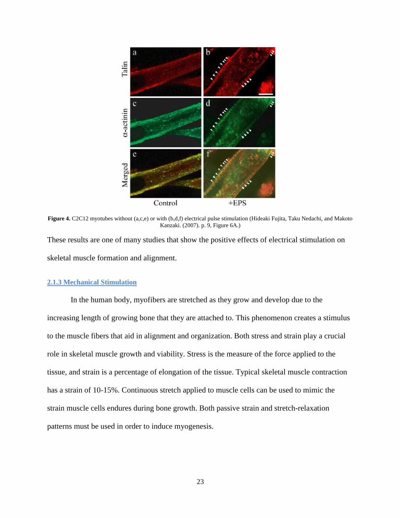
enhance differentiation in skeletal muscle constructs. In a study by Hideaki Fujita, Taku
Nedachi, and Makoto Kanzaki, it was found that electrical pulse stimulation at 40V/60mm, 24ms
and 1Hz was optimal in producing myotube contraction (2007). Electrical pulse stimulation
provided a means of skeletal muscle contraction as well as inducing sarcomere assembly. Figure
4 shows arrows where myotubes have localized levels of talin and α-actinin, which occur in the
early stages of sarcomere formation.
23
Figure 4. C2C12 myotubes without (a,c,e) or with (b,d,f) electrical pulse stimulation (Hideaki Fujita, Taku Nedachi, and Makoto
Kanzaki. (2007). p. 9, Figure 6A.)
These results are one of many studies that show the positive effects of electrical stimulation on
skeletal muscle formation and alignment.
2.1.3 Mechanical Stimulation
In the human body, myofibers are stretched as they grow and develop due to the
increasing length of growing bone that they are attached to. This phenomenon creates a stimulus
to the muscle fibers that aid in alignment and organization. Both stress and strain play a crucial
role in skeletal muscle growth and viability. Stress is the measure of the force applied to the
tissue, and strain is a percentage of elongation of the tissue. Typical skeletal muscle contraction
has a strain of 10-15%. Continuous stretch applied to muscle cells can be used to mimic the
strain muscle cells endures during bone growth. Both passive strain and stretch-relaxation
patterns must be used in order to induce myogenesis.

24
In addition, contractile forces must be produced by cells in order to be self-assembling.
As mentioned previously, fibroblasts can be co-cultured with the myoblasts in order to create the
necessary contractile forces. When introducing mechanical stimulation to in vitro muscle tissue,
especially with fibroblasts, it is necessary to provide an anchor point for the tissue to react
against in order to achieve a uniform direction. As the research suggests, mechanical stimulation
of muscle fibers actively helps the tissue to become functionally more mimetic of their natural
state (Guilak et al., 2003). For example, a study by Powell et al. showed that mechanical
stimulation improves tissue engineered skeletal muscle (2002). A mechanical cell stimulator
was created to perform uniaxial stretching to human bioartificial muscles within a six well plate
by a stepper motor, shown in Figure 5.
Figure 5. Mechanical cell stimulator (Powell et al. (2002), p. 5, Figure 1A).
The muscles were stretched 3.5µm every 10 minutes for 4 days, with the total strain
being 10% of the initial muscle length. Additional stress and relaxation patterns were examined
and found to elicit morphological changes in the muscle fiber, as well. Mechanical conditioning
caused an average 12% increase in myofiber diameter and an overall area increase of 40%. In
addition, mechanically conditioned muscle kept a consistent elastic modulus whereas the control

25
developed a stiffer modulus over time. This shows that mechanical stimulation is necessary in
preventing cross-linking and muscle stiffness. The mechanically stimulated fibers also showed
increased parallel alignment with one another in contrast to fibers that were not mechanically
conditioned. (Powell et al., 2002)
Mechanical stimulation has also been shown to affect myoblast proliferation and
differentiation in additional ways. For example, myoblasts that have been mechanically strained
decrease the production of α-actin if soluble growth factors are not present. Mechanical
stimulation of muscle fibers increases their discharge of insulin-like growth factor-1, which is an
autocrine growth factor for muscle. In summary, mechanical stimulation can provide the means
to cause enhanced cellular alignment and self-assembly.
2.2 Regeneration of Skeletal Muscle
People utilize their skeletal muscles on a regular basis so it is not surprising for these
muscles to become damaged. When the skeletal muscle tissue is torn, it undergoes a regeneration
process which consists of three phases: inflammation, repair, and remodeling (Turner &
Badylak, 2012). Overall, the regeneration process, as illustrated in Figure 6, involves the initial
injury, followed by activation of the satellite cells which then proliferate and differentiate into
new myofibers, followed by the maturation and integration of these myofibers within the
surrounding tissue environment.

26
Figure 6. Regeneration process of the skeletal muscle upon injury (Shi, X. and Garry, D. (2006), p. 1696, Figure 4A).
2.2.1 Inflammation
Upon injury, the myofibers within the skeletal muscle are sheared and torn, thus
triggering the first phase: inflammation (Turner & Badylak, 2012). The inflammation phase
occurs within the first few minutes of injury. Necrosis of the damaged myofibers immediately
occurs as the body releases tumor necrosis factor-α (TNF-α) (Huard et al., 2002). Meanwhile,
phagocytes begin to travel to the damaged site in order to consume the damaged myofibers
amongst any other cell debris formed from the injury. The phagocytes also send signals which
trigger and activate the satellite cells, which are located between the basement membrane and
sarcolemma and are normally quiescent. Neutrophils also travel to the injury site, and the
combination of neutrophils and macrophages releasing cytokines cause an inflammation to the
region, thus characterizing the first phase of the regeneration process (Turner & Badylak, 2012).
Several other growth factors, including insulin-like growth factor-1 (IGF-1), platelet derived
growth factors, and transforming growth factors, all aid the satellite cells in their proliferation
and differentiation into myoblasts (Huard, 2002; Turner & Badylak, 2012).
2.2.2 Repair
About one week post-injury, the repair phase begins. The repair phase peaks after about
two weeks and finishes by the third week (Huard et al., 2002). During this second phase, the
previously activated satellite cells migrate to the site of injury and begin to proliferate and

27
differentiate. The satellite cells differentiate into myoblasts and then into myofibers. These new
myofibers are generated to replace the previously damaged ones and integrate with the
surrounding myofibers (Turner & Badylak, 2012). Fibrosis also begins during this phase as
fibroblasts begin to create scar tissue (Huard et al., 2002).
2.2.3 Remodeling
The final phase of the regeneration process involves the remodeling of the injury site and
surrounding tissue. Continuous regeneration and repair is still ongoing during this phase as the
satellite cells have already formed into myofibers. The new myofibers start to mature and
integrate into the surrounding environment, attaching to the surrounding muscle fibers.
Revascularization and re-innervation are necessary for the regenerated site to obtain blood
supply and properly reconnect to surrounding neurons (Turner & Badylak, 2012).
Although the satellite cells were initially activated to differentiate into new myofibers,
not all of them undergo the differentiation, as only the necessary amount of satellite cells to
regenerate the initial wound is required. The remaining activated satellite cells return to their
inactive state until once again needed. In addition, the initially damaged ends of skeletal muscle
may not be completely reunited due to the accumulation of scar tissue as the fibrosis process
begins two weeks post-injury, during the repair phase and lasts up to four weeks post-injury,
during the remodeling phase (Huard et al., 2002; Turner & Badylak, 2012). The three phases of
the regeneration process allow regrowth and functionality to the previously damaged tissue, but
only to a certain extent.

28
2.3 Clinical Significance
Despite the human body being able to naturally regenerate damaged skeletal muscles,
there are many situations where the damage is too great. The skeletal muscles can be damaged as
a result of physical trauma injuries, such as exercise, sports, combats or accidents, or in
association with myopathies, or muscular diseases. Athletes tend to injure themselves on a
regular basis; 55% of those injuries involve damage to their skeletal muscle (Longo et al., 2012).
Soldiers, like many individuals involved in traumatic accidents, exhibit extensive skeletal muscle
damage. Such physical injuries occur in many forms, such as strains, lacerations, tears, and
contusions (Turner & Badylak, 2012). Many vehicle accidents, surgeries, and other situations
can lead to compartment syndrome, where serious inflammation occurs in the affected area.
Pressure builds up within the affected muscles, and the overall inflammation can cause
permanent damage to the muscle and nerves, potentially leading to amputation (Rekha, 2010).
On the other hand, muscular diseases, or myopathies, can also exhibit skeletal muscle
damage. Myopathies can be inherited genetically or obtained over time. One of the most
common myopathies is Muscular Dystrophy (MD). MD consists of several different diseases,
and exists in 63 individuals per million within the United States (Longo et al., 2012). Some
individuals genetically inherit MD as it involves mutations of the dystrophin gene, a muscle
protein which joins actin filaments and holds the cytoskeleton together. MD causes weakening of
the skeletal muscles and potential necrosis of the muscle cells and tissue. Unfortunately, people
born with MD may first exhibit healthy muscles, which gradually weaken and deteriorate over
time. Eventually, those individuals are restricted to wheel chairs and braces for movement (CDC,
2009).
29
Unlike inherited disorders like MD, people can also be susceptible to myopathies that
occur as a direct result of age. After the age of 30, contractility of sarcopenia increases;
Sarcopenia is a disease characterized by the gradual loss of muscle as one ages. Those who are
physically active are unlikely to contract sarcopenia, but people who are inactive may lose 3-5%
of muscle mass per decade after reaching age 30 (Evans & Campbell, 1993). Eventually, by age
70, the cross-sectional area and strength of the muscle can be reduced by 25-30% and 30-40%
respectively (Close et al., 2005). Even if a person manages to keep their skeletal muscles healthy
with regular activities, the chance of damage increases as they age.
Inflammatory myopathies are another type of myopathies involving chronic muscle
inflammation. A main symptom is weakening of the muscles. Polymyositis, one of the three
types of inflammatory myopathies, affects and weakens the skeletal muscles. The other two
types of inflammatory myopathies, dermatomyositis and inclusion body myositis, involve the
weakening and wasting of the muscles in general over time. (NINDS, 2011)
Damage and weakening of the skeletal muscles take place in day-to-day events and in
several diseases. Unfortunately, the body’s natural regenerative properties are unable to return
full functionality of the skeletal muscles in these more extreme cases. Many of the conservative
treatments used today to aid these cases include physical therapy, RICE protocols (rest, ice,
compression, and elevation), drug therapy, and surgery. Patients use various combinations of
these treatments, but the effects vary for each individual, making the treatments inconsistent.
There are currently no treatments that provide 100% recovery and full functionality of severely
damaged skeletal muscle. Currently, new potential treatments exist only in the research phase.
(Baoge et al., 2012)

30
2.4 Current Methods
Research concentrated on the development of biomimetic skeletal muscle tissue is
ongoing, and researchers continue to utilize different protocols and standards in developing the
tissue constructs and mechanically and electrically stimulating the tissue.
2.4.1 Culture of Tissue Constructs
Different studies have utilized various protocols for culturing 2-dimensional (2D) skeletal
muscle myoblasts into 3-dimensional (3D) tissue constructs. These methods include the use of
scaffolds, sutures, and micro-patterned surfaces. The incorporation of fibroblasts in the
constructs has also been studied.
Scaffolds are commonly used in tissue engineering to culture cells in a 3D environment
in order to properly mimic in vivo cells and their surrounding environment. Skeletal muscle cells
can be seeded onto scaffolds of different biomaterials, including biodegradable ones such as the
polyglycolic acid (PGA) used in Pedrotty et al.’s study (2005). Hydrogels are another method
used since a hydrogel can encapsulate the cells within an extracellular-like matrix and be easily
integrated into a biological system. Hydrogels are also used for their high variability in terms of
the different properties which can be altered, including porosity and mechanical properties, and
can also consist of multiple layers (Elisseeff et al., 2005)
The use of suture pins and fibrin gels can also create 3D constructs of skeletal muscle
tissue. This approach, as shown in Figure 7, involves fixing sutures to a plate layered with a
fibrin gel. The myoblasts are seeded onto the plate and over time, the gel and cells incorporated
in it roll up towards the middle where the suture points are aligned to form the final 3D
construct. (Khodabukus and Baar, 2009; Li et al., 2011)

31
Figure 7. Skeletal muscle cells seeded onto a fibrin gel which roll up into a 3D construct over time. (Lam et al. (2009), p. 1152.
Fig. 1C)
In order to aid the alignment of seeded myoblasts, micro-patterned surfaces are often
utilized, such as PDMS, glass, or other materials. In Ahadian et al.’s study, a hydrogel was given
a micro-patterned surface from PDMS (2012), and patterned PDMS surfaces were also used in
Lam et al.’s research (2009). Meanwhile, glass etching created the linear groove pattern on glass
coverslips in Yamamoto et al.’s research (2008). The grooves and patterns in these surfaces
allowed the myoblasts culturing on said surfaces to differentiate into myofibers while
maintaining a linear structure.
The incorporation of fibroblasts with myoblasts to develop a 3D skeletal tissue model has
also been taken into consideration. Some research studies have cultured C2C12 mouse myoblasts
and mouse embryonic fibroblasts separately, but then, the two were seeded together onto the
seeding surface. Li et. al co-cultured the mouse myoblasts and fibroblasts onto fibrin and
concluded that the fibroblasts aided in the morphology of the myotubes in terms of formation
and viability (2011).
2.4.2 Mechanical Stimulation
The mechanical stimulation of engineered skeletal muscle tissue aids in the alignment
and differentiation of myoblasts into myotubes. There are many studies that incorporate
mechanical strains to culturing skeletal muscle tissue through the use of motors attached to plates
or wells in order to stimulate multiple tissue constructs at once. Various strain percentages,
32
stimulation frequencies, and stimulation durations are used. For example, Pennisi et al. used a
maximum strain of 15% and a stimulation frequency of 0.5 Hz (2011), meanwhile Powell et al.
took the approach of using strains which incremented upwards over time – microstrains of
500µm for 4 days, rest for 3 days, and then 5% to 10% to 15% strain for periods of 2, 2, and 4
days respectively (2002). Standard parameters have yet to be determined as studies continue to
use a wide variety of strain percentages and frequencies.
2.4.3 Electrical Stimulation
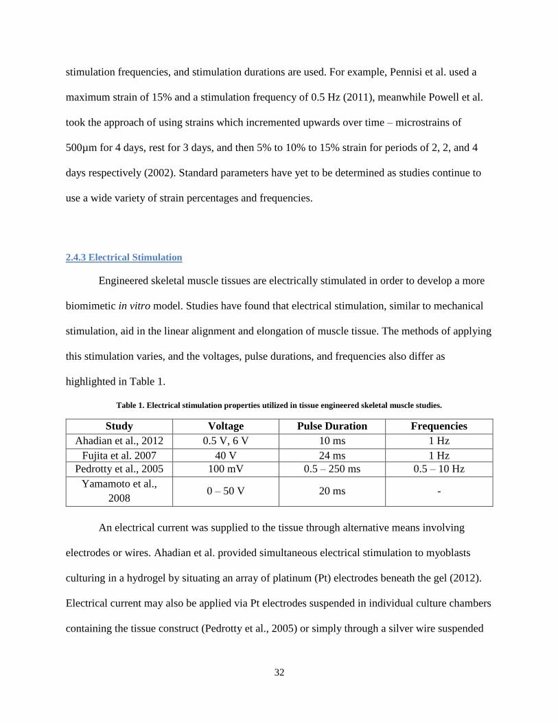
Engineered skeletal muscle tissues are electrically stimulated in order to develop a more
biomimetic in vitro model. Studies have found that electrical stimulation, similar to mechanical
stimulation, aid in the linear alignment and elongation of muscle tissue. The methods of applying
this stimulation varies, and the voltages, pulse durations, and frequencies also differ as
highlighted in Table 1.
Table 1. Electrical stimulation properties utilized in tissue engineered skeletal muscle studies.
Study Voltage Pulse Duration Frequencies
Ahadian et al., 2012 0.5 V, 6 V 10 ms 1 Hz
Fujita et al. 2007 40 V 24 ms 1 Hz
Pedrotty et al., 2005 100 mV 0.5 – 250 ms 0.5 – 10 Hz
Yamamoto et al.,
2008 0 – 50 V 20 ms -
An electrical current was supplied to the tissue through alternative means involving
electrodes or wires. Ahadian et al. provided simultaneous electrical stimulation to myoblasts
culturing in a hydrogel by situating an array of platinum (Pt) electrodes beneath the gel (2012).
Electrical current may also be applied via Pt electrodes suspended in individual culture chambers
containing the tissue construct (Pedrotty et al., 2005) or simply through a silver wire suspended

33
in the differentiation media that the tissue is residing in (Yamamoto et al., 2008). In each of
these studies, the applied electrical current aided in the proliferation and maturation of myoblasts
and myofibers.
2.5 Limitations
Though there are many current technologies being developed in regards to skeletal
muscle tissue engineering, none have been perfected. This lack of accuracy is due to a wide
range of limitations that hamper the ability of these technologies and methods to function to their
fullest potential. In this way, the current gap in knowledge surrounding skeletal muscle tissue
engineering exists in the form of these limitations. The most important limitations that must be
addressed are: cellular alignment, scaffolds, vascularization, and mechanical and electrical
stimulation.
As described in a previous section, skeletal muscle cells grow in a linear pattern due to
the elongation with bone growth and are further guided by the direction of electrical stimulus in
the body. For this reason, cellular alignment of the cells is essential. Aligning the cells promotes
myotube assembly and helps to mimic myotube organization into muscle fibers. Alignment of
the cells facilitates their differentiation into muscle fibers. If the cells are not aligned properly,
contraction cannot occur efficiently. However, despite recent advances and years of research,
uniformly aligning the muscle cells on a 3-dimensional model is still difficult to reproduce and
can be unreliable. (Koning et al. 2009)
In addition to cellular alignment, scaffolds that are currently being used as a support
structure to the skeletal muscle cells are also a current limitation in the field of skeletal muscle
tissue engineering. The purpose of a scaffold is to act as the extracellular matrix (ECM) and
provide the proper structure for the cells to form upon. The scaffold must be 3-dimensional in
34
order to form tissue constructs, and the material must be biocompatible, non-immunogenic and,
often, biodegradable. These are necessary to accurately mimic the structure while still remaining
compatible with the cells. With all of these factors to consider, choosing the material is often a
difficult task and, despite current research, there is still no consensus on the ideal scaffold
material and method that should be used. Many materials have been tested only to find that
some are ideal for certain uses whereas those same materials fall short in other aspects. (Stern-
straeter, et al. 2007; Neumann, et al. 2003)
Skeletal muscle cells, especially after differentiation into fibers, are abundant in
mitochondria, the powerhouses of the cells. Mitochondria provide the muscle cells with the
energy required to contract. However, this leads to a high metabolic demand for oxygen and
other nutrients. Without oxygen and nutrients, differentiation into fibers, as well as muscle
tissue, is a difficult task. In vivo, the cells receive their energy supply through the blood from the
blood vessels that surround them. However, in vitro, it is difficult to provide the cells with the
oxygen and nutrients they require as the current methods and constructs often do not promote
easy diffusion of the oxygen and nutrients through the media which the cells are submerged in.
Vascularization is a method currently studied that aims to address this limitation and provide the
cells with their metabolic requirements. However, these methods are not suitable for purely in
vitro culture of the cells, and are often time consuming and expensive. They are also difficult to
achieve and have not yet been tested with humans. (Koning, et al. 2009)
Recent studies have found mechanical and electrical stimulation to be essential for the
differentiation of skeletal muscle cells into fibers. In vivo, skeletal muscle cells and fibers
increase in strength and push towards differentiation by means of contraction. The electrical
stimulation mimics neuronal activity during myogenesis, and the mechanical stimulation mimics

35
the actual action of contraction. In vitro models are being developed to incorporate these
stimulations into the culture of engineered skeletal muscle to truly mimic the in vivo
environment. However, despite the studies that demonstrate that electrical and mechanical
stimulation are beneficial techniques, there have also been studies that have shown the opposite
to be true (Koning et al., 2009). This contradiction has caused a confusion amongst researchers
who are beginning to understand that the amount of mechanical stress and strain and electrical
voltage and frequency applied can have overwhelming effects—too much, and the cells risk
damage, but too little, and the stimulation barely causes a change. Electrical and mechanical
stimulation must be further studied and optimized in order to discover the ideal stimulation rates
before it can be truly incorporated into tissue engineered skeletal muscle. (Koning, et al. 2009;
Neumann, et al. 2003)
In order to devise a true, in vitro model for tissue engineered skeletal muscle cells and
fibers, these limitations must be addressed. This project aims to address the limitations of
cellular alignment, scaffolding and material, and mechanical and electrical stimulation by
devising a model that may be used to study the formation and maturation of skeletal muscle in
vitro. A model of this type would save money, time, and allow for the study of certain diseases
and injuries.

36
3.0 PROJECT STRATEGY
The client statement is the initial proposal of the project from the perspective of a client
who hopes to make his or her idea into a reality. Often times erring on the extremes of being too
vague or too specific, the client statement can be difficult to decipher. The design process
facilitates the breakdown of a client statement by isolating the important aspects of the project
through identification of the project’s objectives, constraints, and functions. After these have
been defined, the task of revising the client statement into a goal with a more reasonable scope is
easier.
3.1 Initial Client Statement
The following is the client statement that our team received:
Currently, the laboratory uses extruded fibrin microthreads with
human skeletal muscle derived cells seeded onto the surface and
transplanted into SCID mouse skeletal muscle injury models to
study the effect of various cell derivation and culture methods on
functional tissue regeneration. The use of animals is time
consuming and costly which severely limits the number of
parameters that can be evaluated. Currently, the microthreads are
produced first and then cells with myogenic potential are seeded
onto the microthreads using a rotational cell seeding system. The
limitations of this system include the ability to only achieve a cell
density limited to the surface area of the microthreads and the
system is not compatible with long term culture to evaluate the
differentiation potential of the cells in vitro. For cylindrical tissue
such as skeletal muscle fibers to form, the cells must degrade the
microthread material and proliferate and migrate into the core. The
proliferation phase of the cell cycle is not compatible with the
quiescent phase required for cell fusion and matrix synthesis needed
for skeletal muscle tissue formation. This could to lead to premature
breakdown of the tissue structure before the seeded cells can
synthesize new matrix. An optimal situation would involve a
system where cells could be seeded at the density required for cell
fusion and tissue formation. However, the current microthread
37
production process involves a stretching and drying step to produce
axially aligned fibers, which is not compatible with seeding the cells
within the microthreads at the time of formation.
A tissue engineered skeletal muscle system would enable the study
of skeletal muscle tissue formation, maturation and the potentiality
of cells entirely in vitro that could be used to approximate the utility
of their use for the replacement of lost or damaged skeletal muscle
tissue. The goal of this project is to design and produce a system
that recapitulates skeletal muscle fiber structure into which
myogenic cells can be seeded such that skeletal muscle tissue is
formed. The system must be either produced aseptically or must be
sterilizable and fit into an incubator in order to permit study of live
cultures over time. The engineered system should further be
amenable to the study of effect of mechanical strain and electrical
stimulation on muscle fiber maturation and contractile function.
To start, we created a list of questions to present to the client in order to better define the project.
Following this, we listed the objectives, constraints, and functions of the design based off of the
answers received and literature review conducted.
3.2 Objectives, Constraints, and Functions
The following subsections highlight the objectives, constraints, and functions defined.
3.2.1 Objectives
According to Dym & Little, an objective is something which a design should aim to
accomplish, an ideal goal that a design should become (2009). At the base of our objectives is
the following: the design must produce an in vitro model of skeletal muscle tissues. From there,
we were able to produce the following primary objectives and secondary objectives:

38
Biocompatible
Mimic Skeletal Muscle Structure and Function
o Three-dimensional, cylindrical tissue construct
o Provide mechanical and electrical stimulation
o Anchor tissue at fixed points and produce a continuous tissue between
anchored points
Enable Data Acquisition (applied voltages and strains, ultimate tensile strength)
o Precise
o Adjustable parameters
o Real-time data analysis
Marketable
o Easy to use
o Inexpensive
o Sturdy
o Re-usable or inexpensively replaced
If the final design does meet many of these objectives, the product will fail to meet the
demands of the client. Our product must be biocompatible in order to ensure proper cell growth,
viability and function. The design must be mimetic of an in vivo environment in the following
ways: three-dimensional tissue construct and regular mechanical and electrical stimulation via
strains and voltages to prevent atrophy and increase cell strength and maturity. The tissue will
be able to contract upon stimulus and will retain the strength of natural muscle tissue. There
should also be adjustable parameters such as available strain rates and voltages to allow for a
variety of experiments and for different stages of cell growth. Finally, the design must have

39
marketability in mind for those who would utilize a device of this kind in research, particularly
an academic setting. If it is too expensive to build, breaks after a single use, or is too complex to
manage with a basic knowledge of laboratory equipment, the design would be insufficient.
The objectives tree, which organizes the objectives listed above into primary and
secondary levels, can be found in Appendix A. A pairwise comparison chart, which ranks the
objectives in order of importance, can be found in Appendix B.
3.2.2 Constraints
A constraint is a limit on the amount of freedom one has to solve a problem, which
confines the number of options in finding a solution. It is important to evaluate what constraints
apply to your problem in order to allow for a larger focus on more probable solutions. Based on
our initial client statement, our project’s applicable constraints consisted of the following:
Must be within budget ($508)
Has to be sterilizable or made of sterilizable parts that may be assembled
aseptically by autoclave or soaking in alcohol
Must be completed in 25 weeks
Limited to cell line (C2C12 mouse myoblasts and CRL 2097 human dermal
fibroblasts), material (polycarbonate for device, common laboratory materials,
HDPE for base)
Limited by size and accuracy of on campus machines
Fit within the shelf of a standard incubator (0.1524 m (6”) tall, 0.4064 m (16”)
wide, 0.4572 (18”) deep)
Has to be safe for tissue construct and user

40
Must be able to withstand cell culture conditions (37 degrees C, 95% humidity,
5% CO2)
These constraints greatly limit the options in what type of device can be designed. For
example, our budget is highly restrictive because materials such as polycarbonate are costly. The
ability to sterilize is important for our device because without proper sterilization, cells could
become contaminated and die. If the device is to be sterilized by an autoclave, it must be able to
withstand the high temperature (121 degrees C). The size of our device will be confined to the
space of a standard incubator to allow stimulation of tissue constructs while they are in standard
cell culture conditions to prevent shocking tissues with different atmospheres and leading to poor
results or cell death. Naturally, the device components must be able to withstand standard cell
culture conditions or be designed such that the motor and electrical components are outside the
confinements of the incubator.
All of these constraints play a vital role in the drafting of our design as well as our
approach to solving the problem. As we consider these constraints, our device development will
be narrowed down to fewer options and design alternatives.
3.3 Revised Client Statement
The objectives, constraints, and functions described above, as well as the client meeting
questions and answers and literature review helped to create the revised client statement shown
below:
The purpose of this project is to develop and create an in vitro,
tissue-engineered skeletal muscle model that mimics the in vivo
nature of skeletal muscle tissue in both the environment and the
structure and functions of the fibers. The cells should differentiate
and align using the self-assembly approach and should be
mechanically stimulated in order to mimic tissue contraction and
increase strength of the tissue. These parameters, along with any

41
other defined parameters, should be both controllable and adjustable
in order to allow different stimulation rates. Fibroblasts should be
incorporated into the tissue constructs in order to allow for the
production of natural extracellular matrix to enhance tissue
integrity. The device should either be completely sterilizable or
aseptically assembled.

42
4.0 DESIGN ALTERNATIVES
4.1 Needs and Functions Analysis
4.1.1 Needs Analysis
After the client statement was revised, the team divided the functions of the design into
“needs” and “wants” based upon feasibility, time, and cost. The following were identified as
needs, being required of the design:
Able to produce a tissue construct
Able to facilitate axial fiber alignment
Able to mechanically stimulate the tissue construct
Able to electrically stimulate the tissue construct
Able to anchor the ends of the tissue construct
The different methods, or means, which can be utilized to meet each function, were listed in a
function-means chart as seen in Table 2. The following were identified as wants, what are not
necessary to the design but would be ideal to have:
Able to adjust parameters
Able to include real time data analysis

43
Table 2. Function-means chart
Function Means
Produce
myofibers Gels Scaffolds
Self-
aggregation
method
Micro-
patterned
surface
Align cell
growth
Limit
growth/surface
area
Mechanical
stimulation
Electrical
stimulation
Mechanically
stimulate Magnet Motor
Rotating
pulley
Wheel
mechanism
Weight
bearings Buoy
Electrically
stimulate Wire voltage
Electrode
array
Anchor ends
of myofibers Hook Clamp Pins
Dog bone-
shaped
myofibers
(2012 MQP)
Biological
glue
4.1.2 Functions
In order to begin developing a design, a list of functions was created to meet the client’s
needs. Functions define what the product is designed “to do” as stated in Engineering Design: A
Project-Based Introduction (Dym & Little, 2009). The functions of the model include the
following:
Produce a tissue construct from the starting myoblasts
Properly align the orientation of myoblasts (which then allows formation of
aligned tissues)
Mechanically stimulate the muscle cells and tissues
Electrically stimulate the muscle cells and tissues
Anchor ends of the construct
Measure various parameters (stress, strength, strain, contraction, voltage, etc.)

44
The first function which the design must include involves the differentiation of myoblasts
into myofibers. Although the overall goal of the design is to produce in vitro muscle tissue with
similar functions and structures as it is in vivo, the project must begin with the cell culture phase.
A C2C12 mouse myoblast cell line will be used. The myoblasts must be cultured and then
differentiated into the desired skeletal myofibers as needed for the final design before any of the
other components of the project can be incorporated. As the myoblasts are cultured, they also
must be properly aligned to mimic parallel myofibers as occurs in vivo.
Mechanical and electrical stimulation are necessary functions in order to achieve the
objective and client need of being mimetic of an in vivo structure. Mechanical stimulation is
necessary to our model, and according to previous research papers, it should promote further
maturation and alignment of the muscle tissue (Powell, 2002; Ahadian, 2012). Electrical
stimulation mimics the electrical impulses given to the muscle via nerves; these electrical
impulses in turn cause the muscle to contract. Electrical stimulation works to mimic the function
of the nerves on the muscle cells by providing the fiber with an electrical voltage that causes a
contraction to occur.
Anchoring the ends of the tissue construct within the device is necessary due to the
mechanical stimulation that will be placed upon the tissue. As mechanical stimulation includes
pulling the tissue uniaxially, there must be a method of holding onto the fiber, which can be done
by a hook, clamp, or pole. Finally, measuring various parameters of the tissue such as its tensile
and compressive strengths, the stresses and strains applied, the voltages applied, and histological
results using stains such as Masson’s Trichrome, Myosin Heavy Chain counterstained with
Hematoxylin, and antibody stains for fibroblasts within tissue constructs, are necessary in order
45
to validate that the in vitro muscle fiber created successfully mimics the in vivo nature of skeletal
muscle cells.
4.1.3 Specifications
The specifications of the design project include the following:
Utilizing a C2C12 mouse myoblast cell line
Maximum size less than the shelf of a standard incubator (0.1524 m (6”) tall,
0.4064 m (16”) wide, 0.4572 (18”) deep)
Myofiber diameter between 10-100µm (and a myofibril diameter of 1 µm)
Mechanical stimulation in the millimeter range (up to 25% strain ideally)
Electrical stimulation with an electrical field ranging from 0-10 V/cm
For this design project, the main cell line used is C2C12 mouse myoblasts for all initial
experiments. C2C12 myoblasts are the most commonly used cell line for skeletal muscle tissue
engineering due to its high proliferation rate and its differentiation capabilities. Ideally, the
design project will advance to using human myoblasts, but this advancement will not occur until
the project’s protocols and experiments are well established with the C2C12 line. In addition,
there is a limited supply of human myoblasts within the lab.
The full stimulation device must not exceed 0.4064 m wide by 0.4572 long by 0.1524 m
high as that is the dimensions of a standard incubator which is necessary for standard cell
culture. Ideally, in order to fit on the built-in shelves and to successfully share the space with
other groups or experimental tests, the full bioreactor design should not exceed the specified size
above. The diameter of the muscle tissue created in vitro should be within the range of 10-100
µm, and the myofibril diameter should be approximately 1 µm (Fox, 2011). In order to truly

46
mimic the in vivo size of the muscle fibers, the fibers of our tissue should not be smaller or larger
than the specifications provided. Finally, in the sense of mechanical stimulation, the tissue must
be able to withstand elongations (strain) up to 25 percent (adjustable to different percent strains
such as 2, 5, or 10 as well) based on its length (approximately 15 mm in length) (Powell, 2002).
For the electrical stimulation portion of the design, the cultured tissue will be given an
electrical pulse at different rates and for varying duration periods. Based on the parameters
utilized in previous studies, the tissue will be placed in an electrical field ranging from 0-10
V/cm and undergo said pulse for a period of 10 to 20 milliseconds. (Thelen et al., 1997; Radisic
et al., 2004; Pedrotty et al., 2005; Fujita et al., 2007; Yamasaki et al., 2009).
4.2 Conceptual Design Version 1
After many brainstorming sessions and iterations over different design alternatives, our
team developed the first version of our conceptual design. This design uses an agarose mold
rather than the PDMS micro-patterned stamp in order to generate the proper amount of tissue.
The reason for this change comes from the fact that using a PDMS stamp increases preparation
time for an experiment. The agarose culture mold, shown in Figure 8, shows the area where the
C2C12 cells are seeded to form a dog-bone shape. Figure 9 shows how the mold is located in our
device. The dimensions of the device itself are about 154.2 mm lengthwise, 101.6 mm deep, and
76.2 mm tall, including the lid. Figure 9 also shows the locations of the two electrodes that will
be used to provide electrical stimulation to the culture media in which the mold is suspended in,
and the two holes for the flow of the media in and out of the container. The locations of both of
these items are subject to change.

47
Figure 8. Agarose mold for culturing C2C12 cells.
Figure 9. Agarose culture mold location within the device.
After the bone shape is formed, the tissue contracts up onto the two posts, shown in
Figure 10. The two posts have a platform attached to the bottom to prevent the tissue from falling
off after the agarose mold is removed.
Figure 10. Front view of the device’s lid.

48
The two posts are attached to a track on which they can only move uniaxially and
bidirectionally. A top view of this track is shown in Figure 11. Figure 11 also shows the groove
made on the top of the lid which will hold the cap for maintaining an enclosed environment. This
cap is visible in Figure 10.
Figure 11. Top view of the device’s lid.
After the tissue is transferred onto the two posts, the agarose mold can be removed, as
shown in Figure 12. Absent from Figure 12 are grooves at the top of each post that allow
attachment to the lid that can move each post according to the motion of an adjacent stepper
motor. At the time of the making of this design, these features have not yet been included due to
uncertainties in the size and limitations of the motor.

49
Figure 12. Transparent view of the device with the lid cap and culture mold removed.
After the culture period, the muscle tissue attached to the two posts will be electrically
stimulated by the two electrodes. These electrodes will provide a current to the media. The media
can be changed through the two holes in the sides of the device. The two posts will be
mechanically stimulated to using a stepper motor.
4.3 Conceptual Design Version 2
After further evaluation of our first conceptual design, we determined that combining the
culture and mechanical stimulation phases in the same location was not feasible, and due to time
constraints, the pursuit of electrical stimulation incorporation was also not possible. The hollow
agarose posts were difficult to create and further details concerning that can be seen in the later
Feasibility Studies section. In addition, we concluded that the mechanical stimulation setup was
too complex to achieve for a variety of reasons, some including the many interlocking layers of
moving parts and how said parts would be connected to a motor from its current location above

50
the tissue construct. The bioreactor concept was also eliminated as we perceived it as an
unnecessary extra feature, and that it would be simpler, easier, and cheaper to replace media
manually.
Our team took a step back to develop a more feasible, straight-forward design that
allowed easier mechanical stimulation, multiple tests of tissue constructs at once, and utilized
materials readily available within a standard laboratory. Our new conceptual design, as displayed
in Figure 13 basically consisted of a T-shaped bar, T-bar for short, attached to a linkage and
rotor, where the rotor would be directly attached to a stepper motor. The rotation of the stepper
motor and rotor would cause the linkage to move, and in turn, cause the T-bar to move uniaxially
across a single plane.
Figure 13. CAD model of conceptual design version 2 (note only one petri dish is visualized in this image rather than three
which the design could ideally hold, and electrical components are not included).

51
The tissue constructs, which we decided to culture separately in the dog-bone shaped agarose
molds and then transfer into our stimulation device, would be situated within standard 90 mm
diameter petri dishes. The petri dish lids would contain two hooks which the cultured tissue
construct would be suspended across as displayed in Figure 14, and the bottom of the dish would
be filled with differentiation media in order to allow submersion of the tissue.
Figure 14. Close up of the petri dish and T-bar connection – note that the tissue and media are not included.
One of the hooks suspending the tissue will be attached to a separate component from the main
petri dish lid. As the T-bar moves back and forth, so will the movable lids, and in turn, the tissue
suspended on the attached hook will be mechanically stimulated uniaxially.
The general setup we proposed for this conceptual design still had its flaws, but after
feasibility tests and developing a prototype, we were able to devise a more refined final design to
be discussed in a later chapter.

52
4.3.1 Proposed Materials
In regards to manufacturing this second conceptual design, the materials must be
considered. The petri dish utilized throughout our proposed phases (excluding the custom lid)
can be obtained from a standard laboratory setting. Most of the other components, including the
custom lid (the main base and movable portion), T-bar, and linkage would be comprised of
polycarbonate. Polycarbonate will be utilized due to its ease of sterilization via an autoclave,
relatively inexpensive cost, and ease of manufacturing. We did expect to require a base to situate
everything on as certain components must be level with each other in order to work properly.
This base will be made of high-density polyethylene (HDPE) as this material is cheap and easily
accessible.
Other materials which are required for this design include the hooks which the tissue
would be suspended on, the rotor, and pins to hold the interface between the movable lids and T-
bar together. A stainless steel fish hook was selected as they are small and autoclavable. It is
known that the rotor and pins used to hold the movable lids and T-bar together could be made of
materials easily obtainable in a machine workshop as long as the rotor and pins were small
enough to fit the small resolution of our design.
4.4 Feasibility Studies
Although our design team developed a conceptual design for this project, we must
determine whether our proposed design and its components are feasible before finalizing and
manufacturing the assembly.

53
4.4.1 Agarose Molds
As the project calls for testing a skeletal muscle tissue construct that has formed by self-
aggregation, it was essential to create a mold where the cells were positively influenced into an
aligned tissue construct. The use of agarose molds was one method of accomplishing this.
Past projects have utilized and designed an agarose mold in the shape of a dog-bone,
where the tissue construct forms in the middle, and there are two rings of aggregated cells on
either side which, in turn, attach to the mechanical stimulation component of the device. Our
team decided to utilize this main concept for the agarose mold.
In past MQP projects, the agarose molds were cast directly from an ABS plastic negative
mold. However, issues were brought to our attention that included the brittleness of the ABS
plastic due to the manufacturing capabilities of the rapid prototyping machine and the difficulty
in sterilizing these molds. ABS plastic cannot be sterilized via an autoclave, so it must be soaked
in 70% isopropanol. However, ABS plastic is also porous, so it is not possible to completely
sterilize the mold.
In order to avoid these complications, the ABS mold was redesigned as a positive mold.
The intent was to cast a negative PDMS mold from the ABS positive mold and then,
consequently, cast the positive agarose mold from the PDMS mold, and this general process can
be seen in Figure 15.
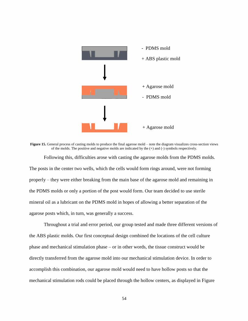
54
Figure 15. General process of casting molds to produce the final agarose mold – note the diagram visualizes cross-section views
of the molds. The positive and negative molds are indicated by the (+) and (-) symbols respectively.
Following this, difficulties arose with casting the agarose molds from the PDMS molds.
The posts in the center two wells, which the cells would form rings around, were not forming
properly – they were either breaking from the main base of the agarose mold and remaining in
the PDMS molds or only a portion of the post would form. Our team decided to use sterile
mineral oil as a lubricant on the PDMS mold in hopes of allowing a better separation of the
agarose posts which, in turn, was generally a success.
Throughout a trial and error period, our group tested and made three different versions of
the ABS plastic molds. Our first conceptual design combined the locations of the cell culture
phase and mechanical stimulation phase – or in other words, the tissue construct would be
directly transferred from the agarose mold into our mechanical stimulation device. In order to
accomplish this combination, our agarose mold would need to have hollow posts so that the
mechanical stimulation rods could be placed through the hollow centers, as displayed in Figure
- PDMS mold
+ ABS plastic mold
+ Agarose mold
- PDMS mold
+ Agarose mold

55
8. Unfortunately despite many trials of re-making the PDMS and agarose molds– we found this
version to not be feasible as the posts were not forming, even with the aid of mineral oil.
Our second version of the ABS plastic (agarose equivalent) mold was a simplified
version of the dog bone shaped mold used last year. As shown in Figure 16, this version has
smaller wells and posts, thus decreasing the diameter and thickness of the tissue ring to be
formed in it. In addition, we decreased the angle between the two end rings and the center
channel in hopes of reducing the stress of the to-be-formed tissue construct when pulled to
failure at a later stage of our project design. The overall size of the plastic/agarose mold was also
decreased in order to allow it to fit within a 6-well plate. Despite using mineral oil once again to
lubricate the surfaces of the wells and posts, the agarose posts would not dislodge from the
PDMS mold. We believe this occurred due to the post diameters being too small so the base of
the agarose mold has less of a grip to pull the posts out.
Figure 16. CAD model of the altered version of the agarose mold situated within a well from a standard 6 well plate (left). Top
view of the mold (right).
As the agarose posts of our mold continued to dislodge from the base after several
attempts, we designed an external replacement for the posts which would be inserted through the
agarose mold. As shown in Figure 17, we designed and created an external PDMS base with
posts which would be situated underneath the agarose molds. The main purpose of the posts in
the mold is to allow the cells to aggregate around a structure to form tissue rings, so the material

56
of the posts was not a concern, as long as it was sterile (PDMS can be easily sterilized in an
autoclave). After the incorporation of the external PDMS posts, the production of the smaller
version agarose molds was easier.
Figure 17. Insertion of external PDMS base and posts into agarose mold (left). Combined view of the agarose mold and PDMS
posts (right).
4.4.2 Stepper Motor and Arduino Setup
A stepper motor was selected as the motor of choice for the purpose of this design
because, through research done by previous project teams, it was shown that the drawback of
regular motors is that the mechanical stimulation applied cannot be precisely controlled. When
using a standard motor, previous groups greatly reduced the rotational speed in order to apply the
minute strains to the attached tissue construct, however, a regular motor only stops moving via
deceleration once the power supply is disrupted. By utilizing a stepper motor, research shows
that we will be able to control the attached mechanical components and allow them to move at
precise distances forward and backward due to the concepts behind a stepper motor.
Electromagnets within stepper motors allow the motor to rotate at specified steps within a full
revolution and stop immediately at a certain step, removing the need for deceleration.

57
Unfortunately, utilizing a stepper motor involves additional components which a standard
motor would not need. A standard motor requires a power source to allow it to rotate and move
attached mechanical components. A stepper motor requires a driver (a programmable circuit
board) in order for it to rotate properly; a stepper motor will not function if simply attached to a
power supply. Stepper motors also require an interface to control different parameters set by the
driver’s program. In order to connect the parts, the team went to Benjamin Dwyer, an Electrical
and Computer Engineering student at Worcester Polytechnic Institute, for assistance. The
following components were obtained for the stepper motor set-up:
Motor driver
Arduino Uno – R3 (microcontroller board based on the ATmega328 which can
be used for a variety of interactive applications)
12 button keypad
Breadboard
LCD kit
Stepper motor (68 oz. in, 400 steps/revolution)
The general setup between these components can be seen in Figure 18, but more detailed
diagrams of all the connections and component specifications can be seen in Appendix D.

58
Figure 18. General diagram of stepper motor and electrical components setup (top) and the same setup viewed in person
(bottom).
Arduinos are easy-to-use electronic prototype boards where free program codes are
provided online for several different applications. An Arduino was incorporated into our stepper
motor as it would facilitate changing parameters of the stepper motor.
The Arduino was set up so any user could easily interact and control the attached stepper
motor. When the Arduino is plugged into an outlet, the LCD screen is prompted by a welcome
message and the main menu appears. The main menu consists of three modes to control the

59
stepper motor: move to position, oscillation, and manual jog, as displayed in Figure 19. All
modes are based around the different steps or intervals that the motor moves. The stepper motor
has 400 steps per revolution, so essentially the Arduino board setup instructs the motor to move
to different specified positions between 0 and 400.
Figure 19. Stepper motor mode functions as displayed on the LCD main menu.
Mode 1, move to position, directs the stepper motor to move to a single specified position
after the number is entered in. Mode 3, manual jog, allows the user to manually control the motor
by pressing 1 repeatedly to move the rotor in one direction or by pressing 3 repeatedly to have it
move the opposite direction. This mode is more useful for making minor adjustments to the
motor position after all the mechanical components are attached. Mode 2, oscillation, is the main
mode which the mechanical stimulation of our design will utilize as it commands the motor to
move back and forth between two step positions as designated by the user. The motor will
oscillate between the positions continuously until the Arduino is reset to the main menu screen.
Earlier in the design process, our team originally thought of placing the motor and
attached electrical components within the incubator in order to mechanically stimulate the
cultured tissue construct. Although all the components could withstand the incubator’s standard
temperature of 37˚C, they would break down from the high level of humidity. There was the
possibility of encasing the electrical components and motor within a plastic Tupperware
container, but the motor would still have to be slightly exposed in order to allow rotation. After

60
looking into other resources, we determined that the entire motor setup could take place outside
of the incubator. The rotor attached to the motor would extend from outside the incubator,
through a bushing sealed in a cork, and the rotor would extend inside the incubator attached to
the remaining ‘culture’ portion of our design.
4.4.3 Mechanical Stimulation Components
Utilizing the rapid prototype machine on campus, we created the mechanical components
utilized in our first conceptual design. Our rapid prototype was created from ABS plastic and due
to the minor resolution errors of the machine, we had to sand down some parts in order to make
the interface connections fit. The rapid prototype of our mechanical system attached to the motor
can be seen in Figure 20.
Figure 20. Rapid prototype of conceptual mechanical system attached to the stepper motor (left). Close-up of the rapid prototype
components (right).
We tested the general movement of the mechanical parts using the different modes of the
Arduino board. The rotor rotated properly as displayed by a marker line we drew on it to
visualize its movement. When the motor was put into oscillation mode, we noticed that although
the rotor moved appropriately, the T-shaped bar connected to the moving lid components did not
move linearly due to a lack of constraints in other axes. After testing the feasibility of this
design, we decided to alter our design by adding in linear bearings and supports to each end of
61
the T-shaped bar so they were positioned parallel to the T-bar’s line of movement, all of which is
shown in detail in Chapter 6. The bearings and supports would keep all the mechanical
components aligned and reduce any loss of linear movement from the motor.
4.4.4 Electrical Testing of Pt/Ir Wire
Our design incorporates electrical stimulation via wires conducting an electrical charge
across the culture medium in a standard petri dish. The material choice for our electrical wire is
important because several types of metals can corrode within the media. Corrosion causes the
release of salts and ions which are cytotoxic to cells (Ratner et al., 2004). In order to reduce the
chances of corrosion, a minimally corrosive material was chosen for the electrical wire; 80%
platinum, 20% iridium (Pt/Ir). Although Pt/Ir is more expensive than other standard metal
materials like copper, the biocompatibility and survival of the cells far exceed in importance over
utilizing an inexpensive wire that can jeopardize cell viability.
Before implementing the Pt/Ir wire into our stimulation device, we needed to test its
electrical properties in a controlled environment. We were concerned about whether a uniform
electrical charge would be present when the wire is electrically stimulated while submerged in a
solution as the tissue will be submerged in cell culture media for electrical stimulation. In order
to test this, two standard 100 mm diameter petri dishes were filled with either 1x phosphate
buffer solution (PBS) or DMEM Ham’s F12 with 10% FC III (the standard cell culture media
used when proliferating C2C12 cells). Two 1 cm pieces of Pt/Ir wire were cut and attached onto
a power supply in an Electrical and Computer Engineering (ECE) lab at Atwater Kent. Different
voltages were applied to the Pt/Ir wire starting at 0 volts and moving upwards at increments of 2
volts. A Digital Multimeter (DMM) was used to measure the varying levels of voltage at five

62
locations throughout the petri dish as shown in Figure 21, where current traveled from location 4
to 2, where the negative and positive ends of the Pt/Ir wires were positioned respectively.
Figure 21. Locations of measured voltage throughout petri dish.
The applied and measured voltages were all recorded in Table 3, along with the
corresponding average and standard deviation values in Table 4. The measured voltage values in
both solutions were not equal to the initially applied voltage due to the solution’s resistance to
the electrical charge, which is to be expected. Based on the collected data, the average and
standard deviation values of all locations (1-5) varied as a whole, but locations 1, 3, and 5 were
more consistent, and average and standard deviations for those locations were calculated
separately as shown in the table. The values obtained at locations 2 and 4 greatly differ due to the
electrical current traveling between those two points. We determined that we should only be
concerned about locations 1, 3, and 5 having similar values, especially location 5, as our tissue
would ideally be linearly aligned and in between the two Pt/Ir wires. Based on the values
obtained from the center locations (1, 3, 5) of the petri dish, our tissue should undergo uniform
electrical stimulation.
1
2
3
4 5

63
Table 3. Measured voltages from Pt/Ir wires at various locations within a 100 mm diameter petri dish.
Measured Voltage (V)
Solution
Voltage Applied
(V) 1 2 3 4 5 1
x P
BS
0.00 -0.13 -0.0005 -0.12 -0.24 -0.84
2.02 0.79 0.77 0.74 0.84 0.81
4.01 1.55 1.48 1.37 1.40 1.39
6.06 2.52 3.10 2.48 2.00 2.59
8.01 3.54 4.33 3.26 2.41 3.23
10.00 4.60 5.49 4.17 2.53 4.09
12.07 5.40 7.34 5.15 3.37 5.34
16.00 7.45 10.46 7.65 4.66 7.46
18.01 8.95 12.00 8.74 5.68 8.56
20.04 10.30 14.00 10.25 6.46 10.29
22.02 10.65 15.69 10.55 7.16 11.62
30.50 15.10 21.90 15.50 9.95 15.70
DM
EM
Ham
's F
12 w
ith 1
0%
FC
III
0.00 -0.38 -0.36 -0.35 -0.33 -0.32
2.07 0.63 0.63 0.62 0.59 0.60
4.03 1.47 1.60 1.48 1.30 1.47
6.01 2.46 3.00 2.46 1.87 2.49
8.00 3.44 4.46 3.52 2.40 3.67
10.11 4.60 6.17 4.72 3.11 4.82
12.08 5.67 7.49 5.82 3.80 6.14
16.05 7.74 11.21 8.20 4.85 8.24
18.04 9.09 12.87 9.23 5.42 9.59
20.03 9.85 14.52 10.32 5.43 10.42
22.04 11.10 15.70 11.10 5.32 11.40
30.13 15.30 24.00 16.00 6.54 16.56

64
Table 4. Correlating average measured voltages in all locations and center locations of a 100 mm diameter petri dish.
Solution
Average
(V) S.D.
Average in
Locations 1,3,5
(V)
S.D. in
Locations 1,3,5
1x
PB
S
-0.27 0.30 -0.36 0.41
0.79 0.03 0.78 0.04
1.44 0.07 1.44 0.10
2.54 0.35 2.53 0.06
3.35 0.62 3.34 0.17
4.18 0.96 4.29 0.27
5.32 1.26 5.30 0.13
7.54 1.84 7.52 0.11
8.79 2.00 8.75 0.20
10.26 2.38 10.28 0.03
11.13 2.73 10.94 0.59
15.63 3.79 15.43 0.31
DM
EM
Ham
's F
12 w
ith 1
0%
FC
III
-0.35 0.02 -0.35 0.03
0.61 0.02 0.62 0.02
1.46 0.10 1.47 0.01
2.46 0.36 2.47 0.02
3.50 0.66 3.54 0.12
4.68 0.97 4.71 0.11
5.78 1.18 5.88 0.24
8.05 2.02 8.06 0.28
9.24 2.36 9.30 0.26
10.11 2.88 10.20 0.30
10.92 3.30 11.20 0.17
15.68 5.55 15.95 0.63
Due to the solution’s resistance, the applied voltages and measured voltages are not
equal. The values obtained from the 1x PBS solution fluctuate in the different petri dish locations
as the applied voltage increases. We believe this is due to PBS being a more concentrated salt
solution than DMEM/F12. Although DMEM/F12 is also a salt solution, it consists of more
components than PBS which, in turn, may impede with voltage traveling through it and reduce
any possible fluctuations. Based on the collected data, we expect to be applying a voltage

65
between the ranges of 0-10 volts (V) in order for the tissue construct placed in the center of the
petri dish to experience a voltage between 0-5 V.
In order to electrically stimulate skeletal muscle tissue, the voltage will be applied to
culture media different from the one utilized in this experiment. Once the tissue construct has
formed and can be electrically stimulated, it will be placed in differentiation media (DMEM
Ham’s F12 with 2% horse serum and 1% insulin transferrin selenium). The cell culture media,
specifically proliferation media, utilized in this experiment was the media that allows the cells to
grow and proliferate prior to differentiating into myotubes. At the time this electrical experiment
was conducted, differentiation media had not yet been prepared, so proliferation media was
utilized in its place. We expect the differentiation media to yield similar results as the
proliferation media as both largely consist of DMEM and Ham’s F12.
Unfortunately despite the electrical feasibility tests conducted, its incorporation of the
device was not possible as a result of time constraints. The use of Pt/Ir wire is certainly feasible
and recommended to be incorporated into the overall device in the future.
4.5 Experimental Tests
4.5.1 Control Tissue Fiber Test
In preparation for future experiments, we needed to have a set of controls readily
available. The control test was designed such that it would accomplish this, as well as feasibly
prove that tissue constructs may be formed within the molds using only the C2C12 cell line. The
experiment was designed to run for five days post seeding to allow the differentiation media to
be fully induced within the construct. Five days was chosen because it is known that
differentiation, once induced, does not occur immediately and proliferation still continues for

66
some time prior to complete differentiation. During culture, the cells were induced towards
differentiation on plates two days prior to seeding as two days was the minimum time necessary
for proliferation to slow and for differentiation to begin having an effect as determined by
previous groups.
The experimental set-up consisted of four tissue constructs. One tissue was to be tested
for contraction via electrical stimulation, another tissue was planned to be embedded in a
paraffin block for histological testing, and one tissue was to be pulled to failure using an Instron
machine in order to obtain tensile strength data on tissues purely made from C2C12 cells with no
stimulation. The last tissue was used as a control in the case that one tissue did not properly
form. These controls are suitable comparisons for future experiments that incorporate the use of
fibroblasts into tissue construct formation, as well as experiments that include mechanical
stimulation.
As the molds that are being used are not very different than the molds used by the
previous teams, we decided to use the same cell seeding density, 3 million cells per mold, due to
the fact that it had worked previously in another MQP design. The molds were equilibrated in
media overnight prior to seeding in order to ensure that the cells would have sufficient nutrients
when settling to the bottom of the mold and contracting and self-aggregating to one another. In
order to visualize the constructs as they form throughout the duration of the experiment, images
were taken.
Images were taken by two methods, microscope images and visual camera pictures.
Visual camera pictures were taken to visualize tissue construct formation. Microscope images
were taken daily at 5X to use as measurements of construct thickness and to visualize any cracks

67
or discontinuities in the cells that are not easily seen by camera photos. Figure 22 demonstrates
how microscope images were taken as the entire construct cannot be seen in one view.
Figure 22. Diagram depicting positions of microscope images to be taken per tissue construct.
4.5.2 Cell Seeding Density Experiment (Mold Version 1)
This experiment was a direct result of the previous experiment, where it was determined
that the cell density seeded was too small for these molds. Three cell densities were determined
for testing: 3 million cells per mold was used as the short end, as it was previously recognized as
not being enough, 5 million cells per mold, and 7 million cells per. For this experiment, there
were six tissue constructs, two for each density. Molds were equilibrated 24 hours prior to
seeding, as described in the previous experiment, and the molds were fitted within the wells of a
standard 6-well plate.
Cells were differentiated at 70% confluency and seeded 24 hours, rather than the previous
48 hours, post differentiation. The earlier seeding was conducted in order to allow the C2C12
cells to have a higher chance of forming a construct within the agarose molds. When
differentiation is induced, the C2C12 cells exit the cell cycle stage and enter the differentiation
stage. Not necessarily all the cells enter the differentiation stage immediately, but stay in the cell
cycle stage for a period of time prior to exiting the cycle. As determined by a previous MQP,

68
there is the concern of determining the time point of seeding the cells where enough cells are
differentiating into myofibers, but a portion of the cells are also proliferating (Aschettino et al.,
2012). The need for the presence of some proliferating cells is necessary in order for the cells
seeded into the molds to properly aggregate and form a cohesive tissue construct.
The experiment was further designed such that it would only run for two days post
seeding, as it was only to visualize tissue construct. Two days was chosen because of
observations made from the previous experiment. Tissue constructs formed after the first day,
with slight changes, if any, occurring by the second day. It was further decided that any tissue
constructs of the ideal density selected would be maintained in culture and used as controls as
described by the previous control experiment. Figure 23 depicts the set-up of the experiment
using a 6-well plate:
Figure 23. Set-up of 6-well plate for Cell Seeding Density Experiment. Labels correlate with labels on the plate. Wells A1 and
B1 have 3 million cells per mold, A2 and B2 have 5 million cells per mold, and A3 and B3 have 7 million cells per mold.
Images were taken as previously described in the control experiment in order to visualize
construct formation.

69
4.5.3 Cell Seeding Density Experiment (Mold Version 2)
The initial cell seeding density experiment concentrated on utilizing the first version of
the agarose mold, but another was conducted on the second version of the mold. Due to the high
cell seeding density of 7 million C2C12 cells required by the original mold, the design was
altered in order to decrease that seeding density. The changes on the mold design included
rounding the edges of the channels, essentially decreasing the presence of sharp corners between
the end rings and center channel junctions and also creating a smaller area for the cells to settle
in by decreasing the well area around the two posts. These alterations allowed the cells to
aggregate in a more concentrated region of the mold and ideally allow less stress and contraction
at the junction points.
Similar to the setup in the experiment involving the first mold, six molds (version 2) were
created and equilibrated overnight prior to seeding. The molds were divided up into three groups,
each with a sample size of 2, at the following cell seeding densities: 2 million C2C12 cells, 3
million C2C12 cells, and 5 million C2C12 cells. C2C12 cells were cultured to about 70%
confluency and then differentiation was induced. 24 hours post differentiation, the cells were
seeded evenly throughout the channels of the equilibrated agarose mold.
Based on the results obtained from this first iteration (further described in Chapter 4.6.3),
the experiment was repeated with different cell seeding densities in order to further determine the
minimum amount required to form a fully-formed tissue construct. Six molds were created and
during this iteration, two molds each were seeded with the following densities: 3 million, 4,
million, and 5 million C2C12 cells. The cells were seeded 24 hours post differentiation and
within 18 hours post seeding, the molds were flooded with media in order to prevent necrosis of
the tissue.

70
4.5.4 Fibroblast Incorporation
After determining the minimum cell seeding density for the second version of the agarose
mold, 4.5 million C2C12 cells, fibroblast incorporation was considered. Previous iterations of
culturing the tissue constructs at various densities have revealed similar results where portions of
the construct will form, but a cohesive dog-bone shape is not achieved. Ideally, incorporating the
fibroblasts will increase natural ECM production and ultimately aid in forming a cohesive tissue
construct. Six of the smaller version agarose molds were seeded with a total of 4.5 million cells,
as determined earlier, where three molds contained 30% CRL2097s, human dermal fibroblasts,
while the remainder molds consisted of solely C2C12s to represent the controls. Human
fibroblasts were utilized in order for ease of imaging and identification later on during the
staining stages of the tissue. Tissue constructs formed within 24 hours post seeding and the
resulting tissue was fixed for further histological analysis in terms of morphology of the tissue
and positive presence of human nuclei from the 30% human fibroblasts seeded.
4.6 Preliminary Data
4.6.1 Control Tissue Fiber Test Results
On Day 1, it was clear that no tissue constructs formed correctly, and the tissues were
maintained until Day 2. Upon seeing no changes, the experiment was ended and tissues were
discarded as they were unusable for electrical stimulation, mechanical testing, or histology.
Figure 24 below is a series of photos taken from a camera that visually depict tissue formation on
Day 2 prior to discarding the constructs.

71
Figure 24. The results of the Control Experiment depicting that none of the fibers formed.
In many cases, the rings formed, but the channels did not while Fiber 4 was an outlier.
When seeding Fiber 4, the cells were suspended in a volume too large for the mold, causing the
suspension to spill out of the mold and into the plate. The following were determined as potential
reasons for construct formation failure: not enough cells were seeded or the angles between the
ring and center channel were too sharp, causing the tissue to break.
Two steps were taken in lieu of these results. New molds were designed that were
rounded and removed the sharp angle. Also, a cell seeding density experiment was designed to
determine the correct number of cells to seed into each mold. It was determined that previous
MQP team, who also had difficulty in forming tissue constructs, did not achieve an optimal
seeding density.

72
4.6.2 Cell Seeding Density Experiment Results (Mold Version 1)
On Day 1, both tissues A3 and B3, which had been seeded with the largest cell density, 7
million cells per mold, had formed. A3 had a visible crack at the right interface between the ring
and fiber, however. None of the 3 million or 5 million cells per mold tissue constructs formed.
On Day 2, no changes were noted in the 3 million or 5 million cells per mold tissue constructs.
Between Day 1 and Day 2, tissue A3 broke where the previous crack had been, resulting in a torn
construct, however, tissue B3 was still fully formed. Images of the six tissue constructs can be
seen in Figure 25.
Tissues A1, A2, B1, and B2 were discarded after imaging while A3 and B3 were
maintained and kept in their molds and differentiation media for four days post differentiation.
B3, the only construct available for use, was embedded in paraffin and used as a histology
control for tissue constructs made purely from C2C12 cells with no stimulation for comparison
with future experiments that incorporate fibroblasts into the tissue construct to aid in construct
formation and constructs that have been stimulated.
Figure 25. Results of the Cell Seeding Density Experiment at Day 2. Only tissue B3, seeded with 7 million cells, properly
formed.
A1 A2 A3
B1 B2 B3

73
As both of the 7 million cells per mold tissue constructs had formed at Day 1, as seen
under a stereoscope in Figure 26, and one survived past Day 1, the team decided that the ideal
seeding density for future tissue constructs would be 7 million cells per mold. Future
experiments would include incorporating fibroblasts into the tissue construct and analyzing its
effects on tissue construct.
Figure 26. 7 million C2C12 tissue construct under stereoscope (Scale bar = 3 mm).
Construct B3 was used as a histology control for tissues made from only C2C12 cells.
The tissue was fixed in 2% paraformaldehyde for 12 hours, processed using the TBS-ATP1
Automatic Tissue Processor on the long cycle with no fix, and embedded into paraffin in
preparation for staining. Images of the stains were taken at 32X and the scale bars represent 50
micrometers. The following three stains were completed: Masson’s Trichrome Blue,
Hematoxylin & Eosin, and Myosin Heavy Chain. After the experiment incorporating human
fibroblasts, slides from this tissue will also be used as a negative control in antibody staining for
fibroblasts.
Masson’s Trichrome Blue exhibits the morphology of the tissue in relation to muscle
components. Black regions depict nuclei, blue regions depict collagen and red regions depict
muscle fibers, as well as other intercellular fibers. As our tissue has been differentiated towards

74
becoming muscle tissue, it was anticipated that the majority of the tissue would exhibit red,
elongated staining with multiple black dots to demonstrate myofibers with multiple nuclei. The
tissue was split into three regions prior to embedding: Left, Middle, and Right. The left side
shows the connecting region between the left hand ring and the fiber construct, the middle shows
the region of most importance to our project, the muscle tissue fiber, and the right side shows the
same as the left side. Figure 27 shows images taken at 32X for the middle region, which is the
most pertinent to our needs.
Figure 27. Masson’s Trichrome stain taken at 32X of construct B3, middle region (Scale bar = 50 µm)
For each section of tissue, the tissue was oriented such that the section that came together
in the V-like point at the bottom of the mold was sectioned first. In the case of this stain, the
section was within the first three cut from the wax block. It can be assumed that these sections
would exhibit the best morphological properties as they were the thinnest regions of the tissue
and were directly aligned with the entirety of the construct. In the stained middle region red,
which exhibits muscle fibers, is visible. This proves that the tissue was successfully
differentiated into muscle. The middle section demonstrates linear alignment with the red stain
on both sides, showing healthy, differentiated tissue. However, the middle of the tissue is not
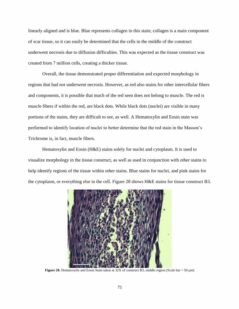
75
linearly aligned and is blue. Blue represents collagen in this stain; collagen is a main component
of scar tissue, so it can easily be determined that the cells in the middle of the construct
underwent necrosis due to diffusion difficulties. This was expected as the tissue construct was
created from 7 million cells, creating a thicker tissue.
Overall, the tissue demonstrated proper differentiation and expected morphology in
regions that had not underwent necrosis. However, as red also stains for other intercellular fibers
and components, it is possible that much of the red seen does not belong to muscle. The red is
muscle fibers if within the red, are black dots. While black dots (nuclei) are visible in many
portions of the stains, they are difficult to see, as well. A Hematoxylin and Eosin stain was
performed to identify location of nuclei to better determine that the red stain in the Masson’s
Trichrome is, in fact, muscle fibers.
Hematoxylin and Eosin (H&E) stains solely for nuclei and cytoplasm. It is used to
visualize morphology in the tissue construct, as well as used in conjunction with other stains to
help identify regions of the tissue within other stains. Blue stains for nuclei, and pink stains for
the cytoplasm, or everything else in the cell. Figure 28 shows H&E stains for tissue construct B3.
Figure 28. Hematoxylin and Eosin Stain taken at 32X of construct B3, middle region (Scale bar = 50 µm)

76
The stain was completed on the fourth slide sectioned from each block, making it such
that the sections were very similar to those performed for the Masson’s Trichrome stain. The
morphology depicted is representative of the morphology that was seen in the Masson’s
Trichrome Blue stain. The sides of the tissue are healthy and linearly aligned, and the center has
entered necrosis and is not linearly aligned. In this stain, the blue dots representing the nuclei are
clear, and it is easy to see that the nuclei are aligned with one another where the stain is red in the
Masson’s Trichrome. This allows us to know that the sides of the tissue do, in fact, consist of
healthy myofibers. As myofibers, the tissue should exhibit myosin, which is a motor protein in
muscle that is used in contraction. To further demonstrate differentiation of the tissue, a Myosin
Heavy Chain counterstained with Hematoxylin for nuclei was completed, as well.
In order to properly analyze the differentiation of the C2C12 mouse myoblasts into
myotubes, a Myosin Heavy Chain immunohistochemistry strain was conducted. The tissue
samples that underwent this stain were from the same tissue construct B3 and were prepared in a
similar manner as the ones previously described in the sections concerning the Masson Trichome
and Hematoxylin and Eosin stains. Myosin MF-20 was used as the primary antibody as we were
interested in seeing myosin expression, meanwhile the secondary antibody was anti-mouse Ig.
Diaminobenzidine (DAB) substrate was used to create the visual cue as the staining involved a
peroxidase enzyme – this enzyme binds to DAB and oxidizes it, thus producing the color brown.
The main purpose of utilizing this stain was to analyze whether differentiated myotubes were
present. The presence of myotubes would be represented by the brown visualized by this stain as
the primary antibodies specifically target myosin, a protein present in skeletal muscle tissue.
Although a Myosin Heavy Chain stain would stain for myosin and visually appear brown
on the microscope images, the presence of myotubes could not be confirmed unless multiple

77
nuclei were present, which is why the tissue samples were then counterstained with
Hematoxylin. The samples that were stained with Myosin Heavy Chain and counterstained with
Hematoxylin were derived from sections of the middle section of the B3 tissue construct –
essentially the center connecting fiber between the two tissue ring ends. As seen in Figure 29, the
brown-stained myosin can be seen along the edges of tissue, while the center area contains
necrotic cells, which was also seen in the previous stains.
Figure 29. Myosin Heavy Chain counterstained with Hematoxylin0 taken at 32X of construct B3, middle region (Scale bar = 50
µm)
The brown linear fibers containing blue circular shapes, the nuclei, represent properly
differentiated myotubes. We expected the outer edges of the tissue construct to differentiate into
myotubes as the differentiation media could easily diffuse into the tissue edges, meanwhile the
necrotic center was malnourished to the point of cell death, thus explaining why no brown, or
differentiation into myotubes, is present in the microscope image. The remnants of brown
present in the center of the tissue are a result of the DAB substrate being exposed and active on
the tissue for a prolonged period of time. When the DAB substrate is active for a prolonged
period of time, it can potentially bleed out to surrounding regions and oxidize the brown colors

78
in the extracellular matrix of other undesired cellular components, thus the brown in the center
tissue can be disregarded.
The controls obtained from these three different stains (Masson’s Trichome, H&E, and
Myosin Heavy Chain) will all compared against tissue constructs made in the future consisting of
fibroblasts and differing mechanical properties as they will be mechanical stimulated by our final
device.
4.6.3 Cell Seeding Density Experiment Results (Mold Version 2)
On Day 1 post seeding, the molds seeded with 3 million and 4 million C2C12 cells did
not form cohesive tissue constructs. All of these molds exhibited similar results – rings partially
formed, but the center channel did not fully form and connect at the intersecting so a cohesive
dog-bone shape was not apparent. The molds seeded with 5 million C2C12 cells did form a
complete construct, and ideally, were to sit in the molds for further testing and analysis, but on
Day 2, the tissue constructs contracted and tore, thus were unusable.
Multiple iterations of this experiment were repeated to further determine whether 5
million cells was indeed the minimum cell seeding density required, or if the number could be
further reduced as a lower number would be more ideal. As 4 million cells was unsuccessful, but
5 million cells demonstrated the opposite, a cell seeding density of 4.5 million cells was
investigated. Fortunately, the tissue constructs were able to fully form at said density as
displayed in Figure 30.

79
Figure 30. 7 million C2C12 tissue construct under stereoscope (Scale bar = 3 mm)
4.6.4 Fibroblast Incorporation
24 hours post seeding 4.5 million cells (30% CRL2097s and 70% C2C12s), the cells
aggregated and formed cohesive tissue constructs within the smaller agarose molds. Due to the
tissue constructs contracting off the PDMS posts 48 hours post seeding, the tissue constructs
were either used towards proof of concept testing, testing of electrical contraction, or were fixed
for further histological analysis. The tissue constructs went under immunohistochemistry in order
to determine whether fibroblasts were incorporated into the tissue and how their presence
affected the overall tissue morphology. After antigen retrieval specific to the human nuclei of the
fibroblasts seeded within the rest of the C2C12 (mouse) cells, DAPI substrate stained the human
nuclei brown as visualized in Figure 31, while the control tissue constructs visualized no brown
and no presence of fibroblasts as expected.

80
Figure 31. Human nuclei antigen retrieval of tissue constructs with a cell seeding density of 4.5 million cells – no fibroblasts
seeded (A) and 30% fibroblasts incorporated (B). (Scale bar = 100 µm)
As seen in the immunohistological stains, the tissue constructs with fibroblasts displayed
a more cohesive morphology in comparison to the control tissue with no fibroblasts. The cross-
section of the tissue with no fibroblasts in Figure 31A consists of a fragmented center, indicating
cell necrosis, meanwhile Figure 31B visualizes healthier tissue apparent by the lack of necrotic
cells visualized. Unfortunately due to the lack of tissue samples during the embedding process,
further stains, like the Myosin Heavy Chain stain, was not conducted to determine the presence
of myosin.
Other tissue samples underwent electrical stimulation to observe for contraction behavior.
Each end of the tissue was suspended in media and attached to leads from a function generator.
A digital multimeter was used to confirm the currents applied to the tissue construct. The tissue
was exposed to a sine wave at 100 Hz and varying voltages of 0 to 5V, but unfortunately, no
contraction occurred. The absence of contraction is most likely due to the necrotic tissue in the
samples with no fibroblasts and the myofibers required more time to further differentiate (as the
samples were induced for 48 hours prior to this test).
A B

81
5.0 DESIGN VERIFICATION
5.1 Tissue Construct Formation
As the molds went through various iterations throughout the course of the year, it was
important to ultimately select a mold design that was able to form a cohesive tissue construct.
The two mold designs ultimately used for final, proof-of-concept testing were the larger, dog-
bone shape mold and the smaller, angled dog-bone shape mold with PDMS posts. In both
molds, tissues were successfully created using the self-assembly method in which the cells were
seeded into the molds and allowed to contract towards one another, forming the dog-bone shape,
as seen in. Figure 32A depicts a tissue formed with the larger mold and Figure 32B depicts a
tissue formed with the smaller mold.
Figure 32. The tissue on the left was formed using the larger mold and the tissue on the right was formed using the smaller mold
with PDMS posts. Both constructs are cohesive constructs, though weaknesses are visible in certain regions. (Scale bar = 3 mm)
As previously described, the tissue also met the requirement of being differentiated into
skeletal muscle by exhibiting myotubes, or linearly aligned fused cells with multiple nuclei and
by exhibiting myosin, which is a product of muscle fibers. Both tissue constructs were able to be
manipulated and removed from the molds in order to transfer to the petri dish lid and hooks and
the larger constructs were able to be stimulated, as well, demonstrating their mechanical
integrity.
A B

82
5.2 Manufacturing Mechanical Device
In order to build our mechanical device, we enlisted the help of the Higgins and
Washburn shops on the Worcester Polytechnic Institute (WPI) campus. The rapid prototyping
machine on campus is limited to printing ABS, which we used for our mechanical prototype in
Figure 20, which is not ideal as it cannot be fully sterilized. An autoclave exposes the pieces to a
heat of 121°C, and ABS melts and deforms at 105°C. Knowing this, we elected to make our
parts out of polycarbonate, which does not begin to melt until 150°C and is known to be easily
manufacturable. In addition to purchasing stock polycarbonate, we purchased stock HDPE for
parts that would not need to be autoclaved. The polycarbonate was purchased from Ultimate
Plastics in Worcester, MA, and the HDPE was ordered online from McMaster-Carr.
The parts for the final device were manufactured using a Haas computer numerical
control (CNC) mill, used for precision manufacturing. Each part was first designed in
Solidworks, a 3D computer-aided design software, and adjusted to appropriate tolerances for fits
and the optimum dimensions for the available drill bits in the shop. The final computer aided
design (CAD) drawings for each part are shown in Appendix C. After each part was optimally
modeled, the designs were moved into ESPRIT computer-aided manufacturing (CAM) software,
where the means of manufacturing was mapped out. This code could then be read by the Haas
CNC mill to carve the wanted dimensions out of the stock loaded into it.
The following parts were milled out of stock polycarbonate:
3 x Petri dish custom static lid
3 x Petri dish custom movable lid
3 x latch piece
1 x cross bar and push link (T-bar shape)
1 x linkage arm

83
2 x 0.5” bushing holder
2 x 0.75” bushing holder
The petri dish components also required custom holes for embedding the stainless steel
fish hooks which would hold the tissue. Each hook had its width measured and its tip truncated
and sharpened to fit in these holes and were carefully pushed into place using an arbor press. The
base upon which everything was held together was milled out of HDPE. The remaining parts
were used from available stock in the shop. Three cylinders of stock mild steel were used for
bushing cylinders and the rotor. Three sizes of stainless steel dowel pins were used to connect
each piece to one another, using either locational fits or sliding fits where appropriate. 0.5” Ø
bronze bushings and 0.75” Ø steel bushings were incorporated as well, to reduce chatter and
increase stability. The sliding motion of the bushing cylinders and the rotation of the rotor were
made easier by a thin layer of white lithium grease applied to each during the final assembly
process.
A late alteration to the design demanded a longer rotor than earlier designs, and a 1.75” Ø
fountain cork was added to allow integration of the device with an incubator in the lab. The
middle of the cork was carefully turned out with a lathe, and steel bushings were pushed into the
opening to allow it to fit with the rotor cylinder without difficulty. Other than the ones fit into the
cork, each bushing was fit to a bushing holding piece which was fastened to the base using ¼-20
hex cap screws. In order to attach the rotor to the stepper motor, the motor’s own rotor had to be
modified. Using a surface grinder, one side of the motor’s rotor was flattened. This allowed a set
screw to lock the two rotors together.

84
6.0 FINAL DESIGN AND VALIDATION
6.1 Agarose Mold Procedure
In order to make 2% agarose in DMEM, the correct grams of agarose powder must be
weighed and poured into the correct amount of DMEM that has been pipetted into a glass bottle.
The mixture should be ran through a wet cycle using an autoclave. Once the agarose has
finished autoclaving, it may be used immediately. PDMS posts should be placed into the hollow
channels of the PDMS negative mold, using sterile mineral oil as a lubricant. While still a hot
liquid, the agarose should be pipetted into the sterile PDMS negative molds in the space where
the PDMS post base and negative mold have a gap, allowing for the agarose to fill the mold until
it reaches the surface of the post base. This should be done in one steady ejection of agarose in
order to prevent the formation of bubbles underneath the post base. After 15 minutes, the agarose
positive may be removed from the PDMS negative and placed individually into the wells of a 6-
well plate. Figure 33 shows the final version of the agarose mold once it has gelled and been
removed from the PDMS negative.
Figure 33. The final agarose mold with the PDMS posts going through the mold. A small PDMS base exists under the agarose
mold that is attached to the posts in order keep the posts sturdy.
Then differentiation media, fungizone and penicillin/streptomyocin (add 1 microliter per
milliliter of media for the antibiotics) is added into each well such that it is not overflowing the
mold, and then it can be placed in the incubator to equilibrate overnight. The media formulation
is different from the agarose, which only contains DMEM, due to the presence of serum and

85
other nutrients. Equilibrating the molds prior to seeding allows for the molds to take in these
nutrients such that when the cells are seeded into the molds, they are surrounded by the nutrients
and may absorb them.
6.2 Cell Culture Phase
The final cell seeding density of the larger molds was 7 million cells, 100% of which
were C2C12 mouse myoblasts; the final cell seeding density of the smaller molds was 4.5
million cells, 70% C2C12 and 30% CRL2097 human dermal fibroblasts. The cell culture
process began with separate cultures of mouse myoblast C2C12 cells cultured using 60%
DMEM/40% Ham’s F12 with 10% Fetal Clone III and Glutamine, and human dermal fibroblasts
CRL2097 cultured using DMEM with 10% Fetal Clone III and Glutamine. At about 60-70%
confluent, 24 hours prior to seeding the cells into molds, both cultures were induced with
differentiation media composed of 60% DMEM/40% Ham’s F12 with 5% Horse Serum, 1%
Insulin Transferin Selenium and Glutamine. All media compositions may be found in Appendix
F.
24 hours post-inducing, the cells are suspended in differentiation media such that 3.15
million C2C12 cells were in 20 µL and 850,000 CRL2097 cells were also in 20 µL, for a total of
4.5 million cells in 40 µL. The suspension was evenly distributed in the molds and placed into
the incubator overnight. For the larger molds, 7 million C2C12 cells are suspended into 150 µL
and evenly distributed into the mold. About 18 hours post seeding, the cells have contracted to
form tissue constructs. For those that have survived, more media is added to the well, flooding
the mold and tissue construct. Fungizone and penicillin/streptomyocin are also added to the
wells at the appropriate ratios. 24 hours post seeding, Day 1 of the tissue constructs, the tissues

86
may be removed from the molds to be placed onto the hooks or they may be left in the molds,
depending on the experiment that the constructs will be used towards.
6.3 Mechanical Stimulation Device
Our final design for the device is a modified and more practical version of the conceptual
design version 2 described in Section 4.3 Conceptual Design Version 2. The design underwent
several modifications and updates, all of which can be seen in Figure 34.
Figure 34. CAD assembly model of the final design with a representative portion of the incubator wall – note that the electrical
components are not included in this model.
This design still provided simultaneous mechanical stimulation to three tissues. Each tissue
construct, once fully grown into the “dog-bone” shape, was suspended manually from truncated
steel fish hooks press fit into the top of two pieces of a custom made petri dish lid. The larger
base part of the lid remained stationary, rigidly constrained by four dowel pins embedded in the

87
base. The smaller part of the lid slid horizontally on top of the main lid base, fit to constrain
movement to one axis. As the smaller lid moved back and forth, the tissue is stretched in
conjunction.
Bronze bushings were incorporated into the design for several reasons. The primary
reason was to reduce unwanted chatter of the device as the mechanism turns. This provided an
extra layer of stability for the device and ensured that the positions of each part were in fact
moving in their intended paths. The cylinders, the bronze bushings and the bushing holders are
shown in Figure 35 along with how they connect to the “T-bar” component.
Figure 35. Close up view of the cylinders which constrained the linear movement of the T-bar.
The three tissues move in sync due to being attached to a T-bar component that laid on an
elevated part of the base. The motion provided to move the T-bar was done by a 400-step stepper
motor and rotor set up with a linkage. The T-bar had a dowel pin in a sliding fit with a
connecting arm attached to the rotor, allowing it to rotate freely. The pin connecting the arm to
the rotor was offset from the center by 3 mm, allowing for a maximum of 6 mm of movement at
180 degrees (or 200 steps).
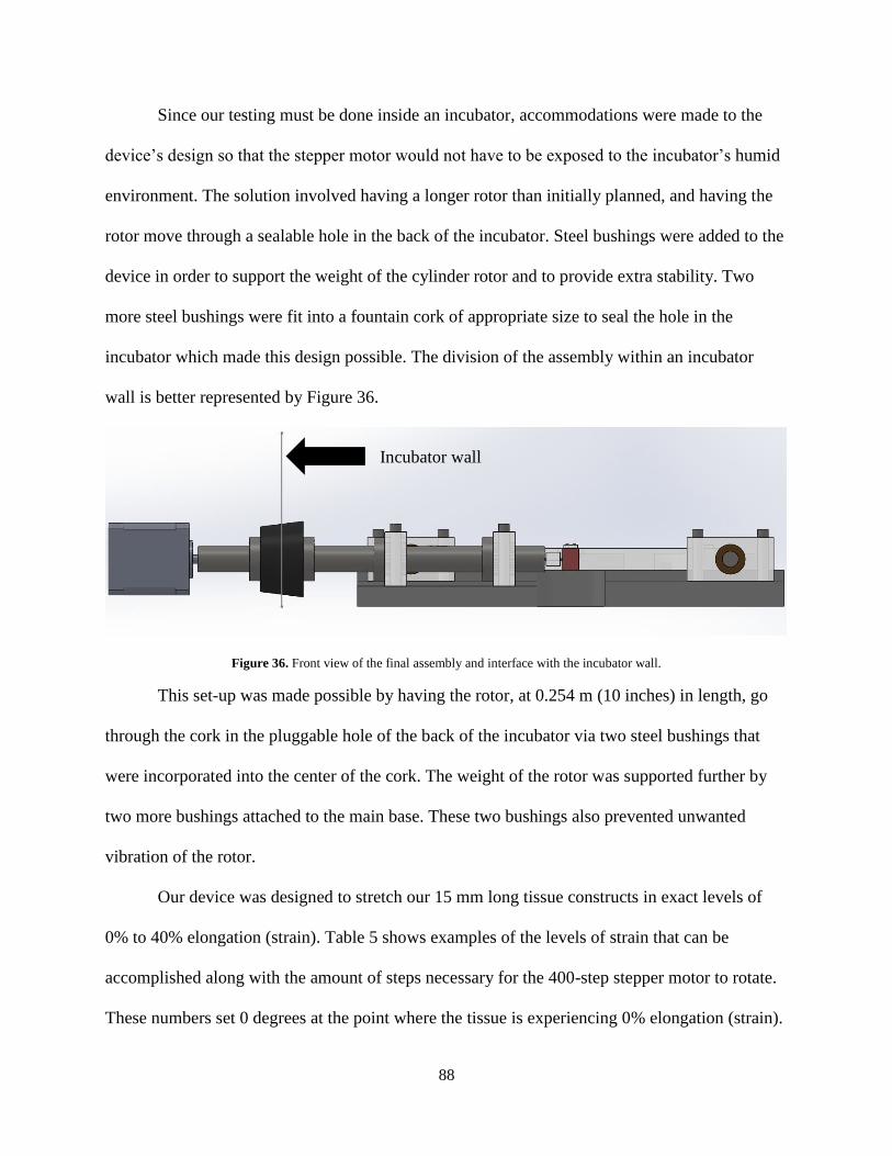
88
Since our testing must be done inside an incubator, accommodations were made to the
device’s design so that the stepper motor would not have to be exposed to the incubator’s humid
environment. The solution involved having a longer rotor than initially planned, and having the
rotor move through a sealable hole in the back of the incubator. Steel bushings were added to the
device in order to support the weight of the cylinder rotor and to provide extra stability. Two
more steel bushings were fit into a fountain cork of appropriate size to seal the hole in the
incubator which made this design possible. The division of the assembly within an incubator
wall is better represented by Figure 36.
Figure 36. Front view of the final assembly and interface with the incubator wall.
This set-up was made possible by having the rotor, at 0.254 m (10 inches) in length, go
through the cork in the pluggable hole of the back of the incubator via two steel bushings that
were incorporated into the center of the cork. The weight of the rotor was supported further by
two more bushings attached to the main base. These two bushings also prevented unwanted
vibration of the rotor.
Our device was designed to stretch our 15 mm long tissue constructs in exact levels of
0% to 40% elongation (strain). Table 5 shows examples of the levels of strain that can be
accomplished along with the amount of steps necessary for the 400-step stepper motor to rotate.
These numbers set 0 degrees at the point where the tissue is experiencing 0% elongation (strain).
Incubator wall

89
For simplicity, the device was designed so that at this point, the linkage arm is level with the T-
bar component.
Table 5. Strain Levels
Tissue Length Displacement (mm) Percent
Elongation (strain)
Rotation
(degrees)
No. of
Steps
15mm 1.5 10 60 67
3 20 97 100
3.75 25 104.5 116
4.5 30 150 167
6 40 180 200
Figure 37 shows the final device assembled outside of the incubator and the stepper
motor and electrical components housing located at the back of the incubator in the position to
do testing. The hole available at the back of the incubator is on the top shelf of the incubator, so a
cardboard stand was constructed to hold these parts to the right height during testing. This setup
was also designed to have the motor weighed down with a paperweight to prevent chatter during
rotation (not shown).
Figure 37. Final device base and all attached components assembled (left) and final design stepper motor outside of incubator
with electrical components housing (right).
The final design was set up within a standard incubator used for cell culture as seen in
Figure 38. The top shelf of the incubator does not make the base level with the hole in the back

90
so during setup, we used leftover HDPE from our stock to be placed underneath the device as
feet to meet the height.
Figure 38. Device setup inside incubator.
Fully assembled, the device demonstrated the desired movement while inside the
incubator. First, the device was tested outside with a rubber band, and later with the tissue
constructs. Figure 39 shows a close-up view of the tissue attached to the hooks, but note that the
image does not display media in the petri dish as the presence of liquid altered the view of the
tissue.
Figure 39. Close up of tissue attached to hooks inside petri dish

91
6.4 Mechanical Stimulation Phase
By wedging a pair of forceps between the agarose mold and the PDMS base and slowly
allowing the forceps to open, the agarose separates from the PDMS, taking the tissue with it and
removing the posts. The agarose may be cut away to allow for the forceps to come in and lift the
tissue out of the mold by looping the rings around the forceps. From there, the tissue is
transferred to the hooks, which are held at an angled position, and the forceps slide the ring onto
the hooks, repeating with the other ring. Once the tissue is securely on the hooks, the lid is
placed down into the base of the petri dish that has been pre-filled with media in order to
submerge the tissue into a bath of nutrients. The petri dish is then transferred to the spot
dedicated to it on the stimulation device and secured. The stimulation device is then placed into
the incubator, allowing the rotor within the cork to go through the hole in the back of the
incubator. After closing the incubator door, the stepper motor is attached to the rotor that is
protruding from the back of the incubator. The electrical components are turned on and
mechanical stimulation may commence.
92
7.0 DISCUSSION
7.1 Tissue Complications
Throughout the duration of the project and experimentation, several problems became
apparent with tissue formation. Although tissues were formed within the agarose molds, the
tissues had a low success rate of formation, did not contract when electrically stimulated, and
broke by day two post seeding.
The low success rate of the tissue construct formation can be attributed to a few possible
explanations. The first reason for low success could be due to the lack of extracellular matrix.
Although fibroblasts were incorporated in order to secrete ECM, the team experimented with
various stages to induce differentiation in order to optimize that secretion. It is possible that the
fibroblasts did not have enough time to settle and secrete ECM prior to the construct breaking
within the agarose mold. This could also be a factor in the integrity of the tissue and limit the
ability for electrical stimulation to occur. Another possible explanation of low tissue formation
could be human error in seeding the cells into the agarose molds. Although cells were visibly
placed within the agarose wells, the wells were very small and it is possible that even distribution
was not always achieved within the entire well and prevented areas from forming a cohesive
structure. In addition, the cells were seeding in a layer method where the C2C12 mouse
myoblasts were seeded and the CRL2097 fibroblasts followed. It is possible that an even mixture
of the cell types may be more beneficial in cell to cell interaction and construct formation than
cell layers. The cells used were cultured separately and then seeded upon reaching ideal
confluency; however studies have co-cultured fibroblasts and myoblasts prior to seeding instead.
Another complication in tissue formation was that the constructs that successfully formed
did not last more than two days post-seeding before breaking. Once a construct broke, it

93
contracted upon itself and was no longer usable for experimentation. This issue could be
attributed to the strength of the PDMS not being flexible enough for contraction in the construct,
meaning that as the tissue matured and began contraction, the posts did not flex with these
contractions and caused a break within the tissue itself. All breaks within tissue constructs were
found in the channel region as opposed to the rings, making this explanation seem more viable.
Lastly, both the 7 million and 4.5 million tissue constructs did not contract upon electrical
stimulation. For the construct containing 7 million cells, visible necrosis in the channel was
present, resulting in dead cells that could not conduct electrical stimulation. The 4.5 million
tissue construct did appear to have necrosis, but electrical contraction could have been prevented
due to the lack of myosin. When the 7 million cell construct was stained for myosin, small
amounts were present at day 2. Since the smaller construct was stimulated at day 2 before it
could contract upon itself, it is likely that there was also small amounts of myosin present.
Myosin is essential in muscle tissue contraction and in previous studies, was noted to be
abundant enough for contraction at day 9 (Dao et al., 2012). This provides reasonable insight as
to why the tissue construct was unable to contract.
7.2 Mechanical Device Complications
Additional issues were observed with our mechanical device that could be considered for
future recommendations. The first problem noticed with our mechanical device was that there
was difficulty in transferring our petri dishes containing the tissue into the stimulation
component. This was due to a tight area in which the petri dish had to be placed in and also the
lack of easily removable attachments. The pins that connected the two components together were
crooked due to imperfections in the manufacturing of certain parts and hard to remove and
replace without significantly moving the petri dish. This would be difficult to do in a sterile

94
manner as the media would be likely to splash onto the lid of the dish when moving it.
Transferring the developed tissue was also difficult as the tissue had to be manually removed
from the PDMS posts and agarose mold, sometimes resulting in breakage of the construct.
In terms of mechanical movement of the rotor being sent to the hooks holding the tissue
construct, there were some errors present. For example, despite greasing the device components
and incorporating bushings, there was still chatter present in the movement along the linkages.
This could be due to non-linear or uneven connections in the mechanical components. This can
be seen elsewhere, as the T-shaped bar and base of the device was not level. Uneven platform
and connections throughout the device could attribute to the chatter and lack of smooth
movement.
Additional general implications were present in the device that could be improved upon.
The length of the rotor component was able to clear the back of the incubator, but the set screw
on the motor was inaccessible. In addition, the petri dish would need to be placed lastly inside of
the incubator, which is very difficult due to low clearance. Another consideration for future
improvements is the method of adjusting the T-bar to a zero position. Since our mechanical
components were outside the incubator and the tissue and T-bar was inside the incubator, it was
necessary to have two people in order to visualize how adjusting the rotor location affected the
tissue.
7.3 System Impacts
There are many topics that can be addressed in terms of our in vitro device and how the
subject is relevant to it. For example, economics can be taken into consideration and relevant to
our device as cell culture is expensive and the devices needed in order to complete are also. The
sterilizable polycarbonate was also costly. Our device can be relevant to environmental impact,

95
as it requires the disposal of biohazards and uses non-renewable energy for operation. The most
significant implication our device has is societal, ethical, and political. Within each area, our
device creates opposition from society due to the use of human cell culture, such as the human-
derived dermal fibroblasts and the cells derived from mice that we use. Politically, stances must
be made on the use of the cells and where they are derived from. Our device could be useless if
access to required cell sources becomes denied. Health and safety can be viewed as an
implication since biohazards are present and could cause danger to those working with them.
Manufacturability is possible but could be difficult in a short amount of time. All components
were custom designed and some handmade, making the manufacturing process longer and more
arduous than normal. Our device is sustainable in how the custom components can be sterilized
and reused, although the petri dish bases cannot. Overall, our device excels in sustainability but
lacks in manufacturability. As with many scientific devices for testing, ethical and societal
impacts are of concern.

96
8.0 CONCLUSIONS AND RECOMMENDATIONS
8.1 Conclusions
After the entire design process, experiments, testing, and validation, our team ultimately
achieved the following accomplishments: optimized the dimensions of the agarose mold used for
forming tissue constructs, optimized the agarose mold making process in terms of making it
more sterile, cultured self-assembled skeletal muscle tissue, and developed a uniaxial stimulation
system.
Initially, we utilized the agarose mold dimensions from a previous MQP, but found that
the version we made required a high cell seeding density of 7 million cells. In order to further
reduce the cell seeding density, we decreased the diameters of the wells which tissue rings would
form, and in addition, created smoother junctions between the rings and center channel. As a
result of the changes made, the cell seeding density was lowered to 4.5 million cells.
Previously, the agarose mold process consisted of creating a negative ABS plastic mold
and then casting the positive agarose mold from it, but this two-step process had a higher chance
of contamination due to the process of sterilizing the ABS plastic molds. The ABS plastic molds
were sterilized via soaking in 70% isopropanol, but the plastic molds carry the chance of holding
residue within its porous inner structure. In order to address this issue, our team created a three
step process involving a positive ABS plastic mold which a negative PDMS mold was cast from.
The PDMS molds could then be easily sterilized within an autoclaved and then the positive
agarose mold can be cast from it. The usage of PDMS allows easier and more dependable
sterilization and repetitive use of its material for a prolonged period of time.
The tissue construct cultured and utilized in this project also exhibited properties evident
within in vivo skeletal muscle tissue. The group successfully cultured skeletal muscle tissue that

97
properly differentiated as indicated by the positive presence and production of myosin, a protein
indicative of myofibers. The cultured tissue also addressed the limitations of current approaches,
as explained in Chapter 2: Literature Review, as the tissue cultured during our project consisted
of its own natural ECM.
Ultimately, the group developed a uniaxial stimulation system, or essentially a
mechanical device which has the ability to suspend a cultured tissue construct and linearly
actuate it. The use of a stepper motor attached to the mechanical device allowed for adjustable,
controllable parameters in regards to the strain applied to the tissue construct. The entire setup
was also incorporated into an incubator environment. Stimulation of the tissue within an
incubator would be more ideal as the incubator environment is more biomimetic to that of which
skeletal muscle typically resides in. The incorporation was possible by positioning all the
electrical components (stepper motor, motor driver, etc.) outside of the incubator, while the
remainder of the system resided within the incubator. The electrical component of the device
remained connected to the main mechanical portions within the incubator via a rotor sealed in a
cork extending from the outside to the inside of the incubator. The device was then able to fully
run without any cautions about the humid environment of the incubator negatively affecting the
motor.
8.2 Future Recommendations
Despite having a year to work on this project, there is always room for improvement.
Different features could be improved upon in regards to the mechanical and stepper motor setup,
the formation of the tissue construct, incorporation of electrical stimulation, and further
validation and experiments.

98
The mechanical device was designed to move uniaxially and the support beams of the
device were incorporated to prevent any unwanted chatter. Unfortunately after further testing,
there is still chatter present as the T-bar will, on occasion, shift from side to side rather than
move linearly forward and back. Friction between a lot of the interfaces of the device,
specifically the ones between the movable lids and the base lid of the petri dishes, are affecting
the accuracy of the applied stimulation. The support beams could be improved upon to address
said issues. In addition, transferring the petri dishes in and out of the mechanical device prove to
be difficult as user error could easily introduce contamination into the petri dishes. The dowel
pin between the rotor and linkage arm had a low fatigue limit which caused it to break, so in
order to increase the life of the device, a thicker dowel pin could be utilized. In order to ease the
setup of the device in the incubator, a longer rotor would be more ideal as it will allow access to
the set screw.
The stepper motor could be improved upon in regards to user accessibility and user ease.
Currently, users of the mechanical device must re-adjust the initial position of the motor prior to
mechanical stimulation, so a limit switch should be integrated into the design so the motor
position is always initialized upon powering on. The programming of the motor could also be
upgraded so the user can enter the exact rates of strain to be applied along with how long the user
wants the motor to run and any other customized time periods (i.e. relaxation periods, alternating
strains, etc.) the user may desire. Currently, the user must calculate the number of steps needed
for the motor to oscillate between to apply said desired strain rate, but this external calculation
could be easily integrated into the motor’s program.
Although the team optimized the agarose mold cell seeding density from 7 million cells
to 4.5 million cells, there is always room for improvement to continually decrease that density.

99
Further variations of the mold could be tested to allow a smaller cell seeding density. In addition,
the geometric shape of the mold could be improved upon to cause less tension within the
construct, as evident in our tissue would contracted and tore two days post seeding. Although the
team created smoother junctions between the end rings and center seeding channel, these
junctions could still be improved upon to further reduce the tension. In addition, the process of
differentiating the cells and seeding them into the molds can be further experimented with,
specifically in terms of additional supplements added to the differentiation media, different
percentages of cell types co-seeded into the molds, and when a more ideal time to seed the cells
would be post differentiation if the percentage of cell types incorporated were altered.
As stated in Chapter 2: Literature Review, mechanical and electrical stimulation of
skeletal muscle tissue further enhances its properties to make the tissue more biomimetic. The
design of this project was unable to fully incorporate electrical stimulation due to time
constraints, but electrical stimulation would certainly aid in the formation of more biomimetic
skeletal muscle tissue.
Future experiments should be conducted with the device in regards to what optimized
strains and currents should be applied to result in a biomimetic skeletal tissue model with similar
mechanical properties and morphology as that in vivo. The experiments can greatly vary in
regards to what strains and currents should be applied, for how long, how often, along with the
inclusion of relaxation periods. Ideally once the device and parameters to be used to develop a
biomimetic tissue model are further determined, then the initially used mouse myoblasts can then
be replaced by human myoblasts.

100
ACRONYMS
ABS Acrylonitrile Butadiene Styrene
CAD Computer-Aided Design
CAM Computer-Aided Manufacturing
DAB Diaminobenzidine
H&E
HDPE
Hematoxylin & Eosin (stain)
High Density Polyethylene
MQP Major Qualifying Project
PBS Phosphate Buffer Solution
PDMS Polydimethylsiloxane
PGA Polyglycolic Acid
PLA Polylactic Acid
WPI Worcester Polytechnic Institute

101
REFERENCES
Aschettino, M., Delfosse, S., Larson, K., Quinn, C. (2011). BioMimeticSkeletal Muscle Tissue
Model. Major Qualifying Project.
Ahadian, S. et al. (2012). Interdigitated array of Pt electrodes for electrical stimulation and
engineering of aligned muscle tissue. Lab Chip, 12: 3491-3503.
Baoge, L. et al. (2012). Treatment of skeletal muscle injury: a review. ISRN Orthopedics, 2012.
CDC. (2009). Prevalence of Duchenne/Becker Muscular Dystrophy among males aged 5-24
years – four states, 2007. Morbidity and Mortality Weekly Report, 58(40): 1119-1122.
Retrieved September 2, 2012 from:
http://www.cdc.gov/mmwr/preview/mmwrhtml/mm5840a1.htm
Close, G. et al. (2005). Skeletal muscle damage with exercise and aging. Sports Med, 35(5): 413-
427.
Dao, K., Lessard, J., Martinez-Betancourt, A., & Yeh, Y. (2012). Assessing differentiation
potential of C2C12 myoblastic cells on hydrogels, and development of stimulation device to
induce contraction on regular and micropatterned C2C12 cells. (MQP Project Report,
Worcester Polytechnic Institute).
Dennis, R.G., Kosnik, P.E., Gilbert, M.E. Faulkner, J.A. (2001). Excitability and contractility of
skeletal muscle engineered from primary cultures and cell lines. Am. J. Physiol. Cell.
Physiol. 280:C288-95.
Dym. L. & Little. P. (2009). Engineering Design: A Project-Based Introduction. John Wiley &
Sons, Inc.
Elisseeff, J., Puleo, C., Yang, F., & Sharma, B. (2005). Advances in skeletal tissue engineering
with hydrogels. Orthod Craniofacial Res, 8: 150-161.
Evans, W. & Campbell, W. (1993). Sarcopenia and age-related changes in body composition and
functional capacity. American Institute of Nutrition, 123: 465-468.
Fox, S. I. (2011). “Mechanisms of contraction.” Chapter 12: Muscle. Human physiology: 12th
edition. McGraw Hill: 361-369.
Fujita, H., Nedachi, T., & Kanzaki, M. (2007). "Accelerated de novo sarcomere assembly by
electric pulse stimulation in C2C12 myotubes." Exp Cell Res 313(9): 1853‐65.
Guilak et al. (2003). Functional Tissue Engineering. Springer-Verlag New York, Inc.

102
Gunnel, S., Higginbottom, S., McCorry, M. C., Ohlson, C. (2011). Mechanical Stimulation
Device for Skeletal Muscle Tissue Engineering. Major Qualifying Project.
Gwyther, T.A., Hu, J.Z., Billiar, K.L., and Rolle, M.W. (2011). Directed cellular self-assembly
to fabricate cell-derived tissue rings for biomechanical analysis and tissue engineering.
Journal of Visualized Experiments (57): e3366.
Huard, J. et al. (2002). Muscle injuries and repair: current trends in research. The Journal of
Bone and Joint Surgery, Incorporated, 84A: 822-832.
Khodabukus, A. and Bar, K. (2009). Regulating fibrinolysis to engineer skeletal muscle from the
C2C12 cell line. Tissue Eng. 15(3): 501-511.
Koning, M. et al. (2009). Current opportunities and challenges in skeletal muscle tissue
engineering. Journal of Tissue Engineering and Regenerative Medicine,3(6): 407-415.
Lam, T. Mai, et al. (2009). Microfeature guided skeletal muscle tissue engineering for highly
organized 3-dimensional free-standing constructs. Biomaterials, 30: 1150-1155.
Larkin, M. Lisa, et al. (2009). Structure and functional evaluation of tendon-skeletal muscle
constructs engineered in vitro. Tissue Eng. 12(11): 3149-3158.
Li et al. (2011). The role of fibroblasts in self-assembled skeletal muscle. Tissue Eng. 17:21.
Longo, U. et al. (2012). Tissue engineered strategies for skeletal muscle injury. Stem Cells
International 2012.
Matthews, Gary (2001). Cellular Physiology of Nerve and Muscle. Malden, MA: Blackwell
Science, Inc.
Neumann, T. et al. (2003). Tissue engineering of skeletal muscle using polymer fiber
arrays. Tissue Eng. 9(5), 995-1003.
NINDS. (2011). Inflammatory Myopathies Fact Sheet. National Institute of Neurological
Disorders and Stroke. Retrieved September 2, 2012 from:
http://www.ninds.nih.gov/disorders/inflammatory_myopathies/detail_inflammatory_myopath
ies.htm
Pedrotty, D. M., Koh, J., Davis, B. H., Taylor, D. A., Wolf, P., & Niklason, L.E. (2005).
“Engineering skeletal myoblasts: roles of three‐dimensional culture and electrical
stimulation." Am JPhysiol Heart CircPhysiol 288(4): H1620‐6.
Pennisi, C.P., Olesen, C.G., de Zee, M., Rasmussen, J., and Zachar, V. (2011). Uniaxial cyclic
strain drives assembly and differentiation of skeletal myocytes. Tissue Eng. 17(19,20): 2543-
2550.

103
Powell, C. et al. (2002). Mechanical stimulation improves tissue-engineered human skeletal
muscle. American Journal of Phsiology-Cell Physiology, 283: C1557-C1565.
Radisic, M., Park, H., Shing, H., Consi, T., Schoen, F.J., Langer, R., Freed, L.E., & Vunjak‐
Novakovic, G. (2004). "Functional assembly of engineered myocardium by electrical
stimulation of cardiac myocytes cultured on scaffolds." ProcNatlAcadSci U S A101(52):
18129‐34.
Ratner, H. et al. (2004). Biomaterials Science.
Rekha, A. (2010). Compartment syndrome. Clinical Reviews and Opinions, 2(2): 28-30.
Shi, X. & Garry, D. (2006). Muscle stem cells in development, regeneration, and disease. Genes
& Development, 20: 1696.
Stern-Straeter, J., et al. (2007). Advances in skeletal muscle tissue engineering. in vivo, 21, 435-
444.
Thelen, M. H., Simonides, W. S., & Hardeveld, C. (1997). "Electrical stimulation of
C2C12myotubes induces contractions and represses thyroid‐hormone‐dependent
transcription of the fast‐type sarcoplasmic‐reticulum Ca2+‐ATPase gene." Biochem J 321(Pt
3): 845‐848.
Turner, N. & Badylak, S. (2012). Regeneration of skeletal muscle. Cell Tissue Res, 347: 750-
774.
Yamamoto, D.L., Csikasz, R.I., Li, Y., Sharma, G., Hjort, K., Karlsson, R., and Bengtsson, T.
(2008). Myotube formation on micro-patterned glass: intracellular organization and protein
distribution in C2C12 skeletal muscle cells. Journal of Histochemistry & Cytochemistry
56(10): 881-892.
Yamasaki, K., Hayashi, H., Nishiyama, K., Kobayashi, H., Uto, S., Kondo, H., Hashimoto, S., &
Fujisato, T. (2009). "Control of myotube contraction using electrical pulse stimulation for
bioactuator." J Artif Organs 12(2): 131‐7.
Zieve, D. (2012). A.D.A.M. Medical Encyclopedia. Kaneshiro, Neil et al. Retrieved from
http://www.ncbi.nlm.nih.gov/pubmedhealth/PMH0002172/

104
APPENDIX A: OBJECTIVES TREE
In Vitro Skeletal Muscle
Model
Mimic structure and function
3-Dimensional
Stimulation
Mechanical
Electrical
Gather Data
Real-time
Precise
Adjustable parameters
Biocompatible
Marketable
Easy to use
Inexpensive
Sturdy
Re-usable

105
APPENDIX B: PAIRWISE COMPARISON CHART

106
APPENDIX C: TECHNICAL DRAWINGS OF DEVICE ASSEMBLY AND
MOLDS

107

108

109

110

111
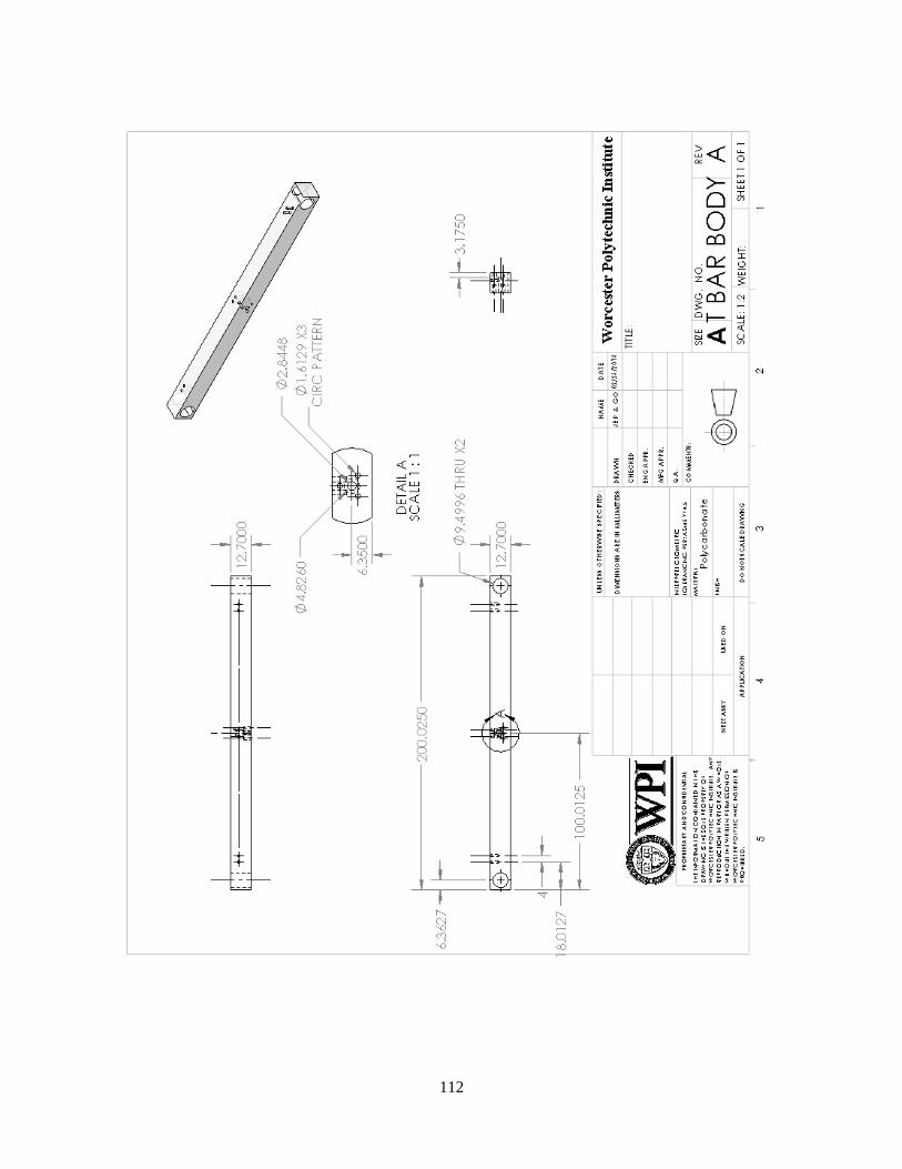
112

113

114

115

116

117

118

119

120

121
APPENDIX D: ARDUINO CODE AND STEPPER MOTOR DIAGRAMS
Arduino Code
/*
For Michelle Tran
By Ben Dwyer
*/
#include <SoftwareSerial.h>
#include <SPI.h>
#include <Keypad.h>
SoftwareSerial lcd(1, 2);
int string1Done = 0;
int string2Done = 0;
int mode = 0;
int looper = 0;
int posn = 0;
int rotation = 0;
int i = 0;
char inputString1[5] = "";
char inputString2[5] = "";
const byte ROWS = 4; // Four rows
const byte COLS = 3; // Three columns
// Define the Keymap
char keys[ROWS][COLS] = {
{'1','2','3'},
{'4','5','6'},
{'7','8','9'},
{'#','0','*'}
};
// Connect keypad ROW0, ROW1, ROW2 and ROW3 to these Arduino pins.
byte rowPins[ROWS] = { 8, 13, 12, 10 };
// Connect keypad COL0, COL1 and COL2 to these Arduino pins.
byte colPins[COLS] = { 9, 7, 11 };
// Create the Keypad
Keypad kpd = Keypad( makeKeymap(keys), rowPins, colPins, ROWS, COLS );
void setup()
{
Serial.begin(57600); // start serial monitor serial
lcd.begin(57600); // start lcd serial (2400)
pinMode(3,OUTPUT);
pinMode(4,OUTPUT);
pinMode(5,OUTPUT);
pinMode(6,OUTPUT);
digitalWrite(5,LOW);
digitalWrite(3,LOW);
digitalWrite(6,LOW);
digitalWrite(4,LOW);
lcd.write(0x01); // need to send one special services command to exit special services mode
clearDisplay();
lcd.print(" Greetings Michelle Tran");
delay(1000);
}
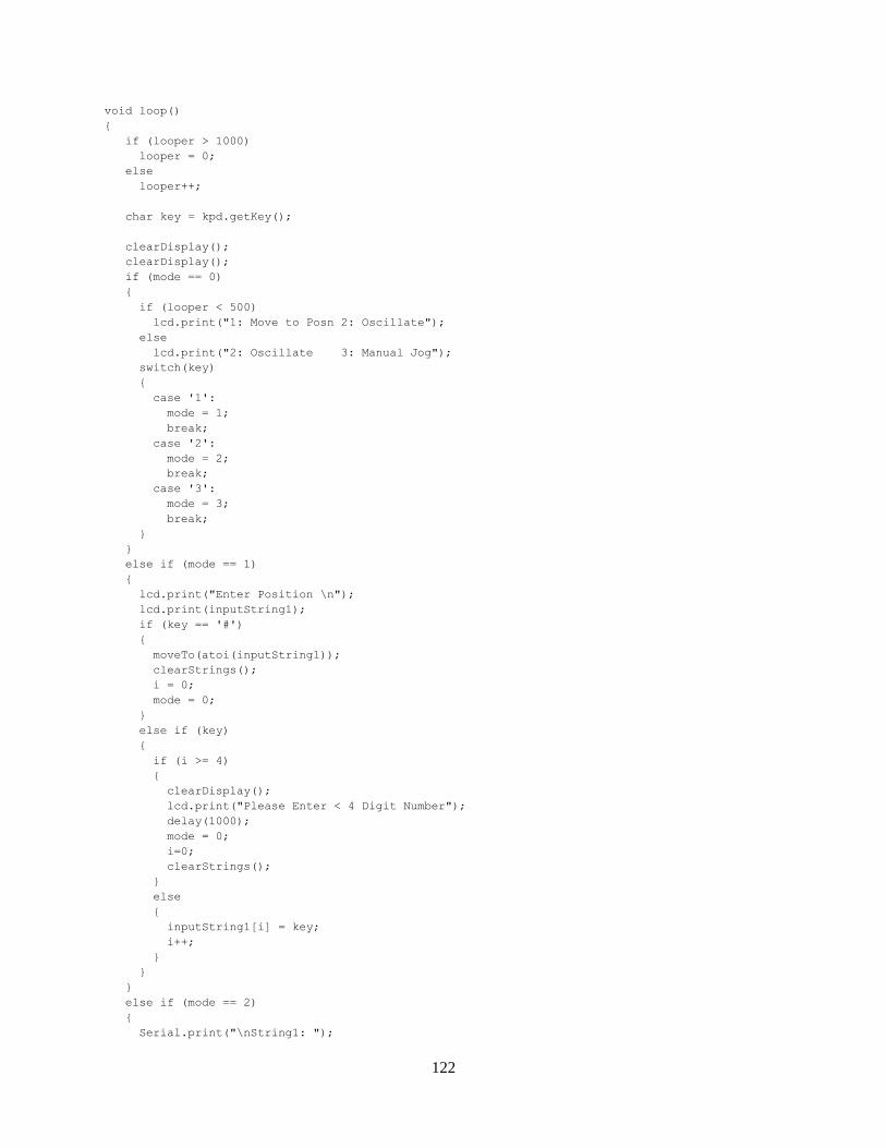
122
void loop()
{
if (looper > 1000)
looper = 0;
else
looper++;
char key = kpd.getKey();
clearDisplay();
clearDisplay();
if (mode == 0)
{
if (looper < 500)
lcd.print("1: Move to Posn 2: Oscillate");
else
lcd.print("2: Oscillate 3: Manual Jog");
switch(key)
{
case '1':
mode = 1;
break;
case '2':
mode = 2;
break;
case '3':
mode = 3;
break;
}
}
else if (mode == 1)
{
lcd.print("Enter Position \n");
lcd.print(inputString1);
if (key == '#')
{
moveTo(atoi(inputString1));
clearStrings();
i = 0;
mode = 0;
}
else if (key)
{
if (i >= 4)
{
clearDisplay();
lcd.print("Please Enter < 4 Digit Number");
delay(1000);
mode = 0;
i=0;
clearStrings();
}
else
{
inputString1[i] = key;
i++;
}
}
}
else if (mode == 2)
{
Serial.print("\nString1: ");

123
Serial.print(string1Done);
Serial.print("\nString2: ");
Serial.print(string2Done);
if (!string1Done && !string2Done)
{
lcd.print("Enter 1st Posn \n");
lcd.print(inputString1);
if (key == '#')
{
string1Done = 1;
i=0;
}
else if (key)
{
if (i >= 4)
{
clearDisplay();
lcd.print("Please Enter < 4 Digit Number");
delay(1000);
mode = 0;
i=0;
clearStrings();
}
else
{
inputString1[i] = key;
i++;
}
}
}
else if (string1Done && !string2Done)
{
lcd.print("Enter 2nd Posn \n");
lcd.print(inputString2);
if (key == '#')
{
string2Done = 1;
i=0;
}
else if (key)
{
if (i >= 4)
{
clearDisplay();
lcd.print("Please Enter < 4 Digit Number");
delay(1000);
mode = 0;
i=0;
clearStrings();
}
else
{
inputString2[i] = key;
i++;
}
}
}
else
{
delay(5);
lcd.print("Press # to stop");
if (key == '#')
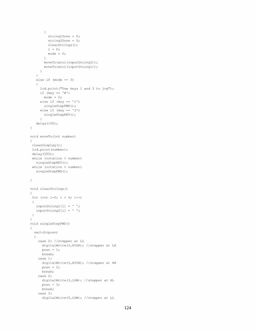
124
{
string1Done = 0;
string2Done = 0;
clearStrings();
i = 0;
mode = 0;
}
moveTo(atoi(inputString2));
moveTo(atoi(inputString1));
}
}
else if (mode == 3)
{
lcd.print("Use keys 1 and 3 to jog");
if (key == '#')
mode = 0;
else if (key == '1')
singleStepFWD();
else if (key == '3')
singleStepREV();
}
delay(100);
}
void moveTo(int number)
{
clearDisplay();
lcd.print(number);
delay(500);
while (rotation > number)
singleStepREV();
while (rotation < number)
singleStepFWD();
}
void clearStrings()
{
for (int i=0; i < 4; i++)
{
inputString1[i] = ' ';
inputString2[i] = ' ';
}
}
void singleStepFWD()
{
switch(posn)
{
case 0: //stepper at LL
digitalWrite(3,HIGH); //stepper at LH
posn = 1;
break;
case 1:
digitalWrite(5,HIGH); //stepper at HH
posn = 2;
break;
case 2:
digitalWrite(3,LOW); //stepper at HL
posn = 3;
break;
case 3:
digitalWrite(5,LOW); //stepper at LL

125
posn=0;
break;
}
rotation++;
clearDisplay();
lcd.print(rotation);
delay(50);
}
void singleStepREV()
{
switch(posn)
{
case 0: //stepper at LL
digitalWrite(5,HIGH); //stepper at HL // good
posn = 3;
break;
case 1: //stepper at LH
digitalWrite(3,LOW); //stepper at LL
posn = 0;
break;
case 2: //stepper at HH
digitalWrite(5,LOW); //stepper at LH
posn = 1;
break;
case 3: //steppper at HL
digitalWrite(3,HIGH); //stepper at HH // good
posn=2;
break;
}
rotation--;
clearDisplay();
lcd.print(rotation);
delay(50);
}
void clearDisplay()
{
lcd.write(0xFE); // LDC Special Services
lcd.write(0x01); // clear LCD
}

126
Stepper Motor Setup and Diagram
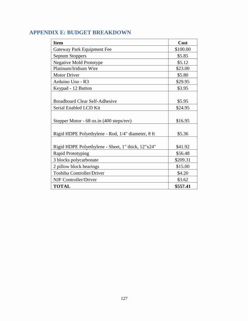
127
APPENDIX E: BUDGET BREAKDOWN
Item Cost
Gateway Park Equipment Fee $100.00
Septum Stoppers $5.85
Negative Mold Prototype $5.12
Platinum/Iridium Wire $23.00
Motor Driver $5.80
Arduino Uno - R3 $29.95
Keypad - 12 Button $3.95
Breadboard Clear Self-Adhesive $5.95
Serial Enabled LCD Kit $24.95
Stepper Motor - 68 oz.in (400 steps/rev) $16.95
Rigid HDPE Polyethylene - Rod, 1/4" diameter, 8 ft $5.36
Rigid HDPE Polyethylene - Sheet, 1" thick, 12"x24" $41.92
Rapid Prototyping $56.48
3 blocks polycarbonate $209.31
2 pillow block bearings $15.00
Toshiba Controller/Driver $4.20
NJF Controller/Driver $3.62
TOTAL $557.41

128
APPENDIX F: MEDIA PROTOCOLS
Proliferation Media Base
DMEM: Ham’s F12
60:40 ratio
+10% Fetal Clone III
DF12 10% FC III (250 ml)
135 mL DMEM
90 mL Ham’s F12
25 mL FC III
0.1461 g Glutamine (powder)
Differentiation Media (DF12 2% HS 1% ITS)
DMEM: Ham’s F12
60:40 ratio
+5% Horse Serum
+1% Insulin Transferin Selenium (ITS)
+0.2922 g Glutamine/500 mL
200 mL total:
120 mL DMEM
80 mL Ham’s F12
10 mL Horse Serum
2 mL ITS
0.117 g Glutamine
Fibroblast Culture Media
DMEM + 10% FCIII + 4mM Glutamine (250 ml)
225 mL DMEM
25 mL FC III
0.1461 g Glutamine (powder)
DMEM + 5% HS + 4mM Glutamine (100 ml)
95 mL DMEM
5 mL Horse Serum
0.058456 g Glutamine (powder)

The Role of the Humanities in Clinical Applications
A Major Qualifying Project
submitted to the Faculty of
WORCESTER POLYTECHNIC INSTITUTE
in partial fulfillment of the requirements for the
Degree of Bachelor of Science
By:
_______________________________________
Bethany Almeida
Date:
25 April 2013
Report Submitted to:
____________________________________
Professor Brenton Faber, Advisor
Department of Professional Writing
Worcester Polytechnic Institute

1 | P a g e
Table of Contents Table of Figures/Tables .................................................................................................................. 3
The Humanities in Relation with BME .......................................................................................... 4
Chapter 1: Today’s Healthcare Problem ......................................................................................... 5
Chapter 1.1: The Health Care System ......................................................................................... 5
Chapter 1.2: Current Treatments............................................................................................... 11
Chapter 1.3: Can the Humanities Help? ................................................................................... 13
Chapter 2: Where Current Treatments Fall Short ......................................................................... 17
Chapter 2.1: NPR Story ............................................................................................................ 17
Chapter 2.2: Trauma ................................................................................................................. 18
Chapter 2.3: What Can the Humanities Teach Us? .................................................................. 18
Chapter 3: Case Study: The Mindfulness-Based Stress Reduction Program ............................... 20
Chapter 3.1: What is It? ............................................................................................................ 20
Chapter 3.2: Religious Origins ................................................................................................. 21
Chapter 4: Mindfulness ................................................................................................................. 25
Chapter 4.1: An Overview ........................................................................................................ 25
Chapter 4.2: Physiological Effects of Mindfulness .................................................................. 26
Chapter 4.3: Experimental Studies ........................................................................................... 30
Chapter 4.4: Quantifying Mindfulness .................................................................................... 34
Chapter 4.5: The Basic Mindfulness Protocol .......................................................................... 39
Chapter 5: Other Humanities-Influenced Intervention Methods .................................................. 41
Chapter 5.1: Cognitive Therapy ................................................................................................ 41
Chapter 5.2: Cognitively-Based Compassion Training (CBCT) .............................................. 46
Chapter 5.3: HEP ...................................................................................................................... 47
Chapter 5.4: The Narrative Approach ....................................................................................... 49
Chapter 6: What Can the Humanities Treat? ............................................................................... 52
Chapter 6.1: Hyperventilation ................................................................................................... 52
Chapter 6.2: Chronic Pain ......................................................................................................... 53
Chapter 6.3: Anxiety and Depression ....................................................................................... 54
Chapter 7: Case Study: The Older Generation and Modern Medication ...................................... 56
Chapter 7.1: Subject Description .............................................................................................. 56
Chapter 7.2: Elderly Opinions of Life and Medication Based on available information ......... 58
Chapter 7.3: How Can We, With the Humanities, Help? ......................................................... 58
Chapter 8: Conclusion................................................................................................................... 60

2 | P a g e
Chapter 8.1: Methods of Study Development........................................................................... 60
Chapter 8.2: Applications to Other Languages ......................................................................... 61
Chapter 8.3: Cost Analysis: Humanities-Based Approaches vs. Pharmaceuticals and Clinical
Treatments................................................................................................................................. 63
Chapter 8.4: Future Recommendations..................................................................................... 65
References ..................................................................................................................................... 67

3 | P a g e
Table of Figures/Tables Table 1: Comparison between Worcester, MA and Boston, MA regions using the Dartmouth
Atlas ................................................................................................................................................ 8 Table 2: Hospital Compare between St. Vincent Hospital and UMASS Medical to compare
similarities and differences between hospitals in the same region ................................................. 9
Table 3: This table shows a summary of the experiments described in chapter 2.2. Overall, the
table shows that mindfulness-based interventions are statistically significant in their intervention
of cognitive and behavioral conditions and diseases, as well as in diseases that cause pain to the
patient by reducing the pain to a manageable amount without the use of medication. ................ 34 Table 4: This table provides a list of the types of quantification tools used to quantify and
analyze results obtained during mindfulness testing. Although not a comprehensive or complete
list, it provides a sense of what is used. ........................................................................................ 35
Table 5: Cost-analysis of humanities-based therapies versus traditional therapies and
pharmaceuticals............................................................................................................................. 65

4 | P a g e
The Humanities in Relation with BME
In Biomedical Engineering (BME), we are taught that science and engineering are the
keys to healthy lives; we are taught that science is the best way to combat illness and disease and
develop lasting treatments that will prolong human life. While science and engineering is an
essential tool to developing promising treatments and should be researched, however, it is not the
only method that can be used. The humanities teach that science isn’t always everything.
Within the humanities, certain skills may be taught that can help people help themselves,
allowing them to make the correct decisions and live healthy lives.
My BME capstone project focuses on developing a skeletal muscle model that is mimetic
of the natural environment of skeletal muscle by actively stimulating the muscle tissue
mechanically and electrically. This type of model may be used in many ways, such as testing the
effects of certain drugs on skeletal muscle and for research on muscle regeneration and diseases
such as Muscular Dystrophy which cause severe muscle loss. By developing this model that
may be used in those ways, we are engineering a scientific approach to the research of treatments
for those uses. However, in illness and disease, it is not always entirely physical. There are
mental and emotional connections with diseases, as well, that affect patients deeply and
potentially even more so than the actual disease, even when the disease is long gone.
To help patients with these mental and emotional side effects of illness, as well as many
of the physical effects, the humanities are useful. The following study will focus on describing
ways in which the humanities has a place in a clinical setting, and may be used to treat certain
diseases and illnesses, including ways in which the humanities are already incorporated into
modern day medicine.

5 | P a g e
Chapter 1: Today’s Healthcare Problem
The price of healthcare has been rising dramatically, it is often difficult to get high
quality healthcare when patients need it, and people seem to be getting sicker, despite the ever-
growing arsenal of drugs and surgeries. The purpose of this review is to study healthcare today,
study the methods employed within healthcare, and determine if new methods may be developed
that are based from the humanities as opposed to the sciences, new methods that may be cheaper
and more beneficial to the patient in the long run.
Chapter 1.1: The Health Care System
The market economics of supply and demand and physician incentives based on free-for-
service work against patients by eroding quality and incentivizing higher medical cost. Hospitals
tend to work within very small financial margins. Due to this, it becomes increasingly important
to fill beds within a hospital and create reasons for in-patients, which is a main source of revenue
in hospitals. The demand for more in-patients is so high, in fact, that Brownlee argues that
physicians and specialists fathom reasons for a patient to stay overnight. Physicians are also paid
according to how many patients they treat and how many resources they use, not according to the
quality of care they provide their patients. As becomes clear, this further provides an incentive
for physicians to create the need for in-patients. Often, they also prescribe too much medicine in
order to combat illnesses that they do not inherently understand or as preventative measures,
without realizing that this could (and in fact most likely is) doing more harm than good to the
patient by providing the patient with a means to develop an immunity to these drugs. However,
this is not an individual physician occurrence. Brownlee also argues that these actions have
become so inherent, that physicians don’t even realize that what they are doing is necessarily

6 | P a g e
wrong or unnecessary. Instead, they are simply doing what they have been taught to do by older
physicians who were doing what they were taught (Brownlee, 2007).
A classic example of this is the case study of appendectomies that Brownlee discusses in
chapter six of Overtreated. There are many conditions that have similar symptoms as a swollen
appendix that is ready to burst, including intestinal blockage, constipation, menstrual cramps,
and so forth (Brownlee 151, 2007). However, as the line to diagnosing appendicitis is often a
thin one, physicians are quick to send patients for CT scans, believing that this method of
imaging will help physicians be able to diagnose the problem. However, despite appendicitis
being visible in a CT scan by appearing swollen with fatty striations, Brownlee insists that the
number of misdiagnosed cases and the number of wrongly ordered appendectomies has not
changed since the development of the CT scan. Brownlee states that physicians see even the
littlest thing and assume it is appendicitis, not wanting to miss it and cause a burst appendix
which can lead to death. Brownlee states that 15 percent of patients that are sent in for an
appendectomy do not have a problem with their appendix, instead having such things as ovarian
cysts or nothing wrong at all. For the elderly, this number jumps up to 35 percent, where the
doctor just removes the appendix anyways while they are already in the middle of surgery.
(Brownlee 150-152, 2007)
Another classic case of performing unnecessary surgeries is heart-related surgeries such
as inserting catheters and cleaning out arteries. However, instead of being something that
physicians do because of excess plaque building in the artery, these are done when it seems
viable to do one, or simply using the excuse of a preventative measure to keep the patients safe
in the long run. In reality, Brownlee argues, these are only done to gain more money in the
physician’s fee for service pay. (Brownlee Chapter 4, 2007)

7 | P a g e
Aside from the effects of over using medicine and poor treatment choices, other factors
have also had an influence in what some to believe to be a failing health care system. This
includes the cost of health care. Health care costs are reaching an all-time high, and they are not
evenly distributed amongst a person’s life.
The Dartmouth Atlas is a project that has been around for more than twenty years. Its
goal is to document and provide data on the distribution of health care resources among different
areas of the United States of America. It serves to show that health care is not uniformly
distributed or evenly accessible to all patients, attempting to create recognition of the need for
reform. Another similar website is Hospital Compare. This website compares regional
variations in cost for different types of procedures and treatments in order to show how the same
procedure can be so much cheaper or expensive in certain areas, regardless of the skill or the
competency of the physician involved. ("The dartmouth atlas"; medicare.gov)
Table 1 exhibits examples taken in order to demonstrate the purpose of these programs
using locations familiar to us. Using the Dartmouth Atlas, the following table was created
comparing the Worcester, MA and Boston, MA regions in some areas: ("The dartmouth atlas")
*Year 2007 Worcester, MA Boston, MA
Total Medicare
Reimbursements per
enrollee
$10,031 $9,704
Inpatient Knee
Replacements per 1000
enrollees
7 7.1
Inpatient Hip Replacements
per 1000 enrollees
3.1 3.4
Inpatient Back Surgery per
1000 enrollees
3.2 3.3
% Deaths of Medicare
patients in hospital
28.6% 28.4%
Average number of inpatient
days per decedent during
last six months of life
9.9 10.7
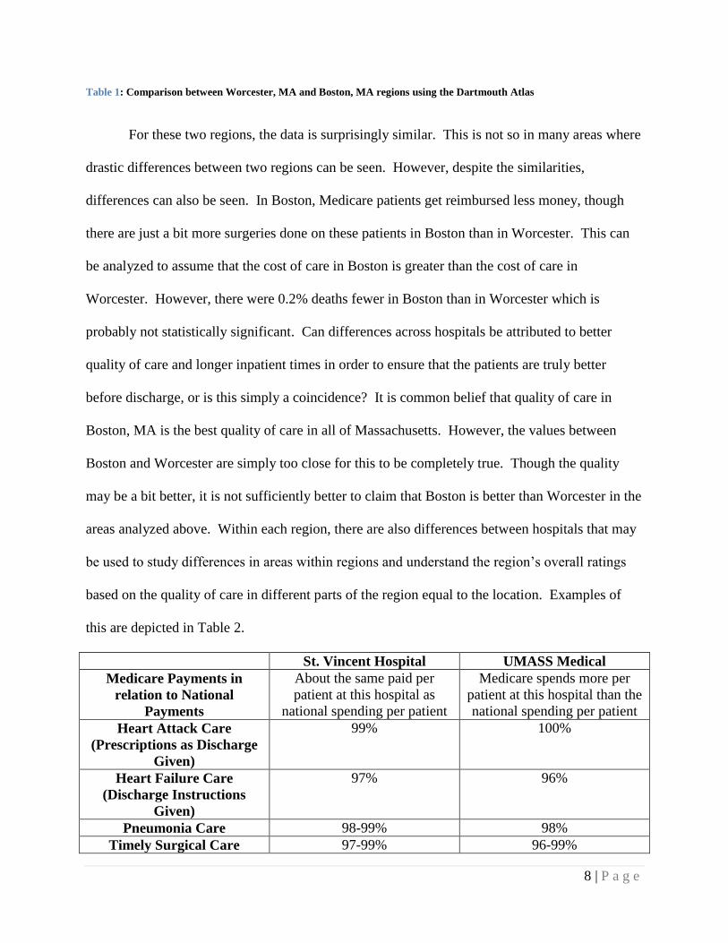
8 | P a g e
Table 1: Comparison between Worcester, MA and Boston, MA regions using the Dartmouth Atlas
For these two regions, the data is surprisingly similar. This is not so in many areas where
drastic differences between two regions can be seen. However, despite the similarities,
differences can also be seen. In Boston, Medicare patients get reimbursed less money, though
there are just a bit more surgeries done on these patients in Boston than in Worcester. This can
be analyzed to assume that the cost of care in Boston is greater than the cost of care in
Worcester. However, there were 0.2% deaths fewer in Boston than in Worcester which is
probably not statistically significant. Can differences across hospitals be attributed to better
quality of care and longer inpatient times in order to ensure that the patients are truly better
before discharge, or is this simply a coincidence? It is common belief that quality of care in
Boston, MA is the best quality of care in all of Massachusetts. However, the values between
Boston and Worcester are simply too close for this to be completely true. Though the quality
may be a bit better, it is not sufficiently better to claim that Boston is better than Worcester in the
areas analyzed above. Within each region, there are also differences between hospitals that may
be used to study differences in areas within regions and understand the region’s overall ratings
based on the quality of care in different parts of the region equal to the location. Examples of
this are depicted in Table 2.
St. Vincent Hospital UMASS Medical
Medicare Payments in
relation to National
Payments
About the same paid per
patient at this hospital as
national spending per patient
Medicare spends more per
patient at this hospital than the
national spending per patient
Heart Attack Care
(Prescriptions as Discharge
Given)
99% 100%
Heart Failure Care
(Discharge Instructions
Given)
97% 96%
Pneumonia Care 98-99% 98%
Timely Surgical Care 97-99% 96-99%
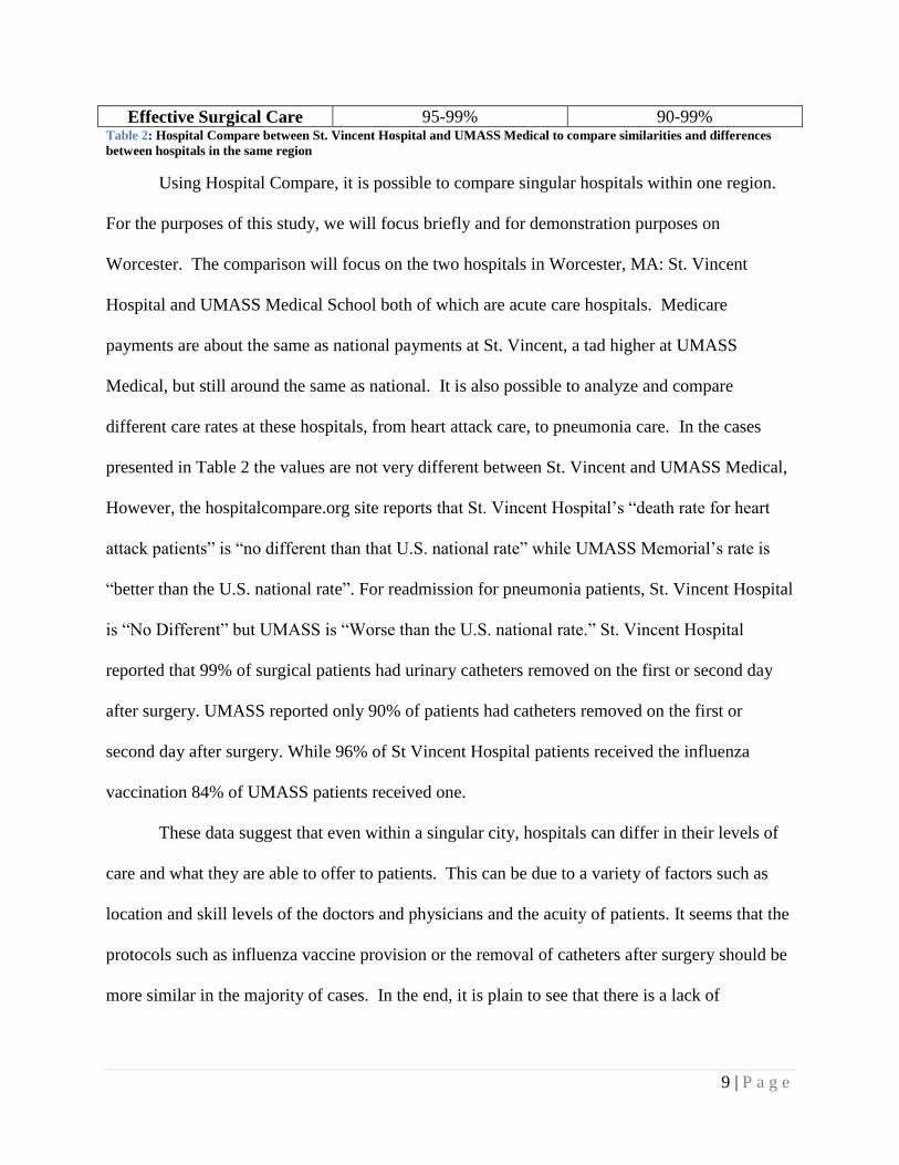
9 | P a g e
Effective Surgical Care 95-99% 90-99% Table 2: Hospital Compare between St. Vincent Hospital and UMASS Medical to compare similarities and differences
between hospitals in the same region
Using Hospital Compare, it is possible to compare singular hospitals within one region.
For the purposes of this study, we will focus briefly and for demonstration purposes on
Worcester. The comparison will focus on the two hospitals in Worcester, MA: St. Vincent
Hospital and UMASS Medical School both of which are acute care hospitals. Medicare
payments are about the same as national payments at St. Vincent, a tad higher at UMASS
Medical, but still around the same as national. It is also possible to analyze and compare
different care rates at these hospitals, from heart attack care, to pneumonia care. In the cases
presented in Table 2 the values are not very different between St. Vincent and UMASS Medical,
However, the hospitalcompare.org site reports that St. Vincent Hospital’s “death rate for heart
attack patients” is “no different than that U.S. national rate” while UMASS Memorial’s rate is
“better than the U.S. national rate”. For readmission for pneumonia patients, St. Vincent Hospital
is “No Different” but UMASS is “Worse than the U.S. national rate.” St. Vincent Hospital
reported that 99% of surgical patients had urinary catheters removed on the first or second day
after surgery. UMASS reported only 90% of patients had catheters removed on the first or
second day after surgery. While 96% of St Vincent Hospital patients received the influenza
vaccination 84% of UMASS patients received one.
These data suggest that even within a singular city, hospitals can differ in their levels of
care and what they are able to offer to patients. This can be due to a variety of factors such as
location and skill levels of the doctors and physicians and the acuity of patients. It seems that the
protocols such as influenza vaccine provision or the removal of catheters after surgery should be
more similar in the majority of cases. In the end, it is plain to see that there is a lack of
10 | P a g e
standardization amongst hospitals, even hospitals within the same city. So, the question remains,
are the current structures of healthcare providing the best care at the best cost? (Medicare.gov)
Generally, a person spends the most money on health care in their first few months of life
and their last few months of life, with little money spent in between. Also, some people with
certain diseases or problems tend to spend more than the average person on medical bills in order
to constantly treat their illnesses. This makes inherent sense; however, it creates a difficulty
when trying to regulate spending on health care for people. (Emanuel & Emanuel, 1994)
In 2009, Atul Gawande sought to put into perspective health care costs for patients, in
order to bring to light the desperate reform that is needed. Most middle class citizens are well
aware of how much they pay for their health care, and often, believe it to be too much.
According to Gawande, in 2009, health care costs composed of 18 cents out of every dollar
earned in a typical family. Gawande further enforces this by noting how average annual
premiums for family insurance coverage rose from $5,800 to $13,400 for employer sponsored
insurance and $5,500 to $11,900 for Medicare funded. These are obviously large sums of
money. Naturally, one would think that the high costs they are paying are due to an exponential
increase in the quality of care that they are receiving. However, as Gawande states in his article,
this is not so. Gawande, like many other professionals, call for a reform in the health care
system, one that will lower costs and increase quality because, in their opinion, higher costs are
not necessary. (Gawande, 2009)
Is this all truly necessary? Do more drugs or medical devices mean better lifestyles?
Unfortunately, what is known, which Brownlee discusses extensively throughout her text, is the
idea that the medical arms race constitutes much of the pharmaceutical and hospital costs that, in
the end, patients must pay for. The medical arms race is just what it sounds like. It is the race

11 | P a g e
between different hospitals and physicians to create and obtain the best medical equipment and
knowledge in industry to provide the best and most up-to-date care for their patients. It also
consists of hospitals purchasing more and more machines that may be unnecessary in quantities
greater than one. However, in order to maintain all of these machines and technologies, the cost
increases, and the patients are, in the end, charged more in order to maintain the upkeep.
Does spending more on more procedures as ‘preventative measures’ mean that illnesses
will be caught sooner and treated easier? As a member of the middle-class, I believe it is safe to
speak for all middle-class citizens when I say that we do not make a large enough income to pay
for all of this. New treatment options must be researched that are less costly in the long run and
will provide better treatment outcomes than current methods. However, the sciences do not
appear to be providing us with the necessary means to devise new treatment methods. Or, is it
simply that physicians do not want to devise new, better and cheaper methods if it means less
money for them? Regardless of this debate, which is a lengthy debate of its own right, there is
the discussion about whether or not the sciences are where researchers should go to obtain new
treatment methods or if there is possibly a different area of study, most notably the humanities,
that can provide outlook into future treatments.
Chapter 1.2: Current Treatments
Current treatment methods include the prescription of pharmaceutical drugs and different
surgical methods used either as corrective procedures or as procedures to implant devices that
function to aid the human body in performing at a normal rate. While it is true that these current
treatments are constantly evolving and new technologies are being developed, this is only aiding
in increasing medical costs. Also, in the case of certain techniques such as imaging techniques
like radiology X-rays for diagnostic imaging, physicians are becoming too dependent on them,

12 | P a g e
utilizing them more and more, even when they are not necessarily beneficial or helpful, further
racking up medical costs.
Another issue in current treatments is the thought that progress only includes building
upon current knowledge to come up with something new and better. However, this can
sometimes provide limitations or narrow the scope of the problem too fully. Sometimes, it is
necessary to go back to the fundamental, biological physiology associated with the issue or
illness and re-analyze how it is understood and the methods that were initially used to solve these
problems. A recent talk on the Worcester Polytechnic Institute campus by Jonathan Gillard, PhD
exemplifies this approach. Gillard takes a new look at arteries and the role of plaque in heart
attacks and strokes. Through his research, he may have discovered something not previously
known about the ways plaque is held in the arteries and causes clogging. If his data and
experiments prove true, his work alone could remove the need for stents and surgically removing
plaque buildup from the inside of arteries. In this way, he would be advancing current
knowledge and technologies by bringing to light a new method of approach to the problem that
does not build off of any previous technologies but could provide better care for these types of
patients in the end.
As previously stated, we will return to the topic of hyperventilation throughout the study
as a model illness for these ideas. Hyperventilation is very much a physiologic illness which
causes changes to occur in the body when an attack takes place. In general terms, as the patient’s
breathing becomes quicker and shallower, the level of carbon dioxide in the lungs of the patient
steadily decreases as the patient is unable to dispel it and intake more oxygen.
In the situation presented at the moment, current technologies for hyperventilation
include medications for relaxation of the patient or techniques devised by physicians in order to

13 | P a g e
steady the patient’s breathing. However, medication can be costly and ineffective and doctor
visits for a simple hyperventilation attack take up both the time of the doctor and patient, as well
as cost money for the patient. New techniques which can be done by the patient at home would
be beneficial in the long run both for better quality care of the patient and cost efficiency.
However, these techniques can only come to light if the fundamental physiology of
hyperventilation is once again examined and understood.
Chapter 1.3: Can the Humanities Help?
This study seeks to determine whether the humanities could play a role in clinical
applications. Do the humanities teach us something that can be altered to fit the needs of a
clinical setting such as a hospital and provide treatment or relief at a rate that is the same or
greater than current treatment methods? The humanities include a wide range of areas of study,
including but not limited to: language, composition, rhetoric, history, philosophy, art and
architecture, society and culture studies, music, and religion. Each of these is composed of an
even wider range of sub-fields.
The humanities with the most promise in providing knowledge that may be attributed to a
clinical setting include philosophy, society and culture studies, and religion, as these generally
already have opinions regarding ways of life and the manners in which people should promote
their well-being. The hypothesis of this study is ‘the humanities can play a role in clinical
interventions’.
This study came into development with the belief that the humanities can contribute to
clinical knowledge and that the humanities would provide valuable assets to the development of
clinical programs and treatments for certain conditions and diseases. This is not to say that the
humanities will be able to solve any health problem—there are clearly many issues that require

14 | P a g e
scientific and medical advancement if we hope to cure or treat the problem. However, beyond
curing or treating the symptoms, the humanities provide a strong foundation for teaching patients
how to best cope with their ailments, as well as understand their own situations and well-being.
A major problem in the world today is the medical and scientific advancement. It is too far
beyond the capability of normal people, especially the elderly who were raised in completely
different settings and have come to fear sickness, to understand all of the different advancements
that have been reached, as well as understand the measures that can be taken to prevent or heal
certain conditions that were once considered deadly. This aspect, in its own right, deserves some
attention. A future chapter will look at a case study involving an elderly woman who I use as a
“representative anecdote” to demonstrate some of the difficulties seniors face when dealing with
medical decisions.
It is understandable that the idea of the humanities having any place in medicine could be
a contentious claim for some. After all, the humanities in an educational setting do not focus on
teaching people to better understand ‘self’, from health to mind to possessions; instead, the
humanities focus on teaching the basics of the core humanities principles: languages (how to
speak it, write it, read it, and understand it), history (what has happened and, to some extent,
why), religion (what types exist and what do they believe)—the list goes on. It is fair, however,
to say that the humanities, if only at least in a University setting, should attempt to teach people
how to understand their own selves so that they better understand how to care for themselves.
However, an attack on the educational system is not what is trying to be accomplished. On the
contrary, the purpose of this study is to provide a basis for understanding that the humanities can
also teach people to understand one’s self, as well as is capable of providing knowledge and skill
to a clinical setting—which does, in fact, already exist. The purpose of this study is to help bring

15 | P a g e
acknowledgement to different programs and treatments that are humanities-based and commonly
used in clinical settings; the program that will be covered most in depth is mindfulness, as it has
had promising results as a treatment for a variety of different ailments and diseases.
However, you may still be wondering: how can the humanities actually be useful in
medicine? Rather than simply try to push my own opinions on you, as the reader, it may be best
to provide an example. Terry Eagleton’s book Literary Theory: An Introduction is the perfect
example of how the humanities go beyond what most people believe them to be. In this book,
the author discusses how literature is shaped by and shapes the world around us. As Eagleton
discusses throughout the introduction of his book, literature is not a solid thing. There are many,
very different, definitions of literature and anything can be read as literature if analyzed in a
certain way. The introduction establishes that the humanities, in particular literature, are not as
straight forward or as clear cut as people would believe. The humanities function as systems that
are meant to be thought-provoking, allowing the person to develop awareness and insight that
may be used to further better themselves. This is close in relation to an opinion that I have given
previously that the humanities teach people to understand self and how to care for themselves.
Also, this wraps easily with the concept of health and medicine. If people were more aware of
themselves and their conditions, they would be better able to care for themselves, limiting
healthcare costs and potentially reducing the amount of medicine needed. (Eagleton, 1983)
An appreciation of the humanities, as well as an understanding of the underlying
principles of the humanities and what isn’t commonly taught in schools is necessary to truly
accept the idea that the humanities have a place in medicine, and that the humanities is, in fact,
already deeply ingrained with clinical culture. It is my belief, as a Biomedical Engineer by
training, that concepts and ideas of humanities should be accepted into medicine with open arms

16 | P a g e
and prized for its capability to provide methods of treatment that limit healthcare costs and
increase patient benefits.

17 | P a g e
Chapter 2: Where Current Treatments Fall Short
Current treatments, though they work well in many situations, tend to fall short in others.
The situations in which they fall short, however, can have devastating effects on the life of the
patient, affecting daily life and performance socially. The following sections highlight areas in
which current treatment falls short and where the humanities may be able to step up and fill the
gap, leading to successful treatment procedures.
Chapter 2.1: NPR Story
Adverse childhood experiences can include mental and emotional trauma such as
emotional, mental and physical abuse and neglect, as well as household dysfunction. These
experiences may cause stressful experiences in the child’s growing brain that can cause
biological effects in the brain which in turn can affect their behavior and learning abilities.
These children exhibit abnormal stress hormones which lead to increased inflammation,
dysfunction of brain circuitry, insulin resistance, and damage to blood vessels, as well as the
mental illnesses such as anxiety and trauma, in their adult lives. These adverse experiences
affect their health well past the experience and may change their lives for the worse. (Tough,
2012; “The adverse childhood”; Raison, 2010)
The skills affected by the stresses created from these adverse childhood experiences are
those of the non-cognitive type, which are generally not taught in schools or hospitals. These
skills are, however, learnable at all ages. They include skills like social skills, personality traits,
mood control and regulation, and conscientiousness. Despite being non-cognitive, these skills
have a profound effect on a child or teen’s ability to learn. Due to the constant stresses from the
adverse childhood experiences, the children grew in a constant ‘fight-or-flight’ state of mind.
Thus, when they are asked difficult questions in class or called on, their mind immediately reacts

18 | P a g e
to it as a threat, causing the child or teen to act out against the teacher or other student. These
children have an increasingly higher rate of dropping out of school, work and marriage as the
number of adverse childhood experiences in their past increases, as well. (Tough, 2012)
Chapter 2.2: Trauma
One area of illness/experience that may see positive results as a cause of the influence of
the humanities is trauma. According to the National Institute of Mental Health, there are two
types of trauma, physical and mental. Physical trauma is the result of the body’s response to
physical injury or damage, whereas mental trauma is a response to fears or negative, frightening
thoughts. Mental trauma is the type that will be discussed throughout the course of this review.
Mental trauma can cause emotional instability in the patient, leading to extreme behavior and
withdrawal from the patient, as well. (National Institute of Mental Health)
Within trauma, there are sub-categories. These include but are not limited to childhood
trauma, adult trauma, trauma due to war, and trauma due to social circumstances. Patients who
have been diagnosed with trauma due to one of these categories are usually diagnosed with Post-
Traumatic Stress Disorder (PTSD). Though there are medications, usually antidepressants like
benzodiazepines and oxidase inhibitors, which are used to treat patients with PTSD, these
medications, much like situations with other cognitive disorders like anxiety and depression
which will be discussed later, oftentimes do not have an effect, have a limited effect, or treat
symptoms only (are not curative). In fact, a consideration that is always taken into consideration
with these medications is removal of the medication if it is not proving efficacious. (Jeffreys,
2009)
Chapter 2.3: What Can the Humanities Teach Us?
19 | P a g e
The purpose of this study is to attempt to consider evidence for including what we are
broadly calling “humanities approaches” to the treatment of acute and chronic disease states. We
are not suggesting fully replacing pharmacology, surgery, or other well-known and successful
interventions. Instead, our goal has been to seek and suggest successful adjuncts to more
expensive and at times less successful interventions. In investigating such approaches, we have
endeavored to apply consistent scientific rigor to an alternative. In other words, an alternative
must have been tested and reported using the same protocols and systems any intervention would
require to be broadly accepted by the medical community.
Initially, we will consider “Mindfulness” as one widely-reported intervention. In recent
years, mindfulness has been gaining a wider audience and as we will show has had decades of
supporting, experimental proof and analysis of its methods.
Mindfulness may be useful as a tool of intervention, for many: the children described in
the NPR story, the war veterans with PTSD, the elderly citizen with chronic pain, or the patient
with anxiety and depression which cause bouts of hyperventilation. Mindfulness focuses on
attention to thought and the present moment, making the patient more cognitively aware of their
surroundings. Thus, undergoing a mindfulness intervention program may assist in the teaching
of these non-cognitive skills to these patients, whether they are children, teens or adults so that
they may combat the stresses and adverse childhood experiences in order to lead successful lives.
The subsequent chapters focus on mindfulness in depth, analyzing and studying the results it has
obtained in order to determine whether it has successfully met its goal and proven the hypothesis
presented in chapter 1.3.
20 | P a g e
Chapter 3: Case Study: The Mindfulness-Based Stress Reduction Program
To date, probably the concept and therapeutic practice of “Mindfulness” represents the
most well developed and extensive application of a traditional Humanities activity within
medicine. For this reason, this study will pay particular attention to Mindfulness as concept and
therapeutic practice though recognizing that there are other methods of treatment that doctors
have been looking into in order to treat these areas where current treatment methods fall short.
This study has also benefitted from the close proximity of the Mindfulness-Based Stress
Reduction Program at the University of Massachusetts (UMASS) Medical School here in
Worcester, MA. This program was one of the first of its kind, a pioneer in the study and use of
mindfulness as a treatment for or method of relief for these patients.
Chapter 3.1: What is It?
The stress-reduction center is a clinic in the University of Massachusetts Medical Center
that has been devoted to the study of and training of mindfulness as a secondary or
complementary form of medicine to treat stress-induced problems such as chronic pain, anxiety
and depression. “[The] mindfulness-based stress reduction (MBSR) is a clinical program,
developed to facilitate adaptation to medical illness, which provides systematic training in
mindfulness meditation as a self-regulatory approach to stress reduction and emotion
management.” (Bishop, 2002) This center has been working towards this goal for since 1979,
and has developed the MBSR (mindfulness-based stress reduction) program—an eight week,
formal program that meets weekly for 2 to 2.5 hours with instructions for homework practice six
days a week for 45-60 minutes. Around week 6, there is also a full day retreat. This program
was developed initially for chronic pain patients but has recently claimed that their methods can
help to alleviate other types of pain including chronic pain patients and patients with cognitive

21 | P a g e
behavioral or emotional disorders. (Bishop, 2004; Carmody, 2008; Dobkin, 2011; Kabat-Zinn, J.
2003; Teasdale, 1995, Baer, 2003)
This program is one of many mindfulness-based programs (other programs include
Cognitive Therapy which will be discussed later, and some programs meant for the treatment of
specific areas). The program teaches patients mindfulness through several yoga positions,
breathing techniques and meditation skills. For the first few weeks, patients are allowed the use
of audiotapes, but they are generally instructed to try and not use them after about three weeks.
The patients are instructed to sit or lay in a comfortable position, especially one that will limit the
stress on the body; however, the goal of the program is that patients are able to use these
mindfulness techniques at any moment when they need to. Patients are instructed to notice any
thoughts they may have, but, instead of disregarding them or categorizing them as ‘negative’ or
‘positive’ thoughts, they are simply instructed to notice them and then return to the ‘present
moment’ which may include focusing on their breathing at the moment or returning focus to
some event that is occurring at the moment. The goal of the program is that, by noticing and
exposing one’s self to the thoughts or conditions that arise, a de-sensitization process will occur
which may cause attitude and mood changes within the patient, as well as increased coping
skills, relaxation and acceptance. (Baer, 2003; Grossman, 2004)
Mindfulness as a generic skill and clinical treatment will be discussed and evaluated in
chapter two. Before a discussion in mindfulness may begin, however, it is essential to
understand the origins of mindfulness in order to better understand how it works and why it is
used.
Chapter 3.2: Religious Origins
22 | P a g e
The practice of mindfulness holds its roots within Buddhism. Buddhism depicts a way of
life for those who follow it using its teachings. By studying the religious origins of mindfulness
treatment through Buddhism, it becomes clear how mindfulness is a treatment developed from
the humanities. Religion is a topic commonly studied in the humanities to understand how
thoughts and beliefs have evolved with time. A clinical practice developed from religion is a
clinical practice developed from the humanities.
The Buddhist concepts of spirituality and awareness of oneself are the founding concepts
of mindfulness treatment, though mindfulness is not religiously orientated. These core Buddhist
teachings provide the framework from which the idea for mindfulness, as well as many of the
techniques used, were developed (Dimidjian & Kleiber, 2012). These teachings were developed
and worked to fit in the context of a clinical setting where it could be used as a technique for
treatment of many illnesses, diseases and conditions through creating in the patient a sense of
awareness of oneself and his or her surroundings, which is fundamentally what Buddhism
embodies.
Jon Kabbat-Zinn, the founder of modern-day mindfulness techniques, especially in the
style of UMASS Medical School’s MBSR program, based his techniques on Buddhist traditions,
despite not explicitly stating this anywhere other than a foreword in one text describing his new
program. According to one article which aims at describing the Buddhist origins of mindfulness
so that the new generation of mindfulness teachers may truly understand the field and better
serve their purpose, takes a new route from Zinn and describes, in detail, some of the traditions
from which mindfulness has hailed.
Mindfulness is based most extensively on Zen meditation, which contains a multitude of
mechanisms of meditation, the different practices and techniques used, as well as different

23 | P a g e
teaching methods that follow the lotus sutra. The lotus sutra states that the teaching of
mindfulness to individuals must be tailored to the individual for best results, and teachers of
mindfulness must also be masters of mindfulness, who live by the techniques and rules they are
teaching and understand the practice completely. In mindfulness treatment, it is still essential
that the teachers of the treatment follow the teachings they are passing on to their students, as
well as practice the techniques religiously outside of the classroom setting. This ensures that
patients are receiving the utmost care and treatment as their teachers are invested in the program
just as much, if not more than, they themselves are.
As has been stated before, mindfulness is considered a skill that must be practiced and
mastered with time. According to tradition, Buddhism is also a skill that can be practiced and
mastered with time. It is not a mantra, nor is it a creed; Buddhism is a way of living that
increases the well-being of the practicing individuals. This practice is able to be translated into
different languages or jargons so that it can be understood by the group of people who are
learning and practicing the technique. In the case of mindfulness treatments, Zinn essentially
restated the traditions of Buddhist thought and practice into a language understood in the
scientific and clinical communities so that they would be accepted by these communities.
A major area in which mindfulness takes from traditional Buddhist thought is the concept
of the four truths, or kalama sutta. The first truth is the observation that there is suffering
(awareness and attention to oneself and his or her surroundings). The second truth is the origin
of suffering (the illness, disease, or so forth). The third truth is the ending of suffering by ending
its origin (ultimate treatment of disease or symptoms and relief of the patient’s stress due to the
illness), and the fourth and final truth is the path leading to the termination of suffering (the
treatment program). The path of the fourth truth is known in Buddhist teachings as the eightfold

24 | P a g e
path and focuses on concepts and ideas such as understanding, virtue, and looking/seeing clearly,
all of which have also been translated into mindfulness treatment.
Finally, Zinn took from the concept of karma—the idea that there are consequences for
one’s behavior and actions. This is also clearly evident in mindfulness treatment as the treatment
focuses on the patients developing an awareness and learning to overcome the issue through this
awareness. This takes from the concept of karma by teaching patients that by simply changing
their thought process, or by not negatively connoting the thoughts, they are able to change how
they ultimately feel, resulting in treatment.
(Maex, 2011)

25 | P a g e
Chapter 4: Mindfulness
Jon Kabat-Zinn is hailed as the founding father of mindfulness training. Since his
foundation of the Mindfulness-Based Stress Reduction clinic at UMASS Medical School, many
forms of mindfulness programs have developed and taken new life. In order to understand what
the modern mindfulness is, it is important to understand mindfulness in its most general context
outside of the MBSR. It is essential to gain a foundation of understanding what mindfulness is,
how it is studied and quantified, its physiological effects on the human body, and examples of
experiments conducted using either the MBSR program or different clinical programs. From this
analysis, it will be possible to obtain a general outline of the basic mindfulness protocol, as well
as obtaining a better understanding of how mindfulness works and how it has used teachings
from the Humanities to sculpt it into a clinical treatment program.
Chapter 4.1: An Overview
Mindfulness, as it was already introduced, includes an awareness of the patient from
paying attention “on purpose, in the present moment, and nonjudgmentally to the unfolding of
experience moment by moment”. (Kabat-Zinn, 2003) It is a psychological process, a generic
skill, a mental training, which patients can practice. (Bishop, 2004)
Mindfulness works through the use of two components: self-regulation of attention, and
orientation to experience. As was already described in chapter 1.1, patients are asked to focus on
the present moment, be it through focusing on one’s breathing or on an event that is occurring at
the moment. They are asked to non-judgmentally notice their thoughts and return to the present
moment without classifying the thought as ‘negative’ or ‘positive’. (Bishop, 2004; Carmody,
2008; Dobkin, 2011; Kabat-Zinn, J. 2003; Teasdale, 1995, Baer, 2003)

26 | P a g e
Mindfulness-based interventions nearly all focus on moment to moment, nonjudgmental
awareness, formal meditation techniques, and the importance of regular practice, regardless of
the program. The methods for mindfulness training are similar in many studies and programs,
however, are not clearly defined. While this is understandable in the sense that these programs
(about 240 in the country) wish to preserve their methods in order to maintain secrecy and their
cliental, it makes quantifying mindfulness far more difficult. The methods of quantifying
mindfulness will be discussed in more detail later. Clinically, it has been found that an average
of 85% of patients complete the mindfulness intervention programs. However, issues in regards
to the experiments done to validate mindfulness-based interventions include a lack of control
groups in many of the experiments, small sample sizes, and a lack of quantification. Despite all
of these, the promise of mindfulness-based interventions has been consistently proven through
experiments that show significant improvement in symptoms and coping, bringing to the table a
promise that mindfulness-based interventions are worth looking into as clinical treatments. The
following section highlights some experimental studies in mindfulness-based interventions.
(Grossman, 2004; Baer, 2003)
Chapter 4.2: Physiological Effects of Mindfulness
Some scholars have criticized mindfulness on the lack of quantifiable data that it
produces. Critiques have also attacked mindfulness on its removal from the classical science.
However, mindfulness has been shown to have physiological effects on the human body. Dr.
David Rock wrote in his article “The Neuroscience of Mindfulness” that “when you understand
the underlying physiology of mindfulness, you begin to see that any discussion about human
change, learning, education, even politics and social issues, ends up being about mindfulness…I
have a problem with something as important as deeper thinking being linked to any religion

27 | P a g e
[because] it’s hard enough getting across the idea that being mindful is useful, without activating
a threat response from the billions of non-buddhists who could benefit from it.” (Rock, “The
neuroscience of”)
In this singular quote, Dr. Rock sums up the entirety of the criticism that mindfulness
training has received: the religious connotations, as well as the proof of it working. These
critics, however, are focusing on the religious connotations and asking for proof without,
seemingly, searching for the proof themselves. The proof of the success of mindfulness as a
clinical program, as well as the proof for its validity as a scientific thought and process and not
simply something taken from religion lies in its physiological connotations. Many critics believe
that there are no underlying physiological processes that occur during mindfulness. However,
recent studies have shown that to be false. To kick off the overview of some of the many
physiological effects of mindfulness, let’s start with the paper that Dr. Rock references in his
work.
In this work “Attending to the present: mindfulness meditation reveals distinct neural
modes of self-reference”, the authors study the neural modes of self-reference, which link to
one’s levels of awareness. They utilize fMRI imaging to examine and monitor specifically two
aspects of awareness, momentary experience (experimental focus) and enduring traits (narrative
focus). They tested both patients who underwent the mindfulness training program and those
who had not. Through their work, they discovered that in those who had not undergone
mindfulness training, the experimental focus showed reductions in self-reference in the cortical
midline regions of the brain that are usually associated with narrative focus. Those who had
underwent mindfulness training, on the other hand, the experimental focus marked omnipresent
reductions in the medial prefrontal cortex but increased engagement of the right-hand brain

28 | P a g e
network, showing a disassociation in the mindfulness group that proves distinct forms of self-
awareness that have been trained to be separate from one another to allow the patient greater
thought and reasoning capacity. This in turn affects how the patients are able to view themselves
and their situations. (Farb, 2007)
Continuing with physiological effects in the brain, a recent study has shown that
mindfulness training has demonstrated a decrease in right amygdala activation in response to
emotional stimuli, a response which occurs long after meditation has ceased and the patient is
simply going about their own business. In other words, mindfulness was shown to provide
lasting improvements in the part of the brain that processes emotions, which in turn affects how
patients react to their circumstances. The consequence of this experiment is such that it provides
proof, or at least some basis, for a claim that states that those who undergo the mindfulness
training program may be able to utilize the immediate benefits of deeper self-awareness and
awareness of emotional stimuli and problems for many years to come in their lives, allowing
them to save money on future treatments and live a better life. (Brauser, 2012)
As further proof of this consequence of mindfulness training in regards to the
neurological activities of the human brain, one study discovered an increase in brain gray matter
in the hippocampus region of the brain in those who have underwent mindfulness training when
compared with those who have not undergone graining. This region, and the others that the
concentration of gray matter increased within, are also involved in learning and memory
processes, emotion regulation, self-referential processing, and perspective taking in the patients,
all of which mindfulness training has been used to increase the effectiveness of humans.
(Holzel, 2011)

29 | P a g e
The mindfulness training program does not only physiologically affect the brain. It has
also been shown to affect other areas of the human body such as the cardiovascular system and
the immune system, as well as conditions such as stress. One study showed that during
mindfulness meditation, the patients, both women and men, exhibited a decrease in diastolic
blood pressure, as well as an increase in cardiac output in comparison with those patients who
underwent other forms of relaxation or no mediation or relaxation. This shows that these
patients, while undergoing mindfulness mediation, were aware of the conditions that were
putting their body and heart under stress and fix their breathing such that their body and heart
released the built up stress, reducing the diastolic pressure, as well as functioning more
effectively by pumping out more blood at any given time. (Ditto, 2006) Immune response has
also been studied extensively by Jon Kabat-Zinn himself in his 2003 article “Alterations in Brain
and Immune Function Produced by Mindfulness Mediation”.
In this study, the authors tested three groups of students: meditating students who were
not under examination stress, non-meditating students who were under examination stress, and
meditating students who were under meditating stress. However, as opposed to the traditional
eight week program, this study was only held for three weeks. Results of the study show that
there is a possibility for success of the treatment, but the three groups were not statistically
different. The consequence of this result is most likely related to the length of the program and
the situation the patients were in. From the results that have been extensively obtained
throughout the course of this research, it is fair to say that mindfulness meditation training,
though it can provide extensive long-term results, this study has shown that, in order to be
effective, it is necessary for mindfulness training to take its full time of seven to eight weeks.
(Myint, 2011) A common result of many experimental studies using mindfulness training is the

30 | P a g e
consideration and fact that perceived stress levels in patients drastically decrease and emotional
stability and awareness drastically increase.
This is not a complete description of the physiological effects of mindfulness meditation
on the human mind and body; it is only a sample of the many studies and research that has been
done and is ongoing. For those who are uncertain about the physiological effects of mindfulness
and wish for more proof than the preceding section, I encourage them to dig deeply and find the
research and primary materials. A good starting place, and where I was able to find a good
number of reference works, is by searching under the key words “physiological effects of
mindfulness” using the search engine of the researcher’s choice.
Chapter 4.3: Experimental Studies
For the purpose of this review, the following six studies were examined for their results.
They were chosen as they each represent a different treatment possibility for mindfulness-based
interventions or they provide a validation for mindfulness. Statistical tests, indexes, and other
methods of evaluation will be described in more detail in chapter 2.3.
Davidson, 2003
o In this controlled, randomized study based on the teachings of the MBSR
program, the authors took 41 patients and separated them into a patient group
(size of 25 people) that underwent the mindfulness program and a control group
(size of 16 people) was a ‘waitlist’. At the end of the eight weeks, the authors
vaccinated all subjects (patient and control) with the flu vaccine. They
hypothesized that those who underwent the mindfulness training would
demonstrate greater antibody titers in response to the vaccine. The methods used
included measuring electrical activity of the brain before, after and in the middle

31 | P a g e
of the program, as well as statistical MANOVA testing to determine the
significance of results. The results of this study proved their hypothesis and,
according to the authors of the paper, demonstrate that the MBSR program was
successful and future studies on the biological effects of the MBSR were not
needed.
Rosenzweig, 2010
o In this experiment, the authors hoped to measure different types of chronic pain
and their success rates using the University of Massachusetts Medical School’s
MBSR program. This experiment built off of earlier experiments that had already
proven the MBSR’s use in significantly addressing pain and symptoms of chronic
pain, but did not differentiate between types of chronic pain; by building off of
these previous experiments, the authors hoped to address these differences. This
experiment had a total of 133 participants: 51 chronic neck/back, 34 arthritis, 32
fibromyalgia, and 27 other. Their methods of determining statistical similarities
and differences used a series of statistical testing and questionnaires, including the
HRQol, SF-36, SCL-90-R, Pearson’s Correlations, and Paired T-tests. Their
results were two-fold: first, they showed that all types of chronic pain had
significantly better results for patients who underwent mindfulness training.
Second, they showed that the results differed amongst conditions and differed
based on amount of home practice. These authors insist that future studies on
homogenous populations are needed to determine which racial population
experiences the most significant results.
Vollestad, 2011

32 | P a g e
o The authors of this experiment designed a randomized, controlled experiment
using a ‘waitlist’ control group, like one of the previously described experiments.
The authors followed the teachings of the University of Massachusetts Medical
School’s MBSR program. This experiment consisted of 76 patients split amongst
the two groups, and their methods for determining statistical soundness of the
program included T-tests, indexes, scales and questionnaires. The authors’ results
showed, much to their contentment, statistical significance of results, despite
these effects being modest; after six months, those who underwent mindfulness
training still maintained the results of their training. However, according to the
authors, it is still unclear how the MBSR works and how it can be used in the
future in prevention and intervention. For these authors, further studies are
required to determine the usefulness of the MBSR.
MacCoon, 2012
o The authors of this experiment sought to test the difference between using an
active control in comparison to the ‘waitlist’ control described in previously
analyzed studies. The authors believed that this would rigorously test the
program and provide numerical values to the results in order to better quantify its
usefulness. In this experiment, the authors had 80 participants, half of which
underwent the MBSR, and the other half who underwent some other type of
intervention program to reduce pain or symptoms (HEP was used in this
experiment). The authors stimulated pain in the subjects who then used the
methods they were learning to overcome/reduce the pain. Statistical methods
included using the SCL-90-R, MSC, and ECQ. The results of the experiment

33 | P a g e
showed that the two intervention programs, HEP and MBSR, were structurally
equivalent and had similar results. However, mindfulness was only present in the
patients who underwent MBSR training. The authors of this experiment insist
that future studies are required using active control groups to determine the true
effectiveness of the MBSR.
Brown & Ryan, 2003
o For this experiment, the authors also developed a mindfulness-based intervention
program but did not specify the program that was the premise for their
experiment. A benefit of this is simply to prove the general concept of
mindfulness and demonstrate whether or not the concept of the intervention is
successful in a clinical environment for cancer. This experiment was a clinical
study on a cancer population of 58 patients with no control group. The authors of
the experiment took measurements before, during and after the study, using
mostly the MAAS scale to measure mindfulness in patients. The results showed
that, over time, as mindfulness increased in the patients, mood disturbance and
stress levels decreased. MAAS scores for the patients also increased over time.
The authors of this experiment acknowledge that future studies in this would be
beneficial to better understand the use of mindfulness in cancer intervention.
Table 3 provides a summary of the experiments described above:
Experiment Program Results
Brain and Immune Response MBSR Statistically significant results
Chronic Pain Conditions MBSR Statistically significant results
differing amongst conditions
Anxiety Disorders MBSR Statistically significant
(modest) results
Active Control Validation MBSR / HEP MBSR and HEP statistically
similar results, mindfulness

34 | P a g e
present in MBSR
Cancer Patients Mindfulness intervention Statistically significant results Table 3: This table shows a summary of the experiments described in chapter 2.2. Overall, the table shows
that mindfulness-based interventions are statistically significant in their intervention of cognitive and
behavioral conditions and diseases, as well as in diseases that cause pain to the patient by reducing the pain to
a manageable amount without the use of medication.
Chapter 4.4: Quantifying Mindfulness
A major difficulty with any mindfulness program is quantitatively proving the method’s
success, which can cause doubt and suspicion to arise regarding the results presented. The
current methods of analysis of results within a mindfulness program include statistical analysis,
surveys, and different scales based on a patient’s response. Table 4 depicts some analysis
methods that have been stumbled upon through research. This is not a complete list, as there are
surely more methods that are not commonly used or were not found during research.
Name Full Name Type What it is
MAAS Mindful attention
awareness scale
Questionnaire Scale Determines how
aware an individual is
to one’s self and what
is occurring in the
present.
PSS Perceived Stress Scale Questionnaire Measures perception
of mental and
emotional stress by
individuals.
CES-D N/A Self-Report Scale Measures depressive
symptoms of
individuals in a
general population by
self-reporting results.
MSCL Medical Symptoms
Related to Stress
(Medical Symptoms
Checklist)
Scale/Checklist Determines what
symptoms a patient
has, as well as how
often, by having
patients check off
symptoms they have,
as well as ranking
them in how often
they occur.
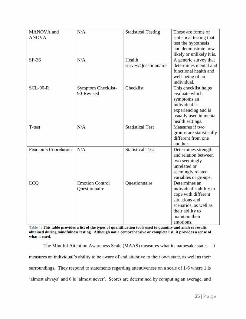
35 | P a g e
MANOVA and
ANOVA
N/A Statistical Testing These are forms of
statistical testing that
test the hypothesis
and demonstrate how
likely or unlikely it is.
SF-36 N/A Health
survey/Questionnaire
A generic survey that
determines mental and
functional health and
well-being of an
individual.
SCL-90-R Symptom Checklist-
90-Revised
Checklist This checklist helps
evaluate which
symptoms an
individual is
experiencing and is
usually used in mental
health settings.
T-test N/A Statistical Test Measures if two
groups are statistically
different from one
another.
Pearson’s Coorelation N/A Statistical Test Determines strength
and relation between
two seemingly
unrelated or
seemingly related
variables or groups.
ECQ Emotion Control
Questionnaire
Questionnaire Determines an
individual’s ability to
cope with different
situations and
scenarios, as well as
their ability to
maintain their
emotions. Table 4: This table provides a list of the types of quantification tools used to quantify and analyze results
obtained during mindfulness testing. Although not a comprehensive or complete list, it provides a sense of
what is used.
The Mindful Attention Awareness Scale (MAAS) measures what its namesake states—it
measures an individual’s ability to be aware of and attentive to their own state, as well as their
surroundings. They respond to statements regarding attentiveness on a scale of 1-6 where 1 is
‘almost always’ and 6 is ‘almost never’. Scores are determined by computing an average, and
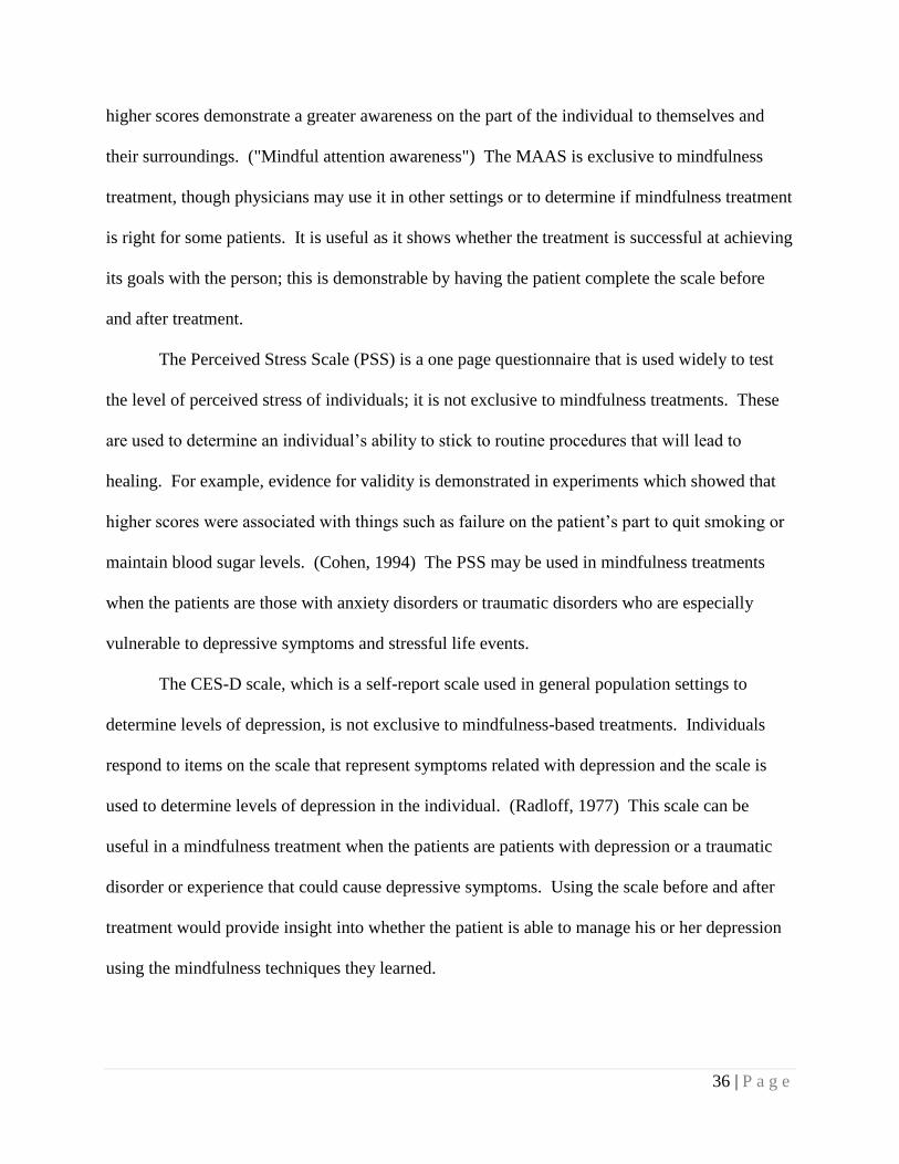
36 | P a g e
higher scores demonstrate a greater awareness on the part of the individual to themselves and
their surroundings. ("Mindful attention awareness") The MAAS is exclusive to mindfulness
treatment, though physicians may use it in other settings or to determine if mindfulness treatment
is right for some patients. It is useful as it shows whether the treatment is successful at achieving
its goals with the person; this is demonstrable by having the patient complete the scale before
and after treatment.
The Perceived Stress Scale (PSS) is a one page questionnaire that is used widely to test
the level of perceived stress of individuals; it is not exclusive to mindfulness treatments. These
are used to determine an individual’s ability to stick to routine procedures that will lead to
healing. For example, evidence for validity is demonstrated in experiments which showed that
higher scores were associated with things such as failure on the patient’s part to quit smoking or
maintain blood sugar levels. (Cohen, 1994) The PSS may be used in mindfulness treatments
when the patients are those with anxiety disorders or traumatic disorders who are especially
vulnerable to depressive symptoms and stressful life events.
The CES-D scale, which is a self-report scale used in general population settings to
determine levels of depression, is not exclusive to mindfulness-based treatments. Individuals
respond to items on the scale that represent symptoms related with depression and the scale is
used to determine levels of depression in the individual. (Radloff, 1977) This scale can be
useful in a mindfulness treatment when the patients are patients with depression or a traumatic
disorder or experience that could cause depressive symptoms. Using the scale before and after
treatment would provide insight into whether the patient is able to manage his or her depression
using the mindfulness techniques they learned.

37 | P a g e
The Medical Symptoms Checklist (MSCL) is used often in clinical settings to help
identify what a patient is suffering from. It consists of two questionnaires—the first of which
focuses on physical symptoms that the patient experiences, as well as their frequency, and the
second of which focuses on emotional thoughts and behaviors of the patient. This checklist is
most often used when the patient is experiencing some levels of stress in order to determine the
root cause of the stress. (“Medical symptoms related”) This checklist has applications in
mindfulness treatment as it can help determine problem areas of focus for the duration of the
treatment. Also, by using it before and after treatment, it can be determined if the symptoms the
patient experiences were treated, or at the very least, helped.
The SF-36 is a general tool used in a clinical setting to determine the overall functional
and mental health and well-being of an individual. It helps to estimate the severity of the illness
or disease that the patient has, as well as understanding the levels of stress and pain they are
experiencing. It is commonly used for such diseases as chronic pain, depression, migraines,
diabetes, kidney or cardiovascular diseases, trauma, and many more. (Ware) The SF-36 is a
beneficial survey for mindfulness treatments as it allows those administering treatment to obtain
a general understanding of what the patient is experiencing, allowing them to understand
necessary measures and plan for the next steps in treatment.
The Symptom Checklist-90-Revised (SCL-90-R), which was devised by Pearson
Assessments, is used to determine a broad range of mental health symptoms in an individual. It
can be used as a means of measuring patient progress and treatment outcomes, as well. It is a
well-rounded scale which consists of 90 items on the checklist, as well as a 5 point scale to
measure severity and frequency of the symptoms. It is also not exclusive to adults and may be
used on any individual aged 13 or above. (Derogatis) This checklist is useful in a mindfulness

38 | P a g e
setting as it, like many of the scales described above, can provide an insight into the patient
regarding his or her mental health levels. It is also beneficial in that it may be used on patients
who are younger but require mindfulness treatment.
The Emotion Control Questionnaire (ECQ) is used to determine an individual’s ability to
cope with different situations and emotions, as well as their ability to maintain their emotions.
This scale differs from others described above, however, as it is a questionnaire in which the
answers are in the form of ‘true’ or ‘false’ instead of numerical ranking values. (ECQ2) The
ECQ is useful in a mindfulness treatment setting as it can help determine the emotional stability
or instability of the patient, which provides insight into their levels of awareness and attention to
responses.
Finally, there are some methods of statistical testing used in analysis of mindfulness
treatment. These include the MANOVA and ANOVA (which is simply a special case of the
MANOVA), the T-Test, and the Pearson’s Correlation. The MANOVA and ANOVA test
hypotheses and differences between groups, the t-test tests how statistically different two groups
are from one another and the Pearson’s Correlation determines whether there is a relation
between two groups. These are useful in mindfulness treatment experiments specifically
designed to prove the efficacy of mindfulness treatment in which there are treatment groups and
control groups. They seek to prove that, though the two groups are related, they are different
from one another in a beneficial manner, essentially proving that mindfulness treatment is
successful. (Acastat; “The t-test”; Juliano & Fader)
Essentially, all of these measures, whether they are scales or questionnaires or statistical
testing, aim to prove that the mindfulness treatment is both viable and successful in delivering
beneficial outcomes to patients of all types. However, despite these measures being taken, critics

39 | P a g e
have found reasons to question the mindfulness technique, presenting the argument that self-
reporting is not a viable option as patients always believe their condition to be worse than may
be accurate. For many, some method needs to be developed that can successfully prove or
disprove the use of mindfulness techniques. However, is developing a method truly a pursuable
option, or will more criticism rise up against any technique that attempts to enter into the field?
Chapter 4.5: The Basic Mindfulness Protocol
Mindfulness-based protocols vary from program to program, experiment to experiment.
It is not possible to obtain the exact protocol used by the UMASS Medical School due to the
program being a for-profit program that aims at bringing in patients and treating them at a cost.
To give out their protocol would be detrimental to their program as patients would be able to
provide their own treatment without needing specialists to guide them through the program.
However, despite the exact protocol not being available, mindfulness-based programs
utilize similar protocols. Mindfulness in itself is not something that can be drastically changed
between programs as they all aim to accomplish the same goals in the same or similar manners.
In 2000, Michael Speca and his colleagues wrote the mindfulness protocol they used into the
article for an experiment that they conducted. Their program was seven weeks long and had
weekly, 90 minute sessions. The authors claim that their program was developed based on the
UMASS Medical School’s program, so it is safe to assume that this protocol is acceptable for
study for these purposes. The best way to describe this program is to provide a summarized,
week-by-week breakdown of the program.
The first week began with introductions of the patients and their reasons for attending the
program. The patients were given their instructions and rules, which included regulatory
information as well as being told to record their home practice. The first week also included

40 | P a g e
teaching the patients the correct breathing techniques for relaxation and mindfulness awareness.
The second week includes an exercise which focuses on visualization to teach the interactions
between mental imagery and bodily responses. Introductory yoga stretches and the principles of
meditation are also introduced.
In week three, discussion about home practice takes place, as well as discussion of how
the body physiologically responds to stress and relaxation. Meditation practices are taught, and
gentle yoga stretches are continued. In week four, breathing exercises that are based off of yoga
are taught and explored in regards to their relationship with emotional responses, and a new form
of meditation is also taught that focuses on meditation while walking. The fifth week teaches
patients about the relationship between cognition and emotion, aiming to teach patients about
awareness of their thoughts. Week six includes monitoring of self-practice, visualization
practice, and concentration on awareness. Finally, week seven includes a review of all material
and forms taught. The patients are then aided in developing their own home plan for continued
practice.
This program aims at teaching the patients the underlying principles that develop
mindfulness to help patients understand why and how mindfulness works. It serves as an
introduction to proper breathing techniques, the importance of awareness, and some yoga and
meditation forms that assist in creating a serene atmosphere that helps the body to undergo
relaxation as opposed to the stress it is usually under. The ultimate goal of this program is to
leave patients with an understanding of how to continue practice on their own post-program so
that they maintain the benefits of treatment throughout the course of their lives. It is a program
that may be applied to many different people with many different conditions, and which can have
an everlasting effect on the patients when taught and enforced well.

41 | P a g e
Chapter 5: Other Humanities-Influenced Intervention Methods
Mindfulness is not the only therapy that has been developed using meditation and similar
techniques that hail from humanities studies to combat many types of disease and injury. In fact,
this field is not a new field. Psychotherapy has been prominent since the 1950s, or possibly
older as some believe that it has origins in Freud. From Psychotherapy, many forms of treatment
have come about and evolved; many hold tight to the reigning features of Psychotherapy while
others are very much different entities. The following sections will highlight other treatment
areas that have combatted with mindfulness for popularity, as well as treatments that are new and
upcoming, yet similar to the ideas of mindfulness. This serves as a showcase that mindfulness is
not the only humanities-based treatment that can be applied in clinical settings.
Chapter 5.1: Cognitive Therapy
Cognitive therapy contains many of the same values as the mindfulness programs, such
as the MBSR, but there are noted differences that set cognitive therapy as its own entity and have
created the need for comparison between the two. Cognitive therapy was originally introduced
as a psychotherapy approach to depression and has been adapted for use in treatment of anxiety
disorders, trauma, and other cognitive disorders. According to Judith S. Beck, “the principle
underlying cognitive therapy is ‘thoughts influence moods’” (Hoffman). It is not used often in
the treatment of physical illnesses such as chronic pain or as a treatment post-surgery. Much like
the existence of the Mindfulness Based Stress Reduction clinic in Worcester, Massachusetts,
there is also the Center for Cognitive Therapy in Boston, Massachusetts. Also like the MBSR,
the Center for Cognitive Therapy offers individual therapy, group therapy, training seminars, and
supervision and consultation with a clinician trained in the practice under teachings by the
Academy of Cognitive Therapy. (McCutchen)

42 | P a g e
Other similarities of cognitive therapy to mindfulness programs include the way the
therapy is taught to patients. In both programs, home practice by the patient is essential for
complete the program successfully and obtain the most complete results for them personally. In
mindfulness, as has been discussed previously, this homework is focused on the patients
practicing the therapy at home regularly for pre-specified time intervals such that the practice of
mindfulness becomes second nature. In this manner, patients may use the techniques learned in
the mindfulness program whenever they need in order to combat the problem. However, these
patients are also able to be aware of their thoughts and the moment in which the problem arises,
allowing them to use that information to their benefit in the future. In Cognitive Therapy,
however, homework is simply oriented at relapse prevention such that the patients understand
what measures they should take should a bout of depression come about. The major difference
between the two programs regarding homework being that the patients who undergo cognitive
therapy are not aware of the circumstances that the bout came about in and are not able to
prevent it in the future utilizing that information. (Beck & Tompkins, 2007)
Many of the features of the program are also similar to the features of the MBSR, as well
as other mindfulness programs. The similar features include identifying and evaluating how one
understands problems, collaboration, step-by-step approaches, feedback and assessments at
regular intervals. The areas in which the Center for Cognitive Therapy differs drastically from
the MBSR and other mindfulness programs are the ideas that symptom reduction is prioritized
and medications are encouraged for use by patients at the same time if the patient desires.
Though mindfulness does focus on reducing symptoms and does allow patients to use
medication should they absolutely require its aide, these are not features or focuses of the
mindfulness programs. Mindfulness programs focus on completely removing symptoms and

43 | P a g e
leading towards a cure of the problem, if possible, without the need for medications so that it is
cheaper for patients and more valuable in the long-run. (McCutchen)
In much the same way that Jon Kabat-Zinn can be credited with being the founder of
modern mindfulness programs through his teachings with the MBSR, the founder of modern
cognitive therapy teachings is Aaron Beck. Both teachings came about roughly during the same
time period, the mid to late 1960s-70s. Similar to how Jon Kabat-Zinn’s original work was
focused on the treatment of chronic pain, Beck’s original work, “Cognitive therapy and the
emotional disorders” (1976) was focused on treating depression. Though the methods and
results of both programs are similar, the focus of their creation, as well as some key differences
including the effects of awareness within the patient, has created two very different programs.
Both programs have suffered a lack of ‘proof’ over the years. However, like mindfulness and the
MBSR, cognitive therapy has also employed some experimental studies that have proven its
efficacy.
One experiment, “Cognitive Therapy vs. Medications in the Treatment of Moderate to
Severe Depression” (2005), was completed by Robert DeRubeis and his colleagues. The authors
hoped that this experiment would provide solid proof that cognitive therapy was at least as good
as anti-depressants (hopefully better) at treating depression symptoms. The authors set their
experiment up such that they had three groups of patients, 120 patients who underwent 16 weeks
of medication, 60 patients who underwent 16 weeks of cognitive therapy, and 60 patients who
took 8 weeks’ worth of a placebo pill they believed to be medication. The medication tested was
up to 50 mg daily of lithium carbonate or desipramine hydrochloride.
The results of the experiment show that at the 8 week mark, both the medicated groups
and the cognitive therapy groups had higher percentages of positive response than the placebo

44 | P a g e
group, with the medicated group having slightly better results than the cognitive therapy group
(though it can be argued that this is due to the different number of patients within the group).
After 16 weeks, both groups had the same percentage of response rates and the remission rates of
the cognitive therapy group were slightly lower than those of the medication group. The results
of this experiment show that cognitive therapy was at least as good as the medication in treating
depression, although the authors do state that the effectiveness of cognitive therapy may depend
on therapist experience or ability, despite the fact that it would be cheaper than medication in the
long run. It is also safe to note that numbers may have skewed slightly due to the different group
sizes. However, ultimately, it can be said that cognitive therapy deserves to be analyzed and
tested more in depth. (DeRubeis, 2005)
The preceding experiment was not, however, the first of its kind. In 1994, David M.
Clark and his colleagues compared cognitive therapy to applied relaxation and imipramine. The
results of this experiment showed that in the short term, 3 months, cognitive therapy was
superior. A few months later, it was the same as medication. However, after a long-term relapse
of the medication patients, 15 months post-experiment, the cognitive therapy group was again
superior (though the results were not as good as they were in the short term). Again, this
experiment, conducted about a decade earlier than the preceding experiment, provided the same
results that cognitive therapy is at least the same as medication, but it continues to have long
term beneficial results (as well as a monetary benefit) that place it one step above medication.
(Clark, 1994)
Unlike mindfulness, where long term experiments are only beginning to take route,
cognitive therapy has had long term experiments demonstrating its effectiveness like the
experiment described above. One essential long term experiment was conducted by Aaron T.

45 | P a g e
Beck, who in 1991 reviewed his previous work and elaborated on its effects and developed a
modernized reconstruction of cognitive therapy 30 years after its introduction. In this review,
Beck discusses the positives and negatives that have come about around his theory of cognitive
therapy. He discusses what different areas have evolved from the original conception of
cognitive therapy, as well as responds to critics and discusses different experiments that have
been conducted over the decades. Ultimately, Beck returns to the question he asked in his 1976
work whether cognitive therapy could stand on its own against other psychotherapy approaches
within a clinical setting—his response for the readers being that cognitive therapy is no longer a
‘fledgling’ therapy and has proven its success and capacity to ‘fly under its own power’. He
believes that cognitive therapy will continue to thrive and provide consistent, beneficial results,
showing that teachings based from the humanities can indeed thrive as clinical treatments.
(Beck, 1991)
Cognitive therapy also focuses on teaching patients ‘emotional intelligence’ which helps
these patients learn non-cognitive skills to help them help themselves improve the quality of their
lives, despite any previous illnesses or trauma. As presented previously in the NPR study, the
use of non-cognitive skills has been examined and has been demonstrated to teach skills to
patients that help them develop fulfilling lives, especially when taught to younger patients and
enforced for the future. Though it may appear ironic that cognitive therapy teaches non-
cognitive skills such as tenacity, self-control, and self-respect, these skills are, in essence,
important when teaching people to understand themselves and be thoughtful of their
circumstances. Through extended preliminary research, it is clear that the use of emotional
intelligence in clinical settings may help to prevent relapses and extra hospital visits, allowing
patients to have a fulfilling life of their own. (Salovey & Mayer, 1990)

46 | P a g e
Emotional intelligence may be described as “a set of skills hypothesized to contribute to
the accurate appraisal and expression of emotion in oneself and in others, the effective regulation
of emotion in self and others, and the use of feelings to motivate, plan, and achieve in one’s life”
(Salovey & Mayer, 1990). Emotional intelligence works by helping patients to understand
themselves and their peers in order to make an accurate judgment on their respective situations
and their emotional responses to those situations in order to understand what steps they must take
to help their situation.
Chapter 5.2: Cognitively-Based Compassion Training (CBCT)
Cognitively-based compassion training (CBCT) focuses on the importance of compassion
for oneself and others. CBCT hails from religious origins and, much like mindfulness training, it
hails from Buddhism—specifically Tibetan Buddhism. However, whereas mindfulness takes
from Buddhism the idea that one should be attentive and aware of oneself and one’s
surroundings, cognitively-based compassion training takes the idea that one should be
compassionate towards oneself and others and understand different situations and problems.
CBCT is “based on the view that self-centered thinking and behavior cause suffering for oneself
and others, while other-centered, altruistic thoughts, emotions, and behaviors ultimately benefit
both oneself and others. Compassion is the heartfelt wish that others be free from suffering and
the readiness to act on their behalf” (Emory University). It focuses on teaching the practitioners
to cultivate thoughts on others and focus their energies in this manner. Like the MBSR, the
CBCT is an 8 week long program. (Emory University)
As was discussed previously, childhood adversity and trauma is an issue that can affect
people throughout their lives and may cause serious effects later on in life. It has been a topic
lately that mindfulness may be used to treat these children and young adults in order to prevent

47 | P a g e
the future issues and problems that may arise from the childhood adversity or trauma. However,
CBCT is already being tested use for this same purpose. Charles Raison from Emory Univeristy,
a university that has been a leading proponent and practitioner of CBCT, provided an update
report on the success of CBCT in relation to this in 2010. Unfortunately, results did not
demonstrate success of the program for self-reported levels of depression, anxiety or emotional
well-being in these young adults, though it did report improved self-reported optimism about
one’s future, which may have been a result of the compassionate training the young adults
received. However, CBCT did show positive physiological effects to the brain similar to
mindfulness, which provides the thought that it may be successful in the future. (Raison, 2010)
However, there are doubts to the CBCT’s success considering Pace et al.’s 2012 work
“Engagement with Cognitively-Based Compassion Training is associated with reduced salivary
C-reactive protein from before to after training in foster care program adolescents” shows only
minute reduction of the protein after the 6 week mark and no differences between control and
experimental groups. The authors suggest long-term follow-up; however, it is unlikely that there
will be any changes. (Pace, 2012) However, like the mindfulness program was 30 years ago, the
CBCT is new and upcoming. In the future, experimental studies may demonstrate use for this
program.
Chapter 5.3: HEP
The home exercise program (HEP) is a program that is used mostly in conjunction with
chronic pain or physical illness conditions. It is a form of exercise training that patients are
informed to complete at home. There are commercially available products such as HEP2GO
which is a rehabilitation program for the computer that walks patients step by step and helps
monitor successes (“HEP2go”). Beyond this, as well as a physician-made routine, there is no

48 | P a g e
professional input. As this is very dependent on patient activity, there has been much concern
for its success, as well as many experimental tests hoping to prove the efficacy of the HEP.
However, because there are many possible routes to take in regards to HEP and many different
forms of the program, there have been very different results over the years.
One article, written by Nadine Fisher and her colleagues, evaluate the HEP on muscle
function and functional capacity of patients with osteoarthritis. Ideally, the home exercise
program would show an increase in muscle function and functional capacity in the patients.
However, the results of this experiment, conducted in 1994, were otherwise. Muscle function
did not increase and, though functional capacity slightly increased, the effects were not
statistically significant. (Fisher, 1994)
Another experiment evaluated the program in response to a chronic condition, low back
pain. This experiment, conducted in 2002, had twenty patients. Ten of the patients went through
an extensive, specified exercise program and the other ten went through a generally given
program. The results of this experiment show that those who underwent the general program did
not benefit; however, those who underwent the extensive, specialized and specific exercise
program (at the appropriate training amounts and levels) did, in fact, benefit. This shows that as
time has progressed, so has the HEP and its ability to be successful. (Descarreaux, 2002)
As if to further demonstrate the varying results of the HEP program, a third experiment,
conducted by Jane Schneiderman-Walker et al, tested cystic fibrosis through a three year trial
HEP. The authors split patients into two groups, a control group that would go about their
normal daily physical activity and an experimental group that was assigned to 20 minutes of
aerobic exercise three times a week. In this experiment, conducted in 2000, patients in the
experimental group reported a sense of well-being and demonstrated a slower decrease in

49 | P a g e
pulmonary function over time. This led the authors to the conclusion that, for cystic fibrosis in
the case of using HEP as aerobic exercise, HEP was successful and may be beneficial in the long
run for patients with cystic fibrosis. (Schneiderman-Walker, 2000)
Through research, it was discovered that many experiments simply had the result that
HEP may be beneficial for some or if conducted in the long-run. This program is unsteady and
variable, leading to the belief that it will not become a standalone program in the future used for
clinical treatment, but a side-program to be used with other programs. In fact, it basically has
been incorporated into other programs. In the case of mindfulness, where patients are expected
to complete homework exercises using the training they were taught, it is as if HEP has become
incorporated into mindfulness programs, an essential component of the program, but not the core
element that the program is based on and formed around.
Chapter 5.4: The Narrative Approach
The narrative approach is a unique approach that combines the literary with the clinical.
The narrative approach accomplishes two things: telling a story such that it allows the narrator to
reflect on and reconstruct the past in order to better understand themselves and their situation and
narrative empathy which allows physicians to improve decision making and healthcare outcomes
for the patient (Harvey & Koteyko 70-73, 2013). This is essential as there is generally an
imbalance of power between the doctor and the patient (Harvey & Koteyko 8-9, 2013). This
imbalance of power usually affects healthcare outcomes in the sense that the physician, who
believes to know more than the patient, will not necessarily provide the best care for the patient.
However, if the physician listens to the patient, they are generally able to understand the
patient’s pain and help the patient get better and not simply treat the illness or disease. As
Harvey and Koteyko state on page 87 of Exploring health Communication: Language in Action,

50 | P a g e
“the narratives presented…offer a counterweight to the perspectives of mental health experts that
dominate research into mental illness.” It is safe to say that this is true for all illness, not only
mental illness, as all illness can affect the patient deeply.
The narrative approach is a useful tool for the patient, as well. It allows the patient a
powerful tool to shape their personal identities and understand what is wrong with themselves
(Harvey & Koteyko 75, 2013). There are implicit social and cultural meanings with illness that
change how patients view themselves and are viewed (Kleinman Chapter 2, 1989). These views
and opinions can hurt the patients more than the illness itself. Thus, the narrative approach
allows the patients to convey their feelings successfully and not only earn better health care but
also allows them to share their stories with others who may be suffering and help those others in
need. It helps to promote a different understanding of the problems—not only the physiological
effects of the issue, but also the mental and emotional effects that affect the patient but aren’t
necessarily treatable by surgery or medication. After all, people always remember the details of
their lives with illness, and the narrative approach allows them a method to get the problems they
personally face off of their chest (Frank Chapter 6, 1997).
There are many uses of the narrative approach in clinical settings. One of the most
widely known uses is the wounded warrior project which focuses on veterans who have finished
their time in service and have either been wounded on the battlefield or are prone to getting
posttraumatic stress disorder (PTSD) (“Wounded warrior project”). Unlike the other programs
and methods which hail from the humanities mostly through religious-based teachings and
meditation, this program utilizes a different humanities approach—the literature and arts
approach. This is done in such a manner where the patients, the warriors who are mostly
suffering from PTSD write about their story and share it with other warriors and families of
51 | P a g e
warriors, creating a support group that works together to combat the PTSD. The program
focuses on four different areas within the warrior patient’s life: mind, body, economic
empowerment and engagement.
The program’s first component “mind” focuses on adjusting the warriors mentally and
creating a support group where warriors may share their experiences and receive support to
overcome the challenges. “Body” focuses on maximizing rehabilitation and teaching warriors
and their families to live a fulfilling, active and healthy lifestyle. “Economic empowerment”
focuses on removing the stress of being unemployed or underemployed and seeks to help every
warrior find employment once their duty to the service is over. Finally, “engagement” is where
the storytelling and narratives really come into play. In order to ensure that the support group is
full of both current service members, recently released members and alumni, there is a peer
mentoring program. In this program, alumni write about and discuss their story of how they
were able to combat their injuries and PTSD and take control of their life, providing the support
to others to do the same.

52 | P a g e
Chapter 6: What Can the Humanities Treat?
Mindfulness has a wide range of applications, including but not limited to non-cognitive
disorders related to trauma and stress, chronic disorders such as hyperventilation and pain, and
cognitive disorders such as anxiety and depression. Despite the wide range of applications,
mindfulness has been shown to positively affect patients in all these areas, as was shown in
chapter 4.2 through the study of different experimental tests. The question that has puzzled
researchers for decades, however, is: how does mindfulness treat these vast applications? How
does the body respond physiologically to mindfulness that combats the physiological response to
the disease? In other words, simply, how does mindfulness work? By looking at the following
types of applications for which mindfulness has been used, it is hoped that a better understanding
of how, potentially, mindfulness works can be obtained.
Chapter 6.1: Hyperventilation
Changes in the carbon dioxide levels in a patient’s body are the cause of hyperventilation.
During times of high stress, a patient may begin to breathe quickly and shallowly as an
emotional response. This, in turn, decreases the carbon dioxide levels in the body by not
allowing the body to constantly dispel carbon dioxide. When carbon dioxide levels lower in the
body, the body is also unable to maintain the oxygen levels needed for metabolism through
alkalosis. In order to combat hyperventilation, the patient must calm down and breathe slower
and deeper. (Mallat, 2012)
With hyperventilation, the key is for the patient to recognize what is happening to them
before they can take the right steps towards finding a solution. The patient needs to understand
that they are not having a heart attack and that their body will be fine in order to calm down
enough to stop the hyperventilation. It is also important for the future for the patient to recognize

53 | P a g e
the signs of a hyperventilation attack and stop it before it happens, understanding that it is only
stress and anxiety and won’t actually hurt them. Mindfulness is a treatment option for
hyperventilation. A main method that is taught in mindfulness programs is breathing techniques
coupled with awareness. Using these mindfulness breathing techniques, patients experiencing
hyperventilation may be able to calm themselves using awareness techniques long enough to
implement proper breathing techniques that will restore proper carbon dioxide levels in the body.
Chapter 6.2: Chronic Pain
Chronic pain was the principle and foremost area of treatment for the Mindfulness-Based
Stress Reduction Program in UMASS Medical School. However, the program has grown to
encompass many areas of treatment since. Chronic pain is most commonly a symptom of some
other underlying problem that causes pain in some part of the patient’s body for more than three
months at a time. It causes a limitation in mobility of the patient, as well as disrupting their
everyday lifestyles. Chronic pain is most commonly experienced in the back, usually the lower
back, and is attributed to the spine. Current methods of treatment for chronic pain include non-
surgical physiotherapy and analgesia, and surgery to achieve spinal fusion. (Ibrahim, 2006)
Lately, surgical methods of treatment have been more commonly used to rid or ease the
patient of their chronic pain. However, according to the article, how and when spinal fusion
surgery should be performed for relief still remains unclear and could be damaging to the patient.
Mindfulness is similar to physiotherapy in that it would work to ease the pain through gentle
yoga stretches and meditation practices. These stretches would work to relieve the patient’s
chronic pain without overextending and further hurting the patient.
Chronic pain may be successfully treated using teachings from the humanities when
patients are able to realize cognitively that pain is a part of life and sometimes does not need

54 | P a g e
elaborate surgeries or pain medications. These patients must understand that, sometimes, the
pain they are experiencing is common in life and will not go away with any sort of treatment.
Naturally, there are extenuating circumstances where the chronic pain does hail from a disease
that may be treated clinically and solved, removing the pain, but many times, the pain will exist
with the patient for the rest of their life. The humanities can teach these patients adaptation and
acceptance of the pain, showing the patients different coping mechanisms that will allow the
patients to spend less money on surgery and medication and could potentially enable them to
learn how to accommodate chronic pain.
Chapter 6.3: Anxiety and Depression
Anxiety and depression are cognitive disorders that affect one’s emotional and mental
state. These cognitive disorders are a sort of panic disorder which disrupts the patient’s mental
state and causes them to worry obsessively over matters, regardless of how trivial, in the case of
anxiety. In the case of depression, it causes the patients to feel sadness and dejection, to feel as if
they are not good enough. In both cases, the patient feels a sense of isolation from the world
which can negatively affect their functionality and productivity. The physiological effect of
these disorders is thought to be based off of chemical and electrical brain signals that are
affecting some mental or emotional aspect of the patient. These disorders can also lead to side
effects such as induced hyperventilation, which causes the effects aforementioned. (Norton &
Price, 2007)
Current treatments for these cognitive disorders include antidepressants and
benzodiazepine treatments. The use of these medications is to alter the electrical and chemical
signals in the brain so that the signals are acting as they would within a normal, healthy human.
However, these treatments are often costly, can cause a dependence from the patient for the rest

55 | P a g e
of the patient’s life, and sometimes has no real effect, which is shown in studies comparing the
drug to a placebo. (Barbui & Cipriani, 2011)
The humanities, and mindfulness in particular, seek to mimic the placebo effect, which
works by teaching the patients how to perceive themselves “as better.” By believing they are
better, the patients become, in fact, better. (Rosenthal & Frank, 1956) As the placebo effect has
demonstrated many times, medications do not always address the problems; also, many times,
when the patient simply believes they are getting better, they in fact do. The humanities hopes to
pull from the placebo effect and teach coping mechanisms to patients by helping them to simply
deal with the anxiety and depression by understanding that life isn’t perfect and looking to the
bright side of things.
Week five of the mindfulness program teaches patients about the relationship between
cognition and emotion. This seeks to inform patients of how cognition and cognitive disorders
effect and are affected by emotions. The patient is taught to control their emotions, recognize
when they are losing control of their emotions, and regain control through mindfulness
techniques. Stress disorders are another type of cognitive disorder with similar conditions to the
anxiety and depression disorders. Anxiety and depression are, in fact, often classified as stress
disorders, which are disorders that cause a form of stress (physical, mental or emotional) within
the patient.

56 | P a g e
Chapter 7: Case Study: The Older Generation and Modern Medication
The humanities are applicable to a clinical setting. There have also been many advances
in medical technology and medicine. The knowledge about these diseases and illnesses is
readily available, and the treatments are commonplace. However, as was stated previously,
many people do not realize this and are stuck in the mindset that a simple illness can be deadly,
unsure of how to perceive their condition, afraid of death, and scared of every day as they walk
further into an unknown world. The gap between medical knowledge and common
understanding of the knowledge is wide. The humanities is a tool that can help these elderly
patients, or even the young patients who simply have had their university training in a different
field, understand one’s self and well-being, how to care for themselves, and understand what
options are out there.
Chapter 7.1: Subject Description
For this study, we will take a look at the life of a common elderly person (“Pat”), devised
such that common ailments are exhibited. For the purpose of this case study, the subject is aged
between 75 and 78 and grew up in a small town in Europe where every day, Pat had to fight for
food and struggle to make ends meet; a simple illness could cost one their life. Today, a resident
of the United States of America, Pat has access to medications that can help Pat’s many different
medical conditions. However, the number of medications Pat is on and Pat’s incapability to be
independent in most situations is a daily challenge faced by Pat and Pat’s family who provide
daily care giving. Pat does not fully understand the extent of these medical conditions or how or
how daily activities can help improve well-being.
The following is a list of Pat’s conditions and medications/treatments as determined and
prescribed by Pat’s physician: illnesses: sjogren’s syndrome, rheumatoid arthritis, acid

57 | P a g e
reflux/gerd, and depression; medications/treatments: hydroxychloroquine (rheumatoid arthritis,
200 mg, once per day), hydrochlorothiazide (kidney stone prevention, 25 mg, once per day),
pilocarpine (dry mouth, 5 mg, twice per day), lovastatin (lowering cholesterol, 20 mg, once per
day), prevacid (GERD, 15 mg, once per day), certraline/Zoloft (depression, 75mg, once per day),
theratears (artificial tears for dry eyes, 1200 mg), omega 3, vitamin B12, and biotene mouthwash
and toothpaste (dry mouth). These were chosen as there are many common illnesses exhibited
that many elderly face on a day-to-day basis, as well as a less common illness, highlighting the
notion that the elderly usually have at least one more serious condition.
“Sjogren’s syndrome is a chronic autoimmune disease in which a person’s white blood
cells attack their moisture-producing glands” (SSF) In essence, this means that the elderly’s
body does not produce things like tears or saliva which can be hazardous to areas such as the
eyes which require the moisture as a protective barrier and may rot the teeth, causing them to fall
out. It also makes dentures difficult, instead requiring that the patient use tooth implants. In
order to combat this, a person with Sjogren’s must constantly carry around a bottle of water and
fake tears to continually moisten eyes and mouth and must use special mouthwashes, tooth
pastes and vitamins designed to aid in bodily production of these fluids. “Rheumatoid arthritis
(RA) is a long-term disease that leads to inflammation of the joints and surrounding tissues”
("Rheumatoid arthritis," 2012). In this case, a person’s joints are constantly swollen and painful,
making easy tasks such as walking and gripping items suddenly difficult and painful.
“Gastroesophageal reflux disease, commonly referred to as GERD or acid reflux, is a condition
in which the liquid content of the stomach regurgitates…into the esophagus” (Medicinenet).
This makes it very painful to consume certain foods without a discomforting, burning sensation

58 | P a g e
in the chest. Finally, depression, could inherited but may also be a result of living with the above
listed illnesses and conditions.
Chapter 7.2: Elderly Opinions of Life and Medication Based on available information
It can be easily assumed that in this case, as is the case for many elderly, the Pat tends to
have a somber outlook on life, refusing to go out and enjoy days and instead choosing to stay
home to avoid the pains and difficulties accompanied with walking and exercising the body. Pat
tends to feel dejected about the amount of medication taken every day, and feels hopeless about
the fact that it is difficult, if not impossible, to take control life and remain self-sufficient..
This dejection leads to feelings of self-depreciation that causes stress, potentially leading
to extended anxiety and depression, which in turn requires higher medication; it is an endless
cycle of pain, particularly of the mental and emotional type. Usually, elderly patients who are
raised in a world much different than the modern one, they tend not to understand the workings
of a modern world. These elderly patients don’t understand that other measures may be taken
aside from these medications. They take for granted that the doctors they visit have all of the
answers and believe that the medicines they take will cure their symptoms and illnesses. What
the elderly don’t realize is that they can do a lot to help themselves.
Chapter 7.3: How Can We, With the Humanities, Help?
Naturally, then, it is the role of the humanities to teach such patients how to help
themselves by making them aware of the different options that exist in the world beyond
medications and doctor visits. In this situation, the humanities also have a different role—
teaching students in a school setting to better understand themselves and their situations so that
59 | P a g e
they may make the correct decisions in the future for themselves and help the elderly in their
family to make educated decisions, as well.
By helping these patients to be cognitively aware of self, patients could potentially limit
the number of medications they take, reduce the frequency and variety of doctor visits, and learn
how to better manage the day-to-day symptoms and co-morbidities of aging. These elderly
patients, and younger patients if it is taught in school, will understand what is out there for
treatment options, as well as what they can do to help themselves. There will no longer be
feelings of helplessness plaguing the elderly and making their situations worse. The humanities
can teach these patients to ask the right questions and help them to create healthy, happy lives for
themselves.

60 | P a g e
Chapter 8: Conclusion
Chapter 8.1: Methods of Study Development
A question that can potentially be brought up in regards to this report may be the question
of how topics and references were selected, as well as why the report was formatted in the
manner that it was. The general topic of the role of humanities in a clinical setting was selected
due to my double-major in Biomedical Engineering (BME) and Professional Writing (PW). As a
BME major, I have been trained to believe that medicine and science are the best ways to treat a
patient. However, as a PW major, I have also gained an appreciation for the humanities and
what it does and doesn’t teach us in our classrooms. I understand the implications of the
humanities and that there are more to the humanities than many believe, allowing it to be useful
in a clinical setting if given the opportunity.
When searching for different humanities-based therapies in clinical settings that exist
today as a foundation for the report, mindfulness was selected as the primary focus because of
the MBSR’s origin at the UMass Medical School here in Worcester, MA. Mindfulness was also
the most promising of all of the humanities-based approaches with the most data and results
demonstrating its usefulness as a clinical program. It is also a good, general humanities-based
program that successfully acknowledges the role of humanities in its creation as well as has
information backing it to acknowledge the biological significance.
The references I used were mostly journal articles from pubmed or science direct on the
topics or they were texts on the subjects by those knowledgeable in the fields. The texts were
provided to me by Professor Faber and Professor Higgins. Internet websites such as webmd
were also used for general background information about topics and the internet was used to find
information about prices and what certain acronyms stood for. Overall, the report was designed

61 | P a g e
in such a way that it would successfully convey how the humanities has a role in clinical settings
by providing examples with a focus on one of the better, more well-known examples. A
secondary case study was included in which an elderly patient (“Pat”) was identified as an
aggregation of a typical elderly patient’s day-to-day experiences in the medical system. To retain
anonymity I created “Pat” as an aggregation of several interviews and research from the
gerontology literature. As such, Pat is what has been termed a “representative anecdote” to
demonstrate a larger theoretical claim. Pat’s experience can suggest how the patient’s role (the
active participation of the patient in medical decision making) defines and contributes to the
problem of medical self-control. Here, I attempted to indicate some ways the humanities can help
to take into account the patient’s thoughts, dispositions, and ability to collaborate on subjects
relative to self-health or the care of the self. Finally, the overall goal of this study was to identify
ways in which the humanities has a role in clinical settings and to insist that these therapies
should be pursued and further studied as they are useful tools to treat illnesses and diseases.
Chapter 8.2: Applications to Other Languages
Language and communication is the major topic of study within the Humanities. The
Humanities work to make each idea conveyable across culture and language barriers, no matter
how difficult the idea may seem to be. An example of this would be idiom phrases. While in
one language it means one thing, literally translated to another language, it may only mean non-
sense. However, that language also has an idiom that means the same thing as the first one. In
that manner, the first language idiom and the second language idiom are translates of one
another.
When treating those of a different culture, for example the Hispanic culture, it is
important to understand similarities and differences between cultural habits such that a

62 | P a g e
practitioner does not inadvertently offend the patient. Communication is an important
component of the clinical field, as well as understanding a patient’s needs and helping them to
help themselves. To do this, it should be customary to learn the customs of cultures relevant to
ones’ own location to better understand how patients think. It is also important to have an idea
of the types of treatments that would be commonly used in the culture’s country. However, this
is not to say that all treatments are different between cultures. Many treatments have been
adapted between cultures and made to fit new cultures.
Mindfulness itself may also be translated into languages such as Spanish, which is the
most commonly spoken language in the world. The mindfulness program is known by Spanish
audiences as “programas de reducción de estrés basados en la atención plena”, which directly
translates to “stress reduction programs based on full attention” (“REBAP International”). The
concept of “la atención plena”, or “full attention” is what refers to the mindfulness program,
which, as has been previously described, is based on creating an attention to awareness of
oneself and one’s surroundings. In fact, Kabat-Zinn’s works have been translated into the
Spanish language, as have the related terminology such that those who teach mindfulness
meditation may teach the program in Spanish. The MBSR at the UMASS Medical Center has
been teaching the classes in Spanish, as well, from 1992-2000 (Kabat-Zinn).
REBAP International is a website that is devoted to translating the mindfulness works of
Kabat-Zinn, as well as the associated techniques, into Spanish. It provides an in depth
description of the history of the mindfulness program, as well as the different forms. It also
discusses the different programs available in Hispanic countries or just in the Spanish language.
(“REBAP International”)

63 | P a g e
Research has shown that the mindfulness program may especially provide benefits to
inner city Hispanics living in the United States. A study was conducted in 2002 that tested 36
Hispanic patients using the MBSR in Worcester, MA. Results shown showed that for the non-
Hispanic patients, chronic pain was reduced. For the Hispanic patients, not only was chronic
pain reduced, but the total medical visits and chronic care visits for the Hispanic patients in the
following year were also greatly decreased. This shows that the mindfulness program may be
useful for providing health benefits to Hispanic citizens who live in the inner city and tend to
have less money and more health problems by providing them with a program that is more cost
effective in the long run and caters to their knowledge by being provided to them in the Spanish
language. (Roth & Stanley, 2002) Further proof to this conclusion can be obtained by analyzing
other experiments. For example, an experiment completed in 2004, which studied 48 Hispanic
patients in comparison with 20 Caucasian patients, showed similar results when it found that
there were significant health improvements and quality of life improvements for the Hispanic,
inner-city patients using the MBSR program (Roth & Robbins, 2004).
Chapter 8.3: Cost Analysis: Humanities-Based Approaches vs. Pharmaceuticals and Clinical Treatments
A feature of humanities-based approaches that makes it more attractive for health care in
the long-run is its cost in comparison with traditional approaches and medications. Table 5
compares different humanities-based approaches, traditional approaches and medications for
different types of diseases and illnesses.
Type Treatment Treatment Cost of Treatment
Humanities MBSR (UMass Medical) Per Program (~8 sessions):
$600 for those who make
greater than $50,000 a year
$525 for those who make
$40,000-49,000 a year

64 | P a g e
$450 for those who make
under $39,000 a year
Tuition Assistance is available
for those who qualify
Only 1 program required in
lifetime
Humanities Cognitive Therapy Per program (10-12 sessions):
$1500
Only 1 program required in
lifetime
Humanities Narrative Approach: Wounded
Warriors Project
Paid for by donations
Clinical Psychologist/Psychiatrist Lower Average:
$100/hour –Usually 1-2
sessions per week with no end
date
Pharmaceutical Anxiety/Depression
Medication
Example Medications
Celexa:
w/insurance-$15.42/month
w/out insurance-$15.92/month
No end date (all life)
Zoloft:
w/insurance-$13.63/month
w/out insurance-$38.82/month
No end date (all life)
Lexapro:
w/insurance-$28.40/month
w/out insurance-$79.70/month
No end date (all life)
Clinical Hyperventilation Treatment Price of average doctor visit or
emergency room cost
Pharmaceutical Chronic Pain Medication Acetaminophen/Ibuprofen:
200 count ~$15.00 (1-2 pills
every 4-6 hours as needed)
Antidepressants (similar to the
above listed medications)
Clinical Chronic Pain Surgery Example surgeries:
ACL: $10,326
Knee Arthroscopy: $5,783

65 | P a g e
Potentially require follow-up
surgeries and medications
Clinical Physical Therapy Dependent on Insurance
Average $150-200 per
session—usually mostly
covered by insurance, co-pay
may exist
Clinical Normal Physician Visit Dependent on Insurance:
Average $95-265 for Family
Practice—usually mostly
covered by insurance, co-pay
up to $20 on average Table 5: Cost-analysis of humanities-based therapies versus traditional therapies and pharmaceuticals
At first glance, it may appear as if some of the non-humanities-based approaches are
cheaper. This may be true in the short term. However, this is not true in the long run.
Pharmaceutical medications are generally life-long pursuits or at least taken for many years.
Surgeries are expensive and may require second surgeries to finalize the problem, as well as
medication after the surgery. Seeing specialist physicians may be cheaper if only a handful of
sessions are attended, but if the patient keeps up with sessions and attends for even a year, the
price is far greater. For the humanities-based approaches, the price is per program. Also, these
programs have been proven to provide results for the long run, meaning that the patient only
needs to undergo one program and will maintain good results for many years to come, limiting
expenses in the future. Thus, the humanities also have the benefit of being cheaper in the long
run for patients who are looking for therapies for their illnesses and conditions.
Chapter 8.4: Future Recommendations
Future recommendations for elaboration of this study include a focus on other
humanities-based programs much like the focus on this study of mindfulness. This will aid in
gaining a better understanding of all options out there. Further steps can also be taken to better

66 | P a g e
understand the cost and other benefits not yet mentioned or studied. Other case studies and
hands-on experiments studying the results of the programs in comparison with traditional
techniques may also be taken. Finally, a next step could also be compiling the information in
such a way that is presentable to those who head school programs to push for the creation of a
humanities course that teaches students to better understand one’s self and different techniques
that may be used to aid in self-knowledge and the care of the self.

67 | P a g e
References
(2012). Rheumatoid arthritis. PubMed Health, Retrieved from
http://www.ncbi.nlm.nih.gov/pubmedhealth/PMH0001467/
(n.d.). Ecq2. Retrieved from
http://www.chcoc.gov/transmittals/TransmittalDetails.aspx?TransmittalID=751
(n.d.). Medical Symptoms Related to Stress.
(n.d.). Mindful attention awareness scale.
acastat. (n.d.). Pearson's product moment correlation coefficient. Retrieved from
http://www.acastat.com/Statbook/correlation.htm
Baer, R. A. (2003). Mindfulness training as a clinical intervention: A conceptual and empirical
review.American Psychological Association, doi: 10.1093/clipsy/bpg015
Barbui, C., & Cipriani, A. (2011). Efficacy of antidepressants and benzodiazepines in minor
depression: systematic review and meta-analysis.The British Journal of
Psychiatry, 198(1), 11-16.
Beck, A. (1976). Cognitive therapy and the emotional disorders.
Beck, A. (1991). Cognitive therapy: A 30-year perspective.American Psychological
Association, 46(4), 368-375. Retrieved from
http://psycnet.apa.org/journals/amp/46/4/368.pdf
Beck, J. S., & Tompkins, M. A. (2007). Cognitive therapy.Handbook of Homework Assignments
in Psychotherapy, 51-63. Retrieved from http://link.springer.com/chapter/10.1007/978-0
387-29681-4_4?LI=true)
Bishop, S. R. (2002). What do we really know about mindfulness-based stress
reduction?.Psychosomatic Medicine, (64), 71-84. doi: 0033-3174/02/6401-0071
Bishop, S. R., et al. (2004). Mindfulness: A proposed operational definition. Clinical
Psychology: Science and Practice, 11(3), 230-241.
Brauser, D. (2012). Meditation may improve emotional processing. Medscape Today, Retrieved
from http://www.medscape.com/viewarticle/774629?src=emailthis
Brown, K. W., & Ryan, R. M. (2003). The benefits of being present: Mindfulness and its role in
psychological well-being. Journal of Personality and Social Psychology, 84(4), 822-848.
Brownlee, S. (2007). Overtreated: Why too much medicine is making us sicker and poorer. (1
ed.). Bloomsbury USA.
68 | P a g e
Carmody, J., et al. (2008). Mindfulness, spirituality, and health-related symptoms. Journal of
Psychosomatic Research, 64, 393-403.
Clark, D. M., et al. (1994). A comparison of cognitive therapy, applied relaxation and
imipramine in the treatment of panic disorder. British Journal of Psychiatry, (164), 759
769. Retrieved from
http://homepage.psy.utexas.edu/HomePage/Class/Psy364/Telch/Lectures/PDF/Treatmen
ofPanic.PDF
Cohen, S. (1994). Perceived stress scale.
Davidson, R. J., et al. (2003). Alterations in brain and immune function produced by mindfulness
meditation.Psychosomatic Medicine, (65), 564-570.
Derogatis, L. R. (n.d.). Scl-90-r (symptom checklist-90-revised). Retrieved from
http://psychcorp.pearsonassessments.com/HAIWEB/Cultures/en
us/Productdetail.htm?Pid=PAg514
DeRubeis, R. J., et al. (2005). Cognitive therapy vs medications in the treatment of moderate
tosevere depression.Arch Gen Psychiatry, 62(4), 409-416. Retrieved from
http://archpsyc.jamanetwork.com/article.aspx?articleid=208460.
Descarreaux, M., et al. (2002). Evaluation of a specific home exercise program for low back
pain. Retrieved from http://members.multimania.co.uk/shiryu01/Pdf/Descarraux.pdf
Dimidjian, S., & Kleiber, B. (2012). Being mindful about the use of mindfulness in clinical
contexts. Cognitive and Behavioral Practice,
Ditto, B., et al. (2006). Short-term autonomic and cardiovascular effects of mindfulness body
scan meditation. Ann Behav Med., 32(3), 227-234. Retrieved from
http://www.ncbi.nlm.nih.gov/pubmed/17107296
Dobkin, P. L., et al. (2011). Increased mindfulness-the active component of the mindfulness
based stress reduction program?. Complementary Therapies in Clinical Practice, 17, 22-
27.
Eagleton, T. (1983). Literary theory: An introduction. (1st ed.). Minneapolis, MN: University of
Minnesota Press.
Emanuel, E. J., & Emanuel, L. L. (1994). The economics of dying -- the illusion of cost savings
at the end of life.The New England Journal of Medicine, (330), 540-544. Retrieved from
http://www.nejm.org/doi/full/10.1056/NEJM199402243300806
Emory University. (n.d.). Cognitive-based compassion training (cbct). Retrieved from
http://tibet.emory.edu/research/

69 | P a g e
Farb, N., et al. (2007). Attending to the present: mindfulness meditation reveals distinct neural
modes of self-reference. (2), 313-322. doi: 10.1093/scan/nsm030
Fisher, N. M., et al. (1994). Quantitative evaluation of a home exercise program on muscle and
functional capacity of patients with osteoarthritis. American Journal of Physical
Medicine & Rehabilitation, Retrieved from
http://journals.lww.com/ajpmr/Abstract/1994/11000/Quantitative_Evaluation_ofA_Ho
e_Exercise_Program.6.aspx
Frank, A. W. (1997). The wounded storyteller: Body, illness, and ethics.
Gawande, A. (2009). Testing, testing. The New Yorker, Retrieved from
http://www.newyorker.com/reporting/2009/12/14/091214fa_fact_gawande
Grossman, P. (2004). Mindfulness-based stress reduction and health benefits: A meta
analysis. Journal of Psychosomatic Research, (54), 35-43.
Harvey, K., & Koteyko, N. (2013). Exploring health communication: Language in action. New
York, New York:
Hep2go. (n.d.). Retrieved from http://www.hep2go.com/
Hoffman, M. (n.d.). Cognitive therapy for depression: Are your thoughts dragging you down?.
Retrieved from http://www.webmd.com/depression/features/cognitive-therapy
Holzel, B. K., et al. (2011). Mindfulness practice leads to increases in regional brain gray matter
density.Psychiatry Research: Neuroimaging, 191(1), 36-43. Retrieved from
http://www.psyn-journal.com/article/S0925-4927(10)00288-X/abstract
Ibrahim, T., et al. (2006). Surgical versus non-surgical treatment of chronic low back pain: a
meta-analysis of randomised trials. International Orthopaedics, (32), 107-113.
Jeffreys, M. (2009, Jan 07). Clinician's guide to medications for ptsd. Retrieved from
http://www.ptsd.va.gov/professional/pages/clinicians-guide-to-medications-for-ptsd.asp
Juliano, S. A., & Fader, J. E. (n.d.). Hypothesis testing in manova. Retrieved from
http://www.entsoc.org/PDF/MUVE/5_MANOVA_Presentation_Stats.pdf
Kabat-Zinn, J. (2003). Mindfulness-based interventions in context: Past, present, and
future. Clinical Psychology: Science and Practice, 10(2), 144-156.
Kabat-Zinn, J. (n.d.). Mindfulness meditation practice cds and tapes with jon kabat-zinn: About
the author. Retrieved from http://www.mindfulnesscds.com/author.html
Kleinman, A. (1989). The illness narratives: Suffering, healing, and the human condition.

70 | P a g e
MacCoon, D. G., et al. (2012). The validation of an active control intervention for mindfulness
based stress reduction (mbsr). Behaviour Research and Therapy, (50), 3-12.
Maex, E. (2011). The buddhist roots of mindfulness training: a practitioners view. Contemporary
Buddhism: An Interdisciplinary Journal, Retrieved from
http://www.tandfonline.com/doi/pdf/10.1080/14639947.2011.564835
Mallat, J. (2012). Effect of acute hyperventilation on the venous-arterial pco2 difference:
Author's response.Critical Care
McCutchen, J. (n.d.). The center for cognitive therapy. Retrieved from
http://www.bostoncognitivetherapy.com/
Medicare.gov. (n.d.). Hospital compare. Retrieved from
http://www.medicare.gov/hospitalcompare/
Medicinenet. (n.d.). Gastroesophageal reflux disease (gerd, acid reflux, heartburn). Retrieved
from http://www.medicinenet.com/gastroesophageal_reflux_disease_gerd/article.htm
Myint, K., et al. (2011). The effect of short-term practice of mindfulness meditation in
alleviating stress in university students. Biomedical Research, 22(2), 165-171. Retrieved
from http://currentneurobiology.com/yahoo_site_admin/assets/docs/3-Myint.8395443.pdf
National Institute of Mental Health. (n.d.). What is trauma?. Retrieved from
http://www.nimh.nih.gov/health/publications/helping-children-and-adolescents-cope
with-violence-and-disasters-parents/what-is-trauma.shtml
Norton, P. J., & Price, E. C. (2007). A meta-analytic review of adult cognitive-behavioral
treatment outcome across the anxiety disorders. The Journal of Nervous and Mental
Disease, 195(6)
Pace, et al. (2012). Engagement with Cognitively-Based Compassion Training is associated with
reduced salivary C-reactive protein from before to after training in foster care program
adolescents. Retrieved from http://www.ncbi.nlm.nih.gov/pubmed/22762896
Radloff, L. S. (1977). The ces-d scale: A self-report depression scale for research in the general
population. Applied Psychological Measurement,1(3), 385-401.
Raison, C. L. (2010). Report from the frontlines: an update on health relevant effects of
compassion meditation.Emory University, Retrieved from
https://www.gascore.com/eblastdocs/CBCTPresentationProviderGMeeting.pdf
Rebap international. (n.d.). Retrieved from http://www.rebapinternacional.com/english.html
Rock, D. The neuroscience of mindfulness. (n.d.). Retrieved from
http://www.psychologytoday.com/blog/your-brain-work/200910/the-neuroscience

71 | P a g e
mindfulness)
Rosenthal, D., & Frank, J. D. (1956). Psychotherapy and the placebo effect. Psychological
Bulletin, 53(4)
Rosenzweig, S., et al. (2010). Mindfulness-based stress reduction for chronic pain conditions:
Variation in treatment outcomes and role of home meditation practice.Journal of
Psychosomatic Research, (68), 29-36.
Roth, B., & Robbins, D. (2004). Mindfulness-based stress reduction and health-related quality of
life: Findings from a bilingual inner-city patient population.Psychosomatic Medicine,
(66), 113-123. Retrieved from
http://www.psychosomaticmedicine.org/content/66/1/113.full.pdf html
Roth, B., & Stanley, T. W. (2002). Mindfulness-based stress reduction and healthcare utilization
in the inner city: preliminary findings. Alternative Therapies in Health and
Medicine, 8(1), Retrieved from
http://europepmc.org/abstract/MED/11795623/reload=0;jsessionid=P1UmpuxpiPJ2frY2
rUE.6
Salovey, P., & Mayer, J. D. (1990). Emotional intelligence.Imagination, Cognition and
Personality, 9(3), 185-211.
Schneiderman-Walker, J., et al. (2000). A randomized controlled trial of a 3-year home exercise
program in cystic fibrosis. The Journal of Pediatrics, 136(3), 304-310. Retrieved from
http://www.sciencedirect.com/science/article/pii/S0022347600121092
SSF. (n.d.). What is sjögren's syndrome?. Retrieved from http://www.sjogrens.org/
Teasdale, J. D., et al. (1995). How does cognitive therapy prevent depressive relapse and why
should attentional control (mindfulness) training help?. Behar. Res. Ter., 33(1), 25-39.
The adverse childhood experiences study. (n.d.). Retrieved from http://acestudy.org/
The dartmouth atlas of healthcare. (n.d.). Retrieved from http://www.dartmouthatlas.org/
The t-test. (n.d.). Retrieved from http://www.socialresearchmethods.net/kb/stat_t.php
Tough, P. (2012, Sep 14). Interview by I. Glass [Web Based Recording]. Back to school: This
american life., Retrieved from http://www.thisamericanlife.org/radio
archives/episode/474/back-to-school
Vollestad, J., et al. (2011). Mindfulness-based stress reduction for patients with anxiety
disorders: Evaluation in a randomized controlled trial. Behaviour Research and Therapy,
(49), 281-288.

72 | P a g e
Ware, J. E. (n.d.). Sf-36 health survey update. Retrieved from www.sf-36.org
Wounded warrior project program. (n.d.). Retrieved from
http://www.woundedwarriorproject.org/programs.aspx