Progress in and From Limb Lengthening
28
Progress in and from Limb Lengthening 5 Dror Paley, MD, FRCSC Director, Paley Advanced Limb Lengthening Institute, St. Mary’s Hospital, FL XINTRODUCTION I n 1988, soon after I started my career as a limb lengthening surgeon, my first article on limb lengthening entitled Current Techniques in Limb Lengthening was published in the Journa l of Pediatric Orthopedics. 1 At that time I did not imagine that this would go on to become number 19 of the 25 most cited articles in pediatric orthopedics. 2 Twenty-five years later, I find myself sitting down to write the sequel “Progress in and from Limb Lengthen ing”. Surgical limb lengthening date s back to the turn of the 20th century with the publica- tion by Codivilla 3 in 1905. Over the first half of the 20th century, the lengthening devices ranged from the traction Thomas splint device of Codivilla, to various bed-mounted and semi-portable external fixation devices. The early limb lengthener s 4–10 employed distrac- tion osteogenesis to fill the distraction gap produced by their fixators. It was not however until the 1950s and 1960s that the biology of distraction osteogenesis became under- stood. This was largely due to Ilizarov and his group in Kurgan, USSR. His pioneering basic science and clinical work demonstrated that distraction osteogenesis was primarily an intramembranous bone formation technique, in which a pluripotential fibrous inter- zone which forms in the middle of the distraction gap between two bone ends that are gradually distracted apart. Longitudinal bony trabeculae-looking like opposing stalac- tites and stalagmites meet at this interzone and are parallel to the direction of distraction. The trabeculae are lined with osteoblastic cells that take their origin in the pluripotential cells of the “fibrous interzone”. In between these trabeculae run longitudinally oriented vascul ar channels, making this “regenerate” mass a very vascul ar tissue. Ilizarov showed that under ideal, stability, rate, and rhythm of distraction, and preservation of the vascu- larity of the bone and its surrounding tissues, the regenerate bone would “directly” form as CPO_Chapter 5.indd 1 3/21/2014 10:48:12 AM
Transcript of Progress in and From Limb Lengthening

7/23/2019 Progress in and From Limb Lengthening
http://slidepdf.com/reader/full/progress-in-and-from-limb-lengthening 1/27
Progress in and fromLimb Lengthening
5Dror Paley, MD, FRCSC
Director, Paley Advanced Limb Lengthening Institute, St. Mary’s Hospital, FL
XINTRODUCTION
In 1988, soon after I started my career as a limb lengthening surgeon, my first article
on limb lengthening entitled Current Techniques in Limb Lengthening was published
in the Journal of Pediatric Orthopedics.1 At that time I did not imagine that this would
go on to become number 19 of the 25 most cited articles in pediatric orthopedics. 2
Twenty-five years later, I find myself sitting down to write the sequel “Progress in and from
Limb Lengthening”.
Surgical limb lengthening dates back to the turn of the 20th century with the publica-
tion by Codivilla3 in 1905. Over the first half of the 20th century, the lengthening devices
ranged from the traction Thomas splint device of Codivilla, to various bed-mounted and
semi-portable external fixation devices. The early limb lengtheners4–10 employed distrac-
tion osteogenesis to fill the distraction gap produced by their fixators. It was not however
until the 1950s and 1960s that the biology of distraction osteogenesis became under-
stood. This was largely due to Ilizarov and his group in Kurgan, USSR. His pioneeringbasic science and clinical work demonstrated that distraction osteogenesis was primarily
an intramembranous bone formation technique, in which a pluripotential fibrous inter-
zone which forms in the middle of the distraction gap between two bone ends that are
gradually distracted apart. Longitudinal bony trabeculae-looking like opposing stalac-
tites and stalagmites meet at this interzone and are parallel to the direction of distraction.
The trabeculae are lined with osteoblastic cells that take their origin in the pluripotential
cells of the “fibrous interzone”. In between these trabeculae run longitudinally oriented
vascular channels, making this “regenerate” mass a very vascular tissue. Ilizarov showed
that under ideal, stability, rate, and rhythm of distraction, and preservation of the vascu-
larity of the bone and its surrounding tissues, the regenerate bone would “directly” form as
CPO_Chapter 5.indd 1 3/21/2014 10:48:12 AM
7/23/2019 Progress in and From Limb Lengthening
http://slidepdf.com/reader/full/progress-in-and-from-limb-lengthening 2/27
2 Current Progress in Orthopedics
described above from this fibrous interzone. When there was more movement or poorer
vascular preservation or too rapid distraction, cartilage or fibrous tissue would form.
Complete failure of tissue formation could also occur resulting in cystic degeneration. Thebiology of distraction osteogenesis was later investigated and corroborated by Aronson
et al.11–14 and Yasui et al.15–17 Based on their biologic work and clinical correlation, Ilizarov
outlined a series of principles to optimize distraction osteogenesis. These included: rate
(between 0.5–1 mm/day); rhythm (usually ¼ mm/4 times a day); low energy osteotomy
with preservation of periosteum and marrow tissues; no initial diastasis or displacement
of the bone ends; latency period prior to distraction; metaphyseal better than diaphyseal
level for osteotomy; and stable fixation.
Failure to follow Ilizarov’s principles, frequently lead to failure of bone formation.
This was the case with the Wagner technique. Wagner’s technique involved an open high
energy diaphyseal osteotomy, cutting through the periosteum and endosteum; initial 1–1.5cm diastasis of the bone ends; and a distraction rate of 1.5 mm per day. The fixation was
with a portable monolateral external fixator that had rotation points at each end that could
bend with loading. Failure of bone formation was common and expected. Bony union was
achieved by bone grafting the distraction gap. A plate was applied to allow removal of the
external fixator. While this technique became the standard in the West, the Ilizarov method
dominated in the East behind the Iron Curtain. It seemed that all that was learned in the
first half century was lost. This did not change until 1981 when Ilizarov was invited to Italy.
Europe adopted and adapted his principles whether using his original fixator or through a
handful of new devices including: the Orthofix and Monotube monolateral external fixa-
tors and the Monticelli-Spinelli, Sequoia, Lima and Fixano circular external fixators. Even
in his own country new devices arose and were used to apply his principles and method-
ology including: Volkov-Oganisyan, Gudushauri, Wasserstein, and Kalberns.
In addition to his now well-known principles Ilizarov introduced new methodologies
for distraction: bilateral, contralateral and ipsilateral lengthening for dwarfism, bifocal
lengthening, lengthening with simultaneous deformity correction, distraction of nonun-
ions, bifocal treatment of nonunions with compression and lengthening at a separate site,
bone transport, bone widening, soft tissue transport, articulated distraction of joints for
contracture and arthritis, etc. These methods that were unknown to the West prior to the
1980s are now known around the world. This occurred secondary to the introduction ofthe Ilizarov method through Italy to Europe in 1981and to North America by Dr. Dror
Paley in 1986. The Richards Company (now Smith and Nephew), played a pivotal role in
the introduction of the Ilizarov method all around the world. Over the course of the next
decade these methodologies will spread to every part of the world.
Progress in limb lengthening over the past 25 years can, therefore, be divided into three
parts: (1) Ilizarov’s methodology learning curve, (2) Byproducts and (3) New products.
Ilizarov’s methodology learning curve
As noted above, since the 1980s limb lengthening with circular and monolateral externalfixators applying Ilizarov’s principles has allowed us to successfully lengthen the upper and
CPO_Chapter 5.indd 2 3/21/2014 10:48:12 AM

7/23/2019 Progress in and From Limb Lengthening
http://slidepdf.com/reader/full/progress-in-and-from-limb-lengthening 3/27
3Progress in and from Limb Lengthening
lower limb bones of children and adults. Lengthening has been successfully applied unilat-
erally for equalization of limb length or bilaterally for short stature for a wide variety of
indications. These include congenital, developmental, post-traumatic, and dysplasia. Whilethe principles of lengthening remain the same, each condition poses different considera-
tions that have to be addressed to make the treatment successful.
To apply the limb lengthening method one must consider not only the discrepancy or
goal of lengthening, but also has to evaluate the associated deformities of rotation, angu-
lation, and translation. One then has to choose whether these can or need to be corrected
and whether to combine their correction with, before, or after the lengthening. The joint
stability of the joint proximal and distal to the lengthening segment should be evaluated
and considered. Lengthening for congenital femoral deficiency (CFD), for example, can
lead to subluxation or dislocation of the knee and/or hip joints. The joint may need to be
spanned at the time of lengthening or preoperatively stabilized (e.g., pelvic osteotomy forcoverage) prior to lengthening.
Each pathology has its own techniques and learning curve. Bone defects can be treated
by bone transport or by shortening of the defect combined with lengthening at another
site. Nonunion treatment depends on the type of nonunion. Stiff hypertrophic ones lend
themselves to distraction, while mobile atrophic ones are best treated open with bone
grafting and lengthening through a separate osteotomy.
The learning curve for the techniques that Ilizarov developed between 1951 and 1981
was learned by surgeons worldwide as they applied these techniques using the Ilizarov or
other devices. The obstacles and challenges they encountered on the way lead to what I
call the byproduct and new product advances of the last 25 years. I shall focus the rest of
this chapter to discuss these advances. I refer the reader to the plethora of publications
(articles, book chapters, books, videos, etc.) on the Ilizarov methodology, all of which
cannot be referenced here. While the devices and even the techniques have changed
significantly, they are all still based on the biologic principles established by Ilizarov.
Byproduct advances of the past 25 years
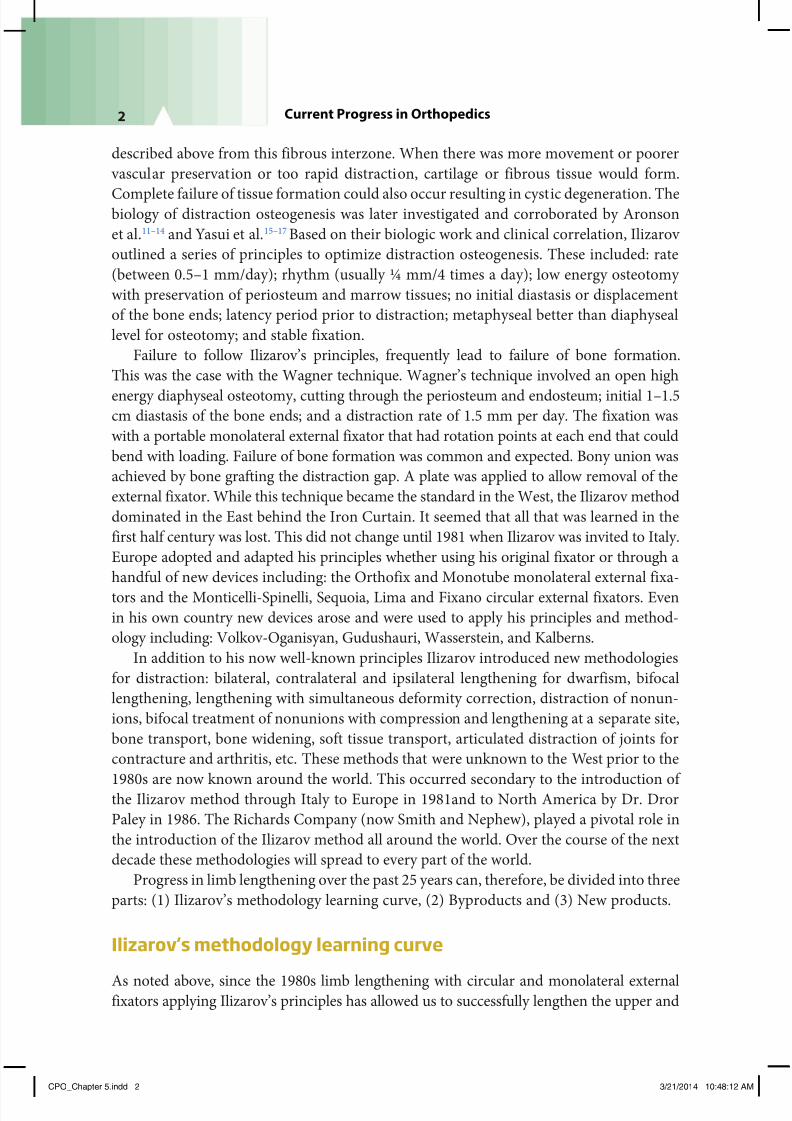
1. Deformity analysis and nomenclature
When I first started applying the Ilizarov method with its various hinges, rotation,and translation attachments, I was puzzled by where to apply the hinge especially for
metaphyseal deformities. The search for the correct location of the hinge, led me to
develop what is now referred to as CORA planning. The CORA (center of rotation
of angulation) is the point where the proximal and distal axes of the bone intersect
(Fig. 5.1a). To define these axes, especially for non-diaphyseal deformities, required
an understanding of frontal and sagittal plane alignment and joint orientation that
did not exist at that time. This lead to a new nomenclature that has been adopted
worldwide defining the mechanical and anatomic axes and their related joint orien-
tation angles (LDFA, MPTA, LPFA, LDTA in the frontal plane Fig. 5.1b, and PPTA,
PDFA, ADTA in the sagittal plane Fig. 5.1c). It established a set of osteotomy rulesbased on sound geometric principles. It introduced the concept of plane of deformity
CPO_Chapter 5.indd 3 3/21/2014 10:48:12 AM
7/23/2019 Progress in and From Limb Lengthening
http://slidepdf.com/reader/full/progress-in-and-from-limb-lengthening 4/27
4 Current Progress in Orthopedics
Figure 5.1
(a) Te center o rotation o angulation (CORA) is the intersection o the proximal and distal axis
lines. (b) Te CORA o a proximal metaphyseal varus deormity is marked. (c) I the osteotomy
is placed distal to the CORA but the correction occurs around an axis going through the CORA
then the osteotomy will angulate and translate. (d) Te CORA method was originally developed
to accurately determine the level o the Ilizarov hinge. (e) Joint orientation angles in the rontal
plane. () Joint orientation angles in the sagittal plane.
a
c
e f
d
b
CPO_Chapter 5.indd 4 3/21/2014 10:48:14 AM

7/23/2019 Progress in and From Limb Lengthening
http://slidepdf.com/reader/full/progress-in-and-from-limb-lengthening 5/27
5Progress in and from Limb Lengthening
and six-axis deformity correction. Current orthopedic deformity nomenclature,
theory, and practice, is all a byproduct of the Ilizarov hinge. Perhaps the best reference
for this is the book I authored, Principles of Deformity Correction.18
2. Age of lengthening surgery
In 1986 when I returned after studying with Ilizarov and with the Italians, the conven-
tional wisdom was that elective lengthening for leg length discrepancy should not be
performed prior to age 6. No objective reason was given for this. Subjective rationale
included: concern regarding growth inhibition in young children; it is hard to discern if
an infant is crying from pain or from other causes; joint or growth plate damage would
occur; technically too small and difficult. Parents of children born with large congen-
ital limb length discrepancy pushed for earlier treatment had to be told that nothing
should be done prior to age 6 for all the reasons mentioned above. In 1990, I had theprivilege to meet Dr. BB Joshi from Mumbai, India. He was applying external fixators
for the correction of clubfoot in children under 1 year of age. He argued against the
conventional wisdom and encouraged me to start lengthening after age 1. Having used
the Ilizarov device at this age for conditions such as congenital pseudarthrosis of the
tibia, I began lengthening in infants as young as 14 months of age for fibular hemimelia
(FH) and soon 2 years of age for CFD (Fig. 5.2a–c). Subsequent study of this group of
patients demonstrated that growth inhibition did not occur for femoral lengthening
in infants irrespective of the amount of lengthening (up to 8 cm) nor did it occur for
tibial lengthenings less than 5 cm.19 Growth inhibition occurred with combined femur
and tibia lengthening and with tibial
lengthening over 5cms. Interestingly
one-third of femurs demonstrated
growth stimulation. Parents had no
difficulty knowing when their child
was in pain and there was not damage
to the growth plate or joint carti-
lage. Lengthening in toddlers was
a byproduct of learning how to treat
CFD and FH in older children by theIlizarov method.
3. Prediction of limb length discrep-
ancy (LLD), timing of epiphysiodesis
and hemi-epiphsiodesis, and stature
With improved methods of limb
lengthening, accurate prediction of
LLD at maturity was required. The
existing methods of Anderson and
Green and Moseley were cumbersome
Figure 5.2
(a) A 2-year-old boy with congenital emoral
deficiency and an 8-cm leg length discrepancy.
(b) Lengthening o the congenitally short emur
with an Ilizarov device at age 2. (c) Afer 7 cm o
lengthening the leg lengths are almost equal.
a b c
CPO_Chapter 5.indd 5 3/21/2014 10:48:15 AM
7/23/2019 Progress in and From Limb Lengthening
http://slidepdf.com/reader/full/progress-in-and-from-limb-lengthening 6/27
6 Current Progress in Orthopedics
and time consuming. A quicker, simpler accurate method was needed. In 1997 Paley
developed the Multiplier method which was published in 2000.18,20 This method recog-
nized that a coefficient could be calculated for each age to represent the reciprocal ofgrowth remaining. That coefficient was independent of percentile, race, nationality, and
generation. For the lower extremity the coefficients (multipliers) for the femur, tibia,
and foot height were the same. Therefore a single set of multipliers could be used to
determine bone or limb length. Formulas designed around these multipliers allow for
calculation of limb length and LLD at skeletal maturity or at any age (forward or back-
wards in time), as well as timing of epiphysiodesis. The accuracy of prediction was also
tested21,22 and found to be accurate to less than 1 cm for both epiphysiodesis and LLD.
Multipliers were also calculated for height, foot length, upper extremity, and fetal bone
growth. They can be used for predictions of LLD from in utero measurements,23 predic-
tion of height,24 or foot length25 during childhood. Upper extremity multipliers can beused for LLD and epiphysiodesis planning the same as in the lower extremity.26 A single
X-ray’s measurements are sufficient for congenital deformity LLD prediction. For devel-
opmental LLD two points in time are required. The Multiplier method is a useful tool to
complement the surgical techniques and decision making for children treated for LLD.
More recently two apps have made this even easier to do (Paley Growth and Multiplier).
The Multiplier method was a byproduct of the need for accuracy and simplicity in limb
lengthening.
4. Lengthening over nails (LONs), lengthening and then nailing (LATN), and length-
ening over plates (LOPs)
The external fixator has been the gold standard of limb lengthening for over a century.
The discomfort of having open pin sites, the tethering of the underlying muscles,
chronic pin infections, pin loosening, etc., all make these devices unappealing to
both the patient and the surgeon. We tolerate the use of these devices as a necessary
means to an end; they get the job done and the patient achieves the goals of treatment.
The longer the treatment with the external fixator the less well they are tolerated. The
external fixation time for lengthening depends on the combination of the distraction
phase (which includes the latency period) and the consolidation phase. The fixator
is removed after the consolidation phase. In children the external fixation time istypically 1month per cm. In adults, the average is 2 months per cm.27 While there
was no alternative to the external fixator as a lengthening device efforts to reduce the
external fixation time have focused on replacing the external fixator with some other
fixation during the consolidation phase. Wagner applied a plate at the end of the
distraction phase and removed the external fixator.28 Wasserstein inserted a cortical
allograft and thin rod to support the distraction gap.29 Bost and Larsen introduced
the idea of a guiding the lengthening with a Rush rod in 1956.10 In 1990, I inserted a
locked intramedullary nail and applied an external fixator at the same time. The idea
was to lengthen over a nail (LON) and then lock it at the end of the distraction phase
and remove the external fixator (Fig. 5.3a,b). Paley et al. published our first series ofLON in 1993 and 1997.30,31 Two years later we tried to do this technique by inserting
CPO_Chapter 5.indd 6 3/21/2014 10:48:15 AM

7/23/2019 Progress in and From Limb Lengthening
http://slidepdf.com/reader/full/progress-in-and-from-limb-lengthening 7/27
7Progress in and from Limb Lengthening
the rod at the end of distraction. We succeeded in doing this in a few cases until
we encountered a deep infection in one case. The excellent success with LON and
one case of intramedullary infection with nailing at the end of distraction stopped
us from pursuing this strategy. Perseverance by one of my former clinical fellows,
Dr. Robert Rozbruch perfected this technique which is now called “lengthening
and then nailing” LATN.32 LATN is a good method in the tibia and offers the advan-
tage of allowing gradual deformity correction of the upper tibia, prior to nailing. The
proximal pins are placed out of the medullary canal and the distal pins, distal to the
tip of the nail. LON and LATN were byproducts of the lengthy consolidation time
in adults by the Ilizarov method and the availability of locking nails. More recently
the same concept has been applied using locking plates.33 Lengthening over plates
(LOPs), has been made possible by the introduction of locking plates. The original
failures of the Wagner method were due to the use of non-locking plates and failure
of bone formation.34 The locking plate is inserted and locked only at one end. The
external fixator is applied avoiding the region of the plate. After distraction the plate
is locked and the fixator removed. This allows early removal of the external fixatorafter first locking the free end of the plate. LOP like LON is a byproduct of lengthy
consolidation time by the Ilizarov method and the advent of locking plate technology.
LOP is a reinvention of the Wagner technique with Ilizarov biology and better plate
technology.
5. Fixator assisted nailing (FAN) and fixator assisted plating (FAP) for deformity
correction
The Ilizarov method for gradual deformity correction has the advantage of being mini-
mally invasive and very accurate due to its adjustability. Similarly, acute correction with
external fixation can also be adjusted in the operating room and check x-rays taken toconfirm that the desired alignment has been achieved.35–39 Both these methods improve
Figure 5.3
(a) Lengthening over a nail (LON). Te nail is inserted and locked proximally. Te external fixatoris applied with its pins posterior to the nail. (b) Afer the completion o lengthening the nail is
locked distally and the external fixator removed.
a b
CPO_Chapter 5.indd 7 3/21/2014 10:48:16 AM

7/23/2019 Progress in and From Limb Lengthening
http://slidepdf.com/reader/full/progress-in-and-from-limb-lengthening 8/27
8 Current Progress in Orthopedics
on the accuracy of deformity correction with internal fixation since the latter is not
adjustable. Following the successful use of LON, in 1992, I applied external fixator pins
out of the path of an intramedullary nail and then performed an osteotomy with acutedeformity correction. After adjusting the alignment using the external fixator, I nailed
the femur with a locking nail. Fixator assisted nailing (FAN) has the adjustability advan-
tages of external fixation combined with the convenience of internal fixation. By using
intramedullary nails it also offers the minimally invasive advantage of external fixation.
The external fixator is removed after the nail is locked (Fig. 5.4a–e). FAN was a byproduct
Figure 5.4
(a) Valgus deormity o the emur treated by
fixator assisted nailing. Te fixator pins are
applied out o line o the intramedullary nail
(IMN). (b) An acute correction is carried out
and an x-ray taken to confirm the joint orien-
tation angle. (c) Te emur is then reamed. (d)
Te nail is inserted and locked. (e) Te external
fixator is removed.
e
c d
a b
CPO_Chapter 5.indd 8 3/21/2014 10:48:17 AM

7/23/2019 Progress in and From Limb Lengthening
http://slidepdf.com/reader/full/progress-in-and-from-limb-lengthening 9/27
9Progress in and from Limb Lengthening
Figure 5.5
(a) Superknee procedure: the ascia lata (FL) is harvested and then split into two parts (FL1 &
2). Te posterior part (FL1) is passed under the lateral collateral ligament, over the top and then
through the proximal tibial epiphysis to be fixed with a screw (Macintosh). (b) Te anterior limb
o the ascia lata (F2) is routed under the patellar tendon, through a medical retinacular tunnel
and around the adductor magnus tendon. It is sutured to itsel and acts as an extra-articular PCL
(reverse Macintosh).
a b
of accurate deformity correction with external fixation with a desire to eliminate the
external fixator after surgery. More recently fixator assisted plating (FAP) was intro-
duced using the same concepts but applying a percutaneous plate instead of a nail.40–42
6. New reconstructive procedures for the joints of children with congenital deficiencies
Superknee (Fig. 5.5a,b): Prior to the introduction of the Ilizarov method only the
mildest CFD, FH, and tibial hemimelia (TH) cases were considered for leg length
equalization surgery. The vast majority were treated by amputation or rotationplasty,
and prosthetic fitting. Prior to 1980s lengthening for these conditions was worse than
no treatment. The complication rates were high with little gain in length achieved and
permanent damage to the hip, knee, and ankle common. Even with Ilizarov’s method
of spanning the joints the outcome of these joints was not good. In 1994, while tryingto reconstruct the knee joint and patella of a CFD patient that had dislocated during
lengthening, I came across an article by Lankenskiold et al. on congenital dislocation
of the patella.43 I combined this method with a technique I learned from Grammont
to move the patellar tendon in immature children. Since the iliotibial band was the
major dislocating force, instead of resecting it I harvested it and used the MacInstosh
procedure (I had learned from its author Dr. David Macintosh , my former professor)
to create an extra and intra-articular anterior cruciate ligament (ACL). Since the dislo-
cation was a posterolateral rotatory dislocation, I sought to prevent this movement of
CPO_Chapter 5.indd 9 3/21/2014 10:48:18 AM
7/23/2019 Progress in and From Limb Lengthening
http://slidepdf.com/reader/full/progress-in-and-from-limb-lengthening 10/27
10 Current Progress in Orthopedics
the tibia. Since I had harvested the entire ITB and only needed half for the Macintosh,
I used the other half as an extra-articular band to prevent external rotation. I ran
it under the patellar tendon and around to the side of the femur. I currently loop itaround the adductor magnus tendon and suture it to itself. I named this medial extra-
articular ligament repair the reverse Macintosh (in honor of my former professor) or
extra-articular posterior cruciate ligament (PCL). This combination of procedures is
named the “superknee” procedure. It can be performed before lengthening to stabilize
a cruciate deficiency knee or after lengthening if a complication occurs. It allows us to
salvage previously unsalvageable, unstable congenitally subluxed, or dislocated knees
and patellae.
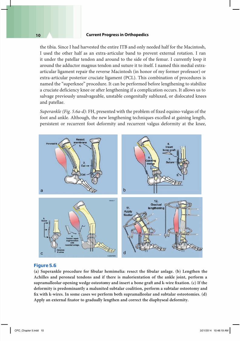
Superankle (Fig. 5.6a-d): FH, presented with the problem of fixed equino-valgus of the
foot and ankle. Although, the new lengthening techniques excelled at gaining length,persistent or recurrent foot deformity and recurrent valgus deformity at the knee,
Figure 5.6
(a) Superankle procedure or fibular hemimelia: resect the fibular anlage. (b) Lengthen the
Achilles and peroneal tendons and i there is malorientation o the ankle joint, perorm a
supramalleolar opening wedge osteotomy and insert a bone graf and k-wire fixation. (c) I the
deormity is predominantly a malunited subtalar coalition, perorm a subtalar osteotomy and
fix with k-wires. In some cases we perorm both supramalleolar and subtalar osteotomies. (d)
Apply an external fixator to gradually lengthen and correct the diaphyseal deormity.
a
c d
b
CPO_Chapter 5.indd 10 3/21/2014 10:48:19 AM

7/23/2019 Progress in and From Limb Lengthening
http://slidepdf.com/reader/full/progress-in-and-from-limb-lengthening 11/27
11Progress in and from Limb Lengthening
remained unsolved problems despite attempts to solve this problem were performed
by many surgeons. The problem again was due to the lack of understanding of the
pathoanatomy. The equino-valgus foot deformity was being treated the way club footdeformity was treated, using circumferential surgical release. This approach failed for
FH. The reason it failed was that the problem is not contracture of the muscles. The
problem is malorientation and dysplasia of the ankle joint and malunion of subtalar
coalition. This pathoanatomic problem went unrecognized because the ankle joint
is invisible radiographically since it is not ossified and the subtalar coalition is also
frequently invisible until a much later age. I was initially treating these by either
distraction or surgical release of the ankle joint. I noticed the malorientation when
I opened the ankle joint to release it. This lead to the “superankle” procedure which
involves an extra-articular soft tissue release of the ankle joint combined with a
supramalleolar varus-extension osteotomy and/or a subtalar coalition osteotomy toslide the calcaneus distal and medial. By reorienting the ankle and/or subtalar joints
which were deformed the foot became plantigrade acutely. The deformity did not
recur as it had happened with all the other methods of treatment. Once again the
problem was solved as a byproduct of failed lengthening surgery. Since the foot could
now be acutely corrected even in the most dysplastic and deficient feet, lengthening
for FH could now be accomplished successfully without recurrent foot deformity. As
such, the results of treatment were so good that it is hard to justify amputation which
previously was the mainstay of treatment.
Superhip 1 (Figs. 5.7a–k, 5.8a–c): At the hip, only ossified proximal femoral neck caseswere lengthenable. The cases with delayed ossification had over 90 degrees of complex
angular deformity. Attempts to correct these deformities were met with complete
recurrence of the angulation. As a byproduct of seeing more and more of these cases,
the pathoanatomy of these CFD cases was unraveled. First, no one had ever seen such
a volume of such rare cases. Second, successes and failures of treatment were care-
fully scrutinized and learned. The natural history study by Sanpera and Sparks44 led
to creation of the Paley classification of CFD in 1996. The “eureka” moment, however
came as I pondered why the coxa vara always recurred. Looking beyond the bony
deformity, I suddenly realized that there was a fixed abduction contracture. Releasing
the abduction contracture allowed full correction of the deformity for the first time.At first I did this distally by elevating the abductors with the quadriceps off of the
greater trochanter (GT) and then resuturing the conjoint tendon to the GT in its new
location. This however led to permanent weakness of the hip abductor muscles. The
problem was solved by allowing the muscle body to slide distally off the ilium with
the apophysis and shortening of the iliac bone. By understanding the pathoanatomy
the complex three-dimensional CFD deformity was unraveled and a new operation to
repair this deformity called the “superhip” was born in 1997. With the superhip proce-
dure we can now acutely reconstruct the upper femoral deformity of CFD irrespective
of the angle of deformation (deformities of greater than 90 degrees of varus and flexion
are common). Recognizing that there was usually a cartilaginous femoral neck present
CPO_Chapter 5.indd 11 3/21/2014 10:48:19 AM

7/23/2019 Progress in and From Limb Lengthening
http://slidepdf.com/reader/full/progress-in-and-from-limb-lengthening 12/27
12 Current Progress in Orthopedics
Figure 5.7
(a) Superhip procedure: make a long incision rom the
iliac crest to the knee. Expose and resect the ascia lata.
(b) Lengthen the psoas and rectus emoris tendons.
(c) Lengthen the piriormis tendon. (d) Split the apo-
physis and allow the abductor muscles to slide distally.
(e) Insert a guide wire rom the tip o the greater tro-
chanter to the center o the emoral head. Insert a sec-
ond guide wire at 45 degrees to the first in line with the
center o the neck and head. () Use a cannulated chisel
and then a cannulated 130 degree angled blade plate.
I it is inserted correctly there is usually a severe varus
and flexion deormity. (g) Osteotomize the emur and
then overlap the bone ends. Shorten the emur to the
necessary amount. (h) Fix the emur with screws and i
there is delayed ossification o the emoral neck insert
BMP into the neck. (i) Perorm a Dega osteotomy. (j)
o repair the apophysis, resect the top o the iliac crest.
(k) Now close the apophysis. Te abduction contracture
has been relieved.
c
d
f
e
h
g
b
a i
j
k
CPO_Chapter 5.indd 12 3/21/2014 10:48:23 AM

7/23/2019 Progress in and From Limb Lengthening
http://slidepdf.com/reader/full/progress-in-and-from-limb-lengthening 13/27
13Progress in and from Limb Lengthening
Figure 5.8
(a) Congenital emoral deficiency, Paley type 1b. Note the lack o neck ossification. (b) Superhip
procedure perormed at age 2. BMP inserted into the neck. (c) Ossification occurs in the superior
neck where the BMP was administered. (d) 8 cm lengthening o the emur 1 year later. (e) Afer
consolidation. Te bone was rodded to prevent racture.
(which can now be confirmed by MRI) allowed us to apply this correction method to
more children at ages as young as 2 years old. Despite the excellent correction and fixa-
tion achieved delayed ossification persisted and led to recurrence of deformity until I
recognized that unossified cartilage could be ossified by the insertion of bone morpho-
genetic protein (BMP). The superhip 1 procedure for treatment of CFD Paley type 1b
cases is a byproduct of failed lengthening surgery for CFD by the Ilizarov method.
Superhip 2 and 3 (Fig. 5.9 a–d) and rotationplasty (Fig. 5.10a–c): Unfortunately CFD
Paley types 2 and 3 were not treatable by the superhip procedure. In 2001, I developed
c d e
a b
CPO_Chapter 5.indd 13 3/21/2014 10:48:25 AM

7/23/2019 Progress in and From Limb Lengthening
http://slidepdf.com/reader/full/progress-in-and-from-limb-lengthening 14/27
14 Current Progress in Orthopedics
a new procedure to address the Paley type 2 cases that had a femoral head and GT but
no femoral neck. This was named the Superhip 2 procedure. This procedure creates a
femoral neck from the upper end of the femur and joins it to the femoral head that is
present in the acetabulum. With the advent of the superhip 1 & 2 procedures length-
ening was now a possibility for virtually all Paley type 1 and 2 cases. The limiting factor
was now the more difficult CFD Paley type 2 and 3. In 2013, the superhip 3 was createdto address these cases. In this procedure a new acetabulum using the triradiate cartilage
as the roof combined with an interposition and trochanteric arthroplasty have success-
fully been done to create a hip joint. It is too early to know the results of the Superhip 3
procedure. The most reliable solution for CFD Paley type 3 remains rotationplasty. The
need to improve this procedure was also a byproduct of failed limb lengthening for CFD
Paley type 3. The classic Van Nes rotationplasty was modified for CFD by Gillespie and
Krajbich from Toronto.45,46 Their technique involved fusion of the knee and leaving the
hip to float free. As a consequence a severe Trendelenberg and recurrent derotation were
frequent complications. Brown, in 199647 modified rotationplasty to use a tumor-like
racquet incision and fuse the femoral remnant to the pelvis converting the knee into
Figure 5.9
(a) Superhip2 procedure: the upper emur is moved on a vascular pedicle. (b) Te proximal
ragment is rotated 135 degrees to be able to act as the emoral neck. It is fixed to the emoralhead with threaded K-wires. (c) Te neck is fixed in place with a Rush rod and tension band
wire. (d) Te construct is neutralized using a spanning external fixator rom the pelvis to the
emur to the tibia.
a
dc
b
CPO_Chapter 5.indd 14 3/21/2014 10:48:27 AM

7/23/2019 Progress in and From Limb Lengthening
http://slidepdf.com/reader/full/progress-in-and-from-limb-lengthening 15/27
15Progress in and from Limb Lengthening
a
cb
a hip, and the ankle into a knee. Paley modified the Brown rotationplasty to include aChiari osteotomy and more distal transfer of the muscle insertions (Fig. 5.10a–c). Most
recently I connected the femoral remnant to the femoral head when it was present.
Both the Brown and Paley modifications of the rotationplasty preserved the knee joint
and converted its function to a hip. Derotation does not occur with the Brown or Paley
modifications but is a known complication of the Gillespie and Krajbich type.45,46
Ulnarization (Fig. 5.11a–e): Treatment of upper extremity deficiencies was similarly
impacted. For radial club hand deformity recurrent deformity and growth arrest of the
distal ulna were common complications48 the usual centralization and even of radiali-
zation procedures done from a dorsal approach. Distraction methods seemed to be the
Figure 5.10
(a) Paley modification o Brown rotationplasty. Trough a racquet incision all o the thigh mus-
cles are detached rom their insertions distally. Te emoral artery and vein are dissected ree
o the surrounding structures. (b) Te emur is rotated 180 degrees and fixed to a Chiari oste-
otomy with screws. (c) All o the muscles are reattached in the rotated position to operate the
knee as a hip joint.
CPO_Chapter 5.indd 15 3/21/2014 10:48:29 AM

7/23/2019 Progress in and From Limb Lengthening
http://slidepdf.com/reader/full/progress-in-and-from-limb-lengthening 16/27
16 Current Progress in Orthopedics
Figure 5.11
(a) Ulnarization o the carpus or radial clubhand.
A volar incision is used and the flexor carpi ulnaris
(FCU) is reflected back with the pisiorm bone. (b)
Te vascular pedicle o the ulnar epiphysis is pre-served and a capsulotomy separating the carpus rom
the ulna is perormed. (c) Te carpus can now be
ulnarized. (d) Te carpus is pinned to the ulna. (e)
Te FCU is transerred to the dorsum o the wrist and
the pisiorm is sutured to the base o the metacarpal.c
b
a
e
d
solution but turned out to be disappointing since they did not change the recurrence
rate.49 Once again experience with these failed procedures including distraction lead to
the development of a new operation through a volar approach. I developed ulnarization
in 1999. Using the flexor carpi ulnaris as a tendon transfer instead of the traditional flexorcarpi radialis, combined with transfer of the carpus to the ulnar side of the ulna (hence
the name ‘ulnarization’), lead to a zero recurrence rate and a much stronger dorsiflexion.
The volar approach also allowed us to identify the vascular pedicle to the distal ulna and
its physis. Therefore, there were no growth arrests. Stabilization of the wrist facilitated
lengthening of the forearm for radial clubhand. Ulnarization was a byproduct of failed
correction of radial clubhand by distraction and by radialization.
Weber patellar arthroplasty (Fig. 5.12a–e): TH treatment had resisted all attempts at
joint reconstruction by the Brown,50 and even by the modified pre-distraction Brown
procedures. Weber recognized that several of these patients had a patella present andthat the patella could be used to substitute the tibial plateau. He developed a vascular
CPO_Chapter 5.indd 16 3/21/2014 10:48:32 AM

7/23/2019 Progress in and From Limb Lengthening
http://slidepdf.com/reader/full/progress-in-and-from-limb-lengthening 17/27
17Progress in and from Limb Lengthening
pedicle method which I refer to as the Weber patellar arthroplasty.51 This has made
stabilization of the knee possible with remodeling to a normal looking tibial plateau
and active knee motion. When a patella is not possible other reconstructive methodscreating an ACL-like ligament are used to prevent the subluxation of the centralized
fibula, which was very common with the Brown. These methods were byproducts of
failed distraction treatment of TH.
7. New reconstructive procedure for congenital pseudarthrosis of the tibia (CPT)
CPT was previously an unsolved problem. Failure to obtain union or recurrent frac-
ture was the norm and not the exception. While the Ilizarov method was used for
many years to treat CPT it was found to be successful to obtain union but not to
maintain union.52 While union rate was high, refracture rate after the Ilizarov was
also high. Combining intramedullary fixation with the Ilizarov method reduced butdid not eliminate failures and refractures.53,54 Recognizing this as a periosteal disease
Figure 5.12
(a) Weber patellar arthroplasty: proximal and distal “visor” flaps are dissected mobilizing the
patella on the proximal flap. (b) Te flaps are moved up and down, so that the patella rests distal
to the end o the emur to act as a tibial plateau. (c) Te flaps are repaired to each other. (d) Peri-
chondrial flaps are created and the ends o the epiphysis o the fibula and the patella are connected
together (e).
a b c
ed
CPO_Chapter 5.indd 17 3/21/2014 10:48:35 AM

7/23/2019 Progress in and From Limb Lengthening
http://slidepdf.com/reader/full/progress-in-and-from-limb-lengthening 18/27
18 Current Progress in Orthopedics
and using periosteal grafting reduced but did not eliminate refractures.54,55 The
finding that there were too many osteoclasts and treatment with bisphosphonates was
a big step forward.56 Identifying that the BMP production was low and using BMP
also helped bone healing56
Probably the most important factor to prevent recur-rence turned out to be creation of a cross union between the tibia and the fibula57
(Fig. 5.13a–g). Combining these factors has produced a union rate of 100% and a
refracture rate of zero. Early treatment which was previously fraught with higher
refracture rates was now practical. This also meant that with early treatment secondary
leg length discrepancy and ankle and foot deformities were less likely to develop. The
end result now can be a completely normal leg and foot. This new method for CPT
is a byproduct of the high refracture rate after Ilizarov treatment.
8. Distraction treatment of joints: e.g., Perthes (Fig. 5.14a–c)
Distraction application to joints according to Ilizarov as well as others like Volkovet al. and Oganesian et al.58,59 lead early investigators like Aledegheri et al and
Figure 5.13
(a, b) Congenital pseudarthrosis o the tibia
in a 1-year-old girl with neurofibromato-
sis. (c)reatment by resection o hamar-
toma, invagination and rodding o the bone
ends o fibula and tibia, periosteal and iliac
crest bone grafing, insertion o BMP, and
external fixation stabilization. Preopera-
tive inusion o Zoledronic acid was given.(d, e) Union and cross union achieved in 3
months. Rods lef in place. () Leg length
discrepancy at end o treatment. (g) Differ-
ential lengthening o emur and tibia with
re-rodding o tibia.
a e
d
b c
f g
CPO_Chapter 5.indd 18 3/21/2014 10:48:39 AM

7/23/2019 Progress in and From Limb Lengthening
http://slidepdf.com/reader/full/progress-in-and-from-limb-lengthening 19/27
19Progress in and from Limb Lengthening
Debatiani et al.60,61 to apply monolateral devices to distract the hip, knee, ankle,
and elbow (arthrodiatasis). Articulated distraction of joints was used to treat joint
contracture, joint stiffness, and joint subluxation. I applied joint distraction to treatan 11-year-old boy with severe extrusion, contracture, and stiffness from Perthes
in 1989. The result was dramatic. It sped up the reossification of the femoral head,
reduced the subluxation, improved the range of motion, and made the femoral head
more spherical. Distraction for Perthes has now been corroborated as an effective
treatment by surgeons from several different countries.18,62 Distraction for Perthes is
a byproduct of the Ilizarov articulated distraction of joints.
New product advances
1. Modularity of monolateral external fixators (Fig. 5.15)Twenty-five years ago only the Ilizarov circular external fixator offered modularity
to address problems of lengthening, angular correction, articulated joint distraction,
bone transport, joint fusion, fracture treatment, etc. It could be modified to correct
angular deformity with hinges, rotational deformity with rotation constructs, trans-
lation deformity with translation constructs, etc. The Ilizarov device could articulate
across joints for protection of a joint during lengthening or for correction of joint
contractures. The Ilizarov device used only wires. In contrast, monolateral external
fixators such as the Orthofix device, had very limited ability to correct deformities and
could not articulate across joints. These devices only used threaded half pins for fixa-
tion. The monolateral devices had limited modularity. Since then the circular external
Figure 5.14
(a) An 11-year-old boy with Perthes disease with significant
extrusion and collapse and adduction contracture. (b) Distrac-
tion treatment o hip with articulated external fixator. Reduc-tion o the extrusion. (c) Radiograph afer 2 years.
b
a
c
Figure 5.15
Monolateral external fixa-
tor with hinges at the hip
and knee or stabilization o
joints while permitting flex-
ion and extension movement
(Modular Rail System, Smith
and Nephew Orthopedics,Memphis, N, USA).
CPO_Chapter 5.indd 19 3/21/2014 10:48:41 AM

7/23/2019 Progress in and From Limb Lengthening
http://slidepdf.com/reader/full/progress-in-and-from-limb-lengthening 20/27
20 Current Progress in Orthopedics
fixators have adapted to use half pins and the monolateral ones added arches to
use wires. The monolateral devices now have hinges to articulate across joints and
deformities. Some monolateral devices can correct angular, rotation, and transla-tion deformities. The two devices now connect with each other. This modularity has
made both circular and monolateral
devices much more user friendly and
has extended the indications of each.
2. Computer dependent external fixation
(Fig. 5.16)
In 1994, Charles and Harold Taylor
presented their new concept of computer-
dependent external fixation. This six-axiscorrection device allowed changes to be
made on the computer screen instead of
on the external fixator. This was a huge
advance in the circular external fixator
technology. Other inventors such as
Claus Seide also developed a similar
device independently.63 These devices
such as the Taylor Spatial Frame or the
Hexapod can correct angulation, rota-
tion, translation, and length deformi-
ties simultaneously. On the basis of the
success of these devices, newer six-axis
correction devices are being developed
and the next few years will see a wider
variety of what are now termed computer-
dependent external fixation devices.
3. Implantable limb lengthening
(Fig. 5.17 a,b)While it is commonly recognized that
the Soviet Union produced the greatest
limb lengthening contributions through
the innovations of Ilizarov, few realize
that implantable limb lengthening also
takes its origin in the Soviet Union.
Alexander Bliskunov from Simferopol,
Ukraine first published his method
in 1983. This was before most of the
world had heard of Ilizarov. Bliskunov
Figure 5.16
Six-axis deormity correction device with
computer-dependent external fixator (aylor
Spatial Frame, Smith and Nephew Orthope-
dics, Memphis, N, USA).
ba
Figure 5.17
(a) Implantable lengthening nail used or
bilateral emoral lengthening or stature (Pre-
cice, Ellipse echnologies, Irvine, CA, USA).
(b) Afer 6.5 cm o bilateral emoral lengthen-
ing with nails.
CPO_Chapter 5.indd 20 3/21/2014 10:48:43 AM
7/23/2019 Progress in and From Limb Lengthening
http://slidepdf.com/reader/full/progress-in-and-from-limb-lengthening 21/27
21Progress in and from Limb Lengthening
developed a telescopic lengthening nail that used a crank shaft connected to the
pelvis to drive his mechanism and lengthen the femur.64 His technology was not
available outside of the Soviet Union. It is not surprising that others soon devel-oped other mechanisms to drive telescopic lengthening nails. Baumgart et al. from
Germany developed a motorized nail in 1991.64,66 Guichet et al. from France,67,68
developed a telescopic nail in 1994 using a ratchet mechanism which rotated the
two segments of the nail through the callus of the bone. Cole used this concept to
develop a double clutch mechanism to lengthen the ISKD (Intramedullary Skeletal
Kinetic Distractor) device marketed by Orthofix.69,70 This was the first FDA-approved
device in 2001. Soubieran developed the Phenix nail which was acquired by Smith
and Nephew and awaits FDA approval and release. Ellipse developed the Precice
nail with Stuart Green. It is the second FDA-approved implantable lengthening nail
and has been in clinical use since 2011. The same company developed the Precice2 with Dror Paley and released this nail in Nov. 2013. In the near future we expect
to see bone transport nails, gradual deformity correction plates, and lengthening
plates. The future of non-invasively adjustable implantable devices is the most
exciting and promising technologic advance in limb lengthening in the past 25 years.
4. Pharmacologic and biologic advances
As the mechanism of bone formation with limb lengthening has become better
understood, the specific pathways for bone and soft tissue regeneration and how
they can be modulated has lead to advances in pharmacology and biologic agents.71–73
The use of bisphosphonate drugs to delay resorption and allow unimpeded boneformation has been useful in treating delayed regenerate bone formation.74 The use
of bone morphogentic protein to speed regenerate bone formation has also been
demonstrated. The role of new therapeutics with concentrated bone marrow stem
cells, frozen embryonic stem cells, etc., are still under investigation.75–81
XSUMMARY
While the history of limb lengthening goes back more than one century, its biggest
advances have all occurred in the past 25 years. Its foundations were set by the
pioneering, landmark original work, and ideas of Gavril Abramovich Ilizarov. Ilizarov’soriginal ideas have been corroborated and reproduced by surgeons all over the globe.
Ilizarov deserves the credit for bringing science to this field. The past 25 years are also
bountiful in new ideas, new devices, and new therapeutics. Distraction histogenesis
is still a young field of study. There is still a lot of room for innovation and advances.
While the first 100 years was the story of external fixation and distraction osteogenesis,
the next 100 years will be the story of implantable distraction devices, biofeedback
controls for internal and external fixation, and the modulation of molecular biology,
and regenerative biology to orthopedics. Many of the genetic conditions currently
being treated will have medical instead of surgical solutions and new applications of
distraction biology will be found.
CPO_Chapter 5.indd 21 3/21/2014 10:48:44 AM

7/23/2019 Progress in and From Limb Lengthening
http://slidepdf.com/reader/full/progress-in-and-from-limb-lengthening 22/27
22 Current Progress in Orthopedics
Key Learning Points
❖ Limb lengthening has evolved from traction bed mounted devices to portable
external fixation to implantable limb lengthening.
❖ Distraction osteogenesis is best achieved by control of rate and rhythm of distrac-
tion of a low energy osteotomy without initial diastasis with preservation of the
periosteal and endosteal elements of the bone.
❖ During the last 25 years the methods of Ilizarov have been reproduced. While over-
coming the obstacles of bone and soft tissue regeneration, new obstacles such as
joint reconstruction of congenital deficiencies required developing a wide variety
of new operations that previously were not considered. These new operations are
called byproducts of the Ilizarov method.
❖ External fixation while a means to an end is not well liked by surgeons and patients
alike. Efforts to shorten the external fixation treatment time lead to the develop-
ment of LON, LOP, FAN, and FAP.
❖ Parallel efforts to eliminate the need for external fixation lead others to develop
fully implantable lengthening devices. We are now in the era of implantable length-
ening nails and await new implantable solutions for bone transport, deformity
correction, etc.
❖ The future of this young field will include dynamic implants and biologic advances
in cellular and molecular technology.
T REFERENCES
1. Paley D. Current techniques of limb lengthening, J Pediatr Orthop 1988; Jan–Feb 8(1): 73–92.
2. Varghese RA, Dhawale AA, Zavaglia BC, Slobogean BL, Mulpuri K. Citation classics in pedi-
atric orthopaedics. J Pediatr Orthop 2013; 33(6): 667–71.
3. Codivilla A. On the means of lengthening, in the lower limbs, the muscles and tissues which
are shortened through deformity. J Bone Joint Surg Am 1905; 2(2): 353–69.
4. Putti V. The operative lengthening of the femur 1921. Clin Orthop Relat Res 1990 Jan; (250):4–7.
5. Allan FG. Bone lengthening. J Bone Joint Surg Br 1948 Aug; 30B(3): 490–505.
6. Allan FG. Leg-lengthening. Br Med J 1951 Feb 3; 1(4700): 218–22.
7. Abbott LC. Lengthening of the lower extremities. Cal West Med 1932 Jan; 36(1): 6–13.
8. Abbott LC, Saunders JB. The operative lengthening of the tibia and fibula: a preliminary report
on the further development of the principles and technic. Ann Surg 1939 Dec; 110(6): 961–91.
9. Anderson WV. Leg lengthening. J Bone Joint Surg Br 1952; 34: 150.
10. Bost FC, Larsen LJ. Experiences with lengthening of the femur over an intramedullary rod.
J Bone Joint Surg Am 1956 Jun; 38-A(3): 567–84.
CPO_Chapter 5.indd 22 3/21/2014 10:48:44 AM
7/23/2019 Progress in and From Limb Lengthening
http://slidepdf.com/reader/full/progress-in-and-from-limb-lengthening 23/27
23Progress in and from Limb Lengthening
11. Aronson J. Temporal and spatial increases in blood flow during distraction osteogenesis.
Clin Orthop Relat Res 1994 Apr; (301): 124–31.
12. Aronson J, Harrison B, Boyd CM, Cannon DJ, Lubansky HJ, Stewart C. Mechanical inductionof osteogenesis. Preliminary studies. Ann Clin Lab Sci 1988 May–Jun; 18(3): 195–203.
13. Aronson J, Harrison BH, Stewart CL, Harp JH Jr. The histology of distraction osteogenesis
using different external fixators. Clin Orthop Relat Res 1989 Apr; (241): 106–16.
14. Aronson J, Harp JH Jr. Factors influencing the choice of external fixation for distraction oste-
ogenesis. Instr Course Lect 1990; 39: 175–83.
15. Kojimoto H, Yasui N, Goto T, Matsuda S, Shimomura Y. Bone lengthening in rabbits by callus
distraction. The role of periosteum and endosteum. J Bone Joint Surg Br 1988 Aug; 70(4):
543–9.
16. Yasui N, Kojimoto H, Shimizu H, Shimomura Y. The effect of distraction upon bone, muscle,
and periosteum. Orthop Clin North Am 1991 Oct; 22(4): 563–7. 17. Yasui N, Sato M, Ochi T, Kimura T, Kawahata H, Kitamura Y, Nomura S. Three modes of
ossification during distraction osteogenesis in the rat. J Bone Joint Surg Br 1997 Sep; 79(5):
824–30.
18. Paley D, Herzenberg JE. Principles of Deformity Correction. 1st ed. Berlin: Springer-Verlag.
2002. (Corr. 3rd printing 2005.)
19. Sabharwal S, Paley D, Bhave A, Herzenberg JE. Growth patterns after lengthening of congeni-
tally short lower limbs in young children. J Pediatr Orthop 2000; 20: 137–145.
20. Paley D, Bhave A, Herzenberg JE, Bowen JR. Multiplier method for predicting limb-length
discrepancy. J Bone Joint Surg Am 2000 Oct; 82-A(10): 1432–46.
21. Aguilar JA, Paley D, Paley J, Santpure S, Patel M, Bhave A, Herzenberg JE. Clinical validation
of the multiplier method for predicting limb length at maturity, part I. J Pediatr Orthop 2005
Mar–Apr; 25(2): 186–91.
22. Aguilar JA, Paley D, Paley J, Santpure S, Patel M, Herzenberg JE, Bhave A. Clinical validation
of the multiplier method for predicting limb length discrepancy and outcome of epiphys-
iodesis, part II. J Pediatr Orthop 2005 Mar–Apr; 25(2): 192–6.
23. Paley J, Gelman A, Paley D, Herzenberg JE. The prenatal multiplier method for prediction of
limb length discrepancy. Prenat Diagn 2005 Jun; 25(6): 435–8.
24. Paley J, Talor J, Levin A, Bhave A, Paley D, Herzenberg JE. The multiplier method for predic-
tion of adult height. J Pediatr Orthop 2004 Nov–Dec; 24(6): 732–7. 25. Lamm BM, Paley D, Kurland DB, Matz AL, Herzenberg JE. Multiplier method for predicting
adult foot length. J Pediatr Orthop 2006 Jul–Aug; 26(4): 444–8.
26. Paley D, Gelman A, Shualy MB, Herzenberg JE. Multiplier method for limb-length predic-
tion in the upper extremity. J Hand Surg Am 2008 Mar; 33(3): 385–91. doi: 10.1016/j.
jhsa.2007.11.007.
27. Fischgrund J, Paley D, Suter C. Variables affecting time to bone healing during limb length-
ening. Clin Orthop Relat Res 1994 Apr; (301): 31–7.
28. Wagner H. Operative lengthening of the femur. Clin Orthop Relat Res 1978 Oct; (136):
125–42.
CPO_Chapter 5.indd 23 3/21/2014 10:48:44 AM

7/23/2019 Progress in and From Limb Lengthening
http://slidepdf.com/reader/full/progress-in-and-from-limb-lengthening 24/27
24 Current Progress in Orthopedics
29. Wasserstein I. Twenty-five years’ experience with lengthening of shortened lower extremities
using cylindrical allografts. Clin Orthop Relat Res 1990 Jan; (250): 150–3.
30. Paley D, Herzenberg JE, Paremain G, Bhave A. Femoral lengthening over an intramedullarynail. A matched-case comparison with Ilizarov femoral lengthening. J Bone Joint Surg Am 1997
Oct; 79(10): 1464–80.
31. Paley D, Herzenberg JE, Maar D, Tetsworth KT. Femoral lengthening by simultaneous
external fixation and intramedullary rodding. J Orthop Trauma 1993; 7:178.
32. Rozbruch SR, Kleinman D, Fragomen AT, Ilizarov S. Limb lengthening and then insertion of
an intramedullary nail: a case-matched comparison. Clin Orthop Relat Res 2008 Dec; 466(12):
2923–32. doi: 10.1007/s11999-008-0509-8. Epub 2008 Sep 18.
33. Iobst CA, Dahl MT. Limb lengthening with submuscular plate stabilization: a case series and
description of the technique. J Pediatr Orthop 2007; Jul–Aug; 27(5): 504–9.
34. Guarniero R, Barros Júnior TE. Femoral lengthening by the Wagner method. Clin OrthopRelat Res 1990 Jan; 250: 154–9.
35. Paley D, Herzenberg JE, Bor N. Fixator-assisted nailing of femoral and tibial deformities. Tech
Orthop 1997; 12: 260–75.
36. Paley D, Herzenberg JE. Principles of Deformity Correction, Hardware and Osteotomy Consid-
erations Chapter 11. Springer-Verlag 1st ed. 2002. Corr. 3rd printing 2005.
37. Jasiewicz B, Kacki W, Tesiorowski M, Potaczek T. [Results of femoral lengthening over an
intramedullary nail and external fixator]. Chir Narzadow Ruchu Ortop Pol 2008 May–Jun;
73(3): 177–83.
38. Kocaoglu M, Eralp L, Bilen FE, Balci HI. Fixator-assisted acute femoral deformity correction
and consecutive lengthening over an intramedullary nail. J Bone Joint Surg Am 2009 Jan; 91(1):
152–9. doi: 10.2106/JBJS.H.00114.
39. Bilen FE, Kocaoglu M, Eralp L, Balci HI. Fixator-assisted nailing and consecutive lengthening
over an intramedullary nail for the correction of tibial deformity. J Bone Joint Surg Br 2010 Jan;
92(1): 146–52.
40. El-Rosasy MA, El-Sallakh SA. Distal tibial hypertrophic nonunion with deformity: treat-
ment by fixator-assisted acute deformity correction and LCP fixation. Strategies Trauma Limb
Reconstr 2013 Apr; 8(1): 31–5. doi: 10.1007/s11751-012-0150-7. Epub 2012 Oct 27.
41. Eidelman M, Keren Y, Norman D. Correction of distal femoral valgus deformities in adoles-
cents and young adults using minimally invasive fixator-assisted locking plating (FALP). J Pediatr Orthop B. 2012 Nov; 21(6): 558–62. doi: 10.1097/BPB.0b013e328358f884.
42. Seah KT, Shafi R, Fragomen AT, Rozbruch SR. Distal femoral osteotomy: is internal fixation
better than external? Clin Orthop Relat Res 2011 Jul;469(7): 2003-11. doi: 10.1007/s11999-010-
1755-0. Epub 2011 Jan 6.
43. Langenskiöld A, Ritsilä V. Congenital dislocation of the patella and its operative treatment.
J Pediatr Orthop 1992 May–Jun;12(3): 315–23.
44. Sanpera I Jr, Sparks LT. Proximal femoral focal deficiency: does a radiologic classification
exist? J Pediatr Orthop 1994 Jan–Feb; 14(1): 34–8.
45. Torode IP, Gillespie R. Rotationplasty of the lower limb for congenital defects of the femur.
J Bone Joint Surg Br . 1983 Nov; 65(5): 569–73.
CPO_Chapter 5.indd 24 3/21/2014 10:48:44 AM

7/23/2019 Progress in and From Limb Lengthening
http://slidepdf.com/reader/full/progress-in-and-from-limb-lengthening 25/27
25Progress in and from Limb Lengthening
46. Krajbich JI. Modified Van Nes rotationplasty in the treatment of malignant neoplasms in the
lower extremities of children. Clin Orthop Relat Res. 1991 Jan; 262: 74–7.
47. Brown KL. Resection, rotationplasty, and femoropelvic arthrodesis in severe congenitalfemoral deficiency. A report of the surgical technique and three cases. J Bone Joint Surg Am
2001 Jan; 83-A(1): 78–85.
48. Buck-Gramcko D. Radialization as a new treatment for radial club hand. J Hand Surg Am 1985
Nov; 10(6 Pt 2): 964–8.
49. Kessler I. Centralisation of the radial club hand by gradual distraction J Hand Surg Br 1989
Feb; 14(1): 37–42.
50. Brown FW. Construction of a knee joint in congenital total absence of the tibia. J Bone Joint
Surg Am 1965; 47: 695–704.
51. Weber M. A new knee arthroplasty versus Brown procedure in congenital total absence of the
tibia: a preliminary report. J Pediatr Orthop B 2002 Jan; 11(1): 53–9. 52. El-Rosasy MA, Paley D, Herzenberg JE. Congenital pseudarthrosis of the tibia. In: Rozbruch
SR, Ilizarov S (eds): Limb Lengthening and Reconstruction Surgery . New York: Informa
Healthcare, 2007, ch 34, pp 485–94.
53. Grill F, Bollini G, Dungl P, Fixsen J, Hefti F, Ippolito E, Romanus B, Tudisco C, Wientroub S.
Treatment approaches for congenital pseudarthrosis of tibia: results of the EPOS multi-
center study: European Paediatric Orthopaedic Society (EPOS). J Pediatr Orthop B. 2000;
9: 75–89.
54. Thabet AM, Paley D, Kocaoglu M, Eralp L, Herzenberg JE, Ergin ON. Periosteal grafting for
congenital pseudarthrosis of the tibia: a preliminary report. Clin Orthop Relat Res 2008 Dec;
466(12): 2981–94.
55. Hermanns-Sachweh B, Senderek J, Alfer J, Klosterhalfen B, Büttner R, Füzesi L, Weber M.
Vascular changes in the periosteum of congenital pseudarthrosis of the tibia. Pathol Res Pract
2005;201: 305–12.
56. Schindeler A, Ramachandran M, Godfrey C, Morse A, McDonald M, Mikulec K, Little DG.
Modeling bone morphogenetic protein and bisphosphonate combination therapy in wild-type
and Nf1 haploinsufficient mice. J Orthop Res 2008; 26: 65−74.
57. Paley D. Congenital Pseudarthrosis of the Tibia: Combined Pharmacologic and Surgical Treat-
ment Using Biphosphonate Intravenous Infusion and Bone Morphogenic Protein with Peri-
osteal and Cancellous Autogenous Bone Grafting, Tibio-Fibular Cross Union, Intramedullary,Bone Grafting . Dr Alessandro Zorzi (Ed.), (2012). ISBN: 978-953-51-0324-0, InTech, DOI:
10.5772/31149.
58. Volkov MV, Oganesian OV. Restoration of function in the knee and elbow with a hinge-dis-
tractor apparatus. J Bone Joint Surg Am 1975 Jul; 57(5): 591–600.
59. Oganesian OV, Istomina IS, Kuzmin VI. Treatment of equinocavovarus deformity in adults
with the use of a hinged distraction++ apparatus. J Bone Joint Surg Am 1996 Apr; 78(4): 546–56.
60. Aldegheri R, Trivella G, Saleh M. Articulated distraction of the hip: Conservative surgery for
arthritis in young patients. Clin Orthop 1994; 301: 94–101.
61. De Bastiani G, Aldegheri R, Renzi-Brivio L, Trivella G. Limb lengthening by callus distraction
(callotasis). J Pediatr Orthop 1987 Mar–Apr; 7(2): 129–34.
CPO_Chapter 5.indd 25 3/21/2014 10:48:44 AM
7/23/2019 Progress in and From Limb Lengthening
http://slidepdf.com/reader/full/progress-in-and-from-limb-lengthening 26/27
26 Current Progress in Orthopedics
62. Paley D. Treatment of Perthes Disease of the Hip by Joint Distraction (in press).
63. Paley D. History and science behind the six-axis correction external fixation devices in
orthopaedic surgery. Oper Tech Orthop 2011; 21(2), 125–28. 64. Bliskunov AI, [Lengthening of the femur using implantable appliances]. Acta Chir Orthop
Traumatol Tech 1984 Dec; 51(6): 454–66.
65. Baumgart R, Betz A, Schweiberer L. [First fully implantable intramedullary system for callus
distraction—intramedullary nail with programmable drive for leg lengthening and segment
displacement. Principles and initial clinical results]. Chirurg 1990 Aug; 61(8): 605–9.
66. Baumgart R, Betz A, Schweiberer L. A fully implantable motorized intramedullary nail for
limb lengthening and bone transport. Clin Orthop Relat Res 1997 Oct; 343: 135–43.
67. Guichet JM, Grammont PM, Trouilloud P. [A nail for progressive lengthening. An animal
experiment with a 2-year follow-up]. Chirurgie 1992; 118(6–7): 405–10.
68. Guichet JM, Casar RS, Mechanical characterization of a totally intramedullary gradual elon-gation nail. Clin Orthop Relat Res 1997 Apr; 337: 281–90.
69. Cole JD, Justin D, Kasparis T, DeVlught D, Knobloch C. The intramedullary skeletal kinetic
distractor (ISKD): first clinical results of a new intramedullary nail for lengthening of the
femur and tibia. Injury 2001 Dec; 32 (Suppl 4): SD129–39.
70. Burghardt RD, Herzenberg JE, Specht SC, Paley D. Mechanical failure of the intramedullary
skeletal kinetic distractor in limb lengthening. J Bone Joint Surg Br 2011 May; 93(5): 639–43.
71. Siddiqui NA, Owen JM. Clinical advances in bone regeneration. Curr Stem Cell Res Ther 2013
May; 8(3): 192–200.
72. Haque T, Hamade F, Alam N, Kotsiopriftis M, Lauzier D, St-Arnaud R, Hamdy RC. Charac-
terizing the BMP pathway in a wild type mouse model of distraction osteogenesis. Bone 2008
Jun; 42(6): 1144–53.
73. Makhdom AM, Hamdy RC. The role of growth factors on acceleration of bone regeneration
during distraction osteogenesis. Tissue Eng Part B Rev . 2013 Oct; 19(5): 442–53. doi: 10.1089/
ten.TEB.2012.0717. Epub 2013 Jun 15.
74. Kiely P, Ward K, Bellemore C M, Briody J, Cowell CT, Little DG. Bisphosphonate rescue in
distraction osteogenesis: a case series. J Pediatr Orthop 2007 Jun; 27(4): 467–71.
75. Lesaichot V, Leperlier D, Viateau V, Richarme D, Petite H, Sailhan F. The influence of bone
morphogenic protein-2 on the consolidation phase in a distraction osteogenesis model. Injury
2011 Dec; 42(12): 1460–6. doi: 10.1016/j.injury.2011.05.039. Epub 2011 Jul 2. 76. Jin L, Li X. Growth differentiation factor 5 regulation in bone regeneration. Curr Pharm Des
2013; 19(19): 3364–73.
77. Sato K, Haruyama N, Shimizu Y, Hara J, Kawamura H. Osteogenesis by gradually expanding
the interface between bone surface and periosteum enhanced by bone marrow stem cell
administration in rabbits. Oral Surg Oral Med Oral Pathol Oral Radiol Endod 2010 Jul; 110(1):
32–40. doi: 10.1016/j.tripleo.2009.11.005. Epub 2010 Feb 26.
78. Zhao ZY, Shao L, Zhao HM, Zhong ZH, Liu JY, Hao CG. Osteogenic growth peptide accel-
erates bone healing during distraction osteogenesis in rabbit tibia. J Int Med Res 2011; 39(2):
456–63.
CPO_Chapter 5.indd 26 3/21/2014 10:48:44 AM

7/23/2019 Progress in and From Limb Lengthening
http://slidepdf.com/reader/full/progress-in-and-from-limb-lengthening 27/27
27Progress in and from Limb Lengthening
79. Latalski M, Elbatrawy YA, Thabet AM, Gregosiewicz A, Raganowicz T, Fatyga M. Enhancing
bone healing during distraction osteogenesis with platelet-rich plasma. Injury 2011 Aug; 42(8):
821–4. 80. Ma D, Mao T.[Cell-based approaches to promote bone regeneration in distraction osteogen-
esis]. Zhongguo Xiu Fu Chong Jian Wai Ke Za Zhi 2012 Dec; 26(12): 1512–5.
81. RolimFilho EL, Larrazabal MC, da Costa LF Jr, dos Santos SM, dos Santos RM, Aguiar JL.
Effect of autologous stem cells on regenerated bone during distraction osteogenesis by Ilizarov
technique in the radius of dogs. Histomorphometric analysis. Acta Cir Bras 2013 Aug; 28(8):
574–81.