Final Report on Major Activities - American Physiological Society

PROGRAM AND ABSTRACT PROGRAM AND ABSTRACT BOOK OF THE
NEBRASKA PHYSIOLOGICAL SOCIETY
12th Annual Meeting Chapter of the American Physiological Society
Physiology - The Science of Life
Chapter of the American Physiological Society
etChapter of the American Physiological Society
y gy
Saturday, September 12, 2009y, p ,Omaha, Nebraska

PROGRAM SPONSORS
The Nebraska Physiological Society would like to take this opportunity to gratefully acknowledge the following contributors for their support of the 2009 Meeting of the Nebraska Physiological Society.
The American Physiological Society
The American Physiological Society is proud to be the major sponsor of the 2009 Nebraska Physiological Society Meeting. APS is a nonprofit devoted to fostering education, scientific research, and dissemination of information in the physiological sciences. The Society was founded in 1887 with 28 members. APS now has over 10,500 members. Most members have doctoral degrees in physiology and/or medicine (or other health professions).
APS is governed by an elected Council consisting of a President, President-Elect, Past President, and nine Councilors. The National headquarters of the Society is based in Bethesda, Maryland, on the campus of the Federation of American Societies for Experimental Biology (FASEB).
The following companies contributed support to the Nebraska Physiological Society:
Biospherix, Ltd.
Data Sciences International
VisualSonics
The American Physiological Society
University of Nebraska Medical Center Department of Cellular and Integrative Physiology
Thank you for your support!

Program Agenda Nebraska Physiological Society
September 12, 2009 Michael Sorrell Center-Campus Event Center
University of Nebraska Medical Center Saturday, September 12, 2009 8:00 – 9:00 AM Breakfast and Registration 9:00 AM Opening Remarks - Dr. Kaushik P. Patel 9:00 – 10:00 AM Research Keynote Address – Dr. Kim Johnson
“Broken Hearts, Sadness and the Dr. Selye’s Other Axis of Evil”
10:00 – 10:15 AM Break and Exhibitor Booths 10:15 – 11:15 AM 11:15 - 11:30 AM Break and Exhibitor Booths 11:30 – 12:30 PM Educational Keynote Address: Dr. Stephen DiCarlo “Understanding is the Residual of THINKING” 12:30 – 1:30 PM 1. Lunch and State of the APS Address including update on curriculum for teachers Dr. Irving H. Zucker, Past-President of the APS Society 2. NPS Business Meeting 3. Poster judging meeting 1:30 – 4:00 PM Poster Viewing/Competition and Exhibitor Viewing 4:00 – 4:15 PM Closing Remarks- Poster Awards
Alternate Sessions – Choose one to attend Young Investigators Scientific Presentation 10:15-10:40 Matthew C. Zimmerman, Ph.D. 10:40-11:10 Sonia M. Rocha-Sanchez, Ph.D. Teacher’s breakout session - Location MSC 3003 and 3004 1. Hands on projects that teachers can perform in classroom 10:15- 10:45 Cardiovascular Activity - Starling's Law Dr. David Holtzclaw, and Dr. Stephen DiCarlo 10:45 -11:05 Molecular Biology Activity - Dr. Barbara Engebretsen
11:05- 11:15 Discussion rubrics, other handouts, answer questions.

Nebraska Physiological Society 2008-2009 Officers
President: Kaushik P. Patel, Ph.D. University of Nebraska Medical Center
Past President: Thomas Pisarri, Ph.D. Creighton University
President-Elect: George J. Rozanski, PhD. University of Nebraska Medical Center
Secretary/Treasurer: Jessica Meendering, Ph.D., ACT, University of Nebraska at Omaha
Councilor: Janet Steele, Ph.D., University of Nebraska at Kearney
Councilor: G. Patrick Lambert, Ph.D. Creighton University
Councilor: Barbara Engebretsen, Ph.D., Wayne State College
Student Council Member: Sarah Clayton, University of Nebraska Medical Center
Executive Director: Cindy R. Norton CPS/CAP University of Nebraska Medical Center

Keynote
Speakers

Educational Keynote Speaker Alan Kim Johnson, Ph.D
University of Iowa
Alan Kim Johnson, Ph.D. is the College of Liberal Arts and Sciences F.
Wendell Miller Distinguished Professor at the University of Iowa. Dr.
Johnson has appointments in the Departments of Psychology, Integrative
Physiology and Pharmacology. He received his Ph.D. in psychobiology
from the University of Pittsburgh in 1970 and was a postdoctoral fellow at
the Institute of Neurological Sciences at the University of Pennsylvania from
1970-73. In 1973, Dr. Johnson joined the faculty of the University of Iowa.
Throughout his career, Dr. Johnson's primary research interest has been
the neural control of body fluid and cardiovascular homeostasis. He began
studying the humoral and hormonal control of thirst as a graduate student
and postdoctoral fellow. His brain ablation studies investigating the mechanisms of the dipsogenic
actions of the hormone angiotensin II led Dr. Johnson and his late colleague, Dr. Michael J. Brody,
and their fellows, Drs. James Buggy and Greg Fink, in the mid 1970's to discover that removal of a
small region in the basal forebrain blocked the development of high blood pressure in virtually all
models of experimental hypertension in the rat. Since that time, his research has broadened to
investigate the relationship between behavioral and stress-related factors and cardiovascular disease.
Recent research in his laboratory has focused on questions addressing why hypertensinogenic ion
sodium is ingested in gross excess by many species, why there is such a remarkably high incidence
in the co-morbidity of psychological depression and heart failure, and what brain mechanisms provide
the female sex steroid estrogen with protective actions against hypertension and heart failure. Dr.
Johnson has published over 350 papers in his fields of research. His research has been funded since
his arrival at the University of Iowa by external sources including the National Institutes of Health,
NASA and the Office of Naval Research. Dr. Johnson has held Research Career Development
Awards from the National Institute of Mental Health and has been a Research Fellow of the Humboldt
Foundation and of the Medical Research Council of Canada. He is a Fellow of the American Heart
Association Council on High Blood Pressure Research and on Basic Cardiovascular Research, and
he is an Honorary Member of the Brazilian Physiological Society. Dr. Johnson is a past Chair of the
International Commission on the Physiology of Food and Fluid Intake of the International
Physiological Union (2001-04). He is an Associate Editor (1996-2001; 2007-present) for the American
Journal of Physiology: Regulatory, Integrative and Comparative Physiology and on the Executive
Board of the Society for the Study of Ingestive Behavior (2009-present).
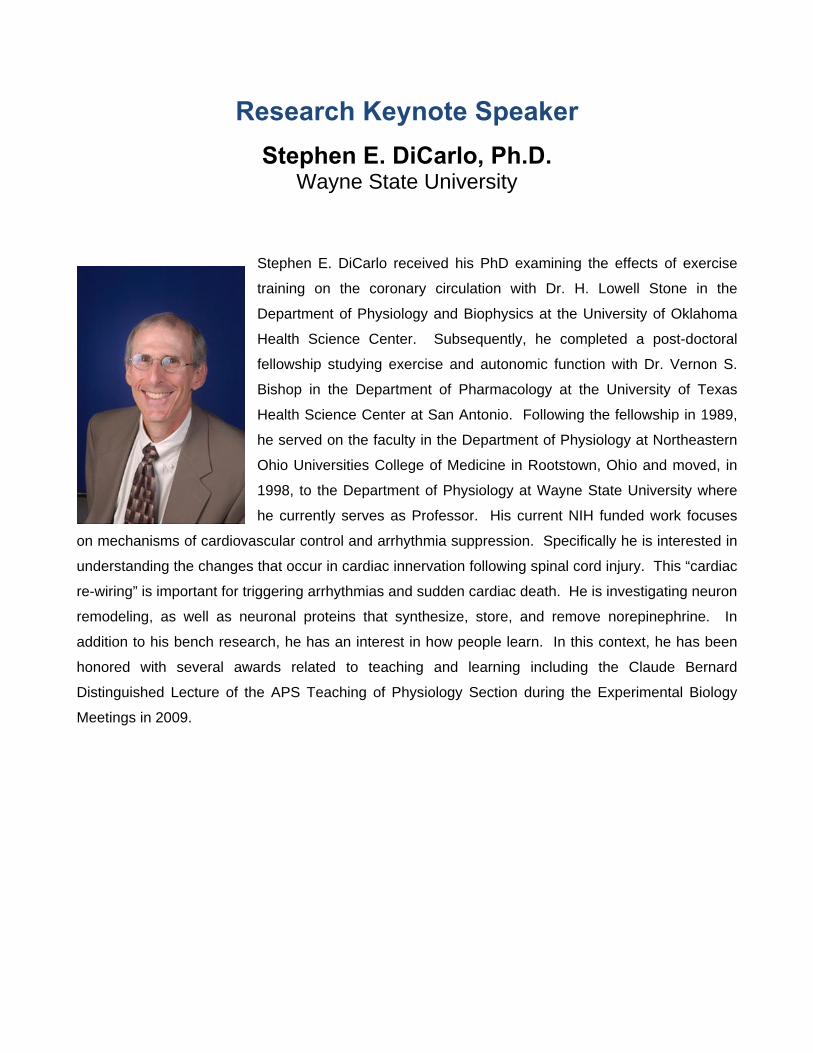
Research Keynote Speaker Stephen E. DiCarlo, Ph.D.
Wayne State University
Stephen E. DiCarlo received his PhD examining the effects of exercise
training on the coronary circulation with Dr. H. Lowell Stone in the
Department of Physiology and Biophysics at the University of Oklahoma
Health Science Center. Subsequently, he completed a post-doctoral
fellowship studying exercise and autonomic function with Dr. Vernon S.
Bishop in the Department of Pharmacology at the University of Texas
Health Science Center at San Antonio. Following the fellowship in 1989,
he served on the faculty in the Department of Physiology at Northeastern
Ohio Universities College of Medicine in Rootstown, Ohio and moved, in
1998, to the Department of Physiology at Wayne State University where
he currently serves as Professor. His current NIH funded work focuses
on mechanisms of cardiovascular control and arrhythmia suppression. Specifically he is interested in
understanding the changes that occur in cardiac innervation following spinal cord injury. This “cardiac
re-wiring” is important for triggering arrhythmias and sudden cardiac death. He is investigating neuron
remodeling, as well as neuronal proteins that synthesize, store, and remove norepinephrine. In
addition to his bench research, he has an interest in how people learn. In this context, he has been
honored with several awards related to teaching and learning including the Claude Bernard
Distinguished Lecture of the APS Teaching of Physiology Section during the Experimental Biology
Meetings in 2009.

State of the American Physiological Address
Irving H. Zucker, Ph.D. Past-President of the American Physiological Society
University of Nebraska Medical Center
Irving H. Zucker, Ph.D. is the Theodore F. Hubbard Professor of
Cardiovascular Research and Chairman of the Department of Cellular and
Integrative Physiology at the University of Nebraska Medical Center in
Omaha, Nebraska. He has been Chairman since 1989. Dr. Zucker received
his Ph.D. from New York Medical College in 1972. He continued his post
doctoral training at the University of Nebraska Medical Center where he
became a faculty member in 1973. Dr. Zucker has been involved in studies
related to the neural regulation of cardiovascular function over the past 35
years. His studies have revolved around cardiovascular reflex control of sympathetic nerve activity in
animal models of chronic heart failure. These investigations focus on the role of central mediators of
sympathetic nerve activity such as angiotensin II and nitric oxide. Dr. Zucker has published over 180
papers in this field and this work has been continuously funded by the National Institutes of Health
and the American Heart Association. He serves on the editorial boards of 10 journals. In addition to
his research, Dr. Zucker is active in administrative activities for the American Physiological Society
and the American Heart Association. He is a member of the National Research Committee of the
American Heart Association. He is the Past-President of the Association of Chairs of Departments of
Physiology and is the Past-President-of the American Physiological Society.

Spotlight
on Young
Investigators

Young Investigator
Matthew C. Zimmerman, Ph.D. University of Nebraska Medical Center
Dr. Matthew C. Zimmerman earned his BS degree in Biology from Marian
College (now Marian University) in Fond du Lac, WI, in 1999. He
attended graduate school at the University of Iowa and in 2004 earned
his PhD degree in Anatomy and Cell Biology under the mentorship of Dr.
Robin Davisson. Dr. Zimmerman’s graduate research, which was funded
by an American Heart Association Predoctoral Fellowship, was aimed at
understanding the role of reactive oxygen species in the central nervous
system in the development of angiotensin II-dependent hypertension.
After completing his doctoral studies, Dr. Zimmerman became a
postdoctoral research fellow in Dr. Larry Oberley’s laboratory in the Free
Radical and Radiation Biology Program at the University of Iowa. During
this time, Dr. Zimmerman was funded by a NIH National Research Service Award while he studied the
role of mitochondrial oxidants and antioxidants in the pathogenesis of amyotrophic lateral sclerosis
(ALS, aka Lou Gehrig’s disease).
In July 2007, Dr. Zimmerman joined the faculty in the Department of Cellular and Integrative
Physiology at the University of Nebraska Medical Center (UNMC) as an Assistant Professor. His lab’s
current research is focused on understanding the role of mitochondria-localized oxidants and
antioxidants in mediating angiotensin II signaling in central neurons. His research is currently funded
by NIH-sponsored Centers of Biomedical Research Excellence (COBRE) grants awarded to the
Redox Biology Center at the University of Nebraska – Lincoln and to the Nebraska Center for
Nanomedicine at UNMC. In addition, Dr. Zimmerman receives funding from an American Heart
Association Scientist Development Grant.
Dr. Zimmerman has received several awards including the University of Iowa Graduate Deans’
Distinguished Dissertation Award, a Merck New Investigator Award from the American Heart
Association Council for High Blood Pressure Research, a Procter & Gamble Professional Opportunity
Award from the American Physiological Society Neural Control & Autonomic Regulation Section, and
a Young Investigator Award from the Society for Free Radical Biology and Medicine. Dr. Zimmerman
has authored several manuscripts published in a variety of scientific journals including, Circulation
Research, Hypertension, Journal of Neurochemistry, Cell, Cardiovascular Research, and the
American Journal of Physiology.

POSTER Y-1 ANGIOTENSIN II INTRA-NEURONAL SIGNALING: ROLE OF MITOCHONDRIA-LOCALIZED OXIDANTS AND ANTIOXIDANTS Shumin Li, Rui-Fang Yang, Jing-Xiang Yin, Harold D. Schultz, Matthew C. Zimmerman
Cellular and Integrative Physiology, University of Nebraska Medical Center, Omaha, NE
Reactive oxygen species, particularly superoxide radicals (O2
-), have been identified as key signaling intermediates in angiotensin II (AngII)-induced neuronal activation and sympathoexcitation associated with hypertension and heart failure. Although NADPH oxidase has been identified as a primary source of O2
- in AngII-stimulated neurons, additional sources including mitochondria have been mostly overlooked. Previous data show that overexpression of the mitochondria-targeted O2
- scavenging enzyme manganese superoxide dismutase (MnSOD) in the brain inhibits the central AngII-induced pressor response, thus suggesting a role for mitochondrial O2
- in AngII-dependent intra-neuronal signaling. Here, we tested the hypothesis that AngII increases O2
- levels in neuron mitochondria, which in turn, mediate AngII intra-neuronal signaling and that overexpression of mitochondria-localized antioxidants inhibits AngII signaling. Using the O2
- sensitive, mitochondria-targeted fluorogenic probe MitoSOX Red and confocal microscopy, we measured mitochondrial O2
- levels in catecholaminergic (CATH.a) neurons before and after AngII (100nM) stimulation. AngII significantly increased MitoSOX fluorescence intensity (4.3 ± 0.3 fold increase; P<0.05 vs. baseline fluorescence), indicating an increase in mitochondrial-localized O2
-. Adenovirus-mediated overexpression of the antioxidants MnSOD (AdMnSOD) or CuZnSOD (AdCuZnSOD) markedly attenuated the AngII-induced increase in MitoSOX fluorescence (2.4 ± 0.2 and 3.3 ± 0.2 fold increase, respectivelyl; P<0.05 vs. non-infected control neurons). Considering MnSOD is efficiently transported into mitochondria, decreased levels of mitochondrial-localized O2
- in MnSOD overexpressing neurons is not all that surprising. In contrast, CuZnSOD is commonly thought of as the cytoplasmic isoform of SOD, and thus reduction of mitochondrial-localized O2
- in CuZnSOD overexpressing neurons was unexpected. To further investigate this observation, we measured CuZnSOD protein and activity levels in mitochondria isolated from AdCuZnSOD-infected neurons and detected expression of this antioxidant in mitochondria. Interestingly, MnSOD and CuZnSOD overexpression similarly inhibited AngII-induced activation of calcium/calmodulin kinase II (CaMKII). Furthermore, the well-characterized AngII-induced inhibition of potassium current (IKv) in neurons (45 ± 4% IKv inhibition) was significantly blunted in neurons overexpressing MnSOD (9 ± 3% IKv inhibition; p<0.05 vs. control) or CuZnSOD (11 ± 3% IKv inhibition; p<0.05 vs. control). These data indicate that mitochondria-produced O2
- mediates, at least in part, AngII intra-neuronal signaling and suggest that MnSOD and CuZnSOD overexpression similarly inhibit this signaling due to their presence in mitochondria.

Young Investigator
Sonia M. Rocha-Sanchez, Ph.D. Creighton University
Dr. Sonia M. Rocha-Sanchez received a B.S. degree from Acre
Federal University in Acre state, Brazil, MS degree from Viçosa
Federal University in Minas Gerais state, Brazil, and a Ph.D.
degree from the Campinas State University, São Paulo, Brazil &
University of Granada, Spain. Following a one year postdoctoral
training in the Creighton University Osteoporosis Research
Center and an NIH postdoctoral fellowship at the Biomedical
Sciences Department, she joined the faculty of the School of
Dentistry in 2006. Dr. Rocha-Sanchez currently serves as the
course director and lecturer for Dental Physiology, besides teaching in Oral Histology &
Embryology, and Cell Biology. Dr. Rocha-Sanchez’s laboratory is primarily interested on
inner ear homeostasis and regeneration of inner ear sensory hair cells.

POSTER Y-2 DIGGING DEEP INTO THE CELLS' POCKET: INSIGHTS FROM THE POCKET PROTEINS FAMILY IN INNER EAR HAIR CELL REGENERATION Sonia M. Rocha-Sanchez1, Joseph M. Miller1, Ekene Nwoye1, Edward Walsh2, Joann McGee2 1. Creighton University School of Dentistry, Omaha, NE 2. Boys Town National Research Hospital, Omaha, NE
The development of the mammalian inner ear depends upon a myriad of irreversible morphogenetic events, culminating with the formation of the cochlea and its epithelial layers of quiescent sensory hair cells (HC) and non sensory supporting cells (SC). Unlike lower vertebrates, mammalian HCs only proliferate during embryogenesis, then exit the cell cycle, differentiate and become functionally mature. Adult mammalian HCs do not proliferate and HC death leads to irreversible neurosensory hearing loss and balance impairment. Unveiling useful mechanisms of cell cycle regulation may offer the tools necessary to generate new cells out of remaining ones, thus providing the cellular basis to induce the production of new HC in the mammalian inner ear. Recent advances have provided proof of principle for two sets of therapies: the use of the cyclin system or pocket protein gene (Rb1) to promote proliferation, and the effectiveness of Atoh1 to induce transdifferentiation (TD) SC into HC. Combined, these two approaches can mimic the ability of lower vertebrates to regenerate HC. However, beyond the proof of principle, current attempts to regulate cell cycle through genetic ablation of Rb1 are not likely to safely repopulate lost HC and SC. The retinoblastoma family of cell cycle genes and proteins (herein called pRB family), composed of Rb1, Rbl1 (p107), and Rbl2 (p130), constitutes a central node controlling G1 to S phase transition in proliferating cells and cannot be bypassed without a pathogenic cost. Compared to other tissues, the biochemical and molecular pathways of the pRBs in the inner ear are relatively unexplored. Over the last three years, our laboratory have acquired experimental evidences on the role of all three pocket proteins in the inner ear and its importance to both HC and SC cell cycle control, progression, and maintenance of their post-mitotic quiescence. Results obtained from our studies are prone to provide insights on the therapeutic applicability of pRB manipulation in SC proliferation and HC regeneration. Financial Support: This work is supported in part by NIH/COBRE/NCRR P20 RR018788 and NIH/NIDCD 1R03DC009989-01A2.
Nebraska Physiological
Society
Poster Session
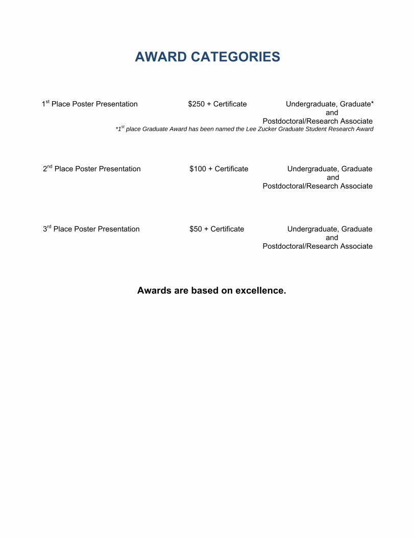
AWARD CATEGORIES
1st Place Poster Presentation $250 + Certificate Undergraduate, Graduate*
and Postdoctoral/Research Associate
*1st place Graduate Award has been named the Lee Zucker Graduate Student Research Award
2nd Place Poster Presentation $100 + Certificate Undergraduate, Graduate
and Postdoctoral/Research Associate
3rd Place Poster Presentation $50 + Certificate Undergraduate, Graduate
and Postdoctoral/Research Associate
Awards are based on excellence.

Undergraduate Posters
Poster U-1 through U-4 to be
considered for the poster award.

POSTER U-1 CENTRAL ANGIOTENSIN-(1-7) ENHANCES BAROREFLEX GAIN IN RABBITS WITH CHRONIC HEART FAILURE Sumit Kar, Pam Curry, Irving H. Zucker Department of Cellular and Integrative Physiology, University of Nebraska Medical Center, Omaha, NE
In chronic heart failure (CHF), arterial baroreflex function is impaired in part by activation of the central renin-angiotensin system. Elevated Angiotensin II (Ang II) has been associated with sympatho-excitation in CHF. A metabolite of Ang II, Ang-(1-7), has been shown to exhibit cardiovascular effects that are in opposition to that of Ang II. However, the action of Ang-(1-7) on sympathetic outflow and baroreflex function is not well understood, especially in CHF. The aim of this study was to determine the effect of intracerebroventricular (ICV) infusion of Ang-(1-7) on baroreflex control of heart rate (HR) and baseline mean arterial pressure (MAP) and HR in rabbits with CHF. We hypothesized that central Ang-(1-7) would improve baroreflex control in CHF. ICV cannulae were implanted and Ang-(1-7) (2 nmol/ 1 μl/ hour) or artificial cerebrospinal fluid (1 μl/ hour) was infused by an osmotic mini-pump for 4 days in sham and pacing-induced CHF rabbits (n=5-6/ group). Experiments were then carried out in the conscious state during which recordings of baseline HR and MAP as well as measurement of baroreflex changes in HR elicited by phenylephrine and sodium nitroprusside were made. CHF rabbits showed elevated resting HR (252.2±8.9 bpm vs. 193.2±6.6 bpm, P<.05) and depressed baroreflex gain (2.5±0.4 bpm/ mm Hg vs. 5.4±0.5 bpm/ mm Hg, P<.05). Ang-(1-7) treatment reduced HR in CHF rabbits (216.4±7.6 bpm vs. 252.2±8.9 bpm, P<.05) and increased baroreflex gain (7.4±1.5 bpm/mm Hg vs. 2.5±0.4 bpm/ mm Hg, P<.05). The results show that central Ang-(1-7) modulates baroreflex control in CHF by lowering minimum heart rate and increasing maximum gain. These data suggest that the modulation of baroreflex function by Ang-(1-7) may be due to increased vagal tone. Further experiments are necessary to determine the autonomic components of this effect.
POSTER U-2 LYSOPHOSPHATIDIC ACID (LPA) RECEPTOR ROLES IN LPA AND SERUM STIMULATION OF LUNG FIBROBLAST PROLIFERATION Bridget A. Leuschen, Nancy A. Schulte, Myron L. Toews. Pharmacology and Experimental Neuroscience, University of Nebraska Medical Center, Omaha, NE
Both LPA and serum stimulate lung fibroblast proliferation and contraction, important components of wound healing and of the pathologic airway remodeling in fibrotic lung diseases. LPA and its LPA1 receptor have been identified as critical mediators of pulmonary fibrosis. We previously showed that the LPA1 agonist NAEPA and the LPA3 agonist OMPT stimulated collagen gel contraction by HFL-1 human lung fibroblasts and that the LPA1+3 antagonist Ki16425 blocked contractions to both LPA and serum (FASEB J 22, A728.9, 2008). The present studies tested these same agents on HFL-1 proliferation. HFL-1 cells were serum starved, stimulated for 24 hr with various agents, and [3H]thymidine incorporation was assessed. Stimulation by LPA was mimicked by both NAEPA and OMPT and was completely blocked by Ki16425. Thus LPA1 and/or LPA3 receptors mediate LPA stimulation of HFL-1 proliferation. In contrast, Ki16425 inhibited stimulation by serum by only about 40%. Though LPA is the major serum component for fibroblast contraction, it makes a smaller but significant contribution to serum stimulation of fibroblast proliferation. Supported by an ASPET SURF Award to B.A.L.

POSTER U-3 BASIC BIOMECHANICAL PARAMETERS IN PATIENTS WITH PERIPHERAL ARTERIAL DISEASE: A CRITICAL REVIEW OF THE LITERATURE Neil B. Huben1, Sara A. Myers1, MS, Nicholas Stergiou1,2, PhD, Iraklis I. Pipinos 2, MD, Jason M. Johanning2, MD 1University of Nebraska at Omaha, Omaha, NE, 2University of Nebraska Medical Center, Omaha, NE
Peripheral arterial disease (PAD) is a manifestation of atherosclerosis in the lower extremities which significantly reduces arterial blood flow causing muscle ischemia during physical activity and leading to abnormal gait. Abnormal gait can be defined according to temporal and spatial parameters. The current study critically reviewed and summarized the data from thirty-five articles retrieved from Pubmed pertaining to basic gait parameters in PAD patients. Previous studies have explored the effects of PAD on gait speed, cadence, and stride length. The current study concluded that PAD results in significantly altered gait compared to controls during ambulation. The majority of papers examining gait parameters in PAD patients and controls documented that PAD patients have significantly decreased gait speed, cadence, and stride length. There is a paucity of data examining gait parameters as compared to (a) rudimentary measurement of initial and absolute claudication times and distances and (b) subjective measurements of the Walking Impairment Questionnaire (WIQ). The baseline gait parameters examined in this study suggest the presence of ambulatory abnormalities in PAD patients; however they provide limited pathophysiological explanations. Future studies should implement the use of advanced biomechanical analysis to overcome current limitations in order to further understand the underlying neuromuscular mechanisms associated with PAD.
POSTER U-4
THE EFFECT OF A SYNTHETIC ESTROGEN, DES, ON THE MALE REPRODUCTIVE TRACT IN THE BROWN NORWAY, ACI AND ACI.BN-EMCA4A CONGENIC RAT STRAINS
Tiffany L Bohlender1, Scott Kurz2, Ningxia Lu1, Racheal Slattery1, Deb Clopton1, James D Shull2 and Andrea S Cupp1 University of Nebraska-Lincoln, Lincoln, NE1 and University of Nebraska Medical Center, Omaha, NE2
Previous research has demonstrated that the ACI rat strain develops mammary tumors when treated with estradiol-17β: however, Brown Norway (BN) rats are resistant to developing mammary tumors. The regions of the genome that contribute to E2-induced mammary tumors have been mapped as Emca3-9. The region Emca4a is orthologous to a portion of the human genome that was recently correlated to breast cancer risk (Schaffer 2006). Interestingly, in this same region of the genome, a QTL conveying estrogen-induced testicular atrophy was mapped in recombinant inbred rat strains (Tachibana 2006). The current study was to investigate how a synthetic estrogen, DES, would affect estrogen-induced testicular atrophy in the ACI, BN and ACI.BN-Emca4a congenic rat strain (Emca4a), which carries the resistant mammary cancer BN alleleles over this region of the genome as well as possibly conferring the testicular atrophy QTL mapped to this same location. Male rats were treated at 9 weeks of age with DES for 12 weeks via an implant while controls received no DES in their implant. After the 12 week treatment, rats were euthanized and testis, epididymal and seminal vesicle tissue were collected from each rat, weighed and stored in Bouin’s for embedding. Tissue was embedded in paraffin, sectioned and stained with hemotoxylin and eosin. All testis, epididymal and seminal vesicle weights were suppressed in DES treated animals compared to their controls and there was no difference in effect among rat strains. Therefore, unlike estrogen induced mammary tumors, DES induced testicular atrophy was not affected by rat strain. Although testis weight was not affected by strain, a difference was observed, when testicular morphology was examined, with the BN rat strain being more resistant to depletion of germ cells from the seminiferous tubules in the testis compared to either the ACI or Emca4a strains. Similar effects were not seen in the epididymis since all DES treated rat strains had less sperm, more mesenchymal tissue between ductal structures, and increased layers of epithelial cells lining the ducts compared to control tissue. Therefore, even though testicular hypertrophy is not different when BN, ACI and Emca4a male rats are treated with DES; the BN strain are resistant to testicular germ cell depletion. Thus, BN germ cells may be able to genetically withstand pharmacologic levels of synthetic estrogens unlike the ACI or Emca4a strains.

Graduate
Posters
Poster G-1 through G-16 to be
considered for the poster award.
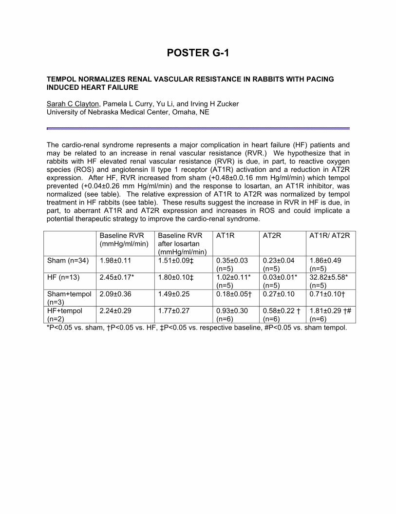
POSTER G-1 TEMPOL NORMALIZES RENAL VASCULAR RESISTANCE IN RABBITS WITH PACING INDUCED HEART FAILURE Sarah C Clayton, Pamela L Curry, Yu Li, and Irving H Zucker University of Nebraska Medical Center, Omaha, NE
The cardio-renal syndrome represents a major complication in heart failure (HF) patients and may be related to an increase in renal vascular resistance (RVR.) We hypothesize that in rabbits with HF elevated renal vascular resistance (RVR) is due, in part, to reactive oxygen species (ROS) and angiotensin II type 1 receptor (AT1R) activation and a reduction in AT2R expression. After HF, RVR increased from sham (+0.48±0.0.16 mm Hg/ml/min) which tempol prevented (+0.04±0.26 mm Hg/ml/min) and the response to losartan, an AT1R inhibitor, was normalized (see table). The relative expression of AT1R to AT2R was normalized by tempol treatment in HF rabbits (see table). These results suggest the increase in RVR in HF is due, in part, to aberrant AT1R and AT2R expression and increases in ROS and could implicate a potential therapeutic strategy to improve the cardio-renal syndrome. Baseline RVR
(mmHg/ml/min) Baseline RVR after losartan (mmHg/ml/min)
AT1R AT2R AT1R/ AT2R
Sham (n=34) 1.98±0.11 1.51±0.09‡ 0.35±0.03 (n=5)
0.23±0.04 (n=5)
1.86±0.49 (n=5)
HF (n=13) 2.45±0.17* 1.80±0.10‡ 1.02±0.11* (n=5)
0.03±0.01* (n=5)
32.82±5.58* (n=5)
Sham+tempol (n=3)
2.09±0.36 1.49±0.25 0.18±0.05† 0.27±0.10 0.71±0.10†
HF+tempol (n=2)
2.24±0.29 1.77±0.27 0.93±0.30 (n=6)
0.58±0.22 † (n=6)
1.81±0.29 †# (n=6)
*P<0.05 vs. sham, †P<0.05 vs. HF, ‡P<0.05 vs. respective baseline, #P<0.05 vs. sham tempol.

POSTER G-2 ANGIOTENSIN-CONVERTING ENZYME 2 ATTENUATES THE ANGIOTENSIN II-INDUCED UPREGULATION OF ANGIOTENSIN II TYPE 1 RECEPTOR IN CATH.A NEURONS Liang Xiao, Lie Gao, Irving H Zucker Department of Cellular and Integrative Physiology, University of Nebraska Medical Center, Omaha, NE
Hyperactivity of sympathetic outflow in chronic heart failure (CHF) is associated with an angiotensin II (AngII)-induced upregulation of AngII type 1 receptor (AT1R) in pre-sympathetic neurons, while a decrease of Angiotensin-Converting Enzyme 2 (ACE2) has been demonstrated in these neurons. ACE2 degrades AngII and forms angiotensin (1-7) (Ang (1-7)), which binds to the mas receptor and possesses vasodilator activity in many tissues. In this study, we hypothesized that ACE2 normalizes the upregulation of AT1R induced by AngII. CATH.a neurons were infected with adenoviral vectors (1 MOI) encoding ACE2 or GFP as a control. Two days later, the neurons were pretreated with either the mas receptor blocker A779, the AT1R blocker losartan or vehicle for 30 min, then stimulated with AngII (100 nM) or vehicle for 6 hours. AT1R expression in cell lysates was measured by Western blotting analysis. In non-infected and AdGFP-infected neurons, AngII increased the AT1R expression, which was inhibited by losartan. However, this effect was normalized in neurons overexpressing ACE2 (See table below). A779 did not alter the effects of AngII and ACE2 overexpression in CATH.a neurons. These data suggest that ACE2 attenuates the AngII-induced upregulation of AT1R in neurons by degradation of AngII and not by Ang (1-7) at least via a mas receptor pathway.
Table. Relative AT1R expression in CATH.a neurons treated with Vehicle or AngII
Control
+ Veh.
Control
+ AngII
AdGFP
+ Veh.
AdGFP
+ AngII
AdACE2
+ Veh.
AdACE2
+ AngII
AT1R
/GAPDH 1 1.47±0.05 * 0.94±0.07 1.41±0.10 * 0.96±0.06 1.01±0.10 †
Values are mean±SEM, * p<0.05 compared to control + veh. group, † p<0.05 compared to AdGFP + AngII group, n=4

POSTER G-3 NANOTECHNOLOGY-DRIVEN DELIVERY OF ACTIVE COPPER ZINC SUPEROXIDE DISMUTASE TO NEURONS Erin Rosenbaugh1, James W. Roat1, Ruifang1 Yang, Elena V. Batrakova2, Alexander V. Kabanov2, Matthew C. Zimmerman1
Department of Cellular and Integrative Physiology1 and the Department of Pharmaceutical Sciences2, University of Nebraska Medical Center, Omaha, NE
Dysregulation of angiotensin II (AngII)-dependent central neural mechanisms and an increase in superoxide (O2
●-) in central neurons is implicated in cardiovascular diseases, such as heart failure and hypertension. Adenoviral-mediated overexpression of the O2
●- scavenging enzyme copper/zinc superoxide dismutase (CuZnSOD) attenuates AngII-induced cardiovascular responses. However, due to toxicity and the inability of adenovirus to target the brain, the therapeutic potential of adenovirus-delivered CuZnSOD is limited. To address these limitations, we developed a nanotechnology-driven delivery system using a polyion complex micelle with a polyethylene glycol (PEG) corona and a polyethyleneimine (PEI) core which, via electrostatic binding, encapsulates CuZnSOD protein (CuZnSOD nanozyme). We hypothesize that CuZnSOD nanozyme delivers active CuZnSOD to central neurons and inhibits the AngII-induced increase in O2
●-. Confocal microscopy images of catecholaminergic CATH.a neurons treated with rhodamine-labeled CuZnSOD nanozyme or pure CuZnSOD protein revealed that the nanozyme increases neuronal uptake of CuZnSOD. Immunohistochemical staining for CuZnSOD confirmed a significant (p<0.05) increase in neuronal uptake of CuZnSOD nanozyme (331.5 ± 37.3 arbitrary fluorescence units (AFU)) compared to PEG-SOD (262.1 ± 15.0 AFU), pure CuZnSOD (195.8 ± 20.7 AFU), or non-treated (193.0 ± 24.3 AFU) CATH.a neurons. The AngII-induced increase in O2
●-, as measured by DHE fluorescence, was markedly attenuated in CuZnSOD nanozyme-treated CATH.a neurons compared to pure CuZnSOD-treated neurons and non-treated neurons. Electron paramagnetic resonance (EPR) studies confirmed that AngII significantly increases (p<0.05) the concentration of O2
●- in CATH.a neurons (6.2 + 0.28 µM O2●-
per 106 cells) compared to basal levels (4.9 + 0.32 µM O2●- per 106 cells), and that CuZnSOD
nanozyme significantly inhibits this response (4.2 + 0.28 µM O2●- per 106 cells). Together, these
data indicate that CuZnSOD nanozyme delivers biologically functional enzyme to CATH.a neurons. Importantly, the PEI-PEG polymer alone did not induce neuronal toxicity. However, pure CuZnSOD, PEG-SOD, and CuZnSOD nanozyme did induce modest but significant toxicity after 6 hour treatment (14.9 + 4.8%; 26.9 + 4.5%, 25.2 + 6.1%, respectively; p<0.05 vs. non-treated neurons). In conclusion, nanomedicine-based delivery of CuZnSOD to neurons inhibits AngII intra-neuronal signaling and may provide a new therapeutic strategy for AngII-dependent cardiovascular disease associated with increased O2
●- production in the brain.
POSTER G-4 HEMODYNAMIC EFFECTS OF CENTRAL ANGIOTENSIN TYPE 2 RECEPTOR STIMULATION IN CONSCIOUS NORMAL RATS Khang Le, Wei Wang, Irving H. Zucker, Lie Gao Department of Cellular & Integrative Physiology, University of Nebraska College of Medicine Omaha, NE
In our previous studies, we have demonstrated that, activation of Angiotension type 2 receptors (AT2R) in the RVLM of normal rats evoked a sympatho-inhibition and hypotension. In addition, we also found a significant down regulation of AT2R in the RVLM and the critical role of this change in AT2R expression in sympatho-excitation in chronic heart failure (CHF). In the current experiment, we observed the effects of intracerebroventricular (IVC) infusions (Micro-osmotic pump, 1007D, alzet) of Compound 21 (C21), the first selective non-peptide AT2R agonist, on the arterial blood pressure (BP), heart rate (HR), and norepinephrine (NE) excretion in conscious normal rats. A radiotelemetry device (model TA11PA-C40, Physiotel, Data Sciences International) was implanted for the measurement of BP and HR in the conscious state. The rats were put in metabolic cage to collect urine. Measurement of urinary NE excretion was done using a Norepinephrine Enzyme Immunoassay kit (Labor Diagnostika Nord GmbH & Co KG). At the end of experiment, the rats were euthanized and AT1R, AT2R, nNOS, and gp91 protein concentrations were measured in the cortex, SFO, PVN, RVLM, and NTS. We found that (1) chronic microinfusion of C21 decreased systolic pressure, diastolic pressure, and mean arterial pressure; (2) the NE concentration in urine and NE excretion from urine also were decreased in the C21 treated rats; (3) C21 down-regulated AT1R protein expression, up-regulated AT2R and nNOS protein expression in the subfornical organ (SFO). These results suggest that activation of central AT2R evoked a hypotension through sympatho-inhibition mechanisms.
POSTER G-5 ROLE OF PKC IN NAD(P)H OXIDASE ACTIVATION INDUCED BY TYPE 1 DIABETES IN RAT RENAL MEDULLARY THICK ASCENDING LIMB Jing Yang, Pascale H. Lane, Jennifer S. Pollock, Pamela K. Carmines. University of Nebraska College of Medicine, Omaha, NE, and Medical College of Georgia, Augusta, GA.
Type 1 diabetes (T1D) accelerates O2
•– production and stimulates protein kinase C (PKC) activation in the renal medullary thick ascending limb (mTAL). As NAD(P)H oxidase is the main source of O2
•– production in the normal mTAL, we hypothesized that T1D activates NAD(P)H oxidase in the mTAL and that this event is PKC-dependent. mTAL suspensions were prepared from rats with streptozotocin (STZ)-induced T1D (20-25 days after onset; blood glucose: 347 ± 20 mg/dl) and from Sham (vehicle-treated) rats. O2
•– production (lucigenin chemiluminescence) was higher in mTAL suspensions from STZ rats (2568 ± 814 RLU/sec/mg protein) than Sham rats (430 ± 31 RLU/sec/mg protein; P<0.05). The NAD(P)H oxidase inhibitor apocynin (100 μM) decreased O2
•– production by STZ mTALs to 544 ± 218 RLU/mg/mg protein, a value that did not differ from untreated or apocynin-treated mTALs from Sham rats. NAD(P)H oxidase activity in mTAL homogenates, measured by lucigenin chemiluminescence in the presence of 100 μM exogenous NADPH, was higher in STZ (27.6 ± 5.5 RLU/sec/µg protein) than in Sham (11.6 ± 1.1 RLU/sec/µg protein; P<0.05). Pan-specific PKC inhibition (1 µM calphostin C) and selective PKC inhibition (10 μM rottlerin) significantly reduced NAD(P)H oxidase activity in both Sham and STZ mTAL homogenates. Calphostin C-sensitive NAD(P)H oxidase activity was greater in STZ homogenates (22.9 ± 4.5 RLU/sec/μg protein) than in Sham homogenates (9.5 ± 1.0 RLU/sec/μg protein; P<0.05). Similarly, rottlerin-sensitive NAD(P)H oxidase activity was 22.8 ± 5.4 and 9.1 ± 1.0 RLU/sec/μg protein in STZ and Sham mTAL homogenates, respectively (P<0.05). In contrast, neither PKCβ inhibition (50 nM indolylmaleimide) nor PKCα/β inhibition (1 μM Gö6976) altered NAD(P)H oxidase activity in Sham or STZ mTAL homogenates. In summary, STZ rats exhibited increased O2
•– production in STZ mTALs that was reversed by NAD(P)H oxidase inhibition. Furthermore, NAD(P)H oxidase activity was increased in mTALs from STZ rats that was reversed by pan-specific PKC inhibition or PKC inhibition (but not by PKCα/β inhibition). We conclude that increased O2
•– production by the mTAL during T1D involves a PKC-dependent increase in NAD(P)H oxidase activity.
POSTER G-6 ROLE OF OXIDATIVE STRESS IN ENHANCED ATP-SENSITIVE K+ CHANNEL REGULATION OF RAT RENAL AFFERENT ARTERIOLAR TONE DURING TYPE 1 DIABETES
Carmen M. Troncoso Brindeiro, Rachel W. Fallet, Pamela K. Carmines. University of Nebraska College of Medicine, Omaha, NE
We previously reported that ATP-sensitive K+ (KATP) channels contribute to the renal afferent arteriolar dilation that accompanies streptozotocin (STZ)-induced type 1 diabetes (T1D) in the rat. KATP channel activity is reportedly augmented by reactive oxygen species. Therefore, we hypothesized that the oxidative stress accompanying T1D underlies the enhanced tonic dilator impact of KATP channels in the afferent arteriole of STZ rats. Sham and STZ rats were left untreated or were chronically-treated with the antioxidant tempol (9.45 mg/day; Sham+T and STZ+T rats). After 27 days, H2O2 excretion by STZ rats (453 ± 68 nmol/day) was greater than that of Sham rats (31 ± 3 nmol/day; P<0.05). H2O2 excretion was reduced in STZ+T rats (195 ± 33 nmol/day; P<0.05 vs untreated STZ) but did not differ between Sham and Sham+T. The in vitro blood-perfused juxtamedullary nephron technique was used to quantify afferent arteriolar lumen diameter responses to the KATP blocker glibenclamide (GLIB). Afferent arteriolar diameter averaged 26.3 ± 1.3 µm in kidneys from STZ rats and 20.5 1.7 μm in Sham rats (P<0.05). GLIB did not alter arteriolar diameter in kidneys from Sham rats ( diameter in response to 30, 100, 300 μM GLIB: 0.4 ± 0.3 μm, 0.7 ± 0.5 μm, 1.3 ± 0.8 μm, respectively). In contrast, GLIB evoked a concentration-dependent constrictor response in the STZ group ( diameter in response to 30, 100, 300 μM GLIB: –1.7 ± 0.6 μm, –2.9 ± 0.5 μm, –5.6 ± 1.2 μm, respectively; each P<0.05 vs. baseline). In STZ rats, arteriolar diameter during exposure to 300 μM GLIB (20.7 ± 1.1 μm) did not differ significantly from untreated or GLIB-treated arterioles from Sham rats. Thus, GLIB reversed the afferent arteriolar dilation evident in STZ rats. Baseline afferent diameter did not differ between Sham+T and STZ+T groups, averaging 21.2 ± 1.3 and 20.4 ± 1.1 μm, respectively. Moreover, GLIB did not significantly alter afferent arteriolar diameter in kidneys from either Sham+T or STZ+T rats. Thus, chronic tempol treatment normalized afferent arteriolar diameter and prevented the contractile response to GLIB that usually accompanies STZ-induced T1D. We conclude that oxidative stress plays a role in the KATP-dependent tonic renal afferent arteriolar dilation that occurs in STZ-induced T1D.
POSTER G-7 EPIDERMAL GROWTH FACTOR RECEPTOR PHOSPHORYLATION AND DOWNSTREAM SIGNALING BY AN AQUEOUS EXTRACT OF HOG BARN DUST IN AIRWAY EPITHELIAL CELLS. P.R. Dodmane, MVSc1, N.A. Schulte, BS1, H. Band, PhD1, D.J. Romberger, MD1,2 and M.L. Toews, PhD1 1University of Nebraska Medical Center, Omaha, NE, and 2Omaha VAMC, Omaha, NE
Workers in swine confinement facilities are prone to chronic inflammatory lung disease. We previously reported that the epidermal growth factor receptor (EGFR) is required for increased IL-6 and -8 secretions on exposure to an aqueous extract of settled dust from these facilities (HDE) in cultured airway epithelial cells. Thus we hypothesized that HDE would lead to EGFR phosphorylation and downstream activation of ERK/MAPK signaling. Beas-2B human bronchial epithelial cells were treated with HDE in the absence or presence of signaling pathway inhibitors and EGFR and ERK1/2 phosphorylation were then assessed by immunoblotting. Exposure of cells to 5% HDE stimulated EGFR phosphorylation at 5 and 15 min and that was sustained up to 18 hr. EGFR phosphorylation induced by 10 ng/ml of EGF at 5 and 15 min was greater in magnitude. The EGFR tyrosine kinase inhibitor AG1478 (10 μM) prevented EGFR phosphorylation indicating autophosphorylation. The Matrix metalloproteinase (MMP) inhibitor GM6001 (25 μM) did not block HDE-induced EGFR phosphorylation. The HDE stimulation of downstream signaling was evidenced by phosphorylation of ERK1/2 upon 5 min exposure. ERK phosphorylation was inhibited by both AG1478 and the MEK inhibitor U0126 (10 μM). HDE induces both rapid and sustained EGFR phosphorylation, and ERK1/2 is in turn activated downstream of EGFR activation. Further studies to identify the MMP-independent pathway by which HDE activates EGFRs and the consequence of HDE activation of ERK1/2 may lead to new insights into disease mechanisms and possible new therapeutic approaches.

POSTER G-8 IMPROVING H2O2 DETECTION METHODS WITH ELECTRON PARAMAGNETIC RESONANCE SPECTROSCOPY Ruth Steadman, Matthew C. Zimmerman Department of Cellular and Integrative Physiology, University of Nebraska Medical Center, Omaha, NE
Aberrant angiotensin II (AngII) signaling in central neurons, which is associated with cardiovascular diseases including hypertension and heart failure, involves elevated levels of reactive oxygen species (ROS), such as superoxide (O2
●-) and hydrogen peroxide (H2O2). To date, the majority of research has focused on O2
●-, while the role of H2O2 and the relationship between these two ROS in AngII intra-neuronal signaling has been mostly overlooked. This oversight is due, in part, to the limitations associated with the current methodology available for the measurement of H2O2 in biological samples. Electron paramagnetic resonance (EPR) spectroscopy is no exception as it detects unpaired electrons (i.e. free radicals), and thus measuring H2O2, which does not contain a free electron, is virtually impossible with EPR. In the present study, we sought to improve the sensitivity of EPR spectroscopy to detect H2O2. To do so, we supplemented the EPR assay buffer with horseradish peroxidase (HRP) and acetamidophenol (AAP), which in the presence of H2O2 induces the oxidization of the cyclic hydroxylamine spin probe 1-hydroxy-3-carboxy-2,2,5,5-tetramethylpyrrolidine (CPH) to its radical form (CP●). Considering the free electron on CP● is easily detectable by EPR, we hypothesize that the newly formulated EPR assay buffer (KDD+) will increase the sensitivity of EPR spectroscopy to detect H2O2. To address this hypothesis, we performed in vitro experiments using known amounts of H2O2 and compared the CP●-dependent EPR spectrum amplitude generated in the KDD+ buffer versus normal KDD buffer. Importantly, the baseline EPR spectrum detected in the KDD+ buffer (1.2e5 ± 2.4e4 EPR arbitrary units (EAU)) was similar to that detected in normal KDD buffer (7.8e4 + 1.7e4 EAU; P>0.05). Adding 10 µM H2O2 to the KDD+ buffer significantly increased the CP●-dependent EPR signal (4.4e6 +4.2e5 EAU; P<0.05 vs. baseline KDD+ buffer), and this response was markedly attenuated when the H2O2 scavenging enzyme, catalase (500U), was introduced to the system. In contrast, 10 µM H2O2 failed to significantly increase the EPR amplitude when added the normal KDD buffer (1.0e5 +3.0e4 EAU, P>0.05 vs. baseline normal KDD buffer). Notably, similar responses were recorded with different EPR spin probes, 1-hydroxy-3-methoxycarbonyl-2,2,5,5-tetramethylpyrrolidine (CMH) and 1-hydroxy-4-phosphono-oxy-2,2,6,6-tetramethylpiperidine (PPH). Together, this data suggest that the HRP and AAP-supplemented EPR assay buffer increases the sensitivity of EPR to detect H2O2 levels. Studies are currently underway in the laboratory in which this new EPR protocol is being utilized to measure H2O2 levels in AngII-stimulated neurons.
POSTER G-9 FLT-3 LIGAND (FLT3L) EXPANDS CD11CHIGHCD11BLOW LUNG DCS FAVORING TH2 SUPPRESSION IN A MOUSE MODEL OF ASTHMA Zhifei Shao, and Devendra K Agrawal Departments of Biomedical Sciences and Internal Medicine, Creighton University School of Medicine, Omaha, NE
Rationale: Dendritic cell subsets display different functional role in regulating immune response and lead to various outcomes such as Th1 versus Th2 or regulatory versus immunity. Administration of Flt3-ligand prevents and reverses allergic airway inflammation and airway hyperresponsiveness in a mouse model. However, the underlying mechanisms are unclear. We examined and characterized the role of lung dendritic cell subsets in the therapeutic effect of Flt3-Ligand. Methods: Dendritic cells were isolated from the lungs of OVA-sensitized and challenged mice treated with Flt3-Ligand. Two populations of CD11c+ cells labeled with fluorochrome-conjugated antibodies were sorted. The ability of the purified cells to stimulate T cell proliferation and the cytokine secretion pattern by different DC subsets were examined. Also, dendritic cells were adoptively transferred in mice to examine their effect on pulmonary function. Results: Two dendritic cell populations, CD11chighCD11blow and CD11clowCD11bhigh, were identified in the lungs of naïve and OVA-sensitized and challenged mice with and without treatment with Flt3-Ligand. The expression levels of CD8α, B220, CD19, F4/80, MHC II, CCR7, CD40, PDL1, PDL2, CD80, and CD86 were distinctly different between the two DC populations. Lung CD11chighCD11blow DCs are prone to induce Th1 response. Administration of Flt3-Ligand increased the numbers of CD11chighCD11blow DCs and IL-10 secretion in the lungs of antigen-sensitized mice, and Flt3-Ligand maximized the regulatory capacity of CD11chighCD11blow lung dendritic cells. Conclusions: CD11chighCD11blow and CD11clowCD11bhigh dendritic cells have regulatory and immunogenic properties, respectively. Under the influence of Flt3-Ligand treatment in OVA-sensitized mice, CD11chighCD11blow DCs induces a Th2 suppression involving an enhanced IL-10 secretion. Funding: NIH R01HL070885

POSTER G-10 VITAMIN D ADMINISTRATION INCREASES CATHELICIDIN EXPRESSION IN EOSINOPHILS AND NEUTROPHILS AND CCR3 EXPRESSION IN EOSINOPHILS Dalia Youssef, Anshu Aggarwal, Benjamin Moore, J. Christopher Gallagher, and Devendra K. Agrawal Departments of Biomedical Sciences, Creighton University School of Medicine, Omaha, NE
Vitamin D increases the expression of the antimicrobial peptide, cathelicidin, in cultured cells. However, the in vivo effects of Vitamin D on cathelicidin in white blood cells (WBCs) and their function are unknown. Here, we examined cathelicidin expression in WBCs and CCR3 expression in eosinophils before and after one year of vitamin D supplementation. Volunteers (ages 59-90y) with vitamin D insufficiency (<20ng/ml) were recruited and administered with Vitamin D (800 – 4,400 IU/day) for 12 months. Lymphocytes, neutrophils, monocytes, and eosinophils were isolated and purified from their blood before (n=87) and after (n=47) Vitamin D administration. The mRNA expression of cathelicidin in purified leukocytes and the mRNA expression of CCR3 in eosinophils was determined by RT-PCR and quantified by densitometric analysis. There was a significant (p<0.0001) increase in cathelicidin mRNA expression in both eosinophils and neutrophils after one year of Vitamin D intake, whereas lymphocytes and monocytes did not show a significant change in cathelicidin mRNA expression. The mRNA expression of CCR3 on eosinophils was also significantly increased (p<0.05) after one year of Vitamin D administration. Upon the breakage of the FDA codes, these data will be correlated with pulmonary function and other outcome measures of the participants in this study. Since neutrophils granules store cathelicidin, it was not surprising to see an increase in cathelicidin mRNA expression in neutrophils after Vitamin D. However, Vitamin D significantly increased mRNA expression of both cathelicidin and CCR3 in eosinophils, suggesting a critical role of cathelicidin and Vitamin D in regulating eosinophil migration in asthma.

POSTER G11 EXPRESSION OF ANGIOPOIETINS AND POTENTIAL REGULATION OF TIE-2 BY TRANSCRIPTION FACTOR ELF-1 IN THE RESPIRATORY EPITHELIUM OF OVALBUMIN-SENSITIZED AND CHALLENGED MICE Toluwalope O Makinde and Devendra K Agrawal Departments of 1Biomedical Sciences, 2Internal Medicine and 3Medical Microbiology and Immunology, Creighton University School of Medicine, Omaha, NE
Tie-2 is a tyrosine kinase receptor and is activated by angiopoietin (Ang)-1, 2, 3 and 4. Tie-2 expression is usually restricted to the vasculature, where it plays a role in the maturation and quiescence of blood vessels. We have previously shown that Tie-2 is expressed in airway epithelial cells in allergic model. However, the regulatory mechanism of Tie-2 and bio-availability of angiopoietins in the airways are unclear. Here, we examined the expression of angiopoietins and transcription factors with binding site on the promoter region of Tie2- gene. Lung tissues were isolated from OVA-sensitized and -challenged mice and PBS control. Lung sections and isolated airway epithelial cells were analyzed for co-localization of Tie-2 expression on the cell membrane and nuclear staining of transcription factors using immuno-flourescence and western-blot. Expression pattern of angiopoietins and vascular endothelial growth factor (VEGF) were also determined. Presence of airway remodeling in the lung was confirmed using H&E, PAS and trichrome staining. Tie-2 expression on the cell membrane was co-localized with nuclear staining of transcription factor E74-like factor 1 (Elf-1) in the airway epithelial cells and alveolar macrophages of OVA-sensitized and challenged mice. Ang2 and VEGF levels were increased in these cells. However, Ang1 expression remained unchanged. In conclusion, extra-vascular Tie-2 expression maybe regulated by transcription factor Elf-1. Deregulation of bio-availability of Ang1 and 2 could lead to aberrant Ang2-induced Tie-2-mediated signaling and consequently contribute to exacerbation of airway hyper-responsiveness and airway remodeling in chronic asthma.

POSTER G-12 IGF-1 DEPENDENT CHANGES IN HISTONE H3 MODIFICATIONS ARE ASSOCIATED WITH AKT SIGNALING AND THE EXPRESSION OF CELL SURVIVAL AND IMMEDIATE EARLY GENES Zhufeng Yang, Jacqueline E. Smith, Taylor Yaw, Jill G. Kerl and Jennifer R. Wood Department of Animal Science, University of Nebraska-Lincoln, Lincoln, NE
Insulin-like growth factor I (IGF-1) regulates cell proliferation, survival, and steroidogenesis in the ovary and female reproductive tract. Likewise, the progression of multiple tumors has been associated with increased activity of the IGF-1 axis. Given that IGF-1 levels are elevated in obese individuals, these studies suggest an important link between obesity-induced abnormalities in female reproduction or cancer development and IGF-1 dependent regulation of cell proliferation and apoptosis. To determine if IGF-1 regulates the expression of genes associated with the cell cycle or apoptosis, HeLa cells were treated with IGF-1 for 2, 4, 8, 16, or 24 hours. After treatment, RNA was collected and quantitative PCR (QPCR) was carried out using primers against to the cell cycle genes CDC2, CDC6, and CCND1 and the pro-apoptosis genes BCL2, BIM, and BAX. IGF-1 stimulated a significant, transient increase in CCND1 and CDC6. Furthermore, BCL2 and BIM mRNA abundance was significantly decreased by IGF-1 treatment of the HeLa cells. Histone modifications at the gene promoter are associated with changes in gene expression. To explore if IGF-1 regulates histone modifications, HeLa cells were treated with IGF-1 for 15, 30, 60, or 120 minutes followed by collection of whole cell extracts and Western blot analysis. IGF-1 stimulated phosphorylation of histone H3 on serine 10 (H3S10p) in a time-dependent manner with maximum phosphorylation at 30 minutes. The phospho-serine 10 modification has been coupled to acetylation of H3 on lysine 14 (H3S10pK14ac). Western blot analysis confirmed that IGF-1 also increased phosphoacetylation of H3 with maximal stimulation at 30 minutes post-treatment. Given that the H3S10pK14ac modification is associated with the induction of immediate-early (IE) gene expression, QPCR analysis was carried out for several IE genes including JUN, FOS, MYC, and RARß. The expression profiles of JUN, IL-6, IL-8 and CASP10 were significantly increased at 2-4 hours post-IGF-1 treatment of the HeLa cells. Both histone H3 phosphorylation and phosphoacetylation have been correlated to increased activity of the kinases Erk1/2, Akt, and p38. Western blot analysis demonstrated that the IGF-1 stimulated phosphorylation of Akt and p38 mimicked the temporal pattern of H3 phosphorylation and phosphoacetylation. Conversely, IGF-1 dependent phosphorylation of Erk1/2 occurred after H3 phosphorylation and phosphacetylation (60-120 minutes). Taken together, these data demonstrate for the first time that IGF-1 stimulates changes in histone H3 phosphorylation and phosphoacetylation. Furthermore, we have demonstrated a correlation between IGF-1 induced H3 modifications, changes in the expression of genes that regulate cell proliferation and apoptosis, and activation of the Akt and p38 signaling pathways. Thus, this study has identified a novel mechanism for IGF-1 dependent regulation of cell survival which may have important implications for the function of the female reproductive tract and/or the process of tumorigenesis and neoplastic growth.

POSTER G-13 FOXP3 +Tr1 REGULATORY T CELLS AS A THERAPEUTIC ANTIDOTE FOR REVERSING AIRWAY HYPER-RESPONSIVENESS IN COCKROACH-SENSITIZED AND CHALLENGED MICE Halvor S. McGee1 and Devendra K. Agrawal1,2,3 Center for Clinical and Translational Science Departments of Biomedical Sciences1,2,3, Medical Microbiology & Immunology 2 and Internal Medicine3, Creighton University School of Medicine, Omaha, NE
Airway hyper-responsiveness (AHR), a hallmark of asthma is characterized by eosinophilia, mucus hyper-secretion, and airway inflammation. Studies have shown that disruption of immune homeostasis is orchestrated by the Th2 cytokines including IL-4, IL-5, IL-9 and IL-13. CD4+CD25+ T regulatory cells (Tregs) and CD4 +CD25- (Tr1) cells that express Foxp3 have shown to be potent modulators of immune homeostasis. Therefore, we adoptively transferred Tregs and Tr1 cells from lungs and spleens of naïve BALB/c mice into cockroach-sensitized and challenged mice, and evaluated if established AHR and airway inflammation could be reversed. Mice were sensitized with i.p. and aerosolized cockroach antigen to induce a murine asthma model. Tregs and Tr1 cells were isolated and purified by cell sorter from lungs and spleens of naïve BALB/c mice and i.v. injected into recipients. Pulmonary function in mice was examined by whole body plethsmography. The lung Treg recipients showed that AHR to methacholine was reversed and the effect lasted for at least 2 weeks and with Tr1 cells for 3 weeks. In contrast, the therapeutic effect of spleen Tregs and Tr1 cells on AHR lasted for at least 4 weeks after adoptive transfer of the cells. Both spleen and lung Tregs and Tr1 cells expressed CD3, CD25, CD62L, CD69, Foxp3, ICOS, high levels of GITR, and minimal amounts of PD-1. Spleen Tregs and Tr1 cells expressed the same phenotype, but with a dramatic increase of PD-1 in Tr1 cells. Both Tr1 cells of the lungs and spleen upregulated CD25+ and expressed the mRNA Foxp3 and neuropilin-1. The protracted suppression of AHR by Treg and Tr1 splenocytes appears to be mediated by the increased levels of PD-1 expression. Therefore, Tr1 and Tregs cells are pivotal in suppression of AHR in cockroach asthma model and could be a plausible panacea for the pathogenesis of asthma.
POSTER G-14 ROLE OF EBP1 AS A MEDIATOR OF ESTROGEN ACTION IN PRIMORDIAL FOLLICLE FORMATION Anindit Mukherjee1 and S. K. Roy1,2, Departments. of Cellular and Integrative Physiology1 and OB/GYN2, UNMC, Omaha, NE
Primordial follicle formation is the first step in mammalian females that determines the lifetime quota of oocytes available for ovulation. Estrogen has been shown to play an important role in mediating primordial follicle formation though the mechanism(s) of its action remains undefined. The objective of this study was to identify protein mediators of estrogen action in primordial follicle formation. We hypothesize that estrogen affects the expression and functions of specific proteins in perinatal ovaries to induce primordial follicle formation. The hypothesis was tested using proteomic evaluation of ovaries obtained from 15-day old fetal (E15, complete absence of primordial follicles), 8-day old postnatal (P8, primordial follicles appear for the first time) hamsters. Proteins from each group of ovarian lyastes were resolved by 2-Dimensional Gel Electrophoresis. The SDS-PAGE gels from the second dimension of separation were scanned and the images were analyzed to compare and quantify the changes in protein expression. On comparison with embryonic day 15 ovaries we found 32 proteins were upregulated in their expression, 42 were newly expressed and 81 were downregulated in their expression in postnatal day 8 ovaries. One of the selected proteins was identified by Mass spectrometric analysis to be ERBB3 binding protein (EBP1) and its expression was downregulated in postnatal day 8 (P8) hamster ovaries. EBP1 is an important transcriptional regulator of protein synthesis, which acts as corepressor of several mitogenic pathways. Spatiotemporal expression of EBP1 was examined in the neonatal and cyclic ovaries by immunofluorescence. We also found estrogen to be downregulating EBP1 expression in ovarian tissue by western blot. These results suggest that EBP1 expression is regulated by estrogen and its downregulation may play an important role in primordial follicle formation. We therefore speculate that estrogen, by downregulating EBP1 expression is activating downstream mitogenic pathways such as one mediated by ERBB3 and bringing about primordial follicle formation.

POSTER G-15 EFFECT OF SERTOLI CELL-SPECIFIC VASCULAR ENDOTHELIAL GROWTH FACTOR (VEGF) LOSS ON GONADAL MORPHOGENESIS AND FUNCTION Ningxia Lu, Debra T Clopton, William E Pohlmeier and Andrea S Cupp. Department of Animal Science, University of Nebraska-Lincoln, Lincoln NE
In last 15 years the increase in male reproductive disorders has led to the development of Testicular Dysgenesis Syndrome (TDS) which may be due to disruptions in embryonic gonadal development. VEGF is present during testis morphogenesis and is expressed by Sertoli cells and germ cells. Inhibition of VEGF disrupts both testis-specific vascular development and cord formation. We hypothesize that production of VEGF isoforms by Sertoli cells is critical for normal testis morphogenesis. Therefore, the objective of the current study was to evaluate the effects of Sertoli cell-specific production of VEGF isoforms on sex-specific vascular development and gonadal morphogenesis. We used Cre-lox technology with a DMRT1-cre mouse crossed to a floxed VEGF mouse to knockout VEGF in Sertoli cells in the testis. The Dmrt1 gene is expressed in the indifferent gonad at 10.5 dpc in precursor cells that differentiate into Sertoli and Granulosa cells. Therefore, we expected potential phenotypes in male and female Sertoli and Granulosa-cell specific knockouts. Mice from these crosses had litters and appeared to have one less mouse than controls. Thus, these mice were fertile/subfertile; however, the penetrance of the DMRT1 Cre still needs to be elucidated. Reproductive organs were collected from adult mice, and weighed (male: control n=7, knockout n=8; female: control n=9, knockout n=10). Tissue sections were stained with hematoxylin and eosin. Testosterone and estrogen levels in blood were determined by Enzyme-linked Immunosorbent Assay (ELISA). In male VEGF conditional knockout mice, the body and testes weights were significant smaller than those from the control (body; 28.52+0.48 VS 32.44+1.57 g, testes; 0.1658+0.0064 VS 0.1867+0.0064 g, P<0.05). The weights of epididymis, seminal vesicles and prostate in knockout mice tended to be smaller than those in control (0.0627+0.0046 VS 0.0768+0.0057 g, P=0.0794; 0.2324+0.0134 VS 0.2855+0.028 g, P=0.098; 0.0104+0.0031 VS 0.0234+0.0055 g, P=0.052). The testosterone concentrations in three knockout male mice were abnormal (13.83, 13.72, 10.88 ng/mL) compared to mean concentrations in control (1.18+0.40 ng/mL). Histological testis sections from two male knockout mice had a majority of seminiferous tubules at the same stage of spermatogenesis instead of a more random pattern that was in the controls. For the female group, the weight of ovaries from the knockout mice were significantly smaller than that of the control (0.0144+0.0014 VS 0.0185+0.0012 g, P<0.05). Two female knockout mice had histologically abnormal structures that were neither CL nor follicles. Furthermore, the estrogen concentrations in the knockout mice were significant lower than those in control (48.83+9.58 VS 91.53+14.75 pg/mL P<0.05). In summary, our findings suggest that removal or reduction of VEGF in Sertoli and Granulosa cells cause mice to be sub-fertile and may result in abnormal morphology in the gonad. Therefore, VEGF plays an important role in reproductive organ morphogenesis and function in mice.

POSTER G-16 TGF-ß INDUCED EXPRESSION OF CLC3 ISOFORMS IN HUMAN BLOOD EOSINOPHILS Rohit Gaurav1, Benjamin T. Moore1, Devendra K. Agrawal1,2,3 Center for Clinical and Translational Science, Departments of 1Biomedical Sciences, 2Internal Medicine and 3Medical Microbiology and Immunology, Creighton University School of Medicine, Omaha, NE
Eosinophils are major inflammatory cells involved in the pathogenesis of asthma. Their association with airway hyperresponsiveness (AHR) and airway remodeling along with TGF-ß is well established owing to their ability to migrate to the site of inflammation. In this study, we investigated the expression of CLC3 isoforms present in human blood eosinophils after stimulation with TGF-ß. Eosinophils were isolated from blood samples of normal healthy donors. Isolated eosinophils were stimulated with different doses of TGF-ß (1-100ng/ml) in vitro, in a time dependent manner (30 min- 24h). Unstimulated cells served as a control. mRNA expression analysis for CLC3 isoforms was done by reverse transcription and real time PCR using specific primers for CLC3b and CLC3e. Change in protein expression was analyzed by Western blotting. Using whole-cell patch clamping experiments, the chloride channel activity was measured by the magnitude of the chloride current on eosinophils after stimulation with TGF-ß. Real time PCR suggested that there is an increased expression of both the isoforms in human blood eosinophils after TGF-ß stimulation. CLC3b expression is considerably higher than the CLC3e expression in stimulated eosinophils. Protein expression analysis and patch clamping data showed increased CLC3 expression and increase in the chloride current respectively, following TGF-ß stimulation. The data suggest that the release of TGF-ß in asthmatic conditions may increase the CLC3 expression, thereby increasing the eosinophil migration to the site of inflammation. However, further experiments, involving asthmatic patients would give a clear picture of the mechanism of TGF-ß induced CLC3 expression and migration of eosinophils in allergic asthma.

Post-doctoral
Posters
Poster P-1 through P-16 to be
considered for the poster award.

POSTER P-1 A MURINE MODEL OF ACUTE HINDLIMB ISCHEMIA/REPERFUSION INJURY Huiyin Tu1, Paul Tran1, Yu-Long LI1,2 Department of 1Emergency Medicine, 2Cellular & Integrative Physiology, University of Nebraska Medical Center, Omaha, NE
15% of preventable combat fatalities are due to hemorrhage from extremity wounds, making emergency tourniquet one of the first-line treatments. While lifesaving, tourniquets can cause serious ischemia-reperfusion (IR) injury. Fundamental to IR injury is mitochondrial production of superoxide. We refined a murine model of acute hind limb IR to simulate the clinical use of tourniquet. Groups of C57/BL6 mice underwent an IR protocol of three hours of ischemia and four hours of reperfusion. IR was achieved by placement and release of a rubber tourniquet around the hind limb, at the greater trochanter level. Muscle blood flow (MBF) was measured using a Doppler probe. Muscle necrosis was measured using TTC stain. Superoxide production was measured using chemiluminescence and DHE staining. MnSOD protein expression and activity were measured using Western blot and a modified nitrite method. Mitochondrial respiratory chain function was measured via spectrophotometry. Significant ischemia and microcirculatory dysfunction were achieved: MBF during IR was < 10% and < 41% of baseline, respectively (n=8), accompanied by >40% necrosis (n=9). Superoxide production was increased (0.05 vs 0.13, p<.05 sham vs IR) accompanied by concomitant increases in MnSOD activity and protein expression (1.51 vs 2.26, p<0.05, sham vs IR, n=8). Mitochondrial complexes I-IV showed signs of dysfunction (Complex IIV:0.13, 0.31, 1.37, 2.76 sham vs 0.08, 0.21, 0.7, 1.97 IR, p<0.05). We have demonstrated an animal model of clinical tourniquet use. IR produces significant mitochondriopathy which is associated with elevated superoxide production and antioxidant activity. The model can be used to mechanistically evaluate antiischemic molecules in efforts to minimize the morbidity and mortality of battlefield injury.

POSTER P-2 INVOLVEMENT OF NADPH OXIDASE-DERIVED SUPEROXIDE ANION IN DIABETES-BLUNTED AORTIC BARORECEPTOR NEURON EXCITABILITY Libin Zhang1, Thai P Tran 1, Robert L Muelleman1, Yu-Long Li1, 2. Department of 1Emergency Medicine, 2Cellular & integrative Physiology, University of Nebraska Medical Center, Omaha, NE
NADPH oxidase-derived superoxide has been identified as key signaling pathway in many pathophysiological processes. Our recent study has indicated that hyperpolarization-activated cyclic nucleotide-gated (HCN) channel function is enhanced, which contributes to the blunted aortic baroreceptor (AB) neuron excitability in diabetic state. Therefore, we investigated if NADPH oxidase-derived superoxide mediates HCN channel activity and cell excitability of AB neurons in streptozotocin (STZ)-induced diabetic rats. Using western blot and lucigenin chemiluminescence method, we found that protein expression of NADPH oxidase (gp91 phox, p22phox, p40phox, p47phox, andp67 phox) and superoxide production were higher in nodose ganglia from diabetic rats than that from sham rats. In whole cell patch-clamp recording, action potential frequency of AB neurons in the diabetic rats was lower than that in the control rats (0.5 ± 0.5 vs. 4.9 ± 0.6 spikes/sec, p<0.05; AB neurons were identified by DiI staining). HCN current density of AB neurons was increased in the diabetic rats, compared with the control rats. In addition, apocynin (30 μM, a NADPH oxidase inhibitor) and tempol (1 mM, a superoxide dismutase mimetic) significantly reduced HCN current density and increased action potential frequency of the AB neurons in diabetic rats. These results indicate that elevation of intracellular NADPH oxidase-derived superoxide generation contributes to the attenuated AB neuron excitability via enhancing HCN channel activation in diabetic state.

POSTER P-3 ALCOHOL-INDUCED EXACEBATION OF ISCHEMIC BRAIN INJURY: ROLE OF NAD(P)H OXIDASE Honggang Zhao, William G. Mayhan, and Hong Sun Department of Cellular and Integrative Physiology, University of Nebraska Medical Center Omaha, NE 68198-5850
Chronic alcohol consumption exacerbates ischemic brain injury. We hypothesized that upregulated NAD(P)H oxidase during alcohol consumption may contribute to exacerbated ischemic brain injury. Sprague/Dawley rats, gp91phox -/- mice, and WT C57BL/6 mice were used. Alcohol was mixed into a liquid diet and given to rats for 8 weeks. NAD(P)H oxidase inhibitor, apocynin (7.5 mg/kg/day), was orally administrated for 4 weeks prior to the experiment. Rats and mice were subjected to middle cerebral artery occlusion (MCAO), and infarct volume, neurological score, superoxide production, and gp91phox expression were assessed at 24 hours of reperfusion. Chronic alcohol consumption produced a larger infarct volume, worse neurological score and higher superoxide production in rats. Apocynin did not alter infarct volume, neurological deficit, and superoxide production in nonalcohol-fed rats, but reduced infarct volume, improved neurological outcome, and attenuated superoxide production in alcohol-fed rats. Compared with wild-type mice, a similar infarct volume was found in gp91phox -/- mice. Upregulated gp91phox expression in parietal cortex during alcohol consumption and in border area of parietal cortex after ischemia/reperfusion was found in cells co-stained with neuronal nuclei marker. Our findings suggest that NAD(P)H oxidase may play an important role in exacerbated ischemic brain injury during chronic alcohol consumption.

POSTER P-4 SODIUM RETENTION AND VOLUME EXPANSION IN Kcnmb4 -/- MICE FED A HIGH K + DIET P. Richard Grimm, J. David Holtzclaw, Debra L. Irsik and Steven C. Sansom University of Nebraska Medical Center, Omaha, NE
Consumption of a K rich diet results in an increased ratio of K secretion to Na absorption in the distal nephron. Large, calcium-activated K channels (BK) are expressed in principal and intercalated cells (PCs and ICs) of the connecting tubule (CNT). The PCs of this segment play a role in fine tuning salt and water balance due to their sensitivity to many hormones and paracrine factors (nitric oxide (NO) and ATP) and the favorable electrochemical gradients established by high levels of Na-K-ATPase expression. BK in the ICs are a complex of pore-forming alpha and accessory beta4 subunits (BKa/ß4). However, the physiological function of BKa/ß4 in ICs has not been established. The goal of this study was to determine if mice having the ß4 subunit (gene Kcnmb4) knocked out (Kcnmb4-/-) have altered renal salt clearance when fed a K-rich diet (5% K) for 10 days. Under control diet conditions (0.6% K) no difference was detected between WT and Kcnmb4-/-. However, when Kcnmb4-/- consumed the K-rich diet they became volume expanded as determined by Hct and weight change (Hct: 45.7 ± 0.4% WT vs 35.4 ± 0.4% Kcnmb4-/-; weight change: 0.42 ± 0.03g WT vs 5.2 ± 0.2g Kcnmb4 -/-). Kcnmb4-/- also had significantly lower urine output (180 ± 7 μl/hour vs 206 ± 6 μl/hour), U V (2061 K ± 32 μmol/day vs 3265 ± 28 μmol/day), U V (188 ± 7 μmol/day vs 433 ± 13 μmol/day), Na FE (93.5 ± 3.2% vs 162.4 ± 2.9%), and FE (0.29 ± 0.03% vs 0.66 ± 0.02%) K Na compared to WT despite having similar aldosterone levels and food and water consumption. Therefore, when consuming a K rich diet, the FE per fraction of Na K absorbed was reduced dramatically in Kcnmb4-/-. The inability of Kcnmb4-/- to minimize Na absorption lead to Na and volume retention, suggesting BKa/ß4 channels of ICs play a role in increasing the amount of K secreted while minimizing the quantity of Na absorbed in mammals on a high K diet. However, it is unlikely that the ICs of the CNT are directly responsible for the increased Na reabsorption due to their low Na-K-ATPase expression. It is possible that ICs release paracrine factors such as NO or ATP that influence Na reabsorption and K secretion in neighboring PCs.

POSTER P-5 OVEREXPRESSION OF SOD PROTEINS NORMALIZES THE EXAGGERATED EXERCISE PRESSOR REFLEX IN CHRONIC HEART FAILURE RATS Han-Jun Wang, Matthew C. Zimmerman, Lie Gao, Wei-Zhong Wang, Irving H. Zucker, Wei Wang Department of Cellular and Integrative Physiology, Nebraska Medical Center, Omaha, NE
It is well established that there is the exaggerated exercise pressor reflex (EPR) in chronic heart failure (CHF) state, which is believed to contribute to the over sympatho-excitation during exercise in CHF. Accumulating evidence also shows that the exaggerated EPR in CHF might be due to the enhanced muscle mechanoreflex. However, what causes the exaggerated EPR and the enhanced mechanoreflex in CHF are not well understood. Here, we hypothesized that muscle oxidative stress in CHF contribute to the exaggerated EPR and the enhanced mechanoreflex. We used the technique of gene transfer to overexpress SOD proteins (AdSOD1 or AdSOD2, 1* 1012 particles in PBS/ml, 50 ml, Adempty as the control) in the triceps surae muscle in CHF rats using adenoviral vectors (2-4 weeks). The EPR was induced by static contraction of electrical stimulation (L4/L5) in decerebrate rats. The mechanoreflex was induced by passive stretch in decerebrate rats. Table 1 showed the effect of overexpression of SOD proteins on the EPR and mechanoreflex in CHF rats. These data indicate that muscle oxidative stress contributes to the exaggerated EPR and the enhanced mechanoreflex in CHF. Table 1. Effect of overexpression of SOD on the pressor response evoked by EPR or the mechanoreflex in CHF rats.
CHF+ CHF+ CHF+ Sham Empty AdSOD1 AdSOD2
EPR (mmHg) 20.6±2.3 32.6±2.6* 20.0±1.4# 21.2±2.1# Mechanoreflex (mmHg) 13.1±1.0 25.8±2.8* 13.6±1.1# 14.8±1.3# Values are Mean±SE, n=6-8 in each group,* P<0.05 vs. Sham, P<0.05 vs. CHF+Empty.
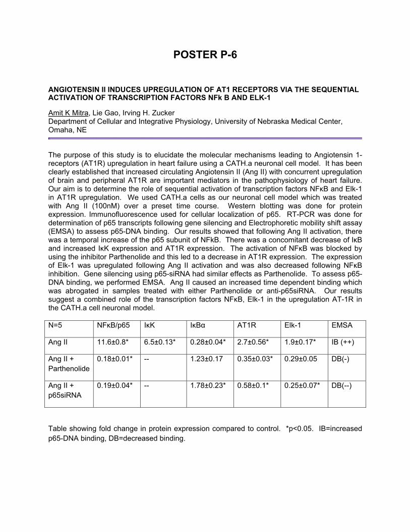
POSTER P-6
ANGIOTENSIN II INDUCES UPREGULATION OF AT1 RECEPTORS VIA THE SEQUENTIAL ACTIVATION OF TRANSCRIPTION FACTORS NFk B AND ELK-1
Amit K Mitra, Lie Gao, Irving H. Zucker Department of Cellular and Integrative Physiology, University of Nebraska Medical Center, Omaha, NE
The purpose of this study is to elucidate the molecular mechanisms leading to Angiotensin 1-receptors (AT1R) upregulation in heart failure using a CATH.a neuronal cell model. It has been clearly established that increased circulating Angiotensin II (Ang II) with concurrent upregulation of brain and peripheral AT1R are important mediators in the pathophysiology of heart failure. Our aim is to determine the role of sequential activation of transcription factors NFκB and Elk-1 in AT1R upregulation. We used CATH.a cells as our neuronal cell model which was treated with Ang II (100nM) over a preset time course. Western blotting was done for protein expression. Immunofluorescence used for cellular localization of p65. RT-PCR was done for determination of p65 transcripts following gene silencing and Electrophoretic mobility shift assay (EMSA) to assess p65-DNA binding. Our results showed that following Ang II activation, there was a temporal increase of the p65 subunit of NFkB. There was a concomitant decrease of IκB and increased IκK expression and AT1R expression. The activation of NFκB was blocked by using the inhibitor Parthenolide and this led to a decrease in AT1R expression. The expression of Elk-1 was upregulated following Ang II activation and was also decreased following NFκB inhibition. Gene silencing using p65-siRNA had similar effects as Parthenolide. To assess p65-DNA binding, we performed EMSA. Ang II caused an increased time dependent binding which was abrogated in samples treated with either Parthenolide or anti-p65siRNA. Our results suggest a combined role of the transcription factors NFκB, Elk-1 in the upregulation AT-1R in the CATH.a cell neuronal model. N=5 NFκB/p65 IκK IκBα AT1R Elk-1 EMSA
Ang II 11.6±0.8* 6.5±0.13* 0.28±0.04* 2.7±0.56* 1.9±0.17* IB (++)
Ang II + Parthenolide
0.18±0.01* -- 1.23±0.17 0.35±0.03* 0.29±0.05 DB(-)
Ang II + p65siRNA
0.19±0.04* -- 1.78±0.23* 0.58±0.1* 0.25±0.07* DB(--)
Table showing fold change in protein expression compared to control. *p<0.05. IB=increased p65-DNA binding, DB=decreased binding.
POSTER P-7 REDOX REGULATION OF L-TYPE CA2+ CHANNELS IN POST-MYOCARDIAL INFARCTION RAT HEARTS: ROLE OF THIOREDOXIN AND PROTEIN PHOSPHATASE 2A Ming-Qi Zheng, Kang Tang, Todd A. Wyatt, George J. Rozanski Department of Cellular and Integrative Physiology, University on Nebraska Medical Center, Omaha NE
Oxidative stress plays an important role in the pathogenesis of heart failure including blunted b-adrenergic contractile responsiveness and dysregulation of intracellular Ca 2+ handling. These functions are tightly controlled by L-type Ca 2+ channels (LTCC), but it is not clear what functional impact oxidative stress has on LTCC under heart failure conditions. Thus, the purpose of the present study was to identify redox mechanisms participating in pathogenic LTCC remodeling. Chronic ventricular dysfunction was induced in rats by myocardial infarction (MI) and studies were done after 6-8wks. Voltage-clamp experiments in isolated left ventricular myocytes from post-MI hearts showed that blunted responsiveness of L-type Ca 2+ current (I ) to forskolin (FSK, 1 μmol/l) was Ca,L reversed after 5 h by treatment with exogenous pyruvate (5 mmol/l), while basal I Ca,L density was similar to sham myocytes. The effect of intracellular cAMP dialysis (10 μmol/l) on I in post-MI and sham myocytes was also consisted with the response to Ca,L FSK, suggesting that generation of cAMP by adenylyl cycles was not impaired post-MI. Biochemical assays of tissue extracts showed that basal and cAMP-stimulated PKA activity were not significantly different between post-MI and sham hearts, indicating that defects downstream from PKA were responsible for impaired responsiveness of I to Ca,L FSK and cAMP. The protein phosphatase 2A (PP2A) inhibitor okadaic acid (OA, 1 μmol/l) increased I in sham myocytes to a similar level as FSK, but the OA effect was Ca,L much smaller in post-MI myocytes. However, pre-treatment of post-MI myocytes with pyruvate reversed the blunted response of I to OA. Finally, the increased Ca,L responsiveness to FSK and OA after pyruvate treatment was also blocked by inhibitors of thioredoxin reductase (auranofin, 10 nmol/l; 13-cis-retinoic acid, 1 μmol/l) implicating a functional role for the thioredoxin system. Our data show that pyruvate reverses the blunted response of I to b-adrenergic stimulation in post-MI hearts which is likely due Ca,L to hyperphosphorylation of LTCC secondary to PP2A inhibition. We postulate that the redox state PP2A is controlled by the thioredoxin system which plays an important role in the remodeling of LTCC during heart failure.
POSTER P-8 THE TRANSLOCATION OF BK CHANNEL ß1 SUBUNIT TO THE BASOLATERAL MEMBRANE OF PRINCIPLE CELLS IS MEDIATED BY ANGIOTENSIN II SIGNALING Liu L, Grimm PR, Holtzclaw D, Irsik D, Sansom SC Department of Cellular/Integrative Physiology, University of Nebraska Medical Center, Omaha, NE
Large-conductance, calcium-activated potassium channels (BK) are expressed in the apical membrane of the distal nephron of the kidney, where they mediate potassium secretion in response to a high K diet and high distal flow conditions. In normal or K-adapted conditions, the BK-a pore associates with the ancillary BK-b1 (gene: Kcnmb1) only in the apical membrane of connecting tubule cells. However, BKa and BKß1 localize on the basolateral membrane of CCD principle cells (PC) when the mice consume a Na-deficient diet. Because Kcnmb1-/- exhibit profound volume depletion when treated with a low Na diet, the localization of BK-a/b1 to the basolateral membrane probably serves to increase the driving force for Na absorption across the apical membrane. The current study investigated the hypothesis that angiotensin II (ANGII) signaling was involved in the BKß1 incorporation into the basolateral membrane of PC. For these studies, the distal nephron cell lines MDCK (PC and IC) and MDCK C7 (PC) cultured on a three dimensional system with Matrigel coated eight well chamber slides. The cells formed cysts within 3 to 5 days. The cells forming the cysts have plasma membrane polarized into functionally distinct apical and basolateral membrane domains, which face the external and internal environments, respectively. The cysts then were fixed with 4% paraformaldehyde and processed for fluorescent immuocytochemistry. MDCK C7 cells were also cultured on T-75 flasks and membrane and cytosolic proteins were isolated for western blotting analysis and biotinylation assay. Immunocytochemical analysis revealed that BKß1 has more expression on the basolateral than apical membrane when ANGII (200nM), but not aldosterone (10nM), was added to the system. Western blotting showed expression of total membrane and cytosolic BK-β1 did not change with addition of ANGII; however, biotinylated BK ß1 (apical membrane) slightly decreased (p=0.06). We conclude that BKß1 translocated from apical to basolateral membrane upon the addition of ANGII, but not with aldosterone stimulation. Incorporating BK into the basolateral membrane would increase the cellular electronegative driving force by recycling potassium, thereby compensating for the low chemical gradient for sodium entry under conditions of low luminal sodium. The ANGII-induced translocation of BKß1 to the basolateral membrane of PC may play a role in sodium absorption in collecting ducts at the condition of sodium deficiency.
POSTER P-9 ADENOVIRUS-MEDIATED GENE TRANSFER OF MN SUPEROXIDE DISMUTASE TO CAROTID BODY NORMALIZES ENHANCED CHEMORECEPTOR FUNCTION IN HEART FAILURE Yanfeng Ding1, Yu-Long Li 2, Jingxiang Yin1,Matthew C Zimmerman1,Harold D Schultz1 1 Department of Cellular & Integrative Physiology, 2Department of Emergency Medicine, University of Nebraska Medical Center, Omaha, NE
Peripheral chemoreflex sensitivity is enhanced in both clinical and experimental chronic heart failure (CHF). We have found CuZn superoxide dismutase (SOD) gene transfer to the carotid body (CB) and tempol (an SOD mimetic) reverse the enhanced chemoreceptor function in heart failure rabbits. Here we investigated the role of MnSOD in the enhanced chemoreceptor activity and function of the CB in CHF rabbits. MnSOD protein expression was lower in CHF CB compared with that in sham CBs. Ad MnSOD (1x10⁸ pfu/ml) gene transfer to the CBs significantly increased MnSOD expression in CHF CBs and reduced the baseline renal sympathetic nerve activity (RSNA) and the response of RSNA to hypoxia in the CHF rabbits. Ad MnSOD decreased the baseline single-fiber discharge from CB chemoreceptors (6.4 1.3 vs.10.5 0.9 imp/s at ~100 mm Hg PO2) and the response to hypoxia (18.4 2.1 vs. 36.7 2.2 imp/s at ~40 mm Hg PO2, p<0.05) in CHF rabbits. In addition, Ad MnSOD normalized the blunted IK in CB glomus cells from CHF rabbits (362 41 vs. 520 65 pA/pF at +70 mV, p<0.05). Ad MnSOD also normalized the blunted IK induced by angiotensin II in CB glomus cells from sham rabbits. These results suggest that decreased MnSOD in the CB may contribute to the enhanced CB chemoreceptors activity and peripheral chemoreflex function in CHF rabbits. Supported by NIH grant PO-1 HL062222
POSTER P-10 EFFECTS OF ACID SENSING ION CHANNELS ON CAROTID CHEMORECEPTOR ACTIVITY Yuhong Tang1,2, Yu Zheng2, Harold D. Schultz1
1 Department of Cellular and Integrative Physiology, University of Nebraska Medical Center, Omaha, NE, and 2 Department of Physiology, School of Preclinical and Forensic Medicine, Sichuan University, Chengdu, Sichuan, China
The carotid bodies (CBs) function as major peripheral chemoreceptors sensing changes in arterial blood oxygen, carbon dioxide, and pH in mammals. Glomus cells are generally accepted as the chemosensitive elements. The molecular mechanism of pH sensing by chemoreceptors is not clear, although it has been proposed to be mediated by a drop in intracellular pH of CBs glomus cells, inhibiting one or more types of K+ channels, leading to membrane depolarization. Acid sensing ion channels (ASICs) are neuronal voltage-insensitive cationic channels activated by extracellular protons. They belong to the ENaC/DEG (epithelial amiloride-sensitive Na+ channel and degenerin) family of ion channel. Six subunits of ASICs have been identified: 1a, 1b, 2a, 2b, 3, and 4. Recently, ASIC1 and ASIC3 were shown to be expressed in rat carotid body by quantitative PCR and immunohistochemistry. Extracellular acidosis evoked transient inward current in glomus cells that was comparable biophysically and pharmacologically to ASICs. And ASIC1a and ASIC3 were found on the primary cultured glomus cells of rat CBs. In this study, we tested the hypothesis that extracellular acidosis directly activates ASICs on CBs glomus cells and induces membrane depolarization; and as a result of acidosis stimulus, the mRNA and/or protein expression of ASICs on carotid body cells can be changed. In preliminary experiments, we confirmed that ASIC3 and ASIC4 protein are expressed in the CBs of Sprague Dawley rats by Western blot. ASIC1 and ASIC3 were shown to be expressed in rat carotid body by immunohistochemistry. In addition, the discharges of carotid sinus nerve (CSN) were recorded in the CB - CSN preparations from New Zealand rabbits in vitro. Superfusion with pH 6.8~7.0 modified Tyrode solution, the spontaneous discharges of CSN increased but could not be block by amiloride. However, lack of appropriate antibodies for ASICs in rabbit prohibits molecular analysis of rabbit CB at this time. The existing evidence suggests that although ASIC channels are expressed in the CB, they do not play a key role in acid sensing of the CB.

POSTER P-11 INCREASING MITOCHONDRIAL LEVELS OF CUZNSOD IN CATH.A NEURONS VIA ADENOVIRUS-MEDIATED GENE TRANSFER INHIBITS ANGIOTENSIN II-DEPENDENT INTRA-NEURONAL SIGNALING Shumin Li, Rui-Fang Yang, Jing-Xiang Yin, Harold D. Schultz, Matthew C. Zimmerman Department of Cellular and Integrative Physiology, University of Nebraska Medical Center, Omaha, NE
Angiotensin (Ang) II, the primary effector peptide of the renin angiotensin system, increases reactive oxygen species (ROS), such as superoxide (O2
●-), in the central nervous system, which mediates, at least in part, the development of brain-related cardiovascular diseases. Numerous studies have demonstrated that overexpression of the O2
●- scavenging enzymes copper/zinc superoxide dismutase (CuZnSOD) or manganese SOD (MnSOD) similarly inhibit AngII-dependent signaling in the brain. MnSOD is the mitochondria-localized SOD isoform, while CuZnSOD is commonly thought to reside in the cytoplasm. However, previous studies have demonstrated that a small fraction of CuZnSOD is also present in mitochondria. Here, we tested the hypothesis that overexpression of CuZnSOD via adenovirus-mediated gene transfer (AdCuZnSOD) increases mitochondrial levels of CuZnSOD and, as a result, inhibits the AngII-induced increase in mitochondria-localized O2
●-. Using mitochondria isolated from CATH.a neurons, Western blot analysis, and a native gel SOD activity assay, we observed a robust increase in CuZnSOD protein expression and activity in mitochondria isolated from AdCuZnSOD-infected neurons compared to non-infected control cells and neurons infected with the control vector, AdEmpty. To examine the biological significance of this finding, we measured AngII-induced changes in mitochondria O2
- levels in control, AdEmpty, and AdCuZnSOD-infected neurons using the O2
- sensitive, mitochondria-targeted fluorogenic probe MitoSOX Red and confocal microscopy. AngII (100 nM, 20 min) significantly increased MitoSOX Red fluorescence in control and AdEmpty-infected neurons (4.3 ± 0.3 fold increase and 4.8 ± 0.3 fold increase, respectively; P<0.05 vs. baseline fluorescence), thus indicating an increase in mitochondria-localized O2
- levels. Overexpression of CuZnSOD significantly attenuated the AngII-induced increase in MitoSOX fluorescence (3.3 ± 0.2 fold increase; P<0.05 vs. control and AdEmpty). Furthermore, CuZnSOD overexpression significantly blunted the well-characterized AngII-induced inhibition of potassium current (IKv) in neurons (11 ± 3% IKv inhibition in AdCuZnSOD-infected neurons versus 45 ± 4% IKv inhibition in control cells, P<0.05). Together, these data indicate that adenovirus-mediated overexpression of CuZnSOD in cultured neurons increases the expression and activity of the protein in mitochondria. This elevated level of mitochondrial-localized CuZnSOD provides a rational explanation why MnSOD and CuZnSOD overexpression similarly inhibit AngII-dependent responses.

POSTER P-12 INTERPLAY BETWEEN ANGIOTENSIN-(1-7) AND ANGIOTENSINII SIGNALING IN NEURONS Rui-Fang Yang, Jing-Xiang Yin, Yu-Long Li, Yan-Feng Ding, Matthew C. Zimmerman and Harold D. Schultz Department of Cellular and Integrative Physiology, University of Nebraska Medical Center, Omaha, NE
Increasing evidence indicates that enhanced reactive oxygen species (ROS), especially superoxide (O2
-), induced by angiotensin II (AngII) plays a vital role in sympathoexcitation in many cardiovascular diseases and chemoreceptor dysfunction in the carotid body (CB). Previously, we demonstrated that AngII reduces outward potassium current (IKv) through increasing superoxide levels in neurons, which mediates, at least in part, neuronal activation. More recent studies have suggested that angiotensin-(1-7) [Ang-(1-7)], a heptapeptide member of the renin-angiotensin-system (RAS), counterbalances the effects of AngII. In the present study, we hypothesized that Ang-(1-7) attenuates the decreased neuronal outward IKv and the increase in O2
- induced by AngII. To test this hypothesis, we first examined and observed Mas receptor [Ang-(1-7) receptor] expression in rabbit CB glomus cells and catecholaminergic CATH.a neurons. Next, we recorded IKv in these neuronal cell types using the whole-cell configuration of the patch-clamp technique. Ang-(1-7) (50-100 nM) increased IKv in both glomus cells and CATH.a neurons and this response was inhibited by the Mas receptor antagonist A779 (1 µM). In contrast, AngII (100 nM) significantly inhibited IKv by 91 ± 5% in glomus cells and 45 ± 4% in CATH.a neurons. However, the AngII-induced inhibition of IKv in CB glomus cells was limited to 12 ± 2% in the presence of Ang-(1-7) (P<0.05 vs. AngII alone), thus, suggesting that Ang-(1-7) signaling opposes AngII signaling. This response is yet to be investigated with CATH.a neurons. To examine the role of ROS in the cross-talk between AngII and Ang-(1-7), O2
- levels were measured by dihydroethidium fluorescence and electron paramagnetic resonance (EPR). AngII significantly increased O2
- levels and this response was markedly attenuated by pretreatment with Ang-(1-7). Together, these findings indicate that Ang-(1-7) attenuates AngII-induced inhibition of IKv and AngII-mediated increase in O2
-. We speculate that this Ang-(1-7)-dependent counterbalance of AngII signaling in neurons may play an important protective role against sympathoexcitation in the pathogenesis of cardiovascular diseases.

POSTER P-13 P53 PRO-APOPTOTIC TARGET, SIVA, REGULATES APOPTOSIS IN ISCHEMIC RENAL INJURY Kurinji Singaravelu and Babu J. Padanilam Department of Cellular and Integrative Physiology, University of Nebraska Medical Center, Omaha, NE
The master regulatory transcription factor, p53, has recently been implicated in inducing apoptosis following acute kidney injury (AKI); nevertheless, the underlying molecular mechanisms remain largely unidentified. We have recently identified a direct transcriptional target of p53 and pro-apoptotic protein, Siva, to be upregulated in AKI along with its cognate receptor, CD27. In this study, we have hypothesized that inhibition of Siva would protect renal tissue from AKI and elucidated the molecular mechanisms involved in Siva-mediated apoptosis in AKI. Inhibition of Siva using antisense oligonucleotides significantly protected renal functions as assessed by blood urea nitrogen levels (30.59 + 3.62 mg/dL vs. 51.76 + 2.3 mg/dL; n=3; p<0.01 ) and significantly reduced level of apoptosis following IRI in mice. Induction of renal IRI in CD27-deficient mice displayed functional protection, and partial inhibition of apoptosis suggesting an incomplete role for CD27 in Siva-mediated apoptosis. To elucidate the molecular mechanism of Siva-apoptosis, we transfected GFP-Siva in a renal tubular epithelial cell line, LLCPK1. Siva translocated to the nucleus at 3hr, mitochondria and 1 binding to CD27 on the plasma membrane at 6 hrs following transfection. Moreover, GFP-Siva overexpression significantly induced mitochondrial permeability, cytochrome c release, caspase 8 and 9 activation, and apoptosis. In addition, for the first time we show that Siva induces the translocation of apoptosis-inducing factor (AIF) to the nucleus. These results indicate that Siva mediates apoptosis via both caspase-dependent and -independent pathways by activating its plasma membrane receptor CD27 and inducing AIF release from mitochondria respectively. Thus, targeting p53 downstream pathways to limit apoptosis in renal injury may help in developing therapeutic strategies without compromising p53-dependent processes important to tissue homeostasis.

POSTER P-14 REACTIVE CARBONYL SPECI ES AND C ARBONYL A DDUCTS AL TER C ONDUCTANCE AND GATING OF CARDIAC RYANODINE RECEPTORS Shouqang Ouyang1, Chengju Tian1, ChunHong Shao, and Keshore R. Bidasee
Department of Pharmacology and Experimental Neuroscience, University of Nebraska Medical Center, Omaha, NE 1 Contributed Equally
Previously, others and we observed an increase in spontaneous Ca2+ sparks in ventricular myocytes isolated from streptozotocin-induced diabetic rat hearts, albeit at lower amplitudes (intensities). To date, molecular mechanisms underlying these deleterious changes remain unknown. During diabetes, circulating levels of reactive carbonyl species (RCS) increases and using mass spectrometry we earlier found carbonyl adducts on select basic amino acid residues of RyR2, the protein through which Ca2+ leave the sarcoplasmic reticulum to generate Ca2+ sparks. In this study, we investigated the effects of the RCS, methylglyoxal (MGO) on the Ca2+ sensitivity, conductance and gating of RyR2 using electrophysiological, molecular and pharmacological techniques. We also singly mutated R1611, K2190 and K2888 to G, W or Y to mimic changes found on them during diabetes, and determine the impact of these modifications on the activity of RyR2. When added to the cis chamber of a K+-symmetric (250mM) lipid bilayer containing RyR2 with 3 µM Ca2+, MGO (160 µM) increased open probability (Po) of the channel 3-fold (Po increased from 0.05 to 0.16) within 10 min. More importantly, MGO also reduce the conductance of RyR2 by 25%. Increasing the concentration of MGO further to 240 µM, reduced channel opening to a Po of 0.02 while maintaining the reduced conductance state. MGO pre-treatment also reduced the amount of [3H]ryanodine bound to RyR2 (at peak 200 µM free Ca2+), consistent with a reduced conductance state channel. Singly mutating R1611, R2190, or K2888 to W or Y to mimic charge neutralization and increased bulk, increase the sensitivity of RyR2 to Ca2+ activation by increasing channel Po. Mutating R1611, R2190, or K2888 to G, to neutralize charge without increasing bulk, had minimal effect on the Ca2+-dependent function of RyR2. From these new data we conclude that bulk increase resulting from modifications of RyR2 by RCS is responsible, at last in part, for the increase in spontaneous Ca2+ sparks frequency (albeit at reduced amplitude) seen during diabetes. (Work supported by a grant from NIH to KRB).
POSTER P-15
VASCULAR ENDOTHELIAL GROWTH FACTOR 165B IN VIVO ADMINISTRATION INDUCES GERM CELL APOPTOSIS IN ADULT MOUSE TESTIS
William E Pohlmeier, Debra T Clopton, Ningxia Lu, Jill Kerl, and Andrea S Cupp Animal Science Department, University of Nebraska, Lincoln, NE
From previous experiments, our laboratory has determined that Vascular Endothelial Growth Factor (VEGF) pro-angiogenic isoforms increase germ cell numbers and anti-angiogenic isoforms reduce the seminiferous cord area and increase the interstitial area of rat testes in vivo. Therefore, our hypothesis was that VEGF pro-angiogenic and anti-angiogenic isoforms would alter germ cell apoptosis in testes of adult mice. Adult mice (60-90 days old) expressing LacZ driven by the Kdr promoter (KDR-lacZ) were injected (IP) with recombinant anti-angiogenic isoform VEGF165b (1ug; n=8), IgG Control (1ug; n=8), an antibody which neutralizes anti-angiogenic isoforms of VEGF (VEGFxxxb, 1ug; n=9) or PBS control (1ug; n=8). Blood samples and testes were collected, at 0, 3 or 9 hours following injection. Blood samples were analyzed for testosterone. There were no differences in testosterone among treatments (P > 0.05). Testes were embedded, sectioned and the number of apoptotic cells was quantified on testis sections with a TUNEL assay. At 3 hours, the VEGF165b treatment group tended (P < 0.06) to have increased number of apoptotic germ cells (16.5±4.2) compared to its PBS control (8.0±1.5). At 9 hours after injection, the VEGF165b treatment group had an increased number of TUNEL positive germ cells (20.0±3.0; P < 0.05) compared to PBS Controls (10.1±1.7). Taken together these data indicate that VEGF165b is detrimental to germ cell survival and may initiate the apoptosis pathway. Further experiments are underway to elucidate the VEGF165b mediated mechanisms of apoptosis in germ cells.

POSTER P-16 SIGNALING MECHANISM OF GPR30-MEDIATED ESTROGEN ACTION ON THE FORMATION OF PRIMORDIAL FOLLICLES: INVOLVEMENT OF AKT AND MAPK PATHWAYS Cheng Wang1 and Shyamal K. Roy1, 2, Departments of Obstetrics and Gynecology1 and Cellular and Integrative Physiology2, University of Nebraska Medical Center, Omaha, NE
We have shown that estradiol (E2) stimulates the formation and development of hamster primordial follicles in vivo and in vitro. A part of the E2 action is mediated by GPR30, but the mechanisms are not known. The objective of this study was to investigate whether GPR30 signaling during primordial follicle formation involved PI3 kinase or ERK-1/2 pathway. Wortmannin or U0126 suppressed the stimulatory effect E2-BSA on primordial follicle formation. PI3 kinase protein was detected in the oocytes of 13-day old fetus (E13), but the enzyme appeared in somatic cells on postnatal day 6 (P6). The intensity of PI3K immunosignal increased in the granulosa cells of newly formed primordial follicles on P8 and primary follicles on P9. PI3K expression correlated well with the formation of primordial follicles, with a higher expression on P8, when the first cohort of primordial follicles appear. The levels of phosphoAkt (pAkt) also increased from P7 and reached a peak on P9, suggesting that PI3K action via Akt might be involved in primordial follicle formation. pAkt immunosignal was located mainly in the oocytes and in adjacent somatic cells before the formation of primordial follicles. After follicle formation, pAkt immunosignal shifted to the granulosa cells of primordial and primary follicles. Similarly, higher ERK-1/2 immunosignal was present in the oocytes and adjacent somatic cells, and increased ERK1/2 phosphorylation correlated with the appearance of the first cohort of primordial follicles. Treatment of cultured P6 ovarian cells with 10 nM E2 significantly increased the levels of pAkt and pERK-1/2 within 10 min, while 1 mM E2 had no effect. 10 nM E2 also stimulated the phosphorylation of cRaf and MEK. Whereas Wortmannin blocked E2-induced Akt phosphorylation without affecting ERK-1/2 phosphorylation, U0126 blocked E2-induced ERK1/2 phosphorylation without affecting Akt phosphorylation. Similar phosphorylation patterns were observed when cells were exposed to G1, a GPR30 specific ligand. siRNA knockdown of GPR30 significantly reduced E2- or G1-induced Akt and ERK1/2 phosphorylation, thus providing additional evidence for a GPR30-mediated action. These results together with our previous findings suggest that the mechanisms of E2-regulated primordial follicle formation may involve two parallel signaling modalities, and GPR30 mediates a part of the E2 action during primordial follicle formation. Key words: Estrogen, GPR30, PI3K, ERK-1/2, signal transduction

Faculty
and Other
Professional
Posters
F1-F8

POSTER F-1 GENE TRANSFER OF AEC2 TO THE PARAVENTRICULAR NUCLEUS IMPROVES ATTENUATED NITRIC OXIDE MECHANISM IN RATS WITH CHRONIC HEART FAILURE Hong Zheng, Xuefei Liu, Joe-Ann S. Moser and Kaushik P. Patel Department of Cellular and Integrative Physiology, University of Nebraska Medical Center, Omaha, NE
In the present study, we investigated the effects of angiotensin converting enzyme 2 (ACE2) gene transfer on nitric oxide (NO) mechanism contributed to the elevated sympathoexcitation in the rats with coronary ligation model of chronic heart failure (CHF). Adenovirus vectors encoding either ACE2 (AdACE2) or EGFP (AdEGFP) were transfered into the paraventricular nucleus (PVN) in vivo. 3 days after application of AdACE2, the expression of ACE2 in the PVN was confirmed by immunohistochemistry. In alpha-chloralose and urethane anesthetized rats, AdACE2 significantly improved the attenuated renal sympathetic nerve activity (RSNA), blood pressure (BP) and heart rate (HR) responses to neuronal NO synthase (nNOS) inhibitor L-NMMA in the rats with CHF (RSNA: 29±5% vs 10±1%, P<0.05) compared to CHF-AdEGFP group. AdACE2 transfection significantly normalized the decreased nNOS protein levels (?30 ±4%) in the PVN in CHF rats. Furthermore, neuronal NG-108 cells were stimulated with AdACE2 or AdEGFP in a dose-dependent manner. nNOS protein expression increased in a dose-dependent manner after AdACE2 transfection. The highest dose showed an approximately 50% increase in nNOS protein expression compared to the control dose. We conclude that the up-regulation of ACE2 by gene transfer significantly stimulates nNOS production and the effects of ACE2 on the NO mechanism in the PVN may play an important role in the altered balance and tone of sympathetic outflow in the CHF condition.
POSTER F-2 ALTERED NORADRENERGIC-MEDIATED SYMPATHETIC ACTIVATION IN THE PARAVENTRICULAR NUCLEUS OF RATS WITH CHRONIC HEART FAILURE Hong Zheng, Xuefei Liu and Kaushik P. Patel Department of Cellular and Integrative Physiology, University of Nebraska Medical Center, Omaha, NE
The purpose of this experiment is to examine if the altered noradrenergic mechanism in the paraventricular nucleus (PVN) of the hypothalamus in rats with chronic heart failure (CHF) contributes to the sympathetic activation of the PVN. Microinjection of norepinephrine (NE, 1-4nmol) in the PVN produced a dose dependent increase in renal sympathetic nerve activity (RSNA), mean arterial pressure (MBP) and heart rate (HR). The responses are potentiated in rats with coronary ligation CHF rats vs. sham rats (deltaRSNA, 49±8% vs. 33±3%, p<0.05). Furthermore, blockade of adrenergic alpha1D receptors in the PVN with BMY 7378 dihydrochloride (1-4nmol) demonstrated a greater decrease in RSNA in rats with CHF vs. sham (deltaRSNA, -29 ±3% vs -13±3%, p<0.05). We also found that mRNA message and protein for alpha1D receptors were significant increased in rats with CHF compared to sham rats. These data demonstrate that there is a tonic and exaggerated noradrenergic activation of the PVN in rats with CHF. This may play an important role in the increased sympathetic outflow in the CHF condition.
POSTER F-3 ATP ACTIVATES BK CHANNEL DEPENDENT POTASSIUM EFFLUX IN INTERCALATED CELLS J. David Holtzclaw, Jakeb D. Riggle, Matthew B. Dennis, Steven C. Sansom. Department of Cellular and Integrative Physiology, University of Nebraska Medical Center; Omaha, NE
Purinergic signaling plays a role in sodium and potassium (K) transport in the distal nephron. Furthermore, luminal nucleotide-triggered K secretion in mouse distal colon was decreased in mice lacking the large conductance, calcium activated potassium channel (BK), or inhibited by iberiotoxin, a specific inhibitor of the BK channel. Therefore, we hypothesized that purinergic signaling might play a role in potassium efflux in the distal nephron by activating the BK channel. Using MDCK cell subclones C7 (principal cell), and C11 (intercalated cell) cells, we examined the affects of ATP on potassium (K) efflux. K efflux was measured by flame photometry and normalized to DNA to adjust for variability in cell number. 10 μM ATP produced significant potassium efflux in C11, but not C7 cells. The potassium efflux was inhibited by 300 μM suramin, suggesting P2 receptor dependent potassium efflux. Furthermore, the potassium efflux was inhibited by 1 μM paxalline, a specific inhibitor of the BK channel, and siRNA for the BK-ß4 subunit, suggesting a BK-ß subunit dependent potassium efflux. Likewise, the addition of 10 μM ATP led to a significant and repetitive spike in intracellular calcium in C7 cells. The removal of extracellular calcium resulted in a single spike in intracellular calcium in C7 cells. Likewise, the addition of 10 μM ATP led to a significant increase in intracellular calcium in C11 cells, which was significantly reduced in the absence of extracellular calcium or the presence of 300 μM suramin suggesting calcium release from both an intracellular and extracellular mechanisms. Also, 10 dynes/cm2 produced potassium efflux in C11 cells, which was inhibited by 300 μM suramin, suggesting that the flow-induced K efflux is also dependent on purinergic signaling. Finally, we also measured intracellular and secreted ATP in C7 and C11 cells. Under static conditions, C11 cells contain 43% more ATP than C7 cells (P<0.01). However, in response to 10 dynes/cm2 for 30 minutes, C7 cells increased their intracellular ATP and mitochondria translocated form the basolateral membrane to the apical membrane. However, C11 cells still secreted 46% more ATP than C7 cells in response to shear stress. These results suggest the presence an autocrine pathway in C11 cells.

POSTER F-4 INCREASED EXPRESSION OF AT2 RECEPTORS IN THE RVLM ATTENUATES RSNA IN RATS WITH CHRONIC HEART FAILURE Lie Gao1, Wei Wang1, Hongwei Li 2, Colin Sumners2, Irving H. Zucker 1 1 Department of Cellular and Integrative Physiology, University of Nebraska Medical Center, Omaha. 2 Department of Physiology and Functional Genomics, University of Florida, Gainesville.
We previously documented that down regulation of AT2 Receptor (AT2R) expression in the rostral ventrolateral medulla (RVLM) contributes to the sympatho-excitation in rats with chronic heart failure (CHF). We also demonstrated that normal rats with adenoviral induced increased expression of AT2R in the RVLM exhibited lower blood pressure and norepinephrine excretion. We therefore hypothesized that increased expression of AT2R in the RVLM of CHF rats would normalize sympathetic nerve activity. Experiments were carried out in Sham and coronary ligation induced CHF rats. Increased AT2R expression was induced by bilateral microinjection of Ad5-SYN-AT2R-IRES-EGFP (0.1 mL of 2.5×10⁶ infection units) into the RVLM, Control rats received similar injections of Ad5-SYN-EGFP. Acute experiments were carried out 6 – 12 days post gene transfer. We found that: (1) in CHF rats, AT2R overexpression significantly decreased baseline renal sympathetic nerve activity (RSNA) compared with the control adenovirus (48.7 ± 7.9 % of Max vs 69.2 ± 9.1 % of Max; P < 0.05). AT2R overexpression also significantly attenuated the sympatho-excitatory response evoked by exogenous AngII (50 pmol in 50 nL) administration into the RVLM (181.4 ± 21.5 % of baseline vs 239.6 ±14.8 % of baseline; P < 0.05); (2) in Sham rats, overexpression of AT2R in the RVLM did not induce a significant change in baseline RSNA, but reversed the sympatho-excitatory response to microinjection of AngII into the RVLM (53.7 ± 8.5 % of baseline vs 152.9 ± 12.3 % of baseline; P < 0.05); (3) Western blot analysis showed that nNOS expression in the RVLM of CHF rats was lower than Sham, and that overexpression of AT2R significantly up regulated nNOS expression in the RVLM of both Sham and CHF rats. These data demonstrate that overexpression of AT2R in the RVLM attenuates sympathetic outflow in CHF, an effect that is potentially mediated by an increase in nNOS expression and nitric oxide production.

POSTER F-5 EFFECT OF CHRONIC ALCOHOL CONSUMPTION ON DELAYED BRAIN DAMAGE FOLLOWING TRANSIENT FOCAL ISCHEMIA Hong Sun, Honggang Zhao, Glenda M. Sharpe, Denise M. Arrick, and William G. Mayhan Cellular and Integrative Physiology, University of Nebraska Medical Center, Omaha, NE
Apoptosis is a major pathway that leads to the process of delayed cell death after cerebral ischemia/reperfusion. Chronic alcohol consumption induces apoptotic neurodegeneration in the brain, and thus may exacerbate ischemic brain damage by enhanced apoptosis. Sprague-Dawley rats were divided into two groups, an alcohol group and a nonalcohol group, and fed a liquid diet with or without alcohol for eight weeks. Focal cerebral ischemia was introduced by occlusion of the middle cerebral artery using a surgically placed intra-luminal filament. After 2 hours of ischemia, reperfusion was established by filament removal. Neurological outcomes were evaluated daily, and infarct volume were assessed on day 3 and day 7. Expression of Bax, release of mitochondrial cytochrome c and TUNEL-positive cell in the penumbra region were measured on day 3. Alcohol-fed rats had significantly larger infarct volume and worse neurological outcomes than nonalcohol-fed rats. Expression of Bax was significantly increased in alcohol-fed rat compared to nonalcohol-fed rats. In addition, chronic alcohol consumption induced an increase in release of mitochondrial cytochrome c. The number of TUNEL positive cell was significantly more in alcohol-fed rats compared to nonalcohol-fed rats. Our findings suggest that chronic alcohol consumption exacerbates delayed brain damage following transient focal ischemia. Increased ischemic brain damage during alcohol consumption may be related to an increased apoptosis.
POSTER F-6 CENTRAL ROS AND NO MECHANISMS IN ALDOSTERONE/SALT-INDUCED HYPERTENSION Baojian Xue1, D. Badaue-Passos Jr.1, T. G. Beltz 1, R.C. Bhalla2, M. Hay4 and A. K. Johnson1,3 Departments of 1Psychology, 2Anatomy, and 3Cardiovascular Center, University of Iowa, 4Department of Physiology, University of Arizona
Increased reactive oxygen species (ROS) production in kidneys and blood vessels has been shown to contribute to elevated blood pressure (BP) in rats with mineralocorticoid excess. Nitric oxide (NO) plays a pivotal role in the regulation of ROS production and BP. However, the role of ROS and NO in the brain in the cardiovascular pathophysiology of mineralocorticoid excess is not clear. Recently we tested the hypothesis that central ROS and NO production are critical for the BP increase produced by aldosterone (ALDO)/salt treatment. BP was measured in male and female rats with the use of DSI® telemetry implants. Rats were infused with ALDO (0.75 μg/h, 28 days) via a subcutaneous osmotic minipump and given access to 1% NaCl as the sole drinking fluid. Twenty-eight day ICV, but not subcutaneous, infusions of a ROS antagonist, apocynin (4 μg/kg/min), attenuated ALDO/salt-induced hypertension in male rats (central n=3, delta 16.5±2.3 vs peripheral n=2, delta 30.4±4.1 mmHg). In a related study, ICV L-NAME infusions (50 μg/kg/day) enhanced the pressor effects in ALDO/salt-treated female rats (n=3, delta 21.7±1.1 mmHg) when compared to ALDO/salt-treated females with peripheral L-NAME (n=2, delta 10.6±1.2 mmHg). Central infusions of apocynin alone or of L-NAME alone had no effect on BP. These results suggest that increased ROS and/or decreased NO in the central nervous system contribute to the development of ALDO/salt-induced hypertension. (Support: NIH HL-59676, HL-62261, HL-14388, & DK-66086)

POSTER F-7 CHOLINERGIC RECEPTOR a4ß2 SUBTYPES MEDIATE A BENEFICIAL EFFECT Katie Hutchinson, Jessica Freeling, Carly LaCroix, Yi-Fan Li Sanford School of Medicine, The University of South Dakota, Vermillion, SD
Nicotinic cholinergic receptors a4ß2 subtypes (a4ß2 nAchRs) broadly express in nervous systems and are involved in neuroprotection. There is evidence that a4ß2 nAchRs also express in the heart, yet, their functional role there remains unknown. In this study, we found that in primary rat neonatal cardiomyocytes, pretreatment with cytisine, a nicotinic agonist with high affinity to a4ß2 AchRs, reduced cell death and mitochondrial damage induced by H2 O2. In addition, treatment with cytisine also reduced the serum withdrawal-induced activation of caspase 3/7, a central signaling pathway for apoptosis. Using a cell line that specifically express a4ß2 AchRs, we attempted to elucidate the link between a4ß2 AchRs and signaling pathways that are associated with cell death and survival. Treatments with a4ß2 AchRs agonist cytisine and varenicline reduced JNK activation but exhibited different effects on ERK and p38. These data suggestion that a4 ß2 AchRs protective effect may involve distinct manipulation of stress-associated signaling pathways. This study is supported by SD BRIN summer undergraduate research program, AHA SDG Grant No. 0835256N, and NIH grant No. 1R03AG033291. The HEK cell lines expressing human and rat a2ß4 nAchRs are gifts from Dr. Joe Henry Steinbach of Washington University School of Medicine at St. Louis.

POSTER F-8 INCREASED TETHERING BETWEEN SUBSARCOLEMMAL MITOCHONDRIA AND SARCOLPLASMIC RETICULUM DURING EARLY DIABETES Chun-Hong Shao1, Ming Li1, Aydin Tay1, Matthew C. Zimmerman2, Lyudmila S. Shlyakhtenko3, William G. Mayhan2, and Keshore R. Bidasee1*
1Departments of Pharmacology and Experimental Neuroscience, 2Cellular and Integrative Physiology, and 3Pharmaceutical Sciences, University of Nebraska Medical Center, Omaha, NE
Myocyte loss is an established feature in hearts of individuals with DM and in several animal models of diabetes. Studies attribute this to apoptosis resulting from elevation in cytoplasmic cytochrome c and activation of caspase-3. However, spatial, structural and functional changes mitochondria undergo that lead to myocyte demise remains poorly characterized. Here we show during early streptozotocin-induced diabetes, a subpopulation of cardiac mitochondria referred to as subsarcolemmal mitochondria (SSM), disaggregate and migrate inwards. Diabetic SSM (dSSM) were also smaller with more compact, lamelliform cristae, possessed higher citrate synthase activity, produced more reactive oxygen species (ROS), increased tethering with sarcoplasmic reticulum (SR), and took up more Ca2+. Forty percent of dSSM also possessed a circumferential “ribbon-like” structure and 12% of these were leaky. dSSM also contained 65% less superoxide dismutase-I and 66% less connexin 43, a protein that regulates the activity of mitochondria KATP channels and opening of the mitochondrial permeability transition pore. Insulin-treatment blunted these changes. Inward migration of SSM during diabetes leaves myocytes vulnerable to effects of a barrage of extracellular agonists. Enhancement in activity, increased ROS production and lower connexin 43 content increases leakiness of dSSM and cytoplasmic levels of apoptosis-inducing factors. From these new data we also propose that compensatory changes likely to ensure adequate ATP production and maintenance of ionic homeostasis during diabetes, switches SSM from protecting myocytes to inducing their demise (Funded in part by grants from NIH (WGM and KRB) and AHA (MCZ)).

Nebraska Physiological Society
Slate of Candidates

Barbara Engebretsen, Ph.D. Nominated for President-Elect
Barbara Engebretsen is Professor of Health, Human Performance
and Sport at Wayne State College, where she has held a faculty
appointment since 1995. She received her BA from the University of
California at Riverside, and her MEd in Exercise Physiology and PhD
in Physiology from Colorado State University. Her PhD was
completed in 2003 under the direction of the late Alan Tucker,
examining hypoxic pulmonary hypertension and high altitude
pulmonary edema (High Altitude Medicine and Biology. 2007.
8(4):312-321). Barbara is a teaching physiologist, teaching Anatomy
& Physiology, Clinical Exercise Physiology and recently developing an Educated Perspective Seminar
- Global Health: Right, Responsibility and Privilege. Since November 2006, she and colleagues in the
departments of HHPS and Life Sciences have been sponsoring “Physiology Understanding (PhUn)
Open House” (The Physiologist, (2007), 50:2). Current research interests include animal models for
hypoxic signaling and the effects of body composition on glucose and insulin responses to exercise in
women. She has been a member of the American Physiological Society since 1999 and attended
Nebraska Physiological Society meetings since 2003. Currently she serves as NPS Councilor.
Barbara is honored to be nominated for NPS President-Elect and welcomes the opportunity to
contribute to the continued excellence of physiology education and research in Nebraska.

G. Patrick Lambert, Ph.D. Nominated for President-Elect
G. Patrick Lambert is Associate Professor of Exercise Science
at Creighton University. He also holds a secondary
appointment in Biomedical Sciences at Creighton. Born in
Michigan, he received his B.S. degree in Exercise and Health
Science from Alma College (1988), his M.S. degree in Exercise
Physiology from Ball State University (1990), and his Ph.D.
degree in Exercise Physiology from the University of Iowa
(2001). At the University of Iowa he was mentored by the late
Dr. Carl Gisolfi and by Dr. Kevin Kregel. His dissertation
examined the effects of heat stress on intestinal barrier
function. Following his Ph.D. work, he conducted postdoctoral
research on the effect of endotoxin on epithelial and immune
function at the University of Nebraska Medical Center under
the direction of Drs. Debra Romberger, Todd Wyatt, Susanna
Von Essen, and Joe Sisson. His current research investigates intestinal epithelial barrier dysfunction
caused by various stresses such as exercise, hyperthermia, dehydration, and non-steroidal anti-
inflammatory drug use. Dr. Lambert has been a professional member of the Nebraska Physiological
Society since 2005 and the American Physiological Society since 2001.

Babu J. Padanilam, Ph.D. Nominated for Secretary/Treasurer
Babu J. Padanilam is an Associate Professor of Cellular and
Integrative Physiology at the University of Nebraska Medical
Center in Omaha, Nebraska, where he has served since his
appointment as Associate Professor in 2000. Born in Kerala,
India, he attended Kerala University, earning a B.Sc and M.Sc
degree in Chemistry. He began his graduate study in 1981 in
the Department of Cellular and Molecular Biology at the Medical
College of Georgia, Augusta and graduated with a PhD in
Biochemistry in 1985. His dissertation research, under the supervision of Dr. Titus H. Huisman,
focused on characterization genetic mutations in beta-thalassemia. He performed his postdoctoral
training from 1986 to 1988 in the Department of Cell Biology at the Baylor College of Medicine,
Houston, Texas. There, he worked on the role of apolipoproteins in development of atherosclerosis
under the mentorship of Dr. Lawrence Chan. In 1988 he joined the Department of Biology at the
University of Iowa as an Assistant Research Scientist and worked on the role of homeobox genes and
growth factors in craniofacial and renal development. In 1994, he joined Washington University in St.
Louis as Research Assistant Professor in the Division of Nephrology. His current research interests
include studies on the mechanism of cellular injury and repair in ischemia induced kidneys and
characterization of genes involved in obesity and type 2 diabetes mellitus.

Hong Sun, M.D., Ph.D. Nominated for Secretary/Treasurer
Hong Sun is Assistant Professor of Cellular and Integrative Physiology at
the University of Nebraska Medical Center. Born in Jiangsu, China, he
received his M.D. from Medical College of Xuzhou, China in 1987 and his
Ph.D. from Mie University, Japan in 1999. He continued his postdoctoral
training under the supervision of Dr. William G. Mayhan at the University of
Nebraska Medical Center where he became a faculty member in 2003. His
research currently focuses on the effects of chronic alcohol consumption on
cerebral vasoreactivity and ischemia. He has published over 30 articles and
his research has been continuously funded by the American Heart
Association. He severs as a reviewer for 14 scientific journals. In addition, he is a member of Peer
Review Committee of the American Heart Association and University of Nebraska Medical Center
Assistantship/Fellowship Competition. Throughout his career he has been extensively involved in the
American Physiological Society and the Nebraska Physiological Society, and now is looking forward
to having opportunity serving the Nebraska Physiological Society.

Keshore R. Bidasee, Ph.D. Nominated for Councilor
Keshore R. Bidasee is currently Associate Professor (with
tenure) and Vice Chair-Graduate Education, Department of
Pharmacology and Experimental Neuroscience, University of
Nebraska Medical Center (UNMC), Omaha, NE. Born in San
Juan, Trinidad, West Indies, he attended the University of the
West Indies, St Augustine, Trinidad, where he earned his B.Sc.
majoring in Chemistry (1986). For his Ph.D (1991), he worked
with WHO/FAO consultant, Dr. Ivan Chang-Yen to subclassify fungi based on chemotaxonomy. In
summer of 1991, he joined the laboratory of Dr. Henry R. Besch (then Chairman), Department of
Pharmacology and Toxicology Indiana University School of Medicine as a postdoctoral fellow to
conduct structure-activity relationship studies on the plant alkaloid ryanodine, a modulator of
intracellular Ca2+. In 1994 he was promoted to Instructor and in 1997 Assistant Professor (p/t) at
Indiana University School of Medicine. In 2000 he received his first R01 award to study the role
ryanodine receptors in diabetic cardiomyopathy, a major shift from his chemical based research.
Two years later he moved to the then Department of Pharmacology, University of Nebraska Medical
Center, Omaha NE as an Associate Professor. Dr. Bidasee has 40 original research publications, 3
book chapters, is a regular member on an NIH study section, is an APS member and his lab is
currently funded by the National Institute of Health and the Nebraska Redox Biology Center to
continue their research work on carbonylation (a type of post-translational modification) of Ca2+
cycling protein during diabetes. Edna Ittner Pediatric Foundation and the American Diabetes
Association also provided research funding. In addition to his active research program, Bidasee is
also involved in graduate and professional teaching at UNMC. He is course director for Receptors
and Cell Signaling (PHAR901), and teaches sections in several team-taught courses including
Pharmacology for Medical, Pharmacy and Physician Assistant students, Cellular Signaling, Advance
Cardio-Respiratory Physiology and Methods in Neuroscience Research. He also oversees all aspects
of graduate student affairs in the Department and is a member of UNMC Graduate Affairs and
Institutional Animal Care and Use Committees.

J. David Holtzclaw, Ph.D. Nominated for Councilor
David Holtzclaw is an Instructor in the Department. of
Cellular and Integrative Physiology at the University of
Nebraska Medical Center where he has been since
2006. David Holtzclaw earned BS degree in
Aerospace Engineering from Purdue University in
1994 and worked for two years in the Aerospace
industry in southern California. He received his MS in
Aerospace Engineering in 1998, and his Ph.D. in
2001 in Biomedical Engineering, both from the
University of Cincinnati. After his doctoral studies, he undertook a teaching and research postdoctoral
fellowship (FIRST, Fellows in Research and Scientific Teaching) performing his research at Emory
University and teaching at Clark Atlanta University (a historically black college) in Atlanta, GA. He
then accepted a postdoc at NASA Johnson Space Center studying the effects of space flight on the
human immune system and the direct effects of altered mechanical force environments on T cells.
Currently, he is studying the effects of fluid mechanics on ion transport in the kidney. David’s research
interest centers on mechanotransduction, the understanding of how mechanical forces modulate
organ behavior and function at the cellular and subcellular level.
David has been a member of the American Physiological Society and Nebraska Physiological
Society since 2007. He has been involved in “Physiology Understanding (PhUn) week” since 2008
and oversees the department’s outreach efforts. He has made over fifteen presentations to
elementary through high school students in the greater Omaha area. As a councilor, he would like to
expand NPS’s outreach efforts to rural Nebraska and continue to build collaborations with primary and
secondary educators.
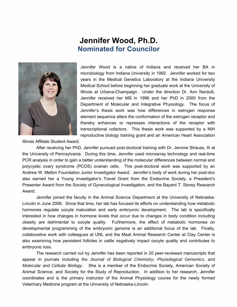
Jennifer Wood, Ph.D. Nominated for Councilor
Jennifer Wood is a native of Indiana and received her BA in
microbiology from Indiana University in 1992. Jennifer worked for two
years in the Medical Genetics Laboratory at the Indiana University
Medical School before beginning her graduate work at the University of
Illinois at Urbana-Champaign. Under the direction Dr. Ann Nardulli,
Jennifer received her MS in 1996 and her PhD in 2000 from the
Department of Molecular and Integrative Physiology. The focus of
Jennifer’s thesis work was how differences in estrogen response
element sequence alters the conformation of the estrogen receptor and
thereby enhances or represses interactions of the receptor with
transcriptional cofactors. This thesis work was supported by a NIH
reproductive biology training grant and an American Heart Association
Illinois Affiliate Student Award.
After receiving her PhD, Jennifer pursued post-doctoral training with Dr. Jerome Strauss, III at
the University of Pennsylvania. During this time, Jennifer used microarray technology and real-time
PCR analysis in order to gain a better understanding of the molecular differences between normal and
polycystic ovary syndrome (PCOS) ovarian cells. This post-doctoral work was supported by an
Andrew W. Mellon Foundation Junior Investigator Award. Jennifer’s body of work during her post-doc
also earned her a Young Investigator’s Travel Grant from the Endocrine Society, a President’s
Presenter Award from the Society of Gynecological Investigation, and the Bayard T. Storey Research
Award.
Jennifer joined the faculty in the Animal Science Department at the University of Nebraska-
Lincoln in June 2006. Since that time, her lab has focused its efforts on understanding how metabolic
hormones regulate oocyte maturation and early embryonic development. The lab is specifically
interested in how changes in hormone levels that occur due to changes in body condition including
obesity are detrimental to oocyte quality. Furthermore, the effect of metabolic hormones on
developmental programming of the embryonic genome is an additional focus of the lab. Finally,
collaborative work with colleagues at UNL and the Meat Animal Research Center at Clay Center is
also examining how persistent follicles in cattle negatively impact oocyte quality and contributes to
embryonic loss.
The research carried out by Jennifer has been reported in 20 peer-reviewed manuscripts that
appear in journals including the Journal of Biological Chemistry, Physiological Genomics, and
Molecular and Cellular Biology. She is a member of the Endocrine Society, American Society of
Animal Science, and Society for the Study of Reproduction. In addition to her research, Jennifer
coordinates and is the primary instructor of the Animal Physiology course for the newly formed
Veterinary Medicine program at the University of Nebraska-Lincoln.
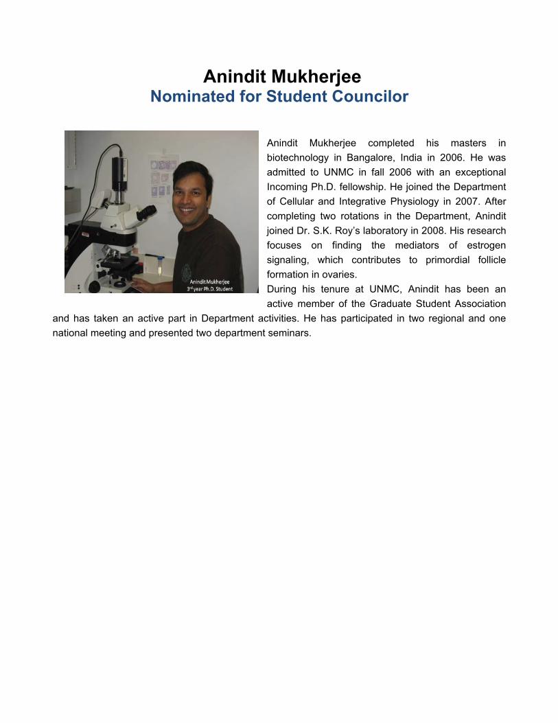
Anindit Mukherjee Nominated for Student Councilor
Anindit Mukherjee completed his masters in
biotechnology in Bangalore, India in 2006. He was
admitted to UNMC in fall 2006 with an exceptional
Incoming Ph.D. fellowship. He joined the Department
of Cellular and Integrative Physiology in 2007. After
completing two rotations in the Department, Anindit
joined Dr. S.K. Roy’s laboratory in 2008. His research
focuses on finding the mediators of estrogen
signaling, which contributes to primordial follicle
formation in ovaries.
During his tenure at UNMC, Anindit has been an
active member of the Graduate Student Association
and has taken an active part in Department activities. He has participated in two regional and one
national meeting and presented two department seminars.

Erin G. Rosenbaugh
Nominated for Student Councilor
During her undergraduate studies, Ms. Rosenbaugh gained two
years of laboratory experience in the INBRE program. She also
received a Biotechnology/ BRTP scholarship to take the first year of
UNMC graduate courses as an undergraduate senior. After
graduating from the University of Nebraska at Omaha with a
bachelor’s degree in Biotechnology, Ms. Rosenbaugh began
graduate school in the summer of 2008 in the Biomedical Research
Training Program, a common entry program for graduate students.
She completed four rotations and started graduate thesis research
in Dr. Matthew C. Zimmerman’s laboratory in January 2009. Her thesis project is focused on the
nanotechnology-driven delivery of active copper zinc superoxide dismutase to central neurons for the
improved treatment of brain-related cardiovascular diseases.
During her first year at UNMC, Erin has been an active member of the Graduate Student
Association and the UNMC Student Senate. She has presented her work in a departmental seminar
and at the 2009 NE-INBRE Conference and has submitted abstracts to the 2009 American Heart
Association High Blood Pressure Conference and the 2009 Nebraska Physiological Society Annual
Meeting.

NEBRASKA PHYSIOLOGICAL SOCIETY BALLOT
PRESIDENT-ELECT - (Vote for One)
� Barbara Engebretsen, Ph.D. Wayne State College
� G. Patrick Lambert, Ph.D. Creighton University
� _______________________________ Write-in (must agree to serve)
SECRETARY/TREASURER – (Vote for One)
� Babu J. Padanilam, Ph.D. University of Nebraska Medical Center
� Hong Sun, M.D., Ph.D. University of Nebraska Medical Center
� _______________________________ Write-in (must agree to serve)
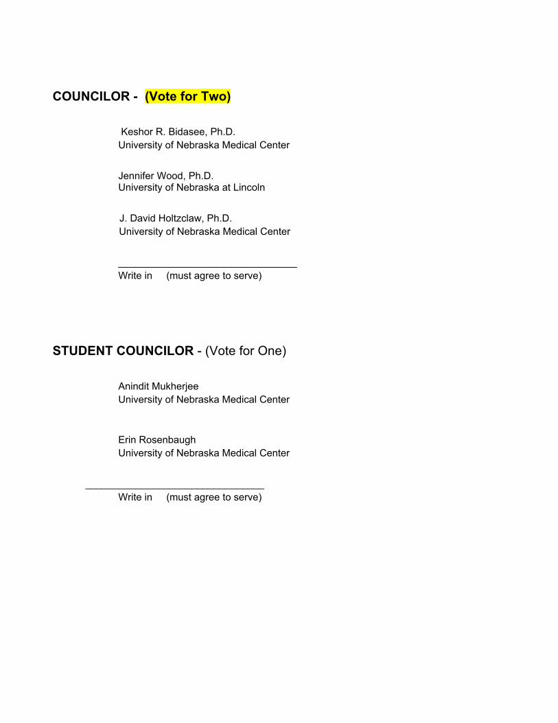
COUNCILOR - (Vote for Two)
� Keshor R. Bidasee, Ph.D. University of Nebraska Medical Center
� Jennifer Wood, Ph.D. University of Nebraska at Lincoln
� J. David Holtzclaw, Ph.D. University of Nebraska Medical Center
� ________________________________ Write in (must agree to serve)
STUDENT COUNCILOR - (Vote for One)
� Anindit Mukherjee University of Nebraska Medical Center
� Erin Rosenbaugh University of Nebraska Medical Center
� ________________________________ Write in (must agree to serve)
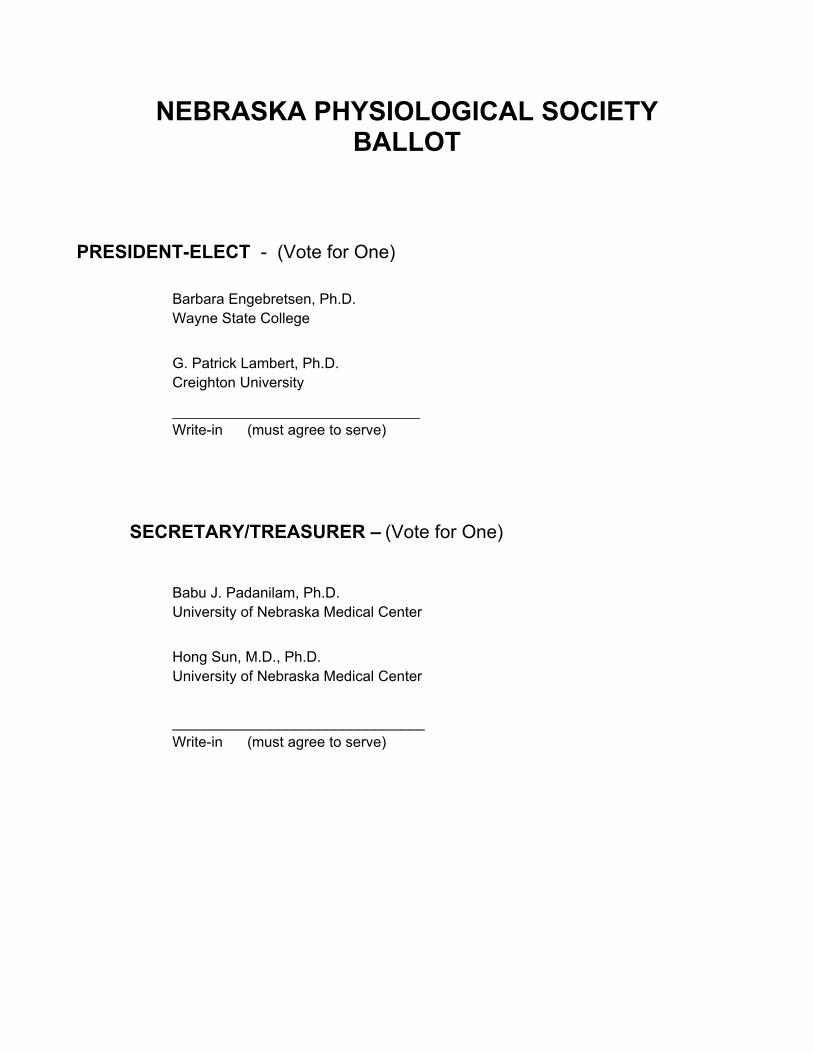

Nebraska Physiological Society
Business Meeting Agenda
September 12, 2009
Item 1: State of American Physiological Society – Dr. Irving Zucker Item 2: Introduction of Slate of Candidates – Dr. Kaushik Patel Item 3: Update on PHUN Week - Dr. David Holtzclaw Item 4: Update on Chapter Advisory Committee – Dr. Harold Schultz Item 5: Treasurer Report - Cindy Norton Item 6: Defining officers and councilors duties – Dr. Kaushik Patel Item 7: Teacher Certificate Presentation – Dr. Kaushik Patel Item 8: Poster judging meeting – Dr. Kaushik Patel

Evaluation Nebraska Physiological Society Annual Meeting
Please indicate your membership status:
○ Professional Member ○ Graduate Student
○ Post-Doctoral/Research Associate ○ Undergraduate Student
Please complete and return evaluation at the conclusion of the program. On a scale of 1 to 5, with 5 being very satisfied
UNMC Michael Sorrell Center Comments
How satisfied were you with the location and accommodations?
1
2
3
4
5
Poster Set-Up Registration/Breakfast UNMC Michael Sorrell Center Comments
Area and amount of time given adequate Help available if you needed it Registration table was well prepared
1 11
2 2 2
3 3 3
4 44
5 5 5
Research Keynote Address Dr. Kim Johnson Comments
How satisfied were you with the presentation?
1
2
3
4
5
Young Investigator Presentation Comments Dr. Matthew Zimmerman
How satisfied were you with the presentation and the time allotted? Dr. Sonia Rocha-Sanchez How satisfied were you with the presentation and the time allotted?
1 1
2 2
3 3
4 4
5 5
Educational Keynote Address Dr. Stephen DiCarlo Comments
How satisfied were you with the presentation? 1
2
3
4
5
State of APS Address, Business Meeting and Lunch Comments
How satisfied were you with the APS presentation? How satisfied were you with the business meeting? How satisfied were you with the lunch?
1 1 1
2 2 2
3 3 3
4 4 4
5 5 5
Poster Session Comments
How satisfied were you with the area & amount of time?
1
2
3
4
5
What type of programs would you like to see NPS do in the future? Additional Comments:
Please return this form at the registration desk. Thank you!

Teacher Evaluation Nebraska Physiological Society Annual Meeting
Please indicate your status:
College Teacher Middle School
High School Teacher Elementary
1. Please rank on a scale of 1-5 (1= poor, 5 = excellent) each of the following teacher breakout session components:
A) Cardiovascular activity (models): ___
B) Molecular biology activity: ___
C) Activity discussion ___
D) Curriculum materials and assessment tools: ____
2. Was the teacher’s breakout session beneficial to you? Yes No
3. What activities or topics would you like to see in future sessions?
4. Would you come back again next year? Yes No
5. What type of programs would you like to see NPS do in the future?
Please return this form at the registration desk.
Thank you!

Acknowledgments
We would like to acknowledge and thank our sponsors who have provided support to the
Nebraska Physiological Society Annual Meeting.
We would especially like to thank Pearl Sorensen, Linda Tegeder, Janine Wilson from the Department of Cellular and Integrative Physiology at the University of Nebraska Medical
Center for their help in organizing the meeting.
For additional information on the NPS sponsors, please visit www.unmc.edu/physiology/nps/sponsors.