Production, stability, gene sequencing and in situ anti-Listeria ... · around the well of the...
12
Production, stability, gene sequencing and in situ anti-Listeria activity of mundticin KS expressed by three Enterococcus mundtii strains Luca Settanni a, * , Rosa Guarcello a , Raimondo Gaglio a , Nicola Francesca a , Aurora Aleo b , Giovanna E. Felis c , Giancarlo Moschetti a a Department of Agricultural and Forestry Science, University of Palermo, Viale delle Scienze 4, 90128 Palermo, Italy b Department of Sciences for Health Promotion and Mother-Child Care “G. D’Alessandro”, University of Palermo, Palermo, Italy c Department of Biotechnology, University of Verona, Strada Le Grazie 15, 37134 Verona, Italy article info Article history: Received 23 April 2013 Received in revised form 5 July 2013 Accepted 16 July 2013 Keywords: Bacteriocins Enterococcus mundtii Food model systems In situ activity Listeria monocytogenes Mundticin KS abstract Three enterococci (WFE3, WFE20 and WFE31) selected as presumptive bacteriocin producers were found to be active against Listeria monocytogenes. In this study, due to their potential industrial/food applica- tions, the three bacterial isolates were extensively characterized. Identification was performed by means of a combined 16S rRNA gene sequencing and multiplex PCR approach, and was confirmed with the sequencing of a partial region of a protein-encoding gene, namely pheS. The three isolates belonged unequivocally to the species Enterococcus mundtii. The randomly amplified polymorphic DNA (RAPD) analysis recognized three distinct strains. The supernatants were mainly active against Listeria spp., but some lactic acid bacteria were also inhibited. The proteinaceous nature of the three supernatants was detected after treatment with proteinase K, protease B and trypsin. The bacteriocins were found to be heat resistant, stable in a large pH range and in presence of ethanol. The bacteriocins were not adsorbed onto the surface of the producer cells and their effect was bactericidal. The production of bacteriocins was higher at neutral pHs and temperatures in the range 30e37 C. The active supernatants did not show cytotoxicity against human erythrocytes and the three strains were susceptible to the action of common antibiotics. The genetic characterization of the bacteriocin genes showed that all three strains produced mundticin KS. They produced it in five food model systems, sterilized by thermal treatment or filtration, prepared from fresh vegetables, cereals, cheeses, meats and fishes. The in situ anti-listerial activities of the strains WFE3, WFE20 and WFE31 were quantitatively different. Ó 2013 Elsevier Ltd. All rights reserved. 1. Introduction Enterococci are considered at the crossroad of food safety (Franz, Holzapfel, & Stiles, 1999); they are reported to be a leading cause of nosocomial infections and to have a significant role in the dissemination and persistence of antimicrobial resistance (Moellering, 1992; Murray, 1990). However, some species within this group are of relevance in food fermentation (Folquié Moreno, Rea, Cogan, & De Vuyst, 2003) and several isolates are commonly employed as probiotics for humans and slaughter animals (Franz, Huch, Abriouel, Holzapfel, & Gálvez, 2011). Enterococci are natu- ral inhabitants of the intestine in warm-blooded animals (Devriese, Collins, & Wirth, 1992), thus, they often occur in foods of animal origin (meat and cheese) (Franz et al., 1999), but they are also commonly found on the above-ground parts of vegetables and cereals (Corsetti et al., 2007; Mundt & Hammer, 1968) and may persist during the fermentation of vegetable products. Further- more, due to their ability to resist to the technological processes used in the food industries (e.g. pasteurization and addition of acids and salt), Enterococcus spp. are usually found in many manufac- tured food products. The enterococcal species most frequently identified in fer- mented foods are Enterococcus faecium and Enterococcus faecalis, but other species such as Enterococcus casseliflavus, Enterococcus durans and Enterococcus mundtii are common in many raw mate- rials and foods (Corsetti et al., 2007; Franciosi, Settanni, Cavazza, & Poznanski, 2009; Settanni et al., 2012). Besides their contribution to the organoleptic properties of fermented food products, entero- cocci of food interest are generally investigated for their ability to produce bacteriocins, because these protein antimicrobials pro- duced by bacteria that enjoy a generally recognized as safe (GRAS) status may be considered as “natural” food preservatives (Settanni, * Corresponding author. Tel.: þ39 091 23896043; fax: þ39 091 6515531. E-mail addresses: [email protected], [email protected] (L. Settanni). Contents lists available at ScienceDirect Food Control journal homepage: www.elsevier.com/locate/foodcont 0956-7135/$ e see front matter Ó 2013 Elsevier Ltd. All rights reserved. http://dx.doi.org/10.1016/j.foodcont.2013.07.022 Food Control 35 (2014) 311e322
Transcript of Production, stability, gene sequencing and in situ anti-Listeria ... · around the well of the...
lable at ScienceDirect
Food Control 35 (2014) 311e322
Contents lists avai
Food Control
journal homepage: www.elsevier .com/locate/ foodcont
Production, stability, gene sequencing and in situ anti-Listeria activityof mundticin KS expressed by three Enterococcus mundtii strains
Luca Settanni a,*, Rosa Guarcello a, Raimondo Gaglio a, Nicola Francesca a, Aurora Aleo b,Giovanna E. Felis c, Giancarlo Moschetti a
aDepartment of Agricultural and Forestry Science, University of Palermo, Viale delle Scienze 4, 90128 Palermo, ItalybDepartment of Sciences for Health Promotion and Mother-Child Care “G. D’Alessandro”, University of Palermo, Palermo, ItalycDepartment of Biotechnology, University of Verona, Strada Le Grazie 15, 37134 Verona, Italy
a r t i c l e i n f o
Article history:Received 23 April 2013Received in revised form5 July 2013Accepted 16 July 2013
Keywords:BacteriocinsEnterococcus mundtiiFood model systemsIn situ activityListeria monocytogenesMundticin KS
* Corresponding author. Tel.: þ39 091 23896043; fE-mail addresses: [email protected], settannil
0956-7135/$ e see front matter � 2013 Elsevier Ltd.http://dx.doi.org/10.1016/j.foodcont.2013.07.022
a b s t r a c t
Three enterococci (WFE3, WFE20 and WFE31) selected as presumptive bacteriocin producers were foundto be active against Listeria monocytogenes. In this study, due to their potential industrial/food applica-tions, the three bacterial isolates were extensively characterized. Identification was performed by meansof a combined 16S rRNA gene sequencing and multiplex PCR approach, and was confirmed with thesequencing of a partial region of a protein-encoding gene, namely pheS. The three isolates belongedunequivocally to the species Enterococcus mundtii. The randomly amplified polymorphic DNA (RAPD)analysis recognized three distinct strains. The supernatants were mainly active against Listeria spp., butsome lactic acid bacteria were also inhibited. The proteinaceous nature of the three supernatants wasdetected after treatment with proteinase K, protease B and trypsin. The bacteriocins were found to beheat resistant, stable in a large pH range and in presence of ethanol. The bacteriocins were not adsorbedonto the surface of the producer cells and their effect was bactericidal. The production of bacteriocinswas higher at neutral pHs and temperatures in the range 30e37 �C. The active supernatants did not showcytotoxicity against human erythrocytes and the three strains were susceptible to the action of commonantibiotics. The genetic characterization of the bacteriocin genes showed that all three strains producedmundticin KS. They produced it in five food model systems, sterilized by thermal treatment or filtration,prepared from fresh vegetables, cereals, cheeses, meats and fishes. The in situ anti-listerial activities ofthe strains WFE3, WFE20 and WFE31 were quantitatively different.
� 2013 Elsevier Ltd. All rights reserved.
1. Introduction
Enterococci are considered at the crossroad of food safety(Franz, Holzapfel, & Stiles, 1999); they are reported to be a leadingcause of nosocomial infections and to have a significant role in thedissemination and persistence of antimicrobial resistance(Moellering, 1992; Murray, 1990). However, some species withinthis group are of relevance in food fermentation (Folquié Moreno,Rea, Cogan, & De Vuyst, 2003) and several isolates are commonlyemployed as probiotics for humans and slaughter animals (Franz,Huch, Abriouel, Holzapfel, & Gálvez, 2011). Enterococci are natu-ral inhabitants of the intestine in warm-blooded animals (Devriese,Collins, & Wirth, 1992), thus, they often occur in foods of animalorigin (meat and cheese) (Franz et al., 1999), but they are also
ax: þ39 091 [email protected] (L. Settanni).
All rights reserved.
commonly found on the above-ground parts of vegetables andcereals (Corsetti et al., 2007; Mundt & Hammer, 1968) and maypersist during the fermentation of vegetable products. Further-more, due to their ability to resist to the technological processesused in the food industries (e.g. pasteurization and addition of acidsand salt), Enterococcus spp. are usually found in many manufac-tured food products.
The enterococcal species most frequently identified in fer-mented foods are Enterococcus faecium and Enterococcus faecalis,but other species such as Enterococcus casseliflavus, Enterococcusdurans and Enterococcus mundtii are common in many raw mate-rials and foods (Corsetti et al., 2007; Franciosi, Settanni, Cavazza, &Poznanski, 2009; Settanni et al., 2012). Besides their contribution tothe organoleptic properties of fermented food products, entero-cocci of food interest are generally investigated for their ability toproduce bacteriocins, because these protein antimicrobials pro-duced by bacteria that enjoy a generally recognized as safe (GRAS)status may be considered as “natural” food preservatives (Settanni,

L. Settanni et al. / Food Control 35 (2014) 311e322312
Valmorri, Suzzi, & Corsetti, 2008). This characteristic is of para-mount importance for their application in strategies of bio-preservation (Settanni & Corsetti, 2008), that refers to the extensionof the shelf-life and improvement of the safety of foods using mi-croorganisms and/or their metabolites (Ross, Morgan, & Hill, 2002).
In comparison to other Enterococcus species, the role of bacte-riocins produced by E. mundtii has been scarcely studied in foodsystems, although their efficacy has been evaluated in mung beansprouts (Bennik, van Overbeek, Smid, & Gorris, 1999) and, veryrecently, in fresh Minas cheese (Vera Pingitore, Todorov, Sesma, &Franco, 2012) and vacuum-packed cold smoked salmon (Bigwoodet al., 2012).
The in situ antimicrobial efficacy of bacteriocinmay be limited bytheir binding to food components (fat or protein particles) and foodadditives (e.g. triglyceride oils), inactivation by proteases or otherinhibitors, changes in solubility and charge, changes in the cell en-velopeof the targetbacteria (Aasenet al., 2003; Settanni et al., 2008).Furthermore, bacteriocin production can be influenced by the cul-ture conditions (Settanni et al., 2008). It is reported that bacteriocinactivities do not always correlate with cell concentration or growthrate of the producer (Kim, Hall, & Dunn,1997), as well as that higherlevels of bacteriocin production may be obtained in sub-optimalconditions (Aasen, Moretro, Katla, Axelsson, & Storro, 2000;Todorov & Dicks, 2004). On the contrary, Settanni et al. (2008)found that stressing conditions and lack or low concentrations ofnutritional factors determined a reduction in bacteriocinproductionby several E. mundtii strains.
This work was performed to evaluate the inhibitory activities ofthree E. mundtii strains, to study their production under severalgrowth conditions and after different enzymatic, thermal andchemical treatments, to genetically investigate their structure, todetermine their production in different food model systems, and tomonitor their anti-Listeria potential in situ during fermentation.
2. Materials and methods
2.1. Strains and growth conditions
Enterococci (isolates WFE3, WFE20, WFE31) scored positive forantimicrobial compound production by well diffusion assay (WDA,see Section 2.3), during a general screening aimed at characterizingLAB from wheat flours (work in preparation), and E. mundtiiPON10063 of flour origin were cultured in MRS (Oxoid, Basing-stoke, England) for 24 h at 30 �C. The bacterial strains used as in-dicators (sensitive to the inhibitory activity) are listed in Table 1.Listeria innocua 4202 (obtained from the culture collection of Na-tional Food Biotechnology Centre, Cork, Ireland) and all Listeriamonocytogenes DHPS strains (belonging to the culture collection ofthe Department of Sciences for Health Promotion andMother-ChildCare “G. D’Alessandro” e University of Palermo, Italy) were prop-agated in Brain Heart Infusion (BHI) (Oxoid) at 37 �C for 24 h,Lactobacillus sakei LMG 2313 (obtained from the Laboratory of Mi-crobial Gene Technology, Ås, Norway) in modified-MRS (mMRS)(maltose and fresh yeast extract were added at final concentrationsof 1% and 10%, respectively, and the final pH was adjusted to 5.6) at30 �C for 24 h, Citrobacter freundii PSS60, Enterobacter spp. PSS11,Escherichia coli PSS2, Klebsiella oxytoca PSS82, Serratia grimesiiPSS72 and Stenotrophomonas maltophilia PSS52 (belonging to theculture collection of the Agricultural Microbiology laboratory e
Department of Agricultural and Forestry Science e University ofPalermo, Italy) were propagated in Nutrient Broth (NB) (DifcoLaboratories, Detroit, MI) at 37 �C for 24 h, Pseudomonas putidaPSS21 (of the same collection) was cultivated in NB at 20 �C for 24 h,while all other strains were propagated as indicated by therespective culture collection.
2.2. Identification of enterococci at species level and straindifferentiation
The DNA from LAB cultures was extracted by the InstageneMatrix kit (Bio-Rad, Hercules, CA) as described by the manufac-turer. Crude cell extracts were used as template DNA for PCR.
Genotypic identification of LABwas first carried out by 16S rRNAgene sequencing. PCRs were performed as described by Weisburg,Barns, Pelletier, and Lane (1991). DNA fragments were visualizedand the amplicons of about 1600 bp were purified by the QIA-quickpurification kit (Qiagen S.p.a., Milan, Italy) and sequenced using thesame primers employed for PCR amplification. DNA sequencingreactions were performed by PRIMM (Milan, Italy). The sequenceswere compared by a BLAST search in GenBank/EMBL/DDBJ databaseand on EzTaxon-e server (http://eztaxon-e.ezbiocloud.net/; Kimet al., 2012). The multiplex PCR assay based on sodA gene re-ported by Jackson, Fedorka-Cray, and Barrett (2004) was applied toconfirm species identity. Finally, pheS partial sequence was ob-tained for strain WFE31 as previously reported (Naser et al., 2005)and compared in GenBank/EMBL/DDBJ database. DNA amplifica-tions were performed by means of T1 Thermocycler (Biometra,Göttingen, Germany).
Strain differentiationwas performed by random amplification ofpolymorphic DNA (RAPD)-PCR analysis in a 25-mL reaction mixusing single primersM13, AB111, and AB106 as reported by Settanniet al. (2012). PCR products were separated by electrophoresis on 2%(w/v) agarose gel (Gibco BRL, Cergy Pontoise, France) and visualizedby UV transillumination after staining with SYBR� safe DNA gelstain (Molecular probes, Eugene, OR, USA). GeneRuler 100 bp PlusDNA ladder (M$Medical Srl, Milan, Italy) was used as a molecularsize marker. RAPD patterns were analyzed using the Gelcompare IIsoftware version 6.5 (Applied-Maths, Sin Marten Latem, Belgium).
2.3. Assays for antibacterial activity
After propagation, the three strains WFE3, WFE20 and WFE31were centrifuged at 10,000�g for 5 min,washed in Ringer’s solution(SigmaeAldrich,Milan, Italy) and re-suspended in the same solutionto achieve an optical density (OD) of ca. 1.00, measured by 6400Spectrophotometer (Jenway Ltd., Felsted Dunmow, UK) at 600 nmwavelength,which approximatelycorresponds to a concentration of109 CFU/mL, to standardize bacterial inocula. Cell suspensions wereinoculated inMRSat afinal concentration of approximately 106 CFU/mL and incubated at 30 �C for 24 h. The antimicrobial activity of theactive supernatants (20 mL) was tested byWDA (Schillinger & Lücke,1989) following the modifications of Corsetti, Settanni, and VanSinderen (2004). L. sakei LMG 2313, L. innocua 4202 andL. monocytogenes ATCC 19114 were used as indicator strains. Inhi-bition was scored positive in presence of a detectable clear areaaround the well of the producer strain. The antibacterial activity ofthe supernatants was measured by the critical dilution assay ofBarefoot and Klaenhammer (1983). The activity was defined as thereciprocal of the highest dilution showing definite inhibition of theindicator strain and was expressed as activity units per milliliter(AU/mL). The inhibitory substances were then characterized fortheir inhibitory spectra against other food related bacteria (Table 1).Tests were carried out in triplicate.
2.4. Characterization of the active supernatants
In order to evaluate the proteinaceous nature of the inhibitorycompounds, the active supernatants, obtained after separation ofthe bacterial cells (10,000�g for 5 min) in the stationary phase ofgrowth, were characterized for their sensitivity to proteolytic en-zymes using proteinase K (12.5 U/mg), protease B (45 U/mg) and

Table 1Inhibitory spectra of E. mundtii WFE3, WFE20, and WFE31 against food-associated bacteria.
Strains used as indicators for the inhibition testsa Source Enterococcus strains tested as bacteriocin producers
PON10063 WFE3 WFE20 WFE31
Enterococcus hirae DSM 20160T Unknown e 10,666� 3695A 12,800� 0A 10,666� 3695AListeria innocua 4202 Unknown e 42,666� 14,780A 51,200� 0A 68,266� 29,560AListeria monocytogenes ATCC 19114 Animal tissue e 273,066� 118,241A 51,200� 0B 819,200� 0CL. monocytogenes DHPS129 Human stool e 51,200� 0A 34,133� 14,780A 85,333� 29,560AL. monocytogenes DHPS131 Human stool e 17,067� 7390A 12,800� 0A 34,133� 14,780AL. monocytogenes DHPS133 Human stool e 204,800� 0A 170,667� 5920A 204,800� 0AL. monocytogenes DHPS179 Salmon e 34,133� 14,780A 17,067� 7390AB 68,266� 29,560ACL. monocytogenes DHPS180 Ricotta cheese e 17,067� 7390A 6,400� 0A 17,067� 7390AL. monocytogenes DHPS182 Ricotta cheese e 25,600� 0A 68,266� 29,560A 34,133� 14,780AL. monocytogenes DHPS184 Rice salad e 85,333� 29,560A 34,133� 14,780AB 136,533� 59,120ACL. monocytogenes DHPS185 Beef e 136,533� 59,120A 136,533� 59,120A 136,533� 59,120AL. monocytogenes DHPS186 Mozzarella salad e 85,333� 29,560A 25,600� 0BC 68,266� 29,560ACL. monocytogenes DHPS187 Roasted chicken e 170,667� 5920A 546,133� 236,483B 409,600� 0BL. monocytogenes DHPS188 Green salad e 204,800� 0A 68,266� 29,560B 68,266� 29,560BL. monocytogenes DHPS1BO Chopped meat e 68,266� 29,560A 17,067� 7390BC 21,333� 7390ACL. monocytogenes DHPS2BO Fresh salami e 68,266� 29,560A 12,800� 0B 17,067� 7390BL. monocytogenes DHPS3BO Fresh salami e 6400� 0A 17,067� 7390AC 25,600� 0BCL. monocytogenes DHPS4BO Ripened salami e 68,266� 29,560A 5333� 1847B 17,067� 7390BL. monocytogenes DHPS5BO Ripened salami e 17,067� 7390A 17,067� 7390A 25,600� 0AL. monocytogenes DHPS6BO Ripened salami e 17,067� 7390A 17,067� 7390A 17,067� 7390AL. monocytogenes DHPS7BO Ripened salami e 17,067� 7390A 6,400� 0A 10,667� 3695AL. monocytogenes DHPS11BO Meat factory e 12,800� 0A 12,800� 0A 12,800� 0AL. monocytogenes DHPS12BO Ripened salami e 12,800� 0A 6,400� 0AB 34,133� 14,780ACL. monocytogenes DHPS13BO Gorgonzola cheese e 12,800� 0A 12,800� 0A 17,067� 7390AL. monocytogenes DHPS20BO Gorgonzola cheese e 204,800� 0A 17,067� 7390B 25,600� 0BL. monocytogenes DHPS22BO Taleggio cheese e 68,266� 29,560A 34,133� 14,780A 51,200� 0AL. monocytogenes DHPS24BO Taleggio cheese e 25,600� 0A 17,067� 7,390AB 68,266� 29,560ACLactobacillus farciminis DSM 20180 Sausage e 1600� 0A 1066� 462B 800� 0CLactobacillus curvatus ssp. curvatus ATCC 25601T Milk e 1066� 462A 200� 0B 167� 58BLactobacillus delbrueckii ssp. bulgaricus ATCC 11842T Yogurt e 667� 231A 200� 0B 400� 0CLactobacillus fermentum DSM 20391 Unknown e 1066� 462A 400� 0B 667� 231CLactobacillus paralimentarius DSM 13238T Sourdough e 3200� 0A 800� 0B 2,133� 924ALactobacillus paraplantarum DSM 10667T Beer e 333� 115A 200� 0A 133� 58ALactobacillus pentosus ATCC 8041T Unknown e 800� 0A 167� 58B 533� 231ALactobacillus pentosus DSM 20199 Unknown e 8533� 3695A 1333� 462B 2667� 924BLeuconostoc mesenteroides DSM 20343T Fermented olives e 25,600� 0A 6400� 0B 8533� 3695BPediococcus acidilactici LMG 11384T Barley e 2667� 924A 667� 231AB 1333� 462ACStatistical significance:Strains ns *** *** ***
The results are expressed in activity units (AU)/mL and indicate mean value� SD of three replicates. The activity was measured in MRS supernatants.e, no inhibition.P value: *, P� 0.05; **, P� 0.01; ***, P� 0.001; ns¼ not significant.Uppercase letters indicate different statistical significances (overall P< 0.05, Tukey’s correction). Means within a given column with the same letter are not statisticallydifferent from each other.
a The following strains were not inhibited by any active supernatant: Citrobacter freundii PSS60, Enterococcus durans DSM 20633T, Enterococcus faecium DSM 20477T,Enterobacter spp. PSS11, Escherichia coli PSS2, Klebsiella oxytoca PSS82, Kocuria varians DSM 20033T, Lactobacillus alimentarius DSM 20181, Lactobacillus amylovorus DSM20531T, Lactobacillus amylolyticus DSM 11664T, Lactobacillus brevis ATCC 14869T, Lactobacillus buchneri LMG 6852T, Lactobacillus casei DSM 20011T, Lactobacillus casei LMG6904T, Lactobacillus delbrueckii ssp. delbrueckiiDSM 20074T, Lactobacillus delbrueckii ssp. lactis ATCC 12315T, Lactobacillus farciminis DSM 20184T, Lactobacillus fermentum ATCC14931T, Lactobacillus fructivorans DSM 20203T, Lactobacillus hilgardii DSM 20051, Lactobacillus hilgardii DSM 20176T, Lactobacillus paracasei ssp. paracasei NCFB 151T,Lactobacillus paracasei ssp. tolerans LMG 9191T, Lactobacillus plantarum ATCC 14917T, Lactobacillus reuteri DSM 20056T, Lactobacillus rhamnosus LMG 6400T, Lactobacillus sakeiLMG 2313, Lactococcus lactis ssp. cremoris DSM 20069T, Lactococcus lactis ssp. lactis DSM 20481T, Pseudomonas putida PSS21, Serratia grimesii PSS72, Stenotrophomonasmaltophilia PSS52, Streptococcus thermophilus DSM 20617T.
L. Settanni et al. / Food Control 35 (2014) 311e322 313
trypsin (10.6 U/mg) at a final concentration of 1 mg/mL in phos-phate buffer (pH 7.0). All enzymes were purchased from SigmaeAldrich (Milan, Italy). The supernatants were incubated for 2 h at37 �C and the remaining activity was determined by well diffusionassay (WDA) (Settanni, Massitti, Van Sinderen, & Corsetti, 2005).The effect of a-amylase and lipase, heat treatment, pH and organicsolvent on the antimicrobial activity was evaluated as described byCorsetti, Settanni, Braga, Lopes, and Suzzi (2008). Tests were carriedout in triplicate.
2.5. Adsorption studies and effect of bacteriocins
The effect of the pH on the adsorption of the active proteins ontoproducer cells was evaluated as reported by Todorov et al. (1999)and Yang, Johnson, and Ray (1992).
The effect of the antimicrobial compounds on the sensitive cellswas evaluated as follows: the supernatants (4 mL) were adjusted topH6.5, treatedwith catalase as reported by Corsetti et al. (2008) andconcentrated under vacuum (Hetovac VR-1, Heto Lab Equipment,Birkerod,Denmark); thedried supernatantswere re-suspended into4 mL of BHI and filtered through a 0.22-mm pore size filter (Milli-pore); the indicator strain (L. monocytogenes ATCC 19114) wasinoculated at a cellular concentration of approximately 103 CFU/mL.If no growth of L. monocytogenesATCC 19114 occurred inpresence ofthe bacteriocins, the cellswere recovered and transferred intoBHI todistinguish between bactericidal and bacteriostatic effect. The su-pernatant of E. mundtii PON10063, treated as above described, wasused as negative control. Cell suspensions were followed bymeasuring the OD at 600 nm at T0, when the supernatants wereadded, 2-h intervals for the first 10 h and then at 24 h for seven days.
Tests were carried out in triplicate.

L. Settanni et al. / Food Control 35 (2014) 311e322314
2.6. Bacteriocin production at different incubation temperaturesand initial growth pH values
To evaluate the effect of temperature and pH on the productionof bacteriocins. Cells of the producer strains were cultivated at 15,30, 37 and 45 �C in MRS. Incubation was for 48 h, except for theassay at 15 �C that was prolonged for five days. The effect of theinitial pH of mediumwas evaluated by adjusting MRS to pH 4.0, 5.0,6.0, 7.0, 8.0 and 9.0 with 5 M NaOH or 5 M HCl. Incubation was at30 �C for 48 h. Tests were carried out in triplicate.
2.7. Evaluation of cellular toxicity
Cellular toxicity of the three active supernatants was assayedfollowing themethodology reported by Xian-guo and Ursula (1994).Each sample (0.8 mL) was placed in a microcentrifuge tube and thefinal volume of 1 mL was reached adding human erythrocytes.Phosphate buffer saline (PBS) (Oxoid) and tap water were used asnegative and positive control, respectively. The tubes were incu-bated at 37 �C for 30 min and hemolysis was observed aftercentrifugation at 3000�g for 5 min. Hemolysis was scored positivewhen the erythrocytes did not form a pellet after centrifugation.
2.8. Evaluation of antibiotic resistance
The antibiotic resistance of E. mundtii strains was tested ac-cording to the guidelines of the Clinical and Laboratory StandardsInstitute (2011) (CLSI) for enterococci, applying the PerformanceStandards for Antimicrobial Susceptibility Testing. According tothose recommendations, the following antibiotics were assayed bythe disk diffusion test: penicillin (10 units) and ampicillin (10 mg)for the “group A primary test and report”; quinupristinedalfo-pristin (15 mg), linezolid (30 mg) and vancomycin (30 mg) for the“group B primary test report selectively”.
2.9. Amplification, cloning and sequencing of bacteriocin-codinggenes
The structural genes for bacteriocin production were analyzedby PCR amplification using primers mapping on the nucleotidesequence of E. mundtii bacteriocin-coding genes. Genomic DNAfrom E. mundtii WFE3, WFE20 and WFE31 were used as templatesfor amplification with the primer pair Mnt-1F (50-TGAGA-GAAGGTTTAAGTTTTGAAGAA-30)/Mnt-1R (50-TCCACTGAAATCCAT-GAATGA-30) mapping upstream of the coding sequence of knownbacteriocins KS (Kawamoto et al., 2002), CRL35 (Saavedra, Minahk,de Ruiz Holgado, & Sesma, 2004), QU2 (Zendo et al., 2005) andMunL (Feng, Guron, Churey, & Worobo, 2009), applying the con-ditions described by Zendo et al. (2005). DNA from E. mundtiiPON10063was used as negative control in PCRs. PCR products wereanalyzed on 1.5% (w/v) agarose gel and visualized as abovereported.
Amplicons generated with the primer pair Mnt-1F/Mnt-1R werepurified by QIA-quick purification kit (Qiagen) and cloned into thepGEM�-T Easy Vector (Promega, Milan, Italy) following manufac-turer’s instructions. Ligation products were transformed into E. coliJM109 high efficiency competent cells and these were plated ontoLuriaeBertani (LB) agar (Oxoid) containing 100 mg/mL ampicillin(SigmaeAldrich), X-Gal (5-bromo-4-chloro-3-indolyl-b-D-galacto-pyranoside, 80 mg/mL) and IPTG (isopropyl-b-D-thiogalactopyrano-side, 0.5 mM) (Eppendorf, Milan, Italy). Recombinant white colonieswere screened by colony-PCR using vector specific primers SP6 (50-ATTTAGGTGACACTATAGAATAC-30) and T7 (50-TAATACGACTCACTATAGGG-30) in a 25-mL reaction mix applying the following amplifi-cation program: 94 �C for 5 min, 4 �C for 4 min, 35 cycles at 94 �C for
30 s, 52 �C for 30 s and 72 �C for 50 s, followed by a final extension at72 �C for 5 min. Insert integrity was confirmed by a nested-amplification with Mnt-1F and Mnt-1R primers using purifiedcolony-PCR amplicons as templates and the previously describedamplification program. In addition, T7/Mnt-1Fand T7/Mnt-1R primerpairswere alternatively employed in a different nested-PCR (35 cyclesat 94 �C for 30 s, 52 �C for 30 s and 72 �C for 40 s, followed by a finalextension at 72 �C for 2 min) to determine the orientation of frag-ments into the cloning vector. PCR products were separated byelectrophoresis in a 1.5% (w/v) agarose gel, stained with SYBR� safeDNA gel.
Sequencing reactions were performed by PRIMM and the se-quences were compared by a BLAST search in GenBank/EMBL/DDBJdatabase. The prediction of the open reading frame (ORF) wasperformed with the softwares ChromasPro v1.6 (Copyright 2003e2012 Technelysium Pty Ltd. Biotech Works Inc.) and pDRAW32v1.1.114 (www.acaclone.com). The ClustalX program (Thompson,Gibson, Plewniak, Jeanmougin, & Higgins, 1997) was used fornucleotide sequence analysis. Sequence alignments were analyzedand adjusted by GeneDoc program v2.5.000 (K.B. Nicholas and H.B.Nicholas, unpublished data).
2.10. Amplification of enterocin CRL35 biosynthetic cluster
The DNA from the strains WFE3, WFE20 andWFE31 were ampli-fied with the primer pair mun1F (50-GCAAACCGATAAGAATGTGGGAT-30)/mun7R (50-TATACATTGTCCCCACAACC-30) (Saavedraet al., 2004), designed to amplify the biosynthetic cluster of enter-ocin CRL35 that has been shown to share high sequence identitywiththe cluster of mundticin KS. The amplification program included:denaturation at 94 �C for 3 min, 30 cycles at 94 �C for 30 s, 55 �C for30 s and 72 �C for 3 min and 40 s, followed by a final extension at72 �C for 4 min. DNA from E.mundtii PON10063was used as negativecontrol in PCRs. PCR products were analyzed on 1% (w/v) agarose geland visualized.
2.11. Bacteriocin production in food model systems
In order to evaluate the effect of different food components onthe inhibitory activity of the E. mundtii strains, five food modelsystems [vegetable broth (VB), meat broth (MB), fish broth (FB),cereal broth (CeB) and cheese broth (ChB)] were developed: VBwith carrot (50 g/L), tomato (50 g/L), zucchini (50 g/L) and celery(50 g/L); MB with pork (50 g/L), calf (50 g/L), chicken (50 g/L) andsheep (50 g/L) meat; FB with salmon (50 g/L), octopus (50 g/L),anchovy (50 g/L) and cod (50 g/L); CeB with wheat bran (50 g/L),wheat (50 g/L), barley (50 g/L) and rice (50 g/L) kernels; ChB withCaciocavallo (50 g/L), Parmigiano (50 g/L), Pecorino (50 g/L) andVastedda (50 g/L) cheeses. The preparation of the five broths was asfollows: all four ingredients of each broth were homogenized witha Sorvall Omni-Mixer (Dupont Instruments, Newtown, CT) at themaximum speed for 1 min, transferred to a Schott Duran bottle,added with distilled H2O (1 L), left under magnetic stirring for 1 hand centrifuged at 10,000�g for 5 min. Each supernatant wasdivided in two aliquots: one was sterilized by autoclaving at 121 �Cfor 20 min (autoclaved broths), while the other aliquot was filter(0.20-mm pore size filter, Sartorius, Aubagne Cedex, France) steril-ized (filtered broths). The broths were subjected to the measure-ment of pH, determined electrometrically using the pH meterBASIC 20þ (Crison Instrument S.A., Barcelona, Spain), and wateractivity (aw), obtained with the AquaLab vapor sorption analyzer(Decagon Devices, Pullman, WA, USA).
Autoclaved and filtered broths were inoculated singly with thestrains WFE3, WFE20 andWFE31 as above reported (Section 2.3) ata final concentration of approximately 106 CFU/mL, after cell
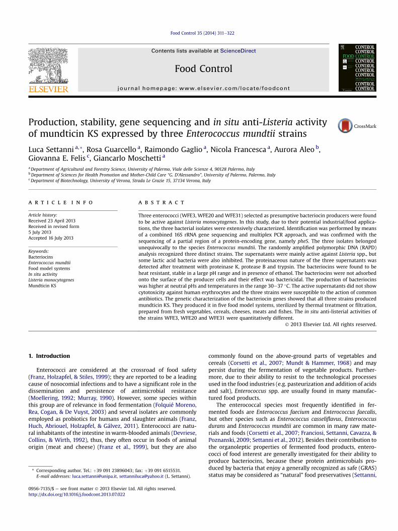
Fig. 1. RAPD-PCR profiles obtained with primer M13. Lanes: 1, GeneRuler 100 bp PlusDNA ladder (M$Medical); 2, E. mundtii WFE3; 3, E. mundtii WFE20; 4, E. mundtiiWFE31; 5, E. mundtii PON10063; 6, PCR negative control.
L. Settanni et al. / Food Control 35 (2014) 311e322 315
washing. MRS was also inoculated as control trial. E. mundtiiPON10063was used as negative control. Incubationwas at 30 �C for24 h. Tests were carried out in triplicate.
2.12. In situ activity of E. mundtii strains against L. monocytogenes
To evaluate the potential of the bacteriocin producing strainsduring the fermentation of different foods, the five food modelsystems were inoculated with dual combinations Enterococcus/L. monocytogenes. Tests in MRS were carried out for comparison.All strains were prepared as reported in Section 2.3. The Entero-coccus strains were inoculated at the final concentration ofapproximately 106 CFU/mL to act as starter cultures, whileL. monocytogenes ATCC 19114 at about 104 CFU/mL to simulate amassive contamination. E. mundtii PON10063 was used as negativecontrol. Incubation was at 30 �C for five days to mimic a commonfood fermentation process. Plate counts were performed toenumerate the surviving cells. The broths (1 mL) were subjected tothe serial decimal dilution in Ringer’s solution and the cell sus-pensions were spread plated (0.1 mL) and incubated as follows: onkanamycin aesculin azide (KAA) agar (Oxoid), incubated aerobi-cally at 37 �C for 24 h, for Enterococcus; on Listeria Selective AgarBase (LSAB) (Oxoid) supplemented with SR0140E (Oxfordformulation), incubated aerobically at 37 �C for 48 h, forL. monocytogenes. In order to verify the specificity of the mediaemployed, the absence of growth of enterococci on LSAB and thatof L. monocytogenes on KAA were verified prior in situ activitydetermination. Tests were carried out in duplicate.
2.13. Statistical analyses
Data of inhibitory activities, effect of enzyme, pH, temperatureand organic solvent treatments on bacteriocins, production ofbacteriocins under different pH and temperature conditions andproduction of bacteriocins in different food models were statisti-cally analyzed using the generalized linear model (GLM) procedure,including the effects of strain, with the program SAS 2008e version9.2 (Statistical Analysis System Institute Inc., Cary, NC, USA). TheStudent “t” test was used for mean comparison. The post-hoc Tukeymethod was applied for pairwise comparison.
The in situ anti-Listeria efficacy of bacteriocins was analyzed bythe Student “t” test and post-hoc Tukey method. Significance levelwas P< 0.05.
3. Results
3.1. Genotypic identification and differentiation of enterococci
The isolates WFE3, WFE20 and WFE31 were identified as E.mundtii by 16S rRNA gene sequencing, and the gene sequenceswere deposited in GenBank under the Acc. No. KC291248eKC291250. Species identification was confirmed by using a specificmultiplex PCR system developed to distinguish among E. mundtii,Enterococcus flavescens and Enterococcus sulfureus (results notshown) and with partial sequencing of pheS gene for WFE31, whichconfirmed the identification as E. mundtii (data not shown).
RAPD-PCR analysis (Fig. 1) recognized the three isolates as threedistinct strains.
3.2. Bacteriocin production
E. mundtiiWFE3,WFE20, andWFE31were active against severalbacterial strains (Table 1). All three Enterococcus strains inhibited thesame sensitive strains, but the inhibitory effect was strain depen-dent, since different activities (P< 0.05), expressed in AU/mL were
registered. The highest inhibitions of all three supernatants weredetected against L. monocytogenes strains: all 25 indicator strainsbelonging to this species and isolated fromdifferent sources resultedsensitive. E. mundtii WFE3 and WFE20 were less active than strainWFE31. No big differences in terms of inhibitory power (P> 0.05)were found among the three strains against Enterococcus hirae DSM20160T, L. innocua 4202, Lactobacillus paraplantarum DSM 10667T
and Pediococcus acidilactici LMG 11384T. Gram-negative bacteriawere not inhibited by any of the active supernatants.
3.3. Effect of different treatments on the antibacterial activity
The antibacterial compounds were all inactivated by proteolyticenzymes (Table 2), confirming their proteinaceous nature. Thethree presumptive bacteriocins were insensitive to a-amylase andlipase. Heat treatment progressively reduced the inhibitory activ-ities of the supernatants; the inhibition was completely lost afterthe sterilization of supernatants. All three bacteriocins retainedalmost the full activity at the different pH and concentration ofethanol tested. After treatment with a-amylase and lipase andexposure at pH 10.0 and 11.0, the supernatants from the strainsWFE3 and WFE20 showed comparable activities (P< 0.05) againstL. monocytogenes ATCC 19114, while the treatment at 100 �Cdetermined a decrease of WFE31 activity which was at the samelevel (P> 0.05) to that of WFE3.
3.4. Absorption to the producing cells and effect of the bacteriocins
The bacteriocins of E. mundtii WFE3, WFE20, and WFE31 werenot absorbed by the cell surface. The effect of the active superna-tants was followed for seven days (Fig. 2). During this period thegrowth of L. monocytogenes ATCC 19114 was completely inhibitedby the addition of all three supernatants. After test, the cells wererecovered from the bacteriocin-containing broths and transferredinto bacteriocin-free BHI. Since no growth of L. monocytogenes ATCC

Table 2Effect of enzymes, heat treatment, pH, and organic solvent on the inhibitory activity of E. mundtii WFE3, WFE20, and WFE31.
Treatment Enterococcus strains
PON10063 WFE3 WFE20 WFE31
Control (supernatant not treated) e 273,066� 118,241A 51,200� 0B 819,200� 0CEnzymes:Proteinase K e e e e
Protease B e e e e
Trypsin e e e e
a-amylase e 85,333� 29,560A 42,667� 14,780A 819,200� 0BLipase e 85,333� 29,560A 51,200� 0A 682,667� 236,483B
Statistical significance:Strains *** *** ***
Heat treatment:100 �C for 20 min e 68,267� 29,560A 25,600� 0B 170,667� 59,120A100 �C for 60 min e 25,600� 0A 10,667� 3695B 42,667� 14,780C121 �C for 15 min e e e e
Statistical significance:Strains ** *** **
pH:3.0 e 170,667� 59,120A 25,600� 0B 682,667� 236,483C4.0 e 204,800� 0A 25,600� 0B 819,200� 0C5.0 e 170,667� 59,120A 21,333� 7390B 819,200� 0C6.0 e 102,400� 0A 25,600� 0B 819,200� 0C7.0 e 102,400� 0A 21,333� 7390B 682,667� 236,483C8.0 e 51,200� 0A 25,600� 0B 819,200� 0C9.0 e 42,667� 14,780A 17,067� 7390B 682,667� 236,483C10.0 e 25,600� 14,780A 12,800� 0A 409,600� 0B11.0 e 25,600� 0A 12,800� 0A 341,333� 118,241B
Statistical significance:Strains *** ** ***
Organic solvent:C2H5OH 5% e 102,400� 0A 25,600� 0B 341,333� 118,241CC2H5OH 10% e 85,333� 29,560A 25,600� 0B 204,800� 0CC2H5OH 15% e 85,333� 29,560A 25,600� 0B 204,800� 0C
Statistical significance:Strains ns ns ns
The results are expressed in activity units (AU)/mL and indicate mean value� SD of three replicates. The activity was measured in MRS supernatants.e, no inhibition.All assays were carried out against L. monocytogenes ATCC 19114.P value: *, P� 0.05; **, P� 0.01; ***, P� 0.001; ns¼ not significant.Uppercase letters indicate different statistical significances (overall P< 0.05, Tukey’s correction). Means within a given column with the same letter are not statisticallydifferent from each other.
L. Settanni et al. / Food Control 35 (2014) 311e322316
19114 was observed after incubation at the optimal temperature,the effect of the three bacteriocins, at the tested concentrations,was assumed to be bactericidal. However, these observationscannot exclude a bacteriostatic behavior at lower concentrations of
Fig. 2. Effect of bacteriocin produced by E. mundtii WFE3, WFE20, and WFE31 evaluated agafrom non bacteriocin producer PON10063; >, with bacteriocin WFE3; ,, with bacteriocinvalue. Bars not visible are smaller than symbol size.
bacteriocins. The supernatant from E. mundtii PON10063 did notshow inhibitory effect, in fact, in its presence, the growth curve ofthe indicator strain was almost superimposable to that obtainedwithout any addition of concentrated supernatant.
inst L. monocytogenes ATCC 19114. Symbols: B, without addition; C, with supernatantWFE20; :, with bacteriocin WFE31. Bars represent the standard deviation of the mean

Table 3Effect of different growth conditions on the inhibitory activity of E. mundtii WFE3, WFE20, and WFE31.
Treatment Enterococcus strains
PON10063 WFE3 WFE20 WFE31
Controla e 273,066� 118,241A 51,200� 0B 819,200� 0CTemperature:15 �C e 204,800� 0A 25,600� 0B 682,667� 236,483C37 �C e 204,800� 0A 34,133� 14,780B 819,200� 0C45 �C e 170,667� 59,121A 25,600� 0B 409,600� 0C
Statistical significance:Strains ns ** **
pH:4.0 e 170,667� 59,121A 17,067� 7390B 273,066� 118,241A5.0 e 170,667� 59,121A 25,600� 0B 409,600� 0C6.0 e 204,800� 0A 25,600� 0B 819,200� 0C7.0 e 204,800� 0A 21,333� 7390B 819,200� 0C8.0 e 204,800� 0A 25,600� 0B 682,667� 236,483C9.0 e 204,800� 0A 25,600� 0B 409,600� 0C
Statistical significance:Strains ns ns *
The results are expressed in activity units (AU)/mL and indicate mean value� SD of three replicates. The activity was measured in MRS supernatants.e, no inhibition.All assays were carried out against L. monocytogenes ATCC 19114.P value: *, P� 0.05; **, P� 0.01; ns¼ not significant.Uppercase letters indicate different statistical significances (overall P< 0.05, Tukey’s correction). Means within a given column with the same letter are not statisticallydifferent from each other.
a Growth in MRS at 30 �C for 24 h.
L. Settanni et al. / Food Control 35 (2014) 311e322 317
3.5. Effect of different incubation temperatures and initial growthpHs on bacteriocin activity
Growth at temperatures different from 30 �C (Table 3) did notaffect (P> 0.05) bacteriocin production for the strain WFE3, butdetermined a consistent decrease (P< 0.05) for the activities ofsupernatants from the strains WFE20 and WFE31. The effect of pHof the MRS medium different from 6.5 on the bacteriocin produc-tionwas significant (P< 0.05) only for the strainWFE31, bacteriocinWFE20 at pH 4.0 and for WFE31 at pH 4.0 and 5.0.
3.6. Evaluation of cytotoxicity and antibiotic sensitivity
Hemolysis of human erythrocytes was negative in PBS andEnterococcus supernatants, showing that the metabolites of thetested strains were not hemolytic.
Fig. 3. PCR products for the structural genes of bacteriocins: (A) amplicons from PCR with MPlus DNA ladder (M$Medical); 2, E. mundtii WFE3; 3, E. mundtii WFE20; 4, E. mundtii WFE3
For the evaluation of the effect of the antibiotics, using CLSIbreakpoints for enterococci, all Enterococcus strains resulted resis-tant to penicillin and susceptible to ampicillin, quinupristinedal-fopristin, linezolid and vancomycin (results not shown).
3.7. Analysis of bacteriocin determinants
In order to analyze the DNA sequences encoding for bacterio-cins, two different PCR amplifications were performed with DNAsextracted from Enterococcus strains. By means of the primer pairsMnt-1F/and Mnt-1R, a 380 bp-long fragment was amplified fromWFE3, WFE20 andWFE31 genomic DNA (Fig. 3A), while only WFE3andWFE31 strains showed a PCR product of approximately 3128 bpwith Mun1R/Mun7R (Fig. 3B). No amplification product was ob-tained from the DNA of the non bacteriocin producer E. mundtiiPON10063.
nt-1F/Mnt-1R; (B) amplicons from PCR with Mun1F/Mun7R. Lanes: 1, GeneRuler 100 bp1; 5, E. mundtii PON10063; 6, PCR negative control.

L. Settanni et al. / Food Control 35 (2014) 311e322318
The sequences of the 380 bp amplicons (Fig. 4) revealed that allthree Enterococcus strains active against L. monocytogenes ATCC19114 possessed bacteriocin-coding genes sharing 99% identitywith mundticin KS (Kawamoto et al., 2002). Moreover, a completeopen reading frame (ORF) was deduced from the structural gene ofthe three bacteriocins. It was found to encode the bacteriocinprecursor (58 amino acid residues). Mature predicted peptidesshowed 100% identity to mundticin KS. Additionally, high sequencesimilarity was observed comparing aminoacidic sequences fromstrains WFE3, WFE20 and WFE31 to different bacteriocins in themundticin group of class IIa, such as mundticin L (98%) and enter-ocin CRL35 (98%). A second partial ORF was also predicted from allthree bacteriocin producing strains and it was found to encode for aputative ATP binding cassette (ABC) transporter.
3.8. Bacteriocin production in food model systems
Bacteriocin production in the different autoclaved and filteredfood broths is reported in Table 4. A very low residual activity(0.01e4.17%) was recovered from the autoclaved food model
Fig. 4. Alignment of the structural bacteriocin-coding genes (A) and am
systems after the growth of bacteriocin producing E. mundtiistrains, while the filtration of the food models allowed a generalhigher retention of inhibitory activities for all strains which reached33.07% for the strain WFE20 in MB. However, no activity wasrecovered from filtered ChB inoculated with the strains WFE20 andWFE31. Except autoclaved CeB and filtered ChB, the best results interms of activity recovery were shown by the strain WFE20. On thecontrary, the strongest reduction of the anti-Listeria inhibition wasregistered for the strain WFE31 in all food model systems sterilizedboth by autoclaving and filtration. The two methodology appliedfor the sterilization of the model systems did not determine sig-nificant differences (P> 0.05) of pH and aw, but the food modelswere statistically different (P< 0.001).
3.9. In situ anti-listerial activity of bacteriocin producing E. mundtiistrains
The effects of the active strains against L. monocytogenes ATCC19114 during growth in the different autoclaved and filtered foodmodel systems are shown in Table 5. Tests performed in MRS
inoacidic sequences; (B) in E. mundtii WFE3, WFE20 and WFE31.
Table 4Bacteriocin productiona by E. mundtii WFE3, WFE20, and WFE31 in food model systems.
Food model systems Characteristics of foodmodelsb
Enterococcus strains
pH aw PON10063 WFE3 (residual activity %) WFE20 (residual activity %) WFE31 (residual activity %)
Autoclaved CeB 6.10� 0.02 0.957� 0.001 e 4267� 1867 (1.56)A 533� 231 (1.04)B 200� 0 (0.02)BFiltered CeB 6.37� 0.02 0.933� 0.001 e 4266� 1847 (1.56)A 800� 0 (1.56)B 8533� 3695 (1.04)A
Autoclaved ChB 5.12� 0.01 0.960� 0.001 e 1600� 0 (0.59)A 2133� 924 (4.17)A 533� 231 (0.06)BFiltered ChB 5.60� 0.02 0.918� 0.001 e 50� 0 (0.02)A 0 (0)B 0 (0)B
Autoclaved FB 6.62� 0.03 0.946� 0.001 e 50� 0 (0.02)A 200� 0 (0.39)B 50� 0 (0.01)AFiltered FB 6.61� 0.01 0.980� 0.001 e 12,800� 0 (4.69)A 8533� 3695 (16.67)AC 4266� 1847 (0.52)BC
Autoclaved MB 6.42� 0.01 0.958� 0.001 e 1067� 462 (0.39)A 400� 0 (0.78)A 50� 0 (0.01)BFiltered MB 5.86� 0.03 0.903� 0.001 e 12,800� 0 (4.69)A 16,933� 7159 (33.07)A 8533� 3695 (1.04)A
Autoclaved VB 5.33� 0.03 0.953� 0.001 e 100� 0 (0.04)A 533� 231 (1.04)B 400� 0 (0.04)BFiltered VB 5.05� 0.02 0.929� 0.001 e 2133� 924 (0.78)A 1600� 0 (3.13)AB 800� 0 (0.10)AC
Statistical significance:Treatment (T) ns ns ns * * ***Broth (B) *** *** ns ns ns nsT * B ns *** ns *** *** ***
e, no inhibition.All assays were carried out against L. monocytogenes ATCC 19114.Abbreviations: CeB, cereal broth; ChB, cheese broth; FB, fish broth; MB, meat broth; VB, vegetable broth.P value: *, P� 0.05; ***, P� 0.001; ns¼ not significant.Uppercase letters indicate different statistical significances (overall P< 0.05, Tukey’s correction). Means within a given column with the same letter are not statisticallydifferent from each other.
a The results are expressed in activity units (AU)/mL and indicate mean value� SD of three replicates.b Evaluated before inoculation.
L. Settanni et al. / Food Control 35 (2014) 311e322 319
confirmed the bactericidal effect of the three bacteriocinogenicstrains against L. monocytogenes ATCC 19114. The growth of theindicator strain reached the maximal level generally at the secondday of incubation, when a concentration of about 107 CFU/mL wasregistered in MRS and in almost all food models except filtered ChBand filtered MB for which a decrease was recorded from the firstday on. Thus, for these two food models the inhibition ofL. monocytogenes ATCC 19114 may be mainly imputable to the lowaw (below 0.92). All levels of concentration registered for the in-dicator strain in absence of E. mundtiiwere comparable (P> 0.05) tothose showed in presence of the non bacteriocin producerE. mundtii PON10063. No differences (P> 0.05) were registered forthe three bacteriocins among autoclaved and filtered MRS, filteredCeB, filtered ChB, autoclaved FB, autoclaved MB, autoclaved VB andfiltered VB. The growth of L. monocytogenes ATCC 19114 wascompletely inhibited within the first 24 h of co-culturing in MRS,filtered CeB, filtered ChB and autoclaved VB, while the highestListeria survival was observed in autoclaved MB and in presence ofall three E. mundtii strains.
4. Discussion
In the present work, the bacterial isolates WFE3, WFE20 andWFE31 of wheat flour origin, presumptively allotted into the groupof enterococci on the basis of growth on a kanamycin containingagar medium and preliminarily screened as producers of antimi-crobial compounds (work in preparation), were characterized fortheir potential application as bio-preservative agents.
The three isolates were identified by a multiple genotypicapproach as three distinct E. mundtii strains. Their supernatantswere tested against pro-technological, spoilage and pathogenbacteria. Several strains of LAB were used as indicators, since theyare commonly used as starter cultures in food fermentations. Abacteriocin active against starter LAB may have detrimental effectsand, for this reason, not applicable in fermented food systems.E. mundtii WFE3, WFE20 and WFE31 were all found to be particu-larly active against Listeria spp. Within this group, L. monocytogenesis a difficult pathogen to control because of its ubiquitous distri-bution, tolerance to high levels of salt and its stability to grow at a
relatively low pH and at refrigeration temperatures (Guinane,Cotter, Hill, & Ross, 2005). The anti-listerial effects of LAB are awanted characteristic, since it strongly contributes to the safety ofthe final foods (Deegan, Cotter, Hill, & Ross, 2006). The three strainsbehaved similarly and they inhibited only a few species within thepro-technological bacterial group, but they were ineffective againstGram-negative bacteria, at least in the non concentrated form. Ourfindings showed the common characteristics of bacteriocins,because they are active mostly on strains closely related to theproducer strains (Cotter, Hill, & Ross, 2005). The activity of somebacteriocins from LAB against Gram-negative bacteria is an unusualphenomenon; only a few bacteriocins possessed this behavior(Kuwano et al., 2005; Todorov & Dicks, 2005).
Proteolytic enzymes determined the loss of inhibitory activityfor all three supernatants, proving their proteinaceous nature, ageneral characteristic of bacteriocins (Jack, Tagg, & Ray, 1995).Treatment with a-amylase and lipase did not alter the antibacterialactivity of the active supernatants, suggesting that the activecompounds did not contain a sugar or lipid moiety. A resistance ofthe bacteriocins to the heat treatments was registered. According tothe classification of Nes et al. (1996), the bacteriocinsWFE3,WFE20and WFE31 were considered members of class II.
The retention of inhibitory power of the E. mundtii active su-pernatants in the wide pH range considered and in presence ofdifferent percentages of ethanol provided evidences of theirpossible application in several food ecosystems. From thisperspective, when a bacteriocinogenic strain is applied duringfermentation it is also important to evaluate the production ofbacteriocin in different conditions that may characterize the foodenvironment during growth (Settanni et al., 2008).
Generally, bacteriocin production by LAB is reported as atemperature-sensitive process, whereby the optimal temperaturefor bacteriocin production does not necessarily coincide with theoptimal growth temperature (Leroy & De Vuyst, 1999). It has beensuggested that bacteriocin production by LAB is enhanced by sub-optimal temperatures (Delgado, Brito, Peres, Noé-Arroyo López, &Garrido-Fernández, 2005). However, in the present study, asalready observed in a previous study by Settanni et al. (2008), theresults showed that temperatures different from those in the range
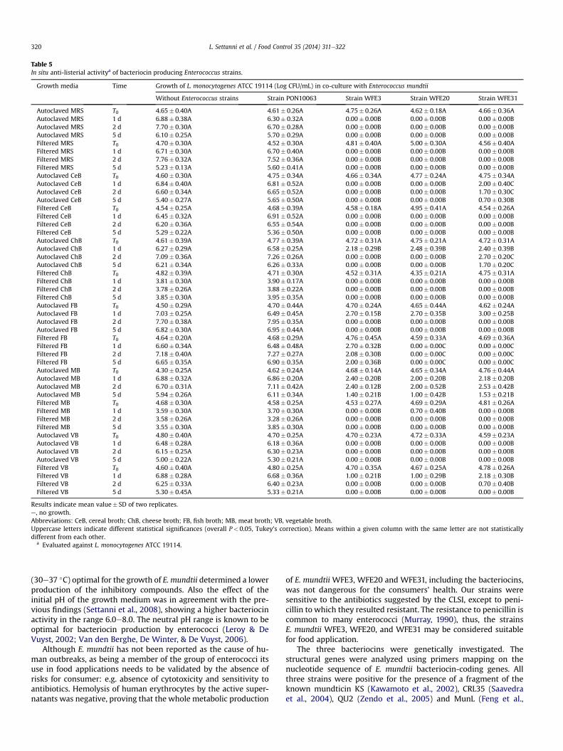
Table 5In situ anti-listerial activitya of bacteriocin producing Enterococcus strains.
Growth media Time Growth of L. monocytogenes ATCC 19114 (Log CFU/mL) in co-culture with Enterococcus mundtii
Without Enterococcus strains Strain PON10063 Strain WFE3 Strain WFE20 Strain WFE31
Autoclaved MRS T0 4.65� 0.40A 4.61� 0.26A 4.75� 0.26A 4.62� 0.18A 4.66� 0.36AAutoclaved MRS 1 d 6.88� 0.38A 6.30� 0.32A 0.00� 0.00B 0.00� 0.00B 0.00� 0.00BAutoclaved MRS 2 d 7.70� 0.30A 6.70� 0.28A 0.00� 0.00B 0.00� 0.00B 0.00� 0.00BAutoclaved MRS 5 d 6.10� 0.25A 5.70� 0.29A 0.00� 0.00B 0.00� 0.00B 0.00� 0.00BFiltered MRS T0 4.70� 0.30A 4.52� 0.30A 4.81� 0.40A 5.00� 0.30A 4.56� 0.40AFiltered MRS 1 d 6.71� 0.30A 6.70� 0.40A 0.00� 0.00B 0.00� 0.00B 0.00� 0.00BFiltered MRS 2 d 7.76� 0.32A 7.52� 0.36A 0.00� 0.00B 0.00� 0.00B 0.00� 0.00BFiltered MRS 5 d 5.23� 0.13A 5.60� 0.41A 0.00� 0.00B 0.00� 0.00B 0.00� 0.00BAutoclaved CeB T0 4.60� 0.30A 4.75� 0.34A 4.66� 0.34A 4.77� 0.24A 4.75� 0.34AAutoclaved CeB 1 d 6.84� 0.40A 6.81� 0.52A 0.00� 0.00B 0.00� 0.00B 2.00� 0.40CAutoclaved CeB 2 d 6.60� 0.34A 6.65� 0.52A 0.00� 0.00B 0.00� 0.00B 1.70� 0.30CAutoclaved CeB 5 d 5.40� 0.27A 5.65� 0.50A 0.00� 0.00B 0.00� 0.00B 0.70� 0.30BFiltered CeB T0 4.54� 0.25A 4.68� 0.39A 4.58� 0.18A 4.95� 0.41A 4.54� 0.26AFiltered CeB 1 d 6.45� 0.32A 6.91� 0.52A 0.00� 0.00B 0.00� 0.00B 0.00� 0.00BFiltered CeB 2 d 6.20� 0.36A 6.55� 0.54A 0.00� 0.00B 0.00� 0.00B 0.00� 0.00BFiltered CeB 5 d 5.29� 0.22A 5.36� 0.50A 0.00� 0.00B 0.00� 0.00B 0.00� 0.00BAutoclaved ChB T0 4.61� 0.39A 4.77� 0.39A 4.72� 0.31A 4.75� 0.21A 4.72� 0.31AAutoclaved ChB 1 d 6.27� 0.29A 6.58� 0.25A 2.18� 0.29B 2.48� 0.39B 2.40� 0.39BAutoclaved ChB 2 d 7.09� 0.36A 7.26� 0.26A 0.00� 0.00B 0.00� 0.00B 2.70� 0.20CAutoclaved ChB 5 d 6.21� 0.34A 6.26� 0.33A 0.00� 0.00B 0.00� 0.00B 1.70� 0.20CFiltered ChB T0 4.82� 0.39A 4.71� 0.30A 4.52� 0.31A 4.35� 0.21A 4.75� 0.31AFiltered ChB 1 d 3.81� 0.30A 3.90� 0.17A 0.00� 0.00B 0.00� 0.00B 0.00� 0.00BFiltered ChB 2 d 3.78� 0.26A 3.88� 0.22A 0.00� 0.00B 0.00� 0.00B 0.00� 0.00BFiltered ChB 5 d 3.85� 0.30A 3.95� 0.35A 0.00� 0.00B 0.00� 0.00B 0.00� 0.00BAutoclaved FB T0 4.50� 0.29A 4.70� 0.44A 4.70� 0.24A 4.65� 0.44A 4.62� 0.24AAutoclaved FB 1 d 7.03� 0.25A 6.49� 0.45A 2.70� 0.15B 2.70� 0.35B 3.00� 0.25BAutoclaved FB 2 d 7.70� 0.38A 7.95� 0.35A 0.00� 0.00B 0.00� 0.00B 0.00� 0.00BAutoclaved FB 5 d 6.82� 0.30A 6.95� 0.44A 0.00� 0.00B 0.00� 0.00B 0.00� 0.00BFiltered FB T0 4.64� 0.20A 4.68� 0.29A 4.76� 0.45A 4.59� 0.33A 4.69� 0.36AFiltered FB 1 d 6.60� 0.34A 6.48� 0.48A 2.70� 0.32B 0.00� 0.00C 0.00� 0.00CFiltered FB 2 d 7.18� 0.40A 7.27� 0.27A 2.08� 0.30B 0.00� 0.00C 0.00� 0.00CFiltered FB 5 d 6.65� 0.35A 6.90� 0.35A 2.00� 0.36B 0.00� 0.00C 0.00� 0.00CAutoclaved MB T0 4.30� 0.25A 4.62� 0.24A 4.68� 0.14A 4.65� 0.34A 4.76� 0.44AAutoclaved MB 1 d 6.88� 0.32A 6.86� 0.20A 2.40� 0.20B 2.00� 0.20B 2.18� 0.20BAutoclaved MB 2 d 6.70� 0.31A 7.11� 0.42A 2.40� 0.12B 2.00� 0.52B 2.53� 0.42BAutoclaved MB 5 d 5.94� 0.26A 6.11� 0.34A 1.40� 0.21B 1.00� 0.42B 1.53� 0.21BFiltered MB T0 4.68� 0.30A 4.58� 0.25A 4.53� 0.27A 4.69� 0.29A 4.81� 0.26AFiltered MB 1 d 3.59� 0.30A 3.70� 0.30A 0.00� 0.00B 0.70� 0.40B 0.00� 0.00BFiltered MB 2 d 3.58� 0.26A 3.28� 0.26A 0.00� 0.00B 0.00� 0.00B 0.00� 0.00BFiltered MB 5 d 3.55� 0.30A 3.85� 0.30A 0.00� 0.00B 0.00� 0.00B 0.00� 0.00BAutoclaved VB T0 4.80� 0.40A 4.70� 0.25A 4.70� 0.23A 4.72� 0.33A 4.59� 0.23AAutoclaved VB 1 d 6.48� 0.28A 6.18� 0.36A 0.00� 0.00B 0.00� 0.00B 0.00� 0.00BAutoclaved VB 2 d 6.15� 0.25A 6.30� 0.23A 0.00� 0.00B 0.00� 0.00B 0.00� 0.00BAutoclaved VB 5 d 5.00� 0.22A 5.30� 0.21A 0.00� 0.00B 0.00� 0.00B 0.00� 0.00BFiltered VB T0 4.60� 0.40A 4.80� 0.25A 4.70� 0.35A 4.67� 0.25A 4.78� 0.26AFiltered VB 1 d 6.88� 0.28A 6.68� 0.36A 1.00� 0.21B 1.00� 0.29B 2.18� 0.30BFiltered VB 2 d 6.25� 0.33A 6.40� 0.23A 0.00� 0.00B 0.00� 0.00B 0.70� 0.40BFiltered VB 5 d 5.30� 0.45A 5.33� 0.21A 0.00� 0.00B 0.00� 0.00B 0.00� 0.00B
Results indicate mean value� SD of two replicates.e, no growth.Abbreviations: CeB, cereal broth; ChB, cheese broth; FB, fish broth; MB, meat broth; VB, vegetable broth.Uppercase letters indicate different statistical significances (overall P< 0.05, Tukey’s correction). Means within a given column with the same letter are not statisticallydifferent from each other.
a Evaluated against L. monocytogenes ATCC 19114.
L. Settanni et al. / Food Control 35 (2014) 311e322320
(30e37 �C) optimal for the growth of E. mundtii determined a lowerproduction of the inhibitory compounds. Also the effect of theinitial pH of the growth medium was in agreement with the pre-vious findings (Settanni et al., 2008), showing a higher bacteriocinactivity in the range 6.0e8.0. The neutral pH range is known to beoptimal for bacteriocin production by enterococci (Leroy & DeVuyst, 2002; Van den Berghe, De Winter, & De Vuyst, 2006).
Although E. mundtii has not been reported as the cause of hu-man outbreaks, as being a member of the group of enterococci itsuse in food applications needs to be validated by the absence ofrisks for consumer: e.g. absence of cytotoxicity and sensitivity toantibiotics. Hemolysis of human erythrocytes by the active super-natants was negative, proving that the whole metabolic production
of E. mundtii WFE3, WFE20 and WFE31, including the bacteriocins,was not dangerous for the consumers’ health. Our strains weresensitive to the antibiotics suggested by the CLSI, except to peni-cillin to which they resulted resistant. The resistance to penicillin iscommon to many enterococci (Murray, 1990), thus, the strainsE. mundtii WFE3, WFE20, and WFE31 may be considered suitablefor food application.
The three bacteriocins were genetically investigated. Thestructural genes were analyzed using primers mapping on thenucleotide sequence of E. mundtii bacteriocin-coding genes. Allthree strains were positive for the presence of a fragment of theknown mundticin KS (Kawamoto et al., 2002), CRL35 (Saavedraet al., 2004), QU2 (Zendo et al., 2005) and MunL (Feng et al.,

L. Settanni et al. / Food Control 35 (2014) 311e322 321
2009), while only E. mundtii WFE20 was negative when analyzedwith the primers designed for the biosynthetic cluster for enterocinCRL35. The nucleotide sequences of the three 381 bp DNA frag-ments obtained after the first PCR amplification showed only fivenucleotides different among the three strains. Furthermore, theaminoacidic sequences did not show any difference among thebacteriocins produced by E. mundtiiWFE3, WFE20, and WFE31 andthey were identical to mundticin KS (Kawamoto et al., 2002). Theabsence of amplification product from WFE20 total DNA with theprimer pairs Mun1F/Mun7R could be due to the sequence poly-morphisms among the three E. mundtii strains. This result reflectsalso the genetic differences highlighted by RAPD analysis.
Mundticin KS belongs to the class IIa (Kawamoto et al., 2002)which contains a consensus YGNGV amino acid motif near the Nterminus. These bacteriocins are active against L. monocytogenes(Ennahar, Sashihara, Sonomoto, & Ishizaki, 2000), a characteristicthat determines their importance in industrial applications. Furtheranalyses are necessary to characterize the entire bacteriocin geneticloci of E. mundtiiWFE3, WFE20, andWFE31. Some mundticins havebeen reported to be encoded by gene clusters carried by plasmids(Feng et al., 2009; Kawamoto et al., 2002), but some E. mundtiistrains have been found to express bacteriocins by chromosomalgenes (Settanni et al., 2008). A character residing on chromosomalDNA is more stable than plasmid encoded information and may besignificant for future applications of these strains at industrial level.
The influence of food components on bacteriocin productionand activity was evaluated in five different food model systemsobtained with fresh vegetables, cereals, cheeses, meats and fishesand subjected to two different sterilization procedures: autoclavingand filtration. Bacteriocin activity was recovered from almost allbroths except filtered cheese broth inoculated with the strainsWFE20 and WFE31. The production was strain dependent, but thebest results were registered in filtered meat and fish broths. Thedifferent production may be imputable to the different presenceand concentration of the food components, as well as to thedifferent pH and aw of the food models. Type and concentration ofcarbon and nitrogen sources are relevant for bacteriocin production(Delgado et al., 2007). Several authors reported that higher bacte-riocin activities are observed with increased nitrogen concentra-tions (Aasen et al., 2000; Kim et al., 1997).
Although with some differences, nine of the ten food modelsystems allowed bacteriocin production. Hence, all broths wereused to test the in situ efficacy of the E. mundtii strains againstL. monocytogenes during the simulation of a common fermentation.The inhibition produced by the growing cultures was often good.This because, in addition to the bacteriocin production, thecompetition for nutrients, initial pH and aw might have beenlimiting for the development of L. monocytogenes. In particular,filtered ChB and filtered MB were characterized by aw below 0.92,which is reported to be the limit for the growth of L. monocytogenesin presence of NaCl and sucrose (Nolan, Chamblin, & Troller, 1992).Some E. mundtii have already been found effective in situ againstL. monocytogenes in vegetable, cheese and fish products (Benniket al., 1999; Bigwood et al., 2012; Vera Pingitore et al., 2012), buttheir bacteriocins have not been characterized for nucleotide oramino acid sequences.
This workwasmainly performed to evaluate the effect of severalfoods (different in composition and nutrient concentrations) on theexpression and activity of bacteriocins produced by three E. mundtiistrains. The results showed that all three strains were able to pro-duce the antimicrobial compounds in different foodmatrices and tocontrol the growth of L. monocytogenes in situ during the fermen-tation process. Works will be prepared to follow the bacteriocinexpression in the different food matrices.
Acknowledgments
This work was financially supported by the project for industrialresearch and training PON01_02249 “Application of molecularbiotechnologies and pro-technological microorganisms for thecharacterization and valorization of dairy and bakery chains oftypical products” of the Italian Ministry of Education, Universityand Research (CUP: B11C11000430005). Prof. Baldassare Portolano(University of Palermo) is thanked for his involvement in therealization of the project and Dr. Aldo Todaro (university ofPalermo) for aw analysis.
References
Aasen, I. M., Moretro, T., Katla, T., Axelsson, L., & Storro, I. (2000). Influence ofcomplex nutrients, temperature and pH on bacteriocin production by Lacto-bacillus sakei CCUG 42687. Applied Microbiology and Biotechnology, 53, 159e166.
Aasen, I. M., Markussen, S., Moretro, T., Katla, T., Axelsson, L., & Naterstad, K. (2003).Interactions of the bacteriocins sakacin P and nisin with food constituents. In-ternational Journal of Food Microbiology, 87, 35e43.
Barefoot, S. F., & Klaenhammer, T. R. (1983). Detection and activity of lactacin B, abacteriocin produced by Lactobacillus acidophilus. Applied and EnvironmentalMicrobiology, 45, 1808e1815.
Bennik, M. H., van Overbeek, W., Smid, E. J., & Gorris, L. G. (1999). Biopreservation inmodified atmosphere stored mungbean sprouts: the use of vegetable-associated bacteriocinogenic lactic acid bacteria to control the growth of Lis-teria monocytogenes. Letters in Applied Microbiology, 28, 226e232.
Bigwood, T., Hudson, J. A., Cooney, J., McIntyre, L., Billington, C., Heinemann, J. A.,et al. (2012). Inhibition of Listeria monocytogenes by Enterococcus mundtii iso-lated from soil. Food Microbiology, 32, 354e360.
Clinical and Laboratory Standards Institute. (2011). Performance standards for anti-microbial susceptibility testing; twenty-first informational supplement. CLSIdocument M100-S21. Wayne, PA: Clinical and Laboratory Standards Institute.
Corsetti, A., Settanni, L., Braga, T., Lopes, M., & Suzzi, G. (2008). An investigation ofthe bacteriocinogenic potential of lactic acid bacteria associated with wheat(Triticum durum) kernels and non-conventional flours. Lwt-Food Science andTechnology, 41, 1173e1182.
Corsetti, A., Settanni, L., Chaves López, C., Felis, G. E., Mastrangelo, M., & Suzzi, G.(2007). A taxonomic survey of lactic acid bacteria isolated fromwheat (Triticumdurum) kernels and non-conventional flours. Systematic and Applied Microbi-ology, 30, 561e571.
Corsetti, A., Settanni, L., & Van Sinderen, D. (2004). Characterization of bacteriocin-like inhibitory substances (BLIS) from sourdough lactic acid bacteria and eval-uation of their in vitro and in situ activity. Journal of Applied Microbiology, 96,521e534.
Cotter, P., Hill, C., & Ross, R. (2005). Bacteriocins: developing innate immunity forfood. Nature Reviews Microbiology, 3, 777e788.
Deegan, L., Cotter, P., Hill, C., & Ross, P. (2006). Bacteriocins: biological tools for bio-preservation and shelf-life extension. International Dairy Journal, 16, 1058e1071.
Delgado, A., Brito, D., Peres, C., Noé-Arroyo López, F., & Garrido-Fernández, A.(2005). Bacteriocin production by Lactobacillus pentosus B96 can be expressedas a function of temperature and NaCl concentration. Food Microbiology, 22,521e528.
Delgado, A., Noé-Arroyo López, F., Brito, D., Peres, C., Fevereiro, P., & Garrido-Fernández, A. (2007). Optimum bacteriocin production by Lactobacillus plan-tarum 17.2b requires absence of NaCl and apparently follows a mixed metab-olite kinetics. Journal of Biotechnology, 130, 193e201.
Devriese, L. A., Collins, M. D., & Wirth, R. (1992). The Genus Enterococcus. InA. Balows, H. G. Trüper, M. Dworkin, W. Harder, & K. H. Schleifer (Eds.), TheProkaryotes (pp. 1465e1481). New York: Springer.
Ennahar, S., Sashihara, T., Sonomoto, K., & Ishizaki, A. (2000). Class IIa bacteriocins:biosynthesis, structure and activity. FEMS Microbiology Reviews, 24, 85e106.
Feng, G., Guron, G. K. P., Churey, J. J., & Worobo, R. W. (2009). Characterization ofmundticin L, a class IIa anti-Listeria bacteriocin from Enterococcus mundtiiCUGF08. Applied and Environmental Microbiology, 75, 5708e5713.
Folquié Moreno, M., Rea, M., Cogan, T., & De Vuyst, L. (2003). Applicability of abacteriocin-producing Enterococcus faecium as a co-culture in Cheddar cheesemanufacture. International Journal of Food Microbiology, 81, 73e84.
Franciosi, E., Settanni, L., Cavazza, A., & Poznanski, E. (2009). Biodiversity andtechnological potential of wild lactic acid bacteria from raw cows’ milk. Inter-national Dairy Journal, 19, 3e11.
Franz, C. M., Holzapfel, W. H., & Stiles, M. E. (1999). Enterococci at the crossroads offood safety? International Journal of Food Microbiology, 47, 1e24.
Franz, C. M., Huch, M., Abriouel, H., Holzapfel, W., & Gálvez, A. (2011). Enterococci asprobiotics and their implications in food safety. International Journal of FoodMicrobiology, 151, 125e140.
Guinane, C., Cotter, P., Hill, C., & Ross, R. (2005). Microbial solutions to microbialproblems; lactococcal bacteriocins for the control of undesirable biota in food.Journal of Applied Microbiology, 98, 1316e1325.
L. Settanni et al. / Food Control 35 (2014) 311e322322
Jack, R. W., Tagg, J. R., & Ray, B. (1995). Bacteriocins of Gram-positive bacteria.Microbiological Reviews, 59, 171e200.
Jackson, C. R., Fedorka-Cray, P. J., & Barrett, J. B. (2004). Use of a genus- and species-specific multiplex PCR for identification of enterococci. Journal of ClinicalMicrobiology, 42, 3558e3565.
Kawamoto, S., Shima, J., Sato, R., Eguchi, T., Ohmomo, S., Shibato, J., et al. (2002).Biochemical and genetic characterization of mundticin KS, an antilisterialpeptide produced by Enterococcus mundtii NFRI 7393. Applied and Environ-mental Microbiology, 68, 3830e3840.
Kim, W. S., Hall, R. J., & Dunn, N. W. (1997). The effect of nisin concentration andnutrient depletion on nisin production of Lactococcus lactis. Applied Microbi-ology and Biotechnology, 48, 449e453.
Kim, O. S., Cho, Y. J., Lee, K., Yoon, S. H., Kim, M., Na, H., et al. (2012). IntroducingEzTaxon-e: a prokaryotic 16S rRNA gene sequence database with phylotypesthat represent uncultured species. International Journal of Systematic andEvolutionary Microbiology, 62, 716e721.
Kuwano, K., Tanaka, N., Shimizu, T., Nagatoshi, K., Nou, S., & Sonomoto, K. (2005).Dual antibacterial mechanisms of nisin Z against Gram-positive and Gram-negative bacteria. International Journal of Antimicrobial Agents, 26, 396e402.
Leroy, F., & De Vuyst, L. (1999). Temperature and pH conditions that prevail duringfermentation of sausages are optimal for production of the antilisterial bacte-riocin sakacin K. Applied and Environmental Microbiology, 65, 974e981.
Leroy, F., & De Vuyst, L. (2002). Bacteriocin production by Enterococcus faecium RZSC5 is cell density limited and occurs in the very early growth phase. Interna-tional Journal of Food Microbiology, 72, 155e164.
Moellering, R. C. (1992). Emergence of Enterococcus as a significant pathogen.Clinical Infectious Diseases, 14, 1173e1176.
Mundt, J. O., & Hammer, J. L. (1968). Lactobacilli on plants. Applied Microbiology, 16,1326e1330.
Murray, B. E. (1990). The life and times of the Enterococcus. Clinical MicrobiologyReviews, 3, 46e65.
Naser, S. M., Thompson, F. L., Hoste, B., Gevers, D., Dawyndt, P., Vancanneyt, M., et al.(2005). Application of multilocus sequence analysis (MLSA) for rapid identifi-cation of Enterococcus species based on rpoA and pheS genes. Microbiology, 151,2141e2150.
Nes, I., Diep, D., Havarstein, L., Brurberg, M., Eijsink, V., & Holo, H. (1996). Biosyn-thesis of bacteriocins in lactic acid bacteria. Antonie Van Leeuwenhoek Interna-tional Journal of General and Molecular Microbiology, 70, 113e128.
Nolan, D. A., Chamblin, D. C., & Troller, J. A. (1992). Minimal water activity levels forgrowth and survival of Listeria monocytogenes and Listeria innocua. InternationalJournal of Food Microbiology, 16, 323e335.
Ross, R. P., Morgan, S., & Hill, C. (2002). Preservation and fermentation: past, presentand future. International Journal of Food Microbiology, 79, 3e16.
Saavedra, L., Minahk, C., de Ruiz Holgado, A. P., & Sesma, F. (2004). Enhancement ofthe enterocin CRL35 activity by a synthetic peptide derived from the NH2-ter-minal sequence. Antimicrobial Agents and Chemotherapy, 48, 2778e2781.
Schillinger, U., & Lücke, F. K. (1989). Antibacterial activity of Lactobacillus sake iso-lated from meat. Applied and Environmental Microbiology, 55, 1901e1906.
Settanni, L., & Corsetti, A. (2008). Application of bacteriocins in vegetable foodbiopreservation. International Journal of Food Microbiology, 121, 123e138.
Settanni, L., Di Grigoli, A., Tornambe, G., Bellina, V., Francesca, N., Moschetti, G., et al.(2012). Persistence of wild Streptococcus thermophilus strains on wooden vatand during the manufacture of a traditional Caciocavallo type cheese. Interna-tional Journal of Food Microbiology, 155, 73e81.
Settanni, L., Massitti, O., Van Sinderen, D., & Corsetti, A. (2005). In situ activity of abacteriocin-producing Lactococcus lactis strain. Influence on the interactionsbetween lactic acid bacteria during sourdough fermentation. Journal of AppliedMicrobiology, 99, 670e681.
Settanni, L., Valmorri, S., Suzzi, G., & Corsetti, A. (2008). The role of environ-mental factors and medium composition on bacteriocin-like inhibitory sub-stances (BLIS) production by Enterococcus mundtii strains. Food Microbiology,25, 722e728.
Thompson, J. D., Gibson, T. J., Plewniak, F., Jeanmougin, F., & Higgins, D. G.(1997). The CLUSTAL-X windows interface: flexible strategies for multiplesequence alignment aided by quality analysis tools. Nucleic Acids Research,25, 4876e4882.
Todorov, S., & Dicks, L. (2004). Effect of medium components on bacteriocin pro-duction by Lactobacillus pentosus ST151BR, a strain isolated from beer producedby the fermentation of maize, barley and soy flour.World Journal of Microbiology& Biotechnology, 20, 643e650.
Todorov, S., & Dicks, L. (2005). Lactobacillus plantarum isolated from molassesproduces bacteriocins active against Gram-negative bacteria. Enzyme and Mi-crobial Technology, 36, 318e326.
Todorov, S., Onno, B., Sorokine, O., Chobert, J., Ivanova, I., & Dousset, X. (1999).Detection and characterization of a novel antibacterial substance produced byLactobacillus plantarum ST 31 isolated from sourdough. International Journal ofFood Microbiology, 48, 167e177.
Van den Berghe, E., De Winter, T., & De Vuyst, L. (2006). Enterocin A production byEnterococcus faecium FAIR-E 406 is characterised by a temperature- and pH-dependent switch-off mechanism when growth is limited due to nutrientdepletion. International Journal of Food Microbiology, 107, 159e170.
Vera Pingitore, E., Todorov, S. D., Sesma, F., & Franco, B. D. (2012). Application ofbacteriocinogenic Enterococcus mundtii CRL35 and Enterococcus faecium ST88Chin the control of Listeria monocytogenes in fresh Minas cheese. Food Microbi-ology, 32, 38e47.
Weisburg, W. G., Barns, S. M., Pelletier, D. A., & Lane, D. J. (1991). 16S ribosomal DNAamplification for phylogenetic study. Journal of Bacteriology, 173, 697e703.
Xian-guo, H., & Ursula, M. (1994). Antifungal compound from Solanum nigrescens.Journal of Ethnopharmacology, 43, 173e177.
Yang, R., Johnson, M. C., & Ray, B. (1992). Novel method to extract large amounts ofbacteriocins from lactic acid bacteria. Applied and Environmental Microbiology,58, 3355e3359.
Zendo, T., Eungruttanagorn, N., Fujioka, S., Tashiro, Y., Nomura, K., Sera, Y., et al.(2005). Identification and production of a bacteriocin from Enterococcusmundtii QU 2 isolated from soybean. Journal of Applied Microbiology, 99,1181e1190.