PROCESS BY WHICH THE PUPARIA OF MANY SPECIES OF …
8
THE PROCESS BY WHICH THE PUPARIA OF MANY SPECIES OF FLIES BECOME FIXED TO A SUBSTRATE G. FRAENKEL AND VICTOR J. BROOKES Department of Entomology, University of Illinois, Urbana, Illinois It is common knowledge among students of the biology of flies that the puparia of many species are firmly fixed to a substrate. This is easy to observe in labora- tory cultures of Drosophila. Toward the end of the larval period the larvae leave the substrate to meander for a short period on the walls of the culture bottle. The puparia are always firmly glued to the walls of the vessel. The process by which the puparia of Drosophila and other flies become fixed to a substrate does not seem ever to have been described. In this paper we shall demonstrate that the puparia are fixed to a substrate by means of a sticky secretion which emanates from the salivary glands. It is well known that the salivary glands of Drosophila and other flies swell to- wards the end of larval life, and that a semi-liquid secretion fills the lumen of the glands at that time. In Drosophila, this is the stage most favorable for a study of the giant chromosomes of the salivary gland cells. The histological and cytological changes in the cells at the time of this secretion have been studied extensively and the presence of this secretion has been recognized and commented upon by a number of investigators (Bodenstein, 1943, 1950; Mellanby, 1938; Painter, 1945; Hsu, 1948; Blumel and Kirby, 1948; Kodani, 1948; Lesher, 1951, 1952). Some of these authors have searched for a possible function of this secretion, but none of them has recognized, or even hinted at what we consider to be the true function, namely the fixation of the puparium. The results of this investigation have been briefly reported (Fraenkel, 1952). In this paper the process of fixation and the changes in the salivary glands will be described, while the identity of the protein in the salivary gland and in the puparial glue will be demonstrated elsewhere (Moorefield and Fraenkel, unpublished data). MATERIAL AND METHODS Most of the observations and experiments described below were carried out on the blowfly, Phormia regina, and on Drosophila melanogaster. The process of fixation to a substrate was studied by direct observation of fully grown larvae under a binocular microscope. All dissections were made in a modified Ringer solution, consisting of H 2 O, 1000 cc. ; NaCl, 7.5 g. ; KC1, 0.35 g. ; and CaCl 2 , 0.21 g. Larvae of Phormia were dissected by cutting off the last segment of the body and pushing the head inwards with the blunt end of a pin while holding the body with a pair of forceps. The larvae were thus turned inside out. The major difficulty of re- moving the salivary glands was loosening them from the tracheae which lie in close association with the gland. If the gland was punctured in any way, it collapsed .and became opaque. No satisfactory method was found other than cautiously break- 442
Transcript of PROCESS BY WHICH THE PUPARIA OF MANY SPECIES OF …

THE PROCESS BY WHICH THE PUPARIA OF MANY SPECIESOF FLIES BECOME FIXED TO A SUBSTRATE
G. FRAENKEL AND VICTOR J. BROOKES
Department of Entomology, University of Illinois, Urbana, Illinois
It is common knowledge among students of the biology of flies that the pupariaof many species are firmly fixed to a substrate. This is easy to observe in labora-
tory cultures of Drosophila. Toward the end of the larval period the larvae leave
the substrate to meander for a short period on the walls of the culture bottle. The
puparia are always firmly glued to the walls of the vessel. The process by which
the puparia of Drosophila and other flies become fixed to a substrate does not seem
ever to have been described. In this paper we shall demonstrate that the puparia are
fixed to a substrate by means of a sticky secretion which emanates from the salivary
glands.It is well known that the salivary glands of Drosophila and other flies swell to-
wards the end of larval life, and that a semi-liquid secretion fills the lumen of the
glands at that time. In Drosophila, this is the stage most favorable for a study of
the giant chromosomes of the salivary gland cells. The histological and cytological
changes in the cells at the time of this secretion have been studied extensively andthe presence of this secretion has been recognized and commented upon by a numberof investigators (Bodenstein, 1943, 1950; Mellanby, 1938; Painter, 1945; Hsu,1948; Blumel and Kirby, 1948; Kodani, 1948; Lesher, 1951, 1952). Some of
these authors have searched for a possible function of this secretion, but none of
them has recognized, or even hinted at what we consider to be the true function,
namely the fixation of the puparium.The results of this investigation have been briefly reported (Fraenkel, 1952).
In this paper the process of fixation and the changes in the salivary glands will be
described, while the identity of the protein in the salivary gland and in the puparial
glue will be demonstrated elsewhere (Moorefield and Fraenkel, unpublished data).
MATERIAL AND METHODS
Most of the observations and experiments described below were carried out onthe blowfly, Phormia regina, and on Drosophila melanogaster. The process of
fixation to a substrate was studied by direct observation of fully grown larvae undera binocular microscope. All dissections were made in a modified Ringer solution,
consisting of H 2O, 1000 cc.; NaCl, 7.5 g. ; KC1, 0.35 g. ;
and CaCl2 ,0.21 g. Larvae
of Phormia were dissected by cutting off the last segment of the body and pushingthe head inwards with the blunt end of a pin while holding the body with a pair of
forceps. The larvae were thus turned inside out. The major difficulty of re-
moving the salivary glands was loosening them from the tracheae which lie in close
association with the gland. If the gland was punctured in any way, it collapsed.and became opaque. No satisfactory method was found other than cautiously break-
442

SALIVARY SECRETION OF FLY LARVAE 443
ing these tracheal connections with a pin. Drosophila larvae were dissected by
submerging in Ringer solution and cutting along the ventral surface with a small
piece of a razor blade.
Salivary glands have also been observed in the living insect in situ by pressing
larvae in water under a cover slip or slide and rolling them back and forth until the
glands could be seen under a microscope.
EXPERIMENTS AND OBSERVATIONS
1. Description of the secretion and fixation process
A. Drosophila melanogaster
Towards the end of the third instar the larva leaves the food and moves alongthe sides of the culture tube. It moves in this manner for about 12 hours, and
then the movements gradually slow down until the larva comes to rest. The head
continues the brushing movements which have gone on throughout larval life but
now this movement describes a semicircle around the thorax. As the body graduallycontracts to the barrel shape of the puparium, this movement slows down. Motion
now all but stops. The mouth hooks are withdrawn but continue to move slightly
back and forth. When the pupal contraction is about completed, the mouth pul-
sates and a clear fluid suddenly pours from it and flows along the area of contact of
the body with the surface. The emission of the fluid is accompanied by a back-and-
forth or pumping movement of the mouth parts and a contraction and expansion of
the body. The fluid begins to harden almost as soon as it is emitted. Once the se-
cretion is completed, the larva becomes motionless and within 30 minutes the pu-
parium begins to darken.
B. Phormia regina
Fully fed third instar larvae leave the meat and wander about in the dry sub-
strate provided for them, sand or sawdust, for about three days. During this
period the crop becomes emptied and very much reduced in size. Several hours
before the formation of the puparium the larva becomes more and more sluggishand opaque. Finally the prepupa contracts slowly to the shape of the puparium. Adescription of the pupal contraction and its underlying processes has been given pre-
viously (Fraenkel and Rudall, 1940). By the time the pupal contraction is nearly
completed and all forward motion has ceased, the anterior segments of the bodyalone are still capable of some movement. Slow rhythmic contractions then start in
about the fourth externally visible segment and move backwards. The larva ex-
FIGURE 1. The larva of Phormia regina (Meig.) in the act of secreting.

444 G. FRAENKEL AND VICTOR J. BROOKES
trudes its proboscis, curves it toward the anterior ventral surface (Fig. 1), and then
alternately inverts and extends the mouth hooks, working back and forth in motions
not unlike the normal feeding motions, but directed toward its own under-surface.
At the same time a pumping motion of structures within the head is visible. After
the above described motions have continued for a few seconds, a clear colorless
fluid emanates from the mouth and is distributed over the anterior under-surface by
moving the head from side to side and back and forth in a painting or brushing move-
ment. The fluid is tacky almost immediately after emission and hardens within a few
minutes. The anterior ventral surface becomes thus firmly fixed to a surface.
The time interval of actual secretion was no longer than thirty seconds.
When larvae are allowed to pupate in sand or sawdust the process of secretion is
exactly the same, and in consequence clumps of sand grains or flakes of wood become
firmly stuck to the anterior ventral surface. In an overcrowded culture, pupariaoften become stuck to each other.
Immediately after completion of this process the anterior end is permanentlywithdrawn into the body and the formation of the white puparium is completed.
Darkening starts 15 to 30 minutes later.
2. The salivary glands of Phormia regina
One of the major difficulties in following salivary gland development is deter-
mination of the maturity of the larva. In this study, the size of the larva, and the
amount of the contents of the crop were used as indication of development. The
salivary glands of Phormia are similar to those described for other fly larvae. Theyare paired sack-like structures, elongated, extending at full size into the second ab-
dominal segment and connected to each other at the posterior end by a small mass of
fat cells.
At the time the larva leaves the food and the crop is still greatly extended with
food, the salivary glands are large, distended and clear (Fig. 2a). The cells are
stretched, the nucleus is distinct and the cytoplasm clear with a very fine granular
consistency. The cells throughout the gland are more or less uniform in size. Asthe gland gets older the cells become more dense and this accounts for a milky ap-
pearance. In older glands the cytoplasm appears to shrink and a clear area is pres-ent around the cell walls.
About twenty hours after the larva leaves the food the glands begin to show a
narrowing of the lumen in the posterior region (Fig. 2b). As the lumen becomes
smaller, the distension of the gland in the anterior region begins to increase, the
gland gets longer and begins to have a milky opacity (Fig. 2c-d). As the larva
approaches the time of pupation the posterior lumen of the gland becomes more andmore occluded, the cells indistinct and the fluid localized in the anterior portion,
causing this area to become bulbous (Fig. 2e-f). At the moment that the larva is
performing the motions as described previously and diagrammed in Figure 1, about
one or two minutes before secretion, the greater part of the gland shows little if anylumen and all of the fluid is concentrated in the swollen anterior region (Fig. 2g).It is this fluid that is expelled, marking the end of larval life.
Figure 2h represents a gland immediately after secretion. The lumen has disap-
peared almost entirely, the cell walls have become indistinct and the whole struc-

SALIVARY SECRETION OF FLY LARVAE 445
ture is now opaque. Only a small region at the anterior end may still be full, trans-
parent and inflated.
There are many deviations from this scheme, some of which can be explained on
the basis of amount of food eaten by the larva. Underfed larvae have glands of nor-
mal length, but the lumen is small and the glands, even before the formation of the
puparium, resemble somewhat those of well fed individuals after the secretion.
To determine how much the volume of secretion varies in different larvae, ten
late third instar larvae were placed in each of ten petri dishes which contained fine
clean sand. When all had pupated, they were collected and the amount of sand
* 1 fl\ tn i
c.
FIGURE 2. The development of the salivary gland of Phormia regina (Meig.) from the
end of the feeding period to the formation of the white pupa, a, The gland of a 5-day old larva
shortly after leaving the food. The lumen is distinct and full of fluid throughout the length of
the gland, b, About 24 hours later. The lumen is beginning to disappear in the posterior
region, c-d, Later in the sixth day. The closing of the lumen is more pronounced and there is
a slight swelling of the anterior portion, e-f, The beginning of the seventh day, several hours be-
fore pupation, g, Immediately before secretion, h, The gland immediately after the pupariumhas become fixed to the substrate.
particles stuck to the surface compared. If sand particles covered more than 50%of the ventral surface or were in large clumps on the head or thorax the secretion was
considered large. When only three or four sand grains were stuck to the bodythe secretion was considered small. The majority of larvae showed sand particles
somewhat between these two. The choice is not as arbitrary as it might seem be-
cause the differences were very distinct. The variation in the amount of secretion of
100 larvae was as follows :
Volume of secretion
Number of larvaelarge29
average48
small
17
none6
The secretion of underfed larvae was usually small or absent.
In another experiment, the salivary glands both of large well fed and underfed
larvae were first observed in the living larvae through the cuticle. Well fed larvae

446 G. FRAENKEL AND VICTOR J. BROOKES
nearly always had large, clear and swollen glands, while those of underfed larvae
were not distended and quite opaque. Fifty-four small larvae with empty glandsand 24 large larvae with distended glands were then placed in a series of petri dishes
which contained sand. Of those with unswollen glands, 10, or 19%, secreted large
amounts, while 15, or 62%, of those with swollen glands secreted large amounts.
However, only three of the larvae with deflated glands emitted amounts comparableto those with swollen glands.
DISCUSSION
From the observations so far reported it can only be concluded that a secretion
which accumulates in the lumen of the salivary gland toward the end of the larval
period is expelled immediately before the formation of the puparium is completed and
that by means of this secretion the puparium becomes firmly fixed to a substrate.
These conclusions are based on the following observations :
1. The secretion into the lumen of the gland develops prior to the formation of
the puparium and has disappeared by the time the puparium is formed and fixed to
a substrate.
2. The size of the gland as observed in the living larva in situ coincides with the
relative volumes secreted by different larvae.
3. In a subsequent investigation (Moorefield and Fraenkel, unpublished data)
a comparison was made between the total ammo acid composition of the secretion
from the lumen of the salivary glands and of the puparial glue collected from fully
formed puparia. The same 15 amino acids were found in the proteins of both ma-
terials, and in addition glucosamine and free lysine, and the two materials provedto be identical within the limits of the paper chromatographic techniques employed.
4. Finally, if we were to argue that the prepupal secretion did not originate in
the salivary glands, what other organ could be offered in their place ? In fly larvae
there are only two other organs large enough to harbor the amount of fluid whichis ultimately discharged, the crop and the mid or hind gut. The crop becomes al-
most entirely reduced prior to the formation of the puparium and what little material
occasionally remains is highly colored. Just before puparium formation the larva
empties the remaining contents of the intestine through the anus. Thus by exclu-
sion, we are again led to the salivary glands.
The enormous increase in size of the salivary glands during, and especially to-
ward the end of the third larval instar has been observed by Ross ( 1939) ,Bodenstein
(1943), Painter (1945), Hsu (1948) and Lesher (1951). Some of these authorsascribe this growth to the accumulation of secretory substances, which have been
variously described as secretory globules (Ross, 1939; Painter, 1945) or deuto-
plasmic substances (Lesher, 1951 ) . At the time of formation of the puparium these
globules have largely disappeared from the cytoplasm of the cells (Painter, 1945).Bodenstein (1943) objected to an interpretation of these cellular inclusions as a
true secretion, since he could not see a function for such a secretion at a time whenfeeding has completely ceased. Instead he claims these changes to be the result of
a beginning histolysis.
Hsu (1948) and Lesher (1951) closely followed this argument. They rec-
ognized the passing out of these granules and their accumulation in the lumen of
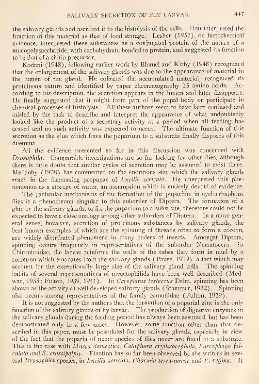
SALIVARY SECRETION OF FLY LARVAE 447
the salivary glands and ascribed it to the histolysis of the cells. Hsu interpreted the
function of this material as that of food storage. Lesher (1952), on histochemical
evidence, interpreted these substances as a conjugated protein of the nature of a
mucopolysaccharide, with carbohydrate bonded to protein, and suggested its function
to be that of a chitin precursor.Kodani (1948), following earlier work by Blumel and Kirby (1948) recognized
that the enlargement of the salivary glands was due to the appearance of material in
the lumen of the gland. He collected the accumulated material, recognized its
proteinous nature and identified by paper chromatography 15 amino acids. Ac-
cording to his description, the secretion appears in the lumen and later disappears.
He finally suggested that it might form part of the pupal body or participate in
chemical processes of histolysis. All these authors seem to have been confused and
misled by the task to describe and interpret the appearance of what undoubtedlylooked like the product of a secretory activity at a period when all feeding has
ceased and no such activity was expected to occur. The ultimate function of this
secretion as the glue which fixes the puparium to a substrate finally disposes of this
dilemma.
All the evidence presented so far in this discussion was concerned with
Drosophila. Comparable investigations are so far lacking for other flies, although
there is little doubt that similar cycles of secretion may be assumed to exist there.
Mellanby (1938) has commented on the enormous size which the salivary glands
reach in the diapausing prepupae of Lucilia sericata. He interpreted this phe-nomenon as a storage of water, an assumption which is entirely devoid of evidence.
The particular mechanisms of the formation of the puparium in cyclorrhaphousflies is a phenomenon singular to this suborder of Diptera. The formation of a
glue by the salivary glands, to fix the puparium to a substrate, therefore could not be
expected to have a close analogy among other suborders of Diptera. In a more gen-
eral sense, however, secretion of proteinous substances by salivary glands, the
best known examples of which are the spinning of threads often to form a cocoon,
are widely distributed phenomena in many orders of insects. Amongst Diptera,
spinning occurs frequently in representatives of the suborder Nematocera. In
Chironimidae, the larvae reinforce the walls of the tubes they form in mud by a
secretion which emanates from the salivary glands (Pause, 1919), a fact which mayaccount for the exceptionally large size of the salivary gland cells. The spinninghabits of several representatives of mycetophilids have been well described (Med-war, 1935; Fulton, 1939, 1941). In Ceroplatus testaceus Dalm. spinning has been
shown as the activity of well developed salivary glands (Stammer, 1932) . Spinningalso occurs among representatives of the family Simuliidae (Fulton, 1939).
It is not suggested by the authors that the formation of a puparial glue is the onlyfunction of the salivary glands of fly larvae. The production of digestive enzymes in
the salivary glands during the feeding period has always been assumed, but has been
demonstrated only in a few cases. However, some function other than that de-
scribed in this paper, must be postulated for the salivary glands, especially in view
of the fact that the puparia of many species of flies never are fixed to a substrate.
This is the case with Musca domestica, Calliphora erythrocephala, Sarcophaga fal-
culata and 6". crassipalpis. Fixation has so far been observed by the writers in sev-
eral Drosophila species, in Lucilia sericata, Phormia terra-novae and P. regina. It

448 G. FRAENKEL AND VICTOR J. BROOKES
is indicated from a few preliminary observations that the salivary glands of species
of flies which do not have this secretion, do not show the spectacular enlargementtowards the end of larval life, as found in the other group. A comparison of the ac-
tivities of salivary glands of many species of fly larvae would appear to be an inter-
esting and necessary subject of study.
Some of the observations recorded in this paper were made by Mrs. E. Lichtwardt
while working in this department. The authors are indebted to Dr. Betty Walshe,
Bedford College, London and Dr. John B. Buck, National Institutes of Health,
Bethesda, Md., for suggestions concerning the spinning habits of Chironomidae and
Mycetophilidae.
SUMMARY
1. The puparia of many flies become fixed to a substrate by means of a secretion
which accumulates in the lumen of the salivary glands toward the end of the larval
period and is expelled immediately before the formation of the puparium is
completed.
2. The process of fixation has been described for Drosophila melanogaster andPhormia rcgina.
3. The changes in the size and appearance of the salivary glands of Phormia
rcgina during the accumulation of the salivary secretion and after its elimination have
been described.
4. The process of secretion in the glands of Drosophila has been previously de-
scribed by other authors, but its true function has never been recognized.
LITERATURE CITED
BLUMEL, J., AND H. KIRBY, 1948. Amino acid constituents of tissues and isolated chromosomes
of Drosophila. Proc. Nat. Acad. Sci.. 34: 561-567.
BODENSTEIN, D., 1943. Factors influencing growth and metamorphosis of the salivary gland in
Drosophila. Biol. Bull., 84 : 13-33.
BODENSTEIN, D., 1950. Chapter 4, in Biology of Drosophila, by M. Demerec, John Wiley & Sons,
Inc., New York.
FRAENKEL, G., 1952. A function of the salivary glands of the larvae of DrosopJiila and other
flies. Biol. Bull, 103 : 285-286.
FRAENKEL, G., AND K. M. RUDALL, 1940. A study of the physical and chemical properties of
the insect cuticle. Proc. Roy. Soc. London, Scr. B. 129: 1-35.
FULTON, B. B., 1939. Lochetic luminous dipterous larvae. /. Elisha Mitchell Sci. Soc., 55 : 289-
293.
FULTON, B. B., 1941. A luminous fly larva with spider traits. Ann. Ent. Soc. Amcr., 34: 288-302.
Hsu, W. S., 1948. The golgi material and mitochondria in the salivary gland of the larva of
Drosophila melanogaster. Quart. J. Micro. Sci., 89: 401-414.
KODANI, M., 1948. The protein of the salivary gland secretion in Drosophila. Proc. Nat. Acad.
Sci., 34: 131-135.
LESHER, S., 1951. Studies on the larval salivary gland of Drosophila. I. The nucleic acids.
Exp. Cell Res.,2: 577-585.
LESHER, S., 1952. Studies on the larval salivary gland of Drosophila. III. The histochemical
localization and possible significance of ribonucleic acid, alkaline phosphatase and poly-saccharide. Anat. Rcc., 114: 633-644.

SALIVARY SECRETION OF FLY LARVAE 449
MEDWAR, S., 1935. The biology and morphology of the immature stages of Macrocera anglica.
Psyche, 42 : 25-34.
MELLANBY, K., 1938. Diapause and metamorphosis of the blowfly Lucilia sericata Meig.
Parasitology. 30: 392-402.
PAINTER, T. S., 1945. Nuclear phenomena associated with secretion in certain gland cells with
special reference to the origin of cytoplasmic nucleic acid. /. Exp. Zool., 100: 523-541.
PAUSE, J., 1919. Beitrage zur Biologic und Physiologic der Larve von Chironomus gregarious.Zool. Jahrb. Abt. Allg. Zool. Physiol, 36: 339^52.
Ross, E. B., 1939. The postembryonic development of the salivary gland of Drosophila melano-
gaster. J. Morphol, 65: 471-496.
STAMMER, H. J., 1932. Zur Biologic und Anatomie der leuchtenden Pilzmiickenlarven von
Ccroplatits tcstaccus Dalm. (Dipt., Fungivoridae). Zeitschr. Morph. Okol. Ticre, 26:
135-146.