Procedures for the reconstruction, primary culture and ...woodcm/Woodblog/wp... · as primary...
9
© 2016 Nature America, Inc. All rights reserved. PROTOCOL 490 | VOL.11 NO.3 | 2016 | NATURE PROTOCOLS INTRODUCTION The freshwater fish gill is a multifunctional organ, and it is the site of gas exchange, osmoregulation, trace metal transport, nitrogenous waste excretion and xenobiotic uptake 1–4 . Fish are commonly used as indicator species to identify environmental hazards and, as their gills are constantly in contact with water, the gill epithelium is a focal point for countless studies that seek to understand deleterious effects of environmental toxicants 2 . Whole-animal studies can use millions of fish worldwide each year, and efforts are focusing on refining, reducing and replacing (3Rs) these numbers 5 . Alternative animal-free methods used in fish toxicology include the use of immortalized cell lines, as well as primary cultures of cells from fish organs. In the present article, we describe a protocol for the primary culture of a freshwater rainbow trout gill epithelium on a flat permeable membrane, via a repeated gill cell seeding protocol. This double-seeded insert (DSI) technique produces a heterogeneous epithelium similar to that found in vivo, and it was first described in Fletcher et al. 6 . We have used the DSI for studies investigating gill physiology and environmental monitoring 7–20 . Development of the DSI technique Primary culture of a rainbow trout gill epithelium was pioneered by Pärt et al. 21 . Wood and Pärt 22 subsequently developed the single-seeded insert (SSI technique), in which isolated gill cells were cultured on permeable membrane inserts for the first time. This provided a two-chamber system that allowed the cells to grow into a polarized epithelium with an apical (water-facing) and basolateral (blood- or medium-facing) surface. The preparation exhibited bar- rier properties that were similar to those of the gill epithelium, and it tolerated apical water exposure for limited time periods. However, the epithelium comprised only a cell population of pavement cells (PVCs), which are the respiratory cells of the gill epithelium. This technique was further developed into the DSI technique 6 whereby primary cells are seeded on inserts over 2 d, producing a gill epithelium of multiple cell layers and types that includes PVCs and mitochondrion-rich cells (MRCs); the latter is associated with the transport of a number of ions and compounds across the fish gill 1 . The DSI procedure produces tight epithelia with transepithe- lial resistance (TER) values of up to 34,000 Ω cm 2 , which are much higher than that of SSI (typically 1,000–5,000 Ω cm 2 ) 23 . The preparation expresses tight junction (TJ) proteins such as occludin, claudins, zonula occludens 7,8,24 and tricellulin 9 . The preparation also exhibits ion movement between the apical and basolateral compartments 10–12 . Active Ca 2+ transport in the cell culture system resembles that of the intact fish 10,12 , although so far it has not been possible to produce the net uptake of Na + and Cl − ions present in freshwater fish in cell culture. Applications of the protocol Primary cultured gill epithelia have been used to assess how hormones and environmental conditions influence TJ protein expression and regulate epithelial permeability 7,8 . Further applications of the gill cell culture technique include studies on cytochrome P450 activity 14 , lipid metabolism 15 , ammonia trans- port 16 , cytotoxicity 25 and metal-binding characteristics 17 . A recent study also used the DSI preparation to analyze the transport of pharmaceuticals across the gill, and found that, for some, facilitated transport processes may have a role at environmentally relevant concentrations 18 . It has also been used as an environ- mental water monitoring tool of urban streams 13 and in metal- polluted rivers 19 . However, these monitoring studies are restricted to a limited time frame, because the cultures can only withstand water exposure of up to 48 h. Thus, there is scope for future devel- opment of the cell culture technique to improve both active ion transport characteristics and extended water tolerance. Generation of reliable in vitro cell assay results as an alternative to whole-animal tests for hydrophobic and volatile compounds has been proven to be difficult. This is because these compounds adhere to the plastic and/or are volatized, which results in exposure con- centrations in the small volumes used in in vitro assays that deviate substantially from the nominal concentrations. In addition, these hydrophobic compounds do not readily dissolve in medium, and to Procedures for the reconstruction, primary culture and experimental use of rainbow trout gill epithelia Sabine Schnell 1,6 , Lucy C Stott 1,6 , Christer Hogstrand 1 , Chris M Wood 2 , Scott P Kelly 3 , Peter Pärt 4 , Stewart F Owen 5 & Nic R Bury 1 1 Division of Diabetes and Nutritional Sciences, Metal Metabolism Group, Faculty of Life Sciences and Medicine, King’s College London, London, UK. 2 Department of Zoology, University of British Columbia, Vancouver, British Columbia, Canada. 3 Department of Biology, York University, Toronto, Canada. 4 European Commission, Joint Research Centre, Institute of Environment and Sustainability, Ispra (Varese), Italy. 5 Global Safety Health and Environment, AstraZeneca, Alderley Park, Macclesfield, Cheshire, UK. 6 These authors contributed equally to this work. Correspondence should be addressed to S.S. ([email protected]), C.H. (christer.hogstrand@kcl. ac.uk) or N.R.B. ([email protected]). Published online 11 February 2016; doi:10.1038/nprot.2016.029 This protocol describes how to reconstruct and culture the freshwater rainbow trout gill epithelium on flat permeable membrane supports within cell culture inserts. The protocol describes gill cell isolation, cultured gill epithelium formation, maintenance, monitoring and preparation for use in experimental procedures. To produce a heterogeneous gill epithelium, as seen in vivo, seeding of isolated gill cells twice over a 2-d period is required. As a consequence, this is termed the double-seeded insert technique. Approximately 5–12 d after cell isolation and seeding, preparations develop electrically tight gill epithelia that can withstand freshwater on the apical cell surface. The system can be used to study freshwater gill physiology, and it is a humane alternative for toxicity testing, bioaccumulation studies and environmental water quality monitoring.
Transcript of Procedures for the reconstruction, primary culture and ...woodcm/Woodblog/wp... · as primary...

©20
16N
atu
re A
mer
ica,
Inc.
All
rig
hts
res
erve
d.
protocol
490 | VOL.11 NO.3 | 2016 | nature protocols
IntroDuctIonThe freshwater fish gill is a multifunctional organ, and it is the site of gas exchange, osmoregulation, trace metal transport, nitrogenous waste excretion and xenobiotic uptake1–4. Fish are commonly used as indicator species to identify environmental hazards and, as their gills are constantly in contact with water, the gill epithelium is a focal point for countless studies that seek to understand deleterious effects of environmental toxicants2. Whole-animal studies can use millions of fish worldwide each year, and efforts are focusing on refining, reducing and replacing (3Rs) these numbers5. Alternative animal-free methods used in fish toxicology include the use of immortalized cell lines, as well as primary cultures of cells from fish organs. In the present article, we describe a protocol for the primary culture of a freshwater rainbow trout gill epithelium on a flat permeable membrane, via a repeated gill cell seeding protocol. This double-seeded insert (DSI) technique produces a heterogeneous epithelium similar to that found in vivo, and it was first described in Fletcher et al.6. We have used the DSI for studies investigating gill physiology and environmental monitoring7–20.
Development of the DSI techniquePrimary culture of a rainbow trout gill epithelium was pioneered by Pärt et al.21. Wood and Pärt22 subsequently developed the single-seeded insert (SSI technique), in which isolated gill cells were cultured on permeable membrane inserts for the first time. This provided a two-chamber system that allowed the cells to grow into a polarized epithelium with an apical (water-facing) and basolateral (blood- or medium-facing) surface. The preparation exhibited bar-rier properties that were similar to those of the gill epithelium, and it tolerated apical water exposure for limited time periods. However, the epithelium comprised only a cell population of pavement cells (PVCs), which are the respiratory cells of the gill epithelium.
This technique was further developed into the DSI technique6 whereby primary cells are seeded on inserts over 2 d, producing a gill epithelium of multiple cell layers and types that includes PVCs and mitochondrion-rich cells (MRCs); the latter is associated with the
transport of a number of ions and compounds across the fish gill1. The DSI procedure produces tight epithelia with transepithe-lial resistance (TER) values of up to 34,000 Ω cm2, which are much higher than that of SSI (typically 1,000–5,000 Ω cm2)23. The preparation expresses tight junction (TJ) proteins such as occludin, claudins, zonula occludens7,8,24 and tricellulin9. The preparation also exhibits ion movement between the apical and basolateral compartments10–12. Active Ca2+ transport in the cell culture system resembles that of the intact fish10,12, although so far it has not been possible to produce the net uptake of Na+ and Cl− ions present in freshwater fish in cell culture.
Applications of the protocolPrimary cultured gill epithelia have been used to assess how hormones and environmental conditions influence TJ protein expression and regulate epithelial permeability7,8. Further applications of the gill cell culture technique include studies on cytochrome P450 activity14, lipid metabolism15, ammonia trans-port16, cytotoxicity25 and metal-binding characteristics17. A recent study also used the DSI preparation to analyze the transport of pharmaceuticals across the gill, and found that, for some, facilitated transport processes may have a role at environmentally relevant concentrations18. It has also been used as an environ-mental water monitoring tool of urban streams13 and in metal- polluted rivers19. However, these monitoring studies are restricted to a limited time frame, because the cultures can only withstand water exposure of up to 48 h. Thus, there is scope for future devel-opment of the cell culture technique to improve both active ion transport characteristics and extended water tolerance.
Generation of reliable in vitro cell assay results as an alternative to whole-animal tests for hydrophobic and volatile compounds has been proven to be difficult. This is because these compounds adhere to the plastic and/or are volatized, which results in exposure con-centrations in the small volumes used in in vitro assays that deviate substantially from the nominal concentrations. In addition, these hydrophobic compounds do not readily dissolve in medium, and to
Procedures for the reconstruction, primary culture and experimental use of rainbow trout gill epitheliaSabine Schnell1,6, Lucy C Stott1,6, Christer Hogstrand1, Chris M Wood2, Scott P Kelly3, Peter Pärt4, Stewart F Owen5 & Nic R Bury1
1Division of Diabetes and Nutritional Sciences, Metal Metabolism Group, Faculty of Life Sciences and Medicine, King’s College London, London, UK. 2Department of Zoology, University of British Columbia, Vancouver, British Columbia, Canada. 3Department of Biology, York University, Toronto, Canada. 4European Commission, Joint Research Centre, Institute of Environment and Sustainability, Ispra (Varese), Italy. 5Global Safety Health and Environment, AstraZeneca, Alderley Park, Macclesfield, Cheshire, UK. 6These authors contributed equally to this work. Correspondence should be addressed to S.S. ([email protected]), C.H. ([email protected]) or N.R.B. ([email protected]).
Published online 11 February 2016; doi:10.1038/nprot.2016.029
this protocol describes how to reconstruct and culture the freshwater rainbow trout gill epithelium on flat permeable membrane supports within cell culture inserts. the protocol describes gill cell isolation, cultured gill epithelium formation, maintenance, monitoring and preparation for use in experimental procedures. to produce a heterogeneous gill epithelium, as seen in vivo, seeding of isolated gill cells twice over a 2-d period is required. as a consequence, this is termed the double-seeded insert technique. approximately 5–12 d after cell isolation and seeding, preparations develop electrically tight gill epithelia that can withstand freshwater on the apical cell surface. the system can be used to study freshwater gill physiology, and it is a humane alternative for toxicity testing, bioaccumulation studies and environmental water quality monitoring.

©20
16N
atu
re A
mer
ica,
Inc.
All
rig
hts
res
erve
d.
protocol
nature protocols | VOL.11 NO.3 | 2016 | 491
increase solubility they are administered with co-solvents that may also exert a cellular response in the assay system. Kramer et al.26 described a solvent-free dosing system for hydrophobic compounds in which the immortalized rainbow trout gill cell line RTgill-W1 was grown on the underside of the membrane of an insert. This allows a polydimethylsiloxane membrane preloaded with test compound to be placed on the bottom of the wells in close proximity to but not touching the cells, which in turn allows for a constant dos-ing concentration26. This technique is termed the double-seeded inverted insert (DSII) technique, and it can therefore be used in the development of a partition-controlled dosing system for hydro-phobic compounds. We describe a modification to the method used by Kramer et al.26 in which primary gill cells are cultured on an inverted insert. Characterization of the resulting primary gill epithe-lium growing in the DSII configuration is shown in Supplementary Figure 1 and was carried out as described in the Supplementary Methods. The advantage of the DSII system is that the cultured epithelia resembles that of the intact gill (Supplementary Fig. 1),
and it can be used in a similar way to the RTgill-W1 cell line when grown on inverted inserts for solvent-free dosing of hydrophobic compounds. However, given that we have only obtained preliminary data to date using the primary cultured epithelium in the DSII con-figuration, this method may require further optimization to ensure that it is optimally set up as required for specific experiments.
Comparison with other techniquesBols et al.27 described the isolation of the RTgill-W1 rainbow trout gill cell line. The main difference between using the cell line and the primary gill cell culture is that when using the primary gill cell culture, both the DSI and DSII contain the different cell types present in the in vivo gill. The epithelium is polarized and electrically tight, with TER values of >20 kΩ cm2 compared with TER values in the range of ~200 Ω cm2 when RTgill W1 cells are used. Importantly, the DSI and DSII tolerate the application of freshwater on the apical surface, which means that these primary cell cultures are better suited for gill physiology and functional studies than the cell line. However, the RTgill-W1 cell line viability28 and cell growth29 have been shown to correlate, in most cases, to both whole fish mortality and growth on exposure to pollutants. Thus, the RTgill-W1 cell line shows promise as a replacement for fish in toxicity testing.
In a number of countries, fish are used in whole effluent toxicity testing as part of environmental monitoring programs. The DSI can be used to assist in environmental monitoring because in addition to tolerating the application of freshwater in the laboratory, the DSI also tolerates the application of river water, as shown in two studies13,19. In one of these studies19, we used the knowledge that the DSI responds to metals (assessed via the
a
b
Figure 1 | Diagram of the primary cultured rainbow trout epithelium. (a,b) Reconstructed as DSI (a) or DSII (b) preparations. The epithelium attaches to the permeable membrane supports and polarizes so as to have an apical and basolateral cell surface (right). Epithelia are bathed on both sides with cell culture medium supplemented with FBS and antibiotics during development (left). For experimental purposes, freshwater can replace cell culture medium on the apical surface, whereas the basolateral surface is bathed in L-15 (without FBS or antibiotics).
PBS
Prepare reagents
Cutfilaments
Separatefilaments
250g4 °C 4 min
250g4 °C
10 min
800 µl1,000 µl
200 µl Day 0
24 h
Wash 2 × 200 µl of PBS
Seed cells1.2 × 106
800 µl1,000 µl
1.5 ml2.0 ml
1.5 ml2.0 ml
200 µl Day 1
Day 2
DSI DSII
24 h
Wash 2 × 200 µl of PBS
Seed cells1.2 × 106
250g4 °C
10 min
250g4 °C 4 min
Excise 8gill arches
2. Trypsinize×2
4. Rinse 5. Culturemedium
3. Stop
Cell count
(agitate,100- µm
cell strainer)
1. WashPBSprewash
1. Wash 1. Wash
Steps 1–4
Steps 5–7
Steps 8–13
Steps 14–19
Steps 20–25
Steps 26 and 27
Condition inserts200 µl of cell culturemedium with FBS
and antibiotics (day 0)1 2 3 4 5
Figure 2 | Summarized flow diagram of rainbow trout gill cell isolation and seeding procedures. The reagents are prepared (see Reagent Setup) before excision of the gills; the filaments are teased apart and washed in PBS (PROCEDURE Steps 1–10). The filaments are centrifuged and washed before being treated with a trypsin solution to isolate cells (PROCEDURE Steps 11–16). The isolated cells are passed through a 100-µm strainer into a stop solution (see Reagent Setup; PROCEDURE Steps 17–21). The cell suspension is centrifuged and resuspended in cell culture medium containing FBS and antibiotics, and the cell density is determined (PROCEDURE Steps 18–25). The cells are seeded onto inserts for the double-seeded insert protocol (PROCEDURE Step 26A(i)) or onto inverted inserts for the double-seeded inverted insert protocol (PROCEDURE Step 26B(i)); this is termed day 0. The gill cell isolation procedure is repeated on day 1, and the inserts from the first seeding are washed with PBS before a second seed is placed on top (PROCEDURE Step 26A(ii–viii), 26B(ii–vii)). On day 2, the cells are washed and for the DSII protocol the insert is placed in the companion wells.
©20
16N
atu
re A
mer
ica,
Inc.
All
rig
hts
res
erve
d.
protocol
492 | VOL.11 NO.3 | 2016 | nature protocols
measurement of an increase in expression of the metal-responsive gene encoding metallothionein), mimics the physiological effect of metals on whole organisms in the laboratory20 and conducted a trial whereby the DSI were exposed to natural metal contaminated water in the field19. The field trial study indicated that the DSI responded to metals in this complex matrix (river water), thereby showing that it is able to detect bioreactive metals. However, fur-ther experiments are required to assess the response of the DSI to other contaminants in more complex contaminant mixtures13 and to determine its versatility for environmental monitoring.
Experimental designThe gill cells from two (~80 g) fish provide ~40 individual culture inserts, but this can increase to 72 depending on the success of the cell isolation, and thus this technique has the potential to reduce the number of organisms used in experiments. Therefore, at a practical level, in order to allow the comparison of toxicologi-cal data from different aquatic ecotoxicology and physiological
studies, a standardized procedure for DSI and DSII production is required. Here we present the most current and readily available standardized DSI technique (Fig. 1a), as well as the development of a DSII (Fig. 1b) technique. The protocols for the DSI and DSII techniques are summarized in a flow diagram (Fig. 2), and typi-cal characteristics of the cultured epithelia are described in the ANTICIPATED RESULTS section. The controls for an experiment using DSI and DSII will depend on their intended use. For a typi-cal experiment in which the cells are to be exposed to a compound in water13,18–20, a set of control inserts that are exposed to either medium or water without the test compound should be set up. By comparing results, such as expression of a gene of interest, between the test and controls, it is possible to determine whether the response is due to the effect of exposure to water, or to water and the test compound. To perform experiments that provide statistical rigor, it is recommended that the experiment include inserts derived from several different preparations—i.e., biologi-cal replicates.
MaterIalsREAGENTS
Juvenile rainbow trout (<100 g) should be acclimatized for at least 2 weeks and maintained according to relevant governmental and institutional guidelines at 13–14 °C in good-quality water with constant 14-h light and 10-h dark photoperiods crItIcal The fish must be kept in optimum conditions. The water must be well aerated and of good quality, and fish should eat well and not show signs of stress. ! cautIon Experiments must conform to relevant institutional and governmental regulations.Leibovitz’s L-15 medium, no phenol red, with l-glutamine (Invitrogen, cat. no. 21083-027) crItIcal We recommend using Leibovitz’s L-15 medium without phenol red, because there has been reports that phenol red may contain estrogenic activity that may influence physiological or toxicological responses30.FBS (Sigma-Aldrich, cat. no. F7524)Penicillin-streptomycin (5,000 units ml−1 penicillin and 5 mg ml−1 streptomycin; Invitrogen, cat. no. 15070063)Gentamicin solution (10 mg ml−1; Invitrogen, cat. no. 15710049)Fungizone (100 mg; Sigma-Aldrich, cat. no. A9528)Trypsin-EDTA, 0.5% (wt/vol), ×10 (Invitrogen, cat. no. 15400054)PBS (used as prewash solution)Ethanol (VWR International, cat. no. 20821.321)Calcium chloride dihydrate (Sigma-Aldrich, cat. no. 223506)Magnesium sulfate heptahydrate (Sigma-Aldrich, cat. no. 230391)Sodium bicarbonate (Sigma-Aldrich, cat. no. S6014)Potassium chloride (Sigma-Aldrich, cat. no. P9333)
EQUIPMENTLaminar flow hood, model no. M51424/2 (Microflow biological safety cabinet)Centrifuge, refrigerated, 5810R, rotor A-4-62 (Eppendorf)Vortex, F20220176 (VELP Scientifica)Dissecting equipment; large sharp knife, forceps (SLS, cat. no. INS4291; 00:SA), scissors (VWR International, cat. no. INS4808; INS4854) and scalpels (Swann-Morton, cat. no. 0501) crItIcal All equipment must be autoclaved or sterilized using 70% (vol/vol) ethanol.Portable gyratory shaker, model IKA-VIBRAX-VXRAdjustable pipettes: P-20 (Gilson, cat. no. FA10003M), P-200 (Gilson, cat. no. FA10005M) and P-1000 (Gilson, cat. no. F123602)Pipette tips: 20 µl (Starlab, cat. no. S1120-1810), 200 µl (Starlab, cat. no. S1120-8810) and 1,000 µl (Starlab, cat. no. S1122-1830)Tissue culture pipettes: 5 ml (Corning, cat. no. CORN4051), 10 ml (Corning, cat. no. CORN4101) and 25 ml (Corning, cat. no. CORN4250)Inverted phase-contrast microscope, TE200, Nikon EclipseCell culture cooling incubator, MIR-1554, Sanyo, 18 °C, no CO2 atmosphere requiredHemocytometer (Neubauer)
•
•
••
•••••••••
•
•••
••
•
•
••
•
EVOM epithelial voltohmmeter modified by the manufacturer to read TER up to 100,000 Ω cm−2 (World Precision Instruments)EVOM ‘chopstick’ electrodes, no. STX-2 (World Precision Instruments)Inserts, 12-well, 0.4 µm, 0.9 cm2 effective growth area, polyethylene terephthalate (PET) membrane (BD Falcon, cat. no. 353180)Companion cell culture plates, 12-well with low evaporation lid, notched (BD Falcon, cat. no. 353503)Sterile tip box with damp cotton wool and glass slides insideSterile syringe filter, 0.2 µm (VWR International, cat. no. 194-2520)Cell strainers, 100 µm, Nylon (BD Falcon, cat. no. 352360)Falcon 50-ml conical tubes (BD Falcon, cat. no. 352070)Sterile Petri dishes (VWR International, cat. no. 25382-166)
REAGENT SETUPPBS PBS is 137 mM sodium chloride (Sigma-Aldrich, cat. no. 746398), 2.7 mM potassium chloride (Sigma-Aldrich, cat. no. P9333), 10 mM disodium hydro-gen phosphate (Sigma-Aldrich, cat. no. 255793) and 1.8 mM potassium dihy-drogen phosphate (Sigma-Aldrich, cat. no. P0662). It is prepared in dH2O and autoclaved. This can be stored for 1 month at room temperature (~20 °C). Before use, place it on ice.Ethanol, 70% Prepare a 70% (vol/vol) solution of ethanol in dH2O.Fungizone Prepare a 10 mg ml−1 solution and store it in 1-ml aliquots at −20 °C until further use.Wash solution Add 1.2 ml of penicillin-streptomycin, 1.2 ml of gentamicin and 90 µl of Fungizone solution to a final volume of 30 ml of PBS. Prepare the solution at room temperature in sterile conditions in a tissue culture hood on the day of cell isolation. Before use, place it on ice.Trypsin solution Dilute 0.5% (wt/vol) trypsin-EDTA to 0.05% by pipetting 650 µl into a conical centrifuge tube and by bringing the volume up to 6.5 ml with PBS. Prepare the solution at room temperature in sterile conditions in a tissue culture hood on the day of cell isolation. Stop solution Pipette 2 ml of FBS into a conical centrifuge tube with 18 ml of PBS. Prepare it at room temperature in sterile conditions in a tissue culture hood on the day of cell isolation. Before use, place it on ice.Rinse solution Add 0.5 ml of FBS in a tube, and make up the final volume to 19.5 ml with PBS. Prepare this solution at room temperature in sterile conditions in a tissue culture hood on the day of cell isolation. Before use, place it on ice.Cell culture medium with antibiotics and FBS (medium A) Pipette 27 ml of FBS, 11 ml of penicillin-streptomycin and 11 ml of gentamicin, all sterile filtered, to 500 ml of L-15 medium. This can be made in advance and stored at 4 °C for up to 1 month.Cell culture medium with FBS (medium B) Pipette 27 ml of FBS, sterile filtered, to 500 ml of L-15 medium. This can be made in advance and stored at 4 °C for up to 1 month.
•
••
•
•••••

©20
16N
atu
re A
mer
ica,
Inc.
All
rig
hts
res
erve
d.
protocol
nature protocols | VOL.11 NO.3 | 2016 | 493
Exposure cell culture medium L-15 without any supplementation can be stored at 4 °C for up to 1 month.Artificial freshwater Freshwater is defined as water prepared to the standard used for maintaining fish, such as that recommended by the Organisation for Economic Co-operation and Development (OECD) and the International Organization for Standardization (ISO). To make up OECD freshwater, prepare the following four solutions (40× stocks). Solution 1: dissolve 11.76 g of CaCl2.2H2O in dH2O, and adjust the volume to 1 liter with dH2O (0.08 M). Solution 2: dissolve 4.93 g of MgSO4.7H2O in dH2O,
and adjust the volume to 1 liter with dH2O (0.02 M). Solution 3: dissolve 2.59 g of NaHCO3 in dH2O, and adjust the volume to 1 liter with dH2O (0.03 M). Solution 4: dissolve 0.23 g of KCl in dH2O, and adjust the volume to 1 liter (0.003 M). Once all the solutions are prepared, mix 25 ml of each solution, and the total volume is made up to 1 liter with dH2O. Aerate the dilution water until oxygen saturation is achieved, and then store it for about 2 d with further aeration before use23.Natural freshwater Filter natural freshwater samples using a 0.2-µm sterile syringe filter before exposure to the cell culture.
proceDurepreparation of solutions and culture inserts tIMInG 1 h, plus 1.5 h of incubation to condition the membrane crItIcal The recommended volumes in this section are appropriate for one cell isolation procedure.1| Prepare the two types of cell culture media with antibiotics (medium A) and without antibiotics (medium B; see Reagent Setup). Take a 50-ml aliquot of each and keep them on ice.
2| Prepare the prewash solution (18 ml), wash solution, trypsin solution, stop solution and rinse solution, and keep them on ice.
3| In the cell culture hood, remove the permeable membrane inserts and companion cell culture plates from their packaging. For the DSI technique, place inserts in the wells of cell culture plates and place the lid on top. For DSII, turn the inserts upside down in a humidified chamber. To generate the humidified chamber, we use a sterile pipette tip box with damp sterile cotton wool. crItIcal step For DSI, the flanges of the inserts rest between the notches of the well to minimize medium wicking and to assure the proper fitting of the plate with the lid. crItIcal step For DSII, microscope slides can be placed on top of the cotton wool in the tip box to create a stable and flat surface on which to place the upside-down inserts. This prevents the medium from spilling over the edge of the inverted insert.
4| Condition permeable membrane inserts by applying cell culture medium with FBS and antibiotics (A) to the surface of the insert on which the cells will be seeded. For DSI preparations, add 200 µl of cell culture medium with FBS and antibiotics (A) in the upper compartment. For DSII, add 200 µl of cell culture medium with FBS and antibiotics (A) onto the top of the upside-down insert. Close the systems and keep them at 18 °C in a cell culture incubator for at least 1.5 h.
rainbow trout dissection and gill cell isolation tIMInG 3 h5| Euthanize one rainbow trout.! cautIon Euthanize rainbow trout according to national and institutional guidelines. We follow UK Home Office Schedule 1 procedures in which the fish is euthanized via the destruction of the brain, which is achieved by a blow to the cranium and the use of a sharp probe.
6| Remove the head behind the operculum with a sharp knife. crItIcal step It is important to perform this procedure quickly so as to reduce the extent of gill hemorrhaging and to avoid excessive bleeding and clotting on the filaments, which will reduce the quality of the gill cells obtained.
7| With dissecting scissors and forceps, remove the operculum (Fig. 3a) and excise out the intact gill arches by cutting the dorsal and ventral cartilage of each gill arch. Place the gill arches into 10 ml of prewash solution in a Petri dish (Fig. 3b). crItIcal step Avoid damage to the filaments by holding the cartilage of the arch and not the filaments.
8| Remove mucus by carefully blotting each gill arch onto tissue paper, and then cut the filaments from the arches and place the filaments into 10 ml of wash solution (prepared in Reagent Setup) in a Petri dish. crItIcal step Do not cut the filaments too close to the cartilage of the gill arch, as this will contaminate the cell culture with fibroblasts.
9| Tease the filaments apart using forceps, and cut them with a scalpel. This should result in filaments separated in bundles of 1–5. This increases the surface areas, which facilitates the action of the trypsin solution later.
10| Pour away this wash solution, and add the filaments to a 10-ml aliquot of wash solution in a 50-ml conical centrifuge tube, and place the tube on ice. Incubate the tube for 10 min. crItIcal step From this step onward, sterile culture technique must be used, and the procedures should therefore take place in a laminar flow hood.
©20
16N
atu
re A
mer
ica,
Inc.
All
rig
hts
res
erve
d.
protocol
494 | VOL.11 NO.3 | 2016 | nature protocols
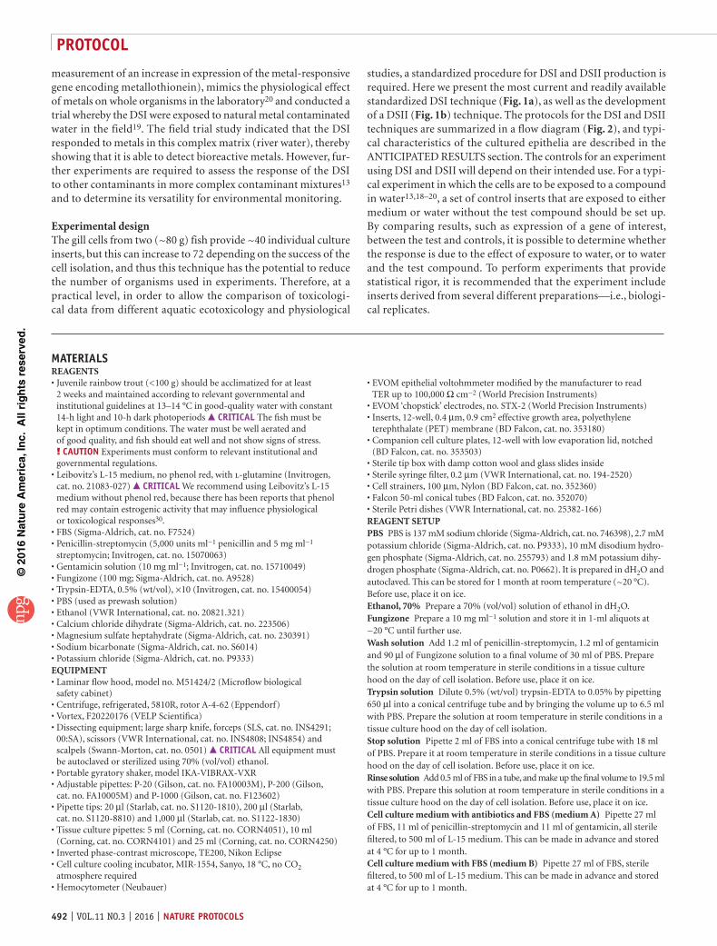
11| Centrifuge the tube for 4 min at 250g at 4 °C to collect the filaments. Aspirate and discard the supernatant.
12| Add the last remaining 10 ml of wash solution and moderately mix to dislodge the filaments. Incubate the tube on ice for a further 10 min.
13| Centrifuge again for 4 min at 250g at 4 °C, and then aspirate and discard the supernatant.
14| Add 500 µl of trypsin solution to the pelleted filaments, and agitate to remove any remaining wash solution. Centrifuge the tube for 4 min at 250g at 4 °C, and then aspirate and discard the supernatant.
15| Add 3 ml of trypsin solution to begin tryptic digestion, and shake for 12 min at 200 r.p.m. at room temperature.
16| To mechanically agitate the cut gill filaments and to improve cell yield, pass the filaments in trypsin solution repeatedly through a wide-bore pipette tip immediately after the 12-min trypsinization period. A wide-bore pipette tip can be prepared by removing the tip of a 1,000-µl pipette tip, so that the bore of the pipette aperture is ~2 mm. Sterilize by storing the cut pipette tip in 70% (vol/vol) ethanol. Before use, blot the wide-bore tip on tissue to remove residual ethanol, and pass the filaments/trypsin solution up and down at least 50 times.
17| Place the 100-µm cell strainer onto the 50-ml conical centrifuge tube containing 20 ml of stop solution (prepared in Reagent Setup) on ice. Pipette the filament/trypsin solution into the strainer, and agitate it to facilitate the collection of gill cells as a single-cell suspension in stop solution. crItIcal step After collection of cells, gently invert the conical centrifuge tube to mix the gill cells with the stop solution.
18| Remove the filaments from the strainer and replace them back into their original conical centrifuge tube. Repeat tryptic digestion by adding the remaining 3 ml of trypsin solution and by shaking for a further 12 min at 200 r.p.m. at room temperature.
19| Place a new 100-µm cell strainer onto the same 50-ml conical centrifuge tube containing the stop solution and the previously trypsinized cells, and repeat the gill cell isolation as in Step 13.
20| Centrifuge the cell suspension for 10 min at 250g at 4 °C. A red pellet should be obtained (Fig. 3c). Aspirate and discard the supernatant.? trouBlesHootInG
21| Resuspend the red pellet in 2 ml of rinse solution (prepared in Reagent Setup), and then add the remaining 18 ml; agitate the mixture briefly and centrifuge it for 10 min at 250g at 4 °C. This should result in another red-colored pellet (Fig. 3d). Aspirate and discard the supernatant.
22| Resuspend the red pellet initially in 2 ml of cell culture medium with FBS and antibiotics (medium A), and flick the tube to dislodge the pellet many times; next, resuspend in 8 ml (for a total of 10 ml) and mix it well.
23| Add 10 µl of cell suspension to 90 µl of trypan blue solution in a 0.5-ml bullet centrifuge tube, and mix thoroughly using a vortex.
24| Count the viable cells using a hemocytometer on the basis of the trypan blue exclusion criterion. crItIcal step Exclude blood cells from the cell count. These appear as ellipsoidal cells, which are smaller than the gill cells.
a b
c d
Figure 3 | Dissection of the gills and isolation of the cells. (a) Four gill arches are located underneath each operculum on the lateral sides of the rainbow trout head. Scale bar, 0.5 cm. (b) Gill arches are excised by cutting the cartilage of the arch, and these are placed into 20 ml of prewash solution (PBS). Scale bar, 1 cm. (c,d) The gill cell pellet is obtained after centrifugation in Step 21 (c) and Step 22 (d). Trapped red blood cells give the red color. Scale bars, 1 cm.

©20
16N
atu
re A
mer
ica,
Inc.
All
rig
hts
res
erve
d.
protocol
nature protocols | VOL.11 NO.3 | 2016 | 495
25| Determine the concentration of cells and adjust cell counts of cell suspension from Step 21 to required seeding density detailed in the following steps.? trouBlesHootInG pause poInt Cells can be left up to 2 h on ice.
cell seeding26| Follow option A to seed the cells for a DSI experiment, and follow option B to seed cells for a DSII experiment.(a) DsI cell seeding tIMInG 30 min (i) Aspirate the medium conditioning the cell culture inserts (from Step 4) before cell seeding. Seed 1.2 × 10⁶ cells
in 800 µl of cell culture medium with FBS and antibiotics (medium A) per insert. In addition, set up three blank inserts (no cells) for TER measurements. Place 1 ml of cell culture medium with FBS and antibiotics (medium A) into the basolateral compartment. This is termed ‘day 0’. crItIcal step It is recommended that when establishing the protocol an insert seeding density gradient be performed to determine the optimum seeding density for producing inserts with consistently tight epithelia10.
(ii) Incubate the cells at 18 °C overnight. (iii) After the overnight incubation, begin a new gill cell preparation using another fish, by repeating Steps 5–25. (iv) To prepare the previously seeded inserts (‘day 0’) for double-seeding (from Step 26A(i)), aspirate the medium first
from the lower compartment and then carefully from the upper compartment. Wash the surface of the inserts by slowly applying 200 µl of PBS down the side of the insert (not directly onto the attached cells to avoid disruption), and tap the edge of the insert gently at this wash stage to dislodge dead cells and mucus. crItIcal step When you are removing or changing the medium, always aspirate the lower compartment first, and then the upper one. This maintains the hydrostatic pressure onto the cells and it prevents them from lifting off.
(v) Repeat the wash as described in Step 26A(iv), with 200 µl of PBS. ? trouBlesHootInG
(vi) Carefully apply the new gill cell preparation (obtained in Step 26A(iii)) directly on top of the previously seeded cells. Seed 1.2 × 10⁶ cells in 800 µl of cell culture medium with FBS and antibiotics (medium A), and add 1 ml in the lower compartment afterward. Also apply medium to your blank insert controls. This is termed ‘day 1’. Incubate the cells at 18 °C.
(vii) After 24 h, wash the inserts again, as described in Step 26A(iv,v). (viii) Add maintenance volumes of culture medium; 1.5-ml cell culture medium with FBS and antibiotics (medium A) in the
upper compartment and then 2.0 ml in the lower compartment. This is termed ‘day 2’. Incubate the cells at 18 °C.(B) DsII cell seeding tIMInG 30 min (i) Aspirate the conditioning medium from the inverted cell culture inserts (from Step 4) before seeding. Seed 1.2 × 10⁶
cells in 200 µl of cell culture medium with FBS and antibiotics (medium A) per inverted insert, and leave them at 18 °C in the sterile tip box. Also set up three blank inserts (no cells) for blank TER measurements. This is termed day 0. crItIcal step It is recommended that when establishing the protocol an insert seeding density gradient be performed to determine the optimum seeding density to produce inserts with consistently tight epithelia10.
(ii) Incubate the cells at 18 °C overnight. (iii) After the overnight incubation and 24 h after Step 26B(i), begin a new gill cell preparation using another fish,
by repeating Steps 5–25. (iv) To prepare the previously seeded inserts (‘day 0’) for double-seeding, wash the inverted inserts by aspirating the
medium and by carefully and slowly applying 200 µl of PBS. (v) Repeat another 200-µl PBS wash.
? trouBlesHootInG (vi) Carefully apply the new gill cell preparation (obtained in Step 26B(iii)) directly on top of the previously seeded cells.
Seed 1.2 × 10⁶ cells in 200 µl of cell culture medium with FBS and antibiotics (medium A), and leave them in the sterile pipette tip box overnight at 18 °C. This is termed ‘day 1’.
(vii) Twenty-four hours later, wash the inserts again, as described in Step 26B(iv,v). The inserts can now be placed the right way up in companion wells. Replace it with maintenance volumes of culture medium: 1.5 ml of cell culture medium with FBS and antibiotics (medium A) in the upper compartment and then 2.0 ml in the lower compartment. Incubate the inserts at 18 °C. This is termed ‘day 2’.
Maintenance tIMInG 5–12 d27| Incubate the inserts until day 4. On day 4, remove the medium and replace it with cell culture medium with FBS (medium B) without antibiotics. Continue to incubate cells at 18 °C, replacing the medium every 48 h. Monitor appropriate growth of the epithelia under a light microscope (Fig. 4). Cell culture inserts should be observed on a daily basis to

©20
16N
atu
re A
mer
ica,
Inc.
All
rig
hts
res
erve
d.
protocol
496 | VOL.11 NO.3 | 2016 | nature protocols
check for (bacterial) contamination. Any contaminated cell culture inserts should be immediately removed, and the associated plate well should be rinsed with 70% (vol/vol) ethanol. From day 4 onward, TER can be monitored daily if desired, as described in the following section. crItIcal step Careful and thorough observation of culture preparations should take place before measuring TER (see following section) or changing the medium. Therefore, if one insert is found to be contaminated, it can be removed without cross-contaminating other epithelia.
Monitoring ter tIMInG 10 min crItIcal From day 4 onward, a daily measurement of TER can be made using a custom-modified (see Equipment) epithelial tissue voltohmmeter fitted with chopstick electrodes, as described in this section.28| Sterilize the electrodes in 70% (vol/vol) ethanol and rinse them in PBS.
29| Confirm that the cells are not contaminated. If the cells are showing no signs of contamination, insert the electrode over the gill cell epithelium of each insert, such that the shorter arm of the electrode is in the upper compartment and the longer is in the lower compartment (Fig. 5a), and measure TER.
30| Calculate the net TER by subtracting the average TER of the blank insert (no cells; Step 26A(i) for DSI and Step 26B(i) for DSII) from the experimental value. Obtain final resistance-area values (Ω cm2) by multiplying the net TER by the effective growth area (0.9 cm2 for 12-well inserts). During days 7–14, DSI and DSII inserts should show TER values of ~5,000–30,000 Ω cm2 (Fig. 5b and supplementary Fig. 1a). Once a TER value of 5,000 Ω cm2 or above is obtained, if desired you can proceed to assaying freshwater, as described in the next section.? trouBlesHootInG pause poInt Epithelia can be stored with medium A in both the apical and basolateral compartments for later use by maintaining them at 4 °C for up to 2 weeks31.
experimental assay tIMInG 30 min31| Remove the medium from the lower compartment, followed by the upper compartment.
32| To wash and remove traces of FBS, apply 800 µl of PBS in the upper compartment, and then apply 1,000 µl to the lower compartment.
33| Aspirate the lower compartment and wash a further two times before the final aspiration.
a b
c d
Figure 4 | Images of the double-seeded insert epithelium. (a) Phase-contrast micrograph of DSI gill epithelial cells. Scale bar, 100 µm. (b) Transmission electron micrograph of DSI gill epithelia showing the characteristic microridges on the apical surface. Scale bar, 200 nm. (c,d) Confocal microscope images of DSI epithelia in which 24 h before cell fixing, epithelia were exposed to symmetrical conditions with L-15 medium on both sides of the epithelium (c) or asymmetrical conditions with freshwater in the upper compartment and L-15 medium in the companion well (d), and cell nuclei were stained with 5 µM Hoechst (blue) and tight junctions with zonula occludens 1 antibody (green). Scale bars, 50 µm. (See supplementary Methods for details of how we stained tight junctions using zonula occluden 1–specific antibodies.)
TE
R (
Ω c
m2 )
Voltohmmeter
40,000
35,000
30,000
25,000
20,000
15,000
10,000
5,000
02 3
Time (d)4 5 6 7 8 9 10 11
a bFigure 5 | Membrane development can be monitored by daily TER measurements. (a) A custom-modified epithelial tissue voltohmmeter is fitted with chopstick electrodes. The electrode arms are inserted above and below the gill cell epithelium of each insert, such that one arm of the electrode is in the upper compartment and the other arm is in the lower (bottom) compartment. (b) Measurements of TER in developing DSI. Freshwater (blue points) was added (after the dotted line) to the companion well compartment (the apical cell surface, while the insert contained L-15 medium) on day 4, resulting in an increase in TER still evident 24 h later on day 5 (n = 6). Values are means ± s.e.m.
©20
16N
atu
re A
mer
ica,
Inc.
All
rig
hts
res
erve
d.
protocol
nature protocols | VOL.11 NO.3 | 2016 | 497
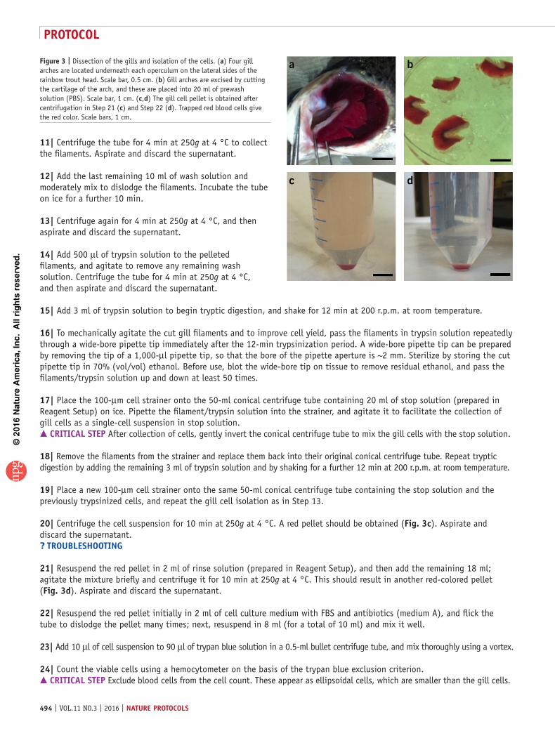
34| Apply the solution of interest, e.g., freshwater, to the test compartment. For DSI, apply 1.5 ml of freshwater into the upper compartment, while leaving the lower compartment in 2.0 ml of L-15 medium. Depending on the experimental design, FBS can be removed from the medium. For DSII, apply 1.5 ml of L-15 medium without FBS into the upper compartment; 2.0 ml of freshwater can now be applied to the lower compartment. The volume in the upper compartment can be reduced if required. Application of freshwater should cause a rise in TER (supplementary Fig. 1a). After freshwater application, inserts around 5,000 Ω cm2 may rise to 30,000 Ω cm2, whereas those already around 25,000–30,000 Ω cm2 may not rise by as much. After freshwater application, TER values should remain above 5,000 Ω cm2 for up to 48 h.
? trouBlesHootInGTroubleshooting advice can be found in table 1.
tIMInG Steps 1–4, preparation of solutions and cell culture inserts: 1 h; inserts also need to be left to condition for at least 1.5 hSteps 5–25, rainbow trout dissection and gill cell isolation: 3 hStep 26, DSI/DSII cell seeding: 30 minSteps 27–30, maintenance and monitoring: this will depend on the number of inserts, and it can take between 30 min and 2 h on the day of medium changes; cell preparation reaches maturity over 5–12 dSteps 31–34, experimental assay: 30 min
antIcIpateD resultsThe epithelia should have distinct apical and basolateral surface consisting of irregularly shaped epithelial (pavement) cells with microridges and plasma membranes (Fig. 4b and supplementary Fig. 1c,d) interspersed with MRCs (supplementary Fig. 1e) typically from day 5 onward while growing in both the DSI and DSII configurations. Both preparations should develop an electrically tight epithelium showing the presence of the TJ proteins, such as zonula occludens (Fig. 4c,d and supplementary Fig. 1f,g), typically from day 5 onward. The DSI can withstand water applied to the apical surface, but it does not tolerate water when exposed via the basolateral compartment31, and these traits are also evident in the DSII prepa-rations (supplementary Fig. 1b). On the basis of rhodamine 123 staining6, the epithelium should consist of between 10 and 15% MRCs, which is consistent with observations of the intact fish gill epithelium; however, PVCs make up the bulk of the epithelium, as is also seen in vivo1.
taBle 1 | Troubleshooting table.
step problem possible reason solution
20 Pellet is not red Excessive trypsinization
Trypsin digestion has not stopped
Check the trypsin concentration and batch, and reduce the trypsinization time Check the FBS, and if it is out of date, remake the stop solution
25 Low cell viability Excessive trypsinization Decrease the trypsinization time
Low donor quality Check the health of stock fish
Low cell number Slow work pace Perform the procedure faster
Low EDTA concentra-tions in trypsin solution (Reagent Setup)
Increase EDTA concentration to 5 mM in the trypsin solution (Reagent Setup)
26A(v), 26B(v) Extra mucus on cells Fish health–related problem Perform an additional PBS wash
30 Low TER Contamination Discard the cells
Seeding density Perform a seeding-density assay
Fish health–related problem Check the health of stock fish and water quality of the fish tank
Slow development Keep monitoring, and discard it if TER is <5,000 Ω cm2 by day 14
Note: Any Supplementary Information and Source Data files are available in the online version of the paper.
acknowleDGMents This work was funded via a grant supporting S.S. from the National Centre for the Replacement, Refinement & Reduction of Animals

©20
16N
atu
re A
mer
ica,
Inc.
All
rig
hts
res
erve
d.
protocol
498 | VOL.11 NO.3 | 2016 | nature protocols
in Research no. 26675, awarded to C.H. and N.R.B. L.C.S. was funded by a studentship from the Biotechnology and Biological Sciences Research Council (BBSRC) co-funded Case Award (BB/J500483/1) supported by the AstraZeneca Global Environment research program to N.R.B. and C.H. C.M.W.’s research is funded by a Natural Sciences and Engineering Research Council of Canada (NSERC) Discovery grant. S.P.K. is funded by an NSERC Discovery Grant. S.F.O. is an employee of AstraZeneca. AstraZeneca is a biopharmaceutical company specializing in the discovery, development, manufacturing and marketing of prescription medicines. Funding bodies had no role in the design of the study or decision to publish. The work aims to identify effective alternatives to reduce, refine and replace the use of live animals to meet environmental regulatory testing needs.
autHor contrIButIons S.S. and L.C.S. contributed equally to the manuscript and generated the data. S.S. developed the double-seeded inverted insert (DSII) technique. C.M.W., S.P.K. and P.P. developed the initial methodology. S.F.O., C.H. and N.R.B. supported the recent developments of the methods. C.H. and N.R.B. received funding for S.S. and L.C.S. that has enabled the development of the methods and expanded the use of the double-seeded insert (DSI) for the replacement of animals in toxicity testing and environmental monitoring. All authors contributed to the manuscript.
coMpetInG FInancIal Interests The authors declare no competing financial interests.
Reprints and permissions information is available online at http://www.nature.com/reprints/index.html.
1. Evans, D.H., Piermarini, P.M. & Choe, K.P The multifunctional fish gill: dominant site of gas exchange, osmoregulation, acid-base regulation, and excretion of nitrogenous waste. Physiol. Rev. 85, 97–177 (2005).
2. Wood, C.M. Toxic responses of the gill. in: Target Organ Toxicity in Marine and Freshwater Teleosts, Vol. 1 (eds. Schlenk, D.W. & Benson, W.H.) 1–89 (Taylor & Francis, 2001).
3. Mckim, J.M. & Erickson, R.J. Environmental impacts on the physiological mechanisms controlling xenobiotic transfer across fish gills. Physiol. Zool. 64, 39–67 (1991).
4. Bury, N.R., Walker, P.A. & Glover, C.N. Nutritive metal uptake in teleost fish. J. Exp. Biol. 206, 11–23 (2002).
5. Scholz, S. et al. A European perspective on alternatives to animal testing for environmental hazard identification and risk assessment. Regul. Toxicol. Pharmacol. 67, 506–530 (2013).
6. Fletcher, M., Kelly, S.P., Pärt, P., O’Donnell, M.J. & Wood, C.M Transport properties of cultured branchial epithelia from freshwater rainbow trout: a novel preparation with mitochondria-rich cells. J. Exp. Biol. 203, 1523–1537 (2000).
7. Kolosov, D., Chasiotis, H. & Kelly, S.P. Tight junction protein gene expression patterns and changes in transcript abundance during development of model fish gill epithelia. J. Exp. Biol. 217, 1667–1681 (2014).
8. Chasiotis, H., Wood, C.M. & Kelly, S.P. Cortisol reduces paracellular permeability and increases occludin abundance in cultured trout gill epithelia. Mol. Cell. Endocrinol. 323, 232–238 (2010).
9. Kolosov, D. & Kelly, S.P. A role for tricellulin in the regulation of gill epithelium permeability. Am. J. Physiol. 304, R1139–R1148 (2013).
10. Walker, P.A., Bury, N.R. & Hogstrand, C. Influence of culture conditions on metal-induced responses in a cultured rainbow trout gill epithelium. Environ. Sci. Technol. 41, 6505–6513 (2007).
11. Zhou, B., Kelly, S.P., Ianowski, J.P. & Wood, C.M. Effects of cortisol and prolactin on Na+ and Cl– transport in cultured branchial epithelia from FW rainbow trout. Am. J. Physiol. 285, R1305–R1316 (2003).
12. Kelly, S.P. & Wood, C.M. Cortisol stimulates calcium transport across cultured gill epithelia from freshwater rainbow trout. In Vitro Cell. Dev. Biol. Anim. 44, 96–104 (2008).
13. Schnell, S. et al. Environmental monitoring of urban streams using a primary fish gill cell culture system (FIGCS). Ecotoxicol. Environ. Saf. 120, 279–285 (2015).
14. Carlsson, C. & Pärt, P. 7-Ethoxyresorufin O-deethylase induction in rainbow trout gill epithelium cultured on permeable supports: asymmetrical distribution of substrate metabolites. Aquat. Toxicol. 54, 29–38 (2001).
15. Hansen, H.J.M., Kelly, S.P., Grosell, M. & Wood, C.M. Studies on lipid metabolism in trout (Oncorhynchus mykiss) branchial cultures. J. Exp. Zool. 293, 683–692 (2002).
16. Kelly, S.P. & Wood, C.M. The cultured branchial epithelium of the rainbow trout as a model for diffusive fluxes of ammonia across the fish gill. J. Exp. Biol. 204, 4115–4124 (2001).
17. Zhou, B., Nichols, J., Playle, R.C. & Wood, C.M. An in vitro biotic ligand model (BLM) for silver binding to cultured gill epithelia of freshwater rainbow trout (Oncorhynchus mykiss). Toxicol. Appl. Pharmacol. 202, 25–37 (2005).
18. Stott, L.C., Schnell, S., Hogstrand, C., Owen, S.F. & Bury, N.R. A primary fish gill cell culture model to assess pharmaceutical uptake and efflux: evidence for passive and facilitated transport. Aquat. Toxicol. 159, 127–137 (2015).
19. Minghetti, M., Schnell, S., Chadwick, M.A., Hogstrand, C. & Bury, N.R. A primary Fish Gill Cell System (FIGCS) for environmental monitoring of river waters. Aquat. Toxicol. 154, 184–192 (2014).
20. Walker, P.A., Kille, P., Scott, A., Bury, N.R. & Hogstrand, C. An in vitro method to assess toxicity of waterborne metals to fish Toxicol. 30, 67–77 (2008).
21. Pärt, P., Norrgren, L., Bergström, B. & Sjöberg, P. Primary culture of epithelial cells from rainbow trout gills. J. Exp. Biol. 175, 219–232 (1993).
22. Wood, C.M. & Pärt, P. Cultured branchial epithelia from freshwater fish gills. J. Exp. Biol. 200, 1047–1059 (1997).
23. Kelly, S.P., Fletcher, M., Pärt, P. & Wood, C.M. Procedures for the preparation and culture of ‘reconstructed’ rainbow trout branchial epithelia. Methods Cell Sci. 22, 153–163 (2000).
24. Sandbichler, A.M., Egg, M., Schwerte, T. & Pelster, B. Claudin 28b and F-actin are involved in rainbow trout gill pavement cell tight junction remodelling under osmotic stress. J. Exp. Biol. 214, 1473–1487 (2011).
25. Sandbacka, M., Pärt, P. & Isomaa, B. Gill epithelial cells as tools for toxicity screening—comparison between primary cultures, cells in suspension and epithelia on filters. Aquat. Toxicol. 46, 23–32 (1999).
26. Kramer, N.I. et al. Development of a partition-controlled dosing system for cell assays. Chem. Res. Toxicol. 23, 1806–1814 (2010).
27. Bols, N.C. et al. Development of a cell-line from primary cultures of rainbow trout, Oncorhynchus mykiss (Walbaum), gill. J. Fish Dis. 6, 601–611 (1994).
28. Tanneberger, K. et al. Predicting fish acute toxicity using a fish gill cell line-based toxicity assay. Environ. Sci. Technol. 47, 1110–1119 (2013).
29. Stadnicka-Michalak, J., Schirmer, K. & Ashauer, R. Toxicology across scales: cell population growth in vitro predicts reduced fish growth. Sci. Adv. 1, e1500302 (2015).
30. Berthios, Y., Katzenellenbogen, J.A. & Katzenellenbogen, B.S. Phenol red in tissue culture media is a weak estrogen: implications concerning the study of estrogen-responsive cells in culture. Proc. Natl. Acad. Sci. USA 86, 2496–2500 (1986).
31. Wood, C.M., Eletti, B. & Pärt, P. New methods for the primary culture of gill epithelia from freshwater rainbow trout. Fish Physiol. Biochem. 26, 329–344 (2002).