Procedure Manual - Grand County EMS · PROCEDURE MANUAL FOR THE i-STAT SYSTEM 2 REV. DATE:...
32
ART: 714446-00P REV. DATE: 23-Apr-14 Procedure Manual for the i-STAT ® System This Procedure Manual is intended to be a template for the Procedure Manual required by CLIA and laboratory accreditation bodies. This Procedure Manual should be customized for site-specific policies and procedures. This Procedure Manual is not intended to replace the System Manual. i-STAT is a registered trademark of the Abbott Group of Companies in various jurisdictions.
Transcript of Procedure Manual - Grand County EMS · PROCEDURE MANUAL FOR THE i-STAT SYSTEM 2 REV. DATE:...

ART: 714446-00P REV. DATE: 23-Apr-14
Procedure Manual for the i-STAT® System
This Procedure Manual is intended to be a template for the Procedure Manual required by CLIA and laboratory accreditation bodies. This Procedure Manual should be customized for site-specific policies and procedures. This Procedure Manual is not intended to replace the System Manual.
i-STAT is a registered trademark of the Abbott Group of Companies in various jurisdictions.

REV. DATE: 23-Apr-14 ART: 714446-00P

PROCEDURE MANUAL FOR THE i-STAT SYSTEM
REV. DATE: 23-Apr-14 ART: 714446-00P
Contents
System Overview ................................................................................................................................................................ 1 Analyzers Analysis Time Cartridges Central Data Station or Data Manager
Supplies and Storage Requirements ............................................................................................................................. 1-2 Cartridges Controls
Blood Specimens ............................................................................................................................................................. 2-5 Blood Collection Equipment Blood Volume Suitable Specimens Specimen Labeling Specimen Collection and Handling Criteria for Specimen Rejection Precautions
Procedure for Analysis ................................................................................................................................................ 5-10 Preparation for Use Procedure for Cartridge Testing Alternative Procedure
Results ......................................................................................................................................................................... 11-17 Calculations Displayed Results Suppressed Results Printing and Transmitting Results Reference Ranges and Reportable Ranges Critical Results Interferences
Quality Control .......................................................................................................................................................... 18-23 Daily Procedures Monthly Procedures Periodic Procedures
Calibration ........................................................................................................................................................................ 23
Clinical Significance .................................................................................................................................................. 24-26
Principles of Measurement ........................................................................................................................................ 27-28
References ......................................................................................................................................................................... 29
Logs ................................................................................................................................................... i-vi

PROCEDURE MANUAL FOR THE i-STAT SYSTEM
1 REV. DATE: 23-Apr-14 ART: 714446-00P
SYSTEM OVERVIEW
The i-STAT System incorporates comprehensive components needed to perform blood analysis at the point of care. The System consists of the following primary components:
i-STAT 1 Analyzer When a sample-filled i-STAT cartridge is inserted into the i-STAT 1 handheld for analysis, the handheld automatically controls all functions of the testing cycle including fluid movement within the cartridge, calibration and continuous quality monitoring.
Analysis Time q ACT cartridge: to detection of end point - up to 1000 seconds (16.7 min.)
q PT/INR cartridge: to detection of end point – up to 300 seconds (5 min.)
q cTnI and BNP cartridges: 600 seconds (10 min.)
q CK-MB Cartridge: 300 seconds (5 min.)
q Other cartridges: typically 130 to 200 seconds
Cartridges A single-use disposable cartridge contains microfabricated sensors, a calibrant solution, fluidics system, and a waste chamber. Sensors for analysis of pH, PCO2, PO2, TCO2, sodium, potassium, chloride, ionized calcium, glucose, lactate, creatinine, urea nitrogen (BUN) and hematocrit are available in a variety of panel configurations. Cartridges are also available for Celite-ACT, Kaolin-ACT, PT/INR, Troponin I/cTnI, CK-MB and BNP (Table 1). A whole blood sample of approximately 1 to 3 drops is dispensed into the cartridge sample well, and the sample well is sealed before inserting it into the analyzer.
Central Data Station or Data Manager A dedicated desktop computer with the i-STAT Central Data application provides the primary information management capabilities for the i-STAT System. Downloaders and Downloader/Rechargers for the i-STAT 1 Analyzer allow for transmission of patient records from a widely distributed network of handhelds to the Central Data Station application. Data can be stored, organized, edited, and transferred to a laboratory information system or other computer system. Cartridge usage and efficiency reports can be generated for management of the System.
SUPPLIES and STORAGE REQUIREMENTS Cartridges Cartridges are sealed in individual pouches or portion packs. Store the main supply of cartridges at a temperature between 2 to 8°C (35 to 46°F). Do not allow cartridges to freeze. Cartridges may be stored at room temperature (18 to 30°C or 64 to 86°F) for the time frame indicated on the cartridge box. Cartridges should not be returned to the refrigerator once they have been at room temperature, and should not be exposed to temperatures above 30°C (86°F). If the pouch has been punctured, the cartridge should not be used. Write the date on the cartridge box or individual cartridge pouches to indicate the room temperature expiration date. Cartridges should remain in pouches until time of use. Do not use after the labeled expiration date.
Note: See the Check Temperature Monitor section for information regarding the four-window temperature indicator shipped with cartridges during transit.

PROCEDURE MANUAL FOR THE i-STAT SYSTEM
2 REV. DATE: 23-Apr-14 ART: 714446-00P
Controls i-STAT Controls for blood gases, electrolytes, and chemistries Store at 2 to 8°C (35° to 46°F). Controls may be stored at room temperature (18 to 30°C or 64 to 86°F) for five days. Do not use after expiration date on the box and ampules.
i-STAT Controls for ACT and PT/INR Store at 2 to 8°C (35° to 46°F). Do not use after expiration date on the box and vials. Controls should be used immediately after reconstitution.
i-STAT Controls for cTnI , BNP, and CK-MB These controls require no reconstitution or frozen storage. They are stable until the expiration date on the vial label when stored unopened at 2 to 8 °C. Once opened, the i-STAT cTnI, BNP, and CK-MB Controls are stable for 30 days when stored tightly capped at 2 to 8°C.
CLINIQA Liquid Cardiac Marker Control for CK-MB This control requires no reconstitution or frozen storage. It is stable until the expiration date on the vial label when stored unopened at 2 to 8°C. Once opened, CLINIQA Liquid Cardiac Marker Control is stable for 30 days when stored tightly capped at 2 to 8°C.
RNA Medical Hematocrit Control RNA Medical Hematocrit Control is stable until the expiration date stated on the ampule when stored at temperatures of 2 to 25°C. Do not freeze or expose ampules to temperatures greater than 30°C. If stored refrigerated, the control material should be equilibrated to room temperature for at least 4 hours prior to testing.
Electronic Simulator Store at room temperature and protect contact pads from contamination by replacing the plastic cap and placing the Electronic Simulator in its protective case after use.
BLOOD SPECIMENS Blood Collection Equipment Cartridges for Blood Gas/Electrolytes/Chemistries/Hematocrit
q Skin puncture: lancet and capillary collection tube (plain, lithium heparin, or balanced heparin for electrolytes and blood gases)
q Venipuncture: lithium or sodium heparin collection tubes and disposable transfer device. q Arterial puncture: Plain syringe or blood gas syringe with heparin and labeled for the assays performed or with the
least amount of heparin to prevent clotting (10 U heparin/mL of blood) Cartridges for ACT
q Skin puncture: not recommended q Venipuncture and arterial puncture: plain plastic syringe without anticoagulant
Cartridges for PT/INR q Skin puncture: lancet only needed. Cartridge can be filled directly from the finger. q Venipuncture: plain plastic syringe without anticoagulant.
Cartridges for Troponin I/ cTnI and CK-MB q Skin puncture: not recommended. q Venipuncture: lithium or sodium heparin collection tubes and disposable transfer device (e.g. 1 cc syringe and a.
Alternately, a plain syringe or plain collection tube and disposable transfer device can be used if the sample is tested within one minute of patient draw.
Cartridges for BNP
PROCEDURE MANUAL FOR THE i-STAT SYSTEM
3 REV. DATE: 23-Apr-14 ART: 714446-00P
q Skin puncture: not recommended. q Venipuncture: plastic EDTA collection tubes and disposable transfer device or plastic EDTA syringe.
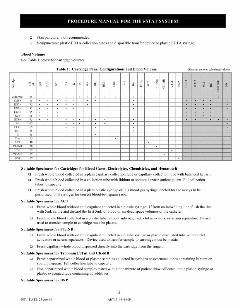
Blood Volume See Table 1 below for cartridge volumes.
Table 1: Cartridge Panel Configurations and Blood Volume (Shading denotes calculated values)
Car
tridg
e
Vol
. µ
L pH
PCO
2
PO2
Na K
Cl
iCa
Glu
BU
N
Cre
at
Lact
Hct
TCO
2
AC
T
PT/IN
R
CK
-MB
cTnI
BN
P
HC
O3
TCO
2
SO2
BE
Ani
on G
ap
H
b
CHEM8+ 95 • • • • • • • • • • • CG8+ 95 • • • • • • • • • • • • • EG7+ 95 • • • • • • • • • • • • EG6+ 95 • • • • • • • • • • • CG4+ 95 • • • • • • • • G3+ 95 • • • • • • •
EC8+ 65 • • • • • • • • • • • • • 6+ 65 • • • • • • •
EC4+ 65 • • • • • E3+ 65 • • • • G 65 •
Crea 65 • ACT 40 •
PT/INR 20 • cTnI 17 •
CK-MB 17 • BNP 17 •
Suitable Specimens for Cartridges for Blood Gases, Electrolytes, Chemistries, and Hematocrit
q Fresh whole blood collected in a plain capillary collection tube or capillary collection tube with balanced heparin. q Fresh whole blood collected in a collection tube with lithium or sodium heparin anticoagulant. Fill collection
tubes to capacity. q Fresh whole blood collected in a plain plastic syringe or in a blood gas syringe labeled for the assays to be
performed. Fill syringes for correct blood-to-heparin ratio.
Suitable Specimens for ACT q Fresh whole blood without anticoagulant collected in a plastic syringe. If from an indwelling line, flush the line
with 5mL saline and discard the first 5mL of blood or six dead space volumes of the catheter.
q Fresh whole blood collected in a plastic tube without anticoagulant, clot activators, or serum separators. Device used to transfer sample to cartridge must be plastic.
Suitable Specimens for PT/INR q Fresh whole blood without anticoagulant collected in a plastic syringe or plastic evacuated tube without clot
activators or serum separators. Device used to transfer sample to cartridge must be plastic.
q Fresh capillary whole blood dispensed directly into the cartridge from the finger.
Suitable Specimens for Troponin I/cTnI and CK-MB q Fresh heparinized whole blood or plasma samples collected in syringes or evacuated tubes containing lithium or
sodium heparin. Fill collection tube to capacity. q Non heparinized whole blood samples tested within one minute of patient draw collected into a plastic syringe or
plastic evacuated tube containing no additives.
Suitable Specimens for BNP

PROCEDURE MANUAL FOR THE i-STAT SYSTEM
4 REV. DATE: 23-Apr-14 ART: 714446-00P
q EDTA whole blood plasma samples collected in plastic syringes or evacuates tubes containing EDTA. Fill collection tubes to capacity.
Specimen Labeling Unless the specimen is analyzed immediately after collection and then discarded, the specimen container must be labeled with the following information:
Patient name, sex, age Patient ID number Time and date of collection Phlebotomist ID Doctor’s name
Specimen Collection and Handling In-Dwelling Line Back flush line with sufficient amount of blood to remove intravenous solution, heparin, or medications that may contaminate the sample. Recommendation: five to six times the volume of the catheter, connectors, and needle. If collecting sample for ACT, clear the line first with 5mL saline and discard the first 5mL of blood or six dead space volumes of the catheter.
Arterial Specimens For cartridge testing of blood gases, electrolytes, chemistries, and hematocrit, fill a plain syringe or fill a blood gas syringe, labeled for the assays to be performed, to the recommended capacity, or use the least amount of liquid heparin anticoagulant that will prevent clotting. Under-filling syringes containing liquid heparin will decrease results due to dilution and will decrease ionized calcium results due to binding. For ionized calcium, balanced or low volume heparin blood gas syringes should be used. Do not expose sample to air or PCO2 may decrease, pH may increase and PO2 may decrease if the value is above or increase if the value is below the PO2 of room air (approximately 150 mmHg).
For cartridge testing of ACT, use only a plain, plastic syringe without anticoagulant.
Mix blood and anticoagulant by rolling syringe between palms for at least 5 seconds each in two different directions, then invert the syringe repeatedly for at least 5 seconds. Discard the first two drops of blood. For blood gas testing, avoid or remove immediately any air drawn into syringe to maintain anaerobic conditions.
Test samples collected without anticoagulant immediately. Test samples for ACT and lactate immediately. For pH, blood gases, TCO2 and ionized calcium, test within 10 minutes of collection. If not tested immediately, remix the sample and discard the first two drops of blood from a syringe before testing. Note that it may be difficult to property remix a sample in a 1.0 cc syringe. For other cartridge tests, test sample within 30 minutes of collection.
Venous Specimens For cartridge testing of electrolytes, chemistries, and hematocrit, collect sample into an evacuated blood collection tube or a syringe containing sodium, lithium, or balanced heparin anticoagulant. For ionized calcium measurements, balanced heparin or 10 U of sodium or lithium heparin/mL of blood is recommended. Fill tubes to capacity; fill syringes for correct heparin-to-blood ratio. Incomplete filling causes higher heparin-to-blood ratio, which will decrease ionized calcium results and may affect other results. The use of partial – draw tubes (evacuated tubes that are adjusted to draw less than the tube volume, e.g. a 5 mL tube with enough vacuum to draw only 3 mL) is not recommended for blood gas or CHEM8+ cartridges because of the potential for decreased PCO2, HCO3 and TCO2 values.
For cartridge testing of ACT or PT/INR, use only a plain, plastic syringe or collection tube containing no anticoagulant. Use a plastic capillary tube, pipette, or syringe to transfer sample from a tube to a cartridge.
For glucose cartridge testing, EDTA is also an acceptable anticoagulant. EDTA is the only acceptable anticoagulant for BNP Cartridge testing.
Mix blood and anticoagulant by inverting a tube gently at least ten times. Roll a syringe vigorously between the palms for at least 5 seconds each in two different directions, then invert the syringe repeatedly for at least 5 seconds, then discard the first two drops of blood. Note that it may be difficult to properly mix a sample in a 1 cc syringe.
Test Sample collected without anticoagulant immediately. Test samples for ACT, lactate and PT/INR immediately. Test samples for pH, PCO2, TCO2 and ionized calcium within 10 minutes of sample draw. If not tested immediately, remix the

PROCEDURE MANUAL FOR THE i-STAT SYSTEM
5 REV. DATE: 23-Apr-14 ART: 714446-00P
sample before testing and discard the first two drops of blood from a syringe before testing. For the glucose test strip and other cartridge tests, test sample within 30 minutes of collection.
Finger and Heelstick Specimens For tests other than PT/INR, wipe away the first drop of blood, which contains excess tissue fluid which can increase the potassium result and decrease other test results. Avoid drawing air into the capillary tube. Use balanced heparin or plain capillary tubes for ionized calcium. Test samples immediately to avoid clotting (especially in neonates). Capillary samples are NOT recommended for ACT, Troponin I/cTnI, CK-MB and BNP.
Criteria For Specimen Rejection q Evidence of clotting q Specimens collected in vacuum tubes with anticoagulant other than lithium or sodium heparin (or EDTA
for BNP or glucose cartridges). q Specimens for ACT or PT/INR collected in glass syringes or tubes or with anticoagulant of any kind q Syringe for pH, PCO2, PO2 and TCO2 with air bubbles in sample q Incompletely filled vacuum tube for the measurement of ionized calcium, PCO2, HCO3 or TCO2 q Other sample types such as urine, CSF, and pleural fluid
Precautions: Avoid the Following Circumstances q Drawing a specimen from an arm with an I.V. q Stasis (tourniquet left on longer than one minute before venipuncture) q Extra muscle activity (fist pumping) q Hemolysis (alcohol left over puncture site, or a traumatic draw) q Icing before filling cartridge q Time delays before filling cartridge, especially lactate, ACT, and PT/INR q Exposing the sample to air when measuring pH, PCO2, PO2 and TCO2.
PROCEDURE FOR ANALYSIS Preparation for Use An individual cartridge may be used after standing 5 minutes, in its pouch, at room temperature. An entire box should stand at room temperature for one hour before cartridges are used.

PROCEDURE MANUAL FOR THE i-STAT SYSTEM
6 REV. DATE: 23-Apr-14 ART: 714446-00P

PROCEDURE MANUAL FOR THE i-STAT SYSTEM
7 REV. DATE: 23-Apr-14 ART: 714446-00P

PROCEDURE MANUAL FOR THE i-STAT SYSTEM
8 REV. DATE: 23-Apr-14 ART: 714446-00P

PROCEDURE MANUAL FOR THE i-STAT SYSTEM
9 REV. DATE: 23-Apr-14 ART: 714446-00P

PROCEDURE MANUAL FOR THE i-STAT SYSTEM
10 REV. DATE: 23-Apr-14 ART: 714446-00P
Procedure for Cartridge Testing
Alternative Procedure Should the i-STAT System become inoperable for any reason, specimens should be collected and submitted to the laboratory in accordance with the Laboratory Procedure Manual.
A level surface includes running the handheld in the downloader/recharger. 7. Review results
i-STAT Cartridge

PROCEDURE MANUAL FOR THE i-STAT SYSTEM
11 REV. DATE: 23-Apr-14 ART: 714446-00P
RESULTS Calculations The i-STAT handheld contains a microprocessor that performs all calculations required for reporting results.
Displayed Results Results are displayed numerically with their units. Electrolyte, chemistry and hematocrit results are also depicted as bar graphs with reference ranges marked under the graphs.
Suppressed Results There are three conditions under which the i-STAT System will not display results:
1. Results outside the System’s reportable ranges are flagged with a < or >, indicating that the result is below the lower limit or above the upper limit of the reportable range respectively. (See the table of Reportable Ranges.) The < > flag indicates that the results for this test were dependant on the result of a test flagged as either > or <. Action: Send specimen(s) to the laboratory for analysis, if necessary.
2. Cartridge results which are not reportable based on internal QC rejection criteria are flagged with ***. Action: Analyze the specimen again using a fresh sample and another cartridge. If the specimen integrity is not in question, the results that are not suppressed should be reported in the usual manner. If the result is suppressed again, send specimen(s) to the laboratory for analysis in accordance with the Laboratory Procedure Manual.
3. A Quality Check message will be reported instead of results if the handheld detects a problem with the sample, calibrant solution, sensors, or mechanical or electrical functions of the handheld during the test cycle. Action: Take the action displayed with the message that identifies the problem. Refer to the i-STAT or i-STAT 1 System Manual’s Troubleshooting section or the “Analyzer Coded Messages” Technical Bulletin if necessary.
Printing and Transmitting Results
Printing Results from the i-STAT 1 Analyzer to the Martel Portable Printer or to the i-STAT Printer Without Downloader or Downloader/Recharger
1. Turn printer on if green power light is not on. 2. Align IR windows of handheld and printer. 3. Display results. 4. Press the Print key. 5. Do not move handheld or printer until printing is complete. 6. If printer is not powered from a wall outlet, turn printer off.
With Downloader or Downloader/Recharger
1. Place handheld in Downloader or Downloader/Recharger that is wired to the printer. 2. Display results. 3. Press the Print key. 4. Do not move handheld or printer until printing is complete.
Printing more than one result
1. Turn the handheld on. 2. Press the Menu key. 3. Press 2 for Data Review. 4. Press 7 for List.
5. Scroll through the test records using the ← and → keys.

PROCEDURE MANUAL FOR THE i-STAT SYSTEM
12 REV. DATE: 23-Apr-14 ART: 714446-00P
6. Press the numbered key for the test record(s). (Press the numbered key again to deselect a record.) 7. Align handheld and printer IR window or place in Downloader or Downloader/Recharger attached to printer.
Press the Print key. 8. Do not move handheld or printer until printing is complete. 9. If printer is not powered from a wall unit using the AC adapter, turn printer off.
Transmitting Results from the i-STAT 1 Analyzer to the Data Manager 1. Place handheld in a Downloader or Downloader/Recharger. 2. Do not move handheld while the message “Communication in Progress” is displayed.

PROCEDURE MANUAL FOR THE i-STAT SYSTEM
13 REV. DATE: 23-Apr-14 ART: 714446-00P
Reference Ranges1,2, Reportable Ranges, and Test Unit Conversions Reference range means the range of test values expected from 95% of fasting individuals presumed to be healthy. Reportable range means the range of test values throughout which the measurement system’s results have been shown to be valid. The following table contains the Reference Ranges (for adults) and Reportable Ranges applicable to the i-STAT System.
Cartridges
ANALYTE UNIT REFERENCE RANGE
(arterial) (venous)
REPORTABLE RANGE
UNIT CONVERSION
Sodium mmol/L (mEq/L) 138 – 146 138 - 146 100 – 180 mmol/L x 1 = mEq/L Example:
140 mmol/L = 140 mEq/L
Potassium mmol/L (mEq/L) 3.5– 4.9 3.5 - 4.9 2.0 – 9.0 mmol/L x 1 = mEq/L
Chloride mmol/L (mEq/L) 98 – 109 98 – 109 65 – 140 mmol/L x 1 = mEq/L
BUN
UREA
mg/dL
mmol/L
8 – 26 8 – 26
2.9 – 9.4 2.9 – 9.4
3 – 140
1 – 50
mg/dL BUN x 0.357 = mmol urea/L Example:
20 mg/dL BUN = 7.1 mmol urea/L
Glucose mg/dL
g/L
mmol/L
70 – 105 70 – 105
0.70 – 1.05 0.70 – 1.05
3.9 – 5.8 3.9 – 5.8
20 – 700
0.20 – 7.00
1.1 – 38.9
mg/dL x 0.055 = mmol/L
Example:
100 mg/dL = 5.55 mmol/L
g/L x 5.556 = mmol/L
Creatinine mg/dL
µmol/L
0.6 - 1.3 0.6 - 1.3
53 - 115 53 - 115
0.2 - 20.0
18 - 1768
mg/dL x 88.4 = µmol/L
Ionized Calcium mmol/L
mg/dL
1.12 – 1.32 1.12 – 1.32
4.5 – 5.3 4.5 – 5.3
0.25 – 2.50
1.0 – 10.0
mmol/L x 4 = mg/dL Example:
1.13 mmol/L x 4 = 4.52 mg/dL
pH 7.35 – 7.45 7.31 – 7.41 6.50 – 8.20 N/A
PCO2 mmHg
kPa
35 – 45 41 – 51
4.67 – 6.00 5.47 – 6.80
5 – 130
0.67 – 17.33
mmHg x 0.133 = kPa Example:
35 mmHg x 0.133 = 4.66 kPa
PO2 mmHg
kPa
80 – 105 10.7 – 14.0
5 – 800
0.7 – 106.6
mmHg x 0.133 = kPa Example:
83 mmHg x 0.133 = 11.04 kPa
TCO2 (on the CHEM8+
cartridge only
mmol/L (mEq/L)
23-27 24-29
5-50
mmol/L x 1 = mEq/L

PROCEDURE MANUAL FOR THE i-STAT SYSTEM
14 REV. DATE: 23-Apr-14 ART: 714446-00P
ANALYTE UNIT REFERENCE RANGE
(arterial) (venous)
REPORTABLE RANGE
UNIT CONVERSION
Hematocrit %PCV
Fraction
38 – 51 38 – 51 0.38 – 0.51 0.38 – 0.51
10 – 75
0.10 – 0.75
% PCV x 0.01 = Volume fraction
Example: 40% PCV = 0.40 PCV
Lactate mmol/L
mg/dL
0.36 –1.25 0.90-1.70
3.2 – 11.3 8.1–15.3
0.30 – 20.00
2.7 – 180.2
mmol/L x 9.01 = mg/dL
HCO3* mmol/L (mEq/L) 22 – 26 23 - 28 1.0 – 85.0 mmol/L x 1 = mEq/L
TCO2* (on all cartridges
but CHEM8+)
mmol/L (mEq/L)
23 – 27 24 - 29
5 – 50
mmol/L x 1= mEq/L
BE* mmol/L (mEq/L) (-2) – (+3) (-2) – (+3) (-30) – (+30)
Anion Gap* mmol/L (mEq/L) 10 – 20 10 – 20 (-10) – (+99)
sO2* % 95 – 98 0 - 100 % x 0.01 = fraction saturated
Hb* g/dL
g/L
mmol/L
12 – 17 12 – 17
120 – 170 120 – 170
7 – 11 7 – 11
3.4 – 25.5
34 – 255
2.1 – 15.8
g/dL x 10 = g/L
Celite ACT seconds 74 – 125 (PREWRM) 74 – 125 (PREWRM)
84 – 139 (NONWRM) 84 – 139 (NONWRM) 50 - 1000
Kaolin ACT seconds 74 – 137 (PREWRM) 74 – 137 (PREWRM)
82 – 152 (NONWRM) 82 – 152 (NONWRM) 50 - 1000
Prothrombin Time/PT
INR 0.9-8.0#
Troponin I/cTnI ng/mL (µg/L) 0.00 – 0.03**
0.00 – 0.08***
0.00 –50.00## ng/mL x 1 = µg/L
Creatine Kinase
MB/ CK-MB
ng/mL (µg/L) 0.0 – 3-5**** 0.0-150.0 ng/mL x 1 = µg/L
B-Type Natriuretic
Peptide/BNP
pg/mL (ng/L) <15-50**** 15-5000 pg/mL x 1 = ng/L
*Calculated values. #Performance characteristics have not been established for INRs above 6.0. **Represents the 0 – 97.5% range of results. Each facility should establish it’s own reference range using the i-STAT cTnI assay. ##Performance characteristics have not been established for cTnI values above 35.00 ng/mL. ***Represents the 0-99% range of results. Each facility should establish it’s own reference range using the i-STAT cTnI assay. ****Represents the 0-95% range of results. Each facility should establish it’s own reference range using the i-STAT assay

PROCEDURE MANUAL FOR THE i-STAT SYSTEM
15 REV. DATE: 23-Apr-14 ART: 714446-00P
Critical Results3 Critical results are test results that fall outside high and low critical limits that define the boundaries of life-threatening values for a test. Critical results represent an emergency condition and must be reported immediately to the patient’s attending physician or nurse.
ANALYTE (units) ADULT CHILDREN NEONATES
Sodium (mmol/L)
low high
120 158
low high
121 156
low high
121 156
Potassium (mmol/L) 2.8 6.2 2.8 6.4 2.8 6.5
Chloride (mmol/L) 75 126 77 121 77 121
TCO2 (mmol/L) 11 40 11 39 – –
Ionized Calcium (mmol/L) 0.78 1.58 0.74 1.57 – –
pH 7.21 7.59 7.21 7.59 – –
PCO2 (mmHg) 19 67 21 66 – –
PO2 (mmHg) 43 – 45 124 37 92
BUN (mg/dL) – 104 – 55 – 55
Glucose (mg/dL) 46 484 46 445 32 328
Creatinine – 7.4 – 3.8 – –
Lactate
Hematocrit (%PCV) 18 61 20 62 33 71
Celite ACT
Kaolin ACT
PT/INR
Troponin I/cTnI
Creatine Kinase MB/ CK-MB
B-Type Natriuretic Peptide/ BNP

PROCEDURE MANUAL FOR THE i-STAT SYSTEM
16 REV. DATE: 23-Apr-14 ART: 714446-00P
Interferences An interferent is a substance which, if present at significant levels in the blood specimen being analyzed, will produce an error in the result of the analyte being measured.
ANALYTE INTERFERENT INTERFERENT CONCENTRATION EFFECT ON ANALYTE RESULT
Sodium Bromide 37.5 mmol/L Increase (↑) Na
Potassium Bromide 37.5 mmol/L Increased rate of star (***) outs.
Chloride Acetylcysteine
Bromide
Bromide (therapeutic)
Salicylate
Thiocyanate
10.2 mmol/L
37.5 mmol/L
2.5 mmol/L
4.34 mmol/L
6.9 mmol/L
Decrease (↓) Cl
Increase (↑) Cl
Increase (↑) Cl
Increase (↑) Cl
Increase (↑) Cl
Ionized Calcium Acetominophen
Magnesium
Acetylcysteine
Bromide
Lactate
Salicylate (therapeutic)
Salicylate
Thiocyanate
1.32 mmol/L
1.0 mmol/L
10.2 mmol/L
37.5 mmol/L
6.6 mmol/L
0.5 mmol/L
4.34 mmol/L
6.9 mmol/L
Decrease (↓) iCa
Increase (↑) iCa by 0.04 mmol/L
Decrease (↓) iCa
Increase (↑) iCa
Decrease (↓) iCa by 0.07 mmol/L
Decrease (↓) iCa by approx. 0.03 mmol/L
Decrease (↓) iCa
Decrease (↓) iCa. Use Another Method.
Glucose
Acetominophen
Acetylcysteine
Bromide
Bromide (therapeutic)
pH
Oxygen
Hydroxyurea
Thiocyanate
1.32 mmol/L
10.2 mmol/L
37.5 mmol/L
2.5 mmol/L
pH: per 0.1 pH units below 7.4 @ 37°C
pH: per 0.1 pH units above 7.4 @ 37°C
PO2 less than 20 mmHg @ 37°C
0.92 mmol/L
6.9 mmol/L
Increase (↑) glucose
Decrease (↓) glucose
Decrease (↓) glucose
Decrease (↓) glucose
Decrease (↓) glucose by 0.9 mg/dL (0.05 mmol/L)
Increase (↑) glucose by 0.8 mg/dL (0.04 mmol/L)
May decrease (↓) glucose
Increase (↑) glucose
Decrease (↓) glucose by approx. 7 mg/dL
Lactate
Bromide
Hydroxyurea
Glycolic Acid
37.5 mmol/L
0.92 mmol/L
10.0 mmol/L
Decrease (↓) lactate
Increase (↑) lactate. Use Another Method.
Increase (↑) lactate. Use Another Method.

PROCEDURE MANUAL FOR THE i-STAT SYSTEM
17 REV. DATE: 23-Apr-14 ART: 714446-00P
ANALYTE INTERFERENT INTERFERENT CONCENTRATION EFFECT ON ANALYTE RESULT
BUN/Urea Bromide
Hydroxyurea
37.5 mmol/L
0.92 mmol/L
Increased rate of star (***) outs
Increase (↑) BUN/Urea results
Creatinine
<2 mg/dL
>2 mg/dL
Acetaminophen
Ascorbate
Bromide (therapeutic) PCO2
PCO2
Hydroxyurea
Acetylcysteine
Creatine
Glycolic Acid
1.32 mmol/L
0.34 mmol/L
2.5 mmol/L Above 40 mmHg
Below 40 mmHg
Above 40 mmHg
Below 40 mmHg
0.92 mmol/L
10.2 mmol/L
0.382 mmol/L
10.0 mmol/L
Increase (↑) creatinine
Increase (↑) creatinine by 0.3 mg/dL
Increase (↑) creatinine
Increase (↑) creatinine by 6.9% per 10 mmHg PCO2
Decrease (↓) creatinine by 6.9% per 10 mmHg PCO2
Decrease (↓) creatinine by 3.7% per 10 mmHg PCO2
Increase (↑) creatinine by 3.7% per 10 mmHg PCO2
Increase (↑) Creatinine. Use Another Method.
Increase (↑) creatinine
Increase (↑) creatinine by 0.2 mg/dL
Decrease (↓) creatinine. Use Another Method.
Hematocrit White Blood Count (WBC)
Total Protein
Lipids
Bromide
Greater than 50,000 WBC/µL
For measured Hct<40% For each g/dL below 6.5 For each g/dL above 8.0
For measured Hct≥40% For each g/dL below 6.5 For each g/dL above 8.0
Abnormally high
37.5 mmol/L
May Increase (↑) hematocrit
Decrease (↓) Hct by 1% PCV Increase (↑) Hct by 1% PCV
Decrease (↓) Hct by 0.75% PCV Increase (↑) Hct by 0.75% PCV
Increase (↑) Hct
Increased rate of star (***) outs
Celite ACT Aprotinin Falsely extends Celite ACT times
PCO2 Propofol (Diprovan®)
Thiopental Sodium
For patients administered propofol or thiopental sodium, i-STAT recommends the use of G3+, CG4+, CG8+, EG6+, and EG7+ cartridges, which are free from clinically significant interference at all relevant therapeutic doses. i-STAT does not recommend the use of EC8+ cartridges for patients receiving propofol or thiopental sodium.
PT/INR Cubicin® (daptomycin for injection)
Chlorhexidine Gluconate
Falsely extends prothrombin time (PT) and INR
May falsely extend prothrombin time (PT) and INR
PROCEDURE MANUAL FOR THE i-STAT SYSTEM
18 REV. DATE: 23-Apr-14 ART: 714446-00P
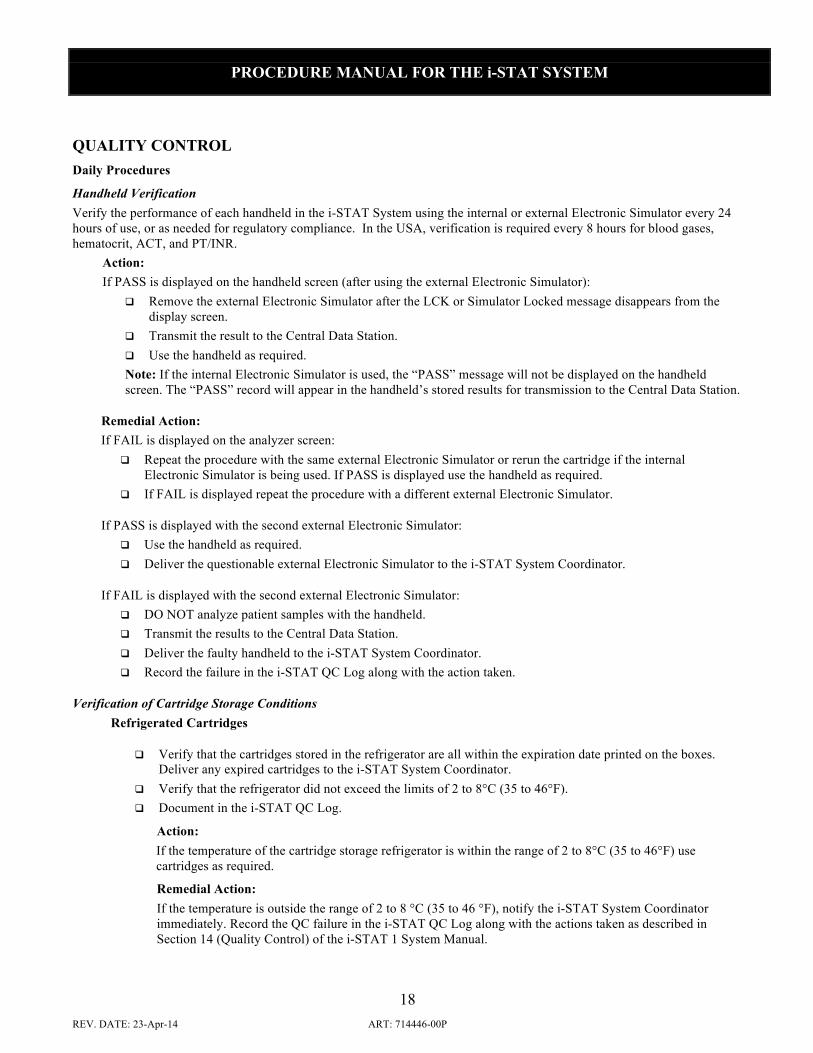
QUALITY CONTROL Daily Procedures
Handheld Verification Verify the performance of each handheld in the i-STAT System using the internal or external Electronic Simulator every 24 hours of use, or as needed for regulatory compliance. In the USA, verification is required every 8 hours for blood gases, hematocrit, ACT, and PT/INR.
Action: If PASS is displayed on the handheld screen (after using the external Electronic Simulator):
q Remove the external Electronic Simulator after the LCK or Simulator Locked message disappears from the display screen.
q Transmit the result to the Central Data Station. q Use the handheld as required. Note: If the internal Electronic Simulator is used, the “PASS” message will not be displayed on the handheld screen. The “PASS” record will appear in the handheld’s stored results for transmission to the Central Data Station.
Remedial Action: If FAIL is displayed on the analyzer screen:
q Repeat the procedure with the same external Electronic Simulator or rerun the cartridge if the internal Electronic Simulator is being used. If PASS is displayed use the handheld as required.
q If FAIL is displayed repeat the procedure with a different external Electronic Simulator.
If PASS is displayed with the second external Electronic Simulator: q Use the handheld as required. q Deliver the questionable external Electronic Simulator to the i-STAT System Coordinator.
If FAIL is displayed with the second external Electronic Simulator: q DO NOT analyze patient samples with the handheld. q Transmit the results to the Central Data Station. q Deliver the faulty handheld to the i-STAT System Coordinator. q Record the failure in the i-STAT QC Log along with the action taken.
Verification of Cartridge Storage Conditions Refrigerated Cartridges
q Verify that the cartridges stored in the refrigerator are all within the expiration date printed on the boxes. Deliver any expired cartridges to the i-STAT System Coordinator.
q Verify that the refrigerator did not exceed the limits of 2 to 8°C (35 to 46°F). q Document in the i-STAT QC Log.
Action: If the temperature of the cartridge storage refrigerator is within the range of 2 to 8°C (35 to 46°F) use cartridges as required.
Remedial Action: If the temperature is outside the range of 2 to 8 °C (35 to 46 °F), notify the i-STAT System Coordinator immediately. Record the QC failure in the i-STAT QC Log along with the actions taken as described in Section 14 (Quality Control) of the i-STAT 1 System Manual.

PROCEDURE MANUAL FOR THE i-STAT SYSTEM
19 REV. DATE: 23-Apr-14 ART: 714446-00P
Room Temperature Cartridges
q Verify that all boxes of cartridges at room temperature have been out of the refrigerator less than the time frame indicated on the cartridge box. Deliver any expired cartridges to the i-STAT System Coordinator.
q Verify that room temperature has not exceeded 30°C. q Document in the i-STAT QC log.
Action: If the measured temperature of the room has been continuously below 30°C (86°F) use cartridges as required.
Remedial Action: If the measured room temperature has exceeded 30°C (86°F) for any period of time: q Quarantine the cartridges. q Notify the i-STAT System Coordinator immediately. q DO NOT USE the cartridges. q Record the out-of-control event in the i-STAT QC Log and the action taken.
Monthly Procedures Print Electronic Simulator Results Print a copy of the Electronic Simulator results from the Central Data Station. Include the report in the i-STAT QC Log.
q CDS version 5 and above: Click on the Simulator Viewer. Print Control Fluid Analysis Results Print results for any control fluids analyzed from the Central Data Station. Include the report in the i-STAT QC Log.
q CDS version 5 and above: Click on the Control Results Viewer.
Periodic Procedures for Cartridges For acceptance of newly received cartridge lots, check the Temperature Monitor and perform integrity testing.
Check Temperature Monitor i-STAT cartridges are shipped refrigerated with a four-window indicator to monitor temperature during transit. Note: All control and calibration verification materials, except for those shipped on dry ice, will also include a four-window indicator to monitor temperature during transit.
Action: q Fill out the record of receipt and forward materials to refrigerator. q If all windows are white or if only the A or B windows are blue or the 1 or 2 windows are red, then transit
temperatures were satisfactory and the cartridges can be used. Remedial Action: If the C or D windows are blue, or the 3 or 4 windows are red: q Quarantine the suspect cartons. q Notify the i-STAT System Coordinator immediately. q DO NOT USE cartridges from the suspect cartons. q Record the out-of-control event in the i-STAT QC Log.

PROCEDURE MANUAL FOR THE i-STAT SYSTEM
20 REV. DATE: 23-Apr-14 ART: 714446-00P
Integrity Testing From each lot of blood gas/chemistry cartridges received, use a representational number of cartridges to analyze i-STAT or TriControls Level 1 and 3 Controls. For CHEM8+ cartridges, analyze i-STAT CHEM8+ or TriControls Control Levels 1 and 3. If not using TriControls and hematocrit is included on the cartridge type being tested, also analyze RNA Medical Hematocrit Control Levels 1 and 3. For ACT cartridges, analyze i-STAT Level 1 and Level 2 ACT Controls. For PT/INR cartridges, analyze i-STAT Level 1 and Level 2 PT Controls. For cTnI cartridges, analyze i-STAT Level 1 and Level 3 cTnI Controls. For CK-MB cartridges, analyze i-STAT Level 1 and Level 3 CK-MB Controls or CLINIQA Cardiac Marker Level 1 and 3 controls. For BNP cartridges, analyze i-STAT BNP Control Levels 1 and 3. Use any verified analyzer for control testing. If available, transmit the results to the Data Manager. Use the expected values published in the Value Assignment Sheet to verify the integrity of the cartridges.
Note: the above information is not a manufacturer’s system instruction; it is a suggestion to comply with regulatory requirements.
Procedure for testing cartridges with i-STAT Level 1 and Level 3 Controls: 1. Prior to testing cartridges that measure PO2, ampules should stand at room temperature a minimum of 4 hours before
use. When testing other cartridges (G, Crea, E3+, EC4+, 6+, or EC8+), ampules may be used once the fluid has reached room temperature, approximately 30 minutes for individual ampules. For best results, ampules, cartridges, and handhelds should be at the same temperature. When using cartridges that contain sensors for measuring ionized calcium, pH, PCO2, or PO2 (G3+, EG6+, EG7+, CG4+, CG8+, or EC8+), a separate ampule must be used for each cartridge being tested; if these sensors are not present (i.e., the 6+ cartridge), the contents of one ampule may be used to fill more than one cartridge as long as the cartridges are filled and inserted into a handheld within 10 minutes of opening the ampule.
2. Immediately before use, shake the ampule vigorously for 5 to 10 seconds to equilibrate the liquid and gas phases. To shake, hold the ampule at the tip and bottom with forefinger and thumb to minimize increasing the temperature of the solution. If necessary, tap the tip of the ampule to send solution back into the bottom section of the ampule. Protect fingers with gauze, tissue, or glove, or use an ampule breaker to snap off the tip of the ampule at the neck.
3. Immediately transfer the solution from the ampule into a plain capillary tube or plain syringe, and then immediately transfer the solution into a cartridge. Immediately seal the cartridge and insert it into a handheld. It is important not to expose the solution to room air since this will alter the results. q When using a capillary tube, fill from the bottom of the ampule. Avoid drawing solution from the surface by
covering the far end of the tube as it is inserted into the ampule. Once the open end of the tube rests at the bottom of the ampule, uncover the other end to allow filling by capillary action.
q When using a syringe (1cc or 3cc syringes with 16 to 20 gauge needles are recommended), slowly draw approximately 1 mL of solution from the bottom of the ampule. If air is trapped between the leading edge of the solution and the plunger, do not invert the syringe to expel it; this will not affect solution near the tip of the syringe. If air bubbles are continually drawn into the syringe, or if a bubble is trapped near the tip of the syringe, discard the ampule and syringe and use a fresh ampule and syringe. Expel one or two drops from the syringe before filling the cartridge.
q Do not use solution left in the syringe, ampule, or capillary tube for additional testing of the cartridges that contain sensors for ionized calcium, pH, PCO2, or PO2. However, cartridges without these sensors may be tested with remaining fluids if within 10 minutes of opening the ampule.
4. Compare results to the value assignment sheet ranges. Check that the lot number on the control ampule matches the lot number on the package insert and that the software version listed on the insert matches the software installed in the handheld. If all results are within expected ranges, use the cartridges as needed. If available, transmit the results to the Data Manager.
Procedure for testing cartridges with TriControls Level 1 and Level 3 Controls:
1 Prior to testing cartridges that measure PO2, ampules should stand at room temperature a minimum of 4 hours before use. When testing other cartridges (G, Crea, E3+, EC4+, 6+, EC8+, or CHEM8+), ampules may be used once the fluid has reached room temperature, approximately 30 minutes for individual ampules. For best results, ampules, cartridges, and handhelds should be at the same temperature. When using cartridges that contain sensors for measuring ionized calcium, pH, PCO2, or PO2 (G3+, EG6+, EG7+, CG4+, CG8+, EC8+, or CHEM8+), a separate ampule must be used for each cartridge being tested; if these sensors are not present (i.e., the 6+ cartridge), the contents of one ampule may be used to fill more than one cartridge as long as the cartridges are filled and inserted into a handheld within 10 minutes of opening the ampule.

PROCEDURE MANUAL FOR THE i-STAT SYSTEM
21 REV. DATE: 23-Apr-14 ART: 714446-00P
2 Immediately before use, shake the ampule vigorously for 5 to 10 seconds to equilibrate the liquid and gas phases. To shake, hold the ampule at the tip and bottom with forefinger and thumb to minimize increasing the temperature of the solution. If necessary, tap the tip of the ampule to send solution back into the bottom section of the ampule. Protect fingers with gauze, tissue, or glove, or use an ampule breaker to snap off the tip of the ampule at the neck.
3 Immediately transfer the solution from the ampule into a plain capillary tube or plain syringe, and then immediately transfer the solution into a cartridge. Immediately seal the cartridge and insert it into a handheld. It is important not to expose the solution to room air since this will alter the results. q When using a capillary tube, fill from the bottom of the ampule. Avoid drawing solution from the surface by
placing a finger over the far end of the tube as it is inserted into the ampule. Once the open end of the tube rests at the bottom of the ampule, uncover the other end to allow filling by capillary action.
q When using a syringe (1cc or 3cc syringes with 16 to 20 gauge needles are recommended), slowly draw approximately 1 mL of solution from the bottom of the ampule. If air is trapped between the leading edge of the solution and the plunger, do not invert the syringe to expel it; this will not affect solution near the tip of the syringe. If air bubbles are continually drawn into the syringe, or if a bubble is trapped near the tip of the syringe, discard the ampule and syringe and use a fresh ampule and syringe. Expel one or two drops from the syringe before filling the cartridge.
q Do not use solution left in the syringe, ampule, or capillary tube for additional testing of the cartridges that contain sensors for ionized calcium, pH, PCO2, or PO2. However, cartridges without these sensors may be tested with remaining fluids if within 10 minutes of opening the ampule.
4. Compare results to the Value Assignment Sheet ranges. Ensure that the lot number printed on the Value Assignment Sheet matches the lot number on the label of the ampule and that the software version above the target value table matches the software version in the handheld. If all results are within expected ranges, use the cartridges as needed. If available, transmit the results to the Data Manager.
Procedure for testing CHEM8+ cartridges with i-STAT CHEM8+ controls.
1. Equilibrate the ampule for approximately 30 minutes at room (ambient) temperature. 2. Immediately before use, shake the ampule vigorously for 5-10 seconds to equilibrate the liquid and gas phases. To
shake, hold the ampule at the tip and bottom with forefinger and thumb to minimize increasing the temperature of the solution. If necessary, tap the tip of the ampule to send solution back into the bottom section of the ampule.
3. Protect fingers with gauze, tissue or glove, or use an ampule breaker to snap off the tip of the ampule at the neck. 4. Immeditely transfer the solution from the ampule into a plain capillary tube or plain syringe, and then immediately
transfer the solution into a cartridge. 5. Immediately seal the cartridge and insert it into a handheld – it is important not to expose the solution to room air
since this will alter the results. q When using a capillary tube, fill from the bottom of the ampule. Avoid drawing solution from the surface by
covering the far end of the tube as it is inserted into the ampule. Once the open end of the tubes at the bottom of the ampule, uncover the other end to allow filling by capillary action.
q When using a syringe (1 cc or 3 cc syringes with 16-20 gauge needles are recommended), slowly draw approximately 1 mL of solution from the bottom of the ampule. If air is trapped between the leading edge of the solution and the plunger, do NOT invert the syringe to expel it; this will not affect solution near the tip of the syringe. If air bubbles are continually drawn into the syringe, or if a bubble is trapped near the tip of the syringe, discard the ampule and syringe and use a fresh ampule and syringe. Expel one or two drops from the syringe before filling the cartridge.
q Do not use solution left in the syringe, ampule, or capillary tube for additional testing of CHEM8+ cartridges. 6. Compare results to the value assignment sheet ranges. Check that the lot number on the control ampule matches the
lot number on the package insert that the software version listed on the insert matches the software installed in the handheld. If all results are within expected ranges, use the cartridges as needed. If available, transmit the results to the Data Manager.

PROCEDURE MANUAL FOR THE i-STAT SYSTEM
22 REV. DATE: 23-Apr-14 ART: 714446-00P
Procedure for testing cartridges with RNA Medical Hematocrit controls 1. If stored refrigerated, the control material should be equilibrated to room temperature for at least four (4) hours
prior to use. If stored at room temperature, no equilibration of the control material is necessary. 2. Gently invert the ampule to mix the solution. Tap the ampule to restore the liquid to the bottom of the ampule. 3. Open the ampule by snapping off the tip at the neck. Use gauze, tissue, gloves, or an appropriate ampule opener to
protect fingers from cuts. 4. Fill and seal a cartridge and insert immediately into the handheld. Note: the Control option from the Quality Tests
menu must be used on the i-STAT 1 Analyzer for RNA Medical Hematocrit Control. 5. Compare the i-STAT System Hematocrit result to the value assignment sheet ranges. Note: the only value assigned
to this fluid is for hematocrit. All other cartridge analyte results obtained with this control material should be ignored.
6. If available, transmit results to the Data Manager.
Remedial Action: If any results are outside the published expected ranges: q DO NOT USE cartridges from the suspect lot. q Quarantine the suspect lot. q Notify the i-STAT System Coordinator immediately. q Record the QC failure in the i-STAT QC Action Log along with the action taken.
Procedure for testing cartridges with i-STAT Level 1 and Level 2 ACT or PT/INR Controls 1. Prior to use, allow one vial each of the lyophilized plasma and calcium chloride reconstituting fluid to stand at room
temperature for a minimum of 45 minutes. 2. Remove the cap and stopper from the vials and pour the entire contents of the calcium chloride vial into the
lyophilized plasma vial. Place the stopper back on the reconstituted vial. 3. Allow the vial to sit for 1 minute and then mix the contents by swirling gently for 1 minute, then inverting slowly for
30 seconds. 4. Use a plastic pipette, syringe, or capillary tube without anticoagulant to transfer the solution to an ACT cartridge. 5. Immediately seal the cartridge and insert it into a handheld. This process must be completed within 30 seconds of the
complete reconstitution of the control sample. 6. Compare results to the value assignment sheet ranges. If results are within the expected ranges, use the cartridges as
needed. If available, transmit results to the Data Manager.
Remedial Action: If any results are outside the published expected ranges:
q DO NOT USE cartridges from the suspect lot. q Quarantine the suspect lot. q Notify the i-STAT System Coordinator immediately. q Record the QC failure in the i-STAT QC Action Log along with the action taken.
Procedures for for testing cartridges with i-STAT cTnI, BNP, and CK-MB controls
1. i-STAT cTnI, BNP, and CK-MB Controls are ready-to-use liquid control requiring no reconstitution or frozen storage. They are stable until the expiration date on the vial label when stored unopened at 2-8°C. Once opened, the i-STAT cTnI, BNP, and CK-MB Controls are stable for 30 days when stored tightly capped at 2-8°C.
2. Access the Control option under Quality Tests in the Administration Menu. Enter the required information. The handheld allows 15 minutes (or the customized timeout period) to insert the cartridge after the last data entry.
3. Immediately before use, gently mix the contents of the control vial to ensure homogeneity. Avoid foaming of the sample.
4. Open the vial and transfer a drop of the solution into the i-STAT cartridge using a plain capillary tube, plain syringe, or plastic transfer pipette. Tightly recap the control vial and store it at 2-8°C.

PROCEDURE MANUAL FOR THE i-STAT SYSTEM
23 REV. DATE: 23-Apr-14 ART: 714446-00P
5. Seal the cartridge and immediately insert it into the i-STAT 1 Analyzer. 6. Compare the result to the Value Assignment Sheet value. Always ensure that the lot number and
software revision on the Value Assignment Sheet matches the lot number of the vial in use and the software revision in the handheld. Should results fall outside the range, refer to the Troubleshooting section of i-STAT 1 System Manual section 14 (Quality Control).
7. If available, transmit results to the Data Manager. Remedial Action
If any results are outside the published expected ranges: q DO NOT USE cartridges from the suspect lot. q Quarantine the suspect lot. q Notify the i-STAT System Coordinator immediately. q Record the QC failure in the i-STAT QC Action Log along with the action taken.
Procedures for for testing CK-MB cartridges with CLINIQA Cardiac Marker controls for i-STAT
1. CLINIQA Liquid QC Cardiac Marker Control is a ready-to-use liquid control requiring no reconstitution or frozen storage. It is stable until the expiration date on the vial label when stored unopened at 2-8°C. Once opened, CLINIQA Liquid QC Cardiac Marker Control is stable for 30 days when stored tightly capped at 2-8°C.
2. Immediately before use, gently mix the contents of the control vial to ensure homogeneity. Avoid foaming of the sample.
3. Open the vial and transfer a drop of the solution into the i-STAT CK-MB cartridge using a plain capillary tube, plain syringe, or plastic transfer pipette. Tightly recap the control vial and store it at 2-8°C.
4. Seal the cartridge and immediately insert it into the i-STAT 1 Analyzer. 5. Compare the i-STAT CK-MB result to the Value Assignment Sheet value. Always ensure that the lot number and
software revision on the Value Assignment Sheet matches the lot number of the vial in use and the software revision in the handheld. Should results fall outside the range, refer to the Troubleshooting section of i-STAT 1 System Manual section 14 (Quality Control).
6. If available, transmit results to the Data Manager. Remedial Action
If any results are outside the published expected ranges: q DO NOT USE cartridges from the suspect lot. q Quarantine the suspect lot. q Notify the i-STAT System Coordinator immediately. q Record the QC failure in the i-STAT QC Action Log along with the action taken.
CALIBRATION For cartridges, calibration is automatically performed as part of the test cycle on each cartridge type, except coagulation and immunoassay cartridges. Operator intervention is not necessary.
CLINICAL SIGNIFICANCE

PROCEDURE MANUAL FOR THE i-STAT SYSTEM
24 REV. DATE: 23-Apr-14 ART: 714446-00P
Analyte Some Causes of Increased Values
Some Causes of Decreased Values
Sodium Dehydration Diabetes insipidus Salt poisoning Skin losses Hyperaldosteronism CNS disorders
Dilutional hyponatremia (cirrhosis) Depletional hyponatremia Syndrome of inappropriate ADH
Potassium Renal glomerular disease Adrenocortical insufficiency Diabetic Ketoacidosis (DKA) Sepsis In vitro hemolysis
Renal tubular disease Hyperaldosteronism Treatment of DKA Hyperinsulinism Metabolic alkalosis Diuretic therapy
Chloride Prolonged diarrhea Renal tubular disease Hyperparathyroidism Dehydration
Prolonged vomiting Burns Salt-losing renal disease Overhydration Thiazide therapy
Ionized Calcium
Dehydration Hyperparathyroidism Malignancies Immobilization Thiazide diuretics Vitamin D intoxication
Hypoparathyroidism Early neonatal hypocalcemia Chronic renal disease Pancreatitis Massive blood transfusions Severe malnutrition
BUN Impaired renal function Prerenal azotemia (e.g. shock) Postrenal azotemia GI bleeding High protein diet
Pregnancy Severe liver insufficiency Overhydration Malnutrition
Glucose Diabetes mellitus Pancreatitis Endocrine disorders (e.g. Cushing’s syndrome) Drugs (e.g. steroids, thyrotoxicosis) Chronic renal failure Stress IV glucose infusion
Insulinoma Adrenocortical insufficiency Hypopituitarism/Massive liver disease Ethanol ingestion/Reactive hypoglycemia Glycogen storage disease
Creatinine Impaired renal function

PROCEDURE MANUAL FOR THE i-STAT SYSTEM
25 REV. DATE: 23-Apr-14 ART: 714446-00P
Analyte Some Causes of Increased Values
Some Causes of Decreased Values
Lactate Hypoxia (shock, hypovolumia, left ventricular failure); diabetes mellitus, neoplasia, liver disease; drug or toxins (ethanol, methanol, salicylates); glycolic acid as a product of ethylene glycol metabolism
pH Respiratory alkalosis Metabolic alkalosis
Respiratory acidosis Metabolic acidosis
PCO2 Acute Respiratory Acidosis:
• Depression of respiratory center • Suppressed neuromuscular system • Pulmonary disorders • Inadequate mechanical ventilation
Chronic respiratory acidosis
• Decreased alveolar ventilation • Hypoventilation
Compensation in metabolic alkalosis
Respiratory alkalosis:
• Increased stimulation of respirator center
• Hypermetabolic states • Mechanical hyperventilation
Compensation in metabolic acidosis
PO2 Breathing oxygen-enriched air Carbon-monoxide exposure Pulmonary disorders Myocardial infarction Congestive heart failure
HCO3 and TCO2
Primary metabolic alkalosis Primary respiratory acidosis
Primary metabolic acidosis Primary respiratory alkalosis
Hematocrit Dehydration Burns Impaired ventilation Renal disorders
Hemolytic anemias Iron deficiency Marrow depression Blood loss
ACT Celite Administration of heparin for medical or surgical procedures. Administration of aprotinin.
PT/INR Administration of oral anticoagulant therapy.

PROCEDURE MANUAL FOR THE i-STAT SYSTEM
26 REV. DATE: 23-Apr-14 ART: 714446-00P
Analyte Some Causes of Increased Values
Some Causes of Decreased Values
ACT Kaolin Administration of heparin for medical or surgical procedures.
cTnI Myocardial Infarction Coronary vasospasm Cardiac contusion/trauma Rhythm disturbance (SVT, AF) Chemotherapy (ex. Adriamycin) Myocarditis/pericarditis Infiltrative diseases (ex. Amyloidosis, sarcoidosis, hemochromatosis, connective tissue disease) Congestive heart failure Heart transplantation Cardiac procedures (PTCA, DC cardioversion) Intracranial hemorrhage/stroke Pulmonary embolism Pulmonary hypertension Chronic renal insufficiency Sepsis Strenuous exercise Certain drug ingestions
Rare antibodies to troponin or its circulating complexes
CK-MB Myocardial Infarction Coronary vasospasm Cardiac contusion/trauma Myocarditis/pericarditis Infiltrative diseases (ex. Amyloidosis, sarcoidosis, hemochromatosis, connective tissue disease) Cardiac procedures (PTCA, DC cardioversion) Intracranial hemorrhage/stroke Pulmonary embolism Pulmonary hypertension Chronic renal insufficiency Sepsis Strenuous exercise Certain drug ingestions (cocaine) Skeletal muscle disease
Lean muscle mass
BNP Congestive heart failure Chronic obstructive pulmonary disease (COPD) Asthma Pulmonary hypertension Cor pulmonale Pulmonary embolism Acute coronary syndrome Chronic renal failure Age Female sex
Obesity (BMI>30 Kg/m2) Flash pulmonary edema (elevation may be delayed)

PROCEDURE MANUAL FOR THE i-STAT SYSTEM
27 REV. DATE: 23-Apr-14 ART: 714446-00P
PRINCIPLES OF MEASUREMENT
Sodium, Potassium, Chloride, Ionized Calcium, pH, and PCO2 are measured by ion-selective electrode potentiometry. Concentrations are calculated from the measured potential through the Nernst equation.
Urea is first hydrolyzed to ammonium ions in a reaction catalyzed by the enzyme urease. The ammonium ions are measured by an ion-selective electrode and the concentration is calculated from the measured potential through the Nernst equation.
Glucose is measured amperometrically. Oxidation of glucose, catalyzed by the enzyme glucose oxidase, produces hydrogen peroxide. The liberated hydrogen peroxide is oxidized at an electrode to produce an electric current which is proportional to the glucose concentration.
Creatinine is hydrolyzed to creatine in a reaction catalyzed by the enzyme creatinine amidohydrolase. Creatine is then hydrolyzed to sarcosine in a reaction catalyzed by the enzyme creatine amidinohydrolase. The oxidation of sarcosine, catalyzed by the enzyme sarcosine oxidase, produces hydrogen peroxide. The liberated hydrogen peroxide is oxidized at the platinum electrode to produce a current which is proportional to the creatinine concentration.
Lactate is measured amperometrically. The enzyme lactate oxidase, immobilized in the lactate biosensor, selectively converts lactate to pyruvate and hydrogen peroxide. The liberated hydrogen peroxide is oxidized at the platinum electrode to produce a current which is proportional to the lactate concentration.
PO2 is measured amperometrically. The oxygen sensor is similar to a conventional Clark electrode. Oxygen permeates through a gas permeable membrane from the blood sample into an internal electrolyte solution where it is reduced at the cathode. The oxygen reduction current is proportional to the dissolved oxygen concentration.
Hematocrit is determined conductometrically. The measured conductivity, after correction for electrolyte concentration, is inversely related to the hematocrit.
ACT is determined amperometrically. The conversion of a thrombin substrate is initiated by mixing a whole blood sample (without anticoagulant) with a particulate clotting activator – either Celite brand diatomaceous earth or kaolin. The substrate used in the electrogenic assay has an amide linkage that mimics the thrombin-cleaved amide linkage in fibrinogen. The product of the thrombin-substrate reaction is the electroactive compound that is detected amperometrically. The time of detection is measured in seconds and the result is reported as a whole blood time (WBT).
PT/INR is determined amperometrically. The conversion of a thrombin substrate is initiated by mixing a whole blood sample (without anticoagulant) with tissue thromboplastin. The substrate used in the electrogenic assay has as amide linkage that mimics the thrombin–cleaved amide linkage in fibrinogen. The product of the thrombin–substrate reaction is the electroactive compound that is detected amperometrically. The time of detection is measured in seconds and reported as INR and/or seconds.
Troponin I/cTnI

PROCEDURE MANUAL FOR THE i-STAT SYSTEM
28 REV. DATE: 23-Apr-14 ART: 714446-00P
is determined amperometrically using a two-site ELISA method. Antibodies specific for human cardiac troponin I (cTnI) are located on an electrochemical sensor fabricated on a silicon chip. Also deposited in another location on the sensor silicon chip is an antibody/alkaline phosphatase enzyme conjugate specific to a separate portion of the cTnI molecule. The whole blood or plasma sample is brought into contact with the sensors allowing the enzyme conjugate to dissolve into the sample. The cTnI within the sample becomes labeled with alkaline phosphatase and is captured onto the surface of the electrochemical sensor during an incubation period of approximately seven minutes. The sample, as well as excess enzyme conjugate, is washed off the sensors. Within the wash fluid is a substrate for the alkaline phosphatase enzyme. The enzyme bound to the antibody/antigen/antibody sandwich cleaves the substrate releasing an electrochemically detectable product. The electrochemical (amperometric) sensor measures this enzyme product which is proportional to the concentration of cTnI within the sample.
Creatine Kinase MB/CK-MB is determined amperometrically using a two-site ELISA method. Antibodies specific for an epitope unique to the CK-MB subunit, that therefore do not bind CK-MM or CK-BB, are located on an electrochemical sensor fabricated on a silicon chip. Also deposited in another location on the sensor silicon chip is an antibody/alkaline phosphatase enzyme conjugate specific to an epitope on the B subunit of creatine kinase. The specificity of the conjugate antibody to the B subunit allows this conjugate to recognize CK-MB and CK-BB, but not CK-MM. The whole blood or plasma sample is brought into contact with the sensors allowing the enzyme conjugate to dissolve into the sample. The CK-MB within the sample becomes labeled with alkaline phosphatase and is captured onto the surface of the electrochemical sensor during an incubation period of approximately three minutes. The sample is washed off the sensors, as well as excess enzyme conjugate. Within the wash fluid is a substrate for the alkaline phosphatase enzyme. The enzyme bound to the antibody/antigen/antibody sandwich cleaves the substrate releasing an electrochemically detectable product. The electrochemical (amperometric) sensor measures this enzyme product which is proportional to the concentration of CK-MB within the sample.
B-Type Natriuretic Peptide/BNP is determined amperometrically using a two-site ELISA method. Antibodies specific for BNP are located on an electrochemical sensor fabricated on a silicon chip. Also deposited in another location on the sensor silicon chip is an antibody/alkaline phosphatase enzyme conjugate specific to a separate portion of the BNP molecule. The whole blood or plasma sample is brought into contact with the sensors allowing the enzyme conjugate to dissolve into the sample. The BNP within the sample becomes labeled with alkaline phosphatase and is captured onto the surface of the electrochemical sensor during an incubation period of approximately seven minutes. The sample is washed off the sensors, as well as excess enzyme conjugate. Within the wash fluid is a substrate for the alkaline phosphatase enzyme. The enzyme bound to the antibody/antigen/antibody sandwich cleaves the substrate releasing an electrochemically detectable product. The electrochemical (amperometric) sensor measures this enzyme product which is proportional to the concentration of BNP within the sample.
TCO2 The measured TCO2 test method is calibrated to the International Federation of Clinical Chemistry (IFCC) TCO2 reference method with an algorithm, based on the Henderson-Hasselbach equation, which uses pH, PCO2, and ionic strength (Na) measurements.
FOOTNOTES

PROCEDURE MANUAL FOR THE i-STAT SYSTEM
29 REV. DATE: 23-Apr-14 ART: 714446-00P
1. Statland, B.E., Clinical Decision Levels for Lab Tests. Medical Economics Books, 1987.
2. Tietz, N.W., Tietz Textbook of Clinical Chemistry, third edition, Ed. C.A. Burtis, E.R. Ashwood, W.B. Saunders Company, Philadelphia, 1999. Table 50 – 20, Appendix.
3. Kost, Gerald J., Using critical limits to improve patient outcome. Medical Laboratory Observer. March 1993; 25(3): 22–27.
Prepared By: ________________________________________________________ Date: _______________________________________
Adopted: ___________________________________________________________ Date: _______________________________________
Reviewed: __________________________________________________________ Date: _______________________________________
Reviewed: __________________________________________________________ Date: _______________________________________
Reviewed: __________________________________________________________ Date: _______________________________________
Revised: ____________________________________________________________ Date: _______________________________________