Probiotic Lactobacillus reuteri Ameliorates Disease Due to Enterohemorrhagic Escherichia coli in
7
INFECTION AND IMMUNITY, Jan. 2011, p. 185–191 Vol. 79, No. 1 0019-9567/11/$12.00 doi:10.1128/IAI.00880-10 Copyright © 2011, American Society for Microbiology. All Rights Reserved. Probiotic Lactobacillus reuteri Ameliorates Disease Due to Enterohemorrhagic Escherichia coli in Germfree Mice Kathryn A. Eaton, 1 * Alexander Honkala, 1 Thomas A. Auchtung, 2 and Robert A. Britton 2 Unit for Laboratory Animal Medicine and Department of Microbiology and Immunology, University of Michigan Medical School, Ann Arbor, Michigan 48109-0614, 1 and Microbial Genomics Laboratory, Department of Microbiology and Molecular Genetics, Michigan State University, East Lansing, Michigan 48824 2 Received 12 August 2010/Returned for modification 29 August 2010/Accepted 13 October 2010 Strains of enterohemorrhagic Escherichia coli (EHEC) are a group of Shiga toxin-producing food-borne patho- gens that cause severe hemorrhagic colitis and can lead to hemolytic-uremic syndrome (HUS), a life-threatening condition that principally affects children and for which there is no effective treatment. We used a germfree mouse model of renal and enteric disease due to EHEC to determine if probiotic Lactobacillus reuteri ATCC PTA 6475 is effective in suppressing disease symptoms caused by EHEC. When germfree Swiss Webster mice are monocolonized with EHEC, they develop disease characterized by weight loss, cecal luminal fluid accumulation, and renal tubular necrosis. When L. reuteri was administered 1 day prior to EHEC challenge and every other day thereafter, EHEC colonization was suppressed and mice were significantly protected from the manifestations of disease. Protection from disease did not require the induction of the antimicrobial compound reuterin in L. reuteri prior to treatment. The twice-daily administration of L. reuteri appeared more effective than every-other-day administration. These data indicated that L. reuteri partially protects mice from disease manifestations of EHEC. Strains of enterohemorrhagic Escherichia coli (EHEC) are food-borne bacterial pathogens of humans that are normal inhabitants of the ruminant intestine (4). These organisms often are transmitted to humans in undercooked meat but also have caused outbreaks associated with contaminated vegeta- bles (6, 25), fruit juices (8), and drinking water (28). Disease symptoms include watery or bloody diarrhea and can be severe enough to cause hospitalization and even death (18). In addi- tion, some affected children (and rarely adults) develop hemo- lytic-uremic syndrome (HUS), a rapidly progressive and some- times fatal form of renal failure that is attributed to the expression of Shiga toxins (Stx) (1, 18). HUS is a rare compli- cation of disease due to EHEC, but its importance is amplified by its severity, tendency to affect young children, and the ab- sence of any specific treatment or preventative measure (3). Thus, novel control and treatment therapies are needed. Recently, several laboratories have explored the role of pro- biotic bacteria in the control and treatment of gastrointestinal diseases (20, 29), including EHEC (2, 5, 10, 11, 14, 15, 19, 23, 31, 32, 37). Probiotic bacteria are live microbes that, when consumed, confer a beneficial effect on the host. Their mech- anism of action is not well understood, but in some cases specific probiotic organisms have been shown to reduce colo- nization by pathogens (20, 23), suppress toxin or other viru- lence factor production by pathogens (2, 11, 24, 32), or con- tribute to host defense mechanisms (20). L. reuteri is one of a few Lactobacillus species that has been shown to be indigenous to the human intestine and also is found in the intestinal tracts of several animal species, including the mouse (26, 34). L. reuteri ATCC 55730 is a commercial probiotic strain that sur- vives in the human intestine (34) and has beneficial effects in clinical trials of infant diarrhea, colic, and IgE-dependent ec- zema (29, 30, 33, 36). L. reuteri was also shown to be effective in protecting interleukin-10 (IL-10)-deficient mice against spontaneous colitis (22). Several potential mechanisms may confer the protective ef- fects observed when L. reuteri is ingested. First, multiple strains of L. reuteri secrete a potent anti-inflammatory activity that reduces the expression of the proinflammatory cytokine tumor necrosis factor alpha (TNF-) by greater than 90% in cultured activated macrophages (17, 21). Second, L. reuteri produces an antimicrobial compound, reuterin, which is bactericidal against many bacteria, including strains of EHEC (7). Reuterin (also known as 3-hydroxypropionaldehyde [3-HPA]) is an interme- diate in the metabolism of glycerol to 1,3-propanediol, which allows the cell to regenerate NAD during carbohydrate me- tabolism (35). It has potent antibacterial, antiviral, and anti- fungal activity, but lactic acid bacteria, including L. reuteri, are much more resistant to reuterin than are enteric pathogens, such as E. coli (7). The goal of this study was to use a germfree mouse model of disease to determine if L. reuteri could be protective against either colonization by EHEC or disease caused by EHEC. Unlike specific-pathogen-free mice, germfree mice are highly susceptible to colonization by pathogenic EHEC of human origin and develop Shiga toxin (Stx)-dependent acute renal disease with features that resemble HUS in human children (9). Mice are susceptible to as few as 100 infectious organisms, and within 5 days the lower bowel is heavily colonized by up to 10 11 organisms/g (9). EHEC strains that produce Stx2 cause a rapidly progressive, sometimes fatal disease that is character- ized by increased cecal luminal fluid accumulation and acute kidney failure with renal tubular necrosis and glomerular fibrin thrombosis. Disease is Stx2 dependent, as indicated by the * Corresponding author. Mailing address: Unit for Laboratory An- imal Medicine, 018 Animal Research Facility, 1150 W. Medical Center Dr., Ann Arbor, MI 48109-0614. Phone: (734) 936-3883. Fax: (734) 936-3235. E-mail: [email protected]. Published ahead of print on 25 October 2010. 185 Downloaded from https://journals.asm.org/journal/iai on 10 February 2022 by 88.199.126.52.
Transcript of Probiotic Lactobacillus reuteri Ameliorates Disease Due to Enterohemorrhagic Escherichia coli in
INFECTION AND IMMUNITY, Jan. 2011, p. 185–191 Vol. 79, No. 10019-9567/11/$12.00 doi:10.1128/IAI.00880-10Copyright © 2011, American Society for Microbiology. All Rights Reserved.
Probiotic Lactobacillus reuteri Ameliorates Disease Due toEnterohemorrhagic Escherichia coli in Germfree Mice�
Kathryn A. Eaton,1* Alexander Honkala,1 Thomas A. Auchtung,2 and Robert A. Britton2
Unit for Laboratory Animal Medicine and Department of Microbiology and Immunology, University of Michigan Medical School,Ann Arbor, Michigan 48109-0614,1 and Microbial Genomics Laboratory, Department of Microbiology and
Molecular Genetics, Michigan State University, East Lansing, Michigan 488242
Received 12 August 2010/Returned for modification 29 August 2010/Accepted 13 October 2010
Strains of enterohemorrhagic Escherichia coli (EHEC) are a group of Shiga toxin-producing food-borne patho-gens that cause severe hemorrhagic colitis and can lead to hemolytic-uremic syndrome (HUS), a life-threateningcondition that principally affects children and for which there is no effective treatment. We used a germfree mousemodel of renal and enteric disease due to EHEC to determine if probiotic Lactobacillus reuteri ATCC PTA 6475 iseffective in suppressing disease symptoms caused by EHEC. When germfree Swiss Webster mice are monocolonizedwith EHEC, they develop disease characterized by weight loss, cecal luminal fluid accumulation, and renal tubularnecrosis. When L. reuteri was administered 1 day prior to EHEC challenge and every other day thereafter, EHECcolonization was suppressed and mice were significantly protected from the manifestations of disease. Protectionfrom disease did not require the induction of the antimicrobial compound reuterin in L. reuteri prior to treatment.The twice-daily administration of L. reuteri appeared more effective than every-other-day administration. These dataindicated that L. reuteri partially protects mice from disease manifestations of EHEC.
Strains of enterohemorrhagic Escherichia coli (EHEC) arefood-borne bacterial pathogens of humans that are normalinhabitants of the ruminant intestine (4). These organismsoften are transmitted to humans in undercooked meat but alsohave caused outbreaks associated with contaminated vegeta-bles (6, 25), fruit juices (8), and drinking water (28). Diseasesymptoms include watery or bloody diarrhea and can be severeenough to cause hospitalization and even death (18). In addi-tion, some affected children (and rarely adults) develop hemo-lytic-uremic syndrome (HUS), a rapidly progressive and some-times fatal form of renal failure that is attributed to theexpression of Shiga toxins (Stx) (1, 18). HUS is a rare compli-cation of disease due to EHEC, but its importance is amplifiedby its severity, tendency to affect young children, and the ab-sence of any specific treatment or preventative measure (3).Thus, novel control and treatment therapies are needed.
Recently, several laboratories have explored the role of pro-biotic bacteria in the control and treatment of gastrointestinaldiseases (20, 29), including EHEC (2, 5, 10, 11, 14, 15, 19, 23,31, 32, 37). Probiotic bacteria are live microbes that, whenconsumed, confer a beneficial effect on the host. Their mech-anism of action is not well understood, but in some casesspecific probiotic organisms have been shown to reduce colo-nization by pathogens (20, 23), suppress toxin or other viru-lence factor production by pathogens (2, 11, 24, 32), or con-tribute to host defense mechanisms (20). L. reuteri is one of afew Lactobacillus species that has been shown to be indigenousto the human intestine and also is found in the intestinal tractsof several animal species, including the mouse (26, 34). L.
reuteri ATCC 55730 is a commercial probiotic strain that sur-vives in the human intestine (34) and has beneficial effects inclinical trials of infant diarrhea, colic, and IgE-dependent ec-zema (29, 30, 33, 36). L. reuteri was also shown to be effectivein protecting interleukin-10 (IL-10)-deficient mice againstspontaneous colitis (22).
Several potential mechanisms may confer the protective ef-fects observed when L. reuteri is ingested. First, multiple strainsof L. reuteri secrete a potent anti-inflammatory activity thatreduces the expression of the proinflammatory cytokine tumornecrosis factor alpha (TNF-�) by greater than 90% in culturedactivated macrophages (17, 21). Second, L. reuteri produces anantimicrobial compound, reuterin, which is bactericidal againstmany bacteria, including strains of EHEC (7). Reuterin (alsoknown as 3-hydroxypropionaldehyde [3-HPA]) is an interme-diate in the metabolism of glycerol to 1,3-propanediol, whichallows the cell to regenerate NAD� during carbohydrate me-tabolism (35). It has potent antibacterial, antiviral, and anti-fungal activity, but lactic acid bacteria, including L. reuteri, aremuch more resistant to reuterin than are enteric pathogens,such as E. coli (7).
The goal of this study was to use a germfree mouse model ofdisease to determine if L. reuteri could be protective againsteither colonization by EHEC or disease caused by EHEC.Unlike specific-pathogen-free mice, germfree mice are highlysusceptible to colonization by pathogenic EHEC of humanorigin and develop Shiga toxin (Stx)-dependent acute renaldisease with features that resemble HUS in human children(9). Mice are susceptible to as few as 100 infectious organisms,and within 5 days the lower bowel is heavily colonized by up to1011 organisms/g (9). EHEC strains that produce Stx2 cause arapidly progressive, sometimes fatal disease that is character-ized by increased cecal luminal fluid accumulation and acutekidney failure with renal tubular necrosis and glomerular fibrinthrombosis. Disease is Stx2 dependent, as indicated by the
* Corresponding author. Mailing address: Unit for Laboratory An-imal Medicine, 018 Animal Research Facility, 1150 W. Medical CenterDr., Ann Arbor, MI 48109-0614. Phone: (734) 936-3883. Fax: (734)936-3235. E-mail: [email protected].
� Published ahead of print on 25 October 2010.
185
Dow
nloa
ded
from
http
s://j
ourn
als.
asm
.org
/jour
nal/i
ai o
n 10
Feb
ruar
y 20
22 b
y 88
.199
.126
.52.

failure of Stx2-deficient mutants to cause disease (9). Becauseboth EHEC and L. reuteri colonize mice well and EHECcauses HUS-like renal disease in germfree mice, we used thismodel to determine if L. reuteri can protect mice against dis-ease due to EHEC.
MATERIALS AND METHODS
Bacterial strains and culture conditions. EHEC strain 86-24 originally wasisolated in association with an outbreak in humans (13) and causes disease ingermfree mice (9). This strain produces Stx2. L. reuteri strain ATCC PTA 6475is a human probiotic isolate that produces reuterin and suppresses TNF-� pro-duction by activated macrophages in vitro (17, 21). It also has endogenouskanamycin resistance, facilitating its recovery from cocolonized mice. E. coli86-24 was cultured on sorbitol MacConkey (SMAC) agar or trypticase soy agar(TSA) with 5% sheep blood (blood agar). For inoculation, single colonies weretransferred into Luria broth (LB) and incubated with shaking until the opticaldensity at 600 nm (OD600) reached 0.6. L. reuteri was grown on Mann-RogosaSharpe (MRS) plates containing 10 �g/ml kanamycin. For inoculation, L. reuteriwas grown for 16 h in MRS broth. For viable counts of inocula and for thequantification of bacteria in mouse cecal contents, 10-fold dilutions were platedon SMAC agar or MRS agar plus 10 �g/ml kanamycin to enumerate EHEC orL. reuteri organisms, respectively. Media were from Difco (SMAC, MRS, andLB) or BBL (TSA).
Mouse experiments. Germfree Swiss-Webster mice, 3 to 4 weeks of age, wereobtained from the breeding colony at the University of Michigan. These animalsare maintained in soft-sided bubble isolators and are free of all bacterial, fungal,viral, and parasitic organisms. For experimental use, mice were aseptically trans-ferred to sterile microisolator cages and housed in sterile laminar-flow hoods,where they can be maintained free from contaminating microorganisms for up to4 weeks. Sterility was verified by terminal aerobic and anaerobic cultures andGram stains of feces and cecal contents. All mice remained bacteriologicallysterile except for the inoculated organism.
Mice were given sterile food, water, and bedding and were weighed daily. For
EHEC infection, mice were orally inoculated once with 1.0 � 106 CFU of EHECstrain 86-24 in LB. For monocolonization with L. reuteri, mice were inoculatedevery other day with approximately 2.0 � 108 CFU/ml in MRS or potassiumphosphate buffer (see below). For cocolonization with L. reuteri and EHEC, micewere inoculated with L. reuteri 1 day prior to EHEC inoculation and again eitherdaily or every other day for the duration of the experiment.
To test the effects of reuterin, some trials were conducted with L. reuterisamples that were incubated with glycerol to induce reuterin production. Forthis, L. reuteri cells were collected by centrifugation, resuspended in 250 mMglycerol in 50 mM potassium phosphate, pH 7.4, and incubated at 37°C for 1 hprior to mouse inoculation. The production of �100 mM reuterin was confirmedby bioassay (as previously described [27]). The treatment groups and number ofmice in each group are shown in Table 1. Mice were weighed prior to the firstinoculation and daily thereafter. They were euthanized 1 or 3 weeks after inoc-ulation or when they became moribund. Mice that died or became moribundearly were grouped according to their time of death. The 1-week group includedmice that died or were euthanized from 6 through 8 days after inoculation, andthe 3-week group included mice that were euthanized or died from 18 through 21days after inoculation. Only seven EHEC-monocolonized mice survived to the3-week time interval (Table 1).
Sample collection. At necropsy, mice were weighed and the cecum asepticallyremoved and weighed. Weighed aliquots of cecal contents were cultured for thequantification of bacterial counts, as described above. Samples of ileum, cecum,and kidney were immersion fixed in neutral-buffered formalin for histologicexamination. Renal lesions were scored on a 0 to 3 scale according to severity(none, mild, moderate, or severe) and extent (none, focal, multifocal, or wide-spread) as previously described (9).
Statistics. Except as noted, groups were compared by Mann-Whitney U test (twogroups) or by analysis of variance (ANOVA) with Bonferroni’s correction for mul-tiple groups. Categorical data were compared using the Fisher’s exact test.
RESULTS
Mono- and coinoculation of mice by EHEC or L. reuteri.EHEC and/or L. reuteri were recovered from all mice inocu-lated with the respective strain regardless of inoculation pro-tocol or experimental interval. No other aerobic or anaerobicbacteria were isolated from any mouse, and the uninoculatedmice remained bacteriologically sterile. As previously reported(9), EHEC colonized mice well, and in monocolonized micebacterial counts reached 1010 to 1011 CFU/g cecal contents by1 week after inoculation (Fig. 1A). In mice monoinoculatedwith L. reuteri, bacterial counts 1 week after inoculation rangedfrom 6.95 log10 CFU/g cecal contents to 10.10 log10 CFU/g(mean, 8.51 � 0.96) (Fig. 1) and did not differ significantlyregardless of inoculation protocol (once [8.45 � 1.17], everyother day [8.20 � 0.48], or daily [9.55 � 0.29]) or experimentalinterval (1 or 2 weeks) (not shown).
TABLE 1. Number of mice in each treatment group
Week No.uninfected
No. withEHECalone
No. withL. reuteri
alone
No. with EHECand L. reuteri byinoculation type
Everyotherdaya
Twicedaily
1 14 22 5 10 (6) 73 NDb 7 NDb 27 (19) NDb
a The numbers in parentheses are the numbers of mice given inocula withreuterin induced (see the text).
b ND, not done.
FIG. 1. Bacterial colonization density in cecal contents of mice inoculated with EHEC and/or L. reuteri. (A) Bacterial counts in EHEC- or L.reuteri-inoculated mice 1 week after inoculation. (B) EHEC counts in coinoculated mice 1 and 3 weeks after inoculation.
186 EATON ET AL. INFECT. IMMUN.
Dow
nloa
ded
from
http
s://j
ourn
als.
asm
.org
/jour
nal/i
ai o
n 10
Feb
ruar
y 20
22 b
y 88
.199
.126
.52.
Both bacterial species were recovered from all mice thatwere coinoculated with EHEC and L. reuteri regardless of theduration of the experiment. One week after inoculation, L.reuteri did not suppress colonization by EHEC (Fig. 1B), but by3 weeks after inoculation, colonization by EHEC was signifi-cantly lower in L. reuteri-coinoculated mice than in the surviv-ing monocolonized mice (Fig. 1B). The density of L. reuteri incoinoculated mice (8.10 � 0.98 log10 CFU/g) did not differfrom that of monoinoculated mice.
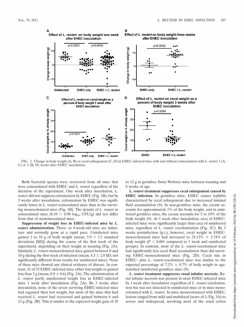
Suppression of weight loss in EHEC-infected mice by L.reuteri administration. Three- to 4-week-old mice are imma-ture and normally grow at a rapid pace. Uninfected micegained 2 to 10 g of body weight (mean, 5.9 � 2.1 standarddeviations [SD]) during the course of the first week of theexperiment, depending on their weight at weaning (Fig. 2A).Similarly, L. reuteri-monocolonized mice gained between 0 and10 g during the first week of infection (mean, 4.3 � 2.8 SD, notsignificantly different from results for uninfected mice). Noneof these mice showed any clinical evidence of disease. In con-trast, 31 of 32 EHEC-infected mice either lost weight or gainedless than 5 g (mean, 0.8 � 0.6) (Fig. 2A). The administration ofL. reuteri partly ameliorated weight loss in EHEC-infectedmice 1 week after inoculation (Fig. 2A). By 3 weeks afterinoculation, none of the seven surviving EHEC-infected micehad regained their lost weight, but most of the mice that hadreceived L. reuteri had recovered and gained between 6 and15 g (Fig. 2B). This is similar to the expected weight gain of 10
to 12 g in germfree Swiss Webster mice between weaning and6 weeks of age.
L. reuteri treatment suppresses cecal enlargement caused byEHEC infection. In germfree mice, EHEC causes typhlitischaracterized by cecal enlargement due to increased luminalfluid accumulation (9). In non-germfree mice, the cecum ac-counts for approximately 1% of the body weight, and in unin-fected germfree mice, the cecum accounts for 5 to 10% of thebody weight (9). At 1 week after inoculation, ceca of EHEC-infected mice were significantly larger than ceca of uninfectedmice, regardless of L. reuteri cocolonization (Fig. 2C). By 3weeks postinfection (p.i.), however, cecal weight in EHEC-monocolonized mice had increased to 24.12% � 3.74% ofbody weight (P � 0.001 compared to 1 week and uninfectedgroups). In contrast, most of the L. reuteri-cocolonized micehad significantly less cecal fluid accumulation than did surviv-ing EHEC-monocolonized mice (Fig. 2D). Cecal size inEHEC- plus L. reuteri-cocolonized mice was similar to theexpected percentage of 7.2% � 0.7% of body weight in age-matched uninfected germfree mice (9).
L. reuteri treatment suppresses renal tubular necrosis. Re-nal tubular necrosis was present in most EHEC-infected miceby 1 week after inoculation regardless of L. reuteri cocoloniza-tion but was not detected in uninfected mice or in mice mono-colonized with L. reuteri. In mice monocolonized with EHEC,lesions ranged from mild and multifocal (score of 1; Fig. 3A) tosevere and widespread, involving most of the renal cortex
FIG. 2. Change in body weight (A, B) or cecal enlargement (C, D) in EHEC-infected mice with and without coinoculation with L. reuteri 1 (A,C) or 3 (B, D) weeks after EHEC inoculation.
VOL. 79, 2011 L. REUTERI IN EHEC INFECTION 187
Dow
nloa
ded
from
http
s://j
ourn
als.
asm
.org
/jour
nal/i
ai o
n 10
Feb
ruar
y 20
22 b
y 88
.199
.126
.52.
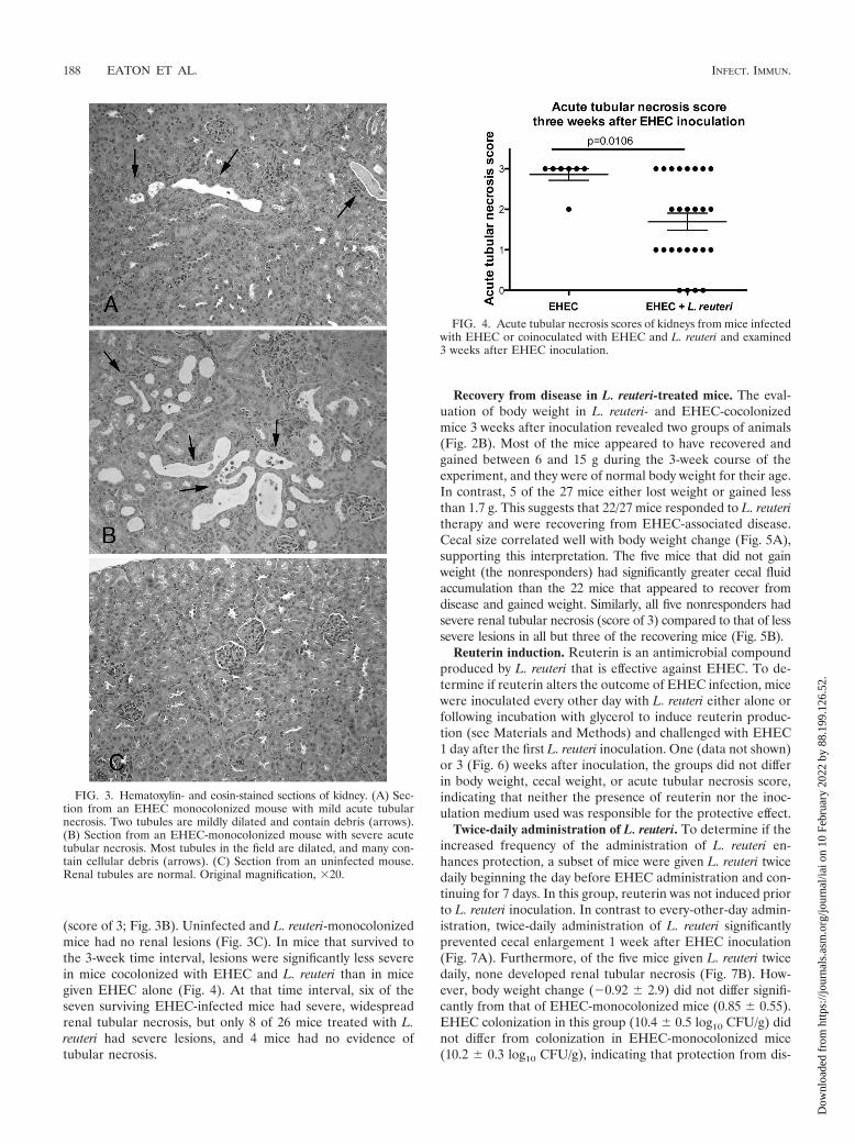
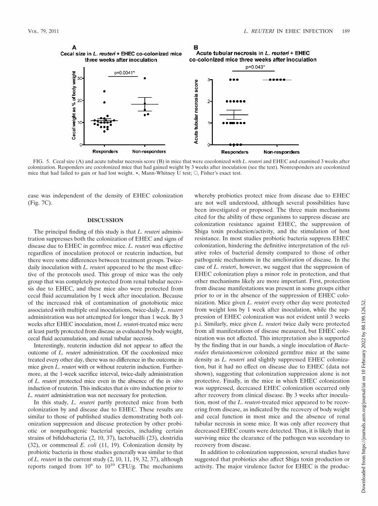
(score of 3; Fig. 3B). Uninfected and L. reuteri-monocolonizedmice had no renal lesions (Fig. 3C). In mice that survived tothe 3-week time interval, lesions were significantly less severein mice cocolonized with EHEC and L. reuteri than in micegiven EHEC alone (Fig. 4). At that time interval, six of theseven surviving EHEC-infected mice had severe, widespreadrenal tubular necrosis, but only 8 of 26 mice treated with L.reuteri had severe lesions, and 4 mice had no evidence oftubular necrosis.
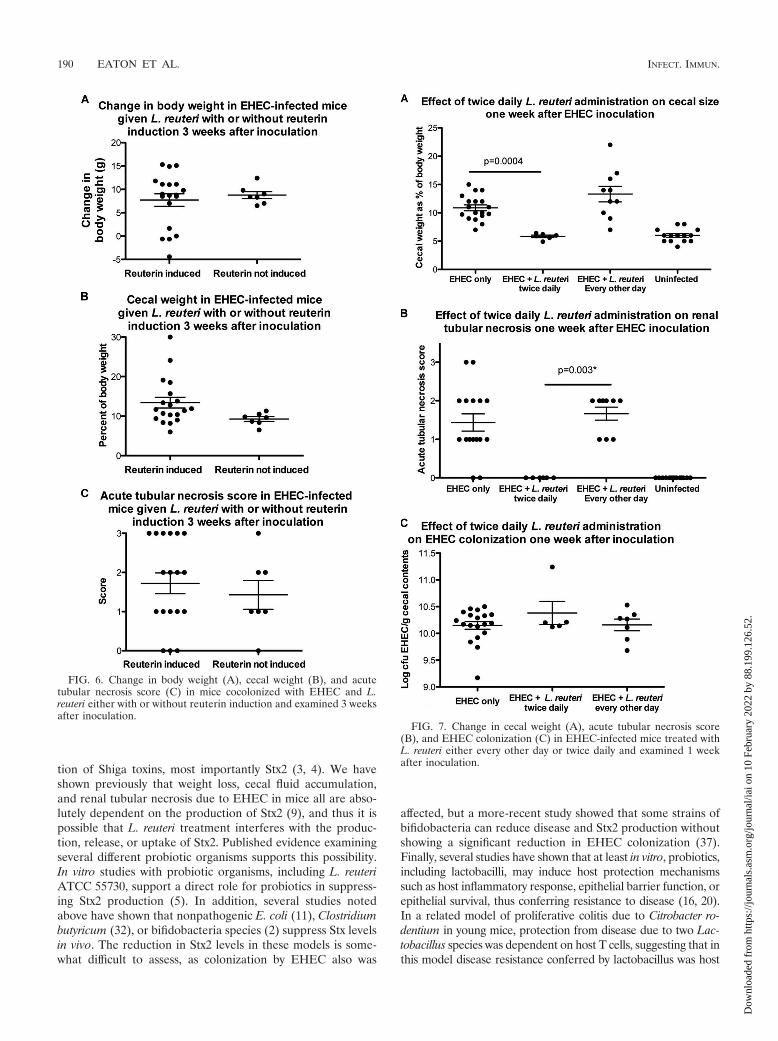
Recovery from disease in L. reuteri-treated mice. The eval-uation of body weight in L. reuteri- and EHEC-cocolonizedmice 3 weeks after inoculation revealed two groups of animals(Fig. 2B). Most of the mice appeared to have recovered andgained between 6 and 15 g during the 3-week course of theexperiment, and they were of normal body weight for their age.In contrast, 5 of the 27 mice either lost weight or gained lessthan 1.7 g. This suggests that 22/27 mice responded to L. reuteritherapy and were recovering from EHEC-associated disease.Cecal size correlated well with body weight change (Fig. 5A),supporting this interpretation. The five mice that did not gainweight (the nonresponders) had significantly greater cecal fluidaccumulation than the 22 mice that appeared to recover fromdisease and gained weight. Similarly, all five nonresponders hadsevere renal tubular necrosis (score of 3) compared to that of lesssevere lesions in all but three of the recovering mice (Fig. 5B).
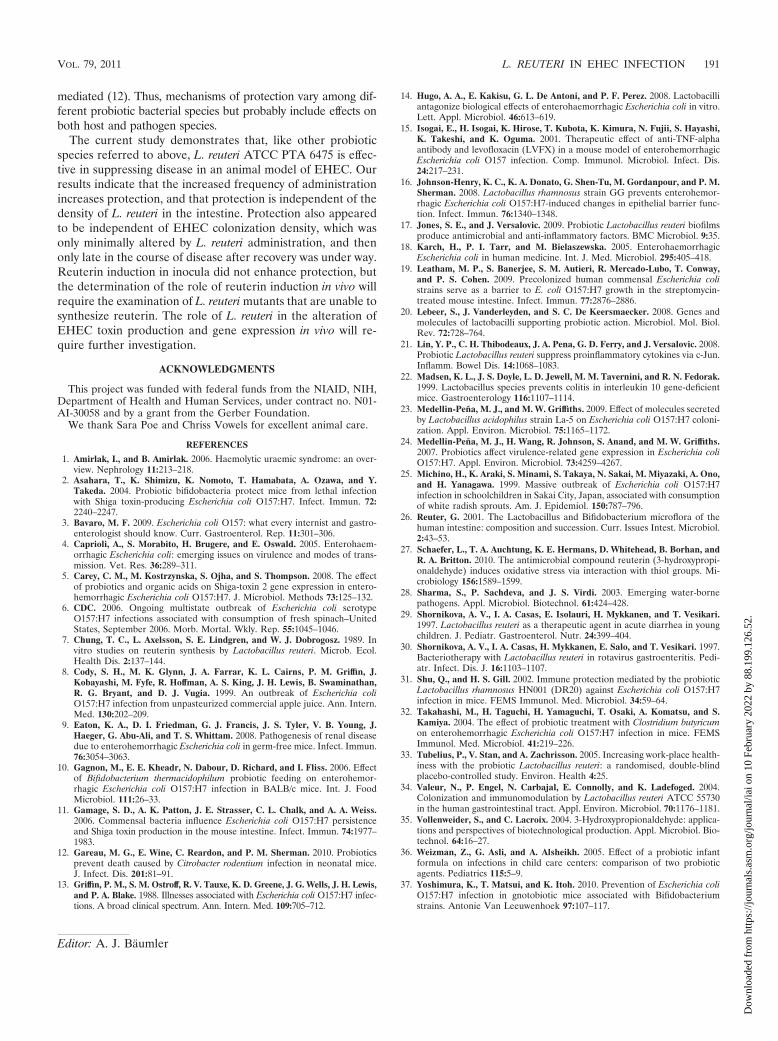
Reuterin induction. Reuterin is an antimicrobial compoundproduced by L. reuteri that is effective against EHEC. To de-termine if reuterin alters the outcome of EHEC infection, micewere inoculated every other day with L. reuteri either alone orfollowing incubation with glycerol to induce reuterin produc-tion (see Materials and Methods) and challenged with EHEC1 day after the first L. reuteri inoculation. One (data not shown)or 3 (Fig. 6) weeks after inoculation, the groups did not differin body weight, cecal weight, or acute tubular necrosis score,indicating that neither the presence of reuterin nor the inoc-ulation medium used was responsible for the protective effect.
Twice-daily administration of L. reuteri. To determine if theincreased frequency of the administration of L. reuteri en-hances protection, a subset of mice were given L. reuteri twicedaily beginning the day before EHEC administration and con-tinuing for 7 days. In this group, reuterin was not induced priorto L. reuteri inoculation. In contrast to every-other-day admin-istration, twice-daily administration of L. reuteri significantlyprevented cecal enlargement 1 week after EHEC inoculation(Fig. 7A). Furthermore, of the five mice given L. reuteri twicedaily, none developed renal tubular necrosis (Fig. 7B). How-ever, body weight change (�0.92 � 2.9) did not differ signifi-cantly from that of EHEC-monocolonized mice (0.85 � 0.55).EHEC colonization in this group (10.4 � 0.5 log10 CFU/g) didnot differ from colonization in EHEC-monocolonized mice(10.2 � 0.3 log10 CFU/g), indicating that protection from dis-
FIG. 3. Hematoxylin- and eosin-stained sections of kidney. (A) Sec-tion from an EHEC monocolonized mouse with mild acute tubularnecrosis. Two tubules are mildly dilated and contain debris (arrows).(B) Section from an EHEC-monocolonized mouse with severe acutetubular necrosis. Most tubules in the field are dilated, and many con-tain cellular debris (arrows). (C) Section from an uninfected mouse.Renal tubules are normal. Original magnification, �20.
FIG. 4. Acute tubular necrosis scores of kidneys from mice infectedwith EHEC or coinoculated with EHEC and L. reuteri and examined3 weeks after EHEC inoculation.
188 EATON ET AL. INFECT. IMMUN.
Dow
nloa
ded
from
http
s://j
ourn
als.
asm
.org
/jour
nal/i
ai o
n 10
Feb
ruar
y 20
22 b
y 88
.199
.126
.52.
ease was independent of the density of EHEC colonization(Fig. 7C).
DISCUSSION
The principal finding of this study is that L. reuteri adminis-tration suppresses both the colonization of EHEC and signs ofdisease due to EHEC in germfree mice. L. reuteri was effectiveregardless of inoculation protocol or reuterin induction, butthere were some differences between treatment groups. Twice-daily inoculation with L. reuteri appeared to be the most effec-tive of the protocols used. This group of mice was the onlygroup that was completely protected from renal tubular necro-sis due to EHEC, and these mice also were protected fromcecal fluid accumulation by 1 week after inoculation. Becauseof the increased risk of contamination of gnotobiotic miceassociated with multiple oral inoculations, twice-daily L. reuteriadministration was not attempted for longer than 1 week. By 3weeks after EHEC inoculation, most L. reuteri-treated mice wereat least partly protected from disease as evaluated by body weight,cecal fluid accumulation, and renal tubular necrosis.
Interestingly, reuterin induction did not appear to affect theoutcome of L. reuteri administration. Of the cocolonized micetreated every other day, there was no difference in the outcome inmice given L. reuteri with or without reuterin induction. Further-more, at the 1-week sacrifice interval, twice-daily administrationof L. reuteri protected mice even in the absence of the in vitroinduction of reuterin. This indicates that in vitro induction prior toL. reuteri administration was not necessary for protection.
In this study, L. reuteri partly protected mice from bothcolonization by and disease due to EHEC. These results aresimilar to those of published studies demonstrating both col-onization suppression and disease protection by other probi-otic or nonpathogenic bacterial species, including certainstrains of bifidobacteria (2, 10, 37), lactobacilli (23), clostridia(32), or commensal E. coli (11, 19). Colonization density byprobiotic bacteria in those studies generally was similar to thatof L. reuteri in the current study (2, 10, 11, 19, 32, 37), althoughreports ranged from 106 to 1010 CFU/g. The mechanisms
whereby probiotics protect mice from disease due to EHECare not well understood, although several possibilities havebeen investigated or proposed. The three main mechanismscited for the ability of these organisms to suppress disease arecolonization resistance against EHEC, the suppression ofShiga toxin production/activity, and the stimulation of hostresistance. In most studies probiotic bacteria suppress EHECcolonization, hindering the definitive interpretation of the rel-ative roles of bacterial density compared to those of otherpathogenic mechanisms in the amelioration of disease. In thecase of L. reuteri, however, we suggest that the suppression ofEHEC colonization plays a minor role in protection, and thatother mechanisms likely are more important. First, protectionfrom disease manifestations was present in some groups eitherprior to or in the absence of the suppression of EHEC colo-nization. Mice given L. reuteri every other day were protectedfrom weight loss by 1 week after inoculation, while the sup-pression of EHEC colonization was not evident until 3 weeksp.i. Similarly, mice given L. reuteri twice daily were protectedfrom all manifestations of disease measured, but EHEC colo-nization was not affected. This interpretation also is supportedby the finding that in our hands, a single inoculation of Bacte-roides thetaiotaomicron colonized germfree mice at the samedensity as L. reuteri and slightly suppressed EHEC coloniza-tion, but it had no effect on disease due to EHEC (data notshown), suggesting that colonization suppression alone is notprotective. Finally, in the mice in which EHEC colonizationwas suppressed, decreased EHEC colonization occurred onlyafter recovery from clinical disease. By 3 weeks after inocula-tion, most of the L. reuteri-treated mice appeared to be recov-ering from disease, as indicated by the recovery of body weightand cecal function in most mice and the absence of renaltubular necrosis in some mice. It was only after recovery thatdecreased EHEC counts were detected. Thus, it is likely that insurviving mice the clearance of the pathogen was secondary torecovery from disease.
In addition to colonization suppression, several studies havesuggested that probiotics also affect Shiga toxin production oractivity. The major virulence factor for EHEC is the produc-
FIG. 5. Cecal size (A) and acute tubular necrosis score (B) in mice that were cocolonized with L. reuteri and EHEC and examined 3 weeks aftercolonization. Responders are cocolonized mice that had gained weight by 3 weeks after inoculation (see the text). Nonresponders are cocolonizedmice that had failed to gain or had lost weight. *, Mann-Whitney U test; E, Fisher’s exact test.
VOL. 79, 2011 L. REUTERI IN EHEC INFECTION 189
Dow
nloa
ded
from
http
s://j
ourn
als.
asm
.org
/jour
nal/i
ai o
n 10
Feb
ruar
y 20
22 b
y 88
.199
.126
.52.
tion of Shiga toxins, most importantly Stx2 (3, 4). We haveshown previously that weight loss, cecal fluid accumulation,and renal tubular necrosis due to EHEC in mice all are abso-lutely dependent on the production of Stx2 (9), and thus it ispossible that L. reuteri treatment interferes with the produc-tion, release, or uptake of Stx2. Published evidence examiningseveral different probiotic organisms supports this possibility.In vitro studies with probiotic organisms, including L. reuteriATCC 55730, support a direct role for probiotics in suppress-ing Stx2 production (5). In addition, several studies notedabove have shown that nonpathogenic E. coli (11), Clostridiumbutyricum (32), or bifidobacteria species (2) suppress Stx levelsin vivo. The reduction in Stx2 levels in these models is some-what difficult to assess, as colonization by EHEC also was
affected, but a more-recent study showed that some strains ofbifidobacteria can reduce disease and Stx2 production withoutshowing a significant reduction in EHEC colonization (37).Finally, several studies have shown that at least in vitro, probiotics,including lactobacilli, may induce host protection mechanismssuch as host inflammatory response, epithelial barrier function, orepithelial survival, thus conferring resistance to disease (16, 20).In a related model of proliferative colitis due to Citrobacter ro-dentium in young mice, protection from disease due to two Lac-tobacillus species was dependent on host T cells, suggesting that inthis model disease resistance conferred by lactobacillus was host
FIG. 6. Change in body weight (A), cecal weight (B), and acutetubular necrosis score (C) in mice cocolonized with EHEC and L.reuteri either with or without reuterin induction and examined 3 weeksafter inoculation.
FIG. 7. Change in cecal weight (A), acute tubular necrosis score(B), and EHEC colonization (C) in EHEC-infected mice treated withL. reuteri either every other day or twice daily and examined 1 weekafter inoculation.
190 EATON ET AL. INFECT. IMMUN.
Dow
nloa
ded
from
http
s://j
ourn
als.
asm
.org
/jour
nal/i
ai o
n 10
Feb
ruar
y 20
22 b
y 88
.199
.126
.52.
mediated (12). Thus, mechanisms of protection vary among dif-ferent probiotic bacterial species but probably include effects onboth host and pathogen species.
The current study demonstrates that, like other probioticspecies referred to above, L. reuteri ATCC PTA 6475 is effec-tive in suppressing disease in an animal model of EHEC. Ourresults indicate that the increased frequency of administrationincreases protection, and that protection is independent of thedensity of L. reuteri in the intestine. Protection also appearedto be independent of EHEC colonization density, which wasonly minimally altered by L. reuteri administration, and thenonly late in the course of disease after recovery was under way.Reuterin induction in inocula did not enhance protection, butthe determination of the role of reuterin induction in vivo willrequire the examination of L. reuteri mutants that are unable tosynthesize reuterin. The role of L. reuteri in the alteration ofEHEC toxin production and gene expression in vivo will re-quire further investigation.
ACKNOWLEDGMENTS
This project was funded with federal funds from the NIAID, NIH,Department of Health and Human Services, under contract no. N01-AI-30058 and by a grant from the Gerber Foundation.
We thank Sara Poe and Chriss Vowels for excellent animal care.
REFERENCES
1. Amirlak, I., and B. Amirlak. 2006. Haemolytic uraemic syndrome: an over-view. Nephrology 11:213–218.
2. Asahara, T., K. Shimizu, K. Nomoto, T. Hamabata, A. Ozawa, and Y.Takeda. 2004. Probiotic bifidobacteria protect mice from lethal infectionwith Shiga toxin-producing Escherichia coli O157:H7. Infect. Immun. 72:2240–2247.
3. Bavaro, M. F. 2009. Escherichia coli O157: what every internist and gastro-enterologist should know. Curr. Gastroenterol. Rep. 11:301–306.
4. Caprioli, A., S. Morabito, H. Brugere, and E. Oswald. 2005. Enterohaem-orrhagic Escherichia coli: emerging issues on virulence and modes of trans-mission. Vet. Res. 36:289–311.
5. Carey, C. M., M. Kostrzynska, S. Ojha, and S. Thompson. 2008. The effectof probiotics and organic acids on Shiga-toxin 2 gene expression in entero-hemorrhagic Escherichia coli O157:H7. J. Microbiol. Methods 73:125–132.
6. CDC. 2006. Ongoing multistate outbreak of Escherichia coli serotypeO157:H7 infections associated with consumption of fresh spinach–UnitedStates, September 2006. Morb. Mortal. Wkly. Rep. 55:1045–1046.
7. Chung, T. C., L. Axelsson, S. E. Lindgren, and W. J. Dobrogosz. 1989. Invitro studies on reuterin synthesis by Lactobacillus reuteri. Microb. Ecol.Health Dis. 2:137–144.
8. Cody, S. H., M. K. Glynn, J. A. Farrar, K. L. Cairns, P. M. Griffin, J.Kobayashi, M. Fyfe, R. Hoffman, A. S. King, J. H. Lewis, B. Swaminathan,R. G. Bryant, and D. J. Vugia. 1999. An outbreak of Escherichia coliO157:H7 infection from unpasteurized commercial apple juice. Ann. Intern.Med. 130:202–209.
9. Eaton, K. A., D. I. Friedman, G. J. Francis, J. S. Tyler, V. B. Young, J.Haeger, G. Abu-Ali, and T. S. Whittam. 2008. Pathogenesis of renal diseasedue to enterohemorrhagic Escherichia coli in germ-free mice. Infect. Immun.76:3054–3063.
10. Gagnon, M., E. E. Kheadr, N. Dabour, D. Richard, and I. Fliss. 2006. Effectof Bifidobacterium thermacidophilum probiotic feeding on enterohemor-rhagic Escherichia coli O157:H7 infection in BALB/c mice. Int. J. FoodMicrobiol. 111:26–33.
11. Gamage, S. D., A. K. Patton, J. E. Strasser, C. L. Chalk, and A. A. Weiss.2006. Commensal bacteria influence Escherichia coli O157:H7 persistenceand Shiga toxin production in the mouse intestine. Infect. Immun. 74:1977–1983.
12. Gareau, M. G., E. Wine, C. Reardon, and P. M. Sherman. 2010. Probioticsprevent death caused by Citrobacter rodentium infection in neonatal mice.J. Infect. Dis. 201:81–91.
13. Griffin, P. M., S. M. Ostroff, R. V. Tauxe, K. D. Greene, J. G. Wells, J. H. Lewis,and P. A. Blake. 1988. Illnesses associated with Escherichia coli O157:H7 infec-tions. A broad clinical spectrum. Ann. Intern. Med. 109:705–712.
14. Hugo, A. A., E. Kakisu, G. L. De Antoni, and P. F. Perez. 2008. Lactobacilliantagonize biological effects of enterohaemorrhagic Escherichia coli in vitro.Lett. Appl. Microbiol. 46:613–619.
15. Isogai, E., H. Isogai, K. Hirose, T. Kubota, K. Kimura, N. Fujii, S. Hayashi,K. Takeshi, and K. Oguma. 2001. Therapeutic effect of anti-TNF-alphaantibody and levofloxacin (LVFX) in a mouse model of enterohemorrhagicEscherichia coli O157 infection. Comp. Immunol. Microbiol. Infect. Dis.24:217–231.
16. Johnson-Henry, K. C., K. A. Donato, G. Shen-Tu, M. Gordanpour, and P. M.Sherman. 2008. Lactobacillus rhamnosus strain GG prevents enterohemor-rhagic Escherichia coli O157:H7-induced changes in epithelial barrier func-tion. Infect. Immun. 76:1340–1348.
17. Jones, S. E., and J. Versalovic. 2009. Probiotic Lactobacillus reuteri biofilmsproduce antimicrobial and anti-inflammatory factors. BMC Microbiol. 9:35.
18. Karch, H., P. I. Tarr, and M. Bielaszewska. 2005. EnterohaemorrhagicEscherichia coli in human medicine. Int. J. Med. Microbiol. 295:405–418.
19. Leatham, M. P., S. Banerjee, S. M. Autieri, R. Mercado-Lubo, T. Conway,and P. S. Cohen. 2009. Precolonized human commensal Escherichia colistrains serve as a barrier to E. coli O157:H7 growth in the streptomycin-treated mouse intestine. Infect. Immun. 77:2876–2886.
20. Lebeer, S., J. Vanderleyden, and S. C. De Keersmaecker. 2008. Genes andmolecules of lactobacilli supporting probiotic action. Microbiol. Mol. Biol.Rev. 72:728–764.
21. Lin, Y. P., C. H. Thibodeaux, J. A. Pena, G. D. Ferry, and J. Versalovic. 2008.Probiotic Lactobacillus reuteri suppress proinflammatory cytokines via c-Jun.Inflamm. Bowel Dis. 14:1068–1083.
22. Madsen, K. L., J. S. Doyle, L. D. Jewell, M. M. Tavernini, and R. N. Fedorak.1999. Lactobacillus species prevents colitis in interleukin 10 gene-deficientmice. Gastroenterology 116:1107–1114.
23. Medellin-Pena, M. J., and M. W. Griffiths. 2009. Effect of molecules secretedby Lactobacillus acidophilus strain La-5 on Escherichia coli O157:H7 coloni-zation. Appl. Environ. Microbiol. 75:1165–1172.
24. Medellin-Pena, M. J., H. Wang, R. Johnson, S. Anand, and M. W. Griffiths.2007. Probiotics affect virulence-related gene expression in Escherichia coliO157:H7. Appl. Environ. Microbiol. 73:4259–4267.
25. Michino, H., K. Araki, S. Minami, S. Takaya, N. Sakai, M. Miyazaki, A. Ono,and H. Yanagawa. 1999. Massive outbreak of Escherichia coli O157:H7infection in schoolchildren in Sakai City, Japan, associated with consumptionof white radish sprouts. Am. J. Epidemiol. 150:787–796.
26. Reuter, G. 2001. The Lactobacillus and Bifidobacterium microflora of thehuman intestine: composition and succession. Curr. Issues Intest. Microbiol.2:43–53.
27. Schaefer, L., T. A. Auchtung, K. E. Hermans, D. Whitehead, B. Borhan, andR. A. Britton. 2010. The antimicrobial compound reuterin (3-hydroxypropi-onaldehyde) induces oxidative stress via interaction with thiol groups. Mi-crobiology 156:1589–1599.
28. Sharma, S., P. Sachdeva, and J. S. Virdi. 2003. Emerging water-bornepathogens. Appl. Microbiol. Biotechnol. 61:424–428.
29. Shornikova, A. V., I. A. Casas, E. Isolauri, H. Mykkanen, and T. Vesikari.1997. Lactobacillus reuteri as a therapeutic agent in acute diarrhea in youngchildren. J. Pediatr. Gastroenterol. Nutr. 24:399–404.
30. Shornikova, A. V., I. A. Casas, H. Mykkanen, E. Salo, and T. Vesikari. 1997.Bacteriotherapy with Lactobacillus reuteri in rotavirus gastroenteritis. Pedi-atr. Infect. Dis. J. 16:1103–1107.
31. Shu, Q., and H. S. Gill. 2002. Immune protection mediated by the probioticLactobacillus rhamnosus HN001 (DR20) against Escherichia coli O157:H7infection in mice. FEMS Immunol. Med. Microbiol. 34:59–64.
32. Takahashi, M., H. Taguchi, H. Yamaguchi, T. Osaki, A. Komatsu, and S.Kamiya. 2004. The effect of probiotic treatment with Clostridium butyricumon enterohemorrhagic Escherichia coli O157:H7 infection in mice. FEMSImmunol. Med. Microbiol. 41:219–226.
33. Tubelius, P., V. Stan, and A. Zachrisson. 2005. Increasing work-place health-iness with the probiotic Lactobacillus reuteri: a randomised, double-blindplacebo-controlled study. Environ. Health 4:25.
34. Valeur, N., P. Engel, N. Carbajal, E. Connolly, and K. Ladefoged. 2004.Colonization and immunomodulation by Lactobacillus reuteri ATCC 55730in the human gastrointestinal tract. Appl. Environ. Microbiol. 70:1176–1181.
35. Vollenweider, S., and C. Lacroix. 2004. 3-Hydroxypropionaldehyde: applica-tions and perspectives of biotechnological production. Appl. Microbiol. Bio-technol. 64:16–27.
36. Weizman, Z., G. Asli, and A. Alsheikh. 2005. Effect of a probiotic infantformula on infections in child care centers: comparison of two probioticagents. Pediatrics 115:5–9.
37. Yoshimura, K., T. Matsui, and K. Itoh. 2010. Prevention of Escherichia coliO157:H7 infection in gnotobiotic mice associated with Bifidobacteriumstrains. Antonie Van Leeuwenhoek 97:107–117.
Editor: A. J. Baumler
VOL. 79, 2011 L. REUTERI IN EHEC INFECTION 191
Dow
nloa
ded
from
http
s://j
ourn
als.
asm
.org
/jour
nal/i
ai o
n 10
Feb
ruar
y 20
22 b
y 88
.199
.126
.52.