Probing the Active Site of Thimet Oligopeptidase: A … the Active Site of Thimet Oligopeptidase: A...
15
Probing the Active Site of Thimet Oligopeptidase: A Study In Removing the Catalytic Metal Abstract Thimet Oligopeptidase(TOP, E.C. 3.4.24.15) is a metallopeptidase that cleaves various peptides involved in many physiological functions including the regulation of blood pressure and pain recognition. TOP is a zincin enzyme of clan MA 1 with the conserved HEXXH binding motif at the active site. Recent crystal structures have elucidated much of the structural information about TOP 2 ; however, little is known about the specific coordinate geometry of Zn 2+ in the active site. In this study, the preparation of spectroscopically active metal substituted derivatives of TOP was attempted in order to better define this coordination and, if possible, extrapolate an explanation for the preferential binding of Zn 2+ as opposed to other divalent metals which have recently been discovered in the active sites of similar peptidases 3 . In addition, different metal substituted derivatives of TOP were prepared in order to determine the kinetic activity of said derivatives, with the intention of using this data to draw conclusions as to a “preferred” coordination of the metal. Introduction Thimet Oligopeptidase(TOP, E.C. 3.4.24.15) is a zincin metalloendopeptidase which is ubiquitous through the body but is most highly concentrated in the brain and gonads. As a peptidase, its physiological function is the cleaving of short peptides(<17 AA)and plays in an important role in regulating blood pressure and has been implicated in the degradation of amyloid-β peptides which contribute to Alzheimer’s disease. 4,5,6 Most importantly for this study, TOP contains the HEXXH binding domain at the active site (see Fig. 1) which is homologous in many other zinc containing metalloenzymes. In the past, studies utilizing metal ion substitution showed that close homologues of TOP, such as thermolysin(E.C. 3.4.24.27 )which contains the HEXXH motif, actually become hyperactive with a cobalt substitution 7 . More recently other peptidases(including, interestingly, a carboxypeptidase) with the HEXXH binding motif have been discovered which natively utilize cobalt in their active site 3,8 . This raises questions as to why the majority of enzymes with this motif natively utilize zinc.
Transcript of Probing the Active Site of Thimet Oligopeptidase: A … the Active Site of Thimet Oligopeptidase: A...
Probing the Active Site of Thimet Oligopeptidase: A
Study In Removing the Catalytic Metal Abstract
Thimet Oligopeptidase(TOP, E.C. 3.4.24.15) is a metallopeptidase that cleaves
various peptides involved in many physiological functions including the regulation of
blood pressure and pain recognition. TOP is a zincin enzyme of clan MA1 with the
conserved HEXXH binding motif at the active site. Recent crystal structures have
elucidated much of the structural information about TOP2; however, little is known about
the specific coordinate geometry of Zn2+ in the active site. In this study, the preparation
of spectroscopically active metal substituted derivatives of TOP was attempted in order to
better define this coordination and, if possible, extrapolate an explanation for the
preferential binding of Zn2+ as opposed to other divalent metals which have recently been
discovered in the active sites of similar peptidases3. In addition, different metal
substituted derivatives of TOP were prepared in order to determine the kinetic activity of
said derivatives, with the intention of using this data to draw conclusions as to a
“preferred” coordination of the metal.
Introduction
Thimet Oligopeptidase(TOP, E.C. 3.4.24.15) is a zincin metalloendopeptidase which
is ubiquitous through the body but is most highly concentrated in the brain and gonads.
As a peptidase, its physiological function is the cleaving of short peptides(<17 AA)and
plays in an important role in regulating blood pressure and has been implicated in the
degradation of amyloid-β peptides which contribute to Alzheimer’s disease.4,5,6 Most
importantly for this study, TOP contains the HEXXH binding domain at the active site
(see Fig. 1) which is homologous in many other zinc containing metalloenzymes. In the
past, studies utilizing metal ion substitution showed that close homologues of TOP, such
as thermolysin(E.C. 3.4.24.27 )which contains the HEXXH motif, actually become
hyperactive with a cobalt substitution7. More recently other peptidases(including,
interestingly, a carboxypeptidase) with the HEXXH binding motif have been discovered
which natively utilize cobalt in their active site3,8. This raises questions as to why the
majority of enzymes with this motif natively utilize zinc.

No spectrophotometric data can be gathered about native zinc enzymes, as Zn2+ has no
absorbance in the visible region, therefore metal substituted derivatives play an important
role in characterizing the coordination geometry of these zincin enzymes.
Spectrophotometric studies on metal substituted derivatives of peptidases simlar to TOP
have yielded interesting results. In a study of dipeptidyl peptidase III (DPP, E.C.
3.4.14.4), which contains the HEXXH motif, UV-vis showed that Co2+-DPP III exhibited
a pentacoordinate geometry. This is only slightly helpful however as the study did not
observe coordination of the metal during catalysis. In a more thorough study of
Thermolysin, an even closer homologue of TOP, the Co2+-substituted form was observed
to adopt a pentacoordinate geometry and was seen to exhibit flexibility in coordination
during catalysis(i.e. adopting different coordinate geometries during catalysis).7
According to the authors, the slight preference for a five-coordinate geometry, which is
close to the transition state, along with the flexibility in coordination, explain the ability
of Co2+-thermolysin to exhibit hyperactivity.
In this study, metal substituted derivatives of TOP were produced and attempted to
be analyzed by quenched fluorescence kinetic assays as well as U.V.-vis spectroscopy.
Materials and Methods
Many methods were attempted in order to remove the divalent zinc ion from TOP;
however, only one was successful. Still, it is illustrative to describe the unsuccessful
methods in order to more thoroughly understand the nature of the Zn2+ as it is substituted
in the enzyme. In addition, the method of substituting in new metals was refined as time
went on. In all studies, TOP was prepared by Marc. J Glucksman of the
Midwest Proteome Center and Department of Biochemistry and Molecular Biology,
Rosalind Franklin University of Medicine and Science in Chicago, IL.
Activity Assay
Activity assay was performed bys using an Perkin Elmer LS 50 B Luminescence
Spetrometer 1980µL of buffer containing 25mM tris, 125mM KCl and 1mM TCEP was
equilibrated with 10µL of MCAi substrate(of varying concentrations) at 23°C. Then,
10µL of .1µM enzyme were added to the chamber and florescence was detected for 150
seconds with and excitation of 325nm and an emission of 400nm. Note: For simple i7‐methoxycoumarin‐4‐acetyl‐Pro‐Leu‐Gly‐Pro‐Lys‐dinitrophenol

binary tests of activity, enzyme concentration was noted(if possible) but not always
.1µM. Often, a total of 2µmols of enzyme was present in the reaction chamber.
Dialysis Against 2,6-Pyridinedicarboxylic Acid
This method was adapted from a paper by Hirose et. al.9 and involved the dialysis of
the enzyme against 50mM tris buffer with 20mM 2,6 Pyridinedicarboxylic Acid(2,6-
PCA) at pH 7.4 with various concentrations of Dithiolreltol(DTT) ranging from .5mM to
3mM. Dialysis was carried out at 4°C for 24 hours. After dialysis, the enzyme was
concentrated using a Millipore centricon and activity was determined by the method
described above.
Dialysis Against Ethylenediaminetetraacetic Acid
A similar method was attempted using Ethylenediamineteraacetic Acid(EDTA). This
was adopted from the method of Barrett and Brown10 who worked with TOP. Using a
dialysis buffer of 50mM tris, 3mM DTT and 100mM EDTA at pH 7.0. The dialysis was
carried for approximately 24 hours at 4°C. The enzyme was then concentrated and
activity was ascertained by quenched fluorescence assay by the method describe above.
Denaturation with an EDTA Buffer
A stock buffer containing 25mM tris, 50mM EDTA, 125mM KCl and 9M urea with
10% glycerol at pH 7.8 was prepared. At the time of use, solid DTT was added to this
buffer to 5mM. 150µL of this buffer was then combined with 50µL of concentrated
enzyme stock in a microfuge tube. The tube was allowed to sit at room temperature for
approximately 1 hour and then was moved to a 4°C where it incubated for 18 hours. The
enzyme was then purified of excess urea and EDTA by using an Amersham Biosciences
PD-10 size exclusion column(PD-10). The new buffer which the enzyme was transferred
to contained 25mM tris and 5mM DTT at pH 7.4. The enzyme concentration was
obtained via –U.V. vis and then activity assay run using the same method described
above. This procedure was also repeated substituting 50mM 2,6-PCA for EDTA.
Incubation with 1,10-Phenanthroline
A buffer containing 25mM tris and approximately 8mM 1,10-Phenanthroline was
prepared. DTT was then added to 200µL of this buffer to 5mM. 150µL of the new buffer
was then combined with 50µL of a concentrated enzyme stock and allowed to sit at room
temperature for 1 hour and then transferred to a 4°C environment for 12 hours. The

sample was then loaded onto a PD-10 column and transferred to a buffer containing
25mM tris and 5mM DTT at pH 7.4. Activity assay was then run using same the method
as described above. This method was also attempted with the chelating agents 2,6-PCA
and EDTA.
General Test for Loss of Metal
If a sample of enzyme was observed to be inactive after a chelation or denaturation,
loss of the catalytic metal was tested by taking an aliquot of the inactive sample and
adding equimolar amounts of ZnCl2 to the enzyme. Activity assay was then performed on
the sample with Zn2+ and without added Zn2+ at regular time intervals. If the activity of
the sample without Zn2+ remained low, and the sample with Zn2+ regained activity, the
sample was determined to be actual apo-enzyme.
Substitution of Metals
Metal substitution was attempted by three methods, dialysis, incubation and
exchange of buffer via a PD-10 column.
Dialysis was performed by transferring the enzyme onto a dialysis chamber with a
metal at .5mM, 50mM tris and 3mM DTT at pH 7.4. The sample was then concentrated.
Incubation was performed by adding concentrated dissolved divalent metals to enzyme
buffered solutions.(Note: inactivation of enzyme was seen with Zn2+ concentrations
higher than 5x of the enzyme concentration). Finally, for the exchange of buffer method,
metal buffers were prepared with .1mM metal, 50mM tris, 3mM TCEP, 125mM KCl at
pH 7.0 were prepared. A PD-10 column was equilibrated to this buffer and the enzyme
was passed down the column in order to exchange the buffer. (Note: DTT could not be
used as it reduced the metals and precipitated them).
Results
As noted above, not all of these methods for removal of the Zn2+ ion were successful.
As such, the results have been divided between the successful and unsuccessful attempts.
Unsuccessful Removal of the Zn2+ Ion
Dialysis of TOP against 2,6-PCA resulted in no decrease in activity of the enzyme.
Initially, this was difficult to determine as 2,6-PCA obscured the 280nm protein peak(as
2,6-PCA absorbs very strongly at 271nm with a distinctive triple peak) and therefore
made it difficult to find the concentration of the enzyme. This problem was overcome

using Low Pressure Chromatography and, later, simple PD-10 columns to separate out
the excess 2,6-PCA
Dialysis for TOP against EDTA initially looked promising. After 24 hours of dialysis
against 100mM EDTA, the TOP was completely inactive(0% activity of holoenzyme),
however, upon an exchange of buffer which occurs as a result of the concentration
process, the enzyme regained nearly full activity. To combat against contamination, two
new samples were dialyzed under the same conditions to inactivation, then one was
transferred directly to a plain buffer(50mM tris, .5mM DTT, pH 7.4) and the other to a
buffer with metal(50mM tris , .5mM TCEP, .5mM CoCl2 pH7.4). After 6 hours, the
enzyme on the buffer without metal had regained activity to 10% of holoenzyme while
the sample on the buffer with metal had regained only 2.6% activity.(See Fig. 2)
Incubation of the enzyme with any chelating agent(i.e. EDTA, 2,6-PCA and
1,10-Phenanthroline) was unsuccessful. Neither 2,6-PCA or EDTA inactivated the
enzyme at all after 24 hours of incubation at 4°C. Incubation with 1,10-Phenanthroline
did decrease the activity of the enzyme to 12% of the t=0 sample. Upon purification and
addition of Zn2+ to the solution, the enzyme did not regain any activity.
Finally, denaturation in the presence of high concentrations of 2,6-PCA was
unsuccessful. Though the enzyme lost activity during the denaturation, it was regained as
soon as the solution was purified of Urea.
Successful Removal of the Zn2+ Ion
The procedure of denaturing the enzyme with urea in the presence of high
concentrations of EDTA was the only one which exhibited success. After 16 hours of
incubation at 4°C in the buffer described above, the enzyme was completely inactive.
Upon purification with a PD-10 column the enzyme remained inactive. Two samples
were then created, one with and one without Zn2+ present. After only 4 hours of
incubation at 4°C, the sample with Zn2+ exhibited full activity, while the sample without
was completely inactive(See Fig. 3). This result, however, was difficult to reproduce.
After apo-enzyme was confirmed to have been prepared, different metals were
substituted. In this way, Co-TOP, Cu-TOP and Zn-TOP(i.e. TOP with Zn2+ reintroduced
to the active site after the loss of the metal) were prepared. After these derivatives were
prepared, a full activity assay was run on each of them and the KM and KCAT determined
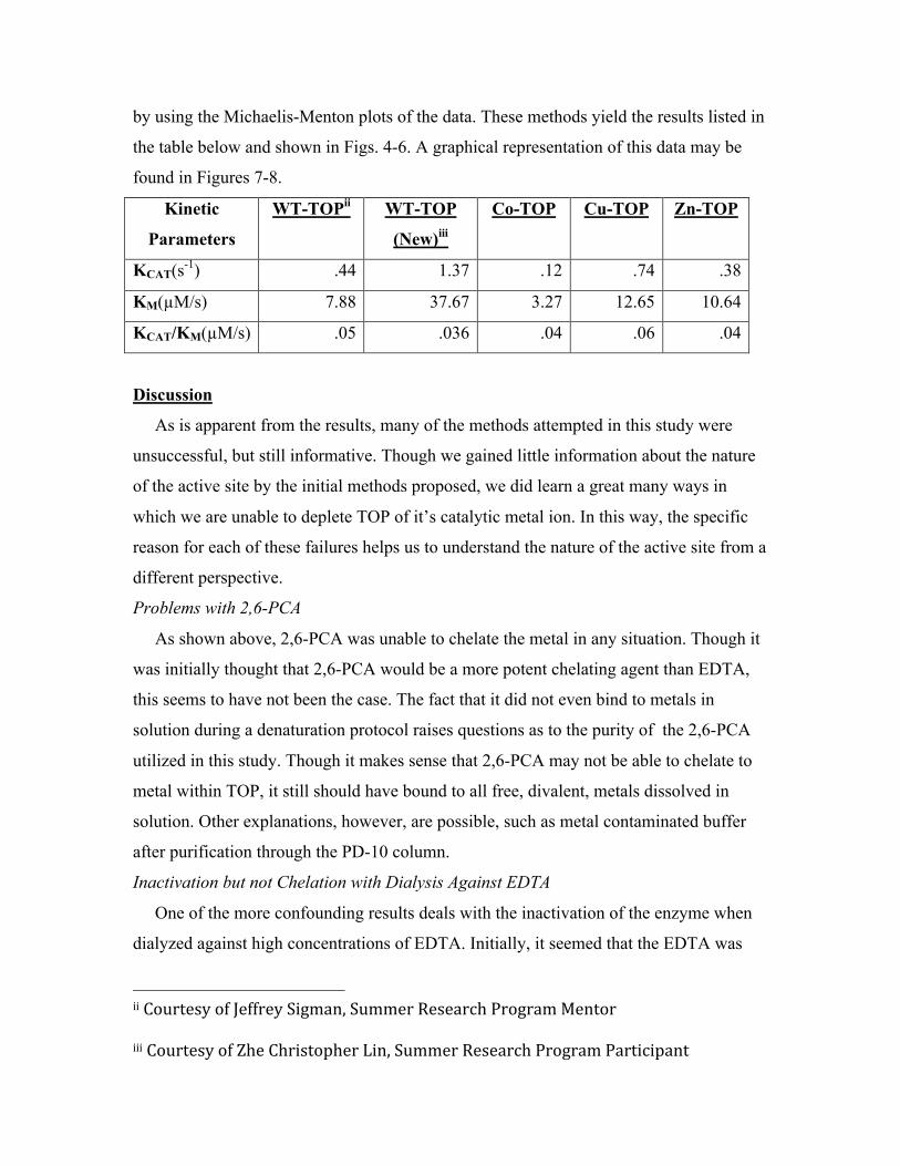
by using the Michaelis-Menton plots of the data. These methods yield the results listed in
the table below and shown in Figs. 4-6. A graphical representation of this data may be
found in Figures 7-8.
Kinetic
Parameters
WT-TOPii WT-TOP
(New)iii
Co-TOP Cu-TOP Zn-TOP
KCAT(s-1) .44 1.37 .12 .74 .38
KM(µM/s) 7.88 37.67 3.27 12.65 10.64
KCAT/KM(µM/s) .05 .036 .04 .06 .04
Discussion
As is apparent from the results, many of the methods attempted in this study were
unsuccessful, but still informative. Though we gained little information about the nature
of the active site by the initial methods proposed, we did learn a great many ways in
which we are unable to deplete TOP of it’s catalytic metal ion. In this way, the specific
reason for each of these failures helps us to understand the nature of the active site from a
different perspective.
Problems with 2,6-PCA
As shown above, 2,6-PCA was unable to chelate the metal in any situation. Though it
was initially thought that 2,6-PCA would be a more potent chelating agent than EDTA,
this seems to have not been the case. The fact that it did not even bind to metals in
solution during a denaturation protocol raises questions as to the purity of the 2,6-PCA
utilized in this study. Though it makes sense that 2,6-PCA may not be able to chelate to
metal within TOP, it still should have bound to all free, divalent, metals dissolved in
solution. Other explanations, however, are possible, such as metal contaminated buffer
after purification through the PD-10 column.
Inactivation but not Chelation with Dialysis Against EDTA
One of the more confounding results deals with the inactivation of the enzyme when
dialyzed against high concentrations of EDTA. Initially, it seemed that the EDTA was
iiCourtesyofJeffreySigman,SummerResearchProgramMentoriiiCourtesyofZheChristopherLin,SummerResearchProgramParticipant

able to chelate the metal, and thus, produce apo-TOP. The experiment in which TOP was
dialyzed against EDTA and then immediately moved to a dialysis chamber which
contained either a “plain” buffer or a metal buffer was inconclusive as both regained
activity. The fact that the sample on a “plain” buffer regained activity more quickly is
most likely because high concentrations of metal often inactivate enzymes, leading to a
more active enzyme on “plain” buffer. A final experiment was performed in which TOP
was dialyzed against 100mM EDTA and then concentrated in a buffer with 10µM EDTA,
a sufficient concentration to get rid of most trace metals in the water. This sample
regained activity to 100% of the holoenzyme.
These results lead to the conclusion that EDTA inactivates the enzyme, but does not
chelate the metal. The mechanism by which this happens is most likely solvation of the
active site. In past experiments, Sigman observed, anecdotally, that high concentrations
of DTT would inactivate the enzyme. This is due to the crowding of the active site by a
large number of DTT molecules. As DTT and EDTA have similar structures, it is likely
that much the same thing is happening. Also, this is consistent with the lack of
inactivation seen when the enzyme was dialyzed against 2,6-PCA. Where DTT and
EDTA have structure such that they may solvate the active site, 2,6-PCA has more of a
winged structure which may coordinate with the metal by a “closing of its wings” over
the metal, but ultimately, block other molecules from getting too close.
Ineffectiveness of Incubation with Chelating Agents
In all experiments in which TOP was incubated with various chelating agents(EDTA,
2,6-PCA, and 1,10-Phenanthroline) inactivation did not occur at all. This is intriguing
because of the results seen with dialysis of EDTA. This could be because, during dialysis,
as EDTA solvates the active site of TOP, the concentration gradient changed and more
EDTA moved into the enzyme chamber, making the concentration around the enzyme
much greater than in an incubation chamber, in which the concentration remains constant.
In addition, concentrations of EDTA used in incubation experiments were 20mM, which
may not have been high enough to cause inactivation.
Also interesting was the loss of activity when TOP was incubated with 1,10-
phenanthroline. As it did not regain activity with incubation with Zn2+, this could be
because the high concentration of 1,10-phenanthroline (8mM, which is saturation in
water) actually damaged the enzyme, inactivating it completely, instead of removing the
metal from TOP.
Problems with 2,6-PCA
2,6-PCA failed to remove the metal from TOP by all methods attempted, dialysis,
incubation and incubation with the denaturant urea. This is a very odd result as in other
metallopeptidases, 2,6-PCA was utilized quite successfully to chelate the metal ion by
simple dialysis9. Most frustrating was the fact that 2,6-PCA seemed incapable of binding
to the metal even when it had “fallen-out” of TOP during denaturation. Because of this,
two possibilities arise. First, the 2,6-PCA, prepared by Sigma-Aldrich, was impure.
Alternatively, the enzyme may have a higher affinity for the metal than the 2,6-PCA. The
latter possibility will be discussed further later.
Successful Preparation of apo-TOP by Denaturation in the Presence of EDTA
As stated earlier, the only method by which apo-TOP was created was by denaturation
in the presence of EDTA. Combined with all of the other results obtained, a fairly clear
mechanistic picture of what occurred may be described. The denaturation by urea
unfolded the enzyme enough for the metal to be lost by TOP. Next, the EDTA solvated
the newly released Zn2+, making it impossible for TOP to re-acquire the metal. Finally,
when the solution was purified down a PD-10 column, the apo-TOP was cleansed of
urea, allowing it to refold without a metal present. The idea that the EDTA solvated the
metal instead of simply binding to it is important, because it bypasses all problems that
may occur because TOP has a high affinity for Zn2+ than EDTA.
Kinetic Parameters of Metal Substituted TOP
Co-TOP, Cu-TOP and Zn-Top(i.e. apo-TOP which then had Zn2+ added to its active
site) were prepared. All of these metal derivatives had overall activities(i.e. KCAT/KM)
that were very similar to the holo enzyme with WT-TOP=.05µM/s, WT-
TOP(New)=.036µM/s, Co-TOP= .04µM/s, Cu-TOP=.06µM/s, and Zn-TOP=.04µM/s.
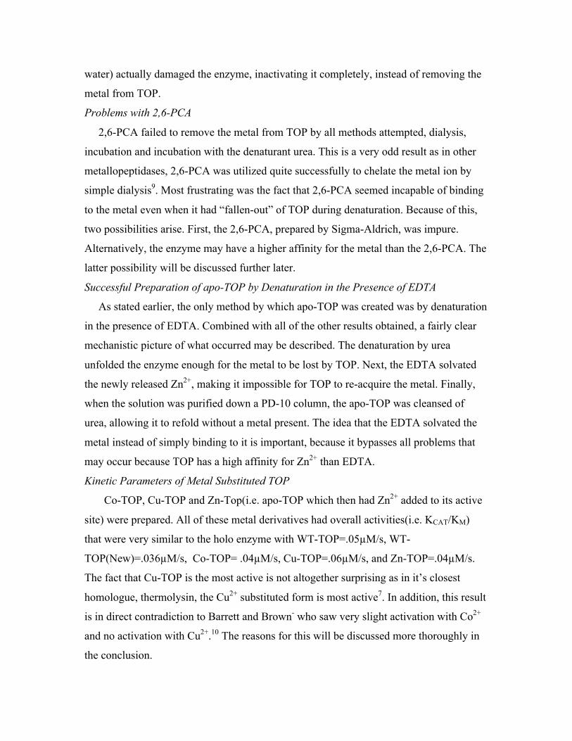
The fact that Cu-TOP is the most active is not altogether surprising as in it’s closest
homologue, thermolysin, the Cu2+ substituted form is most active7. In addition, this result
is in direct contradiction to Barrett and Brown- who saw very slight activation with Co2+
and no activation with Cu2+.10 The reasons for this will be discussed more thoroughly in
the conclusion.

While all of the metal derivatives had similar overall activities, their specific kinetic
parameters were very different. Co-TOP(KM=3.27µM/s) had a much higher binding
affinity than Cu-TOP and Zn-TOP(KM=12.65µM/s and 10.64µM/s respectively). This
result is difficult to interpret but may be due to different coordinate geometries of the
metals. In addition, KCAT for Cu-TOP(.74s-1 )was nearly 5 times larger than Co-
TOP(.12s-1) and nearly twice as large as Zn-TOP(.38s-1). As Cu-TOP has the highest
overall kinetic rate this result, along with its low substrate affinity, probably mean that
Cu-TOP binds weakly (relative to other metal substituted derivatives) to the substrate and
buts acts very quickly.
In addition, it is of interest that the two WT-TOP samples are so different. The kinetic
parameters for WT-TOP were measured quite some time ago, while the parameters for
WT-TOP(New) were measured for this specific batch of enzyme. The much lower
binding affinity seen for WT-TOP(New) is important because it shows that the binding
affinities for all three of the metal substituted enzymes were very much higher than that
of the WT from which they were prepared. In addition, the KCAT for WT-TOP(New) is an
order of magnitude larger than any of the metal substituted samples. This means that the
WT enzyme(for this batch of enzyme) has a much higher turnover rate than that of the
metal substituted derivatives (and the older WT-TOP sample), but the derivatives more
than compensate for this with much higher binding affinities. This difference seems to
imply some sort of change in catalytic mechanism that occurs as a result of metal
substitution, as the KM for Zn-TOP s so different from that for WT-TOP(New).
Additionally, the difference could also be caused by a structural change at the active site
due to re-uptake of metal.
Conclusion
The amount of difficulty experienced in preparing apo-TOP is, at this point, apparent.
Many different methods were attempted and only a very few were successful. One of the
greatest confounding factors in the process may have been an impure water system. Since
such low concentrations of TOP were used, even a slight contamination with any one of
a myriad of divalent metal ions could have caused the enzyme to reactivate during the
denaturation protocols.

In addition to this problem, there is a good probability that TOP has a high affinity for
metal. This can be seen in the complete inefficacy of 2,6-PCA to chelate the metal. In
addition, when Sigman was preparing a denaturation curve, which was measured using a
chelating agent which has a color shift between the bound and unbound forms(i.e. once
the Zn2+ has “fallen out” of TOP due to unfolding, the chelating agent would bind to the
metal and change color) he experienced a great amount of difficulty in achieving
consistent results. One of the reasons for this may have been this high affinity for metal
by the TOP, meaning that TOP could outcompete the chelating agent, even when
partially unfolded. Another explanation is water that was contaminated with metals. This
would change the total amount of metal in solution and give the chelating agent ions to
bind to which were not from TOP. This second explanation also was a likely confounding
factor in this experiment.
After attempting multiple times, the methods of Barrett and Brown10, it can be fairly
well established that they were not able to produce apo-TOP. The reactivation they saw
may have simply been a result of which metals inhibited the enzyme least in solution, as
all of their activity assays were carried out with metals in solution10. Further work must
be done to confirm this. An experiment with a well-studied metallopepdiase (e.g.
Carboxypeptidase A) would allow for the testing of all methods in this paper. If the
method, described by Barrett and Brown and replicated in this paper, is successful then
the results of Barrett and Brown must be inaccurate. That is, if the method leads to a
preparation of apo-Carboxypeptidase A, then it will be known that apo-TOP cannot be
prepared by this procedure for some reason other than a methodological error.
Other further studies that might be attempted are the repetition of all chelation and
denaturation protocols with more pure water. This would eliminated any need problems
that may be associated with metal contamination of the water. In addition, it would be
beneficial to submit a portion of the metal substituted TOP for mass spectroscopy or
elemental analysis, in order to confirm that the substitution worked, as all of the catalytic
data is relatively similar. Finally, after a consistent method is discovered and more metal
substituted TOP is prepared, it will be beneficial to do more activity assays on Co,Cu,
and Zn-TOP in order to verify the results presented in this paper. Also, U.V. –visible
light spectrophotometry can be used on this newly prepared Co-TOP in order to

determine the conformation of ligands around the metal ion, possibly revealing why the
metal is so hard to chelate. Finally, UV-visible spectra of bound forms of TOP(i.e. TOP
which has MCA in the active site and has undergone a conformational change) should be
obtained in order to study the coordination of the metal during catalysis.
Figure 1:
Figure Credit: Bruce L, Sigman J, Randall D, Rodriguez S, Song M, Dai Y, Elmore D, Pabon A, Glucksman M, Wolfson A (2008) Hydrogen bond residue positioning in the 599-611 loop of thimet oligopeptidase is required for substrate selection FEBS J 5607–5617 doi: 10.1111/j.1742-4658.2008.06685.x
Zn2+

Figure 2:
Figure 3:
0
20
40
60
80
100
120
t=0(OnEDTA) t=17(OnEDTA) t=6(OnNewBuffer)
UnsuccessfulChelationw/EDTA
No‐Sub
Sub
0
10
20
30
40
50
60
70
80
t=0 t=1 t=4
PercentActivityofTOPAfterChelation/Denaturationw/EDTA
NoZn
+Zn

Figure 4:
Figure 5:
Figure 5:
0 1 2 3 4 5
[Substrate] uM
0
0.00002
0.00004
0.00006
0.00008
0.0001
Ra
te(u
M/s
)
Michaelis‐MentenforZn‐TOP
0.00 1.00 2.00 3.00 4.00 5.00
[Substrate] uM
0
0.00002
0.00004
0.00006
0.00008
Rate
(uM
/s)
Outlier. Excludedfrom curve fit.
Michaelis‐MentenforCo‐TOP
Michaelis‐MentenforZn‐TOP
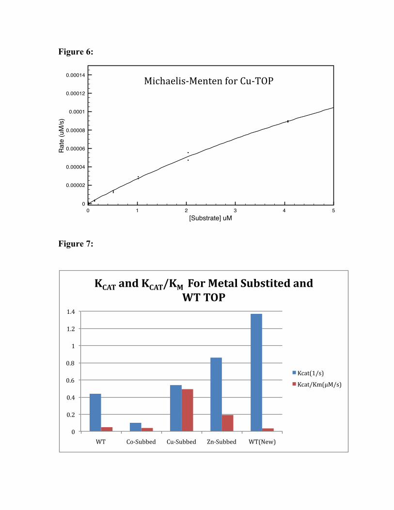
Figure 6:
Figure 7:
0
0.2
0.4
0.6
0.8
1
1.2
1.4
WT Co‐Subbed Cu‐Subbed Zn‐Subbed WT(New)
KCATandKCAT/KMForMetalSubstitedandWTTOP
Kcat(1/s)
Kcat/Km(μM/s)
0 1 2 3 4 5
[Substrate] uM
0
0.00002
0.00004
0.00006
0.00008
0.0001
0.00012
0.00014
Rate
(uM
/s)
Michaelis‐MentenforCu‐TOP
Figure 8:
1Rawlings ND & Barrett AJ (1995) EVOLUTIONARY FAMILIES OF METALLOPEPTIDASES. In Proteolytic Enzymes: Aspartic and Metallo Peptidases, pp. 183-228. Academic Press Inc, San Diego.2Ray K, Hines C, Coll-Rodriguez J & Rodgers D (2004) Crystal structure of human thimet oligopeptidase provides insight into substrate recognition, regulation, and localization. Journal of Biological Chemistry, 20480-20489, doi: DOI 10.1074/jbc.M400795200.3Niemirowicz G, Fernandez D, Sola M, Cazzulo JJ, Aviles FX & Gomis-Ruth FX (2008) The molecular analysis of Trypanosoma cruzi metallocarboxypeptidase 1 provides insight into fold and substrate specificity. Molecular Microbiology 70, 853-866, doi: 10.1111/j.1365-2958.2008.06444.x.4Rioli V, Gozzo F, Heimann A, Linardi A, Krieger J, Shida C, Almeida P, Hyslop S, Eberlin M & Ferro E (2003) Novel natural peptide substrates for endopeptidase 24.15, neurolysin, and angiotensin-converting enzyme. J Biol Chem 278, 8547-8555, doi: M212030200 [pii]5Smith A, Lew R, Shrimpton C, Evans R & Abbenante G (2000) A novel stable inhibitor of endopeptidases EC 3.4.24.15 and 3.4.24.16 potentiates bradykinin-induced hypotension. Hypertension 35, 626-630.6Yamin R, Malgeri E, Sloane J, McGraw W & Abraham C (1999) Metalloendopeptidase EC 3.4.24.15 is necessary for Alzheimer's amyloid-beta peptide degradation. J Biol Chem 274, 18777-18784.7Holland D, Hausrath A, Juers D & Matthews B (1995) Structural analysis of zinc substitutions in the active site of thermolysin. Protein Sci 4, 1955-1965, doi: 10.1002/pro.55600410018Cheng T, Ramakrishnan V & Chan S (1999) Purification and characterization of a cobalt-activated carboxypeptidase from the hyperthermophilic archaeon Pyrococcus furiosus. Protein Sci 8, 2474-2486, doi: 10.1110/ps.8.11.2474.9Hirose J, Iwamoto H, Nagao I, Enmyo K, Sugao H, Kanemitu N, Ikeda K, Takeda M, Inoue M, Ikeda T, et al. (2001) Characterization of the metal-substituted dipeptidyl peptidase III (rat liver). Biochemistry 40, 11860-11865, doi: bi0110903 [pii].10Barrett AJ & Brown MA (1990) CHICKEN LIVER PZ-PEPTIDASE, A THIOL-DEPENDENT METALLOENDOPEPTIDASE. Biochemical Journal 271, 701-706.
0
5
10
15
20
25
30
35
40
WT Co‐Subbed Cu‐Subbed Zn‐Subbed WT(New)
KMForMetalSubstiutedandWTTOP
Km