
Prinsip Sysmex Hematology Analyzer
5
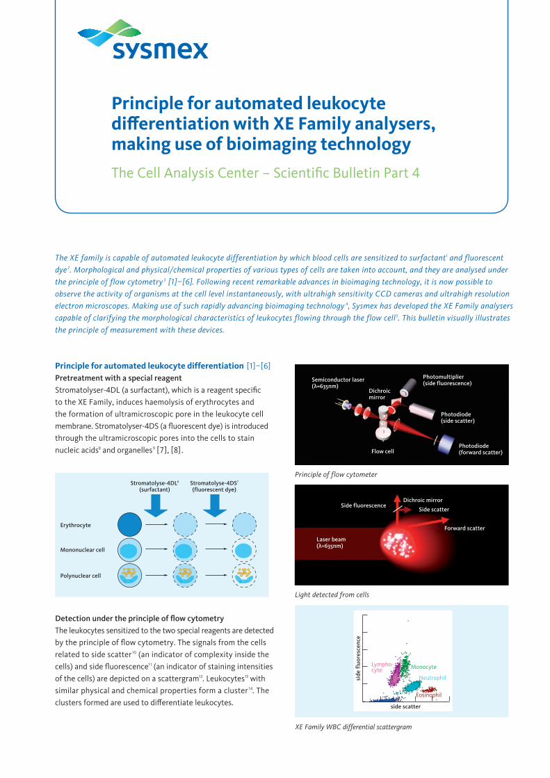
The XE family is capable of automated leukocyte differentiation by which blood cells are sensitized to surfactant 1 and fluorescent dye 2 . Morphological and physical/chemical properties of various types of cells are taken into account, and they are analysed under the principle of flow cytometry 3 [1]–[6]. Following recent remarkable advances in bioimaging technology, it is now possible to observe the activity of organisms at the cell level instantaneously, with ultrahigh sensitivity CCD cameras and ultrahigh resolution electron microscopes. Making use of such rapidly advancing bioimaging technology 4 , Sysmex has developed the XE Family analysers capable of clarifying the morphological characteristics of leukocytes flowing through the flow cell 5 . This bulletin visually illustrates the principle of measurement with these devices. Principle for automated leukocyte differentiation [1]–[6] Pretreatment with a special reagent Stromatolyser-4DL (a surfactant), which is a reagent specific to the XE Family, induces haemolysis of erythrocytes and the formation of ultramicroscopic pore in the leukocyte cell membrane. Stromatolyser-4DS (a fluorescent dye) is introduced through the ultramicroscopic pores into the cells to stain nucleic acids 8 and organelles 9 [7], [8] . Detection under the principle of flow cytometry The leukocytes sensitized to the two special reagents are detected by the principle of flow cytometry. The signals from the cells related to side scatter 10 (an indicator of complexity inside the cells) and side fluorescence 11 (an indicator of staining intensities of the cells) are depicted on a scattergram 12 . Leukocytes 13 with similar physical and chemical properties form a cluster 14 . The clusters formed are used to differentiate leukocytes. Erythrocyte Stromatolyse-4DL 6 (surfactant) Stromatolyse-4DS 7 (fluorescent dye) Mononuclear cell Polynuclear cell XE Family WBC differential scattergram side fluorescence side scatter Lympho- cyte Monocyte Eosinophil Neutrophil Photomultiplier (side fluorescence) Semiconductor laser (λ=635nm) Photodiode (side scatter) Photodiode (forward scatter) Flow cell Dichroic mirror Side fluorescence Dichroic mirror Side scatter Forward scatter Laser beam (λ=635nm) Light detected from cells Principle of flow cytometer Principle for automated leukocyte differentiation with XE Family analysers, making use of bioimaging technology The Cell Analysis Center – Scientific Bulletin Part 4
-
Upload
lince-wijoyo -
Category
Documents
-
view
336 -
download
26
description
clinical pathology
Transcript of Prinsip Sysmex Hematology Analyzer
The XE family is capable of automated leukocyte differentiation by which blood cells are sensitized to surfactant1 and fluorescent dye 2. Morphological and physical/chemical properties of various types of cells are taken into account, and they are analysed under the principle of flow cytometry 3 [1]–[6]. Following recent remarkable advances in bioimaging technology, it is now possible to observe the activity of organisms at the cell level instantaneously, with ultrahigh sensitivity CCD cameras and ultrahigh resolution electron microscopes. Making use of such rapidly advancing bioimaging technology 4, Sysmex has developed the XE Family analysers capable of clarifying the morphological characteristics of leukocytes flowing through the flow cell5. This bulletin visually illustrates the principle of measurement with these devices.
Principle for automated leukocyte differentiation [1]–[6]Pretreatment with a special reagentStromatolyser-4DL (a surfactant), which is a reagent specifi c to the XE Family, induces haemolysis of erythrocytes and the formation of ultramicroscopic pore in the leukocyte cell membrane. Stromatolyser-4DS (a fl uorescent dye) is introduced through the ultramicroscopic pores into the cells to stain nucleic acids8 and organelles9 [7], [8] .
Detection under the principle of fl ow cytometryThe leukocytes sensitized to the two special reagents are detected by the principle of fl ow cytometry. The signals from the cells related to side scatter10 (an indicator of complexity inside the cells) and side fl uorescence11 (an indicator of staining intensities of the cells) are depicted on a scattergram12. Leukocytes13 with similar physical and chemical properties form a cluster 14. The clusters formed are used to diff erentiate leukocytes.
Erythrocyte
Stromatolyse-4DL6
(surfactant)Stromatolyse-4DS7
(fluorescent dye)
Mononuclear cell
Polynuclear cell
XE Family WBC differential scattergram
side
fluo
resc
ence
side scatter
Lympho-cyte Monocyte
Eosinophil
Neutrophil
Photomultiplier(side fluorescence)
Semiconductor laser(λ=635nm)
Photodiode(side scatter)
Photodiode(forward scatter)Flow cell
Dichroicmirror
Side fluorescenceDichroic mirror
Side scatter
Forward scatter
Laser beam(λ=635nm)
Light detected from cells
Principle of flow cytometer
Principle for automated leukocyte diff erentiation with XE Family analysers, making use of bioimaging technology
The Cell Analysis Center – Scientifi c Bulletin Part 4
Principle for automated leukocyte differentiation with XE family analysers, making use of bioimaging technology 2/ 5
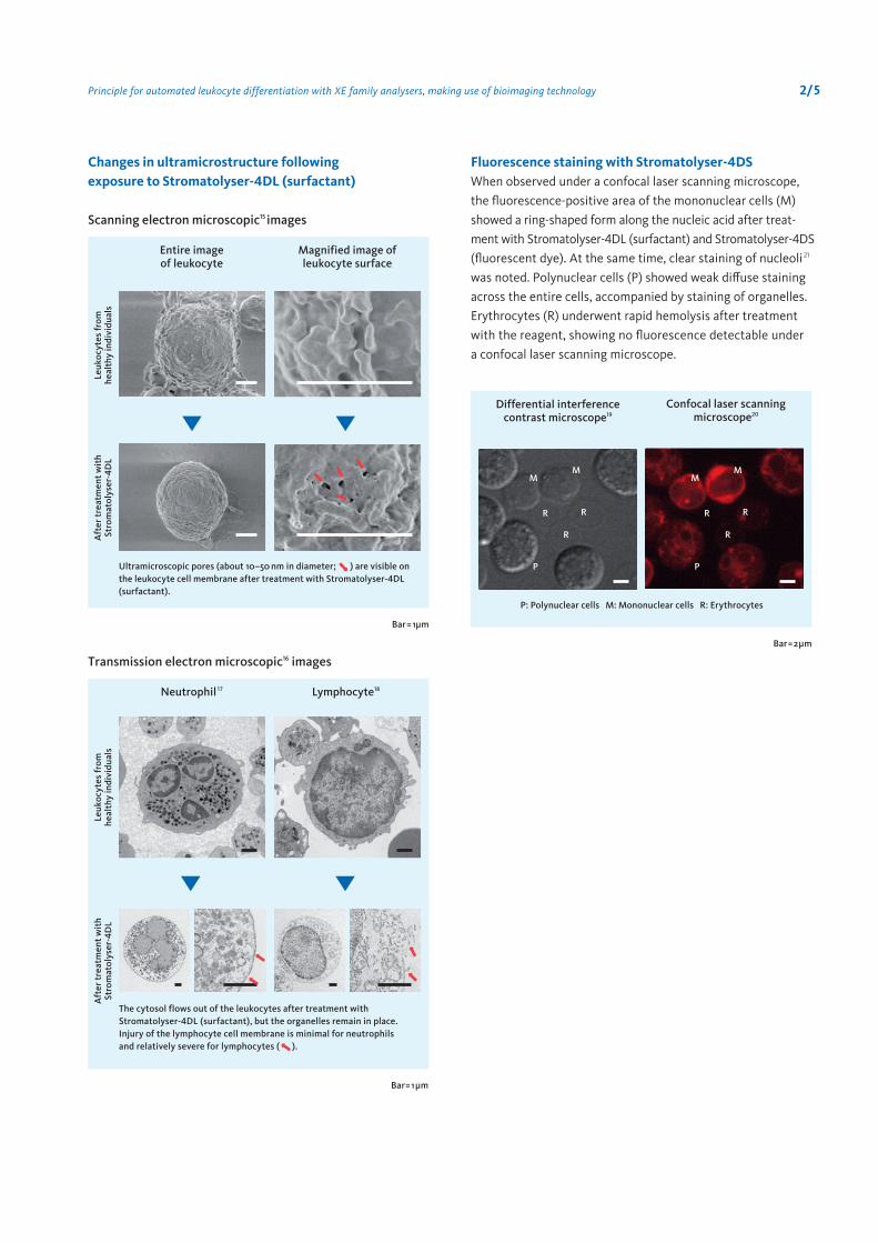
Scanning electron microscopic15 images
Transmission electron microscopic16 images
Changes in ultramicrostructure following exposure to Stromatolyser-4DL (surfactant)
Magnified image of leukocyte surface
Entire image of leukocyte
Leuk
ocyt
es fr
omhe
alth
y in
divi
dual
sA
fter
trea
tmen
t wit
hSt
rom
atol
yser
-4D
L
Ultramicroscopic pores (about 10–50 nm in diameter; ) are visible on the leukocyte cell membrane after treatment with Stromatolyser-4DL (surfactant).
Bar= 1µm
Lymphocyte18Neutrophil 17
Leuk
ocyt
es fr
omhe
alth
y in
divi
dual
sA
fter
trea
tmen
t wit
hSt
rom
atol
yser
-4D
L
The cytosol flows out of the leukocytes after treatment with Stromatolyser-4DL (surfactant), but the organelles remain in place. Injury of the lymphocyte cell membrane is minimal for neutrophils and relatively severe for lymphocytes ( ).
Bar= 1µm
When observed under a confocal laser scanning microscope, the fluorescence-positive area of the mononuclear cells (M) showed a ring-shaped form along the nucleic acid after treat-ment with Stromatolyser-4DL (surfactant) and Stromatolyser-4DS (fluorescent dye). At the same time, clear staining of nucleoli21 was noted. Polynuclear cells (P) showed weak diffuse staining across the entire cells, accompanied by staining of organelles. Erythrocytes (R) underwent rapid hemolysis after treatment with the reagent, showing no fluorescence detectable under a confocal laser scanning microscope.
Fluorescence staining with Stromatolyser-4DS
Differential interference contrast microscope19
Confocal laser scanning microscope20
P: Polynuclear cells M: Mononuclear cells R: Erythrocytes
Bar=2µm
R R
R
P
MM
R R
R
P
MM
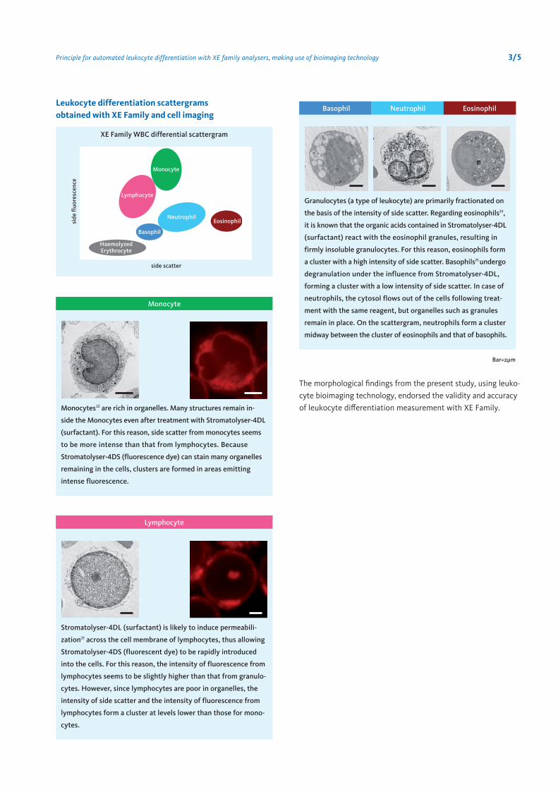
Leukocyte differentiation scattergrams obtained with XE Family and cell imaging
3/ 5
Monocytes22 are rich in organelles. Many structures remain in-
side the Monocytes even after treatment with Stromatolyser-4DL
(surfactant). For this reason, side scatter from monocytes seems
to be more intense than that from lymphocytes. Because
Stromatolyser-4DS (fluorescence dye) can stain many organelles
remaining in the cells, clusters are formed in areas emitting
intense fluorescence.
Stromatolyser-4DL (surfactant) is likely to induce permeabili-
zation23 across the cell membrane of lymphocytes, thus allowing
Stromatolyser-4DS (fluorescent dye) to be rapidly introduced
into the cells. For this reason, the intensity of fluorescence from
lymphocytes seems to be slightly higher than that from granulo-
cytes. However, since lymphocytes are poor in organelles, the
intensity of side scatter and the intensity of fluorescence from
lymphocytes form a cluster at levels lower than those for mono-
cytes.
Granulocytes (a type of leukocyte) are primarily fractionated on
the basis of the intensity of side scatter. Regarding eosinophils24,
it is known that the organic acids contained in Stromatolyser-4DL
(surfactant) react with the eosinophil granules, resulting in
firmly insoluble granulocytes. For this reason, eosinophils form
a cluster with a high intensity of side scatter. Basophils25 undergo
degranulation under the influence from Stromatolyser-4DL,
forming a cluster with a low intensity of side scatter. In case of
neutrophils, the cytosol flows out of the cells following treat-
ment with the same reagent, but organelles such as granules
remain in place. On the scattergram, neutrophils form a cluster
midway between the cluster of eosinophils and that of basophils.
The morphological findings from the present study, using leuko-cyte bioimaging technology, endorsed the validity and accuracy of leukocyte differentiation measurement with XE Family.
XE Family WBC differential scattergram
side scatter
side
fluo
resc
ence
Monocyte
Lymphocyte
NeutrophilEosinophil
Basophil
HaemolyzedErythrocyte
Bar=2µm
Monocyte
Basophil Neutrophil Eosinophil
Lymphocyte
Principle for automated leukocyte differentiation with XE family analysers, making use of bioimaging technology
Terminology1 SurfactantA collective term for substances having both a hydrophilic por-tion (hydrophilic group) and a lipophilic portion (lipophilic and hydrophobic groups) within the molecule. Sysmex’s reagent for leukocyte differentiation (Stromatolyser-4DL) contains cationic and nonionic surfactants, which act on the cell membrane and form ultramicroscopic pores in the membrane.
2 Fluorescent dyeA collective term for substances which, after absorbing electro-magnetic radiation such as light, themselves emit radiation, usually of a longer wavelength than that of the absorbed radiation (e.g. absorbing ultraviolet light and emitting visible light). If a fluorescent dye is bound to particles or substances, it allows accurate location, observation and measurement of potential changes in the target. Sysmex’s reagent for leukocyte differ-entiation (Stromatolyser-4DS) contain fluorescent dyes of the polymethine family, which primarily stain the nucleic acids and organelles.
3 Flow cytometrySmall particles such as cells are dispersed in a fluid, and the fluid is flowed through a small nozzle for optical analysis of individual particles. The Sysmex automated haematology analysers are based on this principle.
4 Bioimaging technologyA technology for observation of the distribution and kinetics of targets in cells or tissues marked with dyes, fluorescent dyes or colloid gold (used for electron microscopy).
5 Flow cellA central unit of a flow cytometer in which cells are dispersed in a fluid flow through a small nozzle and are detected with laser, etc.
6 Stromatolyser-4DLSysmex’s reagent for automated leukocyte differentiation, primarily made of cationic and nonionic surfactants. This reagent induces haemolysis of erythrocytes and formation of ultramicroscopic pores in the leukocyte cell membrane.
7 Stromatolyser-4DSSysmex’s reagent for automated leukocyte differentiation, containing polymethine dyes which are excited by 635 nm laser. It primarily stains nucleic acids and organelles.
8 Nucleic acidA macromolecule found in organisms. Can be divided into DNA and RNA. DNA is associated with genetic information in nuclei, while RNA is involved in expression of genetic information.
9 Organelle Includes mitochondria, endoplasmic reticulum, Golgi apparatus, etc.
10 Side scatterLaser (635 nm) scattered in the right-angle direction when applied to the cells flowing through a flow cell. Serves as an indicator of complexity inside cells (nuclear shape and size, density of organelles).
11 Side fluorescenceFluorescence emitted in the right-angle direction from the cells (stained with a fluorescent dye) flowing through a flow cell due to excitation by the laser applied. Serves as an indicator of the intensity of staining of the cells to the fluorescent dye.
12 ScattergramGraphic representation of optical information of cells collected with a flow cytometer. Physical and chemical properties of cells are presented.
13 LeukocytesIncludes neutrophils, eosinophils, basophils, lymphocytes, and monocytes.
14 ClusterA group of cells with similar physical and chemical properties formed on the scattergram.
15 Scanning electron microscopeA microscope allowing observation of the ultramicrostructure of the cell surface. A special metallic film is formed on the cell surface, and electron beams are applied to it for observation of the cell surface.
16 Transmission electron microscopeA microscope allowing observation of the ultramicrostructure inside cells. The cells are made into thin slices (70 nm) and elec-tron beams are applied to them for visualization of electrons.
17 Neutrophil A type of granulocytes, accounting for about 40–60% of all leukocytes. Contains granules which are positively stained with neutral dyes. Plays an important role in host defense through phagocytosis of bacteria, etc. and disinfection activity.
18 LymphocyteAccounts for about 25% of all leukocytes. Can be divided into NK cell, B cell (B lymphocyte), T cell (T lymphocyte), etc. Plays an important role in the immune system.
4/ 5Principle for automated leukocyte differentiation with XE family analysers, making use of bioimaging technology
5/ 5
19 Differential interference contrast microscopeA microscope capable of three-dimensional observation of cells through interference of ray, making use of polarization.
20 Confocal laser scanning microscopeA microscope with laser serving as a light course, capable of achieving high spatial resolution not possible with a fluorescence microscope. Also capable of providing sectional images of cells stained with fluorescence.
21 NucleolusA region with high molecular density within the nucleus of eukaryote cells. A place for transcription of ribosomal RNA and construction of ribosomes.
22 MonocyteAccounts for about 3–8% of all leukocytes. Plays an important role in initiation of anti-infection immune activity. Mobile through amoeba-like motion. Takes up bacteria and other foreign particles and digests them with intracellular enzymes.
23 PermeabilizationA manipulation to create a hole in the cell membrane which partitions the areas inside and outside the cell, to allow introduction of the target substance into the cell. In this case, Stromatolyser-4DL (surfactant) is used to create an ultra- microscopic pore in the cell membrane.
24 EosinophilAccounts for about 0–5% of all leukocytes. When specimens stained by ordinary methods are observed, the cells are filled with relatively large, round granules of similar features stained orange-red color with eosin. Can cause damages to bacteria and parasites.
25 BasophilAccounts for about 0–2% of all leukocytes. Round cells with a size slightly smaller than neutrophils. Stained dark purple color with aniline blue.
Reference[1] Inoue H. Overview of automated hematology analyzer XE-2100. Sysmex Journal International. 1999; 9: 1 58–64.
[2] Warren G. et al. Optical technology in blood cell counting.Sysmex Journal International. 1999; 9: 1 21–30.
[3] Fujimoto K. Principles of measurement in hematologyanalyzers manufactured by Sysmex Corporation. Sysmex JournalInternational. 1999; 9: 1 31–44.
[4] Herklotz R. et. al. Precision and accuracy of the leukocytedifferential on the Sysmex XE-2100. Sysmex JournalInternational. 2001; 11: 1 8–21.
[5] Jerelyn W. et. al. Performance evaluation of the Sysmex XE-2100 hematology analyzer. Laboratory Hematology. 2000; 6; 83–92.
[6] Rowan R. M. et. al. A picture is worth a thousand words.Sysmex Journal International. 2005; 15: 1 27–38.
[7] Matsumoto H. The technology of reagents in the automatedhematology analyzer Sysmex XE-2100 – Red flourescence reaction –. Sysmex Journal International. 1999; 9: 2 179–185.
[8] Giles I. The Thing about fluorescence technology. SysmexJournal International. 2006; 16: 1 17–18.
[9] Scientific Affairs, Sysmex Corporation. The Cell AnalysisCenter Scientific Bulletin Part 1 Cell analysis and bioimaging technology illustrated. 2007.
[10] Scientific Affairs, Sysmex Corporation. The Cell AnalysisCenter Scientific Bulletin Part 2 Electron microscopy of reticulo-cytes after sorting with magnetic beads. 2007.
Principle for automated leukocyte differentiation with XE family analysers, making use of bioimaging technology
Sysmex Corporation 1-5-1, Wakinohama-Kaigandori, Chuo-ku, Kobe 651-0073, Japan · Phone +81 (78) 265-0500 · Fax +81 (78) 265-0524 · www.sysmex.co.jp
Sysmex Europe GmbH Bornbarch 1, 22848 Norderstedt, Germany · Phone +49 (40) 52726-0 · Fax +49 (40) 52726-100 · [email protected] · www.sysmex-europe.com
Copyright © 2007 by Sysmex Corporation


