Primary myeloid sarcoma of the testicle with t(15;17)
3
Cancer Genetics and Cytogenetics 157 (2005) 148–150 Short communication Primary myeloid sarcoma of the testicle with t(15;17) Shanti Gopal, Sandra Marcussen, Sheila M. Dobin, William Koss, Ludvik R. Donner * Department of Pathology, Scott and White Memorial Hospital and Clinic; Scott, Sherwood and Brindley Foundation; The Texas A&M University Health Science Center College of Medicine, 2401 South 31st Street, Temple, TX 76508 Received 10 May 2004; received in revised form 4 June 2004; accepted 11 June 2004 Abstract The first case of acute promyelocytic leukemia presenting as a solitary testicular mass (myeloid sarcoma) that relapsed in the contralateral testicle is described. The neoplastic cells strongly expressed chloroacetate esterase, myeloperoxidase, CD33, CD43, and weakly, CD117. The presence of many azurophil granules and Auer rods was detected by electron microscopy. Translocation (15;17)(q22;q21.1) was revealed by cytogenetics and was verified by fluorescence in situ hybridiza- tion. Contralateral testicle is a favorite site for recurrence in a subset of testicular myeloid sarcomas. Subclassification of all cases of myeloid sarcoma ought to be attempted. 2005 Elsevier Inc. All rights reserved. 1. Introduction Primary myeloid (granulocytic) sarcomas of the testicle are rare tumors and only a handful of them have been reported [1–8]. The majority of these tumors were neither further subclassified nor studied by cytogenetics or molecu- lar biology, except for 2 tumors that were retrospectively classified as acute myelomonocytic [2] and acute mono- blastic leukemias [5], respectively. Acute promyelocytic leukemia (APL) is defined by the t(15;17)(q22;q21.1) or by its variants. The t(15;17) leads to fusion of the retinoic acid receptor α (RARa) gene on 17q21.1 with a nuclear regulatory factor PML gene on 15q22. The PML gene functions as a tumor suppressor and loss of its function in the leukemic cells renders them less respon- sive to differentiation stimuli, like retinoic acid, and confers on them a proliferative advantage [9,10]. Because dissemin- ated intravascular coagulation occurs in the majority of pa- tients, rapid diagnosis of this leukemia and early retinoic acid treatment are essential [11]. The first case of acute promyelocytic leukemia presenting as a solitary testicular mass is presented in this article. 2. Clinical history A 27-year-old black man presented with a large, painless mass in the left testicle. Radical orchiectomy was performed. * Corresponding author. Tel.: (254) 724-4730; fax: (254) 724-4391. E-mail address: [email protected] (L.R. Donner). 0165-4608/05/$ – see front matter 2005 Elsevier Inc. All rights reserved. doi:10.1016/j.cancergencyto.2004.06.010 The bone marrow and peripheral blood, abdominal sono- gram, computed tomogram of the abdomen, pelvis, and the chest were all normal. The mass was classified as myeloid sarcoma. No further treatment was administered. The patient did well for a year but subsequently developed a mass in the contralateral testicle. A right radical orchiectomy was performed. The recurrent myeloid sarcoma was subclassified as APL and the patient treated with trans-retinoic acid, daun- orubicin, and cytosine-arabinoside. A bone marrow biopsy was performed four months later. The bone marrow was mildly hypercellular, had diploid karyotype, but 1% of the interphase cells had a rearranged RARa gene at band 17q21.1 that was detected by fluorescence in situ hybridization (FISH). 3. Pathology study The left and right testicles contained a 4.5 × 4.0 × 3.0 cm and 6.0 × 4.5 × 4.0 cm firm solid green tumors, respec- tively (Fig. 1A). A 1.4-cm similar tumor was also found on the outer surface of the right spermatic cord. The parenchyma of both testicles was replaced by sheets of neoplastic cells that were entrapping atrophic seminiferous tubules. Their nuclei were bilobed or reniform with fine chromatin, small nucleoli and granular cytoplasm (Fig. 1B and C). A third of the neoplastic cells were strongly chloroacetate esterase positive. They were strongly diffusely positive for myeloper- oxidase and CD43, weakly positive for CD117, and negative for CD34 when studied by immunohistochemistry (Fig. 1D); positive for CD33 and CD117, and negative for CD13 and
-
Upload
shanti-gopal -
Category
Documents
-
view
214 -
download
1
Transcript of Primary myeloid sarcoma of the testicle with t(15;17)
Cancer Genetics and Cytogenetics 157 (2005) 148–150
Short communication
Primary myeloid sarcoma of the testicle with t(15;17)Shanti Gopal, Sandra Marcussen, Sheila M. Dobin, William Koss, Ludvik R. Donner*
Department of Pathology, Scott and White Memorial Hospital and Clinic; Scott, Sherwood and Brindley Foundation;The Texas A&M University Health Science Center College of Medicine, 2401 South 31st Street, Temple, TX 76508
Received 10 May 2004; received in revised form 4 June 2004; accepted 11 June 2004
Abstract The first case of acute promyelocytic leukemia presenting as a solitary testicular mass (myeloidsarcoma) that relapsed in the contralateral testicle is described. The neoplastic cells stronglyexpressed chloroacetate esterase, myeloperoxidase, CD33, CD43, and weakly, CD117. The presenceof many azurophil granules and Auer rods was detected by electron microscopy. Translocation(15;17)(q22;q21.1) was revealed by cytogenetics and was verified by fluorescence in situ hybridiza-tion. Contralateral testicle is a favorite site for recurrence in a subset of testicular myeloid sarcomas.Subclassification of all cases of myeloid sarcoma ought to be attempted. � 2005 Elsevier Inc. Allrights reserved.
1. Introduction
Primary myeloid (granulocytic) sarcomas of the testicleare rare tumors and only a handful of them have beenreported [1–8]. The majority of these tumors were neitherfurther subclassified nor studied by cytogenetics or molecu-lar biology, except for 2 tumors that were retrospectivelyclassified as acute myelomonocytic [2] and acute mono-blastic leukemias [5], respectively.
Acute promyelocytic leukemia (APL) is defined by thet(15;17)(q22;q21.1) or by its variants. The t(15;17) leads tofusion of the retinoic acid receptor α (RARa) gene on17q21.1 with a nuclear regulatory factor PML gene on 15q22.The PML gene functions as a tumor suppressor and lossof its function in the leukemic cells renders them less respon-sive to differentiation stimuli, like retinoic acid, and conferson them a proliferative advantage [9,10]. Because dissemin-ated intravascular coagulation occurs in the majority of pa-tients, rapid diagnosis of this leukemia and early retinoicacid treatment are essential [11]. The first case of acutepromyelocytic leukemia presenting as a solitary testicularmass is presented in this article.
2. Clinical history
A 27-year-old black man presented with a large, painlessmass in the left testicle. Radical orchiectomy was performed.
* Corresponding author. Tel.: (254) 724-4730; fax: (254) 724-4391.E-mail address: [email protected] (L.R. Donner).
0165-4608/05/$ – see front matter � 2005 Elsevier Inc. All rights reserved.doi:10.1016/j.cancergencyto.2004.06.010
The bone marrow and peripheral blood, abdominal sono-gram, computed tomogram of the abdomen, pelvis, and thechest were all normal. The mass was classified as myeloidsarcoma. No further treatment was administered. The patientdid well for a year but subsequently developed a mass inthe contralateral testicle. A right radical orchiectomy wasperformed. The recurrent myeloid sarcoma was subclassifiedas APL and the patient treated with trans-retinoic acid, daun-orubicin, and cytosine-arabinoside. A bone marrow biopsywas performed four months later. The bone marrow wasmildly hypercellular, had diploid karyotype, but 1% of theinterphase cells had a rearranged RARa gene at band 17q21.1that was detected by fluorescence in situ hybridization(FISH).
3. Pathology study
The left and right testicles contained a 4.5 × 4.0 × 3.0cm and 6.0 × 4.5 × 4.0 cm firm solid green tumors, respec-tively (Fig. 1A). A 1.4-cm similar tumor was also found onthe outer surface of the right spermatic cord. The parenchymaof both testicles was replaced by sheets of neoplastic cellsthat were entrapping atrophic seminiferous tubules. Theirnuclei were bilobed or reniform with fine chromatin, smallnucleoli and granular cytoplasm (Fig. 1B and C). A thirdof the neoplastic cells were strongly chloroacetate esterasepositive. They were strongly diffusely positive for myeloper-oxidase and CD43, weakly positive for CD117, and negativefor CD34 when studied by immunohistochemistry (Fig. 1D);positive for CD33 and CD117, and negative for CD13 and

S. Gopal et al. / Cancer Genetics and Cytogenetics 157 (2005) 148–150 149
Fig. 1. (A) Gross appearance of the recurrent testicular tumor; (B) lightmicroscopy, plastic embedded; (C) light microscopy, H&E; and (D) immu-noreactivity for myeloperoxidase.
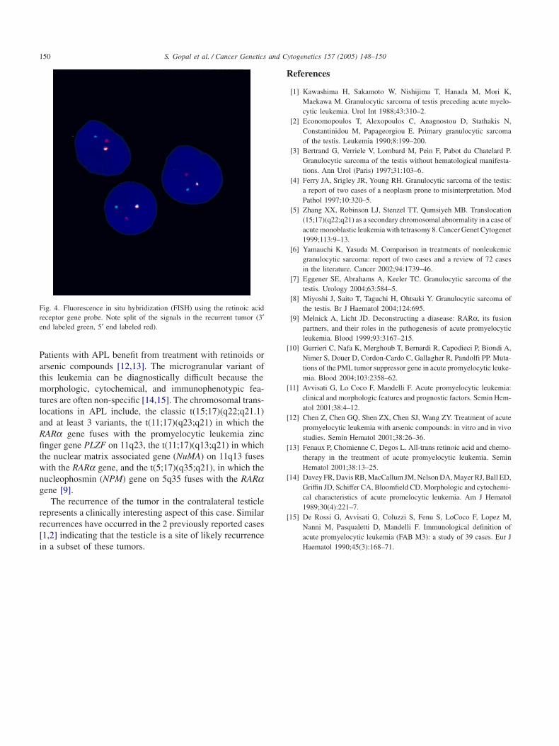
HLA-DR when studied by flow cytometry. A subset of thecells was positive for CD34 when studied by the lattermethod. Numerous azurophil granules and many Auer rodswere detected in the right testicular tumor by electron mi-croscopy (Fig. 2).
4. Cytogenetic study
Six cell cultures were established from the right testicularmass, 2 from explants and 4 from collagenase-treated cells.
Fig. 2. Ultrastructure of the recurrent tumor. Note the presence of numerousazurophilic granules and Auer rods.
Cultures were harvested 1–8 days later. Twenty-one GTG-banded mitotic figures were analyzed. All 6 cultures con-tained a clonal population with the karyotype 46,XY,t(15;17)(q22;q21.1)[18].nuc ish 17q21.1(5′RARAx2,3′RARAx2)(5′RARAsep 3′RARAx1)[178]/46,idem,del(1)(p32p36.2)[2]/46,XY[1].nuc ish 17q21.1(5′RARAx2,3′RARAx2)[21] (Fig. 3). Therearrangement of the RARa gene on 17q21.1 was verifiedwith the LSI RARA dual-color probe (Vysis, Downer Grove,IL) (Fig. 4).
5. Discussion
This case reiterates the importance of subclassification ofmyeloid sarcomas for their optimal therapeutic management.
Fig. 3. Karyotype of the recurrent tumor. The abnormal chromosomes are marked by arrows.
S. Gopal et al. / Cancer Genetics and Cytogenetics 157 (2005) 148–150150
Fig. 4. Fluorescence in situ hybridization (FISH) using the retinoic acidreceptor gene probe. Note split of the signals in the recurrent tumor (3′end labeled green, 5′ end labeled red).
Patients with APL benefit from treatment with retinoids orarsenic compounds [12,13]. The microgranular variant ofthis leukemia can be diagnostically difficult because themorphologic, cytochemical, and immunophenotypic fea-tures are often non-specific [14,15]. The chromosomal trans-locations in APL include, the classic t(15;17)(q22;q21.1)and at least 3 variants, the t(11;17)(q23;q21) in which theRARa gene fuses with the promyelocytic leukemia zincfinger gene PLZF on 11q23, the t(11;17)(q13;q21) in whichthe nuclear matrix associated gene (NuMA) on 11q13 fuseswith the RARa gene, and the t(5;17)(q35;q21), in which thenucleophosmin (NPM) gene on 5q35 fuses with the RARagene [9].
The recurrence of the tumor in the contralateral testiclerepresents a clinically interesting aspect of this case. Similarrecurrences have occurred in the 2 previously reported cases[1,2] indicating that the testicle is a site of likely recurrencein a subset of these tumors.
References
[1] Kawashima H, Sakamoto W, Nishijima T, Hanada M, Mori K,Maekawa M. Granulocytic sarcoma of testis preceding acute myelo-cytic leukemia. Urol Int 1988;43:310–2.
[2] Economopoulos T, Alexopoulos C, Anagnostou D, Stathakis N,Constantinidou M, Papageorgiou E. Primary granulocytic sarcomaof the testis. Leukemia 1990;8:199–200.
[3] Bertrand G, Verriele V, Lombard M, Pein F, Pabot du Chatelard P.Granulocytic sarcoma of the testis without hematological manifesta-tions. Ann Urol (Paris) 1997;31:103–6.
[4] Ferry JA, Srigley JR, Young RH. Granulocytic sarcoma of the testis:a report of two cases of a neoplasm prone to misinterpretation. ModPathol 1997;10:320–5.
[5] Zhang XX, Robinson LJ, Stenzel TT, Qumsiyeh MB. Translocation(15;17)(q22;q21) as a secondary chromosomal abnormality in a case ofacute monoblastic leukemia with tetrasomy 8. Cancer Genet Cytogenet1999;113:9–13.
[6] Yamauchi K, Yasuda M. Comparison in treatments of nonleukemicgranulocytic sarcoma: report of two cases and a review of 72 casesin the literature. Cancer 2002;94:1739–46.
[7] Eggener SE, Abrahams A, Keeler TC. Granulocytic sarcoma of thetestis. Urology 2004;63:584–5.
[8] Miyoshi J, Saito T, Taguchi H, Ohtsuki Y. Granulocytic sarcoma ofthe testis. Br J Haematol 2004;124:695.
[9] Melnick A, Licht JD. Deconstructing a diasease: RARα, its fusionpartners, and their roles in the pathogenesis of acute promyelocyticleukemia. Blood 1999;93:3167–215.
[10] Gurrieri C, Nafa K, Merghoub T, Bernardi R, Capodieci P, Biondi A,Nimer S, Douer D, Cordon-Cardo C, Gallagher R, Pandolfi PP. Muta-tions of the PML tumor suppressor gene in acute promyelocytic leuke-mia. Blood 2004;103:2358–62.
[11] Avvisati G, Lo Coco F, Mandelli F. Acute promyelocytic leukemia:clinical and morphologic features and prognostic factors. Semin Hem-atol 2001;38:4–12.
[12] Chen Z, Chen GQ, Shen ZX, Chen SJ, Wang ZY. Treatment of acutepromyelocytic leukemia with arsenic compounds: in vitro and in vivostudies. Semin Hematol 2001;38:26–36.
[13] Fenaux P, Chomienne C, Degos L. All-trans retinoic acid and chemo-therapy in the treatment of acute promyelocytic leukemia. SeminHematol 2001;38:13–25.
[14] Davey FR, Davis RB, MacCallum JM, Nelson DA, Mayer RJ, Ball ED,Griffin JD, Schiffer CA, Bloomfield CD. Morphologic and cytochemi-cal characteristics of acute promelocytic leukemia. Am J Hematol1989;30(4):221–7.
[15] De Rossi G, Avvisati G, Coluzzi S, Fenu S, LoCoco F, Lopez M,Nanni M, Pasqualetti D, Mandelli F. Immunological definition ofacute promyelocytic leukemia (FAB M3): a study of 39 cases. Eur JHaematol 1990;45(3):168–71.