Primary bone lymphoma with multifocal osteolytic lesions ...€¦ · (SGN-35) for relapsed...
5
Blood Res 2015;50:254-67. bloodresearch.or.kr 256 Letters to the Editor vedotin could be a viable treatment option for refractory CD30-positive ENKL without causing significant toxicity, even in heavily pretreated patients. However, since this case report is the first to show CR in CD30-positive ENKL with brentuximab vedotin, further studies will be required to confirm these results and establish a dosing schedule. This report implies that brentuximab vedotin could be effec- tive in treating CD30-positive non-Hodgkin lymphoma other than ALCL and peripheral T-cell lymphoma. Hee Kyung Kim, Seong Mi Moon, Ji Hoon Moon, Jee Eun Park, Seonggyu Byeon, Won Seog Kim Division of Hematology-Oncology, Department of Internal Medicine, Samsung Medical Center, Sungkyunkwan University School of Medicine, Seoul, Korea Correspondence to: Hee Kyung Kim Division of Hematology-Oncology, Department of Internal Medicine, Samsung Medical Center, Sungkyunkwan University School of Medicine, 81 Irwon-ro, Gangnam-gu, Seoul 06351, Korea E-mail: [email protected] Received on Mar. 5, 2015; Revised on Mar. 24, 2015; Accepted on Oct. 22, 2015 http://dx.doi.org/10.5045/br.2015.50.4.254 AuthorsÊ Disclosures of Potential Conflicts of Interest No potential conflicts of interest relevant to this article were reported. REFERENCES 1. Yamaguchi M, Suzuki R, Kwong YL, et al. Phase I study of dex- amethasone, methotrexate, ifosfamide, L-asparaginase, and eto- poside (SMILE) chemotherapy for advanced-stage, relapsed or refractory extranodal natural killer (NK)/T-cell lymphoma and leukemia. Cancer Sci 2008;99:1016-20. 2. Younes A, Bartlett NL, Leonard JP, et al. Brentuximab vedotin (SGN-35) for relapsed CD30-positive lymphomas. N Engl J Med 2010;363:1812-21. 3. Pro B, Advani R, Brice P, et al. Brentuximab vedotin (SGN-35) in patients with relapsed or refractory systemic anaplastic large-cell lymphoma: results of a phase II study. J Clin Oncol 2012;30:2190-6. 4. Horwitz SM, Advani RH, Bartlett NL, et al. Objective responses in relapsed T-cell lymphomas with single-agent brentuximab vedotin. Blood 2014;123:3095-100. 5. Kwong YL. Natural killer-cell malignancies: diagnosis and treatment. Leukemia 2005;19:2186-94. 6. Falini B, Pileri S, Pizzolo G, et al. CD30 (Ki-1) molecule: a new cytokine receptor of the tumor necrosis factor receptor super- family as a tool for diagnosis and immunotherapy. Blood 1995;85: 1-14. 7. Hong J, Park S, Baek HL, et al. Tumor cell nuclear diameter and CD30 expression as potential prognostic parameter in patients with extranodal NK/T-cell lymphoma, nasal type. Int J Clin Exp Pathol 2012;5:939-47. 8. Tse E, Kwong YL. How I treat NK/T-cell lymphomas. Blood 2013;121:4997-5005. 9. Katz J, Janik JE, Younes A. Brentuximab vedotin (SGN-35). Clin Cancer Res 2011;17:6428-36. Primary bone lymphoma with multifocal osteolytic lesions: a rare case report with review of literature TO THE EDITOR: Primary non-Hodgkin lymphoma (NHL) of bone is a rare disorder [1]. Primary bone lymphoma involving multiple sites is even rarer and in the majority of cases, the diagnosis is diffuse large B-cell lymphoma (DLBCL). Here we report the case of a young patient with unexplained diffuse bone pain that was diagnosed as primary bone lymphoma (B-cell lymphoma, unclassifiable, with fea- tures intermediate between DLBCL and Burkitt lymphoma) with multifocal osteolytic lesions. CASE A 22-year-old male was admitted to the orthopedic ward complaining of pain in the right side of his groin. He had experienced difficulty in walking for 3 months prior fol- lowed by diffuse bone pain in his whole body and weight loss for 2 months. He had 6 brothers and 1 sister; one of his brothers had been treated for spinal tuberculosis 9 years earlier. He was managed with analgesics and proton pump inhibitors. A skeletal survey (Fig. 1) revealed osteo- lytic lesions in multiple long and flat bones. Bone scintig- raphy with technetium-99 showed high accumulation in the skull, vertebrae, ribs, pelvis, both humeri, and the bi- lateral femurs (Fig. 2A). A whole-body positron emission tomography-computed tomography (PET-CT) scan (Fig. 2B, C) revealed multiple metabolically active lytic lesions all over the skeletal system. No other metabolically active le- sions were observed. Serum carcinoembryonic antigen, al- pha-fetoprotein, and prostate-specific antigen levels were normal; the patient’s thyroid profile was also normal. The patient was then referred to the hematology department. There was no history of pallor, bleeding, arthralgia or arthri- tis, nor any history of blood transfusion. On examination, there was mild pallor, but no icterus, pedal edema, or pal- pable lymph nodes. The liver and spleen were not palpable, but bony tenderness was present. The patient was afebrile and his vital signs were stable. The results of hematologic tests were as follows: hemoglobin 12.1 g/dL, red blood cell (RBC) count 4.28×10 12 /L, white blood cell count 11.3×10 9 /L, and platelet count 468×10 9 /L. In addition, a peripheral smear showed normocytic, normochromic RBCs, neutrophils 64%, lymphocytes 29%, monocytes 5%, eosinophils 1%, and baso- phils 1%. Blood biochemistry tests revealed normal serum
Transcript of Primary bone lymphoma with multifocal osteolytic lesions ...€¦ · (SGN-35) for relapsed...
Blood Res 2015;50:254-67. bloodresearch.or.kr
256 Letters to the Editor
vedotin could be a viable treatment option for refractory CD30-positive ENKL without causing significant toxicity, even in heavily pretreated patients. However, since this case report is the first to show CR in CD30-positive ENKL with brentuximab vedotin, further studies will be required to confirm these results and establish a dosing schedule. This report implies that brentuximab vedotin could be effec-tive in treating CD30-positive non-Hodgkin lymphoma other than ALCL and peripheral T-cell lymphoma.
Hee Kyung Kim, Seong Mi Moon, Ji Hoon Moon, Jee Eun Park, Seonggyu Byeon, Won Seog Kim
Division of Hematology-Oncology, Department of Internal Medicine, Samsung Medical Center, Sungkyunkwan
University School of Medicine, Seoul, Korea
Correspondence to: Hee Kyung KimDivision of Hematology-Oncology, Department of
Internal Medicine, Samsung Medical Center, Sungkyunkwan University School of Medicine, 81
Irwon-ro, Gangnam-gu, Seoul 06351, KoreaE-mail: [email protected]
Received on Mar. 5, 2015; Revised on Mar. 24, 2015; Accepted on Oct. 22, 2015
http://dx.doi.org/10.5045/br.2015.50.4.254
AuthorsÊ Disclosures of Potential Conflicts of InterestNo potential conflicts of interest relevant to this article
were reported.
REFERENCES1. Yamaguchi M, Suzuki R, Kwong YL, et al. Phase I study of dex-
amethasone, methotrexate, ifosfamide, L-asparaginase, and eto-
poside (SMILE) chemotherapy for advanced-stage, relapsed or
refractory extranodal natural killer (NK)/T-cell lymphoma and
leukemia. Cancer Sci 2008;99:1016-20.
2. Younes A, Bartlett NL, Leonard JP, et al. Brentuximab vedotin
(SGN-35) for relapsed CD30-positive lymphomas. N Engl J Med
2010;363:1812-21.
3. Pro B, Advani R, Brice P, et al. Brentuximab vedotin (SGN-35)
in patients with relapsed or refractory systemic anaplastic
large-cell lymphoma: results of a phase II study. J Clin Oncol
2012;30:2190-6.
4. Horwitz SM, Advani RH, Bartlett NL, et al. Objective responses
in relapsed T-cell lymphomas with single-agent brentuximab
vedotin. Blood 2014;123:3095-100.
5. Kwong YL. Natural killer-cell malignancies: diagnosis and
treatment. Leukemia 2005;19:2186-94.
6. Falini B, Pileri S, Pizzolo G, et al. CD30 (Ki-1) molecule: a new
cytokine receptor of the tumor necrosis factor receptor super-
family as a tool for diagnosis and immunotherapy. Blood 1995;85:
1-14.
7. Hong J, Park S, Baek HL, et al. Tumor cell nuclear diameter and
CD30 expression as potential prognostic parameter in patients
with extranodal NK/T-cell lymphoma, nasal type. Int J Clin Exp
Pathol 2012;5:939-47.
8. Tse E, Kwong YL. How I treat NK/T-cell lymphomas. Blood
2013;121:4997-5005.
9. Katz J, Janik JE, Younes A. Brentuximab vedotin (SGN-35). Clin
Cancer Res 2011;17:6428-36.
Primary bone lymphoma with multifocal osteolytic lesions: a rare case report with review of literature
TO THE EDITOR: Primary non-Hodgkin lymphoma (NHL) of bone is a rare disorder [1]. Primary bone lymphoma involving multiple sites is even rarer and in the majority of cases, the diagnosis is diffuse large B-cell lymphoma (DLBCL). Here we report the case of a young patient with unexplained diffuse bone pain that was diagnosed as primary bone lymphoma (B-cell lymphoma, unclassifiable, with fea-tures intermediate between DLBCL and Burkitt lymphoma) with multifocal osteolytic lesions.
CASEA 22-year-old male was admitted to the orthopedic ward
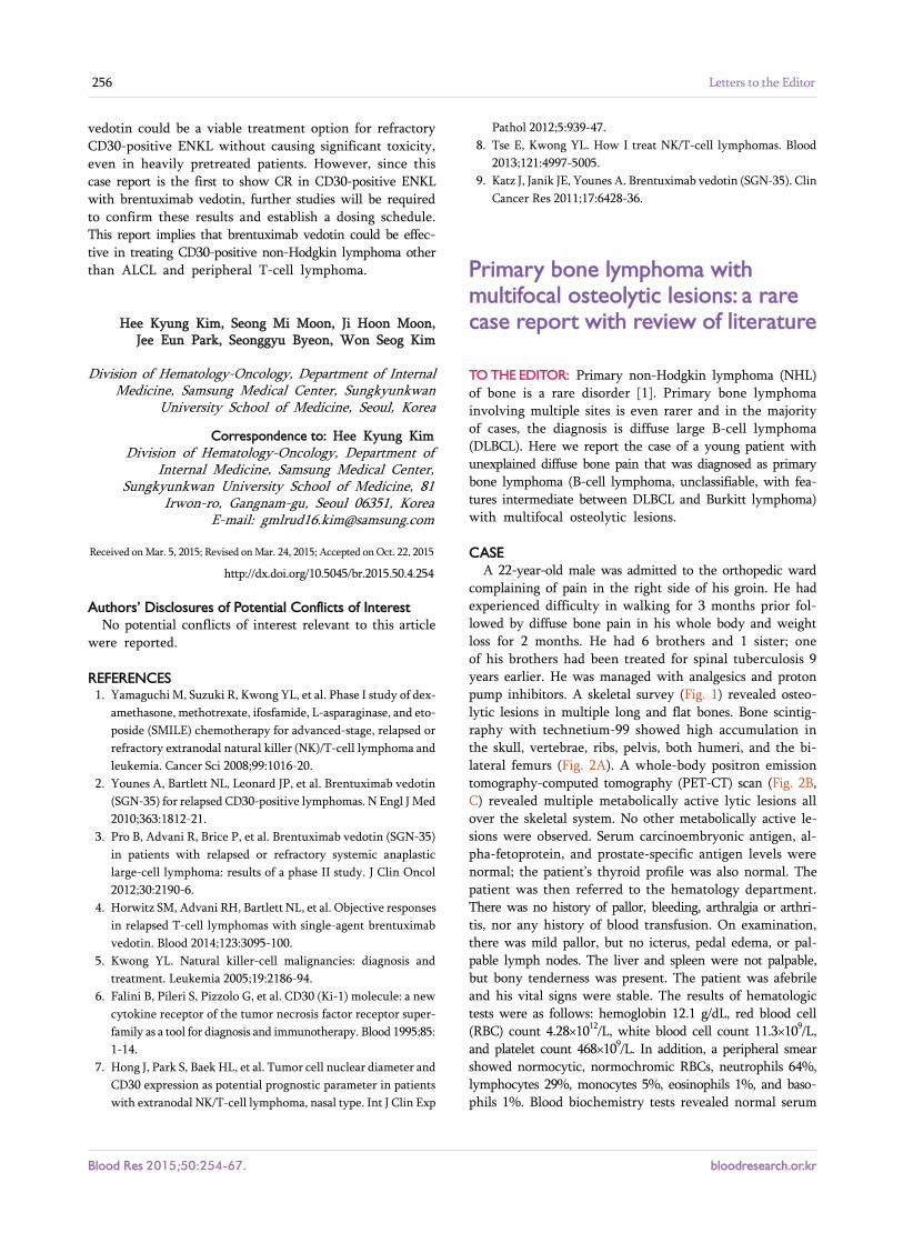
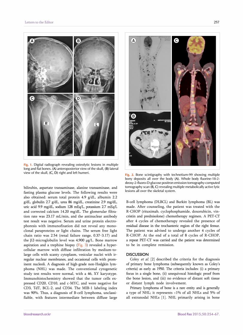
complaining of pain in the right side of his groin. He had experienced difficulty in walking for 3 months prior fol-lowed by diffuse bone pain in his whole body and weight loss for 2 months. He had 6 brothers and 1 sister; one of his brothers had been treated for spinal tuberculosis 9 years earlier. He was managed with analgesics and proton pump inhibitors. A skeletal survey (Fig. 1) revealed osteo-lytic lesions in multiple long and flat bones. Bone scintig-raphy with technetium-99 showed high accumulation in the skull, vertebrae, ribs, pelvis, both humeri, and the bi-lateral femurs (Fig. 2A). A whole-body positron emission tomography-computed tomography (PET-CT) scan (Fig. 2B, C) revealed multiple metabolically active lytic lesions all over the skeletal system. No other metabolically active le-sions were observed. Serum carcinoembryonic antigen, al-pha-fetoprotein, and prostate-specific antigen levels were normal; the patient’s thyroid profile was also normal. The patient was then referred to the hematology department. There was no history of pallor, bleeding, arthralgia or arthri-tis, nor any history of blood transfusion. On examination, there was mild pallor, but no icterus, pedal edema, or pal-pable lymph nodes. The liver and spleen were not palpable, but bony tenderness was present. The patient was afebrile and his vital signs were stable. The results of hematologic tests were as follows: hemoglobin 12.1 g/dL, red blood cell (RBC) count 4.28×10¹²/L, white blood cell count 11.3×10⁹/L, and platelet count 468×109/L. In addition, a peripheral smear showed normocytic, normochromic RBCs, neutrophils 64%, lymphocytes 29%, monocytes 5%, eosinophils 1%, and baso-phils 1%. Blood biochemistry tests revealed normal serum
bloodresearch.or.kr Blood Res 2015;50:254-67.
Letters to the Editor 257
Fig. 2. Bone scintigraphy with technetium-99 showing multiple bony deposits all over the body (A). Whole body fluorine-18-2- deoxy-2-fluoro-D-glucose positron emission tomography-computedtomography scan (B, C) revealing multiple metabolically active lyticlesions all over the skeletal system.
Fig. 1. Digital radiograph revealing osteolytic lesions in multiple long and flat bones. (A) anteroposterior view of the skull, (B) lateralview of the skull, (C, D) right and left humeri.
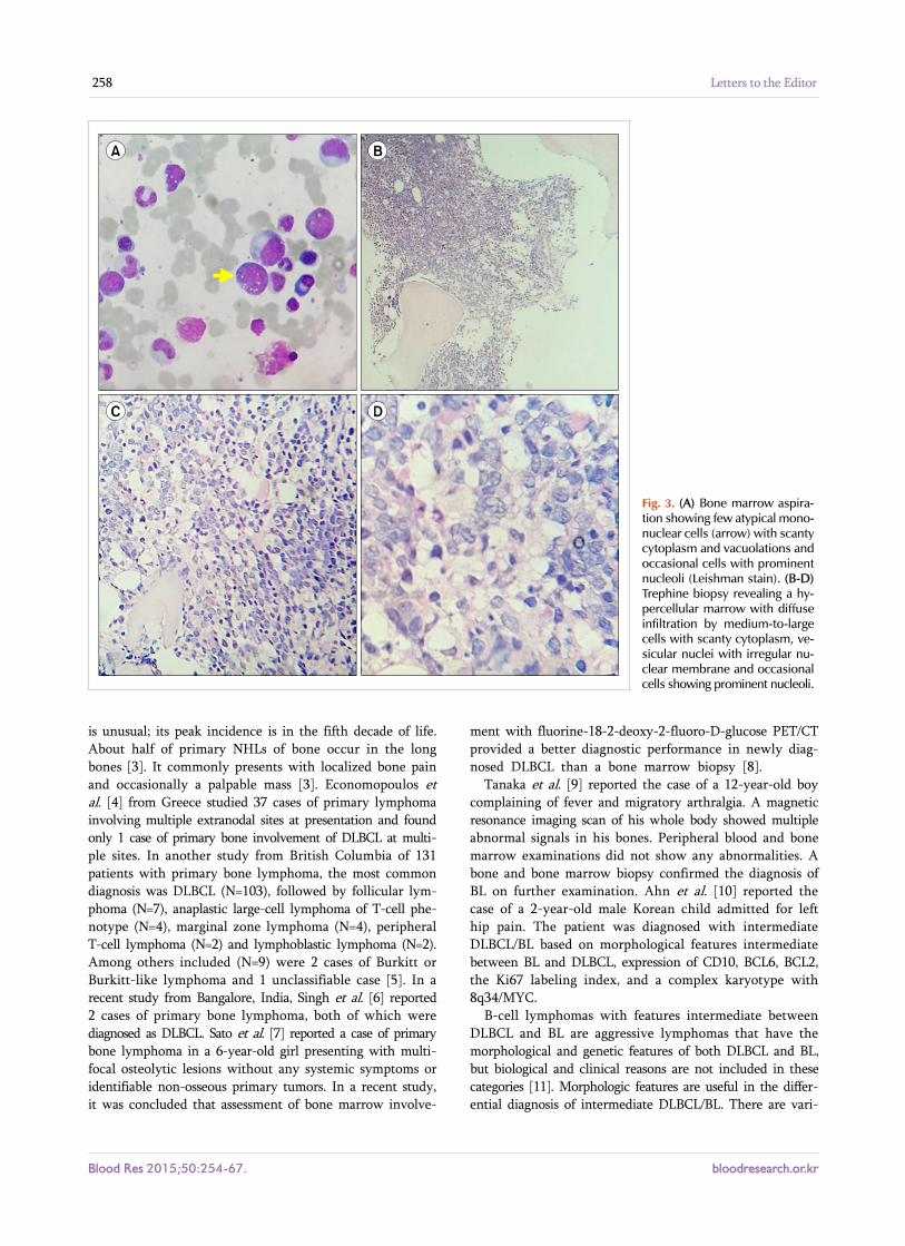
bilirubin, aspartate transaminase, alanine transaminase, and fasting plasma glucose levels. The following results were also obtained: serum total protein 4.9 g/dL, albumin 2.2 g/dL, globulin 2.7 g/dL, urea 86 mg/dL, creatinine 2.9 mg/dL, uric acid 9.9 mg/dL, sodium 128 mEq/L, potassium 2.7 mEq/L and corrected calcium 14.20 mg/dL. The glomerular filtra-tion rate was 23.17 mL/min, and the antinuclear antibody test result was negative. Serum and urine protein electro-phoresis with immunofixation did not reveal any mono-clonal paraproteins or light chains. The serum free light chain ratio was 2.54 (renal failure range, 0.37–3.17) and the 2-microglobulin level was 4,900 μg/L. Bone marrow aspiration and a trephine biopsy (Fig. 3) revealed a hyper-cellular marrow with diffuse infiltration by medium-to- large cells with scanty cytoplasm, vesicular nuclei with ir-regular nuclear membranes, and occasional cells with prom-inent nucleoli. A diagnosis of high-grade non-Hodgkin lym-phoma (NHL) was made. The conventional cytogenetic study test results were normal, with a 46, XY karyotype. Immunohistochemistry showed that the tumor cells ex-pressed CD20, CD10, and c-MYC, and were negative for CD3, TdT, BCL-2, and CD34. The MIB-1 labeling index was 90%. Thus, a diagnosis of B-cell lymphoma, unclassi-fiable, with features intermediate between diffuse large
B-cell lymphoma (DLBCL) and Burkitt lymphoma (BL) was made. After counseling, the patient was treated with the R-CHOP (rituximab, cyclophosphamide, doxorubicin, vin-cristin and prednisolone) chemotherapy regimen. A PET-CT after 4 cycles of chemotherapy revealed the presence of residual disease in the trochanteric region of the right femur. The patient was advised to undergo another 4 cycles of R-CHOP. At the end of a total of 8 cycles of R-CHOP, a repeat PET-CT was carried and the patient was determined to be in complete remission.
DISCUSSIONColey et al. [2] described the criteria for the diagnosis
of primary bone lymphoma (subsequently known as Coley’s criteria) as early as 1950. The criteria includes: (i) a primary focus in a single bone, (ii) unequivocal histologic proof from the bone lesion, and (iii) no evidence of distant soft tissue or distant lymph node involvement.
Primary lymphoma of bone is a rare entity and is generally a type of NHL; it represents <1% of all NHLs and 5% of all extranodal NHLs [1]. NHL primarily arising in bone
Blood Res 2015;50:254-67. bloodresearch.or.kr
258 Letters to the Editor
Fig. 3. (A) Bone marrow aspira-tion showing few atypical mono-nuclear cells (arrow) with scantycytoplasm and vacuolations and occasional cells with prominent nucleoli (Leishman stain). (B-D)Trephine biopsy revealing a hy-percellular marrow with diffuse infiltration by medium-to-large cells with scanty cytoplasm, ve-sicular nuclei with irregular nu-clear membrane and occasional cells showing prominent nucleoli.
is unusual; its peak incidence is in the fifth decade of life. About half of primary NHLs of bone occur in the long bones [3]. It commonly presents with localized bone pain and occasionally a palpable mass [3]. Economopoulos et al. [4] from Greece studied 37 cases of primary lymphoma involving multiple extranodal sites at presentation and found only 1 case of primary bone involvement of DLBCL at multi-ple sites. In another study from British Columbia of 131 patients with primary bone lymphoma, the most common diagnosis was DLBCL (N=103), followed by follicular lym-phoma (N=7), anaplastic large-cell lymphoma of T-cell phe-notype (N=4), marginal zone lymphoma (N=4), peripheral T-cell lymphoma (N=2) and lymphoblastic lymphoma (N=2). Among others included (N=9) were 2 cases of Burkitt or Burkitt-like lymphoma and 1 unclassifiable case [5]. In a recent study from Bangalore, India, Singh et al. [6] reported 2 cases of primary bone lymphoma, both of which were diagnosed as DLBCL. Sato et al. [7] reported a case of primary bone lymphoma in a 6-year-old girl presenting with multi-focal osteolytic lesions without any systemic symptoms or identifiable non-osseous primary tumors. In a recent study, it was concluded that assessment of bone marrow involve-
ment with fluorine-18-2-deoxy-2-fluoro-D-glucose PET/CT provided a better diagnostic performance in newly diag-nosed DLBCL than a bone marrow biopsy [8].
Tanaka et al. [9] reported the case of a 12-year-old boy complaining of fever and migratory arthralgia. A magnetic resonance imaging scan of his whole body showed multiple abnormal signals in his bones. Peripheral blood and bone marrow examinations did not show any abnormalities. A bone and bone marrow biopsy confirmed the diagnosis of BL on further examination. Ahn et al. [10] reported the case of a 2-year-old male Korean child admitted for left hip pain. The patient was diagnosed with intermediate DLBCL/BL based on morphological features intermediate between BL and DLBCL, expression of CD10, BCL6, BCL2, the Ki67 labeling index, and a complex karyotype with 8q34/MYC.
B-cell lymphomas with features intermediate between DLBCL and BL are aggressive lymphomas that have the morphological and genetic features of both DLBCL and BL, but biological and clinical reasons are not included in these categories [11]. Morphologic features are useful in the differ-ential diagnosis of intermediate DLBCL/BL. There are vari-
bloodresearch.or.kr Blood Res 2015;50:254-67.
Letters to the Editor 259
ous cellular forms; those resembling BL cells are smaller than typical DLBCL cells and those resembling DLBCL cells are larger than typical BL cells. Immunophenotypically, the cells are akin to BL with positivity for CD19, CD20, CD22, CD79a, CD10 and BCL6. BCL2 expression may be absent, weak, or strong. The Ki67 labeling index shows varying positivity [12]. Perry et al. [13] studied 39 cases of B-cell lymphoma, unclassifiable (B-UCL), presented at a median age of 69 years. The majority (62%) of patients presented with advanced-stage disease and 54% of patients had high (3-5) International Prognostic Index scores. Genetic heterogeneity was observed; 11 patients had 'dou-ble-hit' lymphomas with rearrangements of both MYC and BCL2 or BCL6. None of the immunohistochemical or genetic features were predictive of survival and the cases were very aggressive and resistant to chemotherapy. The 2008 World Health Organization classification includes provisional bor-derline categories for cases that are not clearly DLBCL or BL. This new category is called B-cell lymphoma, un-classifiable, with features intermediate between DLBCL and BL (intermediate DLBCL/BL) [11]. The justification for this new category is the recognition that these tumors are fre-quently refractory to chemotherapy and the patients have a poor survival rate [14]. Sirelkhatim et al. [15] studied cases with unexplained bone pain and concluded that lym-phoma/leukemia should be kept in mind when investigating a case of unexplained bone pain or an unexplained bone lesion, although it is a rare entity.
Our case presented with unexplained diffuse bone pain and, on evaluation, the patient was diagnosed with primary bone lymphoma (B-cell lymphoma, unclassifiable, with fea-tures intermediate between DLBCL and BL). The features included: a young age at presentation, serum hypercalcemia, multifocal osteolytic lesions present all over the body, and a rare type of histologic diagnosis. The patient achieved complete remission with R-CHOP chemotherapy. Ramadan et al. [5] treated the cases with CHOP (and R-CHOP in the rituximab era), radiotherapy or a combination of both.
NHL presenting as a primary bone tumor with multifocal disease is extremely rare. Primary bone lymphoma should be considered in the differential diagnosis of bony lesions in young patients. A very high index of suspicion, judicious use of the investigative armamentarium, and awareness of provisional borderline categories like B-cell lymphoma, unclassifiable, with features intermediate between DLBCL and BL will help clini-cians to achieve timely diagnosis and management.
Prakas Kumar Mandal, Shuvraneel Baul, Tuphan Kanti Dolai
Department of Hematology, NRS Medical College, Kolkata, India
Correspondence to: Prakas Kumar MandalDepartment of Hematology, NRS Medical College,
8C/1/N, Roy Para Road, Kolkata 700050, IndiaE-mail: [email protected]
Received on Jan. 14, 2015; Revised on Jan. 28, 2015; Accepted on Nov. 12, 2015
http://dx.doi.org/10.5045/br.2015.50.4.256
AuthorsÊ Disclosures of Potential Conflicts of InterestNo potential conflicts of interest relevant to this article
were reported.
REFERENCES1. de Leval L, Braaten KM, Ancukiewicz M, et al. Diffuse large B-cell
lymphoma of bone: an analysis of differentiation-associated anti-
gens with clinical correlation. Am J Surg Pathol 2003;27:1269-
77.
2. Coley BL, Higinbotham NL, Groesbeck HP. Primary retic-
ulum-cell sarcoma of bone; summary of 37 cases. Radiology
1950;55:641-58.
3. Gill P, Wenger DE, Inwards DJ. Primary lymphomas of bone.
Clin Lymphoma Myeloma 2005;6:140-2.
4. Economopoulos T, Papageorgiou S, Rontogianni D, et al.
Multifocal extranodal non-hodgkin lymphoma: a clinicopatho-
logic study of 37 cases in Greece, a Hellenic Cooperative
Oncology Group study. Oncologist 2005;10:734-8.
5. Ramadan KM, Shenkier T, Sehn LH, Gascoyne RD, Connors JM.
A clinicopathological retrospective study of 131 patients with
primary bone lymphoma: a population-based study of succes-
sively treated cohorts from the British Columbia Cancer Agency.
Ann Oncol 2007;18:129-35.
6. Singh T, Satheesh CT, Lakshmaiah KC, et al. Primary bone lym-
phoma: a report of two cases and review of the literature. J Cancer
Res Ther 2010;6:296-8.
7. Sato TS, Ferguson PJ, Khanna G. Primary multifocal osseous lym-
phoma in a child. Pediatr Radiol 2008;38:1338-41.
8. Berthet L, Cochet A, Kanoun S, et al. In newly diagnosed diffuse
large B-cell lymphoma, determination of bone marrow involve-
ment with 18F-FDG PET/CT provides better diagnostic perform-
ance and prognostic stratification than does biopsy. J Nucl Med
2013;54:1244-50.
9. Tanaka C, Nozawa K, Sano A, et al. A case of Burkitt lymphoma
with multifocal bone invasion. Rinsho Byori 2013;61:231-6.
10. Ahn JY, Seo YH, Park PW, et al. A case of B-cell lymphoma, un-
classifiable, with features intermediate between diffuse large
B-cell lymphoma and Burkitt lymphoma in a Korean child. Ann
Lab Med 2012;32:162-6.
11. Swerdlow SH, Campo E, Harris NL, et al, eds. WHO classification
of tumours of haematopoietic and lymphoid tissues. 4th ed. Lyon,
France: IARC Press, 2008:65-6.
12. Carbone A, Gloghini A, Aiello A, Testi A, Cabras A. B-cell lym-
phomas with features intermediate between distinct pathologic
entities. From pathogenesis to pathology. Hum Pathol 2010;
41:621-31.
13. Perry AM, Crockett D, Dave BJ, et al. B-cell lymphoma, un-
classifiable, with features intermediate between diffuse large
B-cell lymphoma and burkitt lymphoma: study of 39 cases. Br J
Haematol 2013;162:40-9.
14. Jack AS, Barrans SL, Qian W, Stenning SP, Mead GM. Bone mar-
row involvement and outcome in Burkitt lymphoma and diffuse
large B-cell lymphoma. Blood 2009;114:486-7.
Blood Res 2015;50:254-67. bloodresearch.or.kr
260 Letters to the Editor
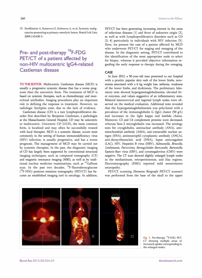
Fig. 1. Pre-therapy 18F-FDG PET/ CT showing multiple areas of increased uptake corresponding tothe enlarged nodes.
15. Sirelkhatim A, Kaiserova E, Kolenova A, et al. Systemic malig-
nancies presenting as primary osteolytic lesion. Bratisl Lek Listy
2009;110:630-5.
Pre- and post-therapy 18F-FDG PET/CT of a patient affected by non-HIV multicentric IgG4-related Castleman disease
TO THE EDITOR: Multicentric Castleman disease (MCD) is usually a progressive systemic disease that has a worse prog-nosis than the unicentric form. The treatment of MCD is based on systemic therapies, such as chemotherapy and mon-oclonal antibodies. Imaging procedures play an important role in defining the response to treatment. However, no radiologic linchpins exist, due to the lack of evidence.
Castleman disease (CD) is a rare lymphoproliferative dis-order first described by Benjamin Castleman, a pathologist at the Massachusetts General Hospital. CD may be unicentric or multicentric. Unicentric CD (UCD), the most common form, is localized and may often be successfully treated with local therapies. MCD is a systemic disease, occurs most commonly in the setting of human immunodeficiency virus (HIV) infection, is usually progressive, and has a worse prognosis. The management of MCD may be carried out by systemic therapies. In the past, the diagnostic imaging of CD has largely been supported by conventional structural imaging techniques, such as computed tomography (CT) and magnetic resonance imaging (MRI), as well as by tradi-tional nuclear medicine examinations, such as 67Gallium scan. In the past two decades, 18F-fluorodeoxyglucose (18F-FDG) positron emission tomography (PET/CT) has be-come an established imaging tool in oncology. In addition,
PET/CT has been generating increasing interest in the areas of infectious diseases [1] and fever of unknown origin [2], as well as with lymphoproliferative disorders such as CD [3, 4] particularly in individuals with HIV infection [5]. Here, we present the case of a patient affected by MCD who underwent PET/CT for staging and restaging of the disease. In the diagnostic setting, PET/CT contributed to the identification of the most appropriate node to select for biopsy, whereas it provided objective information re-garding the early response to therapy during the restaging.
CASEIn June 2012, a 50-year-old man presented to our hospital
with a pruritic papular skin rash of the lower limbs, xero-stomia associated with a 6 kg weight loss, asthenia, weakness of the lower limbs, and dyskinesia. The preliminary labo-ratory tests showed hypergammaglobulinemia, elevated liv-er enzymes, and values suggestive of an inflammatory state. Bilateral laterocervical and inguinal lymph nodes were ob-served on the medical evaluation. Additional tests revealed that the hypergammaglobulinemia was polyclonal with a prevalence of the immunoglobulin G (IgG) cluster (50 g/L) and increases in the light kappa and lambda chains. Moreover, C3 and C4 complement proteins were decreased, whereas beta-2 microglobulin was increased. The serology tests for cryoglobulin, antinuclear antibody (ANA), anti-mitochondrial antibody (AMA), anti-extractable nuclear an-tigen (ENA), antineutrophil cytoplasmic antibody (ANCA), anti-deoxyribonucleic acid (DNA), lupus anticoagulant (LAC), HIV, Hepatitis B virus (HBV), Salmonella, Brucella, Leishmania, Parvovirus, Strongyloides Stercoralis, Bartonella, Epstein-Barr virus (EBV), and cytomegalovirus (CMV) were negative. The CT scan showed slightly enlarged lymph nodes in the mediastinum, retroperitoneum, and iliac regions. Electromyography (EMG) reported mild sensorimotor neuropathy.
PET/CT scanning (Siemens Biograph PET/CT scanner) was performed from the base of the skull to the upper