Primary and Acquired Resistance to Immune Checkpoint … · Review Primary and Acquired Resistance...
12
Review Primary and Acquired Resistance to Immune Checkpoint Inhibitors in Metastatic Melanoma Tuba N. Gide 1,2 , James S. Wilmott 1,2 , Richard A. Scolyer 1,2,3 , and Georgina V. Long 1,2,4,5 Abstract Immune checkpoint inhibitors have revolutionized the treat- ment of patients with advanced-stage metastatic melanoma, as well as patients with many other solid cancers, yielding long- lasting responses and improved survival. However, a subset of patients who initially respond to immunotherapy, later relapse and develop therapy resistance (termed "acquired resistance"), whereas others do not respond at all (termed "primary resis- tance"). Primary and acquired resistance are key clinical barriers to further improving outcomes of patients with metastatic melanoma, and the known mechanisms underlying each involves various components of the cancer immune cycle, and interactions between multiple signaling molecules and pathways. Due to this complexity, current knowledge on resis- tance mechanisms is still incomplete. Overcoming therapy resistance requires a thorough understanding of the mechan- isms underlying immune evasion by tumors. In this review, we explore the mechanisms of primary and acquired resistance to immunotherapy in melanoma and detail potential therapeutic strategies to prevent and overcome them. Clin Cancer Res; 24(6); 1260–70. Ó2017 AACR. Introduction Immune checkpoint inhibitors have revolutionized the treat- ment of advanced melanoma (1–5) and have significant clinical activity across an increasing range of many other solid malignan- cies, including non–small cell lung cancer (6, 7), renal cell carcinoma (8), head and neck cancer (9), Merkel cell carcinoma (10), and bladder cancer (11, 12). Understanding the biology behind response and resistance to immune checkpoint blockade is critical to further improving outcomes of patients with meta- static melanoma. The first immune checkpoint to be clinically targeted, the cytotoxic T-lymphocyte antigen 4 (CTLA-4), is expressed on the surface of activated T cells and binds to its ligands, B7-1 and B7-2, on antigen-presenting cells (APC), resulting in the transmission of inhibitory signals to T cells. In patients with metastatic melano- ma, phase III clinical trials of ipilimumab, a fully human IgG1 monoclonal antibody inhibiting CTLA-4, demonstrated a signif- icant improvement in progression-free survival (PFS) and overall survival (OS) when compared with a gp100 vaccine (13) or dacarbazine chemotherapy (4). Drugs targeting the programmed cell death receptor 1 (PD-1, PDCD1) showed a further increase in response rates, PFS (2), and OS (14–16) compared with anti–CTLA-4 blockade. PD-1 is also expressed on the surface of activated T cells and binds to the programmed cell death ligand 1 (PD-L1, CD274) to negatively regulate T-cell activation and differentiation. PD-L1 is constitu- tively expressed by T cells, macrophages, and dendritic cells (DC), as well as by some tumor cells including melanoma (17). Follow- up data from phase I clinical trials of the fully human IgG4 monoclonal antibody, nivolumab, showed a median OS of 17.3 months, with a 5 year OS rate of 34% (18). In a phase III study of nivolumab versus dacarbazine in patients with BRAF wild-type metastatic melanoma, the median OS was not reached for nivolumab at the most recent analysis, versus 11.2 months for dacarbazine [hazard ratio (HR), 0.43, P < 0.001], and the 1- and 2-year OS rates were 73% and 58%, respectively, for nivolumab (1, 14). Pembrolizumab, a humanized IgG4 monoclonal anti- body against PD-1, also showed 1-, 2-, and 3-year OS rates of 67%, 50%, and 40%, respectively, in a phase I trial of ipilimumab-treated and ipilimumab-na € ve patients with advanced melanoma (3). Furthermore, in a phase III trial of pembrolizumab versus ipilimumab, the 2-year OS rates were 55% versus 43%, respectively (5, 15). More recently, combined anti–CTLA-4 and anti–PD-1 immu- notherapies have shown improved response rates and clinical outcomes in comparison to ipilimumab monotherapy (the study was not powered to compare the two nivolumab treating arms: nivolumab plus ipilimumab and nivolumab alone). A phase III study showed an increase in the median PFS of patients treated with nivolumab and ipilimumab (11.5 months; HR, 0.42, P < 0.001) and nivolumab alone (6.9 months; HR, 0.57, P < 0.001) compared with ipilimumab alone (2.9 months; ref. 2). At a minimum follow-up of 28 months, the median OS had not been reached in the combination or nivolumab-alone groups and was 20 months for ipilimumab alone [HR: combination vs. ipilimumab, 0.55 (P < 0.0001); nivolumab vs. ipilimumab, 1 Melanoma Institute Australia, The University of Sydney, Sydney, NSW, Australia. 2 Sydney Medical School, The University of Sydney, Sydney, NSW, Australia. 3 Royal Prince Alfred Hospital, Sydney, NSW, Australia. 4 Royal North Shore Hospital, Sydney, NSW, Australia. 5 Mater Hospital, North Sydney, NSW, Australia. Note: Supplementary data for this article are available at Clinical Cancer Research Online (http://clincancerres.aacrjournals.org/). Corresponding Author: Georgina V. Long, Melanoma Institute Australia, The Poche Centre, 40 Rocklands Road, North Sydney, NSW, 2060, Australia. Phone: 612-9911-7336; Fax: 612-9954-9290; E-mail: [email protected] doi: 10.1158/1078-0432.CCR-17-2267 Ó2017 American Association for Cancer Research. Clinical Cancer Research Clin Cancer Res; 24(6) March 15, 2018 1260 on March 27, 2020. © 2018 American Association for Cancer Research. clincancerres.aacrjournals.org Downloaded from Published OnlineFirst November 10, 2017; DOI: 10.1158/1078-0432.CCR-17-2267
Transcript of Primary and Acquired Resistance to Immune Checkpoint … · Review Primary and Acquired Resistance...

Review
Primary and Acquired Resistance to ImmuneCheckpoint Inhibitors in Metastatic MelanomaTuba N. Gide1,2, James S.Wilmott1,2, Richard A. Scolyer1,2,3, andGeorgina V. Long1,2,4,5
Abstract
Immune checkpoint inhibitors have revolutionized the treat-ment of patients with advanced-stage metastatic melanoma, aswell as patients with many other solid cancers, yielding long-lasting responses and improved survival. However, a subset ofpatients who initially respond to immunotherapy, later relapseand develop therapy resistance (termed "acquired resistance"),whereas others do not respond at all (termed "primary resis-tance"). Primary and acquired resistance are key clinical barriersto further improving outcomes of patients with metastaticmelanoma, and the known mechanisms underlying each
involves various components of the cancer immune cycle,and interactions between multiple signaling molecules andpathways. Due to this complexity, current knowledge on resis-tance mechanisms is still incomplete. Overcoming therapyresistance requires a thorough understanding of the mechan-isms underlying immune evasion by tumors. In this review, weexplore the mechanisms of primary and acquired resistance toimmunotherapy in melanoma and detail potential therapeuticstrategies to prevent and overcome them. Clin Cancer Res; 24(6);1260–70. �2017 AACR.
IntroductionImmune checkpoint inhibitors have revolutionized the treat-
ment of advanced melanoma (1–5) and have significant clinicalactivity across an increasing range of many other solid malignan-cies, including non–small cell lung cancer (6, 7), renal cellcarcinoma (8), head and neck cancer (9), Merkel cell carcinoma(10), and bladder cancer (11, 12). Understanding the biologybehind response and resistance to immune checkpoint blockadeis critical to further improving outcomes of patients with meta-static melanoma.
The first immune checkpoint to be clinically targeted, thecytotoxic T-lymphocyte antigen 4 (CTLA-4), is expressed on thesurface of activated T cells and binds to its ligands, B7-1 and B7-2,on antigen-presenting cells (APC), resulting in the transmission ofinhibitory signals to T cells. In patients with metastatic melano-ma, phase III clinical trials of ipilimumab, a fully human IgG1monoclonal antibody inhibiting CTLA-4, demonstrated a signif-icant improvement in progression-free survival (PFS) and overallsurvival (OS) when compared with a gp100 vaccine (13) ordacarbazine chemotherapy (4).
Drugs targeting the programmed cell death receptor 1 (PD-1,PDCD1) showed a further increase in response rates, PFS (2), andOS (14–16) compared with anti–CTLA-4 blockade. PD-1 is alsoexpressed on the surface of activated T cells and binds to theprogrammed cell death ligand 1 (PD-L1, CD274) to negativelyregulate T-cell activation and differentiation. PD-L1 is constitu-tively expressed by T cells, macrophages, and dendritic cells (DC),as well as by some tumor cells includingmelanoma (17). Follow-up data from phase I clinical trials of the fully human IgG4monoclonal antibody, nivolumab, showed a median OS of17.3 months, with a 5 year OS rate of 34% (18). In a phase IIIstudy of nivolumab versus dacarbazine in patients with BRAFwild-type metastatic melanoma, the median OS was not reachedfor nivolumab at themost recent analysis, versus 11.2months fordacarbazine [hazard ratio (HR), 0.43, P < 0.001], and the 1- and2-year OS rates were 73% and 58%, respectively, for nivolumab(1, 14). Pembrolizumab, a humanized IgG4 monoclonal anti-body against PD-1, also showed 1-, 2-, and 3-year OS ratesof 67%, 50%, and 40%, respectively, in a phase I trial ofipilimumab-treated and ipilimumab-na€�ve patients withadvanced melanoma (3). Furthermore, in a phase III trial ofpembrolizumab versus ipilimumab, the 2-year OS rates were55% versus 43%, respectively (5, 15).
More recently, combined anti–CTLA-4 and anti–PD-1 immu-notherapies have shown improved response rates and clinicaloutcomes in comparison to ipilimumabmonotherapy (the studywas not powered to compare the two nivolumab treating arms:nivolumab plus ipilimumab and nivolumab alone). A phase IIIstudy showed an increase in the median PFS of patients treatedwith nivolumab and ipilimumab (11.5 months; HR, 0.42, P <0.001) and nivolumab alone (6.9 months; HR, 0.57, P < 0.001)compared with ipilimumab alone (2.9 months; ref. 2). At aminimum follow-up of 28 months, the median OS had not beenreached in the combination or nivolumab-alone groups andwas 20 months for ipilimumab alone [HR: combination vs.ipilimumab, 0.55 (P < 0.0001); nivolumab vs. ipilimumab,
1Melanoma Institute Australia, TheUniversity of Sydney, Sydney, NSW,Australia.2Sydney Medical School, The University of Sydney, Sydney, NSW, Australia.3Royal Prince Alfred Hospital, Sydney, NSW, Australia. 4Royal North ShoreHospital, Sydney, NSW, Australia. 5Mater Hospital, North Sydney, NSW,Australia.
Note: Supplementary data for this article are available at Clinical CancerResearch Online (http://clincancerres.aacrjournals.org/).
Corresponding Author: Georgina V. Long, Melanoma Institute Australia, ThePoche Centre, 40 Rocklands Road, North Sydney, NSW, 2060, Australia. Phone:612-9911-7336; Fax: 612-9954-9290; E-mail: [email protected]
doi: 10.1158/1078-0432.CCR-17-2267
�2017 American Association for Cancer Research.
ClinicalCancerResearch
Clin Cancer Res; 24(6) March 15, 20181260
on March 27, 2020. © 2018 American Association for Cancer Research. clincancerres.aacrjournals.org Downloaded from
Published OnlineFirst November 10, 2017; DOI: 10.1158/1078-0432.CCR-17-2267

0.63 (P < 0.0001); ref. 16]. The two-year OS rates were 64%, 59%,and 45% in the combination, nivolumab, and ipilimumabgroups, respectively (16).
The results of these clinical trials highlight the significantimpact immunotherapies have had on the clinical managementof patients with advanced-stage metastatic melanoma. However,although approximately 35% to 60% of patients have a RECISTresponse (10%–12% a complete response) to anti–PD-1-basedimmunotherapy (2, 14, 15), 40% to 65%have shownminimal orno RECIST response at the outset, and 43%of responders developacquired resistance by 3 years (3). The underlying mechanismsdriving these variations in response are not yet well understood.For an immunotherapy to elicit an efficient antitumor immuneresponse, the cancer immune cycle must be initiated and thesubsequent steps successfully completed. This involves efficient(i) antigen presentation and T-cell activation, (ii) T-cell traffickingand tumor infiltration, and (iii) T-cell killing activity within thetumor microenvironment (Fig. 1). Studies examining possiblepredictive biomarkers of response to immunotherapy havereported a higher density of preexisting cytotoxic T lymphocytesin tumor biopsies of patients who displayed a greater response toanti–PD-1/PD-L1 immunotherapy (19–21), and more signifi-cantly, an increased influx of T cells and PD-L1þ macrophagesearly during treatment (22). In this review,wediscuss the differentforms of immunotherapy resistance, the mechanisms by which
tumors evade the immune system, and strategies to overcome orprevent resistance in the future.
Primary ResistancePrimary resistance to immune checkpoint blockade occurs
in approximately 40% to 65% of patients withmelanoma treatedwith anti–PD-1 based therapy (Fig. 2), depending on whetheranti–PD-1 is given upfront or after progression on other therapies(2, 14, 15), and >70% of those treated with anti–CTLA-4 therapy(4, 13). This key unsolved clinical problem occurs when there isfailure to induce an effective antitumor immune response at anyof the three stages of the cancer immune cycle (Fig. 1). To date, theclinicopathologic factors that have been associated with primaryresistance are elevated levels of baseline serum LDH (23),increased baseline tumor burden (24), lack of PD-L1 expressionin baselinemelanoma tissue samples (Fig. 3; ref. 25), lack of T-cellinfiltration (Fig. 3; ref. 21), the absence of PD-1 T cells and PD-L1macrophages in melanoma biopsies taken early during treatment(22), insufficient neoantigens and low mutational burden (26),the presence of an innate anti–PD-1 resistance signature (IPRES)transcriptional signature (27), or absence of an interferon signa-ture (28). It is currently unknown whether these measures aresurrogates for resistance or have a direct mechanistic role inpreventing response. Here, we discuss the immune escape
Primary resistance• Poor immunogenicity• Impaired DC maturation
Acquired resistance• Loss of B2M
Primary resistance• Upregulation of PD-L1• Induction of IDO• Upregulation of Tregs• Upregulation of CD73/adenosine• Expression of IPRES• Loss-of-function mutations
Acquired resistance• Mutations in JAK1/JAK2• Upregulation of PD-L1• Upregulation of immune checkpoint markers
Primary resistance• Downregulation of chemokines• Upregulation of endothelin B receptor• Overexpression of VEGF
Antigenpresentation
and T-cellactivation
T-cell killing activitywithin the tumor
microenvironment
T-celltraffickingand tumorinfiltration
Therapeutic strategies• Radiotherapy• Oncolytic viruses• CTLA-4 inhibitors• HDAC inhibitors
Therapeutic strategies• Oncolytic viruses• HDAC inhibitors
Therapeutic strategies• PD-1/PD-L1 inhibitors• IDO inhibitors• LAG-3 inhibitors• TIM-3 inhibitors• CD73/A2AR inhibitors
© 2017 American Association for Cancer Research
Figure 1.
The cancer immune cycle. Theinduction of an effective antitumorimmune response requiressuccessful (i) antigen presentationand T-cell activation, (ii) T-celltrafficking and tumor infiltration,and (iii) T-cell killing activity withinthe tumor microenvironment.Various immune escapemechanisms present at each ofthese stages can result in primaryor acquired resistance toimmunotherapy. Potentialtherapeutic strategies can beused at each stage to overcomeimmunotherapy resistance.A2AR, A2A receptor; B2M,beta-2-microglobulin; HDAC,histone deacetylase; JAK1/JAK2,janus kinases 1 and 2; IDO,indoleamine 2,3-dioxygenase;IPRES, innate anti–PD-1 resistancesignature; LAG-3, lymphocyteactivation gene 3; TIM-3, T-cellimmunoglobulin andmucin domain3; Tregs, regulatory T cells; VEGF,vascular endothelial growth factor.
Resistance to Immunotherapy in Melanoma
www.aacrjournals.org Clin Cancer Res; 24(6) March 15, 2018 1261
on March 27, 2020. © 2018 American Association for Cancer Research. clincancerres.aacrjournals.org Downloaded from
Published OnlineFirst November 10, 2017; DOI: 10.1158/1078-0432.CCR-17-2267

mechanisms that can occur at each stage of the cancer immunecycle (Fig. 1), thereby promoting both the growth and metastasisof tumors and resistance to immune checkpoint inhibitortherapies.
Antigen presentation and T-cell activationUpon encountering and engulfing foreign antigens, such as that
of cancer cells, DCs migrate from the tumor to regional lymphnodes where they present the antigens on major histocompati-bility complex (MHC) class I molecules to CD8þ T cells, resultingin activation of the latter. Barriers at this stage of the cancerimmune cycle prevent optimal T-cell priming and activation,hence resulting in evasion of the immune system by the tumor(Table 1).
Poor immunogenicity. Some tumors lack sufficient antigen pre-sentation by the immune system (29, 30) or do not presentantigens that can be recognized as foreign (31, 32). The processof distinguishing tumor cells from normal cells depends on T-cellrecognition of tumor-specific or tumor-associated antigens (TAA;ref. 33). Tumor immune evasion by TAA-negative cells wasreported in patients with melanoma who relapsed after respond-ing to peptide vaccinations (34). Recognition of tumor neoanti-gens by T cells has been associated with increased and durableresponse to immunotherapies and increased tumor regression,indicating a significant role for neoantigens in improving the
outcome of patients with metastatic melanoma (26, 35). Circu-lating CD8þPD-1þ lymphocytes in peripheral blood of patientswith melanoma can target patient-specific neoantigens, andneoantigen-specific T cells can in turn recognize autologoustumors (36). The immunogenicity of neoantigens can be pre-dicted by combining exome sequencing and mass spectrometrydata, thereby facilitating the identification of antigens that can beused to generate active T-cell responses (37).
Analysis of The Cancer Genome Atlas (TCGA) data frommelanoma cases revealed that cutaneous melanoma displays ahigh mutational burden and UV signature (38). In addition toneoantigen recognition, a highmutational loadwas also found tocorrelate with clinical benefit to immune checkpoint blockade(26). Similarly, a positive correlation was observed between ahigher mutational load and increased CD8þ T-cell infiltration(39). Furthermore, an increased mutational burden is associatedwith elevated PD-L1 expression in advancedmelanoma (40). In apooled analysis of 832 patients withmelanoma, an increased PFSwas observed in PD-L1–positive patients treated with nivolumaband ipilimumab combined immunotherapy, aswell as in patientstreated with nivolumab alone, compared with PD-L1–negativepatients (2, 41). Similarly, PD-L1–positive patients treated withpembrolizumab had increased PFS, OS, and overall response rate(ORR), highlighting PD-L1 as a potential biomarker of response(42). As PD-L1 positivity is associated with improved responsein patients with melanoma, a lack of PD-L1 correlates with
A B
© 2017 American Association for Cancer Research
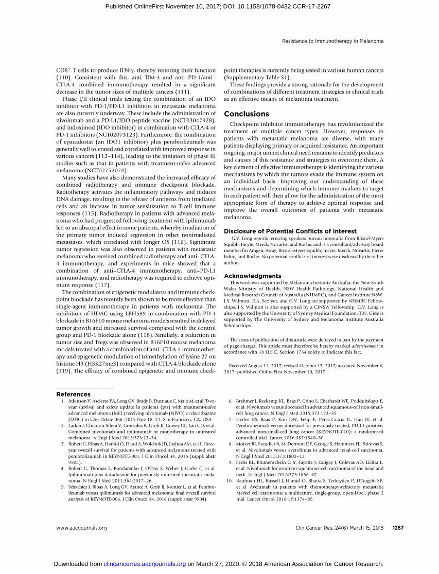
Figure 2.
Primary resistance in metastaticmelanoma. CT scans of a 55-year-oldpatient with metastatic melanomatreated with combined anti–CTLA-4and anti–PD-1 immunotherapy atbaseline (A) and after 12 weeks oftherapy (B), demonstratingwidespread metastatic disease,including significant livermetastases, who did not respond,indicating primary resistance totherapy. The patient did not respondto subsequent chemotherapy anddied 156 days after commencingcombined immunotherapy.
Gide et al.
Clin Cancer Res; 24(6) March 15, 2018 Clinical Cancer Research1262
on March 27, 2020. © 2018 American Association for Cancer Research. clincancerres.aacrjournals.org Downloaded from
Published OnlineFirst November 10, 2017; DOI: 10.1158/1078-0432.CCR-17-2267

primary resistance (Fig. 3). Nevertheless, some patients withPD-L1–positive tumors do not respond to PD-1 blockade, andconversely, some patients with PD-L1–negative tumors respond.For these reasons, intratumoral PD-L1 expression is a suboptimalpredictive biomarker (2, 41). Together, the aforementioned dataindicate that PD-L1 expression is a possible surrogate for lackof immunogenicity, as well as other failures further down theimmune cycle.
ImpairedDCmaturation. Inorder to efficiently activateT cells,DCsmust undergo a process called maturation, where they increasetheir capacity to stimulate T cells by displaying increased expres-sion of various costimulatory molecules required for T-cell acti-vation, such as MHC class I/II, CD80, CD86, and CD40 (43). Thedensity of DCs strongly correlates with activated T cells in mela-noma (44). The function of DCs can be impaired via multiplepathways. Interleukin (IL) 37b, a protein with a critical role in theinhibition of the innate immune response, suppresses DC matu-ration and function by decreasing CD80 and CD86 expression viathe ERK/S6K/NF-kB signaling pathways (45). Furthermore, DCmaturation and tumor infiltration increased significantly in mel-anoma following the inhibition of signal transducer and activatorof transcription3(STAT3),a transcription factor that is required fortumor growth and metastasis (46). STAT3 is also involved in thecross-talk betweenmelanoma cells and immune cells, resulting inthe induction of other immunosuppressive factors such as thevascular endothelial growth factor (VEGF), IL10, regulatory T cells(Treg), and transforming growth factor b (TGF-b), all of whichhave inhibitory effects on DC maturation (47–49).
T-cell trafficking and tumor infiltrationTumors can use a number of immune evasive mechanisms to
prevent T-cell trafficking and infiltration into tumors. Assumingthat the T cells were successfully activated in the previous steps,disruption during this stage is a likely cause for lack of response toimmunotherapy.
Downregulation of chemokines required for T-cell recruitment. Thedifferential expression of chemokine receptors is required foreffective T-cell homing and recruitment in cancer. In particular,
Anti–PD-1 responder Anti–PD-1 nonresponder
Perit
umor
Intr
atum
or
A B
C D
© 2017 American Association for Cancer Research
Figure 3.
Variable expression of immunemarkers in patients with metastaticmelanoma treated with anti–PD-1immunotherapy. Multipleximmunofluorescent imagesillustrating baseline PD-L1expression and tumor-infiltratinglymphocytes (TIL) in a 59-year-oldpatient who responded to anti–PD-1monotherapy (A and C), and lackthereof in a 55-year-old patientwho did not respond toimmunotherapy (B and D).
Table 1. Mechanisms of resistance to immune checkpoint blockade
Mechanisms of resistance Contributing factors References
Insufficient antigen presentationand recognition
Low mutational burden (26)Lack of neoantigenrecognition
(26, 35)
Loss of B2M (99, 100)Loss of MHC class I (99)
Insufficient T-cell activation Lack of mature DCs (44, 45)STAT3 expression (46, 49)
Absence of T cells from tumormicroenvironment
Lack of chemokines (50, 51, 54)VEGF overexpression (63, 64)
Upregulation of immunosuppressivemarkers
PD-L1 (69, 70)IDO (80, 81)Tregs (82–85)
Decreased sensitivity to IFN-gsignaling
Mutations in theJAK/STAT pathway
(93, 101)
Immune checkpoint markers TIM-3 (105)LAG-3 (106)
Abbreviations: B2M, beta-2-microglobulin; IDO, indoleamine 2,3-dioxygenase;IFN-g , interferon gamma; STAT3, signal transducer and activator of transcription3; TIM-3, T-cell immunoglobulin and mucin domain 3; Tregs, regulatory T cells;VEGF, vascular endothelial growth factor.
Resistance to Immunotherapy in Melanoma
www.aacrjournals.org Clin Cancer Res; 24(6) March 15, 2018 1263
on March 27, 2020. © 2018 American Association for Cancer Research. clincancerres.aacrjournals.org Downloaded from
Published OnlineFirst November 10, 2017; DOI: 10.1158/1078-0432.CCR-17-2267

CXCR3 has been identified as an important chemokine receptorcritical for T-cell infiltration. Inmousemelanomamodels, CXCR3was highly expressed on a number of T-cell subsets, and trans-fection with its ligand, CXCL9, resulted in a significant increase inboth CD4þ and CD8þ T-cell infiltration (50). Similarly, humanmelanoma samples with high CD8þ T-cell expression were asso-ciated with increased levels of CXCL9 and CXCL10 (51). Inter-feron gamma (IFN-g , IFNG) has previously been shown to medi-ate trafficking of Tregs, T helper cells, and cytotoxic T cells (52, 53).STAT3 inhibits CXCL10 production by tumor-associatedmyeloidcells and T-cell recruitment into tumors by downregulating IFN-gproduction by CD8þ T cells (54). Conversely, Stat3 ablationincreases CXCR3 expression on CD8þ T cells, allowing T-celltumor infiltration (54).
Epigenetic alterations including DNAmethylation and histonemodifications have alsobeen identified as importantmechanismsof chemokine repression and tumor progression. Epigeneticmodifications are heritable modifications to DNA that result inchanges to the gene expression profiles of tumor cells, therebyallowing them to evade the immune system (55). Epigeneticsilencing resulted in the suppression of CXCL9 and CXCL10 inovarian cancer, and treatment with epigenetic modulatorsincreased chemokine expression and T-cell infiltration intotumors (56). Increased expression of chemokines, T-cell recruit-ment, and tumor regression was also observed in lung cancer celllines and mice treated with the histone deacetylase (HDAC)inhibitor romidepsin (57).
Upregulation of the endothelin B receptor. T-cell trafficking throughthe tumor and lymph nodes is controlled by a number ofendothelial signals, regulating T-cell homing, adhesion, andmigration (58). The interaction between endothelin 1 (ET-1,EDN1) and the endothelin A receptor (ETAR, EDNRA) promotestumorigenesis through various pathways including cell prolifer-ation, invasion, angiogenesis, bone remodeling, and inhibition ofapoptosis (59). The endothelin B receptor (ETBR, EDNRB) coun-terregulates ET-1/ETAR activity via the increased production ofnitric oxide, activation of apoptotic pathways, and clearanceof ET-1 (60). The endothelin system has been implicated in thepathogenesis of a number of cancers, including ovarian cancer,prostate cancer, and colon cancer. Interestingly, ETBR is upregu-lated in melanoma and has also been proposed as a marker ofmelanoma progression, suggesting a role for ETBR in melanomatumorigenesis (61). ETBR inhibition in 10 humanmelanoma celllines using the ETBR antagonist BQ788 resulted in an increase inapoptosis and cell death, as well as an increase in angiogenesis inthe tumors (62). In human ovarian cancers, ETBR was found tocorrelate with an absence of tumor-infiltrating lymphocytes (TIL)as well as decreased patient survival time. Administration ofBQ788 increased T-cell homing to tumors and improved theefficacy of immunotherapy (58), highlighting ETBR as a potentialimmune escape mechanism and future target in patients who failto respond to immunotherapy.
Overexpression of VEGF. Increased levels of the proangiogenicfactor VEGF in plasma and tissue samples have also been asso-ciated with the growth and progression of melanoma (63, 64).VEGF-Adownregulates T-cell adhesion to the endotheliumvia thesuppression of intercellular adhesion molecule 1 (ICAM-1) andvascular cell adhesion molecule 1 (VCAM-1) on endothelial cells(65). Increased expression of VEGF in tumor was associated with
an absence of intratumoral TILs and a shorter OS time in patientswith ovarian carcinoma (66). Inhibition of VEGF resulted in anincrease in T-cell infiltration into B16 melanoma tumors via theupregulation of CXCL10 and CXCL11 chemokines (67). Addi-tionally, VEGF-A, along with prostaglandin E2 (PGE2) and IL10,upregulates the Fas ligand, resulting in CD8þ T-cell death, andsubsequent inhibition of VEGF and PGE2 increased CD8þ T-cellinfiltration (68). Corresponding with these data, in melanomatumor biopsies, increased levels of VEGFA were observed innonresponders to anti–CTLA-4 and anti–PD-1 immunotherapiesin comparison to responders (19).
T-cell killing activity within the tumor microenvironmentPrimary resistance to immunotherapy also occurs when T cells
become successfully activated and infiltrate the tumor; however,their function is hindered by the presence of immunosuppressivemolecules within the tumor microenvironment (31).
Upregulation of PD-L1. Primary resistance can be driven by theconstitutive expression of PD-L1 through oncogenic signaling(69, 70). The increased expression of PD-L1 by cells in the tumormicroenvironment results in decreased function of cytotoxic Tcells and apoptosis, hence providing an immune escape mecha-nism for tumor cells.
Several studies have revealed a correlation between loss of thephosphatase and tensin homolog (PTEN) in cancer and theupregulation of PD-L1, implicating PD-L1 in tumor immuneevasion. PTEN is a tumor suppressor that negatively regulates theP13K/AKT pathway. This pathway is responsible for the regula-tion of cellular processes such as proliferation and survival. Theloss of PTEN and activation of the P13K/AKT pathway in humanglioma cell lines resulted in an increase in posttranscriptionalCD274 expression (71). PD-L1 expressionwas also upregulated inlung squamous cell carcinoma following the simultaneous deple-tion of Pten and Lkb1 [also known as Stk11 (serine–threoninekinase 11); ref. 72]. Inmelanoma, loss of PTEN led to a decrease inT-cell trafficking, infiltration, and T-cell activity (73). However,silencing PTEN did not significantly alter the expression of PD-L1in melanoma cell lines in vitro or in xenograft models in vivo,indicating that PD-L1 regulation may not be the principal mech-anism of immune resistance resulting from a loss of PTEN (73).
Other mechanisms that have been shown to have a role in theconstitutive upregulation of PD-L1 include the transcriptionfactor interferon regulatory factor 1 (IRF-1) and mutations in theepidermal growth factor receptor (EGFR). IRF-1 is responsible forthe regulation of cell proliferation, apoptosis, and immunity (74).The knockdown of IRF-1 using siRNA resulted in the decrease intranscription and translation of PD-L1 in a lung carcinoma cellline (75). Similarly, activationof the EGFRpathway resulted in theincreased expression of PD-L1 in lung cancer cell lines (76, 77)and tissue (76). Increased expression of markers of T-cell exhaus-tion, such as PD-1 and FOXP3, was also observed in the tumormicroenvironment (76). PD-1 blockade increased cytotoxic T-cellnumbers as well as effector T-cell function (76), highlighting therole of the PD-1/PD-L1 axis in immune evasion and its mani-pulation as a therapeutic strategy.
Induction of IDO. Another molecule proposed to play a criticalrole in the negative regulation of T-cell function is indoleamine2,3-dioxygenase (IDO, IDO1). IDO is expressed in awide range ofhuman cancers and is the rate-limiting enzyme responsible for the
Gide et al.
Clin Cancer Res; 24(6) March 15, 2018 Clinical Cancer Research1264
on March 27, 2020. © 2018 American Association for Cancer Research. clincancerres.aacrjournals.org Downloaded from
Published OnlineFirst November 10, 2017; DOI: 10.1158/1078-0432.CCR-17-2267

degradation of tryptophan into kynurenine (78, 79). T lympho-cytes undergo arrest in response to this tryptophan depletion,resulting in the suppression of T-cell proliferation and activity(80). To understand the mechanism of immunosuppressioninduced by IDO, Holmgaard and colleagues (81) developed aB16 melanoma tumor model overexpressing IDO and revealed acorrelation between IDO expression and increased tumor-infil-trating myeloid-derived suppressor cells (MDSC), as well asCD4þFOXP3þ Tregs. This association demonstrated that IDOsuppresses T-cell activity through the recruitment and activationof MDSCs in a Treg-dependent fashion (81). Systemic inhibitionof IDO in mice using a tryptophan analogue, 1-methyl-L-trypto-phan (1MT), reduced tumor progression in a T-cell–dependentmanner (79). Similarly, administration of 1MT in combinationwith anti–CTLA-4 immunotherapy in B16F10 mouse melanomamodels resulted in a significant delay in tumor growth and anincrease in OS (78). These findings provide a strong rationale fortargeting IDO to improve the efficacy of immunotherapies inpatients with melanoma.
Upregulation of Tregs. The upregulation of FOXP3-expressingTregs has been observed in a number of melanoma studies,revealing a possible role for Tregs in melanoma tumorigenesis(82–84). Tregs promote tumor growth by inhibiting the activityof T-cell subsets, either through direct cell-to-cell contact orindirectly through the secretion of anti-inflammatory cyto-kines, such as IL10 and TGF-b (85, 86). The presence ofCD4þCD25þ FOXP3 Tregs was observed amongst TILs inmetastatic melanoma (87) and the transfer of CD25 (IL2RA)-depleted splenic T cells into B16 mouse melanoma modelsresulted in the suppression of tumor growth in vivo (82). Popula-tions of tumor-infiltrating Tregs also significantly correlated withincreased tumor growth in B16BL6 mice (88). Furthermore, adecrease in FOXP3þ Tregs significantly correlated with increasedtumor control and survival in patients with melanoma treatedwith ipilimumab (89), highlighting the immunosuppressivefunction of Tregs in melanoma.
Upregulation of the CD73/adenosine pathway. Elevated levels ofextracellular adenosine and CD73 (NT5E) have also been impli-cated in immune suppression and tumor progression. Adenosineis produced via the conversion of extracellular ATP by the ectoen-zymes CD39 (ENTPD1) and CD73 and binds to the adenosineA2A receptor (A2AR,ADORA2A) to inhibit effector T-cell function(90). Increased CD73 expression correlated with advanced-stagedisease in melanoma (91), and an upregulation in CD73 wasobserved in patients who progressed following treatment withanti–PD-1 immunotherapy (92). A2AR inhibition increasedCD8þ T-cell infiltration and significantly reduced tumor growthinmousemelanomamodels (91), suggesting a role for the CD73/adenosine axis in promoting immune escape.
Expression of IPRES signature. The expression of IPRES has recent-ly been identified as a mechanism of primary resistance toimmunotherapy. Transcriptomal analyses of responding andnonresponding pretreatment melanoma biopsies from patientstreated with anti–PD-1 immunotherapy revealed the coenrich-ment of genes associated with mesenchymal transition, woundhealing, and angiogenesis in nonresponding patient samples(27). This was observed not only in metastatic melanoma butalso in other major cancer types such as pancreatic cancer (27).
Loss-of-function mutations.Mutations in the janus kinases 1 and 2(JAK1/2) have also been shown to be involved in primary resis-tance to anti–PD-1 immunotherapy. JAK1/2 loss-of-functionmutations identified in one of 23 melanoma tumor biopsies,and two of 48 human melanoma cell lines via whole-exomesequencing, resulted in a lack of PD-L1 expression due to aninability to respond to IFN-g signaling (93). Furthermore, therecent development of a two cell type-CRISPR (2CT-CRISPR)screening assay revealed an important role for apelin receptor(APLNR) loss-of-function mutations in disturbing effector T-cellfunction (94). Retroviral overexpression of APLNR correlatedwith an increase in JAK1 as well as tumor sensitivity to effectorT-cell function (94). Conversely, APLNR-knockout cells demon-strated decreased activation of the JAK/STAT pathway followingIFN-g treatment, and Aplnr knockout in mouse melanoma in vivoresulted in a decrease in the efficacy of anti–CTLA-4 immuno-therapy (94). These findings provide a strong rationale for furtherinvestigating APLNR as a potential target to prevent immuneevasion by tumors.
Mechanisms under investigationThe composition of the gut microbiome. Recent studies havehighlighted a possible role for the gut microbiome in patientresponse to immunotherapy. Dysbiosis (an imbalance of themicrobiota) involves decreases in the diversity and stability ofthe microbiome, thereby promoting tumorigenesis (95).Sequencing of the oral and gut microbiome of patients withmetastatic melanoma revealed a correlation between higher gutmicrobiome diversity and response to anti–PD-1 monotherapy(96). Responders also had a significantly different gut micro-biome composition in comparison with nonresponders, and thiscorrelated with differences in PFS (96). The increased abundanceof specific bacteria in the gut microbiome also correlated with ahigher CD8þ T-cell density in responders to anti–PD-1 immuno-therapy (96). Similarly, the composition of the baseline gutmicrobiome inpatientswithmetastaticmelanomawas associatedwith response to ipilimumab, and improved PFS and OS wereassociatedwith specific groups of bacteria such as Faecalibacteriumand other Firmicutes (97). Additionally, a significant decrease inTILs and lack of response to CTLA-4 blockade was observed intumors of mice housed in germ-free conditions (98). The anti-cancer therapeutic effects of the anti–CTLA-4 antibody wererestored upon oral feeding of the germ-free mice with Bacteroidesfragilis (B. fragilis), aBacteroides isolate, aswell aswith the adoptivetransfer of B. fragilis–specific memory T cells (98). The mechan-isms through which the gut microbiome influences the immuneresponse are currently being investigated.
Acquired ResistanceAcquired resistance occurs in patients who relapse after exhibit-
ing an initial response to immunotherapy (Fig. 4). Currently, littleis understood about the mechanisms that give rise to acquiredresistance, and many are likely to be similar to those underlyingprimary resistance (Table 1).
Acquired resistance to immunotherapy can developwhen thereis Darwinian selection of subpopulations of tumor cells withgenetic and epigenetic traits that allow them to evade the immunesystem (32). An example of one such trait is beta-2-microglobulin(B2M), a component of MHC class I molecules that is necessaryfor their functional expression. The loss of B2M expression was
Resistance to Immunotherapy in Melanoma
www.aacrjournals.org Clin Cancer Res; 24(6) March 15, 2018 1265
on March 27, 2020. © 2018 American Association for Cancer Research. clincancerres.aacrjournals.org Downloaded from
Published OnlineFirst November 10, 2017; DOI: 10.1158/1078-0432.CCR-17-2267

reported in melanoma cell lines from five patients who had beentreated with immunotherapy and cytokine–gene therapy (99).This resulted in a loss of MHC class I expression and, therefore, asubsequent decrease in recognition by CD8þ T cells. Archivaltissues taken prior to immunotherapy from three of these patientswere B2M positive, suggesting loss of B2M expression as a mech-anism of acquired resistance (99). Similarly, the loss of B2M hasbeen observed in sequential lesions obtained from a patient withmetastatic melanoma following immunotherapy treatment withDCs transfected with autologous tumor mRNA (100).
JAK1/2 mutations have also recently been identified as geneticmarkers of acquired resistance to immunotherapy in melanoma.These mutations in tumor cells lead to decreased sensitivity toIFN-g , ultimately preventing IFN-g–induced cell growth arrest(101). Upon exposure to IFN-g (produced by activated T cells),JAK1/2 become activated and subsequently phosphorylate atyrosine residue present on STATs (102). This JAK/STAT signalingpathway is responsible for cell proliferation, differentiation, cellmigration, and apoptosis (102). However, IFN-g also results inthe upregulation of PD-L1 on tumor cells, thus inactivatingantitumor T cells (70). Loss-of-function mutations in the genesencoding JAK1 or JAK2 were found in relapsed tumors in two offour patients following whole-exome sequencing of baseline andprogression biopsies; all patients had an objective response totreatment with pembrolizumab and then progressed aftera median of 1.8 years (101). The anti–PD-1-resistant cells har-boring JAK mutations were derived from cells clonally selectedfrom the baseline tumor. These findings demonstrate the roleof the JAK/STAT pathway in promoting acquired resistanceto immunotherapy.
In addition, acquired resistance can also occur on the level ofthe individual cells, whereby tumor cells alter their gene expres-sion in response to immune molecules within the tumor micro-environment (32). For example, PD-L1 can be upregulated bytumor cells in response to immune cytokines, such as IFN-greleased by T cells, hence limiting T-cell function (70), and canoccur in both primary and acquired resistance (32, 103).
Other immune checkpoint markers such as lymphocyte acti-vation gene 3 (LAG-3) and T-cell immunoglobulin and mucindomain 3 (TIM-3, HAVCR2) have also been revealed to interferewith the activity of T cells (70, 104), resulting in acquiredresistance to immunotherapy. In a recent study, TIM-3 upregula-tion was observed in patients who developed adaptive immuneresistance to anti–PD-1 immunotherapy (105). Furthermore,
TIM-3 blockade in mice resulted in a significant increase insurvival time, as well as increased production of IFN-g andproliferation of CD8þ T cells (105). LAG-3 is also overexpressedinPD-L1–positivemelanoma, suggesting LAG-3upregulation as apotential immune evasion mechanism (106).
OvercomingMechanismsofTumor ImmuneEvasion
From the above, it is clear that there exist multiple immuneevasive mechanisms that can be utilized by tumors at each of thedifferent stages of the cancer immune cycle thatmay induce eitherprimary or acquired resistance to immunotherapy. Determiningthe specificmechanisms underlying resistance to immunotherapyin these patients is a crucial step toward effective treatment andultimately producing durable responses for them.
Combinatorial therapiesThe immune escapemechanisms discussed above do not act in
isolation. Together, the overlap between various signaling path-ways and the interactions between several of the immunosup-pressive molecules leads to resistance. It is likely that combina-tions of therapy will be more effective than single-agent therapiesfor a given patient. However, the challenge remains to determinewhich of these combinations are most effective and in whichpatient given interpatient heterogeneity. Currently, multiple clin-ical trials are underway examining the activity and toxicity ofcombined immunotherapies, particularly using an anti–PD-1drug in combination with an agent targeting a complimentarypart of the immune system (Supplementary Table S1).
Dual blockade of PD-1 and LAG-3 in mouse cancer modelsresulted in the regression of tumors in most mice, as well as anincreased survival rate (107). In a recent study, all mice that weretreated with a triple combination of LAG-3 blockade, PD-1blockade, and poxvirus-based immunotherapy demonstratedcomplete tumor regression (108). Phase I clinical trials involvingLAG-3 blockade with and without PD-1 blockade in solid tumorsare currently underway, and in patients who had progressed onanti–PD-1 monotherapy, a response rate of 16% (any tumorreduction in 45%) was observed in those patients whose tumorsexpressed LAG-3 (109). Similarly, in mouse models, combinedTIM-3 and PD-L1 blockade significantly reduced tumor growth incomparison with single-agent immunotherapy (110). Further-more, dual blockade of TIM-3 and PD-L1 increased the ability of
A C DB
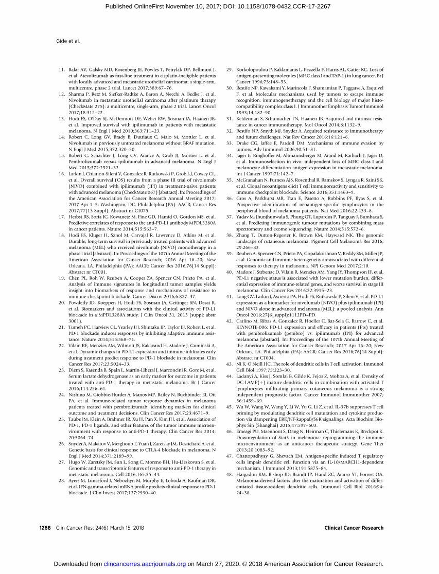
© 2017 American Association for Cancer Research
Figure 4.
Acquired resistance in metastaticmelanoma. Images of a 74-year-oldpatient with metastatic melanomatreatedwith anti–PD-1monotherapy,showing complete resolution ofmelanoma in right-neck lymphnodes from baseline (A), week 4 (B),and week 12 (C). However, acquiredresistance developed in the rightpreauricular region at 12 monthsafter the commencement of anti–PD-1 monotherapy (D). The patientsubsequently received radiotherapyand ipilimumab in addition tocontinuing anti–PD-1, with anexcellent ongoing response 12months after acquired resistance.
Gide et al.
Clin Cancer Res; 24(6) March 15, 2018 Clinical Cancer Research1266
on March 27, 2020. © 2018 American Association for Cancer Research. clincancerres.aacrjournals.org Downloaded from
Published OnlineFirst November 10, 2017; DOI: 10.1158/1078-0432.CCR-17-2267
CD8þ T cells to produce IFN-g , thereby restoring their function(110). Consistent with this, anti–TIM-3 and anti–PD-1/anti–CTLA-4 combined immunotherapy resulted in a significantdecrease in the tumor sizes of multiple cancers (111).
Phase I/II clinical trials testing the combination of an IDOinhibitor with PD-1/PD-L1 inhibitors in metastatic melanomaare also currently underway. These include the administration ofnivolumab and a PD-L1/IDO peptide vaccine (NCT03047928),and indoximod (IDO inhibitor) in combination with CTLA-4 orPD-1 inhibitors (NCT02073123). Furthermore, the combinationof epacadostat (an IDO1 inhibitor) plus pembrolizumab wasgenerally well tolerated and correlated with improved response invarious cancers (112–114), leading to the initiation of phase IIIstudies such as that in patients with treatment-na€�ve advancedmelanoma (NCT02752074).
Many studies have also demonstrated the increased efficacy ofcombined radiotherapy and immune checkpoint blockade.Radiotherapy activates the inflammatory pathways and inducesDNA damage, resulting in the release of antigens from irradiatedcells and an increase in tumor sensitization to T-cell immuneresponses (115). Radiotherapy in patients with advanced mela-noma who had progressed following treatment with ipilimumabled to an abscopal effect in some patients, whereby irradiation ofthe primary tumor induced regression in other nonirradiatedmetastases, which correlated with longer OS (116). Significanttumor regression was also observed in patients with metastaticmelanomawho received combined radiotherapy and anti–CTLA-4 immunotherapy, and experiments in mice showed that acombination of anti–CTLA-4 immunotherapy, anti–PD-L1immunotherapy, and radiotherapy was required to achieve opti-mum response (117).
The combination of epigeneticmodulators and immune check-point blockade has recently been shown to bemore effective thansingle-agent immunotherapy in patients with melanoma. Theinhibition of HDAC using LBH589 in combination with PD-1blockade inB16F10mousemelanomamodels resulted indelayedtumor growth and increased survival compared with the controlgroup and PD-1 blockade alone (118). Similarly, a reduction intumor size and Tregs was observed in B16F10 mouse melanomamodels treated with a combination of anti–CTLA-4 immunother-apy and epigenetic modulation of trimethylation of lysine 27 onhistone H3 (H3K27me3) compared with CTLA-4 blockade alone(119). The efficacy of combined epigenetic and immune check-
point therapies is currently being tested in various human cancers(Supplementary Table S1).
These findings provide a strong rationale for the developmentof combinations of different treatment strategies in clinical trialsas an effective means of melanoma treatment.
ConclusionsCheckpoint inhibitor immunotherapy has revolutionized the
treatment of multiple cancer types. However, responses inpatients with metastatic melanoma are diverse, with manypatients displaying primary or acquired resistance. An importantongoing,major unmet clinical need remains to identify predictorsand causes of this resistance and strategies to overcome them. Akey element of effective immunotherapy is identifying the variousmechanisms by which the tumors evade the immune system onan individual basis. Improving our understanding of thesemechanisms and determining which immune markers to targetin each patient will then allow for the administration of the mostappropriate form of therapy to achieve optimal response andimprove the overall outcomes of patients with metastaticmelanoma.
Disclosure of Potential Conflicts of InterestG.V. Long reports receiving speakers bureau honoraria from Bristol-Myers
Squibb, Incyte, Merck, Novartis, and Roche, and is a consultant/advisory boardmember for Amgen, Array, Bristol-Myers Squibb, Incyte, Merck, Novartis, PierreFabre, and Roche. No potential conflicts of interest were disclosed by the otherauthors.
AcknowledgmentsThis work was supported by Melanoma Institute Australia, the New South
Wales Ministry of Health, NSW Health Pathology, National Health andMedical Research Council of Australia (NHMRC), and Cancer Institute NSW.J.S. Wilmott, R.A. Scolyer, and G.V. Long are supported by NHMRC Fellow-ships. J.S. Wilmott is also supported by a CINSW Fellowship. G.V. Long isalso supported by the University of Sydney Medical Foundation. T.N. Gide issupported by The University of Sydney and Melanoma Institute AustraliaScholarships.
The costs of publication of this article were defrayed in part by the paymentof page charges. This article must therefore be hereby marked advertisement inaccordance with 18 U.S.C. Section 1734 solely to indicate this fact.
Received August 12, 2017; revised October 15, 2017; accepted November 6,2017; published OnlineFirst November 10, 2017.
References1. AtkinsonV, Ascierto PA, LongGV, BradyB,DutriauxC,MaioM, et al. Two-
year survival and safety update in patients (pts) with treatment-na€�veadvanced melanoma (MEL) receiving nivolumab (NIVO) or dacarbazine(DTIC) in CheckMate 066. 2015 Nov 18–21; San Francisco, CA, USA.
2. Larkin J, Chiarion-Sileni V, Gonzalez R, Grob JJ, Cowey CL, Lao CD, et al.Combined nivolumab and ipilimumab or monotherapy in untreatedmelanoma. N Engl J Med 2015;373:23–34.
3. RobertC, RibasA,HamidO,DaudA,Wolchok JD, JoshuaAM, et al. Three-year overall survival for patients with advanced melanoma treated withpembrolizumab in KEYNOTE-001. J Clin Oncol 34, 2016 (suppl; abstr9503).
4. Robert C, Thomas L, Bondarenko I, O'Day S, Weber J, Garbe C, et al.Ipilimumab plus dacarbazine for previously untreated metastatic mela-noma. N Engl J Med 2011;364:2517–26.
5. Schachter J, Ribas A, Long GV, Arance A, Grob JJ, Mortier L, et al. Pembro-lizumab versus ipilimumab for advanced melanoma: final overall survivalanalysis of KEYNOTE-006. J Clin Oncol 34, 2016 (suppl; abstr 9504).
6. Brahmer J, Reckamp KL, Baas P, Crino L, Eberhardt WE, Poddubskaya E,et al. Nivolumab versus docetaxel in advanced squamous-cell non-small-cell lung cancer. N Engl J Med 2015;373:123–35.
7. Herbst RS, Baas P, Kim DW, Felip E, Perez-Gracia JL, Han JY, et al.Pembrolizumab versus docetaxel for previously treated, PD-L1-positive,advanced non-small-cell lung cancer (KEYNOTE-010): a randomisedcontrolled trial. Lancet 2016;387:1540–50.
8. Motzer RJ, Escudier B, McDermott DF, George S, Hammers HJ, Srinivas S,et al. Nivolumab versus everolimus in advanced renal-cell carcinoma.N Engl J Med 2015;373:1803–13.
9. Ferris RL, Blumenschein G Jr, Fayette J, Guigay J, Colevas AD, Licitra L,et al. Nivolumab for recurrent squamous-cell carcinoma of the head andneck. N Engl J Med 2016;375:1856–67.
10. Kaufman HL, Russell J, Hamid O, Bhatia S, Terheyden P, D'Angelo SP,et al. Avelumab in patients with chemotherapy-refractory metastaticMerkel cell carcinoma: a multicentre, single-group, open-label, phase 2trial. Lancet Oncol 2016;17:1374–85.
Resistance to Immunotherapy in Melanoma
www.aacrjournals.org Clin Cancer Res; 24(6) March 15, 2018 1267
on March 27, 2020. © 2018 American Association for Cancer Research. clincancerres.aacrjournals.org Downloaded from
Published OnlineFirst November 10, 2017; DOI: 10.1158/1078-0432.CCR-17-2267
11. Balar AV, Galsky MD, Rosenberg JE, Powles T, Petrylak DP, Bellmunt J,et al. Atezolizumab as first-line treatment in cisplatin-ineligible patientswith locally advanced and metastatic urothelial carcinoma: a single-arm,multicentre, phase 2 trial. Lancet 2017;389:67–76.
12. Sharma P, Retz M, Siefker-Radtke A, Baron A, Necchi A, Bedke J, et al.Nivolumab in metastatic urothelial carcinoma after platinum therapy(CheckMate 275): a multicentre, single-arm, phase 2 trial. Lancet Oncol2017;18:312–22.
13. Hodi FS, O'Day SJ, McDermott DF, Weber RW, Sosman JA, Haanen JB,et al. Improved survival with ipilimumab in patients with metastaticmelanoma. N Engl J Med 2010;363:711–23.
14. Robert C, Long GV, Brady B, Dutriaux C, Maio M, Mortier L, et al.Nivolumab in previously untreated melanoma without BRAF mutation.N Engl J Med 2015;372:320–30.
15. Robert C, Schachter J, Long GV, Arance A, Grob JJ, Mortier L, et al.Pembrolizumab versus ipilimumab in advanced melanoma. N Engl JMed 2015;372:2521–32.
16. Larkin J, Chiarion-Sileni V, Gonzalez R, Rutkowski P, Grob J-J, Cowey CL,et al. Overall survival (OS) results from a phase III trial of nivolumab(NIVO) combined with ipilimumab (IPI) in treatment-na€�ve patientswith advancedmelanoma (CheckMate 067) [abstract]. In: Proceedings ofthe American Association for Cancer Research Annual Meeting 2017;2017 Apr 1–5; Washington, DC. Philadelphia (PA): AACR; Cancer Res2017;77(13 Suppl): Abstract nr CT075.
17. Herbst RS, Soria JC, Kowanetz M, Fine GD, Hamid O, Gordon MS, et al.Predictive correlates of response to the anti-PD-L1 antibodyMPDL3280Ain cancer patients. Nature 2014;515:563–7.
18. Hodi FS, Kluger H, Sznol M, Carvajal R, Lawrence D, Atkins M, et al.Durable, long-term survival in previously treated patients with advancedmelanoma (MEL) who received nivolumab (NIVO) monotherapy in aphase I trial [abstract]. In: Proceedings of the 107th AnnualMeeting of theAmerican Association for Cancer Research; 2016 Apr 16–20; NewOrleans, LA. Philadelphia (PA): AACR; Cancer Res 2016;76(14 Suppl):Abstract nr CT001.
19. Chen PL, Roh W, Reuben A, Cooper ZA, Spencer CN, Prieto PA, et al.Analysis of immune signatures in longitudinal tumor samples yieldsinsight into biomarkers of response and mechanisms of resistance toimmune checkpoint blockade. Cancer Discov 2016;6:827–37.
20. Powderly JD, Koeppen H, Hodi FS, Sosman JA, Gettinger SN, Desai R,et al. Biomarkers and associations with the clinical activity of PD-L1blockade in a MPDL3280A study. J Clin Oncol 31, 2013 (suppl; abstr3001).
21. Tumeh PC, Harview CL, Yearley JH, Shintaku IP, Taylor EJ, Robert L, et al.PD-1 blockade induces responses by inhibiting adaptive immune resis-tance. Nature 2014;515:568–71.
22. Vilain RE, Menzies AM, Wilmott JS, Kakavand H, Madore J, Guminski A,et al. Dynamic changes in PD-L1 expression and immune infiltrates earlyduring treatment predict response to PD-1 blockade in melanoma. ClinCancer Res 2017;23:5024–33.
23. Diem S, Kasenda B, Spain L, Martin-Liberal J, Marconcini R, Gore M, et al.Serum lactate dehydrogenase as an early marker for outcome in patientstreated with anti-PD-1 therapy in metastatic melanoma. Br J Cancer2016;114:256–61.
24. Nishino M, Giobbie-Hurder A, Manos MP, Bailey N, Buchbinder EI, OttPA, et al. Immune-related tumor response dynamics in melanomapatients treated with pembrolizumab: identifying markers for clinicaloutcome and treatment decisions. Clin Cancer Res 2017;23:4671–9.
25. Taube JM, Klein A, Brahmer JR, Xu H, Pan X, Kim JH, et al. Association ofPD-1, PD-1 ligands, and other features of the tumor immune microen-vironment with response to anti-PD-1 therapy. Clin Cancer Res 2014;20:5064–74.
26. Snyder A,MakarovV,MerghoubT, Yuan J, Zaretsky JM,Desrichard A, et al.Genetic basis for clinical response to CTLA-4 blockade in melanoma. NEngl J Med 2014;371:2189–99.
27. Hugo W, Zaretsky JM, Sun L, Song C, Moreno BH, Hu-Lieskovan S, et al.Genomic and transcriptomic features of response to anti-PD-1 therapy inmetastatic melanoma. Cell 2016;165:35–44.
28. Ayers M, Lunceford J, Nebozhyn M, Murphy E, Loboda A, Kaufman DR,et al. IFN-gamma-relatedmRNA profile predicts clinical response to PD-1blockade. J Clin Invest 2017;127:2930–40.
29. Korkolopoulou P, Kaklamanis L, Pezzella F, Harris AL, Gatter KC. Loss ofantigen-presentingmolecules (MHCclass I and TAP-1) in lung cancer. Br JCancer 1996;73:148–53.
30. RestifoNP, Kawakami Y,Marincola F, Shamamian P, Taggarse A, EsquivelF, et al. Molecular mechanisms used by tumors to escape immunerecognition: immunogenetherapy and the cell biology of major histo-compatibility complex class I. J Immunother Emphasis Tumor Immunol1993;14:182–90.
31. Kelderman S, Schumacher TN, Haanen JB. Acquired and intrinsic resis-tance in cancer immunotherapy. Mol Oncol 2014;8:1132–9.
32. Restifo NP, Smyth MJ, Snyder A. Acquired resistance to immunotherapyand future challenges. Nat Rev Cancer 2016;16:121–6.
33. Drake CG, Jaffee E, Pardoll DM. Mechanisms of immune evasion bytumors. Adv Immunol 2006;90:51–81.
34. Jager E, Ringhoffer M, Altmannsberger M, Arand M, Karbach J, Jager D,et al. Immunoselection in vivo: independent loss of MHC class I andmelanocyte differentiation antigen expression in metastatic melanoma.Int J Cancer 1997;71:142–7.
35. McGranahanN, Furness AJS, Rosenthal R, Ramskov S, Lyngaa R, Saini SK,et al. Clonal neoantigens elicit T cell immunoreactivity and sensitivity toimmune checkpoint blockade. Science 2016;351:1463–9.
36. Gros A, Parkhurst MR, Tran E, Pasetto A, Robbins PF, Ilyas S, et al.Prospective identification of neoantigen-specific lymphocytes in theperipheral blood of melanoma patients. Nat Med 2016;22:433–8.
37. YadavM, Jhunjhunwala S, Phung QT, Lupardus P, Tanguay J, Bumbaca S,et al. Predicting immunogenic tumour mutations by combining massspectrometry and exome sequencing. Nature 2014;515:572–6.
38. Zhang T, Dutton-Regester K, Brown KM, Hayward NK. The genomiclandscape of cutaneous melanoma. Pigment Cell Melanoma Res 2016;29:266–83.
39. Reuben A, Spencer CN, Prieto PA,Gopalakrishnan V, Reddy SM,Miller JP,et al. Genomic and immune heterogeneity are associated with differentialresponses to therapy in melanoma. NPJ Genom Med 2017;2:10.
40. Madore J, Strbenac D, Vilain R, Menzies AM, Yang JY, Thompson JF, et al.PD-L1 negative status is associated with lower mutation burden, differ-ential expression of immune-related genes, and worse survival in stage IIImelanoma. Clin Cancer Res 2016;22:3915–23.
41. LongGV, Larkin J, Ascierto PA,Hodi FS, Rutkowski P, Sileni V, et al. PD-L1expression as a biomarker for nivolumab (NIVO) plus ipilimumab (IPI)and NIVO alone in advanced melanoma (MEL): a pooled analysis. AnnOncol 2016;27(6_suppl):1112PD–PD.
42. Carlino M, Ribas A, Gonzalez R, Hoeller C, Bar-Sela G, Barrow C, et al.KEYNOTE-006: PD-L1 expression and efficacy in patients (Pts) treatedwith pembrolizumab (pembro) vs. ipilimumab (IPI) for advancedmelanoma [abstract]. In: Proceedings of the 107th Annual Meeting ofthe American Association for Cancer Research; 2017 Apr 16–20; NewOrleans, LA. Philadelphia (PA): AACR; Cancer Res 2016;76(14 Suppl):Abstract nr CT004.
43. Ni K, O'Neill HC. The role of dendritic cells in T cell activation. ImmunolCell Biol 1997;75:223–30.
44. Ladanyi A, Kiss J, Somlai B, Gilde K, Fejos Z, Mohos A, et al. Density ofDC-LAMP(þ) mature dendritic cells in combination with activated Tlymphocytes infiltrating primary cutaneous melanoma is a strongindependent prognostic factor. Cancer Immunol Immunother 2007;56:1459–69.
45. Wu W, Wang W, Wang Y, Li W, Yu G, Li Z, et al. IL-37b suppresses T cellpriming by modulating dendritic cell maturation and cytokine produc-tion via dampening ERK/NF-kappaB/S6K signalings. Acta Biochim Bio-phys Sin (Shanghai) 2015;47:597–603.
46. Emeagi PU, Maenhout S, Dang N, Heirman C, Thielemans K, Breckpot K.Downregulation of Stat3 in melanoma: reprogramming the immunemicroenvironment as an anticancer therapeutic strategy. Gene Ther2013;20:1085–92.
47. Chattopadhyay G, Shevach EM. Antigen-specific induced T regulatorycells impair dendritic cell function via an IL-10/MARCH1-dependentmechanism. J Immunol 2013;191:5875–84.
48. Hargadon KM, Bishop JD, Brandt JP, Hand ZC, Ararso YT, Forrest OA.Melanoma-derived factors alter the maturation and activation of differ-entiated tissue-resident dendritic cells. Immunol Cell Biol 2016;94:24–38.
Gide et al.
Clin Cancer Res; 24(6) March 15, 2018 Clinical Cancer Research1268
on March 27, 2020. © 2018 American Association for Cancer Research. clincancerres.aacrjournals.org Downloaded from
Published OnlineFirst November 10, 2017; DOI: 10.1158/1078-0432.CCR-17-2267

49. Lindenberg JJ, van de Ven R, Lougheed SM, Zomer A, Santegoets SJAM,Griffioen AW, et al. Functional characterization of a STAT3-dependentdendritic cell-derived CD14þ cell population arising upon IL-10-drivenmaturation. OncoImmunology 2013;2:e23837.
50. Hong M, Puaux AL, Huang C, Loumagne L, Tow C, Mackay C, et al.Chemotherapy induces intratumoral expression of chemokines in cuta-neous melanoma, favoring T-cell infiltration and tumor control. CancerRes 2011;71:6997–7009.
51. Harlin H, Meng Y, Peterson AC, Zha Y, Tretiakova M, Slingluff C, et al.Chemokine expression inmelanomametastases associatedwithCD8þ T-cell recruitment. Cancer Res 2009;69:3077–85.
52. Fu H, Kishore M, Gittens B, Wang G, Coe D, Komarowska I, et al. Self-recognition of the endothelium enables regulatory T-cell trafficking anddefines the kinetics of immune regulation. Nat Commun 2014;5:3436.
53. Kryczek I, Bruce AT, Gudjonsson JE, Johnston A, Aphale A, Vatan L, et al.Induction of IL-17(þ) T cell trafficking and development by IFN-g :mechanism and pathological relevance in psoriasis. J Immunol 2008;181:4733–41.
54. Yue C, Shen S, Deng J, Priceman SJ, LiW,Huang A, et al. STAT3 in CD8þ Tcells inhibits their tumor accumulation by downregulating CXCR3/CXCL10 axis. Cancer Immunol Res 2015;3:864–70.
55. Maio M, Covre A, Fratta E, Di Giacomo AM, Taverna P, Natali PG, et al.Molecular pathways: at the crossroads of cancer epigenetics and immu-notherapy. Clin Cancer Res 2015;21:4040–7.
56. Peng D, Kryczek I, Nagarsheth N, Zhao L, Wei S, WangW, et al. Epigeneticsilencing of Th1 type chemokines shapes tumor immunity and immu-notherapy. Nature 2015;527:249–53.
57. Zheng H, Zhao W, Yan C, Watson CC, Massengill M, Xie M, et al. HDACinhibitors enhance T-cell chemokine expression and augment response toPD-1 immunotherapy in lung adenocarcinoma. Clin Cancer Res2016;22:4119–32.
58. Buckanovich RJ, Facciabene A, Kim S, Benencia F, Sasaroli D, Balint K,et al. Endothelin B receptor mediates the endothelial barrier to T cellhoming to tumors and disables immune therapy. Nat Med 2008;14:28–36.
59. Nelson J, Bagnato A, Battistini B, Nisen P. The endothelin axis: emergingrole in cancer. Nat Rev Cancer 2003;3:110–6.
60. Lalich M, McNeel DG, Wilding G, Liu G. Endothelin receptor antagonistsin cancer therapy. Cancer Invest 2007;25:785–94.
61. Demunter A, De Wolf-Peeters C, Degreef H, Stas M, van den Oord JJ.Expression of the endothelin-B receptor in pigment cell lesions of the skin.Evidence for its role as tumor progression marker in malignant melano-ma. Virchows Arch 2001;438:485–91.
62. Lahav R, Suv�a M-L, Rimoldi D, Patterson PH, Stamenkovic I. Endothelinreceptor B inhibition triggers apoptosis and enhances angiogenesis inmelanomas. Cancer Res 2004;64:8945–53.
63. Redondo P, Bandres E, Solano T,Okroujnov I, Garcia-Foncillas J. Vascularendothelial growth factor (VEGF) and melanoma. N-acetylcysteinedownregulates VEGF production in vitro. Cytokine 2000;12:374–8.
64. Rajabi P, Neshat A, Mokhtari M, Rajabi MA, Eftekhari M, Tavakoli P. Therole of VEGF in melanoma progression. J Res Med Sci 2012;17:534–9.
65. Bouzin C, Brouet A, De Vriese J, DeWever J, Feron O. Effects of vascularendothelial growth factor on the lymphocyte-endothelium interactions:identification of caveolin-1 and nitric oxide as control points of endo-thelial cell anergy. J Immunol 2007;178:1505–11.
66. Zhang L, Conejo-Garcia JR, Katsaros D, Gimotty PA, Massobrio M,Regnani G, et al. Intratumoral T cells, recurrence, and survival in epithelialovarian cancer. N Engl J Med 2003;348:203–13.
67. Huang H, Langenkamp E, Georganaki M, Loskog A, Fuchs PF, DieterichLC, et al. VEGF suppresses T-lymphocyte infiltration in the tumor micro-environment through inhibition of NF-kappaB-induced endothelial acti-vation. FASEB J 2015;29:227–38.
68. Motz GT, Santoro SP, Wang LP, Garrabrant T, Lastra RR, Hagemann IS,et al. Tumor endothelium FasL establishes a selective immune barrierpromoting tolerance in tumors. Nat Med 2014;20:607–15.
69. Dong ZY, Wu SP, Liao RQ, Huang SM, Wu YL. Potential biomarker forcheckpoint blockade immunotherapy and treatment strategy. TumourBiol 2016;37:4251–61.
70. Pardoll DM. The blockade of immune checkpoints in cancer immuno-therapy. Nat Rev Cancer 2012;12:252–64.
71. Parsa AT,Waldron JS, Panner A, Crane CA, Parney IF, Barry JJ, et al. Loss oftumor suppressor PTEN function increases B7-H1 expression and immu-noresistance in glioma. Nat Med 2007;13:84–8.
72. Xu C, Fillmore CM, Koyama S, Wu H, Zhao Y, Chen Z, et al. Loss of Lkb1and Pten leads to lung squamous cell carcinoma with elevated PD-L1expression. Cancer Cell 2014;25:590–604.
73. PengW, Chen JQ, Liu C, Malu S, Creasy C, Tetzlaff MT, et al. Loss of PTENpromotes resistance to T cell-mediated immunotherapy. Cancer Discov2016;6:202–16.
74. Kr€oger A, K€osterM, Schroeder K,Hauser H,Mueller PP. Activities of IRF-1.J Interferon Cytokine Res 2002;22:5–14.
75. Lee SJ, Jang BC, Lee SW, Yang YI, Suh SI, Park YM, et al. Interferonregulatory factor-1 is prerequisite to the constitutive expression and IFN-gamma-induced upregulation of B7-H1 (CD274). FEBS Lett 2006;580:755–62.
76. Akbay EA, Koyama S, Carretero J, Altabef A, Tchaicha JH, Christensen CL,et al. Activation of the PD-1 pathway contributes to immune escape inEGFR-driven lung tumors. Cancer Discov 2013;3:1355–63.
77. ChenN, FangW,Zhan J,Hong S, Tang Y, KangS, et al. Upregulationof PD-L1 by EGFR activation mediates the immune escape in EGFR-drivenNSCLC: implication for optional immune targeted therapy for NSCLCpatients with EGFR mutation. J Thorac Oncol 2015;10:910–23.
78. Holmgaard RB, Zamarin D, Munn DH, Wolchok JD, Allison JP. Indo-leamine 2,3-dioxygenase is a critical resistancemechanism in antitumor Tcell immunotherapy targeting CTLA-4. J Exp Med 2013;210:1389–402.
79. Uyttenhove C, Pilotte L, Theate I, Stroobant V, Colau D, Parmentier N,et al. Evidence for a tumoral immune resistance mechanism based ontryptophan degradation by indoleamine 2,3-dioxygenase. Nat Med2003;9:1269–74.
80. Munn DH, Shafizadeh E, Attwood JT, Bondarev I, Pashine A, Mellor AL.Inhibition of T cell proliferation by macrophage tryptophan catabolism.J Exp Med 1999;189:1363–72.
81. Holmgaard RB, Zamarin D, Li Y, Gasmi B, Munn DH, Allison JP, et al.Tumor-expressed IDO recruits and activates MDSCs in a Treg-dependentmanner. Cell Rep 2015;13:412–24.
82. Gajewski TF. Identifying andovercoming immune resistancemechanismsin the melanoma tumor microenvironment. Clin Cancer Res 2006;12:2326s–30s.
83. Jandus C, Bioley G, Speiser DE, Romero P. Selective accumulation ofdifferentiated FOXP3(þ) CD4 (þ) T cells in metastatic tumor lesionsfrom melanoma patients compared to peripheral blood. Cancer Immu-nol Immunother 2008;57:1795–805.
84. Viguier M, Lemaitre F, Verola O, Cho MS, Gorochov G, Dubertret L, et al.Foxp3 expressing CD4þCD25high regulatory T cells are overrepresentedin humanmetastatic melanoma lymph nodes and inhibit the function ofinfiltrating T cells. J Immunol 2004;173:1444–53.
85. Strauss L, BergmannC, SzczepanskiM,GoodingW, Johnson JT,WhitesideTL. A unique subset of CD4þCD25highFoxp3þ T cells secreting inter-leukin-10 and transforming growth factor-beta1 mediates suppression inthe tumor microenvironment. Clin Cancer Res 2007;13:4345–54.
86. Wang HY, Lee DA, Peng G, Guo Z, Li Y, Kiniwa Y, et al. Tumor-specifichuman CD4þ regulatory T cells and their ligands. Immunity 2004;20:107–18.
87. HarlinH, Kuna TV, Peterson AC,Meng Y,Gajewski TF. Tumor progressiondespite massive influx of activated CD8þ T cells in a patient withmalignant melanoma ascites. Cancer Immunol Immunother 2006;55:1185–97.
88. Wei SC, Levine JH, Cogdill AP, Zhao Y, Anang NAS, Andrews MC, et al.Distinct cellular mechanisms underlie anti-CTLA-4 and anti-PD-1 check-point blockade. Cell 2017;170:1120–33.e17.
89. Simeone E, Gentilcore G, Giannarelli D, Grimaldi AM, Carac�o C, Cur-viettoM, et al. Immunological and biological changes during ipilimumabtreatment and their potential correlation with clinical response andsurvival in patients with advanced melanoma. Cancer Immunol Immun-other 2014;63:675–83.
90. Umansky V, Shevchenko I, Bazhin AV, Utikal J. Extracellular adenosinemetabolism in immune cells in melanoma. Cancer Immunol Immun-other 2014;63:1073–80.
91. Young A, Ngiow SF, Madore J, Reinhardt J, Landsberg J, Chitsazan A, et al.Targeting adenosine in BRAF-mutant melanoma reduces tumor growthand metastasis. Cancer Res 2017;77:4684–96.
Resistance to Immunotherapy in Melanoma
www.aacrjournals.org Clin Cancer Res; 24(6) March 15, 2018 1269
on March 27, 2020. © 2018 American Association for Cancer Research. clincancerres.aacrjournals.org Downloaded from
Published OnlineFirst November 10, 2017; DOI: 10.1158/1078-0432.CCR-17-2267

92. Reinhardt J, Landsberg J, Schmid-Burgk JL, Ramis BB, Bald T, Glodde N,et al. MAPK signaling and inflammation link melanoma phenotypeswitching to induction of CD73 during immunotherapy. Cancer Res2017;77:4697–709.
93. Shin DS, Zaretsky JM, Escuin-Ordinas H, Garcia-Diaz A, Hu-Lieskovan S,Kalbasi A, et al. Primary resistance to PD-1 blockade mediated by JAK1/2mutations. Cancer Discov 2017;7:188–201.
94. Patel SJ, Sanjana NE, Kishton RJ, Eidizadeh A, Vodnala SK, Cam M, et al.Identification of essential genes for cancer immunotherapy. Nature2017;548:537–42.
95. Bhatt AP, Redinbo MR, Bultman SJ. The role of the microbiome in cancerdevelopment and therapy. CA Cancer J Clin 2017;67:326–44.
96. Wargo JA, Gopalakrishnan V, Spencer C, Karpinets T, Reuben A, AndrewsMC, et al. Association of the diversity and composition of the gutmicrobiome with responses and survival (PFS) in metastatic melanoma(MM) patients (pts) on anti-PD-1 therapy. J Clin Oncol 35, 2017 (suppl;abstr 3008).
97. Chaput N, Lepage P, Coutzac C, Soularue E, Le Roux K, Monot C, et al.Baseline gut microbiota predicts clinical response and colitis in metastaticmelanomapatients treatedwith ipilimumab. AnnOncol 2017;28:1368–79.
98. Vetizou M, Pitt JM, Daillere R, Lepage P, Waldschmitt N, Flament C, et al.Anticancer immunotherapy by CTLA-4 blockade relies on the gut micro-biota. Science 2015;350:1079–84.
99. Restifo NP, Marincola FM, Kawakami Y, Taubenberger J, Yannelli JR,Rosenberg SA. Loss of functional beta 2-microglobulin in metastaticmelanomas from five patients receiving immunotherapy. J Natl CancerInst 1996;88:100–8.
100. del Campo AB, Kyte JA, Carretero J, Zinchencko S, Mendez R, Gonzalez-Aseguinolaza G, et al. Immune escape of cancer cells with beta2-micro-globulin loss over the course of metastatic melanoma. Int J Cancer2014;134:102–13.
101. Zaretsky JM, Garcia-Diaz A, Shin DS, Escuin-Ordinas H, Hugo W, Hu-Lieskovan S, et al. Mutations associated with acquired resistance to PD-1blockade in melanoma. N Engl J Med 2016;375:819–29.
102. Dutta P, Li WX. Role of the JAK-STAT signalling pathway in cancer.eLS2013. DOI: 10.1002/9780470015902.a0025214.
103. Sharma P, Hu-Lieskovan S, Wargo JA, Ribas A. Primary, adaptive, andacquired resistance to cancer immunotherapy. Cell 2017;168:707–23.
104. Mellman I, Coukos G, Dranoff G. Cancer immunotherapy comes of age.Nature 2011;480:480–9.
105. Koyama S, Akbay EA, Li YY, Herter-Sprie GS, Buczkowski KA, RichardsWG, et al. Adaptive resistance to therapeutic PD-1 blockade is associatedwith upregulation of alternative immune checkpoints. Nat Commun2016;7:10501.
106. Taube JM, Young GD, McMiller TL, Chen S, Salas JT, Pritchard TS, et al.Differential expression of immune-regulatory genes associated with PD-L1 display in melanoma: implications for PD-1 pathway blockade. ClinCancer Res 2015;21:3969–76.
107. Woo SR, Turnis ME, Goldberg MV, Bankoti J, Selby M, Nirschl CJ, et al.Immune inhibitory molecules LAG-3 and PD-1 synergistically regulate T-cell function to promote tumoral immune escape. Cancer Res 2012;72:917–27.
108. Foy SP, Sennino B, delaCruz T, Cote JJ, GordonEJ, KempF, et al. Poxvirus-based active immunotherapy with PD-1 and LAG-3 dual immune check-point inhibition overcomes compensatory immune regulation, yieldingcomplete tumor regression in mice. PLoS One 2016;11:e0150084.
109. Ascierto PA,Melero I, Bhatia S, BonoP, SanbornRE, Lipson EJ, et al. Initialefficacy of anti-lymphocyte activation gene-3 (anti–LAG-3; BMS-986016)in combination with nivolumab (nivo) in pts with melanoma (MEL)previously treated with anti–PD-1/PD-L1 therapy. J Clin Oncol 35, 2017(suppl; abstr 9520).
110. Sakuishi K, Apetoh L, Sullivan JM, Blazar BR, Kuchroo VK, Anderson AC.Targeting Tim-3 and PD-1 pathways to reverse T cell exhaustion andrestore anti-tumor immunity. J Exp Med 2010;207:2187–94.
111. Ngiow SF, von Scheidt B, Akiba H, Yagita H, Teng MW, Smyth MJ. Anti-TIM3 antibody promotes T cell IFN-gamma-mediated antitumor immu-nity and suppresses established tumors. Cancer Res 2011;71:3540–51.
112. Gangadhar TC, Hamid O, Smith DC, Bauer TM, Wasser JS, Olszanski AJ,et al. Epacadostat plus pembrolizumab in patients with advanced mel-anoma and select solid tumors: updated phase 1 results fromECHO-202/KEYNOTE-037. Ann Oncol 2016;27(6_suppl):1110PD–PD.
113. Gangadhar TC, Schneider BJ, Bauer TM,Wasser JS, Spira AI, Patel SP, et al.Efficacy and safety of epacadostat plus pembrolizumab treatment ofNSCLC: preliminary phase I/II results of ECHO-202/KEYNOTE-037.J Clin Oncol 35, 2017 (suppl; abstr 9014).
114. Smith DC, Gajewski T, Hamid O, Wasser JS, Olszanski AJ, Patel SP, et al.Epacadostat plus pembrolizumab in patients with advanced urothelialcarcinoma: preliminary phase I/II results of ECHO-202/KEYNOTE-037.J Clin Oncol 35, 2017 (suppl; abstr 4503).
115. SpiottoM, FuYX,WeichselbaumRR. The intersectionof radiotherapy andimmunotherapy: mechanisms and clinical implications. Sci Immunol2016;1:eaag1266.
116. Grimaldi AM, Simeone E, Giannarelli D, Muto P, Falivene S, Borzillo V,et al. Abscopal effects of radiotherapy on advanced melanoma patientswho progressed after ipilimumab immunotherapy. Oncoimmunology2014;3:e28780.
117. Twyman-Saint Victor C, Rech AJ, Maity A, Rengan R, Pauken KE, StelekatiE, et al. Radiation and dual checkpoint blockade activate non-redundantimmune mechanisms in cancer. Nature 2015;520:373–7.
118. Woods DM, Sodre AL, Villagra A, Sarnaik A, Sotomayor EM, Weber J.HDAC inhibition upregulates PD-1 ligands in melanoma and augmentsimmunotherapy with PD-1 blockade. Cancer Immunol Res 2015;3:1375–85.
119. Goswami S, Zhao H, Zhang X, Sharma P. Epigenetic changes in T cellsin response to immune checkpoint blockade. J Clin Oncol 34, 2016(suppl; abstr 11549).
Clin Cancer Res; 24(6) March 15, 2018 Clinical Cancer Research1270
Gide et al.
on March 27, 2020. © 2018 American Association for Cancer Research. clincancerres.aacrjournals.org Downloaded from
Published OnlineFirst November 10, 2017; DOI: 10.1158/1078-0432.CCR-17-2267

2018;24:1260-1270. Published OnlineFirst November 10, 2017.Clin Cancer Res Tuba N. Gide, James S. Wilmott, Richard A. Scolyer, et al. in Metastatic MelanomaPrimary and Acquired Resistance to Immune Checkpoint Inhibitors
Updated version
10.1158/1078-0432.CCR-17-2267doi:
Access the most recent version of this article at:
Material
Supplementary
http://clincancerres.aacrjournals.org/content/suppl/2017/11/10/1078-0432.CCR-17-2267.DC1
Access the most recent supplemental material at:
Cited articles
http://clincancerres.aacrjournals.org/content/24/6/1260.full#ref-list-1
This article cites 107 articles, 32 of which you can access for free at:
Citing articles
http://clincancerres.aacrjournals.org/content/24/6/1260.full#related-urls
This article has been cited by 10 HighWire-hosted articles. Access the articles at:
E-mail alerts related to this article or journal.Sign up to receive free email-alerts
Subscriptions
Reprints and
To order reprints of this article or to subscribe to the journal, contact the AACR Publications Department at
Permissions
Rightslink site. Click on "Request Permissions" which will take you to the Copyright Clearance Center's (CCC)
.http://clincancerres.aacrjournals.org/content/24/6/1260To request permission to re-use all or part of this article, use this link
on March 27, 2020. © 2018 American Association for Cancer Research. clincancerres.aacrjournals.org Downloaded from
Published OnlineFirst November 10, 2017; DOI: 10.1158/1078-0432.CCR-17-2267