![[SurgeryB] Surgical Complications - Dr. Guinto (Pacis, Sazon)](https://static.fdocuments.net/doc/165x107/55cf921d550346f57b93bae3/surgeryb-surgical-complications-dr-guinto-pacis-sazon.jpg)
[SurgeryB] Surgical Complications - Dr. Guinto (Pacis, Sazon)

Prevention and Management of Surgical Complications
Larry J. Peterson
PREVENTION OF COMPLICATIONS SOFT TISSUE INJURIES
Tearing Mucosal Flap Puncture Wound of Soft Tissue Stretch or Abrasion Injury
COMPLICATIONS WITH THE TOOTH BEING EXTRACTED Root Fracture Root Displacement Tooth Lost into Oropharynx
INJURIES TO ADJACENT TEETH Fracture of Adjacent Restoration Luxation of Adjacent Teeth Extraction of Wrong Teeth
INJURIES TO OSSEOUS STRUCTURES Fracture of Alveolar Process Fracture of Maxillary Tuberosity
INJURIES TO ADJACENT STRUCTURES Injury t o Regional Nerves lnjury t o Temporomandibular Joint
OROANTRAL COMMUNICATIONS POSTOPERATIVE BLEEDING DELAYED HEALING AND INFECTION
Infection Wound Dehiscence Dry Socket
FRACTURES OF THE MANDIBLE SUMMARY
his chapter discusses the variety of complica- tions of oral surgical procedures. It is divided into two sections, intraoperative and postopera-
tive complications. These aye surgical, not medical, com- plications; the latter are discussed in Chapter 3.
It is axiomatic that the best and easiest way to manage a con~plication is to prevent it from happening. Prevention of surgical complications is best accomplished by a thor- ough preoperative assessment and comprehensive treat- ment plan. Only when these are routinely performed can the surgeon expect to have minimal complications. It is important to realize that even with such planning, com-
plications occasionally occur. In situations in which the dentist has planned carefully, the complication is often expected and can be managed in a routine manner. For example, when extracting a maxillary first premolar, which has long thin roots, it is far easier to remove the buccal root than the palatal root. Therefore the surgeon uses more force toward the buccal root than toward the palatal root. If a root does fracture, it i~ then the buccal root rather than the palatal root, and the subsequent retrieval is easier.
Surgeons must perform surgery that is within their own ability. Surgeons must therefore carefully evaluate their training and ability before deciding to perform a specific surgical task. It is inappropriate for a dentist with limited experience in the management of impacted third

222 PART I1 m Principles of Exoriontin
molars to undertake the surgical extraction of a deeply embedded tooth.
The incidence of operative and postoperative compli- cations is unacceptably high in this situation. Surgeons must be cautious of unwarranted optimism, which clouds their judgment and prevents them from deliver- ing the best possible care to the patient. The dentist must keep in mind that referral to a specialist is an option that should always be exercised if the planned surgery is beyond the dentist's own skill level. In some situations this is not only a moral obligation but also a medicole- gal responsibility.
In planning a surgical procedure, the first step is always a thorough review of the patient's medical histo- ry. Several of the complications to be discussed in this chapter are caused by inadequate attention to medical histories that would have revealed the presence of a com- plicating factor. Patients with compromised physical sta- tus will have local surgical complications that could have been prevented had the surgeon taken a more thorough medical history.
One of the primary ways to prevent complications is by taking adequate radiographs and reviewing them care- fully (see Chapter 7). The radiograph must include the entire area of surgery, including the apices of the roots of the teeth to be extracted and the local and regional anatomic structures, such as the maxillary sinus and the inferior alveolar canal. The surgeon must look for the presence of abnormal tooth root morphology. After care- ful examination of the radiographs, the surgeon must occasionally alter the treatment plan to prevent the com- plications that might be anticipated with a routine for- ceps (closed) extraction. Instead, the surgeon should con- sider surgical approaches to removing teeth in such cases.
After an adequate medical history has been taken and the radiographs have been analyzed, the surgeon must do the preoperative planning. This is not simply a prepara- tion of a detailed surgical plan but is also a plan for man- aging patient anxiety and pain and postoperative recov- ery (instructions and modifications of normal activity for the patient). Thorough preoperative instructions and explanations for the patient are essential in preventing the majority of complications that occur in the postoper- ative period. I f the instructions are not thoroughly explained or their importance made clear, the patient is less likely to follow them.
Finally, to keep complications at a minimum, the sur- geon must always follow the basic surgical principles. There should always be clear visualization and access to the operative field, which requires adequate light, ade- quate soft tissue reflection (including lips, cheeks, tongue, and soft tissue flaps), and adequate suction. The teeth to be removed must have an unimpeded pathway for removal. Occasionally, bone must be removed and teeth must be sectioned to achieve this goal. Controlled force is of paramount importance; this means "finesse," not "force." The surgeon must follow the principles of asepsis, atraumatic handling of tissues, hemostasis, and thorough debridement of the wound after the surgical procedure. Violation of these principles leads to an increased inci- dence and severity of surgical complications.
Prevention of Soft Tissue Injuries
1. Pay strict attention to soft tissue inju, ,,,. 2. Develop adequate-sized flaps. 3. Use minimal force for retraction of soft tissue.
SOFT TISSUE INjURIES
Injuries to the soft tissue of the oral cavity are almost always the result of the surgeon's lack of adequate atten- tion to the delicate nature of the mucosa and the use of excessive and uncontrolled force. The surgeon must con- tinue to pay careful attention to the soft tissue while work- ing primarily on the bone and tooth structure (Box 11-1).
Tearing Mucosal Flap
The most common soft tissue injury is the tearing of the mucosal flap during surgical extraction of a tooth. This is usually the result of an inadequately sized envelope flap, which is retracted beyond the tissue's ability to stretch (Fig. 11-1). This results in a tearing, usually at one end of the incision. Prevention of this complication is twofold: (1) create adequately sized flaps to prevent excess tension on the flap, and (2) use small amounts of retraction force on the flap. If a tear does occur in the flap, the flap should be carefully repositioned once the surgery is complete. In most patients, careful suturing of the tear results in ade- quate but delayed healing. If the tear is especially jagged, the surgeon may consider excising the edges of the torn flap to create a smooth flap margin for closure. This lat- ter step should be performed with caution, because exci- sion of excessive amounts of tissue leads to closure of the wound under tension and probable wound dehiscence.
If the area of surgery is near the apex of a tooth, an increased incidence of envelope-flap tearing exists as a result of excessive retractional forces. In this situation a release incision to create a three-cornered flap should be used to gain access to the bone.
Puncture Wound of Soft Tissue
The second soft tissue injury that occurs with some fre- quency is inadvertent puncturing of the soft tissue. Instruments, such as a straight elevator or periosteal ele- vator, may slip from the surgical field and puncture or tear into adjacent soft tissue.
Once again, this injury is the result of using uncon- trolled force instead of finesse and is best prevented by the use of controlled force, with special attention given to the supporting fingers or support from the opposite hand in anticipation of slippage. If the instrument slips from the tooth or bone, the fingers thus catch the hand before injury occurs (Fig. 11-2). When a puncture wound does occur, the treatment is aimed primarily at preventing infection and allowing healing to occur, usually by sec- ondary intention. If the wound bleeds excessively, it

PrcJifrrltioil ( 7 1 1 ~ 1 Marln~yeinerlt o f S~rr~picnl Cornplicatior1.s m CHAPTER 11 223
FIG. 11-1 Periosteal elevator (Seldin elevator) is used to reflect mucoperiosteal flap. Elevator placed perpendicular to bone and held in place by pushing firmly against bone, not by push- ing it apically against soft tissue (arrow).
should be controlled by direct pressure on the soft tissue. Once hemostasis is achieved, the wound is usually left open and not sutured, so that if a small infection were to occur, there would be an adequate pathway for drainage.
Stretch or Abrasion Injury
Abrasions or burns of the lips and corners of the mouth are usually the result of the rotating shank of the bur rub- bing on the soft tissue (Fig. 11-3). When the surgeon is focused on the cutting end of the bur, the assistant should be aware of the location of the shank of the bur in relation to the cheeks and lips. If such an abrasion does develop, the dentist should advise the patient to keep it covered with Vaseline or an antibiotic ointment. It is important that the patient keeps the ointment only on the abraded area and not spread onto intact skin, because it is quite likely to result in a rash. These abrasions usually take 5 to 10 days to heal. The patient should keep the area moist with the ointment during the entire healing period to pre- vent eschar formation, scarring, and delayed healing, as well as to keep the area reasonably comfortable.
COMPLICATIONS WITH THE TOOTH BEING EXTRACTED
Root Fracture FIG. 1 f -2 Small straight elevator can be used as shoehorn to lux- The most common complication associated with the ate broken root. When straight elevator is used in this position, hand tooth being extracted is fracture of its roots. Long, must be securely supported on adjacent teeth to prevent inadver- curved, divergent roots that lie in dense bone are most tent slippage of instrument from tooth and subsequent injury to likely to be fractured. The main method of preventing adjacent tissue.

A, Abras~on of Ilp as result of sliank of bur rotating on soft tlssue Wound should be kept covered wlth a n t ~ b ~ o t ~ c olntrnent B, Heallng should occur rap~dly, as observed In th~s photograph taken 5 davs later
P r e ~ c n t ~ o n ot Ilool ,rncl I) i \pl ,~cerncnt I r ac ture
1 Always plan for root fracture 2 Use surg~cal (I e , open) extract~on rf hlgh probablllty
of fracture 3 Do not use strong ap~cal force on broken root
fracture of roots is to perform ;In open e~trnc.tion tech- nicluc a n d t o remove bone t o dccre;rse tlic3 a m o u n t of force, nccei\;rr! to rcriio\.v t h c tooth r l<os 1 1-31. I<eco\'er!. of t h e fractuserl root ~ ~ i t l i a sur:;ical al>l,roacli i \ clijcu\\cd in C:haptcr 8 .
Root Displacement
'I'lic tooth root th;rt is niost conimoril!. disl>l;rceel into i rnfa~~or;r l~lc anatomic 5l1;rc.e~ is the mnsill;~r!. molar soot, \chic11 is forced illto the ~ i ~ ; ~ x i l l ; r r ~ ~ \inir\. I t n root ( ~ t n mas- illilr!. molar is Iwing I-v~iio\~ctl, \\.it11 ;I straight cle~.ator Iwing irwd \\.itti c ~ s c i ~ s nl>ic;il Ilr-c\sllrc, ;IS a \ \ ~ ~ l g c iri tllc' pcric>dont;~l lig;iriicnt s l~ri 'c , tlic tootli root can Ilc di\- 1,laccd ilitc? tlic ~na\.illnr! jinirs. I f tliis occurs, tlic surgeon milst niakc sit\.cral a\scs\iiicnt\ to l-rrc~\csil>c~ tticx al-rpropriatc trc;~trnent. F~rst, the' s~rrscori mu$t iclcntif!. tlic si?cX of the root lost into tlic sinus. I t IIILI!. ;I root tip o f \c ' \ . ( ' s~I~ riiil- limctcrs, ;In c9ntire tooth ~rc~ot. o r tlics c,r~lirc tootli. 'l'hc sur- gcon must nest assc3ss it t l l i ~ v 1i;is I1cc.11 :In!. iritcction 0 1 tile tooth or ~,cri;ipic;rl t i sx1~5 . 11 t l ic, tootli is riot infc,ctcd, management i5 c;tsic>r tIi;111 i f tlie tooth has been acutely infected. I:inall!., thc. si~rgc'ori rnu\t ;r\sc,\\ tlie 1>rec)lwr,1tivc
a healthy maxillary sinus, it is easier t o nianage a displaced root than if t h e sinus has been chronicall!. infected.
If t h e tiisplaced too th fra,qmcrlt is a small ( 2 or 3 n r m ) root t ip a n d t h e too th a n d cinirs ha1.e n o ~ ) r e c x i s t i ~ i g infection, t h e surgeon sho~1Ic1 17i;1kc a ~ n i n i m a l a t t empt a t remo\,ing t h e root . First, a radiograph of t h e fractured too th root should bc tahcn t o d o c u ~ n c n t its position a n d size. O n c e t h a t has I,cen ac.coml,lisheti, t h e surgeon should irrigate th rough tlic small open ing in tlie socket a p e s a n d then srrction t h e irrigating solution from t h e s inns \.ia t h e socket. 7'hir occasionally flushes t h e root apex f r o m t h e s inus t h r o u g l ~ t h e socket. 'l'he surgeon should check t h e suct ion solut ion a n d confirm raciio- graphically t h a t tlie root h a \ Iwen r c r n o ~ m i . I f th is tech- n ique is no t successful, n o additional surgical procedure should be performeti tlirou,qIi t h e sockct, a n d t h c root t ip should be left in t h e sinus. .l'lic small, noninfccted root tip c a n be left in place, because it is cluitc ~ ~ n l i k e l ) , that it rcill cause an), t rouhlesornt~ secluclnc. ,-Idditional surger!, in this situation 'icill cause more pat ient mor- bidit)' t h a n leaving t h e root t ip i r l s i t l r . I f t h e root t ip is left in t h e sinus, riieasures should be taken similar t o thaw t ;~ken i ~ h e n leaving an); root t ip in place. T h e ~ x i t i e n t 111ust he informed of tlie dccision a n d gi\'en proper f o l l o ~ v - ~ ~ p illstrirctio~ls.
'l'hc, oroantrnl colnmunicat ion s h o ~ ~ l d I><, managt.tl as tliscirsscd later, rvitl-i a figirrc-of-eight suturc or7er t h e sock- ct, sin115 precautions, ant i l~ioi ics , ant1 ;I t iaj;~l spra)r t o pre- \.cnt ilifcction and keep t h e ostium open . .l'lie n ~ o s t likc- I!, occurrence is that t h e root ;lpex \vill fibrose o n t o t h e siriu$ n ~ e m b r a n e ~vi t l i no sirl)seclirent ~,rol,lenis. I f t he tooth root is infected or tlie patient lins cllronic sinusitis, t h e patient shoilld l)e referred t o a n oral and m;~xillof;rcial surgeon for removal of t h e root tip.
I f n large root f rag~i len t or t h e entire tooth is displaced condition of tlic m;!rillar~\. sinus. I'or tl i t [xrtic~it ~ v h o has into t h e maxillary sinus, it should he removed (Fig. 11-4).
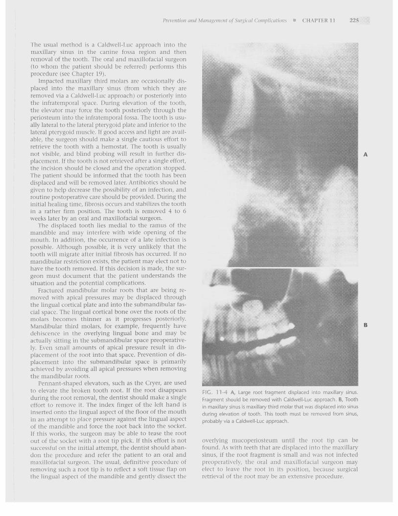
The usual method ic a Caldwell-I.uc approach into the rnasillary sinus in the canine fossa region and then removal of the tooth. The oral and masillofacial surgeon (to whom the patient should be referred) perform5 this procedure (see Chapter 19).
Impacted maxillary third molars are occasionally dis- placed into the maxillar!~ sinus (from which they are removed via a Caldwell-Luc approach) or posteriorly into the infratemporal space. During elevation of the tooth, the e l e~~a to r may force the tooth posteriorly through the periocteuni into the infratemporal fossa. The tooth is usu- ally lateral to the lateral pterygoid plate and inferior to the lateral pterygoid muscle. I f good access and light are avail- able, the surgeon should make a single cautious effort to retrieve the tooth with a hemostat. The tooth is usually not visible, and blind probing will result in further dis- placement. If the tooth is not retricved after a single effort, the incision chould he closed and the operation stopped. The patient should be informed that the tooth has been displaced and ~vill be removed later. /lntibiotics shoi~ld be given to help decrease the possibility of an infection, and routine postoperative care should be provided. During the initial healing time, fibrosis occurs and stabilizes the tooth in a rather firm position. The tooth is removed 1 to 6 weeks later by an oral and rnaxillofacial surgeon.
The displaced tooth lies medial to the ramus of the mandible and may interfere with wide opening of the mouth. In addition, the occurrence of a late infection is possible. ,4Itliough possible, it is very unlikely that the tooth will migrate after initial fibrosis has occurred. I f n o mandibular restriction exists, the patient may elect not to have the tooth removed. If this decision is made, the sur- geon must document that the patient understands the situation and the potential complications.
Fractured ~nandibular molar roots that are being re- moved with apical pressures may be displaced through the lingual cortical plate and into the submandibular fas- cia1 space. The lingual cortical bone over the roots of the niolars becomes thinner as it progresses posteriorly. Mandibular third molars, for example, frequently have dehiscence in the overlying lingual bone and may be actually sitting in the submandibular space preoperative- Iy. Even small amounts of apical pressure result in dis- placement of the root into that space. Prevention of dis- placement into the submandibular space is primarily achie\.eti by avoiding all apical pressures when removing the mandibular roots.
Pennant-shaped elevators, such as the Cryer, are used to elelrate the broken tooth root. I f the root disappears during the root removal, the dentist should make a single effort to remove it. The index finger of the left hand is inserted onto the lingual aspect of the floor of the mouth in an attempt to place pressure against the lingual aspect of the mandible and force the root back into the socket. I f this works, the surgeon may be able to tease the root out of thc socket with a root tip pick. I f this effort is not s~~ccessful on the initial attempt, the dentict should aban- don the proccdi~re and refer the patient to an oral and masillofacial surgeon. The usual, definitive procedure of removing cuch a root tip is to reflect a soft tissue flap on the lingual aspect of the mandible and gently dissect the
A, Large root fraornent diiplac~c! i r i l c i riidx~llary slnus Fragment should be removed w~ th Caldwell-Luc approach B, Tooth In max~llary slnus IS max~llary th~rd molar that was d~splaced Into slnus dur~ng elevat~on of tooth Th~s tooth must be removed from slnus, probably vla a Caldwell-Luc approach.
overlying mucoperiosteum until the root tip can be found. As with teeth that are displaced into the maxillary sinus, if the root fragment is small and ~vas not infected prcolxrati~.cly, the oral and m;~sillof;rci;il \irr,qeon may clect to Icavc the root in its po\itiori, I~ecause surgical retrieval of thc root may be an extensive procedure.

226 PART I1 6 Priricipltr o f Exorlorititr
Tooth Lost into Oropharynx
Occasionally, the crown of a tooth or an entire tooth might be lost down the oropharynx. If this occurs, the patient should be turned toward the dentist, into a mouth-down position, as much as possible. The suction device can then be used to help remove the tooth. The patient should be encouraged to cough and spit the tooth out onto the floor.
In spite of these efforts, the tooth may be swallowed or aspirated. If the patient has no coughing or respiratory distress, it is most likely that the tooth was swallowed and has traveled down the esophagus into the stomach. How- ever, if the patient has a violent episode of coughing that continues, the tooth may have been aspirated beyond the larynx into the trachea.
In either case the patient should be transported to an emergency room and chest and abdominal radiographs taken to determine the specific location of the tooth. I f the tooth has been aspirated, consultation should be requested regarding the possibility of removing the tooth with a bronchoscope. The urgent management of aspira- tion is to maintain the patient's airway and breathing. Supplemental oxygen may be appropriate if respiratory distress appears to be occurring.
If the tooth has been swallowed, it is highly probable that it will pass through the gastrointestinal (GI) tract within 2 to 4 days. Because teeth are not usually jagged or sharp, unimpeded passage occurs in almost all situations. However, it may be prudent to have the patient go to an emergency room and have a radiograph of the abdomen taken to confirm the tooth's presence in the GI tract instead of in the respiratory tract. Follow-up radiographs are probably not necessary, because the usual fate of swal- lowed teeth is passage.
!r\l)URiES TO ADIACENT TEETH
When the dentist extracts a tooth, the focus of attention is on that particular tooth and the application of forces to luxate and deliver it. When the surgeon's total attention is thus focused, likelihood of injury to the adjacent teeth increases. The surgeon should mentally step back from time to time to survey the entire surgical field to prevent injury to adjacent teeth.
Fracture of Adjacent Restoration
The most common injury to adjacent teeth is the inad- vertent fracture of either a restoration or a severely cari- ous tooth while the surgeon is attempting to luxate the tooth to be removed with an elevator (Fig. 11-5). If a large restoration exists, the surgeon should warn the patient preoperatively about the possibility of fracturing it during the extraction. I'revention of such a fracture is primarily achieved by avoiding application of instrumentation and force on the restoration (Box 11-3). This means that the straight elevator should be used with great caution or not used at all to luxate the tooth before extraction. If a
Prevention of Injury t o Adjacent Teeth - -
1. Recognize potential to fracture large restoration. 2. Warn patient preoperatively 3. Employ judicious use of e l e ~ 4. Ask assistant to warn surgeGI y l c , ~ ~ r e on adjacent
teeth.
i- 1 . Mand~bular f ~ r s t molar. If ~t 1s to be removed, surgeon must take care not to frac-
ture amalgam ~n second premolar wlth elevators or forceps.

restoration is dislodged or fractured, the surgeon should make sure that the displaced restoration is removed from the mouth and does not fall into the empty tooth socket. Once the surgical procedure has been completed, the injured tooth should be treated by placement of a tem- porary restoration. The patient should be informed that the fracture has occurred and that a replacement restora- tion must be placed (see Chapter 12).
Teeth in the opposite arch may also be injured as a result of uncontrolled tractional forces. This usually occurs when buccolingual forces inadequately mobilize a tooth and excessive tractional forces are used. The tooth suddenly releases from the socket, and the forceps strikes against the teeth of the opposite arch and chips or frac- tures a cusp. This is more likely to occur with extraction of lower teeth, because these teeth may require more ver- tical tractional forces for their delivery, especially when using the no. 23 (cowhorn) forceps. Prevention of this type of injury can be accomplished by several methods. First and primary, the surgeon should avoid the use of excessive tractional forces. The tooth should be ade- quately luxated with apical, buccolingual, and rotational forces to minimize the need for tractional forces.
Even when this is done, however, occasionally a tooth releases unexpectedly. The surgeon or assistant should protect the teeth of the opposite arch by simply holding a finger or suction tip against them to absorb the blow should the forceps be released in that direction. If such an injury occurs, the tooth should be smoothed or restored as necessary to keep the patient comfortable until a per- manent restoration can be constructed.
Luxation of Adjacent Teeth
Inappropriate use of the extraction instruments may lux- ate the adjacent tooth. This is prevented by judicious use of force with elevators and forceps. If the tooth to be
extracted is crowded and has overlapping adjacent teeth, such as is commonly seen in the mandibular incisor region, thin, narrow forceps such as the no. 286 forceps, may be useful for the extraction (Fig. 11-6). Forceps with broader beaks should be avoided, because it will cause injury and luxation of the adjacent teeth.
If an adjacent tooth is luxated or partially avulsed, the treatment goal is to reposition the tooth into its appro- priate position and stabilize it so that adequate healing occurs. This usually requires that the tooth simply be repositioned in the tooth socket and left alone.
The occlusion should be checked to ensure that the tooth has not been displaced into a hypererupted and traumatic occlusion. Occasionally, the luxated tooth is very mobile. If this is the case, the tooth should be stabi- lized with the least possible rigid fixation to maintain the tooth in its position. A simple silk suture that crosses the occlusal table and is sutured to the adjacent gingiva is usually sufficient. Rigid fixation with circumdental wires and arch bars results in increased chances for external root resorption and ankylosis of the tooth; therefore it should usually be avoided (see Chapter 23).
Extraction of Wrong Teeth
A complication that every dentist believes can never happen-but happens surprisingly often-is extraction of the wrong tooth. This should never occur if appropriate attention is given to the planning and execution of the surgical procedure.
This problem may be the result of inadequate atten- tion to the preoperative assessment. If the tooth to be extracted is grossly carious, it is less likely that the wrong tooth will be removed. The wrong tooth is most com- monly extracted when the dentist is asked to remove teeth for orthodontic purposes, especially from patients who are in mixed dentition stages and whose orthodon-
'FiG. 1 i -6 A, No. 151 forceps, too wide to grasp premolar to be extracted without luxating adjacent teeth. B, Maxillary root forceps, which can be adapted easily to tooth for extraction.

228 PART 11 Prir~ci/~lcc o f Exotlor~tirl
Prevention of Extraction of Wrong Teeth 1 : 1. Focus attention on procedure. i 2. Enlist patient and assistant to ensure correct tooth is 1 being removed. i 3. Check, then recheck, to confirm correct tooth.
tists have asked for unusual extractions. Careful preoper- ative planning and clinical assessment of which tooth is to be removed before the forceps is applied is the main method of preventing this complication (Box 11-4).
I f the wrong tooth is extracted and the dentist realizes this error immediately, the tooth should be replaced immediately into the tooth socket. If the extraction is for orthodontic purposes, the dentist should contact the orthodontist immediately and discuss whether or not the tooth that was removed can substitute for the tooth that should have been removed. If the orthodontist believes the original tooth must be removed, the correct extraction should be deferred for 4 or 5 weeks, until the fate of the replanted tooth can be assessed. If the wrongfully extract- ed tooth has regained its attachment to the alveolar process, then the originally planned extraction can pro- ceed. The surgeon should not extract the contralateral tooth until a definite alternative treatment plan is made.
I f the surgeon does not recognize that the wrong tooth was extracted until the patient returns for a postoperative visit, little can be done to correct the problem. Replanta- tion of the extracted tooth after it has dried cannot be successfully accomplished.
When the wrong tooth is extracted, it is important to inform the patient, the patient's parents (if the patient is a minor), and any other dentist involved with the patient's care, such as the orthodontist. In some situa- tions the orthodontist may be able to adjust the treat- ment plan so that extraction of the wrong tooth necessi- tates only a minor adjustment.
INJUR1ES TO OSSEOUS STRUCTURES
Fracture of Alveolar Process
The extraction of a tooth requires that the surrounding alveolar bone be expanded to allow an unimpeded path- way for tooth removal. However, in some situations the bone fractures and is removed with the tooth instead of expanding. The most likely cause of fracture of the alve- olar process is the use of excessive force with forceps, which fractures large portions of cortical plate. If the sur- geon realizes that excessive force is necessary to remove a tooth, a soft tissue flap should be elevated and controlled amounts of bone removed so that the tooth can be easily delivered. If this principle is not adhered to and the den- tist continues to use excessive or uncontrolled force, frac- ture of the bone will probably occur.
The most likely places for bony fracture are the buccal cortical plate over the maxillary canine, the buccal cortical
d Forceps extraction of these teeth resulted ~n removal of bone and tooth instead of just tooth.
Prevention of Fracture of Alveolar Process I 1
1 . Conduct thorough preoperative clinical and radio- graphic examination.
2. Do not use excessive force. 3. Use surgical (i.e., open) ext chnique to
reduce force required. raction te
plate over the maxillary molars (especially the first molar), the portions of the floor of the maxillary sinus associated with maxillary molars, the maxillary tuberosity, and the labial bone on mandibular incisors (Fig. 11-7). All of these bony injuries are caused by excessive force from the forceps.
The primary method of preventing these fractures is to perform a careful preoperative examination of the alveo- lar process, both clinically and radiographically (Box 11-5). Surgeons should inspect the root form of the tooth to be removed and assess the proximity of the roots to the maxillary sinus (Fig. 11-8). They should also check the thickness of the buccal cortical plate overlying the tooth to be extracted (Fig. 11-9). If the roots diverge widely, i f they lie close to the sinus, or if the patient has a heavy buccal cortical bone, surgeons must take special measures to prevent fracturing excessive portions of bone. Age is a factor to be considered, because the bones of older patients are likely to be less elastic and therefore more likely to fracture rather than expand.
The surgeon who preoperatively determines that a high probability exists for bone fracture should consider performing the extraction by the surgical technique. Using this method the surgeon can remove a smaller,

Pre1'eiitioi7 and .Varingemeilt of S~rr~picnl Complications CHAPTER 11 229
A, Floor of slnus associated wllh roots of teeth I f extraction IS requ~red, tooth should be removed surgically. B, Maxillary molar teeth lmmedlately adjacent to slnus present Increased danger of sinus exposure
more controlled amount of bone, which results in more rapid healing and a more ideal ridge form for prosthetic reconstruction.
When the maxillary molar lies close to the maxillary sinus, surgical exposure of the tooth, with sectioning of the tooth roots into two or three portions, will prevent the removal of a portion of the maxillary sinus floor. This prevents the formation of a chronic oroantral fistula, which requires secondary procedures to close.
In summary, prevention of fractures of large portions of the cortical plate depends on preoperative radiographic and clinical assessment, avoidance of the use of excessive amounts of uncontrolled force, and the early decision to perform an open extraction with removal of controlled amounts of bone and sectioning of multirooted teeth.
During a forceps extraction, if the appropriate amount of tooth mobilization does not occur early, then the wise and prudent dentist will alter the treatment plan to the surgi- cal technique instead of pursuing the closed method.
Management of fractures of the alveolar bone takes sev- eral different routes, depending on the type and severity of the fracture: If the bone has been completely removed from the tooth socket along with the tooth, it should not be replaced. The surgeon should simply make sure that the soft tissue has been replaced and repositioned over the remaining bone to prevent delayed healing. The surgeon must also smooth any sharp edges that may have been caused by the fracture. If such sharp edges of bone exist, the surgeon should reflect a small amount of soft tissue and use a bone file to round off the sharp edges.

230 PART I I a Prirl t iplrc uf Exot/o~ltiil
Patient w ~ t h heavy buccal co r t~ca l plate w h o requlrcs open cxtract lon
-l'ht, wrgron w,ho has been supporting the alveolar procc.?\ \vith the fingers during the extraction will feel the frnct~rrc of thc, I)uccal cortical plate when it occurs. At this time thc bone rernains attached to the periosteum and \ \ . i l l heal i t i t can be separated from the tooth and left attached to the overlying soft tissue. The surgeon must carefully dissect the bone with its attached associated soft tissue ntvay from the tooth. For this procedure the tooth mubt be stabilized with the forceps, and a small sharp instrument, such as a J2roodson periosteal elevator, should I)? used to elevate the buccal bone from the tooth root. It is important to realize that i f the soft tissue flap is reflected from the bone, the blood supply to the overly- ing bone \\.ill be severed and the bone will then undergo necrosis. Once the bone and soft tissue have been elevat- ed from the tooth, the tooth is removed and the bone and soft tissue flap are reapproximated and secured with sutures. When treated in this fashion, it is highly proba- ble that the bone will heal in a more favorable ridge form for prosthetic reconstruction than if the bone had been removed along with the tooth. Therefore it is worth the special effort to dissect the bone from the tooth.
Fracture of Maxillary Tuberosity
fractures. The surgeon using finger support for the alveolar process during the fracture (if the bone remains attached to the periosteum) should take extreme measures to ensure the survival of that bony segment. I f at all possible the bony segment should be dissected away from the tooth and the tooth removed in the usual fashion. The tuberosi- ty is then stabilized with sutures as previouslv indicated.
However, if the tuberosity is excessively mobile and cannot be dissected from the tooth, the surgeon has sev- eral options. The first option is to splint the tooth being extracted to adjacent teeth and defer the extraction for 6 to 8 weeks, during which time the bone will heal. The tooth is then extracted with an open surgical technique. The second option is to section the crown of the tooth from the roots and allow the tuberosity and tooth root section to heal. After 6 to 8 weeks the surgeon can reen- ter the area and remove the tooth roots in the usual fash- ion. If the maxillary molar tooth is infected, these two techniques should be used with caution.
If the maxillary tuberosity is completely separated from the soft tissue, the usual steps are to smooth the sharp edges of the remaining bone and to replace and suture the remaining soft tissue. The surgeon must carefully check for an oroantral communication and treat as necessary.
Fractures of the maxillarv tuberositv should be viewed 1:racture of a large section of bone in the maxillary as a serious complication. The major ;herapeutic goal of tuberosity area is a situation of special concern. The max- management is to maintain the fractured bone in place illary tuberosity is especially important for the construe- and to provide the best possible environment for healing. tion of a stable retentive maxillary denture. If a large This may be a situation that can best be handled by refer- portion of this tuberosity is removed along with the max- ral to an oral and maxillofacial surgeon. illary tooth, denture stability may be compromised. The tracture of the ~nasillary tuberosity most commonly results trom extraction of an erupted maxillarv third INJURIES - - - - TO ADJACENT STRUCTURES molar or from a second molar if it happens to be the last During the process of tooth extraction, it is possible to tooth in the arch (Fig. 11-10). injure adjacent tissues. The prudent surgeon preopera-
If this type of fracture occurs during an extraction, tively evaluates all adjacent anatomic areas and designs a treatment is similar to that just discussed for other bony surgical procedure to prevent injury to these tissues.

< - Tuberosity rernoveti wltti iiiaxillary ,tionti iiiol<lr, \ v ' ~ i c i l
el~mlnates Important prosthetic retent~on area and exposes maxll- lary sinus A, Buccal vlew of bone removed wlth tooth. B, Superlor view, look~ng onto s~nus floor, wh~ch was removed w~th tooth.
Injury to Regional Nerves
The branches of the fifth cranial nerve, which provide innervation to the mucosa and skin, are the structures most likely to be injured during extraction. The most fre- quently involved specific branches are the mental nerve, the lingual nerve, the buccal nerve, and the nasopalatine nerve. The nasopalatine and buccal nerves are frequently sectioned during the creation of flaps for renloval of impacted teeth. The area of sensory innervation of these two nerves is relatively small, and reinnervation of the affected area usually occurs rapidly. Therefore the nasopalatine and long buccal nerves can be surgically sec- tioned without sequelae or complications.
Surgical removal of mandibular premolar roots or impacted mandibular premolars and periapical surgery in
Prevention of Nerve Injury
1. Be aware of nerve anatomy in surgical area. 2. Avoid making incisions or affecting periosteum in
j nerve area.
the area of the mental nerve and mental foramen must be performed with great care. If the mental nerve is injured, the patient will have an anesthesia or paresthesia of the lip and chin. If the injury is the result of flap reflection or simple manipulation, the altered sensation usually disap- pears in a few weeks to a few months. If the mental nerve is sectioned at its exit from the mental foramen or torn along its course, it is likely that mental nerve function will not return, and the patient will have a permanent state of anesthesia. If surgery is to be performed in the area of the mental nerve or the mental foramen, it is imperative that surgeons have a keen awareness of the potential morbidi- ty from injury to this nen7e (Box 11-6). I f surgeons have any question concerning their ability to perform the indi- cated surgical procedure, they should refer the patient to an oral and maxillofacial surgeon. I f a three-corner flap is to be used in the area of the mental nerve, the vertical releasing incision must be placed far enough anterior to avoid severing any portion of the mental nerve. Rarely is it advisable to make the vertical releasing incision at the interdental papilla between the canine and first premolar.
'The lingual nerve is anatomically located directly against the lingual aspect of the mandible in the retrornolar pad region. The lingual ner17e rarely regenerates if it is severely traumatized. Incisions made in the retromolar pad region of the mandible should be placed to avoid severing this nerve. Therefore incisiorls made for surgical exposure of impacted third molars or of bony areas in the posterior molar region should be made well to the buccal aspect of the mandible. Prevention of injury to the lingual nerve is of paramount importance for controlling this difficult complication.
Finally, the inferior alveolar nerve may be traumatized along the course of its intrabony canal. The most common place of injury is the area of the mandibular third molar. Removal of impacted third molars may crush or sharply injure the nerve in its canal. This complication is conlmon enough during the extraction of third molars that it is important to inform patients on a routine basis that it is a possibility. The surgeon must then take every precaution possible to avoid injuring the nerve during the extraction.
Injury to Temporomandibular Joint
Another major structure that can be traumatized during an extraction procedure in the mandible is the temporo- mandibular joint (TM]). Removal of mandibular molar teeth frequently requires the application of a substantial amount of force. If the jaw is inadequately supported dur- ing the extraction, the patient may experience pain in this region. Controlled force and adequate support of the jaw prevents this. The use of a bite block on the contralateral
232 PART I1 m Principles o f Exodontia
Prevention of Injury t o Temporoma Joint
1. Support mandible during extractior 2. Do not open mouth too widely.
side may provide adequate balance of forces so that injury and pain do not occur (Box 11-7). The surgeon must also support the jaw as described earlier. If the patient com- plains of pain in the TMJ immediately after the extraction procedure, the surgeon should recommend the use of moist heat, rest for the jaw, a soft diet, and 1000 mg of aspirin every 4 hours for several days. Patients who cannot tolerate aspirin should be given an aspirin substitute, such as other NSAIDs or acetaminophen.
OROANTRAL COMMUNICATIONS
Removal of maxillary molars occasionally results in com- munication between the oral cavity and the maxillary sinus. If the maxillary sinus is large, if no bone exists between the roots of the teeth and the maxillary sinus, and if the roots of the tooth are widely divergent, then it is increasingly probable that a portion of the bony floor of the sinus will be removed with the tooth. If this com- plication occurs, appropriate measures are necessary to prevent a variety of sequelae. The two sequelae of most concern are postoperative maxillary sinusitis and forma- tion of a chronic oroantral fistula. The probability that either of these two sequelae will occur is related to the size of the oroantral communication and the manage- ment of the exposure.
As with all complications, prevention is the easiest and most efficient method of managing the situation. Preop- erative radiographs must be carefully evaluated for the tooth-sinus relationship whenever maxillary molars are to be extracted. If the sinus floor seems to be very close to the tooth roots and the tooth roots are widely divergent, the surgeon should avoid a closed forceps extraction and perform a surgical removal with sectioning of tooth roots (see Fig. 11-8). Large amounts of force should be avoided in the removal of such maxillary molars (Box 11-8).
Diagnosis of the oroantral communication can be made in several ways: The first is to examine the tooth once it is removed. If a section of bone is adhered to the root ends of the tooth, the surgeon can be relatively cer- tain that a communication between the sinus and mouth exists. If a small amount of bone or no bone adheres to the molars, a communication may exist anyway. To con- firm the presence of a communication, the best tech- nique is to use the nose-blowing test. Pinching the nos- trils together occludes the patient's nose, and the patient is asked to blow gently through the nose while the sur- geon observes the area of the tooth extraction. If a com- munication exists, there will be passage of air through the tooth socket and bubbling of blood in the socket area.
Prevention of Oroantral Communications
examini I. Use sur! ;. Avoid e:
1. Conduct thorouah preoperative radioara~hic
ction earl: al pressur~
d
3tion. gical extra ltcess apic
y and seci e.
2 4
lion roots.
After the diagnosis of oroantral communication has been established, the surgeon must determine the approx- imate size of the communication, because the treatment will depend on the size of the opening. If the communi- cation is small (2 mm in diameter or less), no additional surgical treatment is necessary. The surgeon should take measures to ensure the formation of a high-quality blood clot in the socket and then advise the patient to take sinus precautions to prevent dislodgment of the blood clot.
Sinus precautions are aimed at preventing increases or decreases in the maxillary sinus air pressure that would dislodge the clot. Patients should be advised to avoid blowing the nose, violent sneezing, sucking on straws, and smoking. Patients who smoke and who cannot stop (even temporarily) should be advised to smoke in small puffs, not in deep drags, to avoid pressure changes.
The surgeon must not probe through the socket into the sinus with a periapical curette or a root tip pick. It is possible that the bone of the sinus has been removed with- out perforation of the sinus lining. To probe the socket with an instrument might unnecessarily lacerate the mem- brane. Probing of the communication may also introduce foreign material, including bacteria, into the sinus and thereby further complicate the situation. Probing of the communication is therefore absolutely contraindicated.
If the opening between the mouth and sinus is of moderate size (2 to 6 mm), additional measures should be taken. To help ensure the maintenance of the blood clot in the area, a figure-of-eight suture should be placed over the tooth socket (Fig. 11-11). The patient should also be told to follow sinus precautions. Finally, the patient should be prescribed several medications to help lessen the possibility that maxillary sinusitis will occur. Antibi- otics, usually penicillin or clindamycin, should be pre- scribed for 5 days. In addition, a decongestant nasal spray should be prescribed to shrink the nasal mucosa to keep the ostium of the sinus patent. As long as the ostium is patent and normal sinus drainage can occur, sinusitis and sinus infection are less likely. An oral decongestant is also sometimes recommended.
If the sinus opening is large (7 mm or larger), the den- tist should consider closing the sinus communication with a flap procedure. This usually requires that the patient be referred to an oral and maxillofacial surgeon, because flap development and closure of a sinus opening are somewhat complex procedures that require skill and experience.
The most commonly used flap is a buccal flap. This technique mobilizes buccal soft tissue to cover the open- ing and provide for a primary closure. This technique should be performed as soon as possible, preferably on

Prevention rind Mn~zn~yemrnt o f Slrrgical Compliccltions a CHAPTER 11 233
FIG. 11-1 1 A figure-of-eight stitch is usually performed to help maintain piece of oxidized cellulose in tooth socket.
the same day in which the opening occurred. The same sinus precautions and medications are usually required (see Chapter 19).
The recommendations just described hold true for patients who have no preexisting sinus disease. If a com- munication does occur, it is important that the dentist inquire specifically about a history of sinusitis and sinus infections. If the patient has a history of chronic sinus disease, even small oroantral communications will heal poorly and may result in permanent oroantral communi- cation. Therefore creation of an oroantral communica- tion in patients with chronic sinusitis is cause for referral to an oral and maxillofacial surgeon for definitive care (see Chapter 19).
The majority of oroantral communications treated in the methods just recommended will heal uneventfully. Patients should be followed up carefully for several weeks to ensure that this has occurred. Even patients who return within a few days with a small communication usually heal spontaneously if no maxillary sinusitis exists. These patients should be followed up closely and referred to an oral and maxillofacial surgeon if the communication per- sists for longer than 2 weeks. Closure of oroantral fistulae is important because air, water, food, and bacteria go from the oral cavity into the sinus, often causing a chronic sinusitis condition. Additionally, if the patient is wearing a full maxillary denture, suction is not as strong; therefore retention of the denture is compromised.
POSTOPERATIVE BLEEDING - "" - ."
Extraction of teeth is a surgical procedure that presents a severe challenge to the body's hemostatic mechanism. Several reasons exist for this challenge: First, the tissues of the mouth and jaws are highly vascular. Second, the extraction of a tooth leaves an open wound, with both soft tissue and bone open, which allows additional ooz- ing and bleeding. Third, it is almost impossible to apply dressing material with enough pressure and sealing to prevent additional bleeding during surgery. Fourth, patient5 tend to play with the area of surgery with their tongues and occasionally dislodge blood clots, which ini- tiates secondary bleeding. The tongue may also cause sec- ondary bleeding by creating small negative pressures that suction the blood clot from the socket. Finally, salivary enzymes may lyse the blood clot before it has organized and before the ingrowth of granulation tissue.
Prevention of Postoperative Bleeding
1. Obtain history of bleeding. 2. Use atraurnatic surgical technique. 3. Obtain good hemostasis at surgery. 4. Provide excellent patient instructions.
As with all cbinplications, prevention of bleeding is the best way to manage this problem (Box 11-9). One of the prime factors in preventing bleeding is the taking of a thorough history from the patient regarding this specif- ic potential problem. Several questions should be asked of the patient concerning any history of bleeding. If affir- mative answers to any of these questions are given, the surgeon should take special efforts to control bleeding.
The first question that patients should be asked is if they have ever had a problem with bleeding in the past. The sur- geon should inquire about bleeding after previous tooth extractions or previous surgery (such as a tonsillectomy) and persistent bleeding after accidental lacerations. The sur- geon must listen carefully to the patient's answers to these questions, because the patient's idea of "persistent" may actually be normal. For example, it is quite normal for a socket to ooze small amounts of blood for the first 12 to 24 hours after extraction. However, if a patient relates a histo- ry of bleeding that persisted for more than 1 day or that required special attention from the dentist, then the sur- geon's degree of suspicion should be substantially elevated.
The surgeon should inquire about any family history of bleeding. If anyone in the patient's family has or had a history of prolonged bleeding, further inquiry about its cause should be pursued. Most congenital bleeding disor- ders are familial, inherited characteristics. These congen- ital disorders vary from very mild to very profound, the latter requiring substantial efforts to control.
The patient should next be asked about any medica- tions currently being taken that might interfere with coagulation. Drugs such as anticoagulants may cause prolonged bleeding after extraction. Patients receiving anticancer chemotherapy or who are alcoholics may also tend to bleed.
The patient who has a known or suspected coagulop- athy should be evaluated by laboratory testing before surgery is performed to determine the severity of the dis- order. It is usually advisable to enlist the aid of a hema- tologist if the patient has a familial coagulation disorder.
The means to measure the status of intentional anti- coagulation is to use the International Normalized Ratio (INR). This value takes into account both the patient's prothrombin time (PT) and the control. Normal antico- agulated status for most medical indications will have an INR of 2.0 to 3.0. It is reasonable to perform extractions on patients who have an INR of 2.5 or less without reduc- ing the anticoagulant dose. With special precautions, it is reasonably safe to do minor amounts of surgery in patients with an INR of up to 3.0, if special local hemo- static measures are taken.

234 PART I1 Prirlciplor of E,~vtiotitirl
Primary control of bleeding during routine surgery depends on gaining control of ali factors that may pro- long bleeding. Surgery should be as atraumatic as possi- ble, with clean incisions and gentle management of the soft tissue. Care should be taken not to crush the soft tis- sue, because crushed tissue tends to ooze for long periods. Sharp bony spicules should be smcothed or removed. All granulation tissue should be curetted from the periapical region of the socket and from around the necks of adja- cent teeth and soft tissue flaps. This should be deferred when anatomic restrictions, such as the sinus or inferior alveolar canal, are present (Fig. 11-12). The wound should be carefully inspected for the presence of any specific bleeding arteries. If such arteries exist in the soft tissue, they should be controlled with direct pressure or, if pres- sure fails, by clamping the artery with a hemostat and li- gating it with a resorbable suture. For most oral surgical procedures, direct pressure over the soft tissue bleeding area for 5 minutes results in complete control.
The surgeon should also check for bleeding from the bone. Occasionally, a small, isolated vessel bleeds from a bony foramen. If this occurs, the foramen can be crushed with the closed ends of the hemostat, thereby occluding the bleeding vessel. Once these measures have been accomplished, the bleeding socket is covered with a damp gauze sponge that has been folded to fit directly into the area from which the tooth was extracted. The patient bites down firmly on this gauze for at least 30 minutes. The surgeon should not dismiss the patient from the office until hemostasis has been achieved. This recluires that the surgeon check the patient's extraction socket about 15 minutes after the completion of surgery. The patient should open the mouth widely, the gauze should be removed, and the area should be inspected carefully for any persistent oozing. Initial control should have been achieved. New damp gauze is then folded and placed into position, and the patient is told to leave it in place for an additional 30 minutes.
If bleeding persists but careful inspection of the socket reveals that it is not of an arterial origin, the surgeon should take additional measures to achieve hemostasis. Several different materials can be placed in the socket to help gain hemostasis (Fig. 11-13). The most commonly used and the least expensive is the absorbable gelatin sponge (e.g., Gelfoam). This material is placed in the extraction socket and held in place with a figure eight suture placed over the socket. The absorbable gelatin sponge forms a scaffold for the formation of a blood clot, and the suture helps maintain the sponge in position dur- ing the coagulation process. ,4 gauze pack is then placed over the top of the socket and is held wit11 pressure.
A second material that can be used to control bleeding is oxidized regenerated cellulose (e.g., Surgicel). This material promotes coagulation better than the absorbable gelatin sponge, because it can be packed into the socket under pressure. The gelatin sponge becomes very friable when wet and cannot be packed into a bleeding socket. When the cellulose is packed into the socket, it almost always causes delayed healing of the socket. Therefore packing the socket with cellulose is reserved for more per- sistent bleeding.
If the surgeon has special concerns about the patient's ability to clot, a liquid preparation of topical thrombin (prepared from bovine thrombin) can be saturated onto a gelatin sponge and inserted into the tooth socket. The thrombin bypasses all steps in the coagulation cascade and helps to convert fibrinogen to fibrin enzymatically, which forms a clot. The sponge with the topical thrombin is secured in place with a figure-eight suture. A gauze pack is placed over the extraction site in the usual fashion.
A final material that can be used to help control a bleeding socket is collagen. Collagen promotes platelet aggregation and thereby helps accelerate blood coagula- tion. Collagen is currently available in several different forms. Microfibular collagen (e.g., Avitene) is available as a fibular material that is loose and fluffy but can be
Crarlulorna of second premolar Surgeon should not curettp per~ap~cally around th~s second premolar to remove granuloma because rtsk for slnus perforation IS h~gh.

Preveiltioil niltl ,Mtri~ngeriroilt of Srrr;yitrrl Coii~/~lic.ntioir.s @ CHAPTER 1 1 235
packed into a tooth socket and held in by suturing and gauze packs, as with the other materials. A more highly cross-linked collagen is supplied as a plug (e.g., Collaplug) or as a tape (e.g., Collatape). These materials are more readily packed into a socket (Fig. 11-14) and are easier to use. They are also more expensive.
Even after primary hemostasis has been achieved, patients occasionally call the dentist with bleeding from the extraction site, referred to as secondary bleeding. The patient should be told to rinse the mouth gently with very cold water, then place appropriate-sized gauze over the area and bite firmly. The patient should sit quietly for 30 min- utes, biting firmly on the gauze. If the bleeding persists, the patient should repeat the cold rinse and bite down on a damp tea bag. The tannin in the tea will frequently help stop the bleeding. If neither of these techniques is success- fill, the patient should return to the dentist.
The surgeon must have an orderly, planned regimen to control this secondary bleeding. The patient should be positioned in the dental chair, and all blood, saliva, and fluids should be suctioned from the mouth. Such patients will frequently have large "liver clots" in their mouth, which must be removed. The surgeon should visualize the bleeding site carefully with good light to determine the precise source of bleeding. If it is clearly seen to be a
generalized oozing, the bleeding site is covered with a folded, damp gauze sponge held in place with firm pres- sure by the surgeon's finger for at least 5 minutes.
F l f t Mater~al that can be used In a bleed~rig socket Clockw~se from the canlne tooth: collagen plug, m~crof~bular colla- gen, regenerated oxldlzed cellulose, collagen tape, and absorbable gelat~n sponge
A, Collagen shaped Into the form of a plug IS s~m~lar In slze to the root of a rnax~llary canlne. B and C, The collagen plug IS placed ~nto the socket wlth cotton pl~ers (arrow) D, A f~gure- e~ght suture 1s placed over the socket to ma~nta~n the collagen In the socket.

236 PART I1 8 Priticiples of Exorfotltin
This measure is sufficient to control most bleeding. The reason for the bleeding is usually some secondary trauma that is potentiated by the patient's continuing to suck on the area or to spit blood from the mouth instead of continuing to apply pressure with a gauze sponge.
If 5 minutes of this treatment does not control the bleeding, the surgeon must administer a local anesthetic so that the socket can be treated more aggressively. Block techniques are to be encouraged instead of local infiltra- tion techniques. Infiltration with solutions containing epinephrine cause vasoconstriction and may control the bleeding temporarily. However, when the effects of the epinephrine dissipate, rebound hemorrhage with recur- rent bothersome bleeding may occur.
Once local anesthesia has been achieved, the surgeon should gently curette out the tooth extraction socket and suction all areas of old blood clot. The specific area of bleeding should be identified as clearly as possible. As with primary bleeding, the soft tissue should be checked for diffuse oozing versus specific artery bleeding. The bone tissue should be checked for small nutrient artery bleeding or general oozing. The same measures described for control of primary bleeding should be used. The sur- geon must then decide if a hemostatic agent should be inserted into the bony socket. The use of an absorbable gelatin sponge with topical thrombin held in position with a figure-of-eight stitch and reinforced with applica- tion of firm pressure from a small, damp gauze pack is standard for local control of secondary bleeding. This technique works well in almost every bleeding socket. In many situations an absorbable gelatin sponge and gauze pressure are adequate. The patient should be given spe- cific instructions on how to apply the gauze packs direct- ly to the bleeding site should additional bleeding occur. Before the patient with secondary bleeding is discharged from the office, the surgeon should monitor the patient for at least 30 minutes to ensure that adequate hemosta- tic control has been achieved.
If hemostasis is not achieved by any of the local meas- ures just discussed, the surgeon should consider perform- ing additional laboratory screening tests to determine if the patient has a profound hemostatic defect. The dentist usually requests a consultation from a hematologist, who orders the typical screening tests. Abnormal test results will prompt the hematologist to investigate the patient's hemostatic system further.
A final hemostatic complication relates to intraopera- tive and postoperative bleeding into the adjacent soft tissues. Blood that escapes into tissue spaces, especially subcutaneous tissue spaces, appears as bruising of the overlying soft tissue 2 to 5 days after the surgery. This bruising is termed eccl~ytnosis (see Chapter 10).
DELAYED HEALING AND INFECTION
removal. Careful asepsis and thorough wound debride- ment after surgery can best achieve prevention of infec- tion after surgical flap procedures. This means that the area of bone removal under the flap must be copiously irrigated with saline and that all foreign debris must be removed with a curette. Some patients are predisposed to postoperative wound infections and should be given peri- operative prophylactic antibiotics (see Chapter 15).
Wound Dehiscence
Another problem of delayed healing is wound dehiscence (Box 11-10). If a soft tissue flap is replaced and sutured without an adequate bony foundation, the unsupported soft tissue flap often sags and separates along the line of incision. A second cause of dehiscence is suturing the wound under tension. I f the soft tissue flap is sutured under tension, the sutures cause ischemia of the flap mar- gin with subsequent tissue necrosis, which allows the suture to pull through the flap margin and results in wound dehiscence. Therefore sutures should always be placed in tissue without tension and tied loosely enough to prevent blanching of the tissue.
A common area of exposed bone after tooth extraction is the internal oblique ridge. After extraction of the first and second molar, during the initial healing, the lingual flap becomes stretched over the internal oblique (mylo- hyoid) ridge. Occasionally, the bone will perforate through the thin mucosa, causing a sharp projection of bone in the area.
The two major treatment options are (1) to leave the projection alone, or (2) to smooth it with bone file. If the area is left to heal untreated, the exposed bone will slough off in 2 to 4 weeks. If the irritation of the sharp bone is low, this is the preferred method. If a bone file is used, no flap should be elevated, because this will result in an increased amount of exposed bone. The file is used only to smooth off the sharp projections of the bone. This procedure usually requires local anesthesia. Patients who are quite annoyed by the sharp bone will usually choose this method.
Dry Socket
Dry socket or alveolar osteitis is delayed healing but is not associated with an infection. This postoperative complica- tion causes moderate-to-severe pain but is without the usual signs and symptoms of infection, such as fever, swelling, and erythema. The term dry socket describes the appearance of the tooth extraction socket when the pain
Prevention of Wound Dehiscence
Infection 1. Use aseptic technique. The most common cause of delayed wound healing is 2. Perform atraumatic surgery. infection. Infection is a rare complication after routine 3. Close incision over intact bone. dental extraction and is primarily seen after oral surgery 4. Suture without tension. that involves the reflection of soft tissue flaps and bone

Prevention and Management of Surgical Complications CHAPTER 11 237
begins. In the usual clinical course, pain develops on the third or fourth day after removal of the tooth. On exami- nation the tooth socket appears to be empty, with a par- tially or completely lost blood clot, and the bony surfaces of the socket are exposed. The exposed bone is extremely sensitive and is the source of the pain. The dull, aching pain is moderate to severe, usually throbs, and frequently radiates to the patient's ear. The area of the socket has a bad odor, and the patient frequently complains of a bad taste.
The cause of alveolar osteitis is not absolutely clear, but it appears to be the result of high levels of fibrinolyt- ic activity in and around the tooth extraction socket. This fibrinolytic activity results in lysis of the blood clot and subsequent exposure of the bone. The fibrinolytic activi- ty may be the result of subclinical infections, inflamma- tion of the marrow space of the bone, or other factors. The occurrence of a dry socket after a routine tooth extraction is relatively rare (2% of extractions), but it is quite frequent after the removal of impacted mandibular third molars (20% of extractions).
Prevention of the dry socket syndrome requires that the surgeon minimize trauma and bacterial contamina- tion in the area of surgery. The surgeon should perform atraumatic surgery with clean incisions and soft tissue reflection. After the surgical procedure, the wound should be thoroughly debrided and irrigated with large quantities of saline. Small amounts of antibiotics (e.g., tetracycline) placed in the socket alone or on a gelatin sponge may help to decrease the incidence of dry socket in mandibular third molars. The incidence of dry socket can also be decreased by preoperative and postoperative rinses with antimicrobial mouth rinses, such as chlorhex- idine. Well-controlled studies indicate that the incidence of dry socket after impacted mandibular third molar sur- gery can be reduced by up to 50%.
The treatment of alveolar osteitis is dictated by the single therapeutic goal of relieving the patient's pain dur- ing the period of healing. If the patient receives no treat- ment, no sequela other than continued pain exists (treat- ment does not hasten healing).
Treatment is straightforward and consists of gentle irrigation and insertion of a medicated dressing. First, the tooth socket is gentlv irrigated with saline. The socket
with saline at each dressing change. Once the patient's pain decreases, the dressing should not be replaced, because it acts as a foreign body and further prolongs wound healing.
FRACTURES - OF THE MANDIBLE - - "----- - -- Fracture of the mandible during extraction is a rare com- plication; it is associated almost exclusively with the sur- gical removal of impacted third molars. A mandibular fracture is usually the result of the application of a force exceeding that needed to remove a tooth and often occurs during the use of dental elevators. However, when lower third molars are deeply impacted, even small amounts of force may cause a fracture. Fractures may also occur during removal of impacted teeth from a severely atrophic mandible. Should such a fracture occur, it must be treated by the usual methods used for jaw fractures. The fracture nus st be adequately reduced and stabilized with intermaxillary fixation. Usually this means that the patient should be referred to an oral and maxillofacial surgeon for definitive care.
SUMMARY - - -- -- - - -- Prevention of complications should be a major goal of the surgeon. Skillful management of complications when they do occur is the sine qua non of the wise and mature surgeon.
The surgeon who anticipates a high probability of an unusual specific complication should inform the patient and explain the anticipated management and sequelae. Notation of this should be made in the informed consent that the patient signs.
B l B t l O C R A P H Y - - - "" *- - - - - - - - - - - Birn H: Etiology and pathogenesis of fibrinolytic alveolitis, Int J
Oral S u r ~ 2:211, 1973. Hall HD, Bildman BS, Hand CD: Prevention of dry socket with
local application of tetracycline, Oral Surg 29:35, 1971. Kohn MW, Chase DC, Marciani RD: Surgical misadventures, Dent
" , - should not be curetted down to bare bone, because this Clin North A m 17533, 1973.
'
increases both the amount of exposed bone and the pain. Larsen PE: The effect of chlorhexidine rinse on the incidence of
Usually the entire blood clot is not lysed, and the part alveolar osteitis following the surgical removal of impacted mandibular third molars, J Oral Maxillofac Surg 49:932, 1991.
that is intact should be retained. The socket is carefully Moake JL: Common bleeding problems, Cibn Symp suctioned of all excess saline, and a small strip of iodo- 35(3):1, 1983. form gauze soaked with the medication is inserted into Ngeow WC: Management of the fractured maxillary tuberosity: the socket. The medication contains the following prin- an alternative method, Qzrintessence Int 29:189, 1998. cipal ingredients: eugenol, which obtunds the pain from Osbon DR: Postoperative complications following dentoalveolar the bone tissue; a topical anesthetic, such as benzocaine; surgery, Dent CIin North A m 17:483, 1973. and a carrying vehicle, such as balsam of Peru. The med- Redding SW, Stiegler KE: Dental management of the classic
ication can be made by the surgeon's pharmacist or can hemophiliac with inhibitors, Oral Surg 56:145, 1983.
be obtained as a commercial preparation from dental sup- Steinberg MJ, Moores JF: Use of INR to assess degree of anticoag-
ply houses. ulation in patients who have dental procedures, Oral Surg Om1 Med Oral Path01 Oral Radio1 Enriod 80: 175, 1995.
The medicated gauze is gently inserted the socketf Sweet JB, Butter DP, Drager JL: Effects of lavage techniques with
and the patient usually experiences profound relief from third molar surgery, Oral Surg 41:152, 1976. pain within 5 minutes. The dressing is changed every day Troulis MJ, Head TW, Lederc JC: What is the INR? J Can Dent or every other day for the next 3 to 6 days, depending on Assoc 62(suppl):428, 1996. the severity of the pain. The socket is gently irrigated Waite DE: Maxillary sinus, Dent Clin North A m 15:349, 1971.