Prevalence of Gastrointestinal Parasitic Infections and ...
11
Research Article Prevalence of Gastrointestinal Parasitic Infections and Assessment of Deworming Program among Cattle and Buffaloes in Gampaha District, Sri Lanka Nayana Gunathilaka , 1 Dimuthu Niroshana, 2 Deepika Amarasinghe , 2 and Lahiru Udayanga 3 1 Department of Parasitology, Faculty of Medicine, University of Kelaniya, Ragama, Sri Lanka 2 Department of Zoology and Environment Management, Faculty of Science, University of Kelaniya, Dalugama, Sri Lanka 3 Department of Biosystems Engineering, Faculty of Agriculture and Plantation Management, Wayamba University, Makandura, Sri Lanka Correspondence should be addressed to Nayana Gunathilaka; [email protected] Received 21 March 2018; Revised 21 September 2018; Accepted 25 September 2018; Published 9 October 2018 Academic Editor: Stefano D'Amelio Copyright © 2018 Nayana Gunathilaka et al. is is an open access article distributed under the Creative Commons Attribution License, which permits unrestricted use, distribution, and reproduction in any medium, provided the original work is properly cited. Gastrointestinal (GI) parasitic infection is a serious issue in cattle management. e effects of GI parasites may vary with age, sex of cattle, nutritional condition, and severity of infection. Prevalence of GI parasites among cattle population in Gampaha District has not been studied and there is no published study available. A total of 45 farms rearing cattle were selected randomly in three areas, namely, Kelaniya, Ganemulla, and Welisara, under three Veterinary Surgeon Divisions (VSD) in Gampaha District (Mahara, Gampaha, and Welisara). Freshly voided cattle fecal samples were collected randomly from the selected farms during March 2017–December 2017. Out of 163 cattle and buffaloes examined, 13.39% (n=22) were positive for eggs of one or more species of GI parasites. e prevalence of parasitic infection was higher in buffaloes (31.25%, 5/16) as compared to that of cows (11.56%, 21/147), but the difference was not significant (P >0.05). Hookworms (Bunostomum spp.), whipworms (Trichuris spp.), digenetic trematodes (Paramphistomum spp.), cestodes (Moniezia spp.), and oocysts of protozoans (coccidians) were found during the study. e nontreated animals indicated the highest percentage of parasitic infections accounting for 46.67% (n= 14), followed by partially treated individuals (15.15%, n= 5). GI parasite prevalence in males was higher when compared to that of females, but the difference was nonsignificant (P >0.05). General Linear Modelling (GLM) revealed that the effect of treatment status was significantly associated with the prevalence of GI parasites. e calves and yearlings had the highest rate of GI parasitic infections. e highest infection rate was observed at Kelaniya, followed by Welisara. Future investigations are necessary to evaluate the economic impact of GI parasites in the study areas. 1. Introduction Livestock farming, particularly rearing of cattle (Bos indicus/Bos tarsus) and Ceylon buffalo (Bubalus bubalis migona), is traditionally practiced by rural people in Sri Lanka [1]. Rearing of cattle in the country is catering for draught power, milk production, and meat production. Buffaloes are predominantly used for farm power in the cultivation of rice as well as production of curd [1]. Parasitic diseases caused by intestinal parasites constitute a major impediment to livestock production [2]. All ages of cattle are affected by a diverse set of intestinal parasites. ese infections are rarely associated with high mortality of cattle. However, their effects are usually characterized by lower outputs of animal products, byproducts, manure, and traction, thereby affecting the contributions of cattle in ensuring food security, especially in developing countries [2, 3]. e productivity losses through reduced feed intake and decreased efficiency in feed utilization due to subclinical Hindawi BioMed Research International Volume 2018, Article ID 3048373, 10 pages https://doi.org/10.1155/2018/3048373
Transcript of Prevalence of Gastrointestinal Parasitic Infections and ...

Research ArticlePrevalence of Gastrointestinal Parasitic Infections andAssessment of Deworming Program among Cattle and Buffaloesin Gampaha District, Sri Lanka
Nayana Gunathilaka ,1 Dimuthu Niroshana,2 Deepika Amarasinghe ,2
and Lahiru Udayanga 3
1Department of Parasitology, Faculty of Medicine, University of Kelaniya, Ragama, Sri Lanka2Department of Zoology and Environment Management, Faculty of Science, University of Kelaniya, Dalugama, Sri Lanka3Department of Biosystems Engineering, Faculty of Agriculture and Plantation Management, Wayamba University,Makandura, Sri Lanka
Correspondence should be addressed to Nayana Gunathilaka; [email protected]
Received 21 March 2018; Revised 21 September 2018; Accepted 25 September 2018; Published 9 October 2018
Academic Editor: Stefano D'Amelio
Copyright © 2018 Nayana Gunathilaka et al. This is an open access article distributed under the Creative Commons AttributionLicense, which permits unrestricted use, distribution, and reproduction in any medium, provided the original work is properlycited.
Gastrointestinal (GI) parasitic infection is a serious issue in cattle management. The effects of GI parasites may vary with age,sex of cattle, nutritional condition, and severity of infection. Prevalence of GI parasites among cattle population in GampahaDistrict has not been studied and there is no published study available. A total of 45 farms rearing cattle were selected randomlyin three areas, namely, Kelaniya, Ganemulla, and Welisara, under three Veterinary Surgeon Divisions (VSD) in Gampaha District(Mahara, Gampaha, and Welisara). Freshly voided cattle fecal samples were collected randomly from the selected farms duringMarch 2017–December 2017. Out of 163 cattle and buffaloes examined, 13.39% (n=22) were positive for eggs of one or more speciesof GI parasites. The prevalence of parasitic infection was higher in buffaloes (31.25%, 5/16) as compared to that of cows (11.56%,21/147), but the difference was not significant (P >0.05). Hookworms (Bunostomum spp.), whipworms (Trichuris spp.), digenetictrematodes (Paramphistomum spp.), cestodes (Moniezia spp.), and oocysts of protozoans (coccidians) were found during the study.The nontreated animals indicated the highest percentage of parasitic infections accounting for 46.67% (n= 14), followed by partiallytreated individuals (15.15%, n= 5). GI parasite prevalence in males was higher when compared to that of females, but the differencewas nonsignificant (P >0.05). General Linear Modelling (GLM) revealed that the effect of treatment status was significantlyassociated with the prevalence of GI parasites. The calves and yearlings had the highest rate of GI parasitic infections. The highestinfection rate was observed at Kelaniya, followed byWelisara. Future investigations are necessary to evaluate the economic impactof GI parasites in the study areas.
1. Introduction
Livestock farming, particularly rearing of cattle (Bosindicus/Bos tarsus) and Ceylon buffalo (Bubalus bubalismigona), is traditionally practiced by rural people in SriLanka [1]. Rearing of cattle in the country is catering fordraught power, milk production, and meat production.Buffaloes are predominantly used for farm power inthe cultivation of rice as well as production of curd[1].
Parasitic diseases caused by intestinal parasites constitutea major impediment to livestock production [2]. All agesof cattle are affected by a diverse set of intestinal parasites.These infections are rarely associated with high mortalityof cattle. However, their effects are usually characterizedby lower outputs of animal products, byproducts, manure,and traction, thereby affecting the contributions of cattle inensuring food security, especially in developing countries[2, 3]. The productivity losses through reduced feed intakeand decreased efficiency in feed utilization due to subclinical
HindawiBioMed Research InternationalVolume 2018, Article ID 3048373, 10 pageshttps://doi.org/10.1155/2018/3048373

2 BioMed Research International
or chronic infections are responsible for economic losses inthe livestock industry [4].
In addition, these infections enhance susceptibility tobacterial and viral diseases and losses resulting fromcondem-nation of carcasses and organs, as well as cost of drugs andveterinary care [5]. Gastrointestinal parasites like coccidian,ascarid, strongyle, Setaria, and amphistomes were docu-mented in countries with tropical and temperate climaticconditions such as India, Bangladesh, South Africa, SriLanka, Italy, and Mongolia, with a prevalence rate rangingfrom 20 to 96% [6–11]. Some studies conducted in Sri Lankahave recorded concurrent helminthic and coccidial infectionsat a rate of 78% among the cattle [7].
Anthelmintics and antiprotozoal agents have been usedto control gastrointestinal parasitic infections over the lastten decades [12]. They have succeeded in reducing intestinalparasitic infections, but none has been able to diminishthe reinfestation of diseases [13]. However, excessive use ofanthelmintic drugs has led to developing of anthelmintic-resistant parasites, which are being reported frommany partsof the world. Further, it has resulted in a fear of anthelminticresidues in the milk and meat of livestock animals [14].
In order for an anthelmintic strategy to be successful, in-depth knowledge of pathophysiology and epidemiology ofthe parasite, in the context of immunity and managementof the host, is required. Therefore, periodical monitoring ofparasitic species among livestock animals would be beneficialto control and manage diseases at early stages of infections infarm management practices.
Prevalence of gastrointestinal parasites among cattle pop-ulation in Gampaha District has not been studied and thereis no published study available. In addition, it is important tostudy the present situation of parasitic infections in cattle andassociated risk factors. Hence, the aim of the present studywas to determine the prevalence of single and concurrentinfections of GI parasites among cattle and the intensityof infections in selected farms in Gampaha District of SriLanka.
2. Material and Methods
2.1. Study Area. The District of Gampaha, located in theWestern Province of Sri Lanka covering an area of 1,387km2, was selected as the major study area. The mean annualrainfall of Gampaha remains around 2,398 mm, while themean annual temperature is about 27.3∘C.
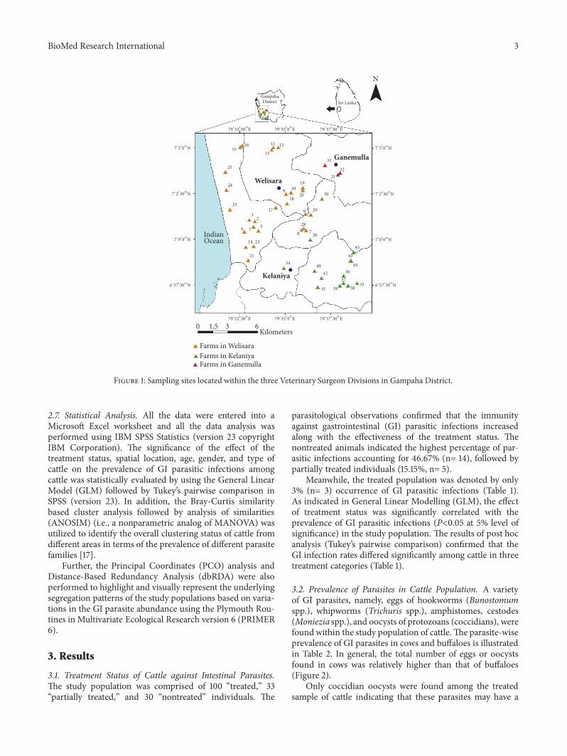
2.2. Selection of Sampling Locations. Based on the registeredcattle farms at Veterinary Investigation Centre, Welisara, 45farms were selected randomly for the study using a randomnumber table method based on the geographic location.These 45 farms fell into three Veterinary Surgeon Divisions(VSD), namely, Mahara VSD, Gampaha VSD, and Welis-ara VSD. Mainly these farms were distributed in Kelaniya,Ganemulla, andWelisara areas.Thegeographical distributionof the selected farms is illustrated in Figure 1. The farms thatrefused to participate in the present study were excluded andreplaced with others in the same area.
2.3. Treatment Procedure and Categorization of TreatmentStatus. The treatment against gastrointestinal (GI) parasitesin calves was administered at 21-day, 3-month, 6-month, and12-month intervals, respectively, with a standard mixture ofAlbendazole and Fenbendazole, in accordancewith the coun-try guidelines based on the bodymass of cattle. Subsequently,a high dosage is provided annually as the deworming practice.Cattle that underwent the above standard procedure wereconsidered as “treated,” while cattle that missed two or moretreatments were grouped as “partially treated.”The rest of thepopulation that were never treated were considered as the“nontreated” sample in the present study.
2.4. Collection and Processing of Fecal Samples. A total of 163freshly voided cattle fecal samples (30 g) from the selectedfarms were collected randomly into 275 ml sterile plasticcontainers with a screwed lid directly from the rectum of thecattle or freshly dropped feces from the ground separately,over a period of nine months from March 2017 to December2017.
Each container was labeled assigning a reference number.About 15 ml of 10% formalin was introduced in situ to eachcollected stool sample in order to prevent embryonationof the parasitic eggs. The preserved samples were trans-ported to the laboratory at the Department of Zoology andEnvironment Management, Faculty of Science, Universityof Kelaniya, Sri Lanka, under cold conditions in Rigifoamboxes with ice cubes. The samples were stored in a bottlecooler at 4∘C, until used for parasitological examination.Information, such as age and sex of the cattle, status ofmanagement, and deworming practices of the farm, wascollected by interviewing the farmer and from the area'sveterinary surgeon.
2.5. Sample Preparation for Parasitological Screening. Fecalsamples were analyzed using standard parasitological screen-ing techniques for intestinal parasites, namely, simple saltfloatation technique followed by sedimentation [15], directsaline and iodine smear observations.
2.6. Morphological Identification and Quantification. Theparasite eggs/oocysts, larvae, and cysts were examined andidentified to the generic level of the parasite by microscopybased on the morphological identification keys describedby Zajac and Conboy [16]. Further, length and width ofeach identified parasitic stage were measured using OPTIKAMicroscope. For quantitative analysis, the modified McMas-ter technique was used to estimate eggs/oocysts per gramof feces (epg/OPG) as described in the following equationby RVC/FAO Guide to Veterinary Diagnostic Parasitol-ogy.
Eggs/oocysts per 1gram of feces
= ∑Ni +∑Nii x 50,(1)
where Ni is number of parasitic stages in chamber 1 andNii is number of parasitic stages in chamber 2.
BioMed Research International 3
N
GampahaDistrict Sri Lanka
Farms in Welisara
Kilometers
Kelaniya
Welisara
Ganemulla
IndianOcean
Farms in GanemullaFarms in Kelaniya
7∘50N
7∘230
N
7∘00N
6∘57
30
N
7∘50N
7∘230
N
7∘00N
6∘57
30
N
79∘55
0E 79
∘57
30
E
79∘52
30
E 79∘55
0E 79
∘57
30
E
7
0 1.5 3 6
34
21
2214
4 15
23
23
24
25
15
16
13
11 12
9
20
6 29
28
8726
40 44
43
45
42
39 3835
37
36
41
30
31
32
33
18
17
10
19
9∘52
30
E
Figure 1: Sampling sites located within the three Veterinary Surgeon Divisions in Gampaha District.
2.7. Statistical Analysis. All the data were entered into aMicrosoft Excel worksheet and all the data analysis wasperformed using IBM SPSS Statistics (version 23 copyrightIBM Corporation). The significance of the effect of thetreatment status, spatial location, age, gender, and type ofcattle on the prevalence of GI parasitic infections amongcattle was statistically evaluated by using the General LinearModel (GLM) followed by Tukey’s pairwise comparison inSPSS (version 23). In addition, the Bray-Curtis similaritybased cluster analysis followed by analysis of similarities(ANOSIM) (i.e., a nonparametric analog of MANOVA) wasutilized to identify the overall clustering status of cattle fromdifferent areas in terms of the prevalence of different parasitefamilies [17].
Further, the Principal Coordinates (PCO) analysis andDistance-Based Redundancy Analysis (dbRDA) were alsoperformed to highlight and visually represent the underlyingsegregation patterns of the study populations based on varia-tions in the GI parasite abundance using the Plymouth Rou-tines in Multivariate Ecological Research version 6 (PRIMER6).
3. Results
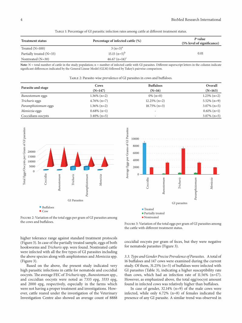
3.1. Treatment Status of Cattle against Intestinal Parasites.The study population was comprised of 100 “treated,” 33“partially treated,” and 30 “nontreated” individuals. The
parasitological observations confirmed that the immunityagainst gastrointestinal (GI) parasitic infections increasedalong with the effectiveness of the treatment status. Thenontreated animals indicated the highest percentage of par-asitic infections accounting for 46.67% (n= 14), followed bypartially treated individuals (15.15%, n= 5).
Meanwhile, the treated population was denoted by only3% (n= 3) occurrence of GI parasitic infections (Table 1).As indicated in General Linear Modelling (GLM), the effectof treatment status was significantly correlated with theprevalence of GI parasitic infections (P<0.05 at 5% level ofsignificance) in the study population. The results of post hocanalysis (Tukey’s pairwise comparison) confirmed that theGI infection rates differed significantly among cattle in threetreatment categories (Table 1).
3.2. Prevalence of Parasites in Cattle Population. A varietyof GI parasites, namely, eggs of hookworms (Bunostomumspp.), whipworms (Trichuris spp.), amphistomes, cestodes(Moniezia spp.), and oocysts of protozoans (coccidians), werefound within the study population of cattle. The parasite-wiseprevalence of GI parasites in cows and buffaloes is illustratedin Table 2. In general, the total number of eggs or oocystsfound in cows was relatively higher than that of buffaloes(Figure 2).
Only coccidian oocysts were found among the treatedsample of cattle indicating that these parasites may have a
4 BioMed Research International
Table 1: Percentage of GI parasitic infection rates among cattle at different treatment status.
Treatment status Percentage of infected cattle (%) P value(5% level of significance)
Treated (N=100) 3 (n=3)a
0.01Partially treated (N=33) 15.15 (n=5)b
Nontreated (N=30) 46.67 (n=14)c
Note: N = total number of cattle in the study population; n = number of infected cattle with GI parasites. Different superscript letters in the column indicatesignificant differences indicated by the General Linear Model (GLM) followed by Tukey’s pairwise comparison.
Table 2: Parasite-wise prevalence of GI parasites in cows and buffaloes.
Parasite and stage Cows Buffaloes Overall(N=147) (N=16) (N=163)
Bunostomum eggs 1.36% (n=2) 0% (n=0) 1.23% (n=2)Trichuris eggs 4.76% (n=7) 12.25% (n=2) 5.52% (n=9)Paramphistomum eggs 1.36% (n=2) 18.75% (n=3) 3.07% (n=5)Moniezia eggs 0.68% (n=1) - 0.61% (n=1)Coccidians oocysts 3.40% (n=5) - 3.07% (n=5)
05000
100001500020000
Tota
l Egg
s/O
ocys
ts pe
r Gra
m o
f GI p
aras
ites
GI Parasites
BuffaloesCow
Coc
cidi
a
Buno
stom
um
Trich
uris
Para
mph
istom
um
Mon
eizia
Figure 2: Variation of the total eggs per gram of GI parasites amongthe cows and buffaloes.
higher tolerance range against standard treatment protocols(Figure 3). In case of the partially treated sample, eggs of bothhookworms and Trichuris spp. were found. Nontreated cattlewere infected with all the five types of GI parasites includingthe above species along with amphistomes andMoniezia spp.(Figure 3).
Based on the above, the present study indicated veryhigh parasitic infections in cattle for nematode and coccidialoocysts.The average FEC of Trichuris spp.,Bunostomum spp.,and coccidian oocysts were noted as 7333 epg, 3333 epg,and 2000 epg, respectively, especially in the farms whichwere not having a proper treatment and investigations. How-ever, cattle reared under the investigation of the VeterinaryInvestigation Centre also showed an average count of 8888
0
2000
4000
6000
8000
10000
Aver
age E
ggs p
er G
ram
s of G
I Par
asite
s
GI parasites
TreatedPartially treatedNontreated
Buno
stom
um
Trich
uris
Para
mph
istom
um
Mon
iezia
Coc
cidi
a
Figure 3: Variation of the total eggs per gram of GI parasites amongthe cattle with different treatment status.
coccidial oocysts per gram of feces, but they were negativefor nematode parasites (Figure 3).
3.3. Type andGender Precise Prevalence of Parasites. A total of16 buffaloes and 147 cows were examined during the currentstudy. Of them, 31.25% (n=5) of buffaloes were infected withGI parasites (Table 3), indicating a higher susceptibility ratethan cows, which had an infection rate of 11.56% (n=17).However, as emphasized above, the total egg/oocyst amountfound in infected cows was relatively higher than buffaloes.
In case of gender, 32.14% (n=9) of the male cows wereinfected, while only 6.72% (n=8) of females indicated thepresence of any GI parasite. A similar trend was observed in

BioMed Research International 5
Table 3: Percentage of GI parasite infection based on gender.
Type Gender Percentage of infectedcattle (%)
Percentage of overallinfection (%)
P value(5% level of significance)
Cows (N=147) Male (N=28) 32.14 (n=9)a 11.56a
0.04Female (N=119) 6.72 (n=8)b (17/147)
Buffaloes (N=16) Male (N=5) 60.00 (n=3)c 31.25b
Female (N=11) 18.18 (n=2)c (5/16)
Overall (N=163) Male (N=33) 36.36 (n=12)a 0.03Female (N=130) 7.69 (n=10)b
Note: N = total number in the study population; n = number of infected individuals with GI parasites. Different superscript letters in a column show significantdifferences (P < 0.05) indicated by Tukey’s pairwise tests after GLM.
buffaloes also, whereby 60.0% (n=3) of males were infected.In general, out of 33males, 12 were infected with GI parasites,denoting that males had a higher prevalence rate of GIparasites (Table 3).
The GLM advocated that males had a significantly highersusceptibility to GI parasites than females (P<0.05), whilebuffaloes had a significantly higher prevalence of GI parasitesthan cows (P<0.05, at 5% level of significance). Interestingly,the interaction among types and genders on the prevalenceof parasites was also significant (P =0.04), in accordance withthe results of GLM.
Amphistome eggs present in male buffaloes (18.75%)were higher than the female buffaloes. Trichuris spp. eggswere found in equal amounts among both male and femalebuffaloes. Oocysts of Coccidia (2.04%) and eggs of Trichurisspp. (3.4%) were more in male cows than the female cows.Eggs of hookworms and amphistomes were observed in bothmale and female cows equally, while eggs of Moniezia spp.were found only in female cows (0.68%).
It was interesting to note that even though all the above-mentioned parasites were found in the infected population ofcows, only Paramphistomum and Trichuris spp. were found inbuffaloes (Figure 2). The results of the GLM confirmed thatthe prevalence rates of GI parasites differed among the cowsand buffaloes (P < 0.05).
3.4. Age-Wise Prevalence of Gastrointestinal Parasites. Thecattle population was defined as calve (≤ 12 months),yearling/heifer (13–60 months), and elderly/matured (>60months) based on their life span. In cows, the calves (17.07%)had a higher susceptibility towards GI infections followedby yearling (14.29%). Interestingly, the elderly cows had thelowest infection rate of 4.0% (Table 4). Among buffaloes,yearlings (50%) had the highest rate of infections, followedby calves (33.33%). The effect of age on the prevalence ofGI parasites was also significant as indicated by the GLM (P<0.05 at 95% level of confidence). Surprisingly, the combinedeffect of age and type was not statistically significant on thesusceptibility of the cattle (P >0.05) in accordance with thetest statistics of GLM (Table 4).
3.5. Effect of Spatial Location on the Parasitic Infection. Thestudy population included 163 cattle selected from threestudy areas, namely, Welisara (n=118), Kelaniya (n=35), and
0
5
10
15
20
25
30
35
Paramphistomum Coccidia Trichuris Moniezia BunostomumPerc
enta
ge p
reva
lenc
e of G
I par
asite
s (%
)
GI Parasites
Buffaloes MaleBuffaloes Female
Cows MaleCows Female
Figure 4: Dendrogram showing the spatial clustering of studiedsites based on the prevalence of GI parasitic infections among cattle.
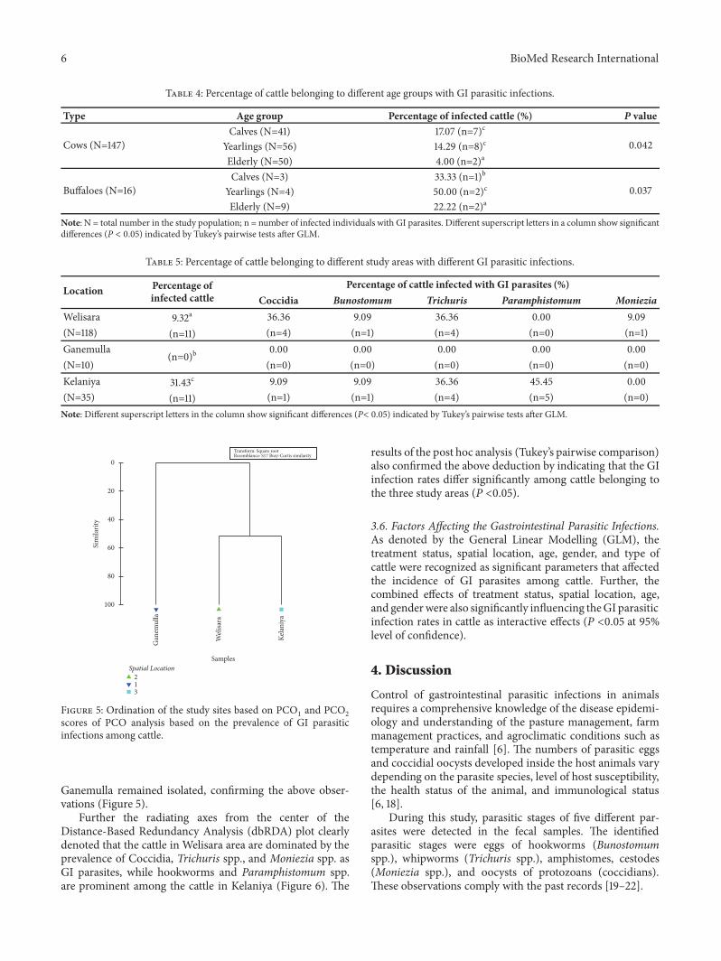
Ganemulla (n=10), covering 45 farms. The highest infectionrate of GI parasites was observed at Kelaniya (31.43%),followed byWelisara (9.32%). Interestingly, none of the cattlefrom the Ganemulla area were infected with any GI parasite.In case of the diversity of the GI parasites, all the observedparasites except for amphistomes were found from the cattlein the Welisara area. Subsequently, Moniezia spp. were notfound from the cattle population in the Kelaniya area, whichwere screened for intestinal parasites (Table 5).
As indicated by the results of GLM, the spatial location ofthe cattle significantly affected the incidence of GI parasitesin cattle (P =0.02, at 95% level confidence). The overallclustering status of cattle from different areas in terms of theprevalence of parasite types is illustrated in Figure 4.
As indicated by theBray-Curtis similarity clustering, bothKelaniya and Welisara share a similarity of 51.4 % in termsof the prevalence and diversity of GI parasites among cattle.Meanwhile, cattle from Ganemulla remain isolated from theabove cluster (Figure 5). The global R value of 0.92 gainedfor the analysis of similarities (ANOSIM) also confirmed theabove observation at a significance level of 5%.
Meanwhile, both PC1(84.1 %) and PC
2(15.9 %) axes
of the Principal Coordinates (PCO) that accounted for thetotal variation (100%) of the GI parasite prevalence amongthe studied cattle population suggested the emergence oftwo major clusters as Kelaniya and Welisara together, while
6 BioMed Research International
Table 4: Percentage of cattle belonging to different age groups with GI parasitic infections.
Type Age group Percentage of infected cattle (%) P value
Cows (N=147)Calves (N=41) 17.07 (n=7)c
0.042Yearlings (N=56) 14.29 (n=8)c
Elderly (N=50) 4.00 (n=2)a
Buffaloes (N=16)Calves (N=3) 33.33 (n=1)b
0.037Yearlings (N=4) 50.00 (n=2)c
Elderly (N=9) 22.22 (n=2)a
Note: N = total number in the study population; n = number of infected individuals with GI parasites. Different superscript letters in a column show significantdifferences (P < 0.05) indicated by Tukey’s pairwise tests after GLM.
Table 5: Percentage of cattle belonging to different study areas with different GI parasitic infections.
Location Percentage ofinfected cattle
Percentage of cattle infected with GI parasites (%)Coccidia Bunostomum Trichuris Paramphistomum Moniezia
Welisara 9.32a 36.36 9.09 36.36 0.00 9.09(N=118) (n=11) (n=4) (n=1) (n=4) (n=0) (n=1)Ganemulla
(n=0)b0.00 0.00 0.00 0.00 0.00
(N=10) (n=0) (n=0) (n=0) (n=0) (n=0)Kelaniya 31.43c 9.09 9.09 36.36 45.45 0.00(N=35) (n=11) (n=1) (n=1) (n=4) (n=5) (n=0)Note: Different superscript letters in the column show significant differences (P< 0.05) indicated by Tukey’s pairwise tests after GLM.
Gan
emul
la
Weli
sara
Kela
niya
Samples
Transform: Square rootResemblance: S17 Bray-Curtis similarity
Spatial Location213
100
80
60
40
20
0
Sim
ilarit
y
Figure 5: Ordination of the study sites based on PCO1and PCO
2
scores of PCO analysis based on the prevalence of GI parasiticinfections among cattle.
Ganemulla remained isolated, confirming the above obser-vations (Figure 5).
Further the radiating axes from the center of theDistance-Based Redundancy Analysis (dbRDA) plot clearlydenoted that the cattle in Welisara area are dominated by theprevalence of Coccidia, Trichuris spp., and Moniezia spp. asGI parasites, while hookworms and Paramphistomum spp.are prominent among the cattle in Kelaniya (Figure 6). The
results of the post hoc analysis (Tukey’s pairwise comparison)also confirmed the above deduction by indicating that the GIinfection rates differ significantly among cattle belonging tothe three study areas (P <0.05).
3.6. Factors Affecting the Gastrointestinal Parasitic Infections.As denoted by the General Linear Modelling (GLM), thetreatment status, spatial location, age, gender, and type ofcattle were recognized as significant parameters that affectedthe incidence of GI parasites among cattle. Further, thecombined effects of treatment status, spatial location, age,and genderwere also significantly influencing theGI parasiticinfection rates in cattle as interactive effects (P <0.05 at 95%level of confidence).
4. Discussion
Control of gastrointestinal parasitic infections in animalsrequires a comprehensive knowledge of the disease epidemi-ology and understanding of the pasture management, farmmanagement practices, and agroclimatic conditions such astemperature and rainfall [6]. The numbers of parasitic eggsand coccidial oocysts developed inside the host animals varydepending on the parasite species, level of host susceptibility,the health status of the animal, and immunological status[6, 18].
During this study, parasitic stages of five different par-asites were detected in the fecal samples. The identifiedparasitic stages were eggs of hookworms (Bunostomumspp.), whipworms (Trichuris spp.), amphistomes, cestodes(Moniezia spp.), and oocysts of protozoans (coccidians).These observations comply with the past records [19–22].

BioMed Research International 7
Transform: Square rootResemblance: S17 Bray-Curtis similarity
Spatial Location213
Similarity20406080
Welisara
Ganemulla
Kelaniya
−40
−20
0
20
40
0 20 40−40−60 −20−80
0#/
2(1
5.9%
of t
otal
var
iatio
n)
0#/1 (84.1% of total variation)
Figure 6: dbRDA plot depicting spatial variation of cattle belonging to different study sites based on the prevalence of GI parasites.
The literature states that nematode infections and coc-cidial infections in cattle are considered highly parasitic ifthey exceed ≥500 eggs per gram (epg) and a count of ≥5000oocysts per gram of feces, respectively [23, 24]. Fecal eggcount (FEC) of 100 eggs per gram (epg) or more fromBunostomum species is likely to indicate severe damage,whereas a count of 500 epg of Cooperia species is expected toproducemild helminthosis in cattle [25]. Based on the above,the present study indicated very high parasitic infections incattle for nematode and coccidial oocysts. The average FECof Trichuris spp., Bunostomum spp., and coccidian oocystswere noted as 7333 epg, 3333 epg, and 2000 epg, respectively,especially in the farms which were not having a propertreatment and investigations. However, cattle reared underthe investigation of the Veterinary Investigation Centre alsoshowed an average count of 8888 coccidial oocysts per gramof feces, but they were negative for nematode parasites.
It is interesting to note that coccidian parasites were theonly abundant parasite identified from treated farms. Thereason for this deviation may be due to the development of ahigher resistance of Coccidia against the standard treatmentprotocol for cattle at 21-day, 3-month, 6-month, and 12-month intervals, respectively, with a standard mixture ofAlbendazole and Fenbendazole based on the body mass.Most farmers (n=100) in this study were registered underthe Veterinary Investigation Centre and reared cattle inhousehold farms. Therefore, restrictions in open grazingand proper management practices may be the reason forlow prevalence of gastrointestinal parasites among treatedcattle. Their control is often achieved by prophylactic use ofanthelmintic treatments.
The results of this study indicated that the highest per-centage of parasitic infections accounting for 46.67% (n=14)was from nontreated cattle. The statistical analysis indicated
that the treatment status has significantly influenced thegastrointestinal parasitic infections (P =0.01 at 5% level ofsignificance). Adding more information to this observation,prevalence of gastrointestinal parasites has been reducedalong with the deworming treatment procedures over time(veterinary surgeon at the Veterinary Investigation Centre,Welisara, Pers.Comm.). According to Rajakaruna and War-nakulasooriya [3], deworming and management practiceslead to low prevalence of gastrointestinal parasites.
However, most of the cattle in nontreated farms wereopen grazing animals and they were almost never treatedfor any GI infections. Grazing often encourages enteringof different parasitic stages into the digestive tract of cattlethrough oral ingestion [3]. Fecal egg counts are highlyimportant as an indicator to decide the period that thecattle have to be given deworming treatments. This canalso be used after deworming treatments to investigatethe effectiveness of a particular anthelmintic. Therefore,unnecessary costs of veterinary services and drugs can bereduced.
When using fecal egg counts, there are some limitationsto determining the significance of the prevalence of flukes.The number of parasitic eggs per gram of feces is influencedby the fecal consistency, total amount of feces produced, andtime of the day feces were collected. When the feces are dried,the parasitic eggs within the feces will be more concentrated.
The severity of gastrointestinal parasitic infections canbe due to the vulnerability of animals to internal parasitesand the poor immunity. The prevalence rate and clinicaldiseases may vary, based on different environmental factorsin different areas. The high prevalence of gastrointestinalnematodes and coccidian oocysts has been reported intropical regions including Sri Lanka, with prevalence ratesranging from 20 to 96% [13].

8 BioMed Research International
During this study the prevalence of parasitic infectionswas higher in buffaloes than cows. Some previous studieshave indicated more than 40% prevalence of GI parasitesamong buffaloes compared to that of cows [23, 24, 26, 27].However, some studies have specified more prevalence ratesof GI parasites among cows than in buffaloes [26]. Thevariation in early findings might be due to the differencein the number of fecal samples examined, period of study,and geoclimatic conditions that favor the survival of infectivestages of the parasites and of intermediate hosts, manage-ment conditions, and deworming practices. In addition, thevariation in the prevalence of GI parasites in cows andbuffaloes may be attributed to differences in feeding andgeneral habitats of the two species [28].
Overall, sex-wise prevalence of GI parasites was higherin males when compared to that of females among bothcows and buffaloes. The higher percentages of infection inmales cannot be explained exactly, but it might be due to theneglected attitude of the farmers toward the management ofmale animals since many of the farms target milk productionthereby focusing more on the health of females. Thesefindings are in agreement with several other studies fromdifferent corners of the world [29–31].
According to Pfukenyi et al. [32] the susceptibility andpathogenicity of GI infections are greater in young animalsthan the matured ones. The present study reveals that mostGI parasites were higher in calves and yearlings than inelderly cows. In case of buffaloes, the yearlings were the mostsusceptible followed by calves. Therefore, the findings of thecurrent study suggest the fact that younger stages of bothcows and buffaloes are more susceptible to GI parasites thanthe elderly stages. A recent study conducted in Ethiopia hasalso reported a similar finding, where the younger animalsremained more susceptible than adults [33]. On the contrary,the increase in the prevalence of GI parasites with age has alsobeen reported by several other researchers [34, 35]. However,the causes for variations in the prevalence of parasites atdifferent age groups are difficult to explain, but they mightbe due to an immunological status of the animals, differencein the grazing area, and management conditions [33].
A significantly higher proportion of calves were infectedwith Coccidia than other age groups. This may be dueto the fact that high humidity and moderate temperaturefacilitate the survival and sporulation of the oocysts. As theirimmunity is also lower than the adult cattle, calves may bemore susceptible to coccidian infections [30]. In addition,Moniezia spp. were seen in elderly cattle. These reared cattleare pasture grazing animals. According to Irie et al.,Oribatulasakamorii (Oribatid mites) act as the intermediate host ofMoniezia spp. [36]. Oribatid mites mainly live in the soilcontaining low biomass, such as those at farming fields,sandbreak plantations along beaches, greenbelts of urbanareas, and organic matter, especially in cattle bedding litter.Therefore, larvae of the mite found in soil or litter may alsobe detected in the rectal feces of a cow, suggesting that thecattle at the farm had ingested them through oral ingestion.In this study, the exact presence of Oribatid mites in the farmsis not known. Perhaps it might be one of the reasons forMoniezia spp. infections. The yearlings of buffaloes had the
highest infections, followed by calves. Buffaloes were infectedwith Trichuris spp. and Paramphistomum. None of the otherparasite stages could be observed among them. According toBilal et al. [30], calves on grazing are heavily infected withGI parasites compared to stall fed calves and the male buffalocalves are more affected than female calves.
The highest GI infection rate (31.43%) was observed inKelaniya, followed by Welisara (9.32%). According to thedistribution pattern, amphistomes were observed from thecattle, which were at Kelaniya, while Moniezia spp. werefound only at the Welisara area. The difference in prevalenceof GI parasitic infections might be due to variation ingeoclimatic conditions of these areas of study. The presentstudy areas are located in the wet zone of Sri Lanka with ahigh species diversity of aquatic snails [37]. Therefore, thesesnails can act as the possible intermediate host for digeneantrematodes. On the other hand, temperature directly affectsthe cercarial output owing to both the stimulating effect oftemperature increasing the emergence from the snail andthe acceleration of cercarial production within the snail host[38].
Overall, the present investigation indicated that the treat-ment status, age, gender, and spatial location were signif-icantly influencing the gastrointestinal parasitic infectionsamong cattle in the selected farms located in GampahaDistrict of Sri Lanka. This survey also highlights howthe deworming and management practices of cattle affectthe prevalence of parasitic infections. Therefore, periodicalmonitoring of the prevalence of GI parasites among farmanimals is essential, in order to achieve the expected goalsin deworming activities, rational use of anthelminthic drugs,and proper farm management.
5. Conclusion
Hookworms (Bunostomum spp.), whipworms (Trichurisspp.), amphistomes, cestodes (Moniezia spp.), and protozoans(coccidians) were diagnosed as gastrointestinal (GI) parasitesfrom the cattle.The effect of treatment status was significantlycorrelated with the prevalence of GI parasitic infectionsin the study population. Males carried significantly higherGI parasites than females. Buffaloes showed significantlyhigher level of susceptibility to GI parasites than cows. Theyearlings and calves of both cows and buffaloes had thehighest rate of GI parasitic infections compared to adultanimals. GI parasites in cattle population varied with spatiallocations among the selected areas of Gampaha District.Future investigations are necessary to evaluate the economicimpact of GI parasites in the study areas.
Data Availability
The collected data will be kept confidential. Data will not beshared in any of the sources.
Conflicts of Interest
The authors declare that they have no conflicts of interest.

BioMed Research International 9
Authors’ Contributions
NayanaGunathilaka contributed to designing of the research,supervision of the research work, and writing of themanuscript. Dimuthu Niroshana contributed to conductinglaboratory experiments. Deepika Amarasinghe contributedin sample identification, supervision of research work, andwriting of the manuscript. Lahiru Udayanga contributed tostatistical analysis and writing of the manuscript. All authorsread and approved the final manuscript.
Acknowledgments
All staff at the Veterinary Investigation Centre, Welisara,Ragama, Sri Lanka, and owners of the selected farms for thepresent study are greatly acknowledged. The authors wouldalso like to acknowledge the technical staff of the Departmentof Parasitology, Faculty of Medicine, University of Kelaniya,and Department of Zoology and Environment Management,Faculty of Science, University of Kelaniya, Sri Lanka.
References
[1] H. Abeygunawardena, D. Rathnayaka, and W. M. A. P. Jayati-lake, “Characteristics of cattle farming systems in Sri Lanka,”Journal of the National Science Council of Sri Lanka, vol. 25, no.1, pp. 25–38, 1997.
[2] H. Hoste, F. Jackson, S. Athanasiadou, S. M. Thamsborg, andS. O. Hoskin, “The effects of tannin-rich plants on parasiticnematodes in ruminants,” Trends in Parasitology, vol. 22, no. 6,pp. 253–261, 2006.
[3] R. S. Rajakaruna andK. N.Warnakulasooriya, “Gastrointestinalparasites in dairy cattle in Kandy district in Sri Lanka,” AnnualResearch Journal of SLA, pp. 92–99, 2011.
[4] M. R. Akanda, M. M. I. Hasan, S. A. Belal et al., “A survey onprevalence of gastrointestinal parasitic infection in cattle of Syl-het division in Bangladesh,”American Journal of Phytomedicineand Clinical Therapeutics, vol. 2, pp. 855–860, 2014.
[5] R. Herlich, “Common gastrointestinal parasites of cattle,” Jour-nal of Veterinary Science, vol. 10, pp. 25–32, 1978.
[6] S. P. A. L Sharma and M. Busang, Prevalence of some gastroin-testinal parasites of ruminants in southern Botswana, 2014.
[7] A. C. M. Faizal and R. P. V. J. Rajapakse, “Prevalence of coccidiaand gastrointestinal nematode infections in cross bred goats inthe dry areas of Sri Lanka,” Small Ruminant Research, vol. 40,no. 3, pp. 233–238, 2001.
[8] F. R. Gwaze, M. Chimonyo, and K. Dzama, “Prevalence andloads of gastrointestinal parasites of goats in the communalareas of the Eastern Cape Province of South Africa,” SmallRuminant Research, vol. 84, no. 1-3, pp. 132–134, 2009.
[9] D. K. Sharma, N. Agrawal, A. Mandal et al., “Coccidia andgastrointestinal nematode infections in semi-intensively man-aged Jakhrana goats of semi-arid region of India,” Tropical andSubtropical Agroecosystems, vol. 11, no. 1, pp. 135–139, 2009.
[10] M. M. Hassan, M. A. Hoque, S. K. M. A. Islam et al., “Aprevalence of parasites in black bengal goats in Chittagong,”International Journal of Livestock Production, vol. 2, pp. 40–44,2011.
[11] E. B. Gebeyehu, M. G. Seo, B. Y. Jung et al., “Prevalenceof gastrointestinal parasites in Korean native goats (Capra
hircusaegagrus),” Journal of Animal and Plant Sciences, vol. 23,no. 4, pp. 986–989, 2013.
[12] H. McL. Gordon, Efficiency of certain drugs againstHaemonchuscontortus: with a note on the treatment oftrichostrongylosis, 1935.
[13] I. Sutherland and I. Scott, Gastrointestinal nematodes of sheepand cattle: biology and control, John Wiley & Sons, 2010.
[14] A. S. Familton, P. Mason, and G. C. Coles, “Anthelmintic-resistant Cooperia species in cattle,”Veterinary Record, vol. 149,no. 23, pp. 719-720, 2001.
[15] G. Kandasamy, R. P. V. J. Rajapakse, and R. S. Rajakaruna,“Gastrointestinal and blood parasites of a free grazing flock ofsheep in Kaithady farm in the Jaffna District,” Journal of theNational Science Foundation of Sri Lanka, vol. 41, no. 3, pp. 195–201, 2013.
[16] Z. M. Anne and G. A. Conboy, Veterinary clinical parasitology,John Wiley & Sons, 2012.
[17] K. R. Clarke and R. M. Warwick, “An approach to statisticalanalysis and interpretation,” Change in Marine Communities,vol. 2, pp. 117–143, 1994.
[18] G.M. Urquhart, J. Armour, J. L. Duncan, A.M. Dunn, and F.W.Jennings, “The laboratory diagnosis of parasitism,” VeterinaryParasitology, vol. 2, pp. 4–11, 1996.
[19] M. A. P. Senadhira, “The parasites of Ceylon. IV. Nematoda. Ahost check list,” Ceylon Veterinary Journal, vol. 15, no. 3, pp. 83–99, 1967.
[20] A. C. M. Faisal, “Helminthiasis in livestock and poultry in SriLanka: a Review of literature,” Sri Lanka Veterinary Journal, vol.42, no. 2, pp. 1–8, 1995.
[21] L. D. Amarasinghe and H. L. Kumara, “Effect of rainfall andtemperature on liver and rumen fluke infestations of bovinesin Sri Lanka,” International Journal of Biological and ChemicalSciences, vol. 1, no. 3, pp. 229–236, 2007.
[22] L. D. Amarasinghe and T. Maduranga, “Intensity and dispersalof rumen flukes of cattle in selected areas of Sri Lanka,” Journalof Global Biosciences, vol. 5, no. 1, pp. 3500–3512, 2016.
[23] A. Gupta, A. K. Dixit, P. Dixit, and C. Mahajan, “Prevalence ofgastrointestinal parasites in cattle and buffaloes in and aroundJabalpur, Madhya Pradesh,” Journal of Veterinary Parasitology,vol. 26, no. 2, pp. 186–188, 2012.
[24] Z. Kashyap, R. S. Sisodia, and P. C. Shukla, “Incidence ofgastrointestinal parasites in cattle and buffaloes inMalwa regionof Madhya Pradesh,” Haryana Veterinarian, vol. 36, pp. 34–36,1997.
[25] E. J. L. Soulsby, Helminths, arthropods and protozoa of domesti-cated animals, Bailliere Tindall, 7 edition, 1982.
[26] M. R. Mir, M. Z. Chishti, M. Rashid et al., “Point Prevalence ofGastrointestinal Helminthiaisis in large Ruminants of Jammu,India,” International Journal of Scientific and Research Publica-tions, vol. 3, no. 3, pp. 5–8, 2013.
[27] Khalil-ur-Rehman, K. Javed, M. T. Tunio, and Z. H. Kuthu,“Passive surveillance of gastrointestinal parasites in bufflaoes ofMandi Bahauddin andGujrat districts of the Punjab,” Journal ofAnimal and Plant Sciences, vol. 19, no. 1, pp. 17–19, 2009.
[28] P. Marskole, Y. Verma, A. K. Dixit, and M. Swamy, “Prevalenceand burden of gastrointestinal parasites in cattle and buffaloesin Jabalpur, India,”Veterinary World, vol. 9, no. 11, pp. 1214–1217,2016.
[29] A. Tigist, B. Bogale, and M. Chanie, “Occurrence of gastrointestinal nematodes of cattle in and around Gondar town,Amhara regional state, Ethiopia,” Acta Parasitologica Globalis,vol. 3, no. 2, pp. 28–33, 2012.
10 BioMed Research International
[30] M. Q. Bilal, A. Hameed, and T. Ahmad, “Prevalence of gastroin-testinal parasites in buffalo and cow calves in rural areas of TobaTek Singh, Pakistan,” Journal of Animal and Plant Sciences, vol.19, no. 2, pp. 67–70, 2009.
[31] R. Fikru, S. Teshale, D. Reta, and K. Yosef, “Epidemiology ofgastro intestinal parasites of ruminants in Western OromiaEthiopia,” International Journal of Applied Research in Veteri-nary Medicine, vol. 4, pp. 51–57, 2006.
[32] D. M. Pfukenyi, S. Mukaratirwa, A. L. Willingham, and J.Monrad, “Epidemiological studies of parasitic gastrointestinalnematodes, cestodes and coccidia infections in cattle in thehighveld and lowveld communal grazing areas of Zimbabwe,”Onderstepoort Journal of Veterinary Research, vol. 74, no. 2, pp.129–142, 2007.
[33] F. Regassa, T. Sori, R. Dhuguma, and Y. Kiros, “Epidemiologyof gastrointestinal parasites of ruminants in Western Oromia,Ethiopia,” International Journal of Applied Research in Veteri-nary Medicine, vol. 4, no. 1, p. 51, 2006.
[34] A. W. Qureshi and A. Tanveer, “Seroprevalence of fasciolosisin buffaloes and humans in some areas of Punjab, Pakistan,”Pakistan Journal of Science, vol. 61, no. 2, pp. 91–96, 2009.
[35] T. Cheru, A. Birhanu, L. Diriba, and E. Eyob, “Prevalence ofgastrointestinal parasitismof cattle in East ShowaZone,OromiaRegional State, Central Ethiopia,” Journal of VeterinaryMedicineand Animal Health, vol. 6, no. 2, pp. 54–62, 2014.
[36] T. Irie, K. Sakaguchi, A. Ota-Tomita et al., “ContinuousMoniezia benedeni infection in confined cattle possibly main-tained by an intermediate host on the farm,” Journal of Veteri-nary Medical Science, vol. 75, no. 12, pp. 1585–1589, 2013.
[37] U. A. Jayawardena, R. S. Rajakaruna, and P. H. Amerasinghe,“Cercariae of trematodes in freshwater snails in three climaticzones in Sri Lanka,” Ceylon Journal of Science (BiologicalSciences), vol. 39, no. 2, p. 95, 2011.
[38] R. Poulin, “Global warming and temperature-mediatedincreases in cercarial emergence in trematode parasites,”Parasitology, vol. 132, no. 1, pp. 143–151, 2006.

Hindawiwww.hindawi.com
International Journal of
Volume 2018
Zoology
Hindawiwww.hindawi.com Volume 2018
Anatomy Research International
PeptidesInternational Journal of
Hindawiwww.hindawi.com Volume 2018
Hindawiwww.hindawi.com Volume 2018
Journal of Parasitology Research
GenomicsInternational Journal of
Hindawiwww.hindawi.com Volume 2018
Hindawi Publishing Corporation http://www.hindawi.com Volume 2013Hindawiwww.hindawi.com
The Scientific World Journal
Volume 2018
Hindawiwww.hindawi.com Volume 2018
BioinformaticsAdvances in
Marine BiologyJournal of
Hindawiwww.hindawi.com Volume 2018
Hindawiwww.hindawi.com Volume 2018
Neuroscience Journal
Hindawiwww.hindawi.com Volume 2018
BioMed Research International
Cell BiologyInternational Journal of
Hindawiwww.hindawi.com Volume 2018
Hindawiwww.hindawi.com Volume 2018
Biochemistry Research International
ArchaeaHindawiwww.hindawi.com Volume 2018
Hindawiwww.hindawi.com Volume 2018
Genetics Research International
Hindawiwww.hindawi.com Volume 2018
Advances in
Virolog y Stem Cells International
Hindawiwww.hindawi.com Volume 2018
Hindawiwww.hindawi.com Volume 2018
Enzyme Research
Hindawiwww.hindawi.com Volume 2018
International Journal of
MicrobiologyHindawiwww.hindawi.com
Nucleic AcidsJournal of
Volume 2018
Submit your manuscripts atwww.hindawi.com