PREVALANCE OF RADIOLOGICAL CHANGES IN THE SPINES OF …
84
1 PREVALANCE OF RADIOLOGICAL CHANGES IN THE SPINES OF SOUTH AFRICAN FEMALE ARTISTIC GYMNASTS Adele Geldenhuys-Koolen 0000691G A research report submitted to the Faculty of Health Sciences, University of the Witwatersrand, in fulfilment of the requirements for the degree of Master of Science (Medicine) in the field of Exercise Science Johannesburg 2012
Transcript of PREVALANCE OF RADIOLOGICAL CHANGES IN THE SPINES OF …

1
PREVALANCE OF RADIOLOGICAL CHANGES IN THE SPINES OF SOUTH
AFRICAN FEMALE ARTISTIC GYMNASTS
Adele Geldenhuys-Koolen
0000691G
A research report submitted to the Faculty of Health Sciences, University of the
Witwatersrand, in fulfilment of the requirements for the degree
of
Master of Science (Medicine) in the field of Exercise Science
Johannesburg 2012

2
DECLARATION
I, Adele Geldenhuys-Koolen declare that this dissertation is my own work. It is being
submitted for the degree of Master of Science in Medicine in the field of Exercise
Science at the University of the Witwatersrand, Johannesburg. It has not been
submitted before for any degree or examination at this or any other university.
Signed at Fourways on this 30th day of October 2012.
Adele Geldenhuys-Koolen

3
AKNOWLEDGEMENTS
Our Heavenly Father for guidance and enabling me.
My supervisors, Prof. Y. Coopoo and Prof. D. Constantinou for
encouragement, guidance and patience throughout this project.
The coaches, gymnasts and parents from JGC, Visions and Centurion
gymnastics clubs for their contribution to the study.
Dr. Peter Goldschmidt, for the endless hours of analysing the X-Rays.
Dr. Conidaris & Partners, for the use of their facility for the taking of the X-
Rays.
Ms. Juliana van Staden, statistician, for assistance with data analysis, and
many Sunday afternoon meetings.
Susan Benedict and Sunè Bezuidenhout, for the editing of the final product.
My husband, mom, and employees, for the encouragement, understanding
and guidance throughout this project.

4
TABLE OF CONTENTS Page
DECLARATION 2
ACKNOWLEDGEMENTS 3
TABLE OF CONTENTS 4 - 6
LIST OF TABLES 7
LIST OF APPENDICES 8
1.0 CHAPTER 1: Introduction
1.1 Introduction 9 - 12
1.2 Aim 12
1.3 Objectives 12
1.4 Hypothesis 12
1.5 Limitations 12
1.6 Assumptions 12
1.7 Definitions of Terms 13 - 14
2.0 CHAPTER 2: Literature Review
2.1 Introduction 15 - 16
2.2.1 Growth and development 16 - 18
2.2.2Effect of nutrition on growth and development 18
2.2.3 Effect of exercise on growth and development 18 - 21
2.2.4 Effect of exercise on bone mineral density 21
2.3 Prevalence of back injuries 21 - 23
2.3.1.1 Spondylolysis and spondylolisthesis 23 - 25
2.3.1.2 Aetiology of spondylolysis and spondylolisthesis 26
2.3.1.3 Pathology of spondylolysis and spondylolisthesis 26 - 27
2.3.1.4 Conclusion 27
2.3.2 Discogenic pain 27 - 28
2.3.2.1 Aetiology of discogenic pain 28

5
2.3.2.2 Pathology of discogenic pain 28 – 29
2.3.3 Treatment of lumbar injuries in gymnasts 28 - 29
3.0 CHAPTER 3: Methodology
3.1 Study design 30
3.2 Population 30
3.3 Sample selection 30
3.3.1 Inclusion criteria 30 - 31
3.3.2 Exclusion criteria 31
3.4 Main Study 31
3.5 Questionnaires 31 - 32
3.5.1 Content and construct validity 32
3.6 Procedure 32
3.6.1 Pilot study 32 - 33
3.6.1.1 Results of the pilot study 33
3.6.1.1.1 Clarity and ambiguity of questions 33
3.6.1.1.2 Duration 33
3.6.1.1.3 Test-retest reliability of the questionnaires 33
3.7 Radiological testing (X-Rays) 33
3.7.1 Procedures 33
3.7.2 Views 33 - 34
3.7.2.1 Antero-posterior views 34
3.7.2.2 Oblique views (left and right) 34
3.7.2.3 Lateral views 34 - 35

6
3.7.2.4 Lateral coned L5,S1 angle view 35 - 37
3.8 Ethical consideration 37
4.0 CHAPTER 4: Statistical Analysis
4.1 Description of the sample 38 - 40
4.2 Prevalence of injuries 40 - 56
4.3 Prevalence of radiological changes 56 - 59
4.4 Summary 59
5.0 CHAPTER 5: Discussion 60 - 64
REFERENCES 65 - 73

7
LIST OF TABLES Page
Table
4.1 Analysis of level of competition of sample 39
4.2 Reasons for retirement 40
4.3 Areas of peripheral injury and types of injuries sustained 40 - 51
4.4 Type of back injury sustained 52
4.5 Peripheral injury and back injury profile cross-tabulated with 53 - 55
level of competition
4.6 Scoliosis on X-Ray for the gymnasts with back injury 56
4.7 Degenerative changes on X-Ray for the gymnasts with back 56
Injury
4.8 Spondylolysis changes on X-Ray for the gymnasts with back 57
injury
4.9 Spondylolisthesis changes on X-Ray for the gymnasts with back 57
injury
4.10 Scoliosis changes on X-Ray for gymnasts without back injury 57
4.11 Degenerative changes on X-Ray for gymnasts without back 58
injury
4.12 Spondylolysis changes on X-Ray for gymnasts without back 58
injury
4.13 Spondylolisthesis changes on X-Ray for gymnasts without 58
back injury

8
LIST OF APPENDICES Page
A. Consent form 74 - 77
B. Questionnaire (current gymnasts) 78 - 80
C. Questionnaire (retired gymnasts) 81 - 84
D. Ethical clearance certificate 85
9
PREVALANCE OF RADIOLOGICAL CHANGES IN THE SPINES OF SOUTH
AFRICAN FEMALE ARTISTIC GYMNASTS
Chapter One
1.1 Introduction
In South Africa, women’s gymnastics participation is on the increase both locally
and internationally (Chadwick, 2004 as cited in Adamson,2006). Competition
season for Level 1 - 4 gymnasts starts as early as March and finishes in June,
whereas Level 5 and higher start their competitions in June and continue through
September. The Junior and Senior Olympic entry girls start with testing
competitions as early as January, and the first major competition takes place in
February. Qualification trials for world championships and the All Africa Games
take place in South Africa throughout the year. The girls also are selected for
numerous international competitions based on their performances and their
rankings with the South African Gymnastics Federation.
Gymnastics appears to be associated with a high incidence of injury when
compared to most other sporting activities (Kolt and Kirby, 1996) as the amount
of impact that is applied to the body is relatively high in comparison with other
sports (Daly et al, 2001). The skeletal immaturity of gymnasts also allows for a
unique physiological predisposition to injuries because of growing spines, limbs,
ankles and wrists (Winkler, 2001).Studies have shown that lower back (spinal)
injuries account for approximately 12% of injuries in women’s gymnastics (Sands,
et al., 1993, Caine and Nassar, 2005). Case studies that have been published
show that back injuries tend to have a gradual onset, and involve predominantly
advanced level gymnasts (Caine, et al., 1996). These studies also have indicated
that an increase in skill and competitive level are risk factors for injury (Caine, et
al., 1996, Caine and Nassar, 2005).
The researcher has found in her experience working with gymnasts that the
majority of back injuries in gymnasts are confined to the lower back. These
clinical findings concur with that of prior research. Previous imaging studies
reported in the medical literature have shown that degenerative disc disease and

10
spinal injuries are more frequent in competitive female gymnasts than in
asymptomatic non-athletic people of the same age (Goldstein, et al., 1991; Tertti,
et al., 1990; Swärd, et al., 1990; Swärd, et al., 1991). Although these findings are
more prevalent in the competitive gymnast, other studies suggest that they may
not be of clinical significance (Tertti, et al., 1990). A study documenting Magnetic
Resonance Imaging (MRI) findings in symptomatic and asymptomatic Olympic
gymnasts was performed in the United States of America (USA), and reported
that symptomatic patients exhibited radiological changes that were absent from
asymptomatic patients (Bennett, et al., 2006).
Due to the lack of recent research on X-ray changes in gymnasts and the
questions relating to their clinical significance, there is a need to investigate the
prevalence of radiological changes in female artistic gymnasts in South Africa. It
is also important to note that, to date, there have been no research studies done
on artistic gymnasts in South Africa because the sport has only recently become
popular (Cameron-Smith, 2005 as cited in Adamson, 2006).
The focus of the study is on X-rays rather than Magnetic Resonance Imaging
(MRI) because MRI scans in South Africa is very expensive and impractical,
especially for the screening of asymptomatic subjects. MRI scans need to be
referred and motivated for by a specialist in the field, and are usually only
performed if the patient is symptomatic and when X-rays show radiological
changes and further investigation is indicated.
Sports related injuries are of two types. Macro trauma occurs as an acute,
perhaps dramatic, event like a concussion, spinal cord injury, fracture, or
dislocation. Micro trauma occurs as a repeated injury, usually not noticed initially
because the injury is microscopic in magnitude, but in which the cumulative
trauma leads to pain and, in some cases, significant disability. Stress fractures
and the so-called overuse syndromes are examples of micro trauma.
Any injury can be caused by any sport, but there are injuries that are recognized
to be particular to a specific sport (Boden et al, 2001). In gymnastics,
spondylolysis occurs frequently due to the hyperextended position and rotational
forces in gymnastic routines, e.g. back walk-overs (Bruggeman, 1999).

11
Spondylolysis may represent a form of stress fracture. Prevention includes
abdominal and spinal muscle strengthening (Yancy and Micheli, 1994; Standaert,
2002 and Miller et al, 2004). However, more recent evidence regarding specific
prevention and treatment is required.
Elbow injuries occur from excessive weight-bearing with the elbow in an
exaggerated valgus position and can lead to inflammation of the medial
epicondyle with micro tears of the flexor tendons (so-called “Little League elbow”)
(Caine and Nassar, 2005).
Wrist pain is frequent in young gymnasts due to constant stress on the lower
radial epiphysis or growth plate at the wrist. If ignored, this stress can cause
premature closure of the growth plate, leading to a Madelung deformity (Caine
and Nassar, 2005). Knee pain is very frequent in young gymnasts, especially
Osgood-Schlatter disease, Patellar tendonopathy, and Patello-femoral syndrome
(Caine and Nassar, 2005).
A study by Cohen and Stuecker (2005) showed the importance of detecting and
monitoring the early onset of spondylolysis which is the degeneration of the pars
interarticularis of the vertebrae. This condition is not isolated to gymnastics,
however it has a higher prevalence in sports where the lumbar spine is placed
under stress in hyper extension positions, i.e. gymnastics, ballet, swimming
(butterfly) or in unilateral sports such as bowling in cricket (Bruggeman, 1999).
According to the study by Cohen and Stuecker (2005) bracing and the avoidance
of strenuous activity prevented the formation of pars defects in all of their
subjects. Spondylolisthesis results from a bilateral pars defect (stress fracture).
Due to the dissolution of the pars on both sides, the vertebrae become unstable,
and movement of one vertebra on top of another can occur (Ciullo and Jackson,
1985).
The long term prognosis of spondylolisthesis, as defined and described by Miller,
et al., (2004), indicated that 91% had good to excellent outcomes up to 11 years
after conservative treatment. Surgery is indicated only if patients have persistent
neurological symptoms, have refractory pain, or have progressed to a Grade III or
Grade IV spondylolisthesis (>50-100% vertebral shift). Lumbar vertebral fusion
12
for slips usually requires 6 to 12 months hiatus before an athlete can compete in
non-contact sports (Rubery and Bradford, 2002; Eddy et al., 2005).
One of the benefits of this study is ascertaining the state of spinal health of this
group of gymnasts. Based on the results, group exercise programmes can be
created to prevent further back injuries, and to prevent progression of current
back injuries where they exist.
1.2 Aim
1.2.1. The aim of the study was determine the prevalence and nature of lower
back injuries in South African elite female artistic gymnast.
1.3 Objectives of the study
The primary objective of the study was to determine the prevalence of
radiological changes in South African female artistic gymnasts, ages 10 to
30 years.
A secondary objective was to determine if these radiological changes
occur more frequently in symptomatic vs. asymptomatic subjects.
A third objective was to determine if these radiological changes were
associated with the amount of time spent training gymnastics.
1.4 Hypothesis
Most female artistic gymnasts will have spinal problems as a result of the training
intensity.
1.5 Limitations
The study is limited to female non-professional artistic gymnasts, ages 10 to 30,
who reside in 1 province of the Republic of South Africa. All subjects were
obtained from three gymnastics clubs.
1.6 Assumptions
All radiological changes in the spine are a result of the gymnastics.
1.7 Definitions of Terms

13
Scoliosis: A lateral deviation of the normal vertical line of the spine. It may or
may not include rotation or deformity of the vertebrae.
Spondylosis: Degeneration of the disc spaces between the vertebrae.
Spondylolysis: The breaking down (dissolution) of a portion of a bony building
block of the spine (a vertebra). The portion that is affected is called the pars
interarticularis, which is located between the superior and inferior articular
processes of a lumbar vertebra. Therefore, spondylolysis is a separation of the
pars interarticularis.
Spondylolisthesis: Forward movement of one vertebrae of the spine in relation
to an adjacent vertebra. The spondylolisthesis can be either stable; i.e. there is
no translation, or unstable, there is translation. It can be either antero; i.e.
forward, or retro; i.e. backward, and is classified into four grades according to the
percentage of slippage of one vertebral body on the one below.
Grade I: 25% slippage of the vertebral body on the vertebra below.
Grade II: 50% slippage of the vertebral body on the vertebra below.
Grade III: 75% slippage of the vertebral body on the vertebra below.
The following classification will be used in terms of this study:
Mild Scoliosis: A lateral deviation when measured by X-ray is less than 5
degrees.
Moderate Scoliosis: A lateral deviation when measured by X-ray is between
5 and 10 degrees.
Severe Scoliosis: A lateral deviation when measured by X-ray is greater than
10 degrees.
Level of competition: In gymnastics the gymnasts start competing at Level 1.
The levels are both age and skill appropriate. In some cases when the
gymnasts show potential, they may skip a level or go into the high
performance program.

14
High Performance (HP): The high performance (HP) program is more difficult
for gymnasts of the same age. The program is geared for those gymnasts
identified by the coaches with the talent and potential to compete at
international level in events such as World Cups, World Championships and
the Olympic Games.

15
CHAPTER 2
LITERATURE REVIEW
2.1 INTRODUCTION
The literature review includes a discussion of participation in gymnastics,
growth and development of females, prevalence of back injuries, and a discussion of
the injuries associated with gymnastics. This discussion includes common locations
of injuries, severity, activities during which the injuries occurred, and mechanisms of
injury. Extrinsic factors that contribute to injuries, such as skill level and training
exposure, are also reviewed.
Competitive women’s gymnastics is well-established in Europe, Australia,
New Zealand, China and, to a limited extent, in Africa (Marshall et al, 2007; Kolt and
Kirkby, 1999; Richards et al, 2007; Caine et al, 1989). Children are participating in
organized sports at younger ages and in increasing numbers. This trend is
particularly evident in women’s gymnastics. According to Garrick and Requa (1980),
there was an increase of 461% in interscholastic participants in gymnastics in the
United States between 1974 and 1980. Since 1980, the number of interscholastic
participants has decreased while the number of gymnastics clubs who have younger
participants has increased significantly (Johnson, 1985). As early participation in
women’s gymnastics has become characteristic, the average age of champions and
Olympic gold medallists has decreased markedly over the past two decades. Before
1981, the minimum age for competition at senior level sanctioned by the
International Gymnastics Federation (FIG) was 14 years.
(www.aafla.org/Olympicinformationcenter/olympicreview). During the earlier years
of competition, gymnasts tended to be in their 20’s. In 1956, the Hungarian gymnast
Agnes Keleti won the individual all round Olympic gold medal at the age of 35.
Larissa Latynina from the Soviet Union, won her first Olympic all-round medal at age
21, and proceeded to win a second at age 25, and a third at 29 years of age. During
the 1970s, the average age of competitors gradually began to decrease. As difficulty
level increased, it became the norm to see teenagers competing. In July 1980, in
response to the changing demands of the sport, the FIG decided during their 58th
Congress to raise the age limit from 14 to 15 years. This rule was affected in 1981.
The rule stated that gymnasts had to turn at least 15 years of age in the calendar

16
year to compete at senior level. This limit remained in place until 1997, when the
age was once again raised by one year, from 15 to 16 years.
These age restrictions were designed to help prevent child athletes from
injury. There is no maximum age restriction for competition. The oldest international
female gymnast currently competing is Germany’s Oksana Chusovitina, who was
born in 1975 and was 33 years of age at the 2008 Beijing Summer Olympic Games.
She was 17 years of age when she competed in her first Olympic Games in 1992.
2.2.1. GROWTH AND DEVELOPMENT
It is of great importance to understand the growing musculoskeletal system in
order to understand the injuries that occur in children and adolescents. Linear
growth and physical maturation are dynamic processes encompassing molecular,
cellular, somatic, and organizational changes. Customarily, stature is primarily used
for growth assessment, but changes in body proportion and composition are
essential elements of growth, especially of maturation (Caine et al, 2003).
Growth and development are classified into stages from birth as infancy,
childhood, adolescence, and adulthood. Malina and Bourchard, (1991) defined
adolescence as the phase in which the child undergoes massive changes physically,
physiologically, and psychologically. Adolescence is also the transition period from
childhood to adulthood which starts after the onset of puberty and during which the
reproductive systems develop to maturity. Puberty is a dynamic period of
development marked by rapid changes in size, shape, and composition, all of which
are sexually dimorphic (Rogol et al., 2000).
Puberty maturation can be described in terms of sequence, tempo (puberty), and
timing. These stages consist of a series of predictable events, which are followed by
changes in secondary sexual characteristics, which have been categorized by
several groups. Marshal and Tanner (1983) published a staging system, which is
utilised most frequently and commonly referred to as the Tanner Stages (Tanner,
1968).
Rogol et al., (2000) found that, on average, girls enter and complete each stage of
puberty earlier than boys do. It has been noted that puberty begins earlier for girls
than boys with puberty for girls beginning between the ages 11 and 14 years while
17
that for boys occurs between 13 and 16 years (Gordon and Laufer, 2005). The
difference in the onset of puberty in girls and boys is mainly because of body
composition, which includes relative proportions of water, muscle, fat, and bone. The
activity of the pituitary gland at this stage of development leads to increased
secretion of hormones leading to a growth spurt alongside other physiological
effects. This results in the body achieving most of its adult height and weight in
about two years thereby contributing to the changes in muscle, fat, and bone.
Beilina and Fireman (1999) reviewed Piaget’s theories and determined that
adolescence is the onset of formal operational thought, characterized by thinking that
revolves around deductive logic. According to Piaget (2000), at puberty a child has a
perceptual-cognitive approach to his/her role in sports; e.g., for a gymnast, this is the
act before the actual performance. This would minimize injuries and stress
associated with failure. Research by Hall (1986), however, shows that the ability to
solve complex problems at puberty depends on accumulated learning and education.
He asserted that adolescence is a stage of emotional stress, resulting from the rapid
and extensive physiological changes arising at puberty. This emotional stress,
coupled with the growth spurt, may be a cause of difficulty for gymnasts who haven’t
mastered enough spatial awareness relative to recognize their growing body size.
Growth and development are also influenced by physical activity and sports.
On investigating growth parameters in the adolescent female gymnast, a consistency
was found with these girls being shorter and lighter and having a significantly lower
percentage of body fat than age-matched control girls or athletes participating in less
strenuous sports such as swimming. Girls participating in sports such as swimming
are generally taller and mature earlier than the non-sport participating group (Malina,
1994; Theintz et al., 1993; Constantini and Warren, 1995). A cohort study done by
Theintz et al. (1993) of gymnasts and swimmers over a 2-3 year period concluded
that gymnasts had significantly lower growth velocities from the skeletal age of 11 –
13 years, thus the predicted heights of the gymnasts decreased with time, but those
of the swimmers did not change. Slower growth velocities were also observed by
Lindholm et al., (1994).
Claessens et al., (1992) found that the median age of menarche to be 15.6 +/-
2.1 years among a group of gymnasts and 13.2 +/- 1.2 years among the control

18
group. Theintz et al., (1993) observed that only 7.4% of the 11 – 13 year old
gymnasts in their study had experienced menarche, in contrast with 50% of the age-
matched swimmers. The gymnasts in this study, however, had a significant delay in
skeletal age, but the swimmers had comparable chronologic and skeletal ages.
”Although moderate exercise has a stimulating effect on growth, intensive physical
training represents a chronic stress capable of attenuating growth” (Georgopoulos et
al., 1999).
2.2.2 EFFECT OF NUTRITION ON GROWTH AND DEVELOPMENT
Nutrition plays an important role in normal growth and development and
therefore it is vital to have adequate nutrition during athletic training and performance
(Borer, 1995). In the case of insufficient nutrition during high levels of activity, the
energy demands of training may compete with those of the cellular processes that
underlie normal growth and maturation (Borer, 1995). It has frequently been reported
that advanced level, competitive adolescent females restrict their energy intake.
This during a pubertal growth spurt which is a very important time in growth and
development where nutrition plays a vital role (Caine et al, 2001).
Dietary assessments of gymnasts indicate that many are not consuming the
recommended caloric intake for their respective ages which can cause failure to
grow and develop normally (Kirchner et al, 1995).
2.2.3 EFFECT OF EXERCISE ON GROWTH AND DEVELOPMENT
Exercise has several effects on the human body and these effects can be
divided into remote and immediate effects and also permanent and temporary
effects. The general well-being of the human body is one of the temporary effects of
the exercises one does. The exercise may take different forms ranging from running,
walking, bicycling or horse riding, playing, and even doing gymnastics of any kind
(Beyer, 2000).
One immediate and direct consequence of exercise of any kind is that one
sleeps better and thinks clearer; e.g., reacts and associates ideas quicker. Moreover,
we see, hear, and taste more distinctly. The functions of the skin and the kidneys are
also increased. Digestion of foods is greatly improved as is the expansion of the
lungs. Even the contraction of the heart is stronger (Micheli, 1985).

19
A study by Caine et al., (1989) concluded that biomechanical efficiencies may
be gained with particular physiques: decreased height and weight elicit a greater
ratio of strength to weight, greater stability, and a decreased moment of inertia. Body
fat adds to mass without adding to power-producing capability, therefore fat mass is
detrimental to the gymnast. There is an emphasis on leanness for aesthetics from
coaches and judges in the gymnastics society. Therefore, there may be an
increased risk of injury during the periods of rapid body growth due to “increased
moments of inertia, increased muscle-tendon tightness, and decreased epiphyseal
strength” (Caine et al., 1996).
Lindner and Caine (1990) characterised ”injury-prone” gymnasts as those having
rapid growth, with greater body size, age, and body fat. Caine et al., (1996),
however, found that the greater body size and increase in body fat was characteristic
of the older gymnasts who have had more years of training and compete at higher
levels.
The female athlete triad is defined as a female athlete having amenorrhea,
and eating disorder and osteoporosis (Barrow and Saha, 1988). As an effort to
minimize body fat and maintain athletic performance athletes commonly develop
eating disorders during puberty (Barry et al, 2001). Inadequate nutrition results in
irregular or less frequent menstrual cycles (oligomenorrhea) or amenorrhea (three or
more missed cycles). The onset of the menstrual cycle is also delayed as a result of
poor nutrition. Irregular menstrual cycles results in less oestrogen production which
in turn has an effect on bone density. This increases the risk of stress fractures and
overuse injuries and the development of osteoporosis. (Barrow and Saha, 1988)
Although data from a study by Malina (1994) suggest a relationship between
intense athletic training and growth, they are not conclusive. In interpreting growth
and development, numerous variables need to be considered including the intensity
of training. The individual’s general state of health is critical for normal growth and
development. Malina (1994) found that genetic predisposition also plays an
important role. The short stature of gymnasts is often familial, and a positive
correlation has been found between menarche age in mothers and daughters
(Baxter-Jones et al., 1994). Psychological and emotional stressors may also
influence growth and pubertal timing (Malina, 1994).Although the whole body is

20
involved when doing gymnastics, the lower extremities are most important due to the
skill required for balance during landings from dismounts and tumbling. The lower
extremities also provide the locomotive power and speed for the entire body, which
exposes them to tensile and compressive stresses from soft tissues and ground
reaction forces. During peak linear growth, adolescents are vulnerable to injury due
to the imbalance in strength and flexibility and changes in the biomechanical
properties of the bone (Sharma et al., 2003). Holschen (2004) described the typical
female physique with a wider pelvis, femoral anteversion, genu valgus, and external
tibial torsion, all which may lead to specific types of musculoskeletal injuries. The
combination of growth and changes in female adolescents will alter cutting, jumping,
and landing skills (Swartz et al., 2005; Quatman et al., 2006) and lead to increased
risk of injuries in the lower extremity (Quatman et al.,2006).
In gymnastics, the lower extremities, like the spine, are involved in absorbing
large repetitive forces over a long period of time. Takei (1991) concluded that the
magnitude of these forces is approximately four times body weight (BW) for takeoffs,
and 12 times BW for landings (Panzer et al., 1988). Brown et al., (1996) performed a
study that specifically analysed ground reactive forces (GRF) on dismounts from the
balance beam. They found that the GRF of a simple dismount to be 10 times BW. A
follow up study of more difficult landings was found to have GRF 13 times BW. From
these studies it is concluded that during landings and interactions with apparatuses,
the trunk and pelvis absorb the forces that have not been attenuated through the
extremities. This leads to trunk injuries or to the aggravation of current injuries
during landings that include controlling large forces, rotation, or hyperextension
(Weber and Woodall, 1991; Micheli and Wood, 1995).
Data from prospective studies indicate that lower extremity injuries typically
occur suddenly from a ”missed move” (Lindner and Caine, 1990), and are most often
ankle sprains, lower leg strains (Caine et al., 1989), and knee dislocations (Lindner
and Caine, 1990). Quatman et al., (2006) found that when landing during a vertical
jump test, adolescent females had higher ground reaction forces and higher loading
forces compared to adolescent males, which lead to poor dissipation of forces
through the lower extremity, especially the knee joints. The difference in these two
forces can be explained by the differences between angles of the hip, knee, and
ankle between the two sexes.

21
2.2.4 EFFECT OF EXERCISE ON BONE MINERAL DENSITY
Bone mineral density is affected by environmental factors such as exercise
and nutritional intake (Munoz et al, 2004). High impact weight bearing activity and
intensive exercise has been shown to be beneficial for the load bearing sites of the
skeleton (Lima et al, 2001) by increasing density of skeletal areas and bone mass
due to a change in bone size (Henderson, White and Eisman, 1998).
As stated previously, the prevalence of exercise-associated amenorrhea and
oligomenorrhea and inadequate nutritional intake is higher in athletic than non-
athletic females. These conditions have been found to be related to reduced bone
mineral density in female athletes with particular concern at the spine (Georgopoulos
et al, 1999).
However, research suggest that the mechanical forces generated from the
high impact loading and strong muscular contraction during gymnastics training have
strong osteogenic effects that might counteract the increased bone resorption that
has been shown to result from oligomenorrhea and amenorrhea and poor nutritional
intake (Robinson et al, 1995; Nickols-Richardson et al, 1999; Courteix et al, 1999).
Therefore, leading to higher bone mineral density gains in gymnasts especially
before puberty.
2.3. PREVALENCE OF BACK INJURIES
There is little doubt that, although not as common as lower extremity injuries,
the severity of lower back injuries in gymnastics is of considerable concern. Gerbino
and Micheli (1995) concluded that lumbar spine injuries were common in gymnastics
because of the repetitive hyperextension and excessive training. They found that
gymnasts’ complaints may initially be contributed to muscular strains, but persistent
pain was frequently caused by stress fractures of the pars interarticularis. Although
disc herniations are rare in young athletes, they must be considered. Lumbar spine
injury prevention includes optimizing the level of conditioning of the back extensors
and abdominal muscles and ensuring proper performance of technique. Limiting the
number of extension elements in any given routine can prevent injuries. If pain
develops, immediate discontinuation of extension can prevent more serious injuries.

22
In a comparison of prospective studies done on injury location data collected
from female competitive gymnasts, injuries of the lower extremities were the most
common (54.1% - 70.1%), whereas the spine/trunk region sustained 7.5% - 16.7% of
injuries. Of the lower extremity injuries, ankle injuries were most common, followed
by the knee. Injuries to the spine and trunk were most frequently to the lower back
(Caine et al., 1989; Lindner and Caine, 1990; Garrick and Requa, 1993; Weiker,
1985; Bak et al., 1994; Wadley and Albright, 1993, Solomon et al, 1999; Bronner et
al, 2003).
Case studies have indicated that lower back injuries in gymnasts have a
gradual onset and this slow onset may reduce the reported incidence of back
problems (Caine et al, 1996). These studies have also shown that the injuries
involve primarily the advanced level gymnast (Caine et al., 1996). This would
implicate experience and level of competition as possible risk factors for injury.
Cumulative effects of back injuries can lead to more serious injuries, such as
spondylolysis and spondylolisthesis, as well early retirement from the sport
(Bruggeman, 1999). Sward et al. (1990), in their survey of Swedish elite athletes,
found that 65% of female gymnasts experienced back pain.
Micheli (1985) found that the demands on the lower back in gymnasts are
greater than in any other athletes. In a study by Goldstein et al. (1991) comparing
spine injuries in gymnasts and swimmers, 63% of Olympic-level gymnasts were
found to have spinal abnormalities. Therefore, it can be concluded from these
studies that gymnasts are at risk for spine injury and/or pain due to the high force
load transmission that occurs across the trunk and the repetitive end ranges of
motion that are required.
Gymnasts sustain injuries to the spine that are seen in non-athletic and other
athletic populations such as swimming, football, weightlifting, ballet, dance and
cricket (Patel and Nelson, 2000; Congeni et al, 1997); however, the relative
frequency of these injuries is higher in the gymnasts. Micheli and Wood (1995) found
a significant difference in the aetiology of lower back pain in adolescent athletes
compared to adults. In their study, the authors retrospectively reviewed 100 files of
adolescent athletes complaining of low back pain. These findings were then
compared with files from an adult back pain clinic. This review concluded that

23
discogenic pain was more common in adults (48%) compared to adolescents (11%).
Spondylolysis and spondylolisthesis were found to be the most common (47%)
causes of adolescent back pain. Jackson et al. (1976) found that adolescents
participating in gymnastics had an increased risk of incidence of spondylolysis and
spondylolisthesis compared to the normal population. This finding has also been
confirmed in more recent research (Boden et al, 2001).
Other common sites of lower back injuries include vertebral bodies and
intervertebral discs. More severe injuries include vertebral endplate abnormalities
and pars interarticularis damage with resultant spondylolysis and spondylolisthesis
(Caine et al., 1996). Gymnastic movements most likely to result in injury to the lower
back are chronic repetitive flexion, extension, and rotation demanded of the spine
and its associated structures (Hall, 1996; Patel and Nelson, 2000; Dunn et al, 2006).
In addition, the extreme loading forces resulting from dismount and tumbling
landings place the spine and lower extremities under enormous stress (Caine et. al.,
1996). Bruckner and Khan (1993) found that this stress has been implicated in pars
interarticularis spondylolysis and spondylolisthesis and in the genesis of vertebral
growth plate disorders which may disrupt growth and/or lead to chronic degenerative
changes in the spine. Discussion of some of the most common gymnastic specific
spine injuries follows below.
2.3.1.1 SPONDYLOLYSIS AND SPONDYLOLISTHESIS
Spondylolysis and spondylolisthesis are common causes of back and leg pain
in the athletic population including gymnasts (Patel and Nelson, 2000).
Spondylolysis, or stress fracture of the pars interarticularis, originates most often in
children between 5 and 10 years of age with an overall incidence in the general
population of approximately 5% (Muschik et al, 1996; Patel and Nelson, 2000;
Cogeni et al, 1997; Solomon et al, 2000). Athletically-acquired spondylolysis is a
stress fracture through that part of the lamina between the superior and inferior
articular facets named the pars interarticularis. Many types of athletic activities
requiring lumbar extension or extension and rotation place the immature spine at risk
for developing these lesions (Congeni et al, 1997).
Spondylolysis is present in 2.3% of the general non-athletic white population.
It has, however, been detected in 11% of female competitive gymnasts (Zetaruk,

24
2000; Ciullo and Jackson, 1985; Jackson et al., 1976). Saal (1990) described a
symptom of spondyloysis as vague low back pain that may radiate down to the
buttock or thigh. He also found that this pain was exacerbated with movements that
involved extension or extension plus rotation of the spine. Walkovers are found to be
the main culprit for bringing on symptoms (Ciullo and Jackson, 1985; Micheli, 1985;
Weiker, 1989). These spondylotic injuries are most common at the L5 level (Soler
and Calderon, 2000). Soler and Calderon (2000), on examination of X-rays, found
84% of the lesions to be located at L5 level and 12% at L4 level.
Spondylolisthesis is an anterior or posterior slipping or displacement of one
vertebra or another (Ikata et al, 1996). Wiltse et al., (1976) classified
spondylolisthesis into the following five different classes, known as the Newman
classification:
Congenital (dysplastic) - Congenital malformation of the sacrum or neural
arch of L5,
Isthmic - Stress fracture, elongation, or acute fracture of the pars,
Degenerative - Long-standing arthritic process of the zygapophyseal joints,
Traumatic - Neural arch fracture excluding the pars region, or
Pathologic - Bone disease - Paget's, metastatic disease, or osteoporosis.
An isthmic spondylolisthesis is the most common spondylolisthesis that occurs in
gymnasts. This is a listhesis with an underlying aetiology being a defect in the pars
interarticularis. This defect may be either a lytic fracture or an elongated pars
interarticularis (Reitman, Gertzbein and Francis, 2002).
Amundson et al. (1999) found that genetics and environmental stress play a
role in the development of pars defects. They also found that the defect is rarely
seen before the age of 5 years, and an incidence of 5% has been cited for children
between the ages of 5 and 7 years, with an increase of 6-7% found in 18 year olds.
Gender plays a vital role in the likelihood of development of a pars defect. Males
showed an overall higher incidence, but a high-grade slip is four times more common
in females (Amundson et al., 1999).
25
Soler and Calderon (2000) examined the prevalence of spondylolysis in over
3000 Spanish elite athletes. All these athletes received a standard anterior-posterior
(AP) view and lateral X-ray of the lumbar spine, regardless of history or absence of
low back pain. The overall incidence of spondylolysis was 8%. Artistic gymnastics
accounted for 17% of the incidence. In 1976 a study performed by Jackson et al., X-
ray images were obtained from 100 gymnasts. These gymnasts were selected
without knowledge of their history of back pain. Eleven of the 100 demonstrated
bilateral L5 pars interarticularis defects. Six of these 11 also demonstrated a Grade I
spondylolisthesis of L5 on S1. This 11% incidence was four times higher than the
previously documented rate of 2.3% reported by Douglas et al. (1976) in the general
female Caucasian population.
Soler and Calderon (2000) noted that only 10% of the general population with
spondylotic changes are symptomatic. They found in their study that 46% of athletes
with spondylosis had some history of low back pain in comparison with 24% of
symptomatic athletes without spondylotic changes. Amongst gymnasts, 53% of
those with spondylotic defects reported a history of back pain. These findings
correlated with those obtained by Jackson et al. (1976) who found that 55% of
gymnasts with spondylotic changes had history of back pain and 23% of the
symptomatic gymnasts were without spondylolysis.
According to the study by Amundson et al. (1999), the rate of progression to
spondylolisthesis in athletes is low. These authors found that children and
adolescents with an already high-grade slip were at increased risk for progression.
Women were also more likely to show progression than men. Progression of a slip
is highly unlikely after skeletal maturity. In a study by Ikata et al. (1996), 77 athletes
were examined for radiographic risk factors for the progression of a spondylotic
lesion to spondylolisthesis. It was found that advanced pars defects characterized
as wide defects with sclerosis and hypertrophy was more likely to progress than a
hairline defect.
2.3.1.2 AETIOLOGY OF SPONDYLOLYSIS
Although much debate has arisen regarding the true nature of isthmic
spondylolysis, there is little doubt that physical forces are major factors in its
production. Wiltse (1976) theorized that the defect has two causes operating

26
dependently. Mazel (2006) found the lumbar lordosis necessary for bi-pedal
locomotion imparts stress on a neural arch weakened by an inherited defect in its
cartilage model resulting in failure. This is supported by the absence of neural arch
defects in other primates and mammals. In a study of non-ambulatory adult cerebral
palsy patients, not a single pars defect was identified. Conversely, the importance of
physical forces in the pathogenesis of isthmic spondylolysis is suggested by its high
incidence in adolescent athletes participating in sports such as gymnastics, diving,
football, weight lifting, and wrestling (Wiltse, 1976). The likelihood that regions of
high stress intensity in the vertebra will fracture over time in vivo is supported by
numerous biomechanical studies (Micheli, 1985)
There is definite evidence that many of these lesions have some basis in
heredity. Spondylolisthesis has been described in identical twins, and a high familial
incidence has been reported in the literature, with up to 50% of first-degree relatives
of index cases being similarly afflicted. A prevalence of up to 50% among Alaskan
natives has been found. On the other hand, a fourfold increase in the prevalence of
spondylolysis was found in athletes compared with the general nonathletic
population. Also, athletically acquired spondylolysis usually produces symptoms and
presents later than the "silent" pars fracture often picked up incidentally on screening
radiographs in childhood. Although heredity probably plays some part, spondylolysis
in the athlete, in some respects, is a unique entity (Kirkcaldy-Willis et al., 1989).
2.3.1.3 PATHOLOGY OF SPONDYLOLYSIS AND SPONDYLOLISTHESIS
The majority of patients with spondylolysis and spondylolisthesis are
asymptomatic. In a series of 415 patients, only 9% sought medical attention as
children or adolescents. In a long-term follow-up study by Kirkcaldy-Willis et al.,
(1989), only 13% of subjects reported periods of disabling pain. Again, athletes
present a different picture. They will first note pain associated with certain activities
during their training regimen (Kirkcaldy-Willis et al., 1989). They later will complain of
a chronic midline ache at the lumbosacral junction worsened by extension
manoeuvres. The pain may radiate to the buttocks and thighs. Although more leg
pain may be noted with higher grade slips, many patients can be completely
symptom-free. With minimal slippage, the physical examination is often normal. A
palpable or even visible discontinuity at the lumbosacral junction may be found with
27
higher grade slips. With hypertrophy of the fibro-cartilaginous mass at the defect fifth
lumbar or first sacral nerve root, irritation may ensue (Bradford & Hensinger , 1990).
2.3.1.4CONCLUSION
Persistence of low back pain in the skeletally-immature athlete is
spondylolysis or spondylolisthesis until proved otherwise. Most patients will respond
favourably to non-surgical measures, and periodic observation of children and
adolescents is necessary to exclude asymptomatic progression. In the rare patient
with progressive slippage, severe deformity, and refractory symptoms, good long-
term results are reported from fusion in-situ (Kornberg 1998).
2.3.2 DISCOGENIC PAIN
Moreland (1994) found that a diagnosis of posterior element overuse
syndrome could be made once spondylolysis was ruled out. Although the posterior
elements such as muscle-tendon units, ligaments, joint capsules, and facet joints
appear to be the most common site of injuries in the adolescent gymnasts, the discs
are also at risk of injury.
The gymnasts may suffer from both acute discogenic pain with or without
radicular symptoms, as well as chronic discogenic pain related to degenerative
changes. There is no epidemiologic data on the frequency of disc herniation in
gymnasts. Micheli and Wood (1995) found that almost half of the general population
suffered from discogenic lower back pain compared to 11% of young athletes. In
1995, they reviewed the records of 100 adolescent athletes and 9 of the 11
diagnosed with discogenic pain were diagnosed with disc herniation. These authors
noted that the presentation of the discogenic pain in a young gymnast might differ
from that occurring in an adult. A child might have minimal complaints of pain, but
complain more about the loss of hamstring flexibility.
In a study by Yancy and Micheli (1994), it was found that gymnasts are at risk
of developing discogenic low back pain due to repetitive flexion and axial loading of
the spine, especially during vaulting and tumbling. Saal (1990) found that these
movements that involve repetitive flexion may create micro-trauma to the annular
fibres of the disc. Continued trauma can lead to tears and eventually to disc
herniation.

28
The type of resulting disc injury is thought to be a function of the position of
the spine at the time of the injury. McGill (1998) explained that a posterior disc
herniation occurs when the spine is in the fully flexed position and an intraosseous
disc herniation occurs when the spine is in a neutral position.
2.3.2.1 AETIOLOGY OF DISCOGENIC PAIN
Almost everything we humans do affects our backs. This includes any weight-
bearing exercise, standing, walking, and even sitting. The complex movements of
our daily work, sports - especially gymnastics and high impact aerobics, jogging, and
running - all may damage the spine. Dancing, especially ballet, due to jumps and
lifts, is also an important source of injuries. The most common cause of low back
injuries is trauma from motor vehicle accidents and falls from a height (Cluett, 2008 ).
2.3.2.2 PATHOLOGY OF DISCOGENIC PAIN
These injuries may result in fractures of the vertebrae, spinal cord
compression, and/or complete severance resulting in paralysis. Other injuries may
cause soft tissue strain or tear, disk rupture, and other structural deformities resulting
in nerve damage, sensory and movement dysfunction, chronic pain, and sometimes
permanent disabilities.
Symptoms and signs of low back injuries include pain, impairment of
sensation in the lower extremity, muscle weakness including partial or full paralysis,
and, in milder cases, difficulty with standing and walking. Pain is either local or
radiates to the lower extremity (Micheli, 1995).
2.3.3 TREATMENT OF LUMBAR INJURIES IN GYMNASTS
Controversy exist regarding the most appropriate and effective treatment of
spondylolysis and spondylolisthesis (Standaert, 2002). The goals of treatment in
these cases are to alleviate pain and prevent progression and instability (Dunn et al,
29
2006). Accepted treatment includes relative rest, analgesics, physiotherapy and
possibly bracing (Cassas and Cassettari-Wayhs, 2006).
It has been suggested that bracing should be used for symptomatic patients after a
two to four week period of rest and should continue until radiographs show a healed
lesion or until the patient is completely asymptomatic which may take nine months to
a year (Solomon et al, 2000). Suggested rehabilitation after bracing should consist
of physiotherapy with a focus on abdominal and core strengthening, anti-lordotic and
flexion exercises and hamstring and lumbodorsal fascia stretches (Yancy and
Mitchell, 1994; Standaert, 2002; Cassas and Cassettari-Wayhs, 2006).
Some experts believe that bracing should be used as a means of activity restriction
rather than one of immobilization to allow for bone healing (Standaert, 2002).
Gymnasts with spondylolysis can return to sport once they have pain free range of
movement, normal strength and conditioning and is able to perform sport related
skills without experiencing pain (Standaert, 2002).
Treatment of disc injuries includes an initial period of bed rest in the case of
severe pain. Micheli (1985) suggests a 6–12 months break from vigorous training
following an episode of discogenic back pain and sciatica. In his study, he suggests
bracing with a 15° lumbar lordosis which will facilitate earlier return to activity. The
bracing is continued until symptoms subside. A rehabilitation programme focusing
on abdominal strengthening and lumbar and hamstring flexibility must be
incorporated as soon as the gymnast can tolerate it. The prognosis for a pain-free
return to full training is approximately 50%, which is substantially less than for those
with spondylolysis (Micheli, 1985; Yancy and Micheli, 1994).

30
CHAPTER 3
3.0 METHODOLOGY
3.1 STUDY DESIGN
The purpose of this descriptive study was to determine the prevalence of
radiological changes in South African elite female artistic gymnasts, 10 to 30 years of
age, current group ages 10 to 17 and retired group no age limit.
A secondary objective was to determine if these radiological changes occur
more frequently in symptomatic vs. asymptomatic subjects.
A third objective was to determine if these radiological changes are
associated with the amount of time spent training gymnastics.
This descriptive study used both a questionnaire and radiographic tests for
the data collection. The questionnaire provided a history of the subjects’ training
type, experience, and injuries. The radiographic tests were to ascertain the
presence of spinal abnormalities and abnormalities of the lower extremities.
3.2 POPULATION
Although there are 20 registered gymnastics clubs in Gauteng, only seven
clubs offered a high performance programme. Of these, three agreed to participate
in the study. The other four clubs did not have any elite gymnasts at the time of the
study.
3.3 SAMPLE SELECTION
All the clubs offering the high performance programme in Gauteng, South
Africa were contacted to participate in this study.
3.3.1 INCLUSION CRITERIA
All female artistic gymnasts born between 1990 and 2000, currently training at
least 15 hours per week for minimum of three years were considered for inclusion.
The training programme had to be the high performance programme.
A cohort of former elite level gymnasts who had retired from the sport was also
included in the study in order to gather radiological data and reasons for retirement

31
from the sport. These results were then compared to those of the current
participating gymnasts. There was no age limit for the retired group; the only
requirement was that they trained for at least 3 years, and for 15 hours or more per
week in the high performance programme.
Only gymnasts who assented to be in the study and whose parents consented for
their participation were included in the study.
3.3.2 EXCLUSION CRITERIA
Exclusion criteria were for the currently training gymnasts born before 1997
and after 2000 those who trained less than 15 hours per week, and those who had
been doing gymnastics for less than 3 years.
3.4 MAIN STUDY
The coaches of each club were contacted via e-mail to determine if their clubs
offered the high performance programme. Appointments were made to meet with
the parents of the gymnasts currently training in the high performance programme to
gain consent for participation in the study (Appendix A). Once consent was
obtained, participants were given a questionnaire to complete (Appendix B). The
participants were able to take the questionnaires home to complete with the help of
their parent/guardian. The participants were divided into three groups and taken to
the Glynwood Hospital in Benoni for the X-rays on three consecutive days. The
questionnaires were collected on the days of the X-rays. Data collection took place
during October 2007.
Retired gymnasts were contacted via email, and consent forms (Appendix C)
and questionnaires (Appendix D) were e-mailed to them. The completed
questionnaires were collected on the day of X-rays.
3.5 THE QUESTIONNAIRES
Two questionnaires were developed by the researcher to meet the objectives of
the study and suit the study design. Each questionnaire consisted of 22 multiple
choice questions. One questionnaire was for the current gymnasts (Appendix B)
and the other was for the retired gymnasts (Appendix D). The questionnaires
obtained the following data:

32
Demographic data
Training loads with respect to the amount of hours trained per week.
Level of competition
History of injury associated with gymnastics. These questions were divided
into peripheral joints and back injuries.
The questionnaires for the retired gymnasts (Appendix D) obtained the same
information as well as:
Reasons for retiring
Age of retiring
3.5.1 CONTENT AND CONSTRUCT VALIDITY
“Construct validity is a means of validation that relies on the theoretical
context in which a test or measure is utilized” (Sim and Wright, 2000). ”Content
validity is concerned with a scope of a tool: the extent to which it taps the full domain
of content of a concept or phenomenon” (Sim and Wright, 2000). The questionnaire
was sent to ”experts” in the field of gymnastics to validate the content. This included
10 gymnasts (not participating in the study) and two gymnastic coaches. Input was
also received from a biokineticist and sports physician who worked with gymnasts.
Consensus was obtained on the format and content of the questionnaires.
3.6 PROCEDURE
3.6.1 PILOT STUDY
Once the experts had refined and commented on the questionnaires, a pilot
study was conducted at one of the clubs (Centurion) in Gauteng. Ten questionnaires
were distributed.
The purpose of the pilot study was to:
Determine the clarity of the questions used in the questionnaire.
Determine the time it took to explain the aims of the study and how to
complete the questionnaire.

33
Determine the time it took to complete the questionnaire.
Establish test-retest reliability of the questionnaire.
3.6.1.1 RESULTS OF THE PILOT STUDY
3.6.1.1.1 CLARITY AND AMBIGUITY OF QUESTIONS
Several items were modified as a result of the pilot study, including questions
about the sites and types of injuries so that participants would be more specific in
their answers. Questions were added to allow the participants to record the injuries
to their back separately. Wording was modified into simple English for the
participants to have a better understanding of the questions.
3.6.1.1.2 DURATION
It took an average of 20 minutes to explain and administer the questionnaire.
3.6.1.1.3 TEST-RETEST RELIABILITY OF THE QUESTIONNAIRES
The test was taken back to the same participants one week after completion
of the initial questionnaire to check for reliability of the responses. Ten
questionnaires were again distributed and there was a 100% agreement of all the
questions after the ambiguous questions were removed. It took 15 minutes to
explain and administer the questionnaire. The pilot study was conducted 3 months
before the main study, at the beginning of the competition season of 2007.
3.7. RADIOLOGICAL TESTING (X-RAYS)
3.7.1 PROCEDURES
X-rays were taken by 3 radiographers, following the same procedures for each view.
The X-Ray machines that were used were the Phillips Omnidiagnost, Siemens
Iconos 100 and Siemens Sieragraph B. The films were processed in a Konical
Minolta SRX201, using Fujifilm HR U30.
3.7.2 VIEWS
The X-rays were taken in the following views, using the procedure as set out in
Clark’s positioning in Radiography. Each subject underwent five X-Ray views,

34
namely Antero-Posterior (AP), lateral, oblique, stress views (extension and flexion)
and lateral coned L5,S1 views.
3.7.2.1 ANTERO-POSTERIOR VIEW:
Film used: 35.56cm x 43.18cm for general survey examinations
Position of patient
o The patient can be either erect or in the recumbent position. For the
purpose of this study, all subjects were in the erect position for this
view.
3.7.2.2 OBLIQUE VIEWS (LEFT AND RIGHT):
Film used: 27.94cm x 35.56cm lengthwise; 20.32cm x 25.4cm for the last
apophysial joints
Position of patient
o The patient can be either erect or in the recumbent position. The latter
is generally preferred as it facilitates immobilization. For the purpose of
this study, all subjects were in the recumbent position for this view.
o Greater ease in positioning the patient, and a resultant higher
percentage of success in duplicating results, make the semiprone
position preferable to the semisupine position.
o The patient is turned to a semiprone position and supported herself on
the forearm and flexed knee of the elevated side.
o The body is aligned so as to center the previously marked plane of the
elevated side to the midline of the table.
o The central X-ray is directed perpendicularly to the midpoint of the film.
Structures shown:
o An oblique view of the lumbar and/or the lumbosacral vertebrae,
demonstrating the articular facets of the side farther from the film.
o The articulation between the twelfth thoracic and first lumbar vertebrae,
having the same direction as those in the lumbar region is shown on
the larger film.
o The last intervertebral foramen is usually well shown in the oblique
projections.
3.7.2.3 LATERAL VIEW:

35
Film used: 35.56cm x 43.18cm for general survey examinations.
The high kilovoltage techniques that are used currently allow for adequate
exposure to the lumbosacral area without overexposure of the lumbar
vertebrae. This allows for the use of one film, and one exposure per patient.
Position of patient
o The patient can be either erect or in the recumbent position. For the
purpose of this study, all subjects were in the erect position for this
view.
o The film is centered as for the posteroanterior projection, and the
subject is asked to turn to the lateral position. The midaxillary line of
the body to the midline of the Potter-Bucky grid is then centered.
Structures shown
o A lateral view of the lumbar bodies and their interspaces
o Spinous processes
o Lumbosacral junction
o Sacrum and coccyx
o Upper four lumbar intervertebral foramina
o Last lumbar foramina is not well visualized due to their oblique position
3.7.2.4 LATERAL CONED L5, S1 ANGLE VIEW:
Film used: 20.32cm x 25.4cm lengthwise
Position of patient:
o The subjects were placed in the recumbent position.
o In order to center the laterally positioned lumbosacral joint to the film,
the subject’s body is aligned in such a way that the coronal plane
passes 1 ½ inches posterior to the midaxillary line is centered to the
midline of the table or of the vertical Potter-Bucky diaphragm.
o With the subject in the recumbent position, the pillow is adjusted to
place the median sagittal plane of the head coextensive with that of the
spine.
o With the elbow flexed, the dependent arm is adjusted in a position at
right angles to the body.

36
o The subject grasps the side of the table with the opposite hand, and
then adjusts the position to place the scapulae in the same vertical
plane.
o It is desirable to have the hips fully extended for this study.
o With the cassette in the Potter-Bucky tray, the film is centered at the
level of the transverse plane that passes midway between the iliac
crests and the anterior superior iliac spines.
o The central ray is directed perpendicularly to the midpoint of the film.
Structures shown:
o A lateral view of the lumbosacral joint
o Lower one or two lumbar vertebrae
o Upper sacrum
Each of the above views was examined for as follows:
Antero-posterior views were examined for:
Scoliosis
Degenerative changes of the spine
Oblique views were examined for:
Spondylolysis
Degenerative changes of the spine
Lateral view was examined for:
Spondylolisthesis
Degenerative changes of the spine
Stress views were examined for:
Degree of instability of the mal-alignment found on the lateral view

37
Lateral coned L5,S1 angle view was examined for:
True disc space integrity
The X-rays were examined and reported on by a radiologist from the practice that
took the X-rays. The radiologist was given an X-ray report form with criteria to which
reporting had to be done. Once all the X-rays were examined and reported, they
were taken to an independent radiologist, who, using the same criteria, re-examined
and reported on the films. Both radiologists concurred in their findings, without
having knowledge of the others’ findings.
3.8 ETHICAL CONSIDERATION
Consent was obtained from the parents (Appendix A) and assent from the
participants (Appendix C). A detailed explanation of the study was given in writing to
the coaches, parents and participants. Ethical clearance was obtained from the
University of Witwatersrand, protocol number M070404 (Appendix E).

38
CHAPTER 4
STATISTICAL ANALYSIS
A purpose of this study was to determine the prevalence of radiological changes in
South African female artistic gymnasts, 10 to 30 years of age. A second purpose
was to determine if these changes were more prevalent in symptomatic than
asymptomatic subjects. A third purpose was to determine if these radiological
changes were associated with number of hours spent in training weekly.
The following chapter describes the analysis of the data collected for the study. The
data were analysed using the statistical package, Predictive Analytics Software
(PASW) 18. This descriptive study had a cross-sectional design and used
descriptive statistics including frequencies and percentages.
4.1. DESCRIPTION OF THE SAMPLE
The sample consisted of 40 females, who were members of three South African
gymnastics clubs. Of these, 31 were currently training as gymnasts and 9 had
retired from the sport. The subjects ranged in age from 10 to 30 years with a mean
age of 15.2 years. Currently training gymnasts ranged from 10 to 17 years of age.
The mean age of the current gymnasts was 13.3 years and the mean age of the
retired gymnasts was 21.6 years.
The gymnasts began training in gymnastics, on average, at the age of 5.5 years.
The youngest began at age 2 years, with almost one-quarter (n=9, 22.5%) beginning
by age 5 years. Overall, the retired gymnasts began at a later age, 6.2 years, than
the current gymnasts who began, on average at 5.2 years.
Almost half (n=4, 44.4%) of retired gymnasts quit participation at the age of 16 years.
The youngest to retire was 14 and the oldest at retirement age was 21 years.
The average number of years all subjects participated in gymnastics was 8.6 years.
Interestingly, the group of current gymnasts were approaching that mean, with an
average of 8.16 years already spent in the sport. The retired gymnasts participated
for an average of 10.2 years.

39
Sixty-five percent (n=26) of the sample (N=40) reported having started their
menstrual cycle. 100% of the nine retired gymnasts and 54.8% of the 31 current
gymnasts had started their menstrual cycle before the time of data collection.
Almost three-quarters (70.97%, n=22) of the current gymnasts reported having a
regular cycle, i.e., a monthly menstruation, and all of the retired gymnasts reported
having a regular cycle. The mean age that the sample started their menstrual cycle
was 13.9 years, with current gymnasts starting at a mean age of 13.4 years and
retired gymnasts experiencing menarche at the mean age of 13.3 years.
Table 4.1 Analysis of the level of competition of the sample.
LOC* Tot* % CG* RG*
4 1 2.5 1 0
6 2 5.0 2 0
6HP 5 12.5 4 1
7 5 12.5 5 0
8 3 7.5 3 0
8HP 2 5.0 2 0
9HP 2 5.0 1 1
10 6 15.0 4 2
10HP 2 5.0 2 0
OE* 2 5.0 2 0
JO* 5 12.5 4 1
SO* 5 12.5 1 4
Total 40 100.0 31 9
LOC – Level of Competition; *Tot - Total; * CG – Current Gymnasts; *RG –
Retired gymnasts; * OE – Olympic Entry; * JO – Junior Olympic; * SO –
Senior Olympic; * HP – High Performance
The majority of the current gymnasts at the time of the study competed at Level 7
(12.5%) and 8% competed at Junior Olympic level. Of the retired group of
gymnasts, 8% competed at Senior Olympic level.

40
Table 4.2 Reasons for retirement
RFR* Tot* %
Injury 3 33.3
Achieved my
goal 3 33.3
Other sports 1 11.1
Academic
progression 1 11.1
Financial factors 1 11.1
Total 9 100.0
RFR – Reasons for retirement; * –Tot - Total
Of the 9 retired gymnasts, 33.3% retired after achieving their goal or due to an injury.
Fifty-five percent (n=22) of the sample trained for more than 25 hours per week.
45% (n=18) of the sample trained for 24 hours or less per week. Of this 45%, 10%
(n=4) trained for less than 14 hours per week. Over three-quarters (77.7%, n=7) of
the retired gymnasts trained for more than 25 hours per week.
4.2. PREVALENCE OF INJURIES
The injury profile of the sample appears as follows: Ninety percent (n=36) of the
gymnasts reported having sustained a peripheral injury (all injuries excluding back
injury). Of the 90% reported, the retired group of gymnasts accounted for 22.2%
(n=8) of the injuries sustained.
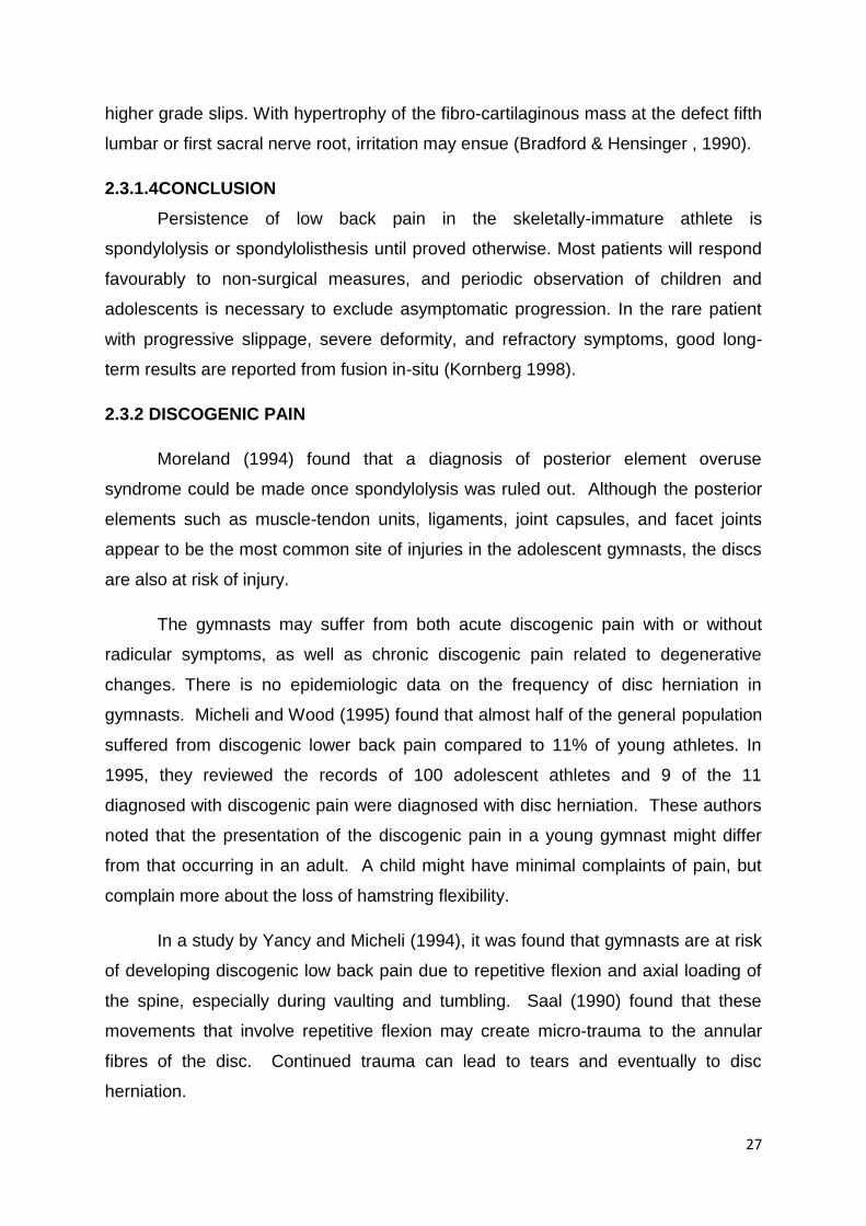
Table 4.3 Areas of peripheral injuries and types of injury sustained
41
The total number of peripheral injuries sustained by the sample was 36. Ligament
tears accounted for the most prevalent injury reported, 12/36 (30.5%)
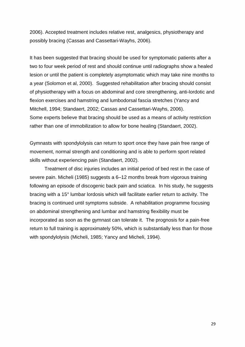
There was 5 injuries reported in the shoulder, this accounted for 13.8% of the peripheral injury
profile.
05
10152025303540
Total injuries according to type of injury
Total
0
1
2
3
4
5
6
Shoulder
Sh*

42
Three injuries were located in the elbow, contributing to 8.3% of the injury profile.
Dislocations of the elbow was the most prevalent reported elbow injury.
Four injuries were located in the wrist, contributing to 11.1% of the injury profile.
Tenosynovitis/Tendinitis of the wrist was the most prevalent reported wrist injury.
0
0.5
1
1.5
2
2.5
3
3.5
Elbow
Elb*
0
0.5
1
1.5
2
2.5
3
3.5
4
4.5
Wrist
Wr*

43
Three injuries were located in the fingers, contributing to 8.3% of the injury profile.
Six injuries were located in the knee, contributing to 16.6% of the injury profile.
Osgood Schlatters disease was the most prevalent reported knee injury.
0
0.5
1
1.5
2
2.5
3
3.5
Fingers
Fin*
0
1
2
3
4
5
6
7
Knee
Kn*

44
One injury was located in the groin, contributing to 2.7% of the injury profile.
Tenosynovitis/Tendinitis of the groin was the most prevalent reported groin injury.
One injury was located in the shin, contributing to 2.7% of the injury profile.
0
0.2
0.4
0.6
0.8
1
1.2
Groin
Gro*
0
0.2
0.4
0.6
0.8
1
1.2
Shin
Shin
45
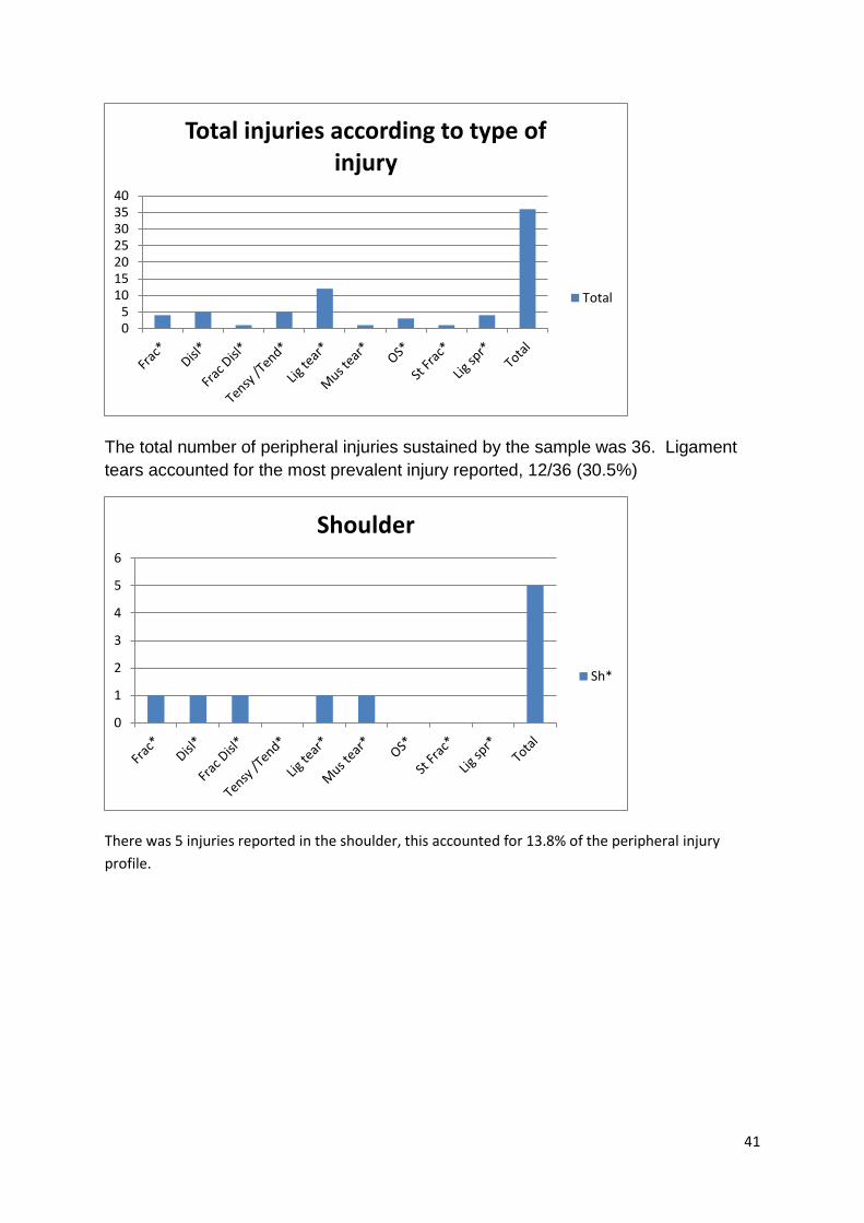
One injury was located in the Achilles tendon, contributing to 2.7% of the injury
profile.
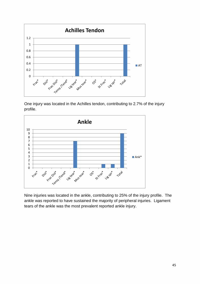
Nine injuries was located in the ankle, contributing to 25% of the injury profile. The
ankle was reported to have sustained the majority of peripheral injuries. Ligament
tears of the ankle was the most prevalent reported ankle injury.
0
0.2
0.4
0.6
0.8
1
1.2
Achilles Tendon
AT
0123456789
10
Ankle
Ank*

46
Three injuries were located in the foot, contributing to 8.3% of the injury profile.
The total number of peripheral injuries sustained by the sample was 36. The majority
of injuries were found in the ankle, 9/36 (25%).
0
0.5
1
1.5
2
2.5
3
3.5
Foot
Foot
0
5
10
15
20
25
30
35
40
Sh* Elb* Wr* Fin* Gro* Kn* Shin Ank* AT Foot Total
Total injuries according to area of injury
Total

47
Four fractures were reported and were found in the shoulder, elbow, fingers and foot.
Five dislocations were reported, with elbow dislocations being the most prevalent.
0
0.5
1
1.5
2
2.5
3
3.5
4
4.5
Sh* Elb* Wr* Fin* Gro* Kn* Shin Ank* AT Foot Total
Fracture
Frac*
0
1
2
3
4
5
6
Sh* Elb* Wr* Fin* Gro* Kn* Shin Ank* AT Foot Total
Dislocation
Disl*

48
One fracture dislocation was reported in the shoulder.
Five tenosynovitis/tendinitis was reported, with the majority being found in the wrist.
0
0.2
0.4
0.6
0.8
1
1.2
Sh* Elb* Wr* Fin* Gro* Kn* Shin Ank* AT Foot Total
Fracture dislocation
Frac Disl*
0
1
2
3
4
5
6
Sh* Elb* Wr* Fin* Gro* Kn* Shin Ank* AT Foot Total
Tenosynovitis/Tendinitis
Tensy /Tend*

49
Twelve ligament tears were reported, with the majority being found in the ankle.
One muscle tear was reported in the shoulder.
0
2
4
6
8
10
12
14
Sh* Elb* Wr* Fin* Gro* Kn* Shin Ank* AT Foot Total
Ligament tear
Lig tear*
0
0.2
0.4
0.6
0.8
1
1.2
Sh* Elb* Wr* Fin* Gro* Kn* Shin Ank* AT Foot Total
Muscle tear
Mus tear*

50
Three cases of Osgood Schlatters were reported, and is found only in the knee.
One stress fracture was reported in the ankle.
0
0.5
1
1.5
2
2.5
3
3.5
Sh* Elb* Wr* Fin* Gro* Kn* Shin Ank* AT Foot Total
Osgood Schlatters
OS*
0
0.2
0.4
0.6
0.8
1
1.2
Sh* Elb* Wr* Fin* Gro* Kn* Shin Ank* AT Foot Total
Stress fracture
St Frac*
51
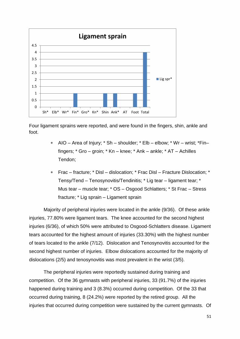
Four ligament sprains were reported, and were found in the fingers, shin, ankle and
foot.
AIO – Area of Injury; * Sh – shoulder; * Elb – elbow; * Wr – wrist; *Fin–
fingers; * Gro – groin; * Kn – knee; * Ank – ankle; * AT – Achilles
Tendon;
Frac – fracture; * Disl – dislocation; * Frac Disl – Fracture Dislocation; *
Tensy/Tend – Tenosynovitis/Tendinitis; * Lig tear – ligament tear; *
Mus tear – muscle tear; * OS – Osgood Schlatters; * St Frac – Stress
fracture; * Lig sprain – Ligament sprain
Majority of peripheral injuries were located in the ankle (9/36). Of these ankle
injuries, 77.80% were ligament tears. The knee accounted for the second highest
injuries (6/36), of which 50% were attributed to Osgood-Schlatters disease. Ligament
tears accounted for the highest amount of injuries (33.30%) with the highest number
of tears located to the ankle (7/12). Dislocation and Tenosynovitis accounted for the
second highest number of injuries. Elbow dislocations accounted for the majority of
dislocations (2/5) and tenosynovitis was most prevalent in the wrist (3/5).
The peripheral injuries were reportedly sustained during training and
competition. Of the 36 gymnasts with peripheral injuries, 33 (91.7%) of the injuries
happened during training and 3 (8.3%) occurred during competition. Of the 33 that
occurred during training, 8 (24.2%) were reported by the retired group. All the
injuries that occurred during competition were sustained by the current gymnasts. Of
0
0.5
1
1.5
2
2.5
3
3.5
4
4.5
Sh* Elb* Wr* Fin* Gro* Kn* Shin Ank* AT Foot Total
Ligament sprain
Lig spr*

52
the 40 gymnasts who completed the questionnaire, 50% (n=20) reported sustaining
a back injury, either before the time of data collection (current n=16, 80%) or before
the time of retirement (retired, n=4, 20%). Of the 20 gymnasts with back injuries,
75% (n=15) reported the injuries to be located in the lumbar spine, with 15% (n=3)
located in the thoracic spine and only 10% (n=2) were located in the cervical spine.
Of the 15 with lumbar spine injuries, 3 (20%) were reported by the retired gymnasts.
Table 4.4 Type of back injury sustained
20 of the 40 gymnasts reported having sustained a back injury. Of the 20, 16 were
current gymnasts and 4 were retired.
Muscle strain or tear, was reported to be the most prevalent injury sustained in the
sample (14/20) as well as in the current (11/14) and retired group (3/4).
Sixteen of the 20 back injuries sustained were by current gymnasts. Muscle strain or
tears accounted for the highest amount of injury in both the current (11/16) and the
retired group (3/4).
Twenty-eight current gymnasts reported sustaining a peripheral injury and 16
(52%) reported a back injury. Of the 28 with peripheral injury, 14 (50%) trained for
24 or less hours per week and 14 (50%) trained for 25 hours or more per week. Of
the 16 current gymnasts who reported a back injury, 9 (56%) trained for 24 hours or
less per week and 7 (44%) trained for 25 or more hours per week. All 16 of the
gymnasts with back injury reported being symptomatic.
0
5
10
15
20
25
Type of back injury
Frequency
Current
Retired

53
Of the 16 current gymnasts who trained for less than 25 hours per week, 14
(87.5%) reported a peripheral joint injury, and 9 (56.3%) reported sustaining a back
injury. All 9 (100%) with back injuries were symptomatic. Of the 14 gymnasts with
peripheral injuries, 9 (64.3%) also had back injuries.
Fifteen current gymnasts trained for 25 hours or more per week. Of these, 14
(93.3%) reported peripheral injuries and 7 (46.7%) reported back injuries. All 7
(100%) with back injuries were symptomatic. Of the 14 with peripheral injuries, 7
(50%) also had back injuries.
Of the 28 current gymnasts that reported injury, 18 (64.3%) had participated for 9
years or less and 10 (35.7%) for more than 10 years. Of the 16 current gymnasts
that reported back injury, 4 (25%) participated for 9 years or less and 12 (75%)
trained for 10 or more years. All (100%) of the gymnasts were symptomatic.
Table 4.5 Peripheral injury and back injury profile cross-tabulated with level of
competition.
The level of competition of the sample, showed that the majority of the gymnasts
competed at Level 10. The majority of the current gymnasts competed at Level 7,
and the majority of the retired gymnasts competed at Senior Olympic level.
0
5
10
15
20
25
30
35
40
45
Level of competition of sample
Total
CG*
RG*

54
The level 7 gymnasts sustained the highest amount peripheral injuries, 5/27, in the
current gymnasts group.
The Senior Olympic gymnasts sustained the highest amount of peripheral injuries,
4/9, in the retired gymnasts group.
0
5
10
15
20
25
30
4 6 6HP 7 8 8HP 9HP 10 10HP OE* JO* SO* Total
Level of competition against peripheral injuries sustained in the
current gymnasts
PICG*
0
2
4
6
8
10
4 6 6HP 7 8 8HP 9HP 10 10HP OE* JO* SO* Total
Level of competition against peripheral injuries sustained in retired
gymnasts
PIRG*

55
The Level 7 gymnasts sustained the highest amount of back injuries in the current
gymnasts sample, 4/16.
The Senior Olympic gymnasts sustained the highest amount of back injury in the
retired gymnast group, 2/5.
LOC –level of competition; * CG – Current gymnasts; * RG – Retired
gymnasts; * PICG – Peripheral injury current gymnasts; * BICG – Back injury
current gymnasts; * BIRG – Back injury retired gymnasts; * OE – Olympic
Entry; * JO – Junior Olympic; * SO – Senior Olympic
Of the 31 current gymnasts, 28 reported peripheral injury, with the majority of the
gymnasts competing in Level 7. This could be attributed to the increase in difficulty
at this level as well as the increase in hours spent training. Of the 9 retired
0
2
4
6
8
10
12
14
16
18
Level of competition against back injury sustained by current gymnasts
BICG*
0
1
2
3
4
5
6
Level of competition against back injury sustained by retired gymnasts
BIRG*

56
gymnasts, all reported having sustained peripheral injury during their gymnastics
career, with the majority (4/9) having competed at the Senior Olympic level. Senior
Olympic level is the highest level of competition, and the routines are of high difficulty
and intensity. 16 of the 31 current gymnasts reported having sustained a back
injury, with the level 7 gymnasts sustaining the highest number of back injuries
(4/16). Only 5 of the 9 retired gymnasts reported sustaining a back injury, with the
Senior Olympic girls reporting the majority of injury (2/5).
4.3 PREVALENCE OF RADIOLOGICAL CHANGES
Spinal X-rays were taken of each gymnast (n=40) and analysed for the following
changes:
Scoliosis;
Spondylolysis;
Spondylolisthesis; and
Degenerative changes.
Table 4.6 Scoliosis on X-ray for the gymnasts with back injury
Scoliosis Total Percentage Current Retired
Yes 15 75 11 4
No 5 25 5 0
Total 20 100 16 4
Of the 20 gymnasts who reported back injury that underwent X-Rays, 15 showed
radiological changes in the form of Scoliosis. Of the 15 with scoliosis, 11 were
current and 4 retired.
Table 4.7 Degenerative changes on X-ray for the gymnasts with back injury
Degenerative
changes Total Percentage Current Retired
Yes 14 70 11 3
No 6 30 5 1
Total 20 100 16 4

57
Of the 20 gymnasts who reported back injury, that underwent X-Rays, 14 showed
radiological changes in the form of degenerative changes. Of the 14 with X-Rays
changes, 11 were current and 3 were retired.
Table 4.8 Spondylolysis changes on X-ray for the gymnasts with back injury
Spondylolysis Total Percentage Current Retired
Yes 3 15 2 1
No 17 85 14 3
Total 20 100 16 4
Of the 20 gymnasts who reported back injury, that underwent X-rays, 3 showed
radiological changes in the form of spondylolysis. Of the 3 with X-ray changes, 2
were current and 1 was retired.
Table 4.9 Spondylolisthesis changes on X-ray for the gymnasts with back
injury
Spondylolisthesis Total Percentage Current Retired
Yes 4 20 3 1
No 16 80 13 3
Total 20 100 16 4
Of the 20 gymnasts who reported back injury, that underwent X-rays, 4 showed
radiological changes in the form of spondylolisthesis. Of the 4 with X-ray changes, 3
were current and 1 was retired.
Analysis of X-rays was done on those gymnasts who didn’t report a back
injury and were asymptomatic (n=20). X-ray analysis of these gymnasts showed
changes as follow: 15(75%) of the group had scoliosis (current =12 and retired = 3),
3 (15%) of the group had spondylolysis (current = 1 and retired = 2), 3 (15%) of the
group had spondylolisthesis (current = 2 and retired = 1) and 17 (85%) had
degenerative changes (current = 12 and retired = 5).
Table 4.10 Scoliosis changes on X-ray for the gymnasts without back injury
Scoliosis Total Percentage Current Retired
Yes 15 75 12 3
No 5 25 3 2
Total 20 100 15 5

58
Table 4.11 Degenerative changes on X-ray for the gymnasts without back
injury
Degenerative changes Total Percentage Current Retired
Yes 17 85 12 5
No 3 15 3 0
Total 20 100 15 5
Table 4.12 Spondylolysis changes on X-ray for the gymnasts without back
injury
Spondylolysis Total Percentage Current Retired
Yes 3 15 1 2
No 17 85 14 3
Total 20 100 15 5
Table 4.13 Spondylolisthesis changes on X-ray for the gymnasts without back
injury
Spondylolisthesis Total Percentage Current Retired
Yes 3 15 2 1
No 17 85 13 4
Total 20 100 15 5
Objectives of the Study
1. To determine the prevalence of radiological changes in South African female
artistic gymnasts, 10 to 30 years of age.
Twenty-eight of the 40 gymnasts (70%) had radiological evidence of
degenerative changes.
2. To determine if these changes were more prevalent in symptomatic than
asymptomatic subjects.
Twenty of the 40 gymnasts reported being symptomatic. Of these, 14 (70%) had
degenerative changes demonstrated by radiological examination and 6 (30%) did
not.

59
Twenty of the 40 gymnasts reported being asymptomatic. Of these, 17 (85%)
had degenerative changes demonstrated by radiological examination and 3 (15%)
did not.
3. To determine if these radiological changes were associated with number of
hours spent in training weekly.
The 40 gymnasts were divided into two groups: those training less than 25 hours
per week and those training 25 or more hours per week. Of the 18 gymnasts in the
lower training group, 13 (72.2%) had degenerative changes on X-ray. Of the 22
gymnasts training 25 or more hours per week, 15 (68.2%) had degenerative changes
on X-ray.
4.4 Summary
The prevalence rate of radiological evidence of degenerative changes in the
spines of South African female gymnasts was determined to be 70%. Half of all
subjects reported being symptomatic and, of these, 70% had radiological evidence of
degenerative changes in the spine. Interestingly, gymnasts who trained 25 or more
hours per week were less likely to have evidence of degenerative changes in the
spine, 68.2% vs. 72.2%.

60
CHAPTER 5
DISCUSSION
South African women’s gymnastics participation is on the increase locally and
internationally. Lumbar injuries are known to be prevalent in gymnasts (Caine and
Nassar, 2005); however, there is still a lack of research regarding the prevalence
and clinical significance of radiological changes in South African gymnasts
(Chadwick, 2004 as cited in Adamson, 2006).
A literature review was conducted to examine the aetiology of lumbar injuries in
gymnasts and to evaluate the effect that gymnastics have on normal growth. The
research shows that in recent years gymnasts started competing at younger ages,
mostly when entering adolescence and puberty. When compared to age matched
non gymnasts it was found that gymnasts are shorter and lighter, have a lower
percentage of body fat and also show lower growth velocities. This might be
attributed to the chronic stress that intensive physical training places on the body
which is capable of attenuating growth. Adolescents are also vulnerable to injury due
to imbalance in strength, flexibility and due to changes in the biomechanical
properties of bone during the period of peak linear growth. It has also been shown
that injury is more prevalent in gymnasts demonstrating rapid growth, greater body
size, age and percentage of body fat (Caine et al, 2003). Gymnastics have also
shown to increase prepubertal bone mineral density due to the mechanical forces
generated from high impact loading and strong muscular contraction.
Research clearly shows that there is a high prevalence of lumbar spine injuries in
gymnastics due to repetitive hyperextension, rotation and excessive training. It is of
a gradual onset due to repetitive load and involves primarily advanced level
gymnasts. This might implicate experience and the level of competition as possible
risk factors for injury. It has been proven that spondylolysis and spondylolisthesis
are one of the most common causes of adolescent back pain with increased risk in
gymnasts. This may be attributed to demands on the lower back being far greater in
gymnastics compared to other sports due to high force load transmission across the
trunk and repetitive end of range motion into flexion, extension and rotation.
Vertebral growth plate disorders and disc prolapsed can also occur leading to growth

61
disruption or chronic degenerative changes that may also force early retirement from
the sport.
Research has shown that conservative treatment of spondylolysis and
spondylolisthesis in gymnasts has been fairly effective. This has consisted of hard
and soft bracing along with abdominal strengthening exercises, anti-lordotic
exercises and stretching of the hamstrings and lumbodorsal fascia. These
interventions limit progression of the injury and showed symptom free return to
gymnastics. However, the interventions differed in studies and therefore it is still not
known what the optimal conservative treatment regime is regarding rehabilitation
intensity, the use of soft vs. rigid bracing and the amount of activity restriction. If
conservative treatment fails, a fusion in situ is needed. Discogenic pain has a 50%
prognosis of pain free return to activity and the rehabilitation also consists of bracing,
activity restriction and abdominal strengthening.
As a result of the lack of current research undertaken in South African gymnasts, a
descriptive study was done in 2007. The aim was to determine the prevalence of
radiological changes in South African gymnast between the ages of ten and thirty,
determine whether or not the changes are more prevalent in symptomatic or
asymptomatic gymnasts and lastly to determine if the changes are associated with
exposure time to the sport. The study included 31 current gymnasts and 9 retired
gymnast with high performance level experience.
Questionnaires and radiographic tests were used to investigate the history and type
of training, experience and injuries sustained in the participating gymnasts. The
radiographic tests consisted of X-rays taken in different planes do determine spinal
abnormalities and abnormalities of the lower extremities.
Ninety percent of gymnasts in this study reported sustaining a peripheral injury, most
of which occurred during training. Results correlated with previous research where
the majority of peripheral injury was sustained in the ankle (Caine and Nassar,
2005). Results showed that current gymnasts are demonstrating a higher prevalence
of both peripheral and back injury compared to retired gymnasts. It is possible that
performance expectations are now higher, leading current gymnasts to undertake
more strenuous activities during performances. It is also possible that the training
programmes of current gymnasts are less preparatory for performance than the
62
programmes of the retired gymnasts. The high prevalence of injury may also
indicate a need for better training to avoid injuries and the use of proper rehabilitation
protocols before returning to the sport. Increased time spent training might explain
why most injuries occurred during training another reason might be that there is
increased pressure on skill acquisition and perfection of routines during training.
Back injury was reported by 50% of gymnasts included in the study with 75% of
these located in the lumbar spine. As with peripheral injuries, current gymnast also
showed a higher prevalence of back injury compared to retired gymnasts. The
reason for this could be because the demand on the body has changed rapidly as
current gymnasts are not just training for longer hours but are also doing much more
difficult moves.
Radiographic tests showed that 70% of the gymnasts that reported back injury had
evidence of degenerative changes. Radiographic evidence of degenerative changes
was also found in 70% of the gymnasts that did not report back injury and was
asymptomatic. Therefore, it can be said that 70% of the gymnast included in the
study showed degenerative changes in the spine.
Changes correlating with scoliosis occurred in 75% of the symptomatic gymnasts
compared to 65% in the asymptomatic gymnasts. Spondylolysis occurred equally
(15%) in symptomatic and asymptomatic gymnasts and spondylolisthesis were more
prevalent in symptomatic gymnasts (20%). However 15% of the asymptomatic
gymnasts also showed radiographic evidence of spondylolisthesis. This shows that
there can be significant structural deficit in the lumbar spines of gymnasts even
without pain or back injury. A study done on professional fast bowlers in cricket also
showed that there were significant spondylotic changes in the lumbar spine even
without symptoms (Ranson et al, 2005). This concludes that radiographic changes of
degeneration or structural defect are more likely to exist in symptomatic gymnast, but
that it is still possible to have these changes without symptoms. This can indicate
that asymptomatic gymnast will continue excessive training and competing at high
levels which can lead to faster progression of changes and can lead to more severe
injury. Therefore regular X-rays should be performed on gymnasts to monitor spinal
changes and prevent more severe injury.

63
The findings of this study showed that exposure time to gymnastics also had an
effect on the prevalence of back injuries in the gymnasts. Back injuries occurred
more in gymnasts that trained for 24 hours or less a week. However, the results of
other studies concluded that greater exposure to training was directly related to an
increased risk of injury (Adamson, 2006; Caine et al, 1989). Therefore, the results
from this study might indicate that gymnasts training less than 25 hours per week
were less conditioned, were younger or that they had reduced skeletal maturity.
Results showed that gymnasts training and competing at higher levels experienced
more back injuries. This was also found in retired gymnast who competed at senior
Olympic level. This correlates with findings of other studies (Adamson,?; Caine et al
1989) which might implicate age as another risk factor for injury because gymnasts
competing at higher levels are generally older.
Conclusion
The results of the study bring to mind again that lumbar spine injuries are extremely
prevalent in gymnasts of all ages. It also demonstrates that degenerative and other
structural changes such as scoliosis, spondylolysis and spondylolisthesis occur
commonly and can also be present in gymnasts who are asymptomatic. Training
intensity and duration affects the prevalence of changes in the lumbar spine and can
be related to conditioning and experience.
Therefore, it is important to monitor radiographic spinal changes in gymnasts that are
training at high level performance level and competing professionally by doing
regular X-rays to determine the presence of any abnormalities whether gymnasts are
symptomatic or asymptomatic. The importance of proper rehabilitation after injury
before return to high level activity is also highlighted to prevent further injury and
reduce the rate of early retirement from the sport. Proper conditioning of gymnasts is
also needed to prevent injury during skill acquisition and performance.
Limitations of the study include the small number of participants used and that the
study only took place in one province of South Africa. There was also no
consideration of whether participant where Caucasian or African as genetic
predisposition might also affect the results. The exact gymnastic routines weren’t

64
described or distinguished between gymnasts which can be seen as a limitation as
different moves and routines could have different effects on the spine.
RECOMMENDATIONS BY THE AUTHOR
After completion of this study, the author found that more is necessary in this field.
The following recommendations can be considered for future research:
1. A follow up study using different radiological interventions such as MRI or CT
scans.
2. A follow up study using a larger sample i.e. country wide and combining
men’s and women’s artistic gymnastics.
3. A study can be done on the impact of the sport on the bone density of the
gymnast.
4. A study can be done on the influence of nutrition on the bone density of the
gymnast.
5. As there is very little research on management of the different radiological
changes found in this study, there is place for future research, concentrating
on rehabilitation and prevention, as well as conservative treatment, such as
bracing.
6. A strengthening and conditioning program should be developed and
incorporated at the clubs to assist with injury prevention.
7. Pre-screening of gymnasts when they start competing should be implemented
and continued on an annual basis to identify any pre-existing conditions and
manage them accordingly, and to prevent any further injury.

65
REFERENCES
8. Adamson, I. Gymnastics injuries: A quantitative profile of athletes in the
greater Durban area. Dissertation submitted in partial compliance with the
requirements for the Master’s Degree in Technology: Chiropractic in the
Department of Chiropractic at the Durban Institute of Technology derived from
www.dut.ac.za, 2006.
9. Amundson G, Edwards CC, Garfin SR: Spondylolisthesis. In: Herkowitz HN
et al, eds. The Spine, 4th edn. Philadelphia: WB Saunders, 835 – 885, 1999.
10. Bak K, Kalms SB, Olesen S, et al.: Epidemiology of injuries in gymnastics.
Scand J Med Sci Sports 4: 148 – 154, 1994.
11. Barrow, G.W. and Saha, S. Menstrual irregularity and stress fractures in
collegiate female distance runners. The American Journal of Sports Medicine,
16: 209-216, 1988.
12. Baxter-Jones AD, Helms P, Baines-Preece J, Preece M: Menarch in
intensively trained gymnast, swimmers and tennis players. Annuals of Human
Biology 21: 407 – 415, 1994.
13. Beilin H, Fireman G: The foundation of Piaget’s theories: mental and physical
action. Adv Child Dev Behav 27: 221 – 246, 1999.
14. Bennett D.L, Nassar L.G, DeLano M.C: Lumbar spine MRI in the elite-level
female gymnast with low back pain. Skeletal Radiol 35: 503 – 509, 2006.
15. Beyer HG: Influence of exercise on growth, United States Navy, pg 546 – 558,
2000.
16. Boden, B.P., Osbahr, D.C. and Jimenez, C. Low-risk stress fractures.
American Journal of Sports Medicine, 29(1): 100-111, 2001.
17. Borer, K.T, The effect of exercise on growth. Sports Med. 20(6): 375-397,
1995.
18. Bradford D, Winter R et al: Management of sever spondylolisthesis in children
and adolescents. Spine 5: 608 – 612, 1994.
19. Bronner, S., Ojofeitimi, S., and Rose, D. Injuries in a modern dance company.
Effect of comprehensive management on injury incidence and time loss.
American Journal of Sports Medicine, 31(3): 365-373, 2003.Brown EW,
Witten WA, Weise MJ,

66
20. Bruggeman, G.P. Mechanical load in artistic gymnastics and its relation to
apparatus and performance. In: Symposium Medico-Technique, Leglise M
(Ed.). Lyss, Switzerland: International Gymnastics Federation, 1999, pp. 17-
27.
21. Brukner P, Khan K: Clinical Sports Medicine. Sydney: McGraw-Hill Book
Company, 1993.
22. Caine, D., Bass, S., Daly, R. Does elite competition inhibit growth and delay
maturation in some gymnasts? quite possibly. Pediatric exercise science,
15(4): 360-372, 2003.
23. Caine D, Cochrane B, Caine C, et al: An epidemiologic investigation of
injuries affecting young competitive female gymnasts. Am J Sports Med 17:
811 - 820, 1989.
24. Caine, D., Lewis, R., O’Connor, P., Howe, W., Bass, S. Does gymnastics
training inhibit growth of females? Clin. J. Sport. Med. 11:260-270, 2001.
25. Caine D.J, Lindner K.J, Mandelbaum B.R, Sands W.A: Gymnastics. In:
Epidemiology of Sports Injuries, edited by Caine D.J, Caine C.G and Lindner
K.J. Champaign, IL: Human Kinetics, 1996, p. 213 – 246.
26. Caine, D.J., and Nassar, L. Gymnastic injuries. Med Sport Sci, 48: 18-58,
2005.
27. Cassas, K.J., and Cassettari-Wayhs, A. Childhood and Adolescent Sports-
related overuse injuries. Am Fam Physician, 73: 1014-1022, 2006.
28. Ciullo J.V, Jackson D.W: Pars interarticularis stress reaction, spondylolysis,
and spondylolisthesis in gymnasts. Clin Sports Med 4(1): 95 – 110, 1985.
29. Claessens A.L, Malina R.M, Lefevre J, Beunen G, Stijnen V, Maes H, et al.,:
Growth and menarcheal status of eliste female gymnasts. Med Sci Sports
Exerc 24: 755 – 763, 1992.
30. Clark KC: Positioning in radiography.
31. Cluett J: Low back pain in elite rhythmic gymnasts. Med Sci Sports Exerc 11:
1686 – 1688, 1999.

67
32. Cohen E and Stuecker RD: Magnetic resonance imaging in diagnosis and
follow-up of impending spondylolysis in children and adolescents: early
treatment may prevent pars defects. J Pediatr Ortho 14: 63 – 67, 2005.
33. Congeni, J., McCulloch, J., and Swanson,K. Lumbar spondylolysis. A study of
natural progression in athletes. Am J Sports Med, 25: 248-253, 1997.
34. Constantini NW, Warren MP: Menstrual dysfunction in swimmers: a distinct
entity. J Clin Endocrinol Metab 80(9): 2740 – 2744, 1995
35. Courteix, D., Lespessailles, E., Loiseau Peres, S., Obert, P., Germain, P., and
Benhamou, C.L. Effect of physical training on bone mineral density in
prepubertal girls: A comparative study between impact-loading and non-
impact-loading sports. Osteoporosis International, 8(2): 152-158, 1998.
36. Daly, R.M., Bass, S.L., and Finch, C.F. Balancing the risk of injury to
gymnasts: how effective are the counter measures? British Journal of Sports
Medicine, 35(1): 8-19, 2001.
37. Douglas WJ, Leon LW, Cirincione RJ: Spondylolysis in the female gymnast.
Clin Orthop Rel Res 117: 68 – 72, 1976.
38. Dunn, I.F., Proctor, M.R., and Day, A.L. Lumbar spine injuries in athletes.
Neurosurg Focus 21(4): E4, 2006.
39. Eddy D, Congeni J and Loud K: A review of spine injuries and return to play.
Clin J Sport Med 15(6): 453 – 458, 2005
40. Espinoza D, Wisner DM, Learman J et al.,: Attenuation of ground reaction
forces in salto dismounts from the balance beam. In J.M.C.S Abrantes (Ed),
Proceedings of the XIV international symposium on biomechanics in sport pp.
336 – 338. Lisboa Codex, Portugal: Edicoes FMH, 1996.
41. Garrick JG, Requa RK: Epidemiology of women’s gymnastics injuries. Am J
Sports Med 8: 261 – 264, 1980.
42. Garrick JG, Requa RK: Ballet injuries – An analysis of epidemiology and
financial outcome. Am J Sports Med,21(4):586-590, 1993.
43. Georgopoulos N, Markou K, Theodoropoulou A, Paraskevopoulou, Varaki Z,
Kazantzi Z, Leglise M and Vagenakis AG: Growth and pubertal development
68
in elite female gymnasts. The Journal of Clinical Endocrinology and
Metabolism 84(12): 4525 – 4530, 1999.
44. Georgopoulos N.A, Markou K.B, Theodoropoulou A, Bernadot D, Leglise M,
Vagenakis A.G: Growth retardation in artistic compared with rhythmic elite
female gymnasts. Journal of Clinical Endocrinology and Metabolism 87:
3169 – 3173, 2002.
45. Gerbino PG II, Micheli LJ: Back injuries in the young athlete. Clin Sports Med
14: 571 – 590, 1995.
46. Goldstein J.D, Berger P.E, Windler G.E, Jackson D.W: Spine injuries in
gymnasts and swimmers: An epidemiologic investigation. Am J Sports Med
19(5): 463 – 468, 1991.
47. Gordon CM, Laufer MR: Chapter 4: Physiology of puberty. In Emans SJH,
Goldstein DP, Laufer MR, eds. Pediatric and Adolescent Gynecology (5th ed).
Philadelphia: Lippincott, Williams & Wilkins. pg. 120 – 155, 2005
48. Hall S.J: Mechanical contribution to lumbar stress injuries in female gymnasts.
Med Sci Sports Exerc 18(6): 599 – 602, 1986.
49. Henderson, N.K., White, C.P., and Eisman, J.A. The roles of exercise and fall
risk reduction in the prevention of osteoporosis. Endocrinology and
Metabolism Clinics of North America, 27: 369–387, 1998.
50. Holschen JC: The female athlete. Southern Medical Journal 97(9): 852 – 858,
2004.
51. Ikata T, Miyake R, Katoh S, et al: Pathogenesis of sports related
spondylolisthesis in adolescents. Am J Sports Med 24: 94 – 97, 1996.
52. Jackson DW, Wiltse LL, Cirincione RJ: Spondylolysis in the female gymnast.
Clin Orthop Rel Res 117: 68 – 73, 1976.
53. Johnson K.M: Where have all the gymnasts gone? JOPERD: 28 – 29, 1985.
54. Kirchner, E.M., Lewis, R.D., and O’Connor, P.J. Bone mineral density and
dietary intake of female college gymnasts. Medicine and Science in Sports
and Exercise, 27: 496-502, 1995.
55. Kirkaldy-Willis WH, ed. The pathology and pathogenesis of low back pain.
Managing Low Back Pain. New York, NY: Churchill Livingstone; 49, 1988.

69
56. Kolt G.S, Kirkby R.J: Epidemiology of injury in elite and subelite female
gymnasts: a comparison of retrospective and prospective findings. Br J Sports
Med 33: 312 – 318, 1999.
57. Kolt, G., and Kirby, R. Injury in Australian gymnasts: A psychological
perspective. Aust. J. Physiother. 42: 121-126, 1996.
58. Kornberg C: Spine injuries in gymnasts and swimmers: An epidemiologic
investigation. Am J Sports Med 5: 463 – 468, 1991.
59. Lima, F., De Falco, V., Baima, J., Carazzato, J.G., and Pereira, R.M. Effect of
impact load and active load on bone metabolism and body composition of
adolescent athletes. Medicine and Science in Sports and Exercise, 33: 1318–
1323, 2001.
60. Lindholm C, Hagenfeld K, Ringertz BM: Pubertal development in elite juvenile
gymnasts: Effects of physical training. Acta Obstet Gynecol Scand 73(3): 269
– 273, 1994.
61. Lindholm T.S, Ragni P, Ylikoski M, et al.,: Lumbar isthmic spondylolisthesis in
children and adolescents: radiologic evaluation and results or operative
treatment. Spine 15: 1350 – 1355, 1990.
62. Lindner K.J, Caine D.J: Injury patterns of female competitive club gymnasts.
Can J Sports Sci 15: 254 – 261, 1990.
63. Malina RM and Bouchard C: Growth maturation and physical activity.
Champaign, IL Human Kinetics publishers, 1991.
64. Malina RM: Benefits of physical activity from a lifetime perspective. In:
Quinney HA, Gauvin L, Wall AET, eds. Towards active living: proceedings of
the international conference on physical activity, fitness, and health.
Champaign, IL: Human Kinetics, 47 – 53: 1994.
65. Marshall S.W, Covassin T, Dick R, Nassar L.G, Agel J: Descriptive
Epidemiology of Collegiate Women’s Gymnastics Injuries: National collegiate
Athletic Association Injury Surveillance System, 1988 – 1989 through 2003 –
2004. J Athl Train 42(2): 234 – 240, 2007.
66. Marshall W.A, Tanner J.M: Puberty, in Falkner F, Tanner JM (eds): Human
Growth: A Comprehensive Treatise. New York, Plenum Press, 1983.
67. Marshall WA, Tanner JM: Variations in pattern of pubertal changes in girls.
Arch Dis Child 44: 291, 1969.

70
68. Mazel C: Etiology of isthmic spondylolisthesis. ArgoSpine News & Journal
23(2): 85 – 87, 2006.
69. McGill SM: Low back exercises. Phys Ther 78: 754 – 765, 1998.
70. Micheli L, Wood R: Back pain in young athletes: significant differences from
adults in causes and patterns. Arch Pediatr Adolesc Med 149: 15 – 18, 1995.
71. Micheli L.J: Back injuries in gymnastics. Clin Sports Med 4(1): 85 -94, 1985.
72. Miller S, Congeni J, Swanson K: Long-term functional and anatomical follow-
up of early detected spondylolysis in young athletes. Am J Sports Med 32:
928 – 933, 2004.
73. Moreland M.S: special concerns of the pediatric athlete. In Fu FH, Stone DA
(eds): Sports Injuries: Mechanisms, Prevention, Treatment. Baltimore,
Williams & Wilkins, p135 – 151, 1994.
74. Munoz, M.A., de la Piedral, C., Barrios, V., Garrido, G., and Argente, J.
Changes in bone density and bone markers in rhythmic gymnasts and ballet
dancers: implications for puberty and leptin levels. European Journal of
Endocrinology, 151: 491-496, 2004.
75. Muschik, M., Hahnel, H., Robinson, P.N., Perka, C. and Muschik, C.
Competetive Sports and the progression of spondylolisthesis. J Pediatric
Orthop, 16: 364-369, 1996.
76. Nickols-Richardson, S.M., O’Connor, P.J., Shapses, S.A., Lewis, R. D.
Longitudinal bone mineral density changes in female child artistic gymnasts.
Journal of Bone and Mineral Research, 14(6): 994-1002, 1999.
77. Panzer V.P, Wood G.A, Bates B.T, Mason B.R: Lower extremity loads in
landings of elite gymnasts. In: de Groot G, Hollander A.P, Huijing P.A, van
Ingen Schenau G.J (Eds.) Biomechanics XI-B. Amsterdam: Free University
Pres, 727 – 735, 1988.
78. Patel, D.R., and Nelson, T.L. Sports injuries in adolescents. Med Clin North
Am, B4: 983-1007, 2000.

71
79. Quatman CE, Ford KE, Myer and Hewett TE: Maturation leading to gender
differences in landing force and vertical jump performance: A longitudinal
study. Am J Sports Med 34(5): 806 – 813, 2006.
80. Ranson, C.A., Kerslake, R.W., Burnett, A. F., Batt, M.E., and Abdi, S.
Magnetic resonance imaging of the lumbar spine in asymptomatic
professional fast bowlers in cricket. Journal of Bone and Joint Surgery, 87-
B(8): 1111-1116, 2005.
81. Reitman, C.A., Gertzbein, S.D., and Francis, W.R. Jr. Lumbar isthmic defects
in teenagers resulting from stress fractures. Spine Journal, 2(4): 303-306,
2002.
82. Richards R, Williams S, Poulton R and Reeder AI: Tracking club sport
participation from childhood to early adulthood. Res Q Exerc Sport 78(5): 413
– 419, 2007.
83. Robinson, T.L., Snow-Harter, C., Taaffe, D.R., Gillis, D., Shaw, J. and
Marcus, R. Gymnasts exhibit higher bone mass than runners despite similar
prevalence of amenorrhea and oligomenorrhea. J Bone Miner Res, 10(1): 26–
35, 1995.
84. Rogol AD, Clark PA and Roemmich N: Growth and pubertal development in
children and adolescents: effects of diet and physical activity. Am J Clin
Nutrition 72: 521S – 528S, 2000.
85. Rubery PT and Bradford DS: Athletic activity after spine surgery in children
and adolescents: results of a survey. Spine 27: 423 – 427, 2002.
86. Saal JA: Lumbar injuries in gymnastics. Spine: State of the Art Review 4: 426
– 440, 1990.
87. Sands W.A, Shultz B.B, Newman A.P: Women’s gymnastics injuries: A 5-year
study. Am J Sports Med 21(2): 271 – 276, 1993.
88. Sharma P, Luscombe KL and Maffulli N: Sports injuries in children. Trauma
5: 245 – 259, 2003.
89. Sim J, Wright C: Research in Health Care Concepts, Designs and Methods.
Stanley Thomas publishers, pg 126 – 129, 2000.
90. Soler T, Calderon C: The Prevalence of Spondylolysis in the Spanish Elite
Athlete. Am J Sports Med 28(1): 57 – 62, 2000.
72
91. Solomon, R., Brown, T., Gerbino, P.G. The young dancer. Clin Sports Med,
19: 717-739, 2000.
92. Solomon, R., Solomon, J., and Mitcheli, L.J. The “cost” of injuries in a
professional ballet company: A five year study.Med Probl Perform Artists, 14:
164-169, 1999.
93. Standaert, C.J. New Strategies in the management of low back injuries in
gymnasts. Curr. Sports. Med. Rep. 1(5): 293-300, 2002.
94. Standaert, C.J. Spondylolysis in the adolescent athlete. Clin J Sport Med, 12:
119-122, 2002.
95. Swärd L, Hellstrom M, Jacobsson B, et al.,: Back pain and radiologic changes
in the thoraco-lumbar spine of athletes. Spine 15: 124 – 129, 1990.
96. Swärd L, Hellstrom M, Jacobsson B, et al.,: Disc degeneration and associated
abnormalities of the spine in elite gymnasts: A magnetic resonance imaging
study. Spine 16: 437 – 443, 1991.
97. Swartz EE, Decostert LC, Russell PJ and Croce RV: Effects of developmental
stage and sex on lower extremity kinematics and vertical ground reaction
forces during landing. J Athl Train 40(1): 9 – 14, 2005.
98. Takei Y: A comparison of techniques in performing the men’s compulsory
gymnastic vault at 1988 Olympics. Intl J Sport Biomech 7(1): 54 – 75, 1991.
99. Tanner JM: Growth in adolescence (2ed) Oxford: Blackwell Scientific
Publications, 1968.
100. Tertti M, Paajanen H, Kujala U.M, Alanen A, Salmi T.T, Kormano M:
Disc degeneration in young gymnasts. Am J Sports Med 18(2): 206 – 208,
1990.
101. Theintz GE, Howald H, Weiss U and Sizonenko PC: Evidence for a
reduction of growth potential in adolescent female gymnasts. J Pediatr
122(2): 306 – 313, 1993.
102. Wadley G.H, Albright J.P: Women’s intercollegiate gymnastics: Injury
patterns and “permanent” medical disability. Am J Sports Med 21: 314 – 320,
1993.
103. Weber MD, Woodall WR: Spondylogenic disorders in gymnasts. J
Orthop Sports Phys Ther 14: 6 – 13, 1991

73
104. Weiker G.G: Evaluation and treatment of common spine and trunk
problems. Clin Sports Med 8: 399 – 417, 1989.
105. Weiker GG: Injuries in club gymnastics. Physician Sportsmed 13(4): 63
– 66, 1985.
106. Wiltse L, Newman P, Macnab I: Classification of spondylolysis and
spondylolisthesis. Clin Orthop Relat Res 117: 23 – 29, 1976.
107. Wiltse LL, Newman PH and Macnab I: Classification of spondylolysis
and spondylolisthesis. Clin Orthop Relat Res 117: 23 – 29, 1976.
108. Winkler, K. Born to fly: kids, gymnastics and the will to win. Journal of
the American Chiropractic Association, 38(2), 2001.
109. www.aafla.org/Olympicinformationcenter/olympicreview
110. www.childgrowthfoundation.org/psm_tanner_stages.htm
111. www.education.com/reference/article/jean-piaget/
112. Yancy R.A, Micheli L.J: Thoraco-lumbar injuries in pediatric sports. In
Stanitski CL, DeLee JC, Drez DD Jr (Eds.): Pediatric and Adolescent Sports
Medicine, vol 3, Philadelphia, WB Saunders, p 162 – 174, 1994.
113. Zetaruk M.N: The Young Gymnast. Clin Sports Med 19(4): 757 – 780,
2000.

74
ADELE GELDENHUYS PHYSIOTHERAPY
BSc. PHYSIOTHERAPY (WITS)
PT 0083941
PRACTICE NO. 014637
082 777 6921
Dear Parents/Gymnast
I am reading for my Masters in Medicine in the field of Exercise Science through
WITS University this year. My supervisors for the study are Dr. Demitri Constantinou
and Prof. Y. Coopoo. I am researching lower back health in active and retired
female gymnasts who have been doing gymnastics for more than five years.
The aim of the study is to determine the radiological (X-Ray) changes in female
artistic gymnasts and to correlate these findings with the asymptomatic and
symptomatic subject.
The study will be a complete blind study, which means that, each gymnast will only
be identified by a number. They will complete a questionnaire and undergo a set of
lumbar spine X-Rays. The questionnaire and X-rays will have their number only and
the identity of the gymnast will remain anonymous.
If parents would like to see the results, an interview will be arranged with the
researcher. The results will remain totally confidential and no other person in the
medical team or coach will have access to these results, unless permission is
granted by the parents and gymnast. In no way will this study influence your child’s
training and there will be no cost involved to you as parents/gymnasts.
The study will be of benefit to your child in context that although her results will be
unknown to me, I can ascertain the health of the back of gymnasts in general, and
design a specific rehabilitation and prevention program to strengthen the back and
possibly prevent future injuries.

75
If you are comfortable with your daughter taking part in my research, kindly sign the
attached consent form. Please note that your daughter is free to withdraw from the
study at any time.
Yours faithfully
Adele Geldenhuys

76
INFORMED CONSENT
INCIDENCE OF RADIOLOGICAL CHANGES IN FEMALE ARTISTIC GYMNASTS
1. In order to assess the integrity of the lower back, the undersigned
hereby voluntarily consents to his/her daughter engaging in the
following tests namely: X-rays of the lumbar spine and pelvis, and
a questionnaire documenting history of back pain over the last 12
months.
2. EXPLANATION OF TESTS
a. The X-rays will be done at a specified hospital, by a fully
trained radiographer. The radiologist reporting on the X-
rays, will have no knowledge of who the subject is, and all X-
rays will be done using a number only. The questionnaire
will also be filled out using the specific number allocated.
Parent can help with completing the questionnaire.
3. RISKS AND DISCOMFORTS
a. There might be some discomfort in the symptomatic subject
during the stress view X-rays. Risks are considered minimal,
as exposure will be once off.
4. EXPECTED BENEFITS FROM TESTING
a. These X-rays will allow me to assess the health of the back,
and to create exercise programs to assist with injury
prevention.
5. ENQUIRIES
a. Questions about the procedure are encouraged. If you have
any further questions or need additional information, please
ask me to explain further.
6. FREEDOM OF CONSENT

77
a. Your permission to perform these tests on your daughter is
strictly voluntary. You are free to deny consent if you so
desire. You are free to withdraw from the study and any
time.
I have read this form carefully and fully understand the test procedures. I
consent to my daughter participating in these tests.
Signature of gymnast/parent if Witness
under 18 years of age
Date
Questions:

78
1. RESEARCH NUMBER: 2. DATE OF BIRTH: 3. AGE:
10 11 12
13 14 15
16
4. HOW OLD WERE YOU WHEN YOU FIRST STARTED DOING GYMNASTICS?
4 years 5 years 6 years
7 years 8 years 9 years or older
5. HOW MANY YEARS HAVE YOU BEEN DOING GYMNASTICS FOR?
3 4 5
6 7 8
9 10 11
12 13 14
15
6. WHAT LEVEL DO YOU CURRENTLY COMPETE AT?
1 2 3
4 4HP 5
6 6HP 7
8 8HP 9
9HP 10 10HP
APPENDIX A
PREVALANCE OF RADIOLOGICAL CHANGES IN THE LUMBAR SPINE OF SOUTH AFRICAN FEMALE ARTISTIC
GYMNASTS TRAINING AND SYMPTOM QUESTIONNAIRE PLEASE TICK THE ANSWER THAT APPLIES TO YOU

79
Olympic Entry Junior Olympic Senior Olympic
8. HOW MANY HOURS A WEEK DO YOU SPEND ON GYMNASTICS TRAINING?
14 hours or less 15 - 24 hours 25 hours or more
9. HAVE YOU INJURED YOURSELF DOING GYMNASTICS?
Yes No
10. IF YOU ANSWERED YES TO QUESTION 9, PLEASE INDICATE WHERE
YOUR INJURY WAS
Shoulder Elbow Wrist
Hand Fingers Hip
Groin Quadriceps Hamstrings
Knee Shin Calf
Ankle Achilles tendon Foot
Toes
11. WHAT WAS YOUR INJURY?
Fracture Dislocation Fractured-dislocation
Tendinitis/Tenosynivitis Ligament tear Muscle tear
Osgood Schlatters Severs disease Stress fracture
Ligament sprain Muscle strain
12. WHEN DID YOU INJURY YOURSELF?
Training Competing At school
At home Doing other sports
13. HAVE YOU INJURED YOUR BACK DOING GYMNASTICS?
Yes No
14. WHICH PART OF YOUR BACK DID YOU INJURE?
Neck (Cervical) Middle back (Thoracic) Lower back (Lumbar)
15. WHAT WAS YOUR INJURY?
Muscle strain/tear “Slipped” disc Stress fracture
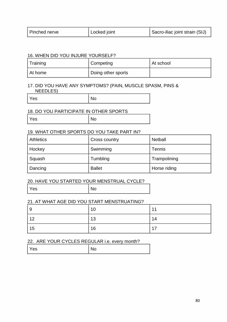
80
Pinched nerve Locked joint Sacro-iliac joint strain (SIJ)
16. WHEN DID YOU INJURE YOURSELF?
Training Competing At school
At home Doing other sports
17. DID YOU HAVE ANY SYMPTOMS? (PAIN, MUSCLE SPASM, PINS &
NEEDLES)
Yes No
18. DO YOU PARTICIPATE IN OTHER SPORTS
Yes No
19. WHAT OTHER SPORTS DO YOU TAKE PART IN?
Athletics Cross country Netball
Hockey Swimming Tennis
Squash Tumbling Trampolining
Dancing Ballet Horse riding
20. HAVE YOU STARTED YOUR MENSTRUAL CYCLE?
Yes No
21. AT WHAT AGE DID YOU START MENSTRUATING?
9 10 11
12 13 14
15 16 17
22. ARE YOUR CYCLES REGULAR i.e. every month?
Yes No

81
4. RESEARCH NUMBER: 5. DATE OF BIRTH: 6. AGE:
10 11 12 13 14 15
16 17 18 19 20 21
22 23 24 25 26 27
28 29 30
5. HOW OLD WERE YOU WHEN YOU FIRST STARTED DOING GYMNASTICS?
4 years 5 years 6 years
7 years 8 years 9 years or older
6. HOW MANY YEARS DID YOU DO GYMNASTICS FOR?
3 4 5 6 7
8 9 10 11 12
13 14 15 16 17
18 19 20
7. AT WHAT AGE DID YOU RETIRE?
12 13 14 15 16
17 18 19 20 21
22 23 24 25
APPENDIX B
PREVALANCE OF RADIOLOGICAL CHANGES IN THE LUMBAR SPINE OF SOUTH AFRICAN FEMALE ARTISTIC
GYMNASTS TRAINING AND SYMPTOM QUESTIONNAIRE FOR RETIRED GYMNASTS PLEASE TICK THE ANSWER THAT APPLIES TO YOU

82
9. WHAT PROMPTED YOU TO RETIRE?
Injury No motivation Achieved my goal
Other sports Academic progression
Financial factors
Poor parental support
Parental pressure
Coaching pressure
Public pressure
9. WHAT LEVEL DID YOU COMPETE AT?
1 2 3 4
4HP 5 6 6HP
7 8 8HP 9
9HP 10 10HP Olympic Entry
Junior Olympic Senior Olympic
10. HOW MANY HOURS A WEEK DID YOU SPEND ON GYMNASTICS TRAINING?
14 hours or less 15 - 24 hours 25 hours or more
11. DID YOU INJURE YOURSELF DOING GYMNASTICS?
Yes No
10. IF YOU ANSWERED YES TO QUESTION 9, PLEASE INDICATE WHERE
YOUR INJURY WAS
Shoulder Elbow Wrist
Hand Fingers Hip
Groin Quadriceps Hamstrings
Knee Shin Calf
Ankle Achilles tendon Foot
Toes
13. WHAT WAS YOUR INJURY?
Fracture Dislocation Fractured-dislocation
Tendinitis/Tenosynivitis Ligament tear Muscle tear
Osgood Schlatters Severs disease Stress fracture

83
Ligament sprain Muscle strain
14. WHEN DID YOU INJURY YOURSELF?
Training Competing At school
At home Doing other sports
15. DID YOU INJURE YOUR BACK DOING GYMNASTICS?
Yes No
16. WHICH PART OF YOUR BACK DID YOU INJURE?
Neck (Cervical) Middle back (Thoracic) Lower back (Lumbar)
17. WHAT WAS YOUR INJURY?
Muscle strain/tear “Slipped” disc Stress fracture
Pinched nerve Locked joint Sacro-iliac joint strain (SIJ)
18. WHEN DID YOU INJURE YOURSELF?
Training Competing At school
At home Doing other sports
19. DID YOU HAVE ANY SYMPTOMS? (PAIN, MUSCLE SPASM, PINS & NEEDLES)
Yes No
20. DID YOU PARTICIPATE IN OTHER SPORTS WHILST DOING GYMNASTICS?
Yes No
21. WHAT OTHER SPORTS DID YOU TAKE PART IN?
Athletics Cross country Netball
Hockey Swimming Tennis
Squash Tumbling Trampolining
Dancing Ballet Horse riding

84
22. HAVE YOU STARTED YOUR MENSTRUAL CYCLE?
Yes No
23. AT WHAT AGE DID YOU START MENSTRUATING?
9 10 11
12 13 14
15 16 17
24. ARE YOUR CYCLES REGULAR i.e. every month?
Yes No