Presentation1, radiological imaging of colitis.
70
RADIOLOGICAL IMAGING OF COLITIS. Dr/ ABD ALLAH NAZEER. MD. Ischemic colitis
-
Upload
abdellah-nazeer -
Category
Health & Medicine
-
view
643 -
download
0
Transcript of Presentation1, radiological imaging of colitis.
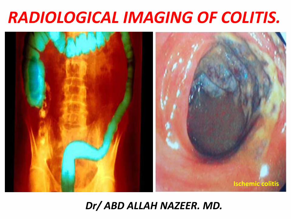
RADIOLOGICAL IMAGING OF COLITIS
Dr ABD ALLAH NAZEER MD
Ischemic colitis
TYPES OF COLITIS
bull IBD UC and Crohnrsquos bull Infectious colitisbull Ischemic colitis
bull Diverticulitis (DDX EA) bull Neutropenic colitis bull Drug-related colitis
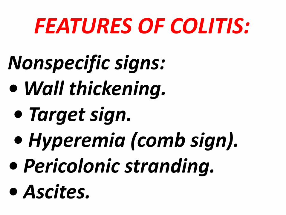
FEATURES OF COLITIS
Nonspecific signs bull Wall thickeningbull Target sign bull Hyperemia (comb sign)
bull Pericolonic stranding bull Ascites
FEATURES OF COLITIS
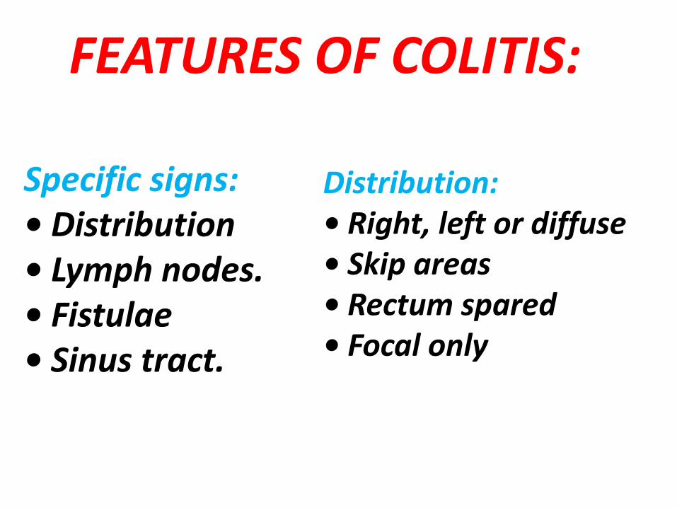
Specific signs bull Distributionbull Lymph nodesbull Fistulaebull Sinus tract
Distribution bull Right left or diffuse bull Skip areasbull Rectum sparedbull Focal only
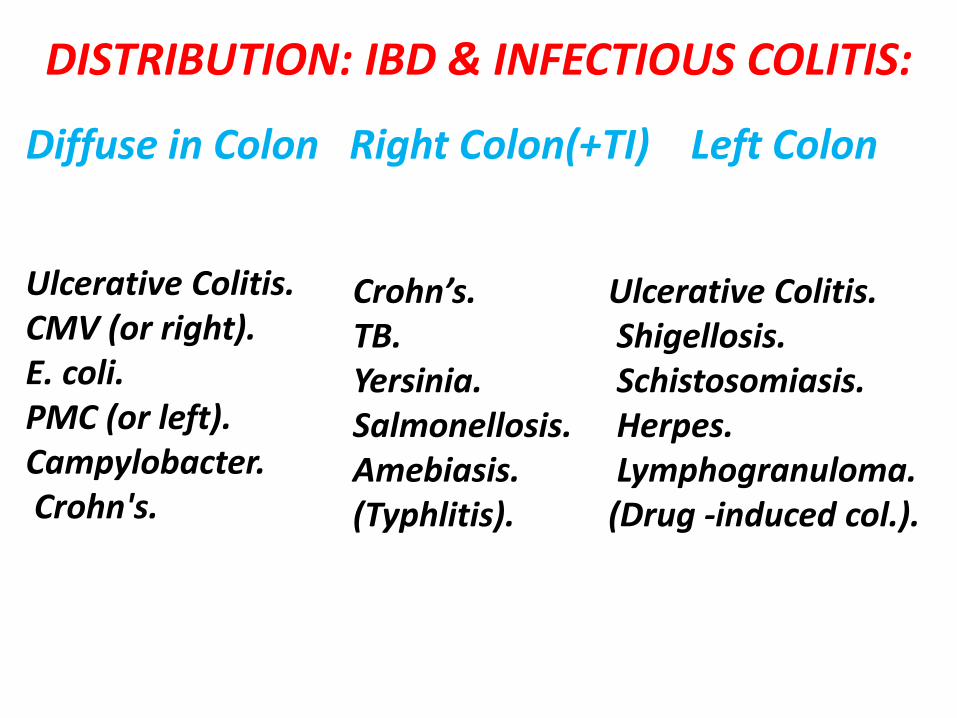
DISTRIBUTION IBD amp INFECTIOUS COLITIS
Ulcerative Colitis CMV (or right)E coli PMC (or left) Campylobacter Crohns
Diffuse in Colon Right Colon(+TI) Left Colon
Crohnrsquos TBYersiniaSalmonellosisAmebiasis(Typhlitis)
Ulcerative ColitisShigellosisSchistosomiasis HerpesLymphogranuloma
(Drug -induced col)
Segment Thickness ExtensionSmallBowel
RectumHaloSign
Ischemia GgtD 8-10 Long + - +
Crohns DgtG 10-15 Long +++ + +++
UlcerativeColitis
G 6-8 Long - +++ +++
InfectiousColitis
DgtG 8-10 Long ++ + +++
Pseudo-MembranousColitis
GgtD 10-15 Long - - +++
ETIOLOGIC DIAGNOSIS
Bowel wall thickening
Degree of mural thickening
Crohns disease is an idiopathic inflammatory bowel disease (IBD) characterized by widespread gastrointestinal tract involvement typically with skip lesions It is also known as regional enteritis and frequently there is systemic involvementRadiographic Features FluoroscopyFeatures on barium small bowel follow-through includemucosal ulcers
aphthous ulcers initiallydeep ulcers (more than 3mm depth)longitudinal fissurestransverse stripeswhen severe leads to cobblestone appearancemay lead to sinus tracts and fistulae
widely separated loops of bowel due to fibro-fatty proliferation (creeping fat)thickened folds due to edemaPseudo-diverticula formation due to contraction at the site of ulcer with ballooning of the opposite sitestring sign tubular narrowing due to spasm or stricture depending on chronicitypartial obstructionon control films presence of gall stones renal oxalate stones and sacroiliac joint or lumbosacral spine changes should be sought
CT examination can be carried out with both intravenous and intraluminal contrast (positive or negative)fat halo signcomb signbowel wall enhancementbowel wall thickening (1-2 cm) which is most frequently seen in the terminal ileum (present in up to 83 of patients)strictures and fistulaemesentericintra-abdominal abscess or phlegmon formationabscesses are eventually seen in 15-20 of patients
MRI enterography (MRE)MR enterography can be a useful technique for evaluation of the bowel Inflamed loops of bowel demonstrate thickening and contrast enhancementExtramural disease is where MRI excelsfibrofatty proliferation
thickening of extramural fat which separates bowel loopsequivalent to the fat halo sign on CT
vascular engorgement comb signstenosis and strictures
Crohns Disease ndash Barium Images
FEATURES OF COLITIS
COLITIS
CROHNrsquoS DISEASE SMALL BOWEL
CT and MR Enterography for Crohns Disease
Crohns disease Coronal fluid sensitive (T2) T2 fat suppressed and T1 post contrast images Red arrows outline the diseased colon with wall thickening abnormal edema and ascites (thin arrow) and intense post contrast enhancement Yellow arrows highlight proliferation of mesenteric vessels in creeping fat commonly referred to as the ldquocombrdquo sign
Ulcerative colitis is an inflammatory bowel disease that not only
predominantly affects the colon but also has extraintestinal manifestations Radiographic featuresInvolvement of the rectum is almost always present (95) with the disease involving variable amounts of the most proximal colon in continuity The entire colon may be involved in which case edema of the terminal ileum may also be present (so-called backwash ileitis)Plain radiographNon-specific findings but may show evidence of mural thickening (more common) with thumbprinting also seen in more severe casesFluoroscopyMucosal inflammation leads a granular appearance to the surface of the bowel As inflammation increases the bowel wall and haustra thickenedMucosal ulcers are undermined (button-shaped ulcers) When most of the mucosa has been lost islands of mucosa remain giving it a pseudopolyp appearanceIn chronic cases the bowel becomes featureless with the loss of normal haustral markings luminal narrowing and bowel shortening (lead pipe sign)Small islands of residual mucosa can grow into thin worm-like structures (so-called filiform polyps)Colorectal carcinoma in the setting of ulcerative colitis is more frequently sessile and may appear to be a simple stricture
CT CT will reflect the same changes that are seen with a barium enema
with the additional advantage of being able to directly visualise the colonic wall the terminal ileum and identify extra-colonic complications such as perforation or abscess formation It is important to note however that CT is insensitive to early mucosal diseaseIn chronic cases fat submucosal deposition is seen particularly in the rectum (fat halo sign) Also in this region extramural deposition of fat leads to thickening of the perirectal fat and widening of the presacral space Strictures are also common and are not all malignant These are predominantly due to marked muscularis mucosa hypertrophy which is alsoin part responsible for the lead pipe sign
MRI The most striking abnormalities in ulcerative colitis are wall thickening
and increased enhancementThe median wall thickness in ulcerative colitis ranges from 47 to 98 mm In general the more severe the inflammation the thicker the colonic wall A colonic wall thickness lt3 mm is usually considered as normal 3-4 mm as a gray zone and gt4 mm as pathologicalEnhancement of the mucosa with no or less enhancement of the submucosa producing a low SI stripemdash the so-called submucosal stripe
Ulcerative colitis with lead pipe appearance
Ulcerative colitis
MRI Images with ulcerative colitis
MRI Images with ulcerative colitis
Cytomegalovirus (CMV) is a member of the Herpes viridae family along with herpes simplex viruses 1 and 2 Epstein-Barr virus and varicella-zoster virus It is a double-stranded DNA virus with a protein coat and lipoprotein envelope Similar to other herpes viruses CMV is icosahedral and replicates in the hosts nucleus Replication in the host cell typically manifests pathologically with large intra-nuclear inclusion bodies and smaller cytoplasmic inclusions and is accompanied by the presence of CMV viral particles in the plasma Radiographic featuresBarium studiesCMV oesophagitisSmall well-circumscribed ulcers are present with the mucosa between them appearing normal Larger (~2cm) superficial mid-esophageal ulcers are said to be relatively characteristic of CMV oesophagitis Deep ulceration is uncommon CMV gastritisTypically the antrum is involved and it has a nodular mucosal pattern with luminal narrowingCTCT is particularly useful in CMV enterocolitis The appearances are similar to that of inflammatory bowel disease with mural thickening and surrounding stranding although often the thickening is patchy and not circumferential Ascites is seen in almost half of cases Both diffuse and segmental involvement is encountered In some instances the appearances are essential normal and biopsy is therefore still required when clinical symptoms are suspiciousInvolvement of the small bowel is less frequent seen in only 42 of casesLymph node enlargement is usually not presentPerforation has the usual imaging hallmarks of free intraperitoneal fluid and gas
CMV ileocolitis in a 30 years old patient with aids important wall thickening related to submucosal edema CMV inclusions showed are shown at biopsy
Circumferential ulcerated strictureMarkedly thickened folds of the transverse and descending
colon consistent with pseudo- membrane formation
INFECTIOUS COLITISbullDiagnosis clinical aspects suggesting some infections (tuberculosis amoeba CMV)bullConfirmed by specific lab tests + colonoscopy and biopsybullCT featuresndashRight colon + ileum = salmonella amoeba tuberculosis yersiniandashLeft colon = schistosoma shigella herpes syphilisndashPan colonic CMV E Coli
bullTuberculosis (may mimic Crohns disease)ndashClinical contextndashCaecum granuloma with necrosis on biopsyndashIleocaeligcal locationndashTransmural injuryndashWall thickeningndashEnlarged lymph nodes with low attenuationndashAscitesndashFistulaeabscess
bullAmebiasisndashEndemic countriesndashDifferential diagnosis appendicitisndashDiffuse ulcerations at biopsy with presence of the parasite ndashAcute fulminant colitisndashIgraveleocaecal is the preferred location but the entire colon can be involvedndashLiver abscesses help in the positive diagnosis
Tuberculosis colitis in a 25 years old patient wall thickening of the caecum enlarged lymph nodes with low attenuated center and ascites are seen
Amoeba colitis of the Caecum in a 40 years old patient liver abscesses are seen
Right -sided colitis in a patient with cirrhosis and portal hypertension The endoscopic images are of different patient with right-sided colitis
bullPseudomembranous colitis- Mean age 50 yearsndashDiarrheandashAbdominal painndashFever anorexia nausea dehydrationbullRecent use of antibiotics or recent surgerybullClostridium difficile toxins in the stoolsbullColonoscopyndashUlcerations creamy white and elevated plaques or nodules volcano like eruptions of fibrin and leukocyte from mucosal cryptbullEntire colon+++ involvementbullSmall bowel can be involvedbullImportant wall thickening (mean 11mm)bullAccordion sign nodularpolypoid = submucosal edemabullThumbprintingbullPericolonic fat strandingbullAscites
Pseudomembranous colitis in a 34 years old patient with recent use of antibiotics follow up J4 aspecific colonic wall thickening
Pseudomembranous colitis (Left) Axial CT scan of the mid-abdomen utilizing oral but not intravenous contrast demonstrates marked thickening of the colonic wall (white arrows) producing the so-called accordion sign Thereis a small amount of pericolonic stranding (red arrow) and ascites (green arrow) (Right) Axial CT scan through the pelvis shows marked thickening of the wall of the rectum (yellow arrows) indicating this is a pan-colitis
Pseudomembranous Colitis with dilatation of the sigmoid Pseudomembranous Colitis
Pseudomembranous colitis
Pseudomembranous colitis
DRUG INDUCED COLITISbullChemotherapy (mucosal ulcerations inflammation)bullAntibiotics (clostridium difficile)bullVasoconstrictors drug for hypertension oral contraceptives (ischemia inflammation) NSAID
Toxic colitis involving the entire colon in a 40 years old patient 48H after the colonoscopy
NEUTROPENIC COLITISbullClinical aspectsndashImmunodepression leukemia HIV transplantation chemotherapyndashBacteria viruses and fungi grow profusely in the absence of neutrophils bullLab tests Immunodepression non specificbullColonoscopyndashSame features than ulcerative colitis or infectious colitis but only the right colon is involvedndashHyperemia edema and superficial ulcerationsbullIgraveleocaecal and right colon involvementbullImportant circumferential wall thickening (up to 3 cm for the caecum 4mm for ileum)bullParietal low attenuation submucosal edemabullParietal pneumatosis (severity sign)bullPericolonic strandingbullSepsis abscess intestinal necrosis hemorrhage may occur
Neutropenic colitis involving the entire colon and ileum in a 30 years old patient ascites circumferential wall thickening are shown
Neutropenic colitis Computed tomography (CT) in an 18-year-old man with acute myelogenous leukemia fever and neutropenia A Axial CT image shows the marked thickening of the caecum and a small amount of free fluid B Coronal CT reformat shows pancolitis affecting the right colon to a greater extent
Typhlitis in a patient with neutropenia
Radiation induced colitis in a 70 years old woman with radiation exposure left colon wall thickening stricture of is caliber are shown
Diverticulitis is one of the presentations of diverticular disease and is most often a complication of colonic diverticulosis Differentiating one from the other is critical since uncomplicated diverticulosis is mostly asymptomatic and acute diverticulitis is a potentially life-threatening illness Radiographic featuresCT is the modality of choice for the diagnosis and staging of diverticulitis Appearances includepericolic stranding often disproportionately prominent compared to the amount of bowel wall thickeningsegmental thickening of the bowel wallenhancement of the colonic wall
usually has inner and outer high-attenuation layers with a thick middle layer of low attenuation
diverticular perforationextravasation of air and fluid into the pelvis and peritoneal cavity
abscess formation (seen in up to 30 of cases)may contain fluid gas or both
fistula formationgas in the bladderdirect visualization of a fistulous tract
Sigmoid diverticulitis with abscess formation sigmoid colon displaying mural thickening diverticulosis and pericolic fat stranding (arrow) Adjacent low attenuation septated collection (circle) representing abscess formation with adhesion noted to adjacent small bowel loops
Perforated sigmoid diverticulitis sigmoid colon displaying diverticulosis mural thickening and pericolic inflammatory fat stranding (arrow) with adjacent collection of intra-abdominal free air and adjacent inflammatory fat stranding (circle) again representative of active diverticulitis with perforation
Ischemic colitis refers to inflammation of the colon secondary to vascular
insufficiency and ischemia It sometimes considered under the same spectrum of intestinal ischemia The severity and consequences of the disease are highly variable
EpidemiologyIschemic bowel is typically a disease of the elderly (age gt60 years) where atherosclerotic disease or low flow states are usually the cause 2 In younger individuals the disease is more likely to be related to vasculitis or hyper coagulable statesThe causes can be categorized as follows
arterial occlusionarteriosclerosisvasculitidesarterial emboli
venous thrombosishyper coagulative states including malignancy and OCP useprimary mesenteric venous thrombosis
low flow stateshypotensioncongestive heart failurecardiac arrhythmias
otherssickle cell diseaseradiation therapy
Radiographic featuresPlain film abdominal radiographAbdominal radiographs are often normal but signs includedilatation due to ileusthumbprinting due to mucosal edemahemorrhagelocalized intramural gas (pneumatosis coli) if necroticfree intraperitoneal gas if perforatedFluoroscopy barium studiesContrast enema is abnormal in 90 but is rarely used for diagnostic purposessegmental region of abnormalitythumbprinting which is classically obliterated by air insufflationspasmulcerations serated mucosarsquostricture from fibrosis as a late complication of ischemia
CTContrast enhanced imaging is the modality of choice Features includesegmental region of abnormalitysymmetrical or lobulated thickening of bowel wallirregularly narrowed lumenSubmucosal edema may produce low-density ring bordering lumen (target sign)intramural or portal venous gasMesenteric edemasuperior mesenteric artery or vein thrombusocclusion may be demonstrated
AngiographyCan show mesenteric artery occlusion if present Otherwise angiography may show increased arterial caliber accelerated arteriovenous transit time and dilated draining veins due to the inflammatory response In mesenteric venous thrombosis the veins may not be visualized and collateral venous filling may be seen
UltrasoundUltrasound is of limited use due to bowel gas but may showluminal thickening over the affected segment with or without stratificationhypoechoic wall due to edemaareas of increased echogenicity if hemorrhageechogenic foci with shadowing if intramural gasreduced peristalsis may be observedDoppler imaging of the SMA origin can be useful in assessing for stenosis
Nuclear medicineIncreased uptake of Tc99m (V) DMSA tracer in the ischaemic bowel may be present but is unreliable
Ischaemic colitis
Ischemic colitis parietal pneumatosis is seen on the right side of the scout view
Ischemic colitis CT coronal view showing wall thickening with pericolonic fat stranding
CT Images for Ischemic colitis
CT Images for Ischemic colitis
CT Images for Ischemic colitis
CT Images for Ischemic colitis
Magnetic resonance imaging follow-up of a patients with ischemic colitis resolved promptly Ischemic colitis (IC) of left side colon in a 57 year old woman with a recent history of acute hypertensive crisis who presented with left lower quadrant pain and massive rectal bleeding A Endoscopic procedure showed multiple necrotic area B and C Contrast-enhanced computed tomography (CT) and axial T2 fast-recovery fast-spin echo sequence (FRFSE) magnetic resonance imaging (MRI) after 32 h from CT showed acute IC (Type I CT and MRI) with wall thickening three layer sandwich sign and a mild amount of free fluid in the parabolic gutter D and E 2D coronal reformat CT and coronal T2 FRFSE MRI at the same time showed the entire involved tract F Ischemia resolved without complications with conservative therapy as shown in the follow-up MRI
Magnetic resonance imaging follow-up of a patient with ischemic colitis and worsening of clinical symptoms Ischemic colitis (IC) of sigmoid colon in a 62-year-old man with left lower quadrant pain and elevate lactate dehydrogenase levels who presented with melena and a recent history of stenting procedures for ischemic cardiopathy A Endoscopic procedure showed multiple necrotic area B 2D (two dimensional) coronal reformat contrast-enhanced computed tomography (CT) showed acute IC (Type I CT) C-E The patient had 2 magnetic resonance examinations (C and D-E) with an interval of 48 h due to worsening of clinical symptoms with an increase of the length and thickness of the involved tract (D-E) F The ischemic process resolved without complication after parenteral nutrition as showed in the follow-up magnetic resonance imaging performed after 384 h from the date of CT examination
7T magnetic resonance imaging investigation A Image of a 7T magnetic resonance imaging (MRI) abdominal scan before inferior mesenteric artery (IMA) ligation B A 7T MRI abdominal scan 1 h after IMA ligation C At 4 h after IMA ligation D At 6 h after IMA ligation E At 8 h after IMA ligation F Image of 7T MRI colon enema
Thank You

TYPES OF COLITIS
bull IBD UC and Crohnrsquos bull Infectious colitisbull Ischemic colitis
bull Diverticulitis (DDX EA) bull Neutropenic colitis bull Drug-related colitis
FEATURES OF COLITIS
Nonspecific signs bull Wall thickeningbull Target sign bull Hyperemia (comb sign)
bull Pericolonic stranding bull Ascites
FEATURES OF COLITIS
Specific signs bull Distributionbull Lymph nodesbull Fistulaebull Sinus tract
Distribution bull Right left or diffuse bull Skip areasbull Rectum sparedbull Focal only
DISTRIBUTION IBD amp INFECTIOUS COLITIS
Ulcerative Colitis CMV (or right)E coli PMC (or left) Campylobacter Crohns
Diffuse in Colon Right Colon(+TI) Left Colon
Crohnrsquos TBYersiniaSalmonellosisAmebiasis(Typhlitis)
Ulcerative ColitisShigellosisSchistosomiasis HerpesLymphogranuloma
(Drug -induced col)
Segment Thickness ExtensionSmallBowel
RectumHaloSign
Ischemia GgtD 8-10 Long + - +
Crohns DgtG 10-15 Long +++ + +++
UlcerativeColitis
G 6-8 Long - +++ +++
InfectiousColitis
DgtG 8-10 Long ++ + +++
Pseudo-MembranousColitis
GgtD 10-15 Long - - +++
ETIOLOGIC DIAGNOSIS
Bowel wall thickening
Degree of mural thickening
Crohns disease is an idiopathic inflammatory bowel disease (IBD) characterized by widespread gastrointestinal tract involvement typically with skip lesions It is also known as regional enteritis and frequently there is systemic involvementRadiographic Features FluoroscopyFeatures on barium small bowel follow-through includemucosal ulcers
aphthous ulcers initiallydeep ulcers (more than 3mm depth)longitudinal fissurestransverse stripeswhen severe leads to cobblestone appearancemay lead to sinus tracts and fistulae
widely separated loops of bowel due to fibro-fatty proliferation (creeping fat)thickened folds due to edemaPseudo-diverticula formation due to contraction at the site of ulcer with ballooning of the opposite sitestring sign tubular narrowing due to spasm or stricture depending on chronicitypartial obstructionon control films presence of gall stones renal oxalate stones and sacroiliac joint or lumbosacral spine changes should be sought
CT examination can be carried out with both intravenous and intraluminal contrast (positive or negative)fat halo signcomb signbowel wall enhancementbowel wall thickening (1-2 cm) which is most frequently seen in the terminal ileum (present in up to 83 of patients)strictures and fistulaemesentericintra-abdominal abscess or phlegmon formationabscesses are eventually seen in 15-20 of patients
MRI enterography (MRE)MR enterography can be a useful technique for evaluation of the bowel Inflamed loops of bowel demonstrate thickening and contrast enhancementExtramural disease is where MRI excelsfibrofatty proliferation
thickening of extramural fat which separates bowel loopsequivalent to the fat halo sign on CT
vascular engorgement comb signstenosis and strictures
Crohns Disease ndash Barium Images
FEATURES OF COLITIS
COLITIS
CROHNrsquoS DISEASE SMALL BOWEL
CT and MR Enterography for Crohns Disease
Crohns disease Coronal fluid sensitive (T2) T2 fat suppressed and T1 post contrast images Red arrows outline the diseased colon with wall thickening abnormal edema and ascites (thin arrow) and intense post contrast enhancement Yellow arrows highlight proliferation of mesenteric vessels in creeping fat commonly referred to as the ldquocombrdquo sign
Ulcerative colitis is an inflammatory bowel disease that not only
predominantly affects the colon but also has extraintestinal manifestations Radiographic featuresInvolvement of the rectum is almost always present (95) with the disease involving variable amounts of the most proximal colon in continuity The entire colon may be involved in which case edema of the terminal ileum may also be present (so-called backwash ileitis)Plain radiographNon-specific findings but may show evidence of mural thickening (more common) with thumbprinting also seen in more severe casesFluoroscopyMucosal inflammation leads a granular appearance to the surface of the bowel As inflammation increases the bowel wall and haustra thickenedMucosal ulcers are undermined (button-shaped ulcers) When most of the mucosa has been lost islands of mucosa remain giving it a pseudopolyp appearanceIn chronic cases the bowel becomes featureless with the loss of normal haustral markings luminal narrowing and bowel shortening (lead pipe sign)Small islands of residual mucosa can grow into thin worm-like structures (so-called filiform polyps)Colorectal carcinoma in the setting of ulcerative colitis is more frequently sessile and may appear to be a simple stricture
CT CT will reflect the same changes that are seen with a barium enema
with the additional advantage of being able to directly visualise the colonic wall the terminal ileum and identify extra-colonic complications such as perforation or abscess formation It is important to note however that CT is insensitive to early mucosal diseaseIn chronic cases fat submucosal deposition is seen particularly in the rectum (fat halo sign) Also in this region extramural deposition of fat leads to thickening of the perirectal fat and widening of the presacral space Strictures are also common and are not all malignant These are predominantly due to marked muscularis mucosa hypertrophy which is alsoin part responsible for the lead pipe sign
MRI The most striking abnormalities in ulcerative colitis are wall thickening
and increased enhancementThe median wall thickness in ulcerative colitis ranges from 47 to 98 mm In general the more severe the inflammation the thicker the colonic wall A colonic wall thickness lt3 mm is usually considered as normal 3-4 mm as a gray zone and gt4 mm as pathologicalEnhancement of the mucosa with no or less enhancement of the submucosa producing a low SI stripemdash the so-called submucosal stripe
Ulcerative colitis with lead pipe appearance
Ulcerative colitis
MRI Images with ulcerative colitis
MRI Images with ulcerative colitis
Cytomegalovirus (CMV) is a member of the Herpes viridae family along with herpes simplex viruses 1 and 2 Epstein-Barr virus and varicella-zoster virus It is a double-stranded DNA virus with a protein coat and lipoprotein envelope Similar to other herpes viruses CMV is icosahedral and replicates in the hosts nucleus Replication in the host cell typically manifests pathologically with large intra-nuclear inclusion bodies and smaller cytoplasmic inclusions and is accompanied by the presence of CMV viral particles in the plasma Radiographic featuresBarium studiesCMV oesophagitisSmall well-circumscribed ulcers are present with the mucosa between them appearing normal Larger (~2cm) superficial mid-esophageal ulcers are said to be relatively characteristic of CMV oesophagitis Deep ulceration is uncommon CMV gastritisTypically the antrum is involved and it has a nodular mucosal pattern with luminal narrowingCTCT is particularly useful in CMV enterocolitis The appearances are similar to that of inflammatory bowel disease with mural thickening and surrounding stranding although often the thickening is patchy and not circumferential Ascites is seen in almost half of cases Both diffuse and segmental involvement is encountered In some instances the appearances are essential normal and biopsy is therefore still required when clinical symptoms are suspiciousInvolvement of the small bowel is less frequent seen in only 42 of casesLymph node enlargement is usually not presentPerforation has the usual imaging hallmarks of free intraperitoneal fluid and gas
CMV ileocolitis in a 30 years old patient with aids important wall thickening related to submucosal edema CMV inclusions showed are shown at biopsy
Circumferential ulcerated strictureMarkedly thickened folds of the transverse and descending
colon consistent with pseudo- membrane formation
INFECTIOUS COLITISbullDiagnosis clinical aspects suggesting some infections (tuberculosis amoeba CMV)bullConfirmed by specific lab tests + colonoscopy and biopsybullCT featuresndashRight colon + ileum = salmonella amoeba tuberculosis yersiniandashLeft colon = schistosoma shigella herpes syphilisndashPan colonic CMV E Coli
bullTuberculosis (may mimic Crohns disease)ndashClinical contextndashCaecum granuloma with necrosis on biopsyndashIleocaeligcal locationndashTransmural injuryndashWall thickeningndashEnlarged lymph nodes with low attenuationndashAscitesndashFistulaeabscess
bullAmebiasisndashEndemic countriesndashDifferential diagnosis appendicitisndashDiffuse ulcerations at biopsy with presence of the parasite ndashAcute fulminant colitisndashIgraveleocaecal is the preferred location but the entire colon can be involvedndashLiver abscesses help in the positive diagnosis
Tuberculosis colitis in a 25 years old patient wall thickening of the caecum enlarged lymph nodes with low attenuated center and ascites are seen
Amoeba colitis of the Caecum in a 40 years old patient liver abscesses are seen
Right -sided colitis in a patient with cirrhosis and portal hypertension The endoscopic images are of different patient with right-sided colitis
bullPseudomembranous colitis- Mean age 50 yearsndashDiarrheandashAbdominal painndashFever anorexia nausea dehydrationbullRecent use of antibiotics or recent surgerybullClostridium difficile toxins in the stoolsbullColonoscopyndashUlcerations creamy white and elevated plaques or nodules volcano like eruptions of fibrin and leukocyte from mucosal cryptbullEntire colon+++ involvementbullSmall bowel can be involvedbullImportant wall thickening (mean 11mm)bullAccordion sign nodularpolypoid = submucosal edemabullThumbprintingbullPericolonic fat strandingbullAscites
Pseudomembranous colitis in a 34 years old patient with recent use of antibiotics follow up J4 aspecific colonic wall thickening
Pseudomembranous colitis (Left) Axial CT scan of the mid-abdomen utilizing oral but not intravenous contrast demonstrates marked thickening of the colonic wall (white arrows) producing the so-called accordion sign Thereis a small amount of pericolonic stranding (red arrow) and ascites (green arrow) (Right) Axial CT scan through the pelvis shows marked thickening of the wall of the rectum (yellow arrows) indicating this is a pan-colitis
Pseudomembranous Colitis with dilatation of the sigmoid Pseudomembranous Colitis
Pseudomembranous colitis
Pseudomembranous colitis
DRUG INDUCED COLITISbullChemotherapy (mucosal ulcerations inflammation)bullAntibiotics (clostridium difficile)bullVasoconstrictors drug for hypertension oral contraceptives (ischemia inflammation) NSAID
Toxic colitis involving the entire colon in a 40 years old patient 48H after the colonoscopy
NEUTROPENIC COLITISbullClinical aspectsndashImmunodepression leukemia HIV transplantation chemotherapyndashBacteria viruses and fungi grow profusely in the absence of neutrophils bullLab tests Immunodepression non specificbullColonoscopyndashSame features than ulcerative colitis or infectious colitis but only the right colon is involvedndashHyperemia edema and superficial ulcerationsbullIgraveleocaecal and right colon involvementbullImportant circumferential wall thickening (up to 3 cm for the caecum 4mm for ileum)bullParietal low attenuation submucosal edemabullParietal pneumatosis (severity sign)bullPericolonic strandingbullSepsis abscess intestinal necrosis hemorrhage may occur
Neutropenic colitis involving the entire colon and ileum in a 30 years old patient ascites circumferential wall thickening are shown
Neutropenic colitis Computed tomography (CT) in an 18-year-old man with acute myelogenous leukemia fever and neutropenia A Axial CT image shows the marked thickening of the caecum and a small amount of free fluid B Coronal CT reformat shows pancolitis affecting the right colon to a greater extent
Typhlitis in a patient with neutropenia
Radiation induced colitis in a 70 years old woman with radiation exposure left colon wall thickening stricture of is caliber are shown
Diverticulitis is one of the presentations of diverticular disease and is most often a complication of colonic diverticulosis Differentiating one from the other is critical since uncomplicated diverticulosis is mostly asymptomatic and acute diverticulitis is a potentially life-threatening illness Radiographic featuresCT is the modality of choice for the diagnosis and staging of diverticulitis Appearances includepericolic stranding often disproportionately prominent compared to the amount of bowel wall thickeningsegmental thickening of the bowel wallenhancement of the colonic wall
usually has inner and outer high-attenuation layers with a thick middle layer of low attenuation
diverticular perforationextravasation of air and fluid into the pelvis and peritoneal cavity
abscess formation (seen in up to 30 of cases)may contain fluid gas or both
fistula formationgas in the bladderdirect visualization of a fistulous tract
Sigmoid diverticulitis with abscess formation sigmoid colon displaying mural thickening diverticulosis and pericolic fat stranding (arrow) Adjacent low attenuation septated collection (circle) representing abscess formation with adhesion noted to adjacent small bowel loops
Perforated sigmoid diverticulitis sigmoid colon displaying diverticulosis mural thickening and pericolic inflammatory fat stranding (arrow) with adjacent collection of intra-abdominal free air and adjacent inflammatory fat stranding (circle) again representative of active diverticulitis with perforation
Ischemic colitis refers to inflammation of the colon secondary to vascular
insufficiency and ischemia It sometimes considered under the same spectrum of intestinal ischemia The severity and consequences of the disease are highly variable
EpidemiologyIschemic bowel is typically a disease of the elderly (age gt60 years) where atherosclerotic disease or low flow states are usually the cause 2 In younger individuals the disease is more likely to be related to vasculitis or hyper coagulable statesThe causes can be categorized as follows
arterial occlusionarteriosclerosisvasculitidesarterial emboli
venous thrombosishyper coagulative states including malignancy and OCP useprimary mesenteric venous thrombosis
low flow stateshypotensioncongestive heart failurecardiac arrhythmias
otherssickle cell diseaseradiation therapy
Radiographic featuresPlain film abdominal radiographAbdominal radiographs are often normal but signs includedilatation due to ileusthumbprinting due to mucosal edemahemorrhagelocalized intramural gas (pneumatosis coli) if necroticfree intraperitoneal gas if perforatedFluoroscopy barium studiesContrast enema is abnormal in 90 but is rarely used for diagnostic purposessegmental region of abnormalitythumbprinting which is classically obliterated by air insufflationspasmulcerations serated mucosarsquostricture from fibrosis as a late complication of ischemia
CTContrast enhanced imaging is the modality of choice Features includesegmental region of abnormalitysymmetrical or lobulated thickening of bowel wallirregularly narrowed lumenSubmucosal edema may produce low-density ring bordering lumen (target sign)intramural or portal venous gasMesenteric edemasuperior mesenteric artery or vein thrombusocclusion may be demonstrated
AngiographyCan show mesenteric artery occlusion if present Otherwise angiography may show increased arterial caliber accelerated arteriovenous transit time and dilated draining veins due to the inflammatory response In mesenteric venous thrombosis the veins may not be visualized and collateral venous filling may be seen
UltrasoundUltrasound is of limited use due to bowel gas but may showluminal thickening over the affected segment with or without stratificationhypoechoic wall due to edemaareas of increased echogenicity if hemorrhageechogenic foci with shadowing if intramural gasreduced peristalsis may be observedDoppler imaging of the SMA origin can be useful in assessing for stenosis
Nuclear medicineIncreased uptake of Tc99m (V) DMSA tracer in the ischaemic bowel may be present but is unreliable
Ischaemic colitis
Ischemic colitis parietal pneumatosis is seen on the right side of the scout view
Ischemic colitis CT coronal view showing wall thickening with pericolonic fat stranding
CT Images for Ischemic colitis
CT Images for Ischemic colitis
CT Images for Ischemic colitis
CT Images for Ischemic colitis
Magnetic resonance imaging follow-up of a patients with ischemic colitis resolved promptly Ischemic colitis (IC) of left side colon in a 57 year old woman with a recent history of acute hypertensive crisis who presented with left lower quadrant pain and massive rectal bleeding A Endoscopic procedure showed multiple necrotic area B and C Contrast-enhanced computed tomography (CT) and axial T2 fast-recovery fast-spin echo sequence (FRFSE) magnetic resonance imaging (MRI) after 32 h from CT showed acute IC (Type I CT and MRI) with wall thickening three layer sandwich sign and a mild amount of free fluid in the parabolic gutter D and E 2D coronal reformat CT and coronal T2 FRFSE MRI at the same time showed the entire involved tract F Ischemia resolved without complications with conservative therapy as shown in the follow-up MRI
Magnetic resonance imaging follow-up of a patient with ischemic colitis and worsening of clinical symptoms Ischemic colitis (IC) of sigmoid colon in a 62-year-old man with left lower quadrant pain and elevate lactate dehydrogenase levels who presented with melena and a recent history of stenting procedures for ischemic cardiopathy A Endoscopic procedure showed multiple necrotic area B 2D (two dimensional) coronal reformat contrast-enhanced computed tomography (CT) showed acute IC (Type I CT) C-E The patient had 2 magnetic resonance examinations (C and D-E) with an interval of 48 h due to worsening of clinical symptoms with an increase of the length and thickness of the involved tract (D-E) F The ischemic process resolved without complication after parenteral nutrition as showed in the follow-up magnetic resonance imaging performed after 384 h from the date of CT examination
7T magnetic resonance imaging investigation A Image of a 7T magnetic resonance imaging (MRI) abdominal scan before inferior mesenteric artery (IMA) ligation B A 7T MRI abdominal scan 1 h after IMA ligation C At 4 h after IMA ligation D At 6 h after IMA ligation E At 8 h after IMA ligation F Image of 7T MRI colon enema
Thank You
FEATURES OF COLITIS
Specific signs bull Distributionbull Lymph nodesbull Fistulaebull Sinus tract
Distribution bull Right left or diffuse bull Skip areasbull Rectum sparedbull Focal only
DISTRIBUTION IBD amp INFECTIOUS COLITIS
Ulcerative Colitis CMV (or right)E coli PMC (or left) Campylobacter Crohns
Diffuse in Colon Right Colon(+TI) Left Colon
Crohnrsquos TBYersiniaSalmonellosisAmebiasis(Typhlitis)
Ulcerative ColitisShigellosisSchistosomiasis HerpesLymphogranuloma
(Drug -induced col)
Segment Thickness ExtensionSmallBowel
RectumHaloSign
Ischemia GgtD 8-10 Long + - +
Crohns DgtG 10-15 Long +++ + +++
UlcerativeColitis
G 6-8 Long - +++ +++
InfectiousColitis
DgtG 8-10 Long ++ + +++
Pseudo-MembranousColitis
GgtD 10-15 Long - - +++
ETIOLOGIC DIAGNOSIS
Bowel wall thickening
Degree of mural thickening
Crohns disease is an idiopathic inflammatory bowel disease (IBD) characterized by widespread gastrointestinal tract involvement typically with skip lesions It is also known as regional enteritis and frequently there is systemic involvementRadiographic Features FluoroscopyFeatures on barium small bowel follow-through includemucosal ulcers
aphthous ulcers initiallydeep ulcers (more than 3mm depth)longitudinal fissurestransverse stripeswhen severe leads to cobblestone appearancemay lead to sinus tracts and fistulae
widely separated loops of bowel due to fibro-fatty proliferation (creeping fat)thickened folds due to edemaPseudo-diverticula formation due to contraction at the site of ulcer with ballooning of the opposite sitestring sign tubular narrowing due to spasm or stricture depending on chronicitypartial obstructionon control films presence of gall stones renal oxalate stones and sacroiliac joint or lumbosacral spine changes should be sought
CT examination can be carried out with both intravenous and intraluminal contrast (positive or negative)fat halo signcomb signbowel wall enhancementbowel wall thickening (1-2 cm) which is most frequently seen in the terminal ileum (present in up to 83 of patients)strictures and fistulaemesentericintra-abdominal abscess or phlegmon formationabscesses are eventually seen in 15-20 of patients
MRI enterography (MRE)MR enterography can be a useful technique for evaluation of the bowel Inflamed loops of bowel demonstrate thickening and contrast enhancementExtramural disease is where MRI excelsfibrofatty proliferation
thickening of extramural fat which separates bowel loopsequivalent to the fat halo sign on CT
vascular engorgement comb signstenosis and strictures
Crohns Disease ndash Barium Images
FEATURES OF COLITIS
COLITIS
CROHNrsquoS DISEASE SMALL BOWEL
CT and MR Enterography for Crohns Disease
Crohns disease Coronal fluid sensitive (T2) T2 fat suppressed and T1 post contrast images Red arrows outline the diseased colon with wall thickening abnormal edema and ascites (thin arrow) and intense post contrast enhancement Yellow arrows highlight proliferation of mesenteric vessels in creeping fat commonly referred to as the ldquocombrdquo sign
Ulcerative colitis is an inflammatory bowel disease that not only
predominantly affects the colon but also has extraintestinal manifestations Radiographic featuresInvolvement of the rectum is almost always present (95) with the disease involving variable amounts of the most proximal colon in continuity The entire colon may be involved in which case edema of the terminal ileum may also be present (so-called backwash ileitis)Plain radiographNon-specific findings but may show evidence of mural thickening (more common) with thumbprinting also seen in more severe casesFluoroscopyMucosal inflammation leads a granular appearance to the surface of the bowel As inflammation increases the bowel wall and haustra thickenedMucosal ulcers are undermined (button-shaped ulcers) When most of the mucosa has been lost islands of mucosa remain giving it a pseudopolyp appearanceIn chronic cases the bowel becomes featureless with the loss of normal haustral markings luminal narrowing and bowel shortening (lead pipe sign)Small islands of residual mucosa can grow into thin worm-like structures (so-called filiform polyps)Colorectal carcinoma in the setting of ulcerative colitis is more frequently sessile and may appear to be a simple stricture
CT CT will reflect the same changes that are seen with a barium enema
with the additional advantage of being able to directly visualise the colonic wall the terminal ileum and identify extra-colonic complications such as perforation or abscess formation It is important to note however that CT is insensitive to early mucosal diseaseIn chronic cases fat submucosal deposition is seen particularly in the rectum (fat halo sign) Also in this region extramural deposition of fat leads to thickening of the perirectal fat and widening of the presacral space Strictures are also common and are not all malignant These are predominantly due to marked muscularis mucosa hypertrophy which is alsoin part responsible for the lead pipe sign
MRI The most striking abnormalities in ulcerative colitis are wall thickening
and increased enhancementThe median wall thickness in ulcerative colitis ranges from 47 to 98 mm In general the more severe the inflammation the thicker the colonic wall A colonic wall thickness lt3 mm is usually considered as normal 3-4 mm as a gray zone and gt4 mm as pathologicalEnhancement of the mucosa with no or less enhancement of the submucosa producing a low SI stripemdash the so-called submucosal stripe
Ulcerative colitis with lead pipe appearance
Ulcerative colitis
MRI Images with ulcerative colitis
MRI Images with ulcerative colitis
Cytomegalovirus (CMV) is a member of the Herpes viridae family along with herpes simplex viruses 1 and 2 Epstein-Barr virus and varicella-zoster virus It is a double-stranded DNA virus with a protein coat and lipoprotein envelope Similar to other herpes viruses CMV is icosahedral and replicates in the hosts nucleus Replication in the host cell typically manifests pathologically with large intra-nuclear inclusion bodies and smaller cytoplasmic inclusions and is accompanied by the presence of CMV viral particles in the plasma Radiographic featuresBarium studiesCMV oesophagitisSmall well-circumscribed ulcers are present with the mucosa between them appearing normal Larger (~2cm) superficial mid-esophageal ulcers are said to be relatively characteristic of CMV oesophagitis Deep ulceration is uncommon CMV gastritisTypically the antrum is involved and it has a nodular mucosal pattern with luminal narrowingCTCT is particularly useful in CMV enterocolitis The appearances are similar to that of inflammatory bowel disease with mural thickening and surrounding stranding although often the thickening is patchy and not circumferential Ascites is seen in almost half of cases Both diffuse and segmental involvement is encountered In some instances the appearances are essential normal and biopsy is therefore still required when clinical symptoms are suspiciousInvolvement of the small bowel is less frequent seen in only 42 of casesLymph node enlargement is usually not presentPerforation has the usual imaging hallmarks of free intraperitoneal fluid and gas
CMV ileocolitis in a 30 years old patient with aids important wall thickening related to submucosal edema CMV inclusions showed are shown at biopsy
Circumferential ulcerated strictureMarkedly thickened folds of the transverse and descending
colon consistent with pseudo- membrane formation
INFECTIOUS COLITISbullDiagnosis clinical aspects suggesting some infections (tuberculosis amoeba CMV)bullConfirmed by specific lab tests + colonoscopy and biopsybullCT featuresndashRight colon + ileum = salmonella amoeba tuberculosis yersiniandashLeft colon = schistosoma shigella herpes syphilisndashPan colonic CMV E Coli
bullTuberculosis (may mimic Crohns disease)ndashClinical contextndashCaecum granuloma with necrosis on biopsyndashIleocaeligcal locationndashTransmural injuryndashWall thickeningndashEnlarged lymph nodes with low attenuationndashAscitesndashFistulaeabscess
bullAmebiasisndashEndemic countriesndashDifferential diagnosis appendicitisndashDiffuse ulcerations at biopsy with presence of the parasite ndashAcute fulminant colitisndashIgraveleocaecal is the preferred location but the entire colon can be involvedndashLiver abscesses help in the positive diagnosis
Tuberculosis colitis in a 25 years old patient wall thickening of the caecum enlarged lymph nodes with low attenuated center and ascites are seen
Amoeba colitis of the Caecum in a 40 years old patient liver abscesses are seen
Right -sided colitis in a patient with cirrhosis and portal hypertension The endoscopic images are of different patient with right-sided colitis
bullPseudomembranous colitis- Mean age 50 yearsndashDiarrheandashAbdominal painndashFever anorexia nausea dehydrationbullRecent use of antibiotics or recent surgerybullClostridium difficile toxins in the stoolsbullColonoscopyndashUlcerations creamy white and elevated plaques or nodules volcano like eruptions of fibrin and leukocyte from mucosal cryptbullEntire colon+++ involvementbullSmall bowel can be involvedbullImportant wall thickening (mean 11mm)bullAccordion sign nodularpolypoid = submucosal edemabullThumbprintingbullPericolonic fat strandingbullAscites
Pseudomembranous colitis in a 34 years old patient with recent use of antibiotics follow up J4 aspecific colonic wall thickening
Pseudomembranous colitis (Left) Axial CT scan of the mid-abdomen utilizing oral but not intravenous contrast demonstrates marked thickening of the colonic wall (white arrows) producing the so-called accordion sign Thereis a small amount of pericolonic stranding (red arrow) and ascites (green arrow) (Right) Axial CT scan through the pelvis shows marked thickening of the wall of the rectum (yellow arrows) indicating this is a pan-colitis
Pseudomembranous Colitis with dilatation of the sigmoid Pseudomembranous Colitis
Pseudomembranous colitis
Pseudomembranous colitis
DRUG INDUCED COLITISbullChemotherapy (mucosal ulcerations inflammation)bullAntibiotics (clostridium difficile)bullVasoconstrictors drug for hypertension oral contraceptives (ischemia inflammation) NSAID
Toxic colitis involving the entire colon in a 40 years old patient 48H after the colonoscopy
NEUTROPENIC COLITISbullClinical aspectsndashImmunodepression leukemia HIV transplantation chemotherapyndashBacteria viruses and fungi grow profusely in the absence of neutrophils bullLab tests Immunodepression non specificbullColonoscopyndashSame features than ulcerative colitis or infectious colitis but only the right colon is involvedndashHyperemia edema and superficial ulcerationsbullIgraveleocaecal and right colon involvementbullImportant circumferential wall thickening (up to 3 cm for the caecum 4mm for ileum)bullParietal low attenuation submucosal edemabullParietal pneumatosis (severity sign)bullPericolonic strandingbullSepsis abscess intestinal necrosis hemorrhage may occur
Neutropenic colitis involving the entire colon and ileum in a 30 years old patient ascites circumferential wall thickening are shown
Neutropenic colitis Computed tomography (CT) in an 18-year-old man with acute myelogenous leukemia fever and neutropenia A Axial CT image shows the marked thickening of the caecum and a small amount of free fluid B Coronal CT reformat shows pancolitis affecting the right colon to a greater extent
Typhlitis in a patient with neutropenia
Radiation induced colitis in a 70 years old woman with radiation exposure left colon wall thickening stricture of is caliber are shown
Diverticulitis is one of the presentations of diverticular disease and is most often a complication of colonic diverticulosis Differentiating one from the other is critical since uncomplicated diverticulosis is mostly asymptomatic and acute diverticulitis is a potentially life-threatening illness Radiographic featuresCT is the modality of choice for the diagnosis and staging of diverticulitis Appearances includepericolic stranding often disproportionately prominent compared to the amount of bowel wall thickeningsegmental thickening of the bowel wallenhancement of the colonic wall
usually has inner and outer high-attenuation layers with a thick middle layer of low attenuation
diverticular perforationextravasation of air and fluid into the pelvis and peritoneal cavity
abscess formation (seen in up to 30 of cases)may contain fluid gas or both
fistula formationgas in the bladderdirect visualization of a fistulous tract
Sigmoid diverticulitis with abscess formation sigmoid colon displaying mural thickening diverticulosis and pericolic fat stranding (arrow) Adjacent low attenuation septated collection (circle) representing abscess formation with adhesion noted to adjacent small bowel loops
Perforated sigmoid diverticulitis sigmoid colon displaying diverticulosis mural thickening and pericolic inflammatory fat stranding (arrow) with adjacent collection of intra-abdominal free air and adjacent inflammatory fat stranding (circle) again representative of active diverticulitis with perforation
Ischemic colitis refers to inflammation of the colon secondary to vascular
insufficiency and ischemia It sometimes considered under the same spectrum of intestinal ischemia The severity and consequences of the disease are highly variable
EpidemiologyIschemic bowel is typically a disease of the elderly (age gt60 years) where atherosclerotic disease or low flow states are usually the cause 2 In younger individuals the disease is more likely to be related to vasculitis or hyper coagulable statesThe causes can be categorized as follows
arterial occlusionarteriosclerosisvasculitidesarterial emboli
venous thrombosishyper coagulative states including malignancy and OCP useprimary mesenteric venous thrombosis
low flow stateshypotensioncongestive heart failurecardiac arrhythmias
otherssickle cell diseaseradiation therapy
Radiographic featuresPlain film abdominal radiographAbdominal radiographs are often normal but signs includedilatation due to ileusthumbprinting due to mucosal edemahemorrhagelocalized intramural gas (pneumatosis coli) if necroticfree intraperitoneal gas if perforatedFluoroscopy barium studiesContrast enema is abnormal in 90 but is rarely used for diagnostic purposessegmental region of abnormalitythumbprinting which is classically obliterated by air insufflationspasmulcerations serated mucosarsquostricture from fibrosis as a late complication of ischemia
CTContrast enhanced imaging is the modality of choice Features includesegmental region of abnormalitysymmetrical or lobulated thickening of bowel wallirregularly narrowed lumenSubmucosal edema may produce low-density ring bordering lumen (target sign)intramural or portal venous gasMesenteric edemasuperior mesenteric artery or vein thrombusocclusion may be demonstrated
AngiographyCan show mesenteric artery occlusion if present Otherwise angiography may show increased arterial caliber accelerated arteriovenous transit time and dilated draining veins due to the inflammatory response In mesenteric venous thrombosis the veins may not be visualized and collateral venous filling may be seen
UltrasoundUltrasound is of limited use due to bowel gas but may showluminal thickening over the affected segment with or without stratificationhypoechoic wall due to edemaareas of increased echogenicity if hemorrhageechogenic foci with shadowing if intramural gasreduced peristalsis may be observedDoppler imaging of the SMA origin can be useful in assessing for stenosis
Nuclear medicineIncreased uptake of Tc99m (V) DMSA tracer in the ischaemic bowel may be present but is unreliable
Ischaemic colitis
Ischemic colitis parietal pneumatosis is seen on the right side of the scout view
Ischemic colitis CT coronal view showing wall thickening with pericolonic fat stranding
CT Images for Ischemic colitis
CT Images for Ischemic colitis
CT Images for Ischemic colitis
CT Images for Ischemic colitis
Magnetic resonance imaging follow-up of a patients with ischemic colitis resolved promptly Ischemic colitis (IC) of left side colon in a 57 year old woman with a recent history of acute hypertensive crisis who presented with left lower quadrant pain and massive rectal bleeding A Endoscopic procedure showed multiple necrotic area B and C Contrast-enhanced computed tomography (CT) and axial T2 fast-recovery fast-spin echo sequence (FRFSE) magnetic resonance imaging (MRI) after 32 h from CT showed acute IC (Type I CT and MRI) with wall thickening three layer sandwich sign and a mild amount of free fluid in the parabolic gutter D and E 2D coronal reformat CT and coronal T2 FRFSE MRI at the same time showed the entire involved tract F Ischemia resolved without complications with conservative therapy as shown in the follow-up MRI
Magnetic resonance imaging follow-up of a patient with ischemic colitis and worsening of clinical symptoms Ischemic colitis (IC) of sigmoid colon in a 62-year-old man with left lower quadrant pain and elevate lactate dehydrogenase levels who presented with melena and a recent history of stenting procedures for ischemic cardiopathy A Endoscopic procedure showed multiple necrotic area B 2D (two dimensional) coronal reformat contrast-enhanced computed tomography (CT) showed acute IC (Type I CT) C-E The patient had 2 magnetic resonance examinations (C and D-E) with an interval of 48 h due to worsening of clinical symptoms with an increase of the length and thickness of the involved tract (D-E) F The ischemic process resolved without complication after parenteral nutrition as showed in the follow-up magnetic resonance imaging performed after 384 h from the date of CT examination
7T magnetic resonance imaging investigation A Image of a 7T magnetic resonance imaging (MRI) abdominal scan before inferior mesenteric artery (IMA) ligation B A 7T MRI abdominal scan 1 h after IMA ligation C At 4 h after IMA ligation D At 6 h after IMA ligation E At 8 h after IMA ligation F Image of 7T MRI colon enema
Thank You
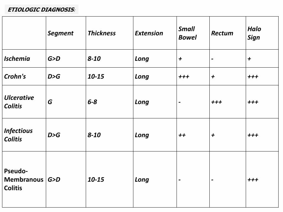
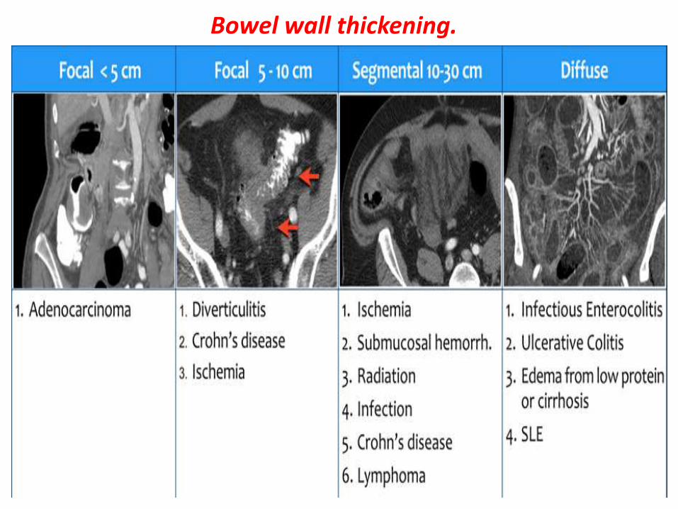
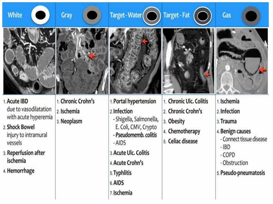
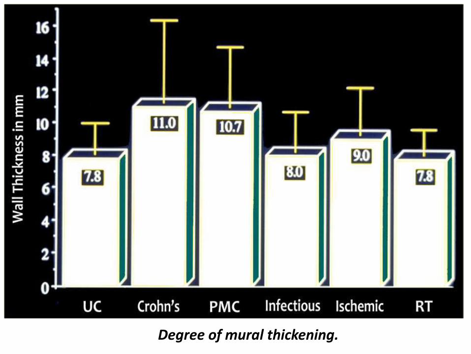
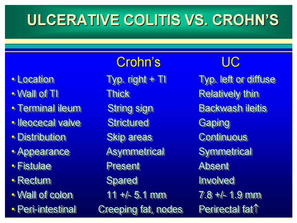
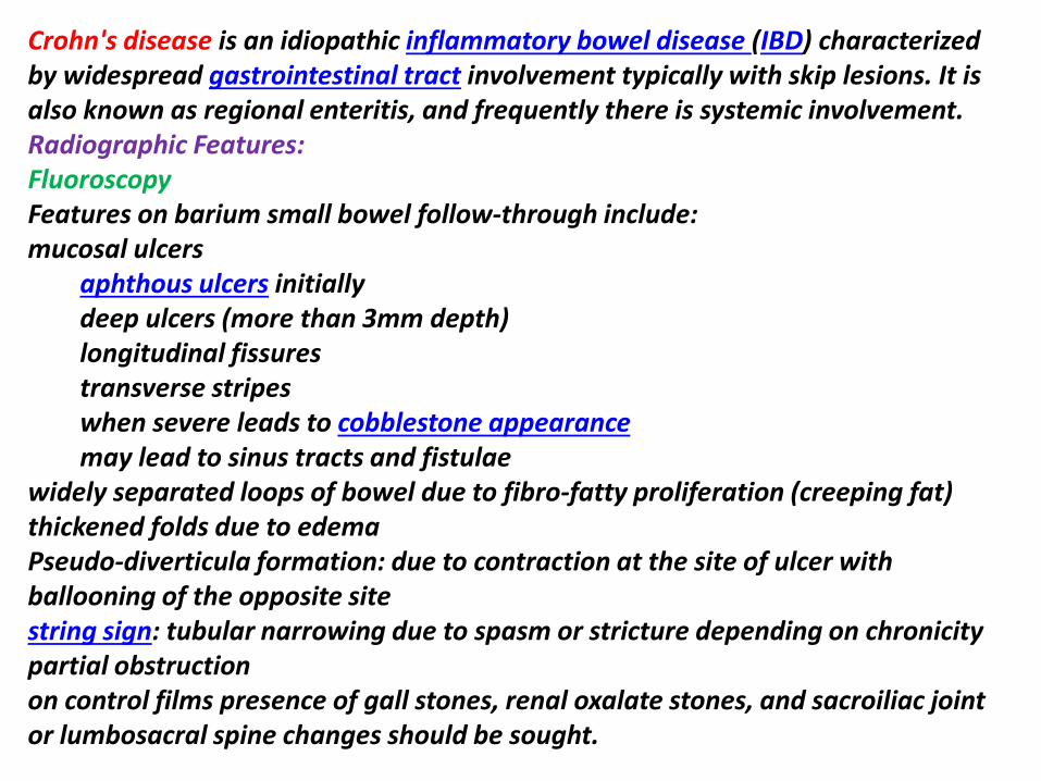
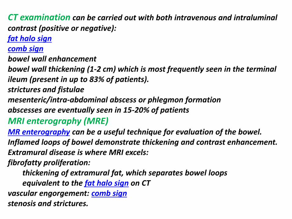
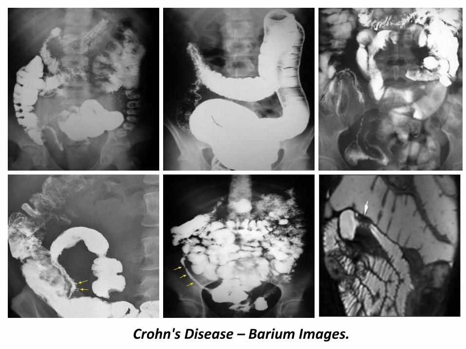
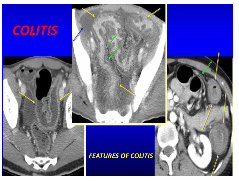
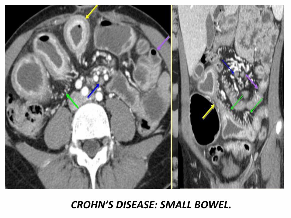
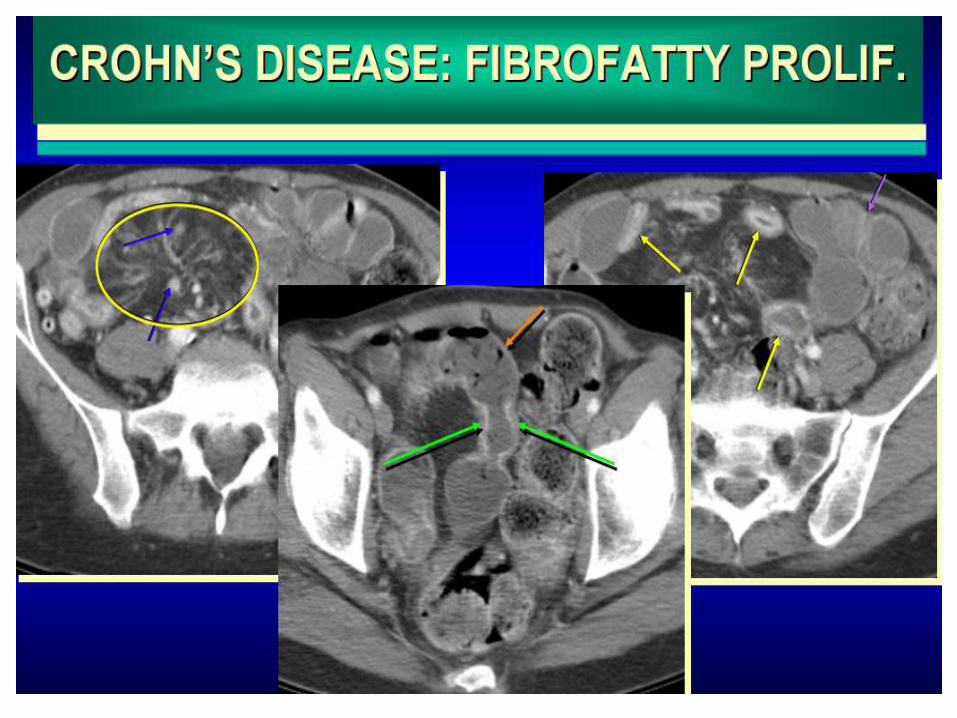
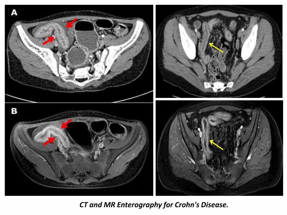
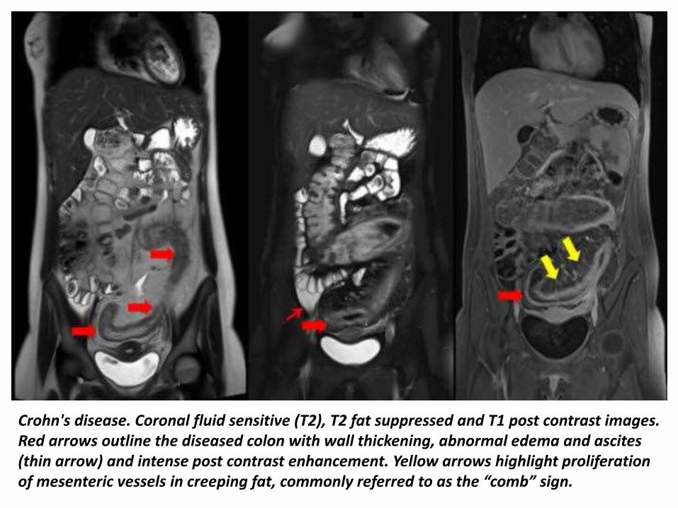
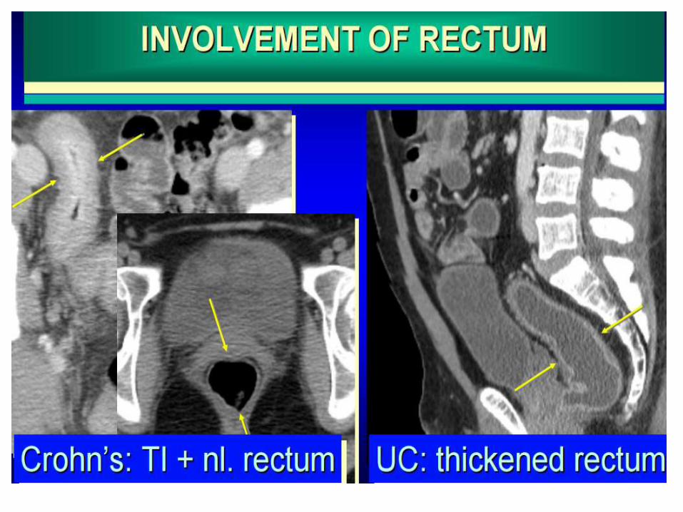
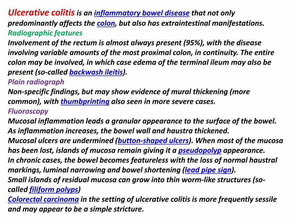
MRI Images with ulcerative colitis
MRI Images with ulcerative colitis
Cytomegalovirus (CMV) is a member of the Herpes viridae family along with herpes simplex viruses 1 and 2 Epstein-Barr virus and varicella-zoster virus It is a double-stranded DNA virus with a protein coat and lipoprotein envelope Similar to other herpes viruses CMV is icosahedral and replicates in the hosts nucleus Replication in the host cell typically manifests pathologically with large intra-nuclear inclusion bodies and smaller cytoplasmic inclusions and is accompanied by the presence of CMV viral particles in the plasma Radiographic featuresBarium studiesCMV oesophagitisSmall well-circumscribed ulcers are present with the mucosa between them appearing normal Larger (~2cm) superficial mid-esophageal ulcers are said to be relatively characteristic of CMV oesophagitis Deep ulceration is uncommon CMV gastritisTypically the antrum is involved and it has a nodular mucosal pattern with luminal narrowingCTCT is particularly useful in CMV enterocolitis The appearances are similar to that of inflammatory bowel disease with mural thickening and surrounding stranding although often the thickening is patchy and not circumferential Ascites is seen in almost half of cases Both diffuse and segmental involvement is encountered In some instances the appearances are essential normal and biopsy is therefore still required when clinical symptoms are suspiciousInvolvement of the small bowel is less frequent seen in only 42 of casesLymph node enlargement is usually not presentPerforation has the usual imaging hallmarks of free intraperitoneal fluid and gas
CMV ileocolitis in a 30 years old patient with aids important wall thickening related to submucosal edema CMV inclusions showed are shown at biopsy
Circumferential ulcerated strictureMarkedly thickened folds of the transverse and descending
colon consistent with pseudo- membrane formation
INFECTIOUS COLITISbullDiagnosis clinical aspects suggesting some infections (tuberculosis amoeba CMV)bullConfirmed by specific lab tests + colonoscopy and biopsybullCT featuresndashRight colon + ileum = salmonella amoeba tuberculosis yersiniandashLeft colon = schistosoma shigella herpes syphilisndashPan colonic CMV E Coli
bullTuberculosis (may mimic Crohns disease)ndashClinical contextndashCaecum granuloma with necrosis on biopsyndashIleocaeligcal locationndashTransmural injuryndashWall thickeningndashEnlarged lymph nodes with low attenuationndashAscitesndashFistulaeabscess
bullAmebiasisndashEndemic countriesndashDifferential diagnosis appendicitisndashDiffuse ulcerations at biopsy with presence of the parasite ndashAcute fulminant colitisndashIgraveleocaecal is the preferred location but the entire colon can be involvedndashLiver abscesses help in the positive diagnosis
Tuberculosis colitis in a 25 years old patient wall thickening of the caecum enlarged lymph nodes with low attenuated center and ascites are seen
Amoeba colitis of the Caecum in a 40 years old patient liver abscesses are seen
Right -sided colitis in a patient with cirrhosis and portal hypertension The endoscopic images are of different patient with right-sided colitis
bullPseudomembranous colitis- Mean age 50 yearsndashDiarrheandashAbdominal painndashFever anorexia nausea dehydrationbullRecent use of antibiotics or recent surgerybullClostridium difficile toxins in the stoolsbullColonoscopyndashUlcerations creamy white and elevated plaques or nodules volcano like eruptions of fibrin and leukocyte from mucosal cryptbullEntire colon+++ involvementbullSmall bowel can be involvedbullImportant wall thickening (mean 11mm)bullAccordion sign nodularpolypoid = submucosal edemabullThumbprintingbullPericolonic fat strandingbullAscites
Pseudomembranous colitis in a 34 years old patient with recent use of antibiotics follow up J4 aspecific colonic wall thickening
Pseudomembranous colitis (Left) Axial CT scan of the mid-abdomen utilizing oral but not intravenous contrast demonstrates marked thickening of the colonic wall (white arrows) producing the so-called accordion sign Thereis a small amount of pericolonic stranding (red arrow) and ascites (green arrow) (Right) Axial CT scan through the pelvis shows marked thickening of the wall of the rectum (yellow arrows) indicating this is a pan-colitis
Pseudomembranous Colitis with dilatation of the sigmoid Pseudomembranous Colitis
Pseudomembranous colitis
Pseudomembranous colitis
DRUG INDUCED COLITISbullChemotherapy (mucosal ulcerations inflammation)bullAntibiotics (clostridium difficile)bullVasoconstrictors drug for hypertension oral contraceptives (ischemia inflammation) NSAID
Toxic colitis involving the entire colon in a 40 years old patient 48H after the colonoscopy
NEUTROPENIC COLITISbullClinical aspectsndashImmunodepression leukemia HIV transplantation chemotherapyndashBacteria viruses and fungi grow profusely in the absence of neutrophils bullLab tests Immunodepression non specificbullColonoscopyndashSame features than ulcerative colitis or infectious colitis but only the right colon is involvedndashHyperemia edema and superficial ulcerationsbullIgraveleocaecal and right colon involvementbullImportant circumferential wall thickening (up to 3 cm for the caecum 4mm for ileum)bullParietal low attenuation submucosal edemabullParietal pneumatosis (severity sign)bullPericolonic strandingbullSepsis abscess intestinal necrosis hemorrhage may occur
Neutropenic colitis involving the entire colon and ileum in a 30 years old patient ascites circumferential wall thickening are shown
Neutropenic colitis Computed tomography (CT) in an 18-year-old man with acute myelogenous leukemia fever and neutropenia A Axial CT image shows the marked thickening of the caecum and a small amount of free fluid B Coronal CT reformat shows pancolitis affecting the right colon to a greater extent
Typhlitis in a patient with neutropenia
Radiation induced colitis in a 70 years old woman with radiation exposure left colon wall thickening stricture of is caliber are shown
Diverticulitis is one of the presentations of diverticular disease and is most often a complication of colonic diverticulosis Differentiating one from the other is critical since uncomplicated diverticulosis is mostly asymptomatic and acute diverticulitis is a potentially life-threatening illness Radiographic featuresCT is the modality of choice for the diagnosis and staging of diverticulitis Appearances includepericolic stranding often disproportionately prominent compared to the amount of bowel wall thickeningsegmental thickening of the bowel wallenhancement of the colonic wall
usually has inner and outer high-attenuation layers with a thick middle layer of low attenuation
diverticular perforationextravasation of air and fluid into the pelvis and peritoneal cavity
abscess formation (seen in up to 30 of cases)may contain fluid gas or both
fistula formationgas in the bladderdirect visualization of a fistulous tract
Sigmoid diverticulitis with abscess formation sigmoid colon displaying mural thickening diverticulosis and pericolic fat stranding (arrow) Adjacent low attenuation septated collection (circle) representing abscess formation with adhesion noted to adjacent small bowel loops
Perforated sigmoid diverticulitis sigmoid colon displaying diverticulosis mural thickening and pericolic inflammatory fat stranding (arrow) with adjacent collection of intra-abdominal free air and adjacent inflammatory fat stranding (circle) again representative of active diverticulitis with perforation
Ischemic colitis refers to inflammation of the colon secondary to vascular
insufficiency and ischemia It sometimes considered under the same spectrum of intestinal ischemia The severity and consequences of the disease are highly variable
EpidemiologyIschemic bowel is typically a disease of the elderly (age gt60 years) where atherosclerotic disease or low flow states are usually the cause 2 In younger individuals the disease is more likely to be related to vasculitis or hyper coagulable statesThe causes can be categorized as follows
arterial occlusionarteriosclerosisvasculitidesarterial emboli
venous thrombosishyper coagulative states including malignancy and OCP useprimary mesenteric venous thrombosis
low flow stateshypotensioncongestive heart failurecardiac arrhythmias
otherssickle cell diseaseradiation therapy
Radiographic featuresPlain film abdominal radiographAbdominal radiographs are often normal but signs includedilatation due to ileusthumbprinting due to mucosal edemahemorrhagelocalized intramural gas (pneumatosis coli) if necroticfree intraperitoneal gas if perforatedFluoroscopy barium studiesContrast enema is abnormal in 90 but is rarely used for diagnostic purposessegmental region of abnormalitythumbprinting which is classically obliterated by air insufflationspasmulcerations serated mucosarsquostricture from fibrosis as a late complication of ischemia
CTContrast enhanced imaging is the modality of choice Features includesegmental region of abnormalitysymmetrical or lobulated thickening of bowel wallirregularly narrowed lumenSubmucosal edema may produce low-density ring bordering lumen (target sign)intramural or portal venous gasMesenteric edemasuperior mesenteric artery or vein thrombusocclusion may be demonstrated
AngiographyCan show mesenteric artery occlusion if present Otherwise angiography may show increased arterial caliber accelerated arteriovenous transit time and dilated draining veins due to the inflammatory response In mesenteric venous thrombosis the veins may not be visualized and collateral venous filling may be seen
UltrasoundUltrasound is of limited use due to bowel gas but may showluminal thickening over the affected segment with or without stratificationhypoechoic wall due to edemaareas of increased echogenicity if hemorrhageechogenic foci with shadowing if intramural gasreduced peristalsis may be observedDoppler imaging of the SMA origin can be useful in assessing for stenosis
Nuclear medicineIncreased uptake of Tc99m (V) DMSA tracer in the ischaemic bowel may be present but is unreliable
Ischaemic colitis
Ischemic colitis parietal pneumatosis is seen on the right side of the scout view
Ischemic colitis CT coronal view showing wall thickening with pericolonic fat stranding
CT Images for Ischemic colitis
CT Images for Ischemic colitis
CT Images for Ischemic colitis
CT Images for Ischemic colitis
Magnetic resonance imaging follow-up of a patients with ischemic colitis resolved promptly Ischemic colitis (IC) of left side colon in a 57 year old woman with a recent history of acute hypertensive crisis who presented with left lower quadrant pain and massive rectal bleeding A Endoscopic procedure showed multiple necrotic area B and C Contrast-enhanced computed tomography (CT) and axial T2 fast-recovery fast-spin echo sequence (FRFSE) magnetic resonance imaging (MRI) after 32 h from CT showed acute IC (Type I CT and MRI) with wall thickening three layer sandwich sign and a mild amount of free fluid in the parabolic gutter D and E 2D coronal reformat CT and coronal T2 FRFSE MRI at the same time showed the entire involved tract F Ischemia resolved without complications with conservative therapy as shown in the follow-up MRI
Magnetic resonance imaging follow-up of a patient with ischemic colitis and worsening of clinical symptoms Ischemic colitis (IC) of sigmoid colon in a 62-year-old man with left lower quadrant pain and elevate lactate dehydrogenase levels who presented with melena and a recent history of stenting procedures for ischemic cardiopathy A Endoscopic procedure showed multiple necrotic area B 2D (two dimensional) coronal reformat contrast-enhanced computed tomography (CT) showed acute IC (Type I CT) C-E The patient had 2 magnetic resonance examinations (C and D-E) with an interval of 48 h due to worsening of clinical symptoms with an increase of the length and thickness of the involved tract (D-E) F The ischemic process resolved without complication after parenteral nutrition as showed in the follow-up magnetic resonance imaging performed after 384 h from the date of CT examination
7T magnetic resonance imaging investigation A Image of a 7T magnetic resonance imaging (MRI) abdominal scan before inferior mesenteric artery (IMA) ligation B A 7T MRI abdominal scan 1 h after IMA ligation C At 4 h after IMA ligation D At 6 h after IMA ligation E At 8 h after IMA ligation F Image of 7T MRI colon enema
Thank You

MRI Images with ulcerative colitis
Cytomegalovirus (CMV) is a member of the Herpes viridae family along with herpes simplex viruses 1 and 2 Epstein-Barr virus and varicella-zoster virus It is a double-stranded DNA virus with a protein coat and lipoprotein envelope Similar to other herpes viruses CMV is icosahedral and replicates in the hosts nucleus Replication in the host cell typically manifests pathologically with large intra-nuclear inclusion bodies and smaller cytoplasmic inclusions and is accompanied by the presence of CMV viral particles in the plasma Radiographic featuresBarium studiesCMV oesophagitisSmall well-circumscribed ulcers are present with the mucosa between them appearing normal Larger (~2cm) superficial mid-esophageal ulcers are said to be relatively characteristic of CMV oesophagitis Deep ulceration is uncommon CMV gastritisTypically the antrum is involved and it has a nodular mucosal pattern with luminal narrowingCTCT is particularly useful in CMV enterocolitis The appearances are similar to that of inflammatory bowel disease with mural thickening and surrounding stranding although often the thickening is patchy and not circumferential Ascites is seen in almost half of cases Both diffuse and segmental involvement is encountered In some instances the appearances are essential normal and biopsy is therefore still required when clinical symptoms are suspiciousInvolvement of the small bowel is less frequent seen in only 42 of casesLymph node enlargement is usually not presentPerforation has the usual imaging hallmarks of free intraperitoneal fluid and gas
CMV ileocolitis in a 30 years old patient with aids important wall thickening related to submucosal edema CMV inclusions showed are shown at biopsy
Circumferential ulcerated strictureMarkedly thickened folds of the transverse and descending
colon consistent with pseudo- membrane formation
INFECTIOUS COLITISbullDiagnosis clinical aspects suggesting some infections (tuberculosis amoeba CMV)bullConfirmed by specific lab tests + colonoscopy and biopsybullCT featuresndashRight colon + ileum = salmonella amoeba tuberculosis yersiniandashLeft colon = schistosoma shigella herpes syphilisndashPan colonic CMV E Coli
bullTuberculosis (may mimic Crohns disease)ndashClinical contextndashCaecum granuloma with necrosis on biopsyndashIleocaeligcal locationndashTransmural injuryndashWall thickeningndashEnlarged lymph nodes with low attenuationndashAscitesndashFistulaeabscess
bullAmebiasisndashEndemic countriesndashDifferential diagnosis appendicitisndashDiffuse ulcerations at biopsy with presence of the parasite ndashAcute fulminant colitisndashIgraveleocaecal is the preferred location but the entire colon can be involvedndashLiver abscesses help in the positive diagnosis
Tuberculosis colitis in a 25 years old patient wall thickening of the caecum enlarged lymph nodes with low attenuated center and ascites are seen
Amoeba colitis of the Caecum in a 40 years old patient liver abscesses are seen
Right -sided colitis in a patient with cirrhosis and portal hypertension The endoscopic images are of different patient with right-sided colitis
bullPseudomembranous colitis- Mean age 50 yearsndashDiarrheandashAbdominal painndashFever anorexia nausea dehydrationbullRecent use of antibiotics or recent surgerybullClostridium difficile toxins in the stoolsbullColonoscopyndashUlcerations creamy white and elevated plaques or nodules volcano like eruptions of fibrin and leukocyte from mucosal cryptbullEntire colon+++ involvementbullSmall bowel can be involvedbullImportant wall thickening (mean 11mm)bullAccordion sign nodularpolypoid = submucosal edemabullThumbprintingbullPericolonic fat strandingbullAscites
Pseudomembranous colitis in a 34 years old patient with recent use of antibiotics follow up J4 aspecific colonic wall thickening
Pseudomembranous colitis (Left) Axial CT scan of the mid-abdomen utilizing oral but not intravenous contrast demonstrates marked thickening of the colonic wall (white arrows) producing the so-called accordion sign Thereis a small amount of pericolonic stranding (red arrow) and ascites (green arrow) (Right) Axial CT scan through the pelvis shows marked thickening of the wall of the rectum (yellow arrows) indicating this is a pan-colitis
Pseudomembranous Colitis with dilatation of the sigmoid Pseudomembranous Colitis
Pseudomembranous colitis
Pseudomembranous colitis
DRUG INDUCED COLITISbullChemotherapy (mucosal ulcerations inflammation)bullAntibiotics (clostridium difficile)bullVasoconstrictors drug for hypertension oral contraceptives (ischemia inflammation) NSAID
Toxic colitis involving the entire colon in a 40 years old patient 48H after the colonoscopy
NEUTROPENIC COLITISbullClinical aspectsndashImmunodepression leukemia HIV transplantation chemotherapyndashBacteria viruses and fungi grow profusely in the absence of neutrophils bullLab tests Immunodepression non specificbullColonoscopyndashSame features than ulcerative colitis or infectious colitis but only the right colon is involvedndashHyperemia edema and superficial ulcerationsbullIgraveleocaecal and right colon involvementbullImportant circumferential wall thickening (up to 3 cm for the caecum 4mm for ileum)bullParietal low attenuation submucosal edemabullParietal pneumatosis (severity sign)bullPericolonic strandingbullSepsis abscess intestinal necrosis hemorrhage may occur
Neutropenic colitis involving the entire colon and ileum in a 30 years old patient ascites circumferential wall thickening are shown
Neutropenic colitis Computed tomography (CT) in an 18-year-old man with acute myelogenous leukemia fever and neutropenia A Axial CT image shows the marked thickening of the caecum and a small amount of free fluid B Coronal CT reformat shows pancolitis affecting the right colon to a greater extent
Typhlitis in a patient with neutropenia
Radiation induced colitis in a 70 years old woman with radiation exposure left colon wall thickening stricture of is caliber are shown
Diverticulitis is one of the presentations of diverticular disease and is most often a complication of colonic diverticulosis Differentiating one from the other is critical since uncomplicated diverticulosis is mostly asymptomatic and acute diverticulitis is a potentially life-threatening illness Radiographic featuresCT is the modality of choice for the diagnosis and staging of diverticulitis Appearances includepericolic stranding often disproportionately prominent compared to the amount of bowel wall thickeningsegmental thickening of the bowel wallenhancement of the colonic wall
usually has inner and outer high-attenuation layers with a thick middle layer of low attenuation
diverticular perforationextravasation of air and fluid into the pelvis and peritoneal cavity
abscess formation (seen in up to 30 of cases)may contain fluid gas or both
fistula formationgas in the bladderdirect visualization of a fistulous tract
Sigmoid diverticulitis with abscess formation sigmoid colon displaying mural thickening diverticulosis and pericolic fat stranding (arrow) Adjacent low attenuation septated collection (circle) representing abscess formation with adhesion noted to adjacent small bowel loops
Perforated sigmoid diverticulitis sigmoid colon displaying diverticulosis mural thickening and pericolic inflammatory fat stranding (arrow) with adjacent collection of intra-abdominal free air and adjacent inflammatory fat stranding (circle) again representative of active diverticulitis with perforation
Ischemic colitis refers to inflammation of the colon secondary to vascular
insufficiency and ischemia It sometimes considered under the same spectrum of intestinal ischemia The severity and consequences of the disease are highly variable
EpidemiologyIschemic bowel is typically a disease of the elderly (age gt60 years) where atherosclerotic disease or low flow states are usually the cause 2 In younger individuals the disease is more likely to be related to vasculitis or hyper coagulable statesThe causes can be categorized as follows
arterial occlusionarteriosclerosisvasculitidesarterial emboli
venous thrombosishyper coagulative states including malignancy and OCP useprimary mesenteric venous thrombosis
low flow stateshypotensioncongestive heart failurecardiac arrhythmias
otherssickle cell diseaseradiation therapy
Radiographic featuresPlain film abdominal radiographAbdominal radiographs are often normal but signs includedilatation due to ileusthumbprinting due to mucosal edemahemorrhagelocalized intramural gas (pneumatosis coli) if necroticfree intraperitoneal gas if perforatedFluoroscopy barium studiesContrast enema is abnormal in 90 but is rarely used for diagnostic purposessegmental region of abnormalitythumbprinting which is classically obliterated by air insufflationspasmulcerations serated mucosarsquostricture from fibrosis as a late complication of ischemia
CTContrast enhanced imaging is the modality of choice Features includesegmental region of abnormalitysymmetrical or lobulated thickening of bowel wallirregularly narrowed lumenSubmucosal edema may produce low-density ring bordering lumen (target sign)intramural or portal venous gasMesenteric edemasuperior mesenteric artery or vein thrombusocclusion may be demonstrated
AngiographyCan show mesenteric artery occlusion if present Otherwise angiography may show increased arterial caliber accelerated arteriovenous transit time and dilated draining veins due to the inflammatory response In mesenteric venous thrombosis the veins may not be visualized and collateral venous filling may be seen
UltrasoundUltrasound is of limited use due to bowel gas but may showluminal thickening over the affected segment with or without stratificationhypoechoic wall due to edemaareas of increased echogenicity if hemorrhageechogenic foci with shadowing if intramural gasreduced peristalsis may be observedDoppler imaging of the SMA origin can be useful in assessing for stenosis
Nuclear medicineIncreased uptake of Tc99m (V) DMSA tracer in the ischaemic bowel may be present but is unreliable
Ischaemic colitis
Ischemic colitis parietal pneumatosis is seen on the right side of the scout view
Ischemic colitis CT coronal view showing wall thickening with pericolonic fat stranding
CT Images for Ischemic colitis
CT Images for Ischemic colitis
CT Images for Ischemic colitis
CT Images for Ischemic colitis
Magnetic resonance imaging follow-up of a patients with ischemic colitis resolved promptly Ischemic colitis (IC) of left side colon in a 57 year old woman with a recent history of acute hypertensive crisis who presented with left lower quadrant pain and massive rectal bleeding A Endoscopic procedure showed multiple necrotic area B and C Contrast-enhanced computed tomography (CT) and axial T2 fast-recovery fast-spin echo sequence (FRFSE) magnetic resonance imaging (MRI) after 32 h from CT showed acute IC (Type I CT and MRI) with wall thickening three layer sandwich sign and a mild amount of free fluid in the parabolic gutter D and E 2D coronal reformat CT and coronal T2 FRFSE MRI at the same time showed the entire involved tract F Ischemia resolved without complications with conservative therapy as shown in the follow-up MRI
Magnetic resonance imaging follow-up of a patient with ischemic colitis and worsening of clinical symptoms Ischemic colitis (IC) of sigmoid colon in a 62-year-old man with left lower quadrant pain and elevate lactate dehydrogenase levels who presented with melena and a recent history of stenting procedures for ischemic cardiopathy A Endoscopic procedure showed multiple necrotic area B 2D (two dimensional) coronal reformat contrast-enhanced computed tomography (CT) showed acute IC (Type I CT) C-E The patient had 2 magnetic resonance examinations (C and D-E) with an interval of 48 h due to worsening of clinical symptoms with an increase of the length and thickness of the involved tract (D-E) F The ischemic process resolved without complication after parenteral nutrition as showed in the follow-up magnetic resonance imaging performed after 384 h from the date of CT examination
7T magnetic resonance imaging investigation A Image of a 7T magnetic resonance imaging (MRI) abdominal scan before inferior mesenteric artery (IMA) ligation B A 7T MRI abdominal scan 1 h after IMA ligation C At 4 h after IMA ligation D At 6 h after IMA ligation E At 8 h after IMA ligation F Image of 7T MRI colon enema
Thank You
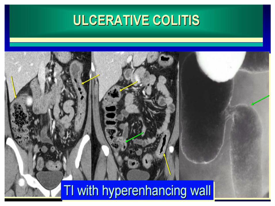
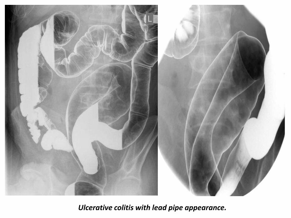
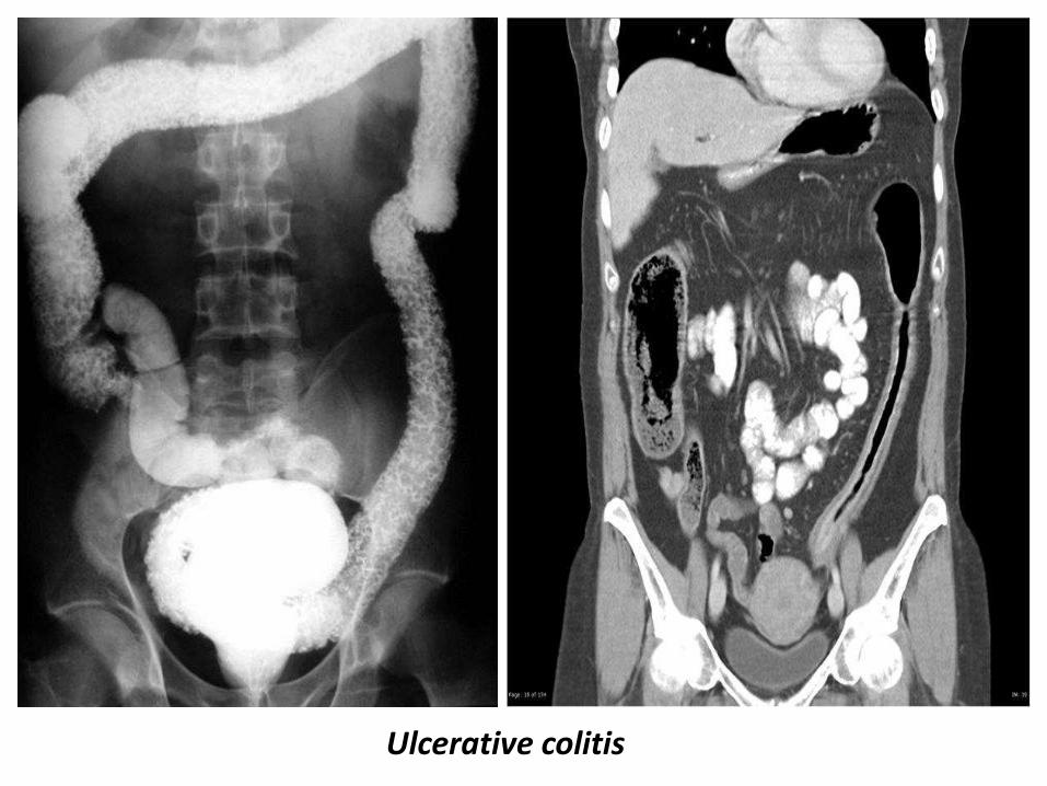
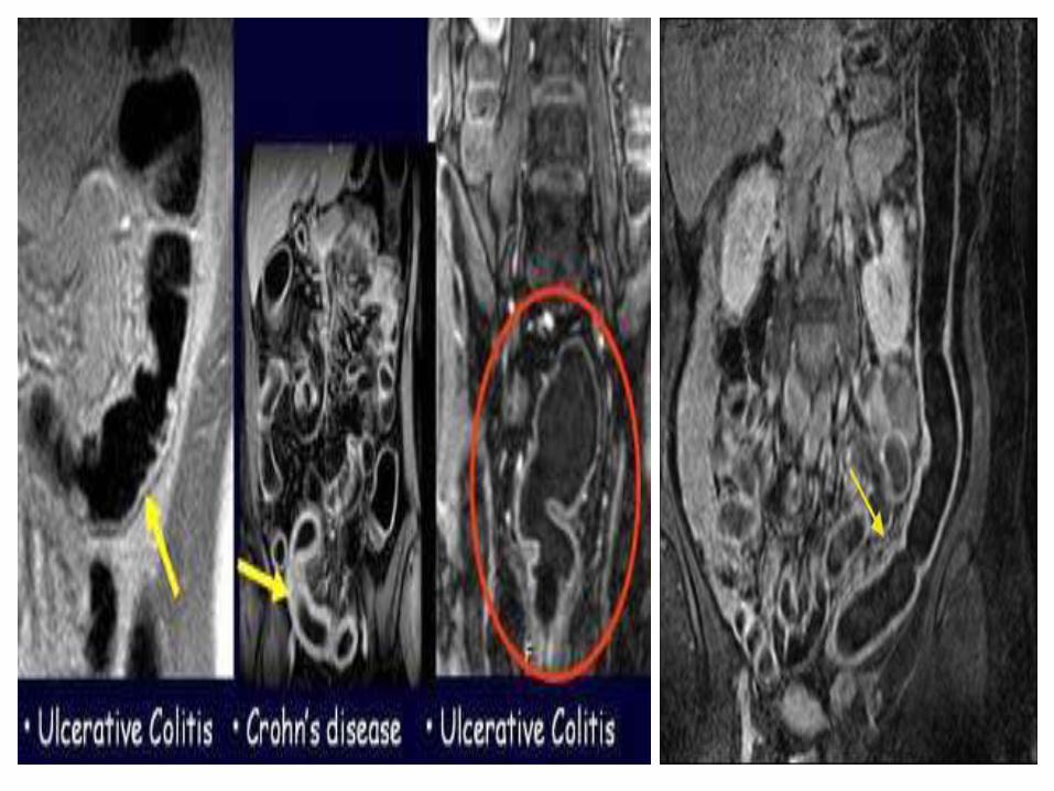
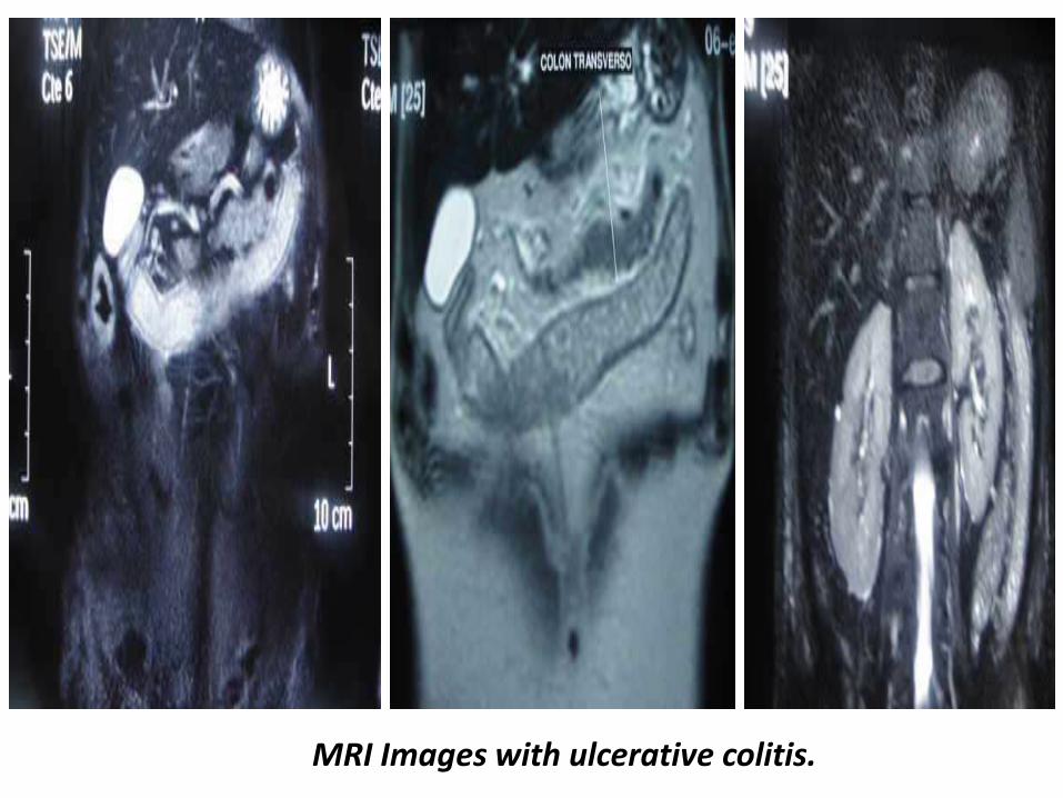
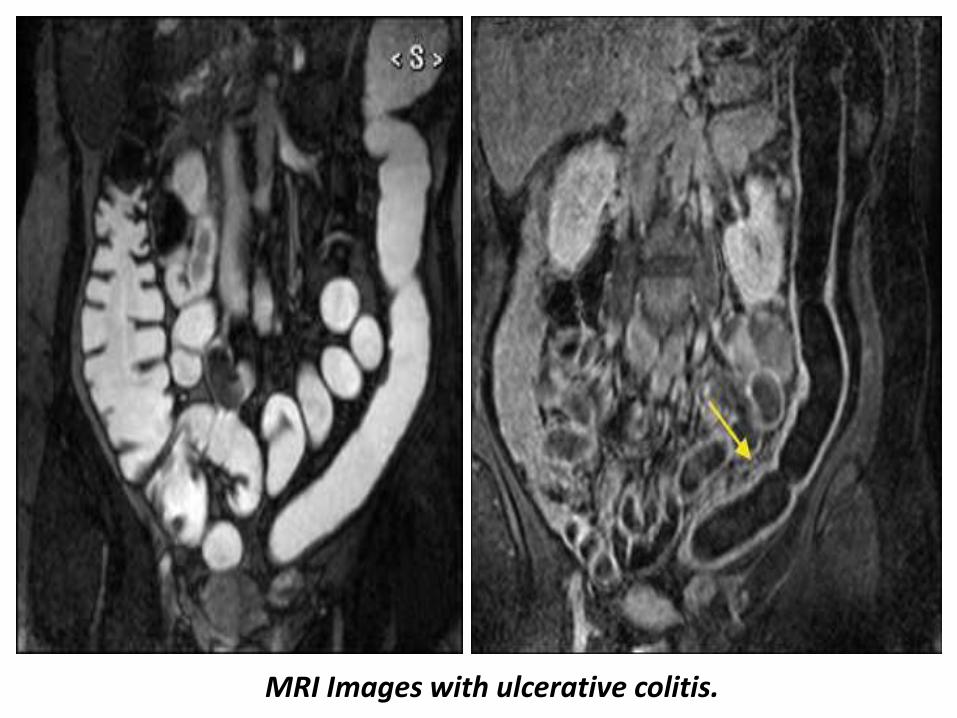
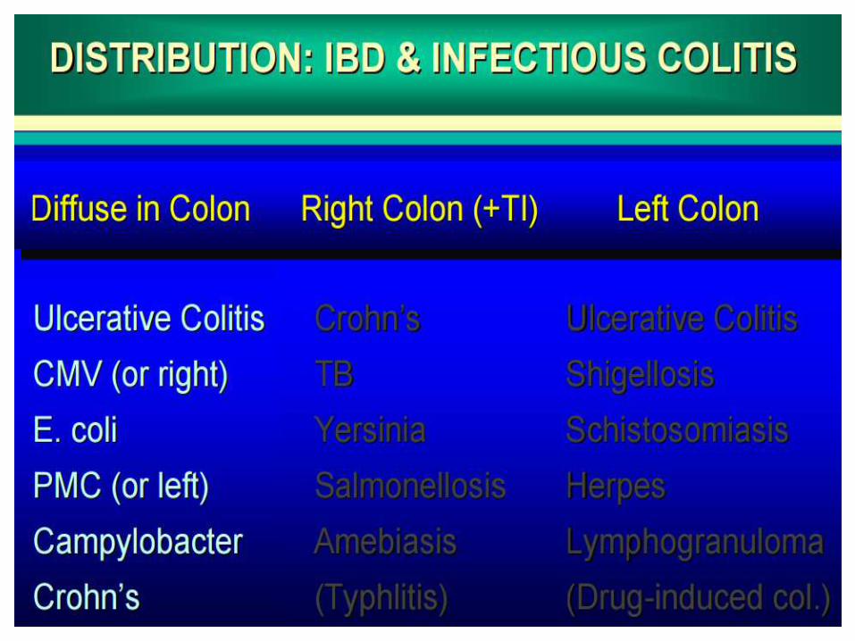
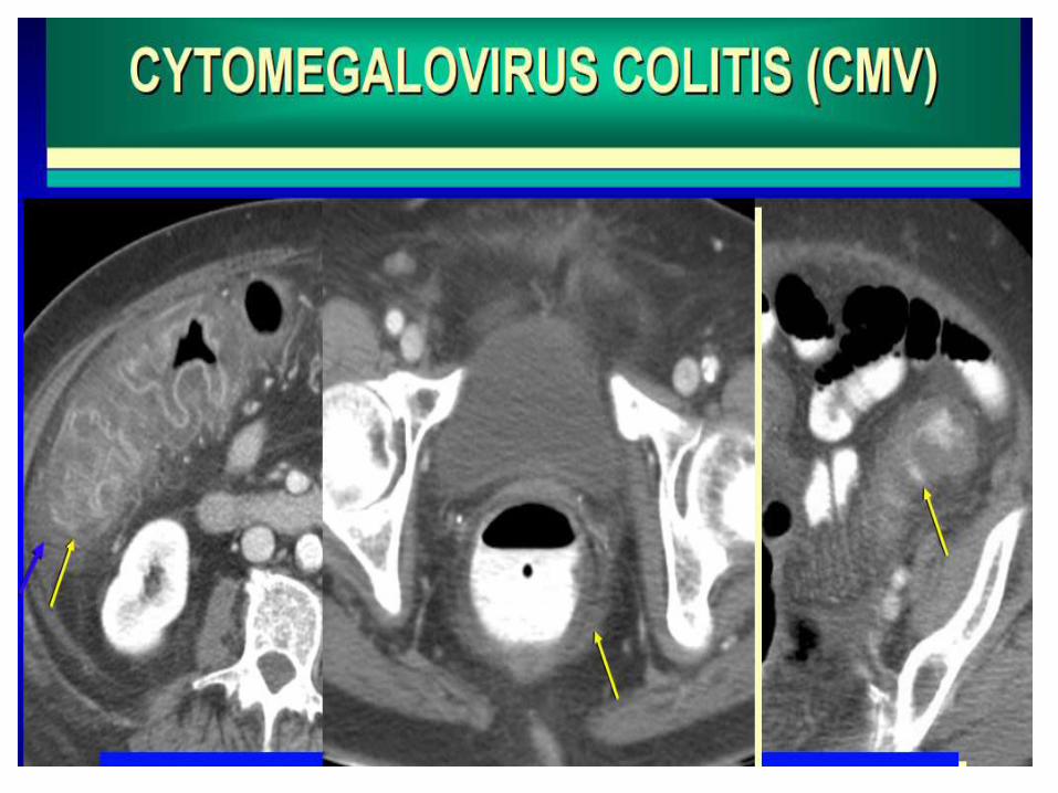
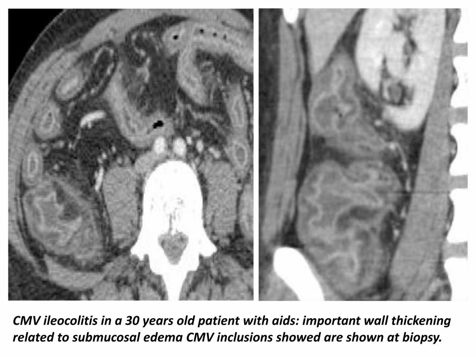
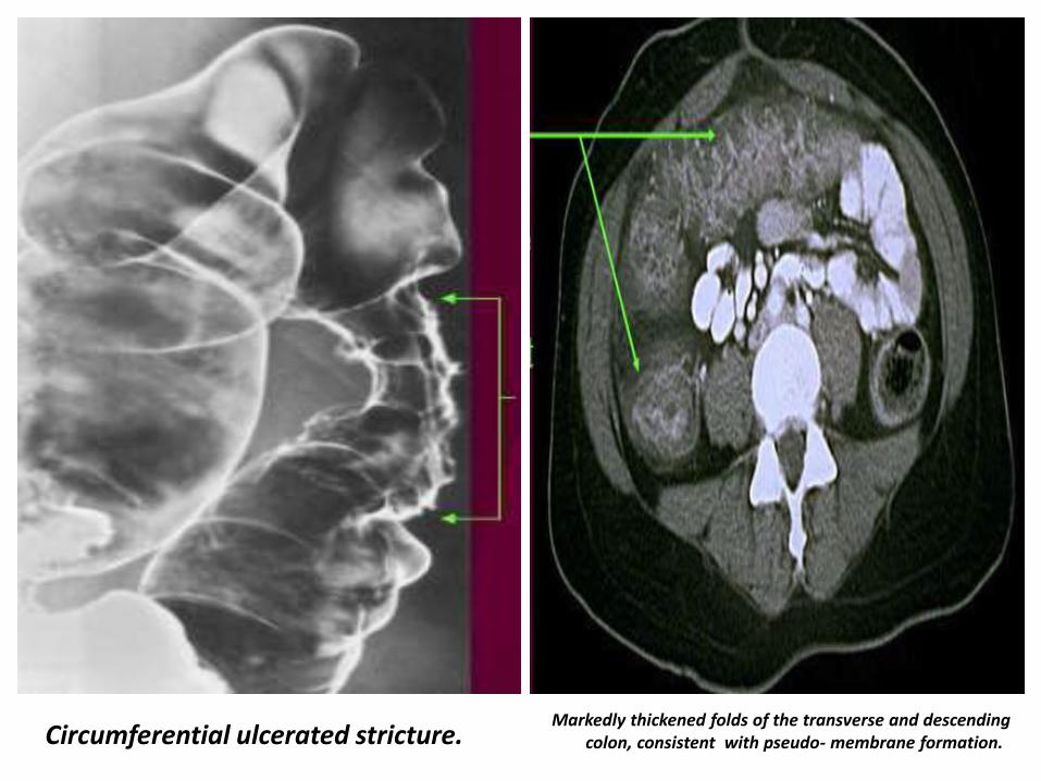
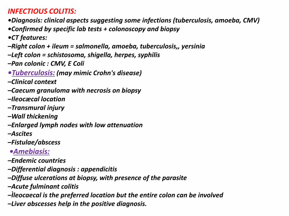
Pseudomembranous colitis
Pseudomembranous colitis
DRUG INDUCED COLITISbullChemotherapy (mucosal ulcerations inflammation)bullAntibiotics (clostridium difficile)bullVasoconstrictors drug for hypertension oral contraceptives (ischemia inflammation) NSAID
Toxic colitis involving the entire colon in a 40 years old patient 48H after the colonoscopy
NEUTROPENIC COLITISbullClinical aspectsndashImmunodepression leukemia HIV transplantation chemotherapyndashBacteria viruses and fungi grow profusely in the absence of neutrophils bullLab tests Immunodepression non specificbullColonoscopyndashSame features than ulcerative colitis or infectious colitis but only the right colon is involvedndashHyperemia edema and superficial ulcerationsbullIgraveleocaecal and right colon involvementbullImportant circumferential wall thickening (up to 3 cm for the caecum 4mm for ileum)bullParietal low attenuation submucosal edemabullParietal pneumatosis (severity sign)bullPericolonic strandingbullSepsis abscess intestinal necrosis hemorrhage may occur
Neutropenic colitis involving the entire colon and ileum in a 30 years old patient ascites circumferential wall thickening are shown
Neutropenic colitis Computed tomography (CT) in an 18-year-old man with acute myelogenous leukemia fever and neutropenia A Axial CT image shows the marked thickening of the caecum and a small amount of free fluid B Coronal CT reformat shows pancolitis affecting the right colon to a greater extent
Typhlitis in a patient with neutropenia
Radiation induced colitis in a 70 years old woman with radiation exposure left colon wall thickening stricture of is caliber are shown
Diverticulitis is one of the presentations of diverticular disease and is most often a complication of colonic diverticulosis Differentiating one from the other is critical since uncomplicated diverticulosis is mostly asymptomatic and acute diverticulitis is a potentially life-threatening illness Radiographic featuresCT is the modality of choice for the diagnosis and staging of diverticulitis Appearances includepericolic stranding often disproportionately prominent compared to the amount of bowel wall thickeningsegmental thickening of the bowel wallenhancement of the colonic wall
usually has inner and outer high-attenuation layers with a thick middle layer of low attenuation
diverticular perforationextravasation of air and fluid into the pelvis and peritoneal cavity
abscess formation (seen in up to 30 of cases)may contain fluid gas or both
fistula formationgas in the bladderdirect visualization of a fistulous tract
Sigmoid diverticulitis with abscess formation sigmoid colon displaying mural thickening diverticulosis and pericolic fat stranding (arrow) Adjacent low attenuation septated collection (circle) representing abscess formation with adhesion noted to adjacent small bowel loops
Perforated sigmoid diverticulitis sigmoid colon displaying diverticulosis mural thickening and pericolic inflammatory fat stranding (arrow) with adjacent collection of intra-abdominal free air and adjacent inflammatory fat stranding (circle) again representative of active diverticulitis with perforation
Ischemic colitis refers to inflammation of the colon secondary to vascular
insufficiency and ischemia It sometimes considered under the same spectrum of intestinal ischemia The severity and consequences of the disease are highly variable
EpidemiologyIschemic bowel is typically a disease of the elderly (age gt60 years) where atherosclerotic disease or low flow states are usually the cause 2 In younger individuals the disease is more likely to be related to vasculitis or hyper coagulable statesThe causes can be categorized as follows
arterial occlusionarteriosclerosisvasculitidesarterial emboli
venous thrombosishyper coagulative states including malignancy and OCP useprimary mesenteric venous thrombosis
low flow stateshypotensioncongestive heart failurecardiac arrhythmias
otherssickle cell diseaseradiation therapy
Radiographic featuresPlain film abdominal radiographAbdominal radiographs are often normal but signs includedilatation due to ileusthumbprinting due to mucosal edemahemorrhagelocalized intramural gas (pneumatosis coli) if necroticfree intraperitoneal gas if perforatedFluoroscopy barium studiesContrast enema is abnormal in 90 but is rarely used for diagnostic purposessegmental region of abnormalitythumbprinting which is classically obliterated by air insufflationspasmulcerations serated mucosarsquostricture from fibrosis as a late complication of ischemia
CTContrast enhanced imaging is the modality of choice Features includesegmental region of abnormalitysymmetrical or lobulated thickening of bowel wallirregularly narrowed lumenSubmucosal edema may produce low-density ring bordering lumen (target sign)intramural or portal venous gasMesenteric edemasuperior mesenteric artery or vein thrombusocclusion may be demonstrated
AngiographyCan show mesenteric artery occlusion if present Otherwise angiography may show increased arterial caliber accelerated arteriovenous transit time and dilated draining veins due to the inflammatory response In mesenteric venous thrombosis the veins may not be visualized and collateral venous filling may be seen
UltrasoundUltrasound is of limited use due to bowel gas but may showluminal thickening over the affected segment with or without stratificationhypoechoic wall due to edemaareas of increased echogenicity if hemorrhageechogenic foci with shadowing if intramural gasreduced peristalsis may be observedDoppler imaging of the SMA origin can be useful in assessing for stenosis
Nuclear medicineIncreased uptake of Tc99m (V) DMSA tracer in the ischaemic bowel may be present but is unreliable
Ischaemic colitis
Ischemic colitis parietal pneumatosis is seen on the right side of the scout view
Ischemic colitis CT coronal view showing wall thickening with pericolonic fat stranding
CT Images for Ischemic colitis
CT Images for Ischemic colitis
CT Images for Ischemic colitis
CT Images for Ischemic colitis
Magnetic resonance imaging follow-up of a patients with ischemic colitis resolved promptly Ischemic colitis (IC) of left side colon in a 57 year old woman with a recent history of acute hypertensive crisis who presented with left lower quadrant pain and massive rectal bleeding A Endoscopic procedure showed multiple necrotic area B and C Contrast-enhanced computed tomography (CT) and axial T2 fast-recovery fast-spin echo sequence (FRFSE) magnetic resonance imaging (MRI) after 32 h from CT showed acute IC (Type I CT and MRI) with wall thickening three layer sandwich sign and a mild amount of free fluid in the parabolic gutter D and E 2D coronal reformat CT and coronal T2 FRFSE MRI at the same time showed the entire involved tract F Ischemia resolved without complications with conservative therapy as shown in the follow-up MRI
Magnetic resonance imaging follow-up of a patient with ischemic colitis and worsening of clinical symptoms Ischemic colitis (IC) of sigmoid colon in a 62-year-old man with left lower quadrant pain and elevate lactate dehydrogenase levels who presented with melena and a recent history of stenting procedures for ischemic cardiopathy A Endoscopic procedure showed multiple necrotic area B 2D (two dimensional) coronal reformat contrast-enhanced computed tomography (CT) showed acute IC (Type I CT) C-E The patient had 2 magnetic resonance examinations (C and D-E) with an interval of 48 h due to worsening of clinical symptoms with an increase of the length and thickness of the involved tract (D-E) F The ischemic process resolved without complication after parenteral nutrition as showed in the follow-up magnetic resonance imaging performed after 384 h from the date of CT examination
7T magnetic resonance imaging investigation A Image of a 7T magnetic resonance imaging (MRI) abdominal scan before inferior mesenteric artery (IMA) ligation B A 7T MRI abdominal scan 1 h after IMA ligation C At 4 h after IMA ligation D At 6 h after IMA ligation E At 8 h after IMA ligation F Image of 7T MRI colon enema
Thank You
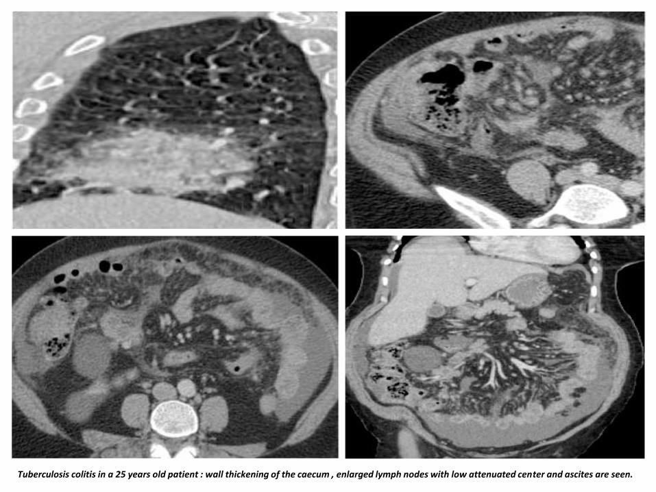
Pseudomembranous colitis
DRUG INDUCED COLITISbullChemotherapy (mucosal ulcerations inflammation)bullAntibiotics (clostridium difficile)bullVasoconstrictors drug for hypertension oral contraceptives (ischemia inflammation) NSAID
Toxic colitis involving the entire colon in a 40 years old patient 48H after the colonoscopy
NEUTROPENIC COLITISbullClinical aspectsndashImmunodepression leukemia HIV transplantation chemotherapyndashBacteria viruses and fungi grow profusely in the absence of neutrophils bullLab tests Immunodepression non specificbullColonoscopyndashSame features than ulcerative colitis or infectious colitis but only the right colon is involvedndashHyperemia edema and superficial ulcerationsbullIgraveleocaecal and right colon involvementbullImportant circumferential wall thickening (up to 3 cm for the caecum 4mm for ileum)bullParietal low attenuation submucosal edemabullParietal pneumatosis (severity sign)bullPericolonic strandingbullSepsis abscess intestinal necrosis hemorrhage may occur
Neutropenic colitis involving the entire colon and ileum in a 30 years old patient ascites circumferential wall thickening are shown
Neutropenic colitis Computed tomography (CT) in an 18-year-old man with acute myelogenous leukemia fever and neutropenia A Axial CT image shows the marked thickening of the caecum and a small amount of free fluid B Coronal CT reformat shows pancolitis affecting the right colon to a greater extent
Typhlitis in a patient with neutropenia
Radiation induced colitis in a 70 years old woman with radiation exposure left colon wall thickening stricture of is caliber are shown
Diverticulitis is one of the presentations of diverticular disease and is most often a complication of colonic diverticulosis Differentiating one from the other is critical since uncomplicated diverticulosis is mostly asymptomatic and acute diverticulitis is a potentially life-threatening illness Radiographic featuresCT is the modality of choice for the diagnosis and staging of diverticulitis Appearances includepericolic stranding often disproportionately prominent compared to the amount of bowel wall thickeningsegmental thickening of the bowel wallenhancement of the colonic wall
usually has inner and outer high-attenuation layers with a thick middle layer of low attenuation
diverticular perforationextravasation of air and fluid into the pelvis and peritoneal cavity
abscess formation (seen in up to 30 of cases)may contain fluid gas or both
fistula formationgas in the bladderdirect visualization of a fistulous tract
Sigmoid diverticulitis with abscess formation sigmoid colon displaying mural thickening diverticulosis and pericolic fat stranding (arrow) Adjacent low attenuation septated collection (circle) representing abscess formation with adhesion noted to adjacent small bowel loops
Perforated sigmoid diverticulitis sigmoid colon displaying diverticulosis mural thickening and pericolic inflammatory fat stranding (arrow) with adjacent collection of intra-abdominal free air and adjacent inflammatory fat stranding (circle) again representative of active diverticulitis with perforation
Ischemic colitis refers to inflammation of the colon secondary to vascular
insufficiency and ischemia It sometimes considered under the same spectrum of intestinal ischemia The severity and consequences of the disease are highly variable
EpidemiologyIschemic bowel is typically a disease of the elderly (age gt60 years) where atherosclerotic disease or low flow states are usually the cause 2 In younger individuals the disease is more likely to be related to vasculitis or hyper coagulable statesThe causes can be categorized as follows
arterial occlusionarteriosclerosisvasculitidesarterial emboli
venous thrombosishyper coagulative states including malignancy and OCP useprimary mesenteric venous thrombosis
low flow stateshypotensioncongestive heart failurecardiac arrhythmias
otherssickle cell diseaseradiation therapy
Radiographic featuresPlain film abdominal radiographAbdominal radiographs are often normal but signs includedilatation due to ileusthumbprinting due to mucosal edemahemorrhagelocalized intramural gas (pneumatosis coli) if necroticfree intraperitoneal gas if perforatedFluoroscopy barium studiesContrast enema is abnormal in 90 but is rarely used for diagnostic purposessegmental region of abnormalitythumbprinting which is classically obliterated by air insufflationspasmulcerations serated mucosarsquostricture from fibrosis as a late complication of ischemia
CTContrast enhanced imaging is the modality of choice Features includesegmental region of abnormalitysymmetrical or lobulated thickening of bowel wallirregularly narrowed lumenSubmucosal edema may produce low-density ring bordering lumen (target sign)intramural or portal venous gasMesenteric edemasuperior mesenteric artery or vein thrombusocclusion may be demonstrated
AngiographyCan show mesenteric artery occlusion if present Otherwise angiography may show increased arterial caliber accelerated arteriovenous transit time and dilated draining veins due to the inflammatory response In mesenteric venous thrombosis the veins may not be visualized and collateral venous filling may be seen
UltrasoundUltrasound is of limited use due to bowel gas but may showluminal thickening over the affected segment with or without stratificationhypoechoic wall due to edemaareas of increased echogenicity if hemorrhageechogenic foci with shadowing if intramural gasreduced peristalsis may be observedDoppler imaging of the SMA origin can be useful in assessing for stenosis
Nuclear medicineIncreased uptake of Tc99m (V) DMSA tracer in the ischaemic bowel may be present but is unreliable
Ischaemic colitis
Ischemic colitis parietal pneumatosis is seen on the right side of the scout view
Ischemic colitis CT coronal view showing wall thickening with pericolonic fat stranding
CT Images for Ischemic colitis
CT Images for Ischemic colitis
CT Images for Ischemic colitis
CT Images for Ischemic colitis
Magnetic resonance imaging follow-up of a patients with ischemic colitis resolved promptly Ischemic colitis (IC) of left side colon in a 57 year old woman with a recent history of acute hypertensive crisis who presented with left lower quadrant pain and massive rectal bleeding A Endoscopic procedure showed multiple necrotic area B and C Contrast-enhanced computed tomography (CT) and axial T2 fast-recovery fast-spin echo sequence (FRFSE) magnetic resonance imaging (MRI) after 32 h from CT showed acute IC (Type I CT and MRI) with wall thickening three layer sandwich sign and a mild amount of free fluid in the parabolic gutter D and E 2D coronal reformat CT and coronal T2 FRFSE MRI at the same time showed the entire involved tract F Ischemia resolved without complications with conservative therapy as shown in the follow-up MRI
Magnetic resonance imaging follow-up of a patient with ischemic colitis and worsening of clinical symptoms Ischemic colitis (IC) of sigmoid colon in a 62-year-old man with left lower quadrant pain and elevate lactate dehydrogenase levels who presented with melena and a recent history of stenting procedures for ischemic cardiopathy A Endoscopic procedure showed multiple necrotic area B 2D (two dimensional) coronal reformat contrast-enhanced computed tomography (CT) showed acute IC (Type I CT) C-E The patient had 2 magnetic resonance examinations (C and D-E) with an interval of 48 h due to worsening of clinical symptoms with an increase of the length and thickness of the involved tract (D-E) F The ischemic process resolved without complication after parenteral nutrition as showed in the follow-up magnetic resonance imaging performed after 384 h from the date of CT examination
7T magnetic resonance imaging investigation A Image of a 7T magnetic resonance imaging (MRI) abdominal scan before inferior mesenteric artery (IMA) ligation B A 7T MRI abdominal scan 1 h after IMA ligation C At 4 h after IMA ligation D At 6 h after IMA ligation E At 8 h after IMA ligation F Image of 7T MRI colon enema
Thank You
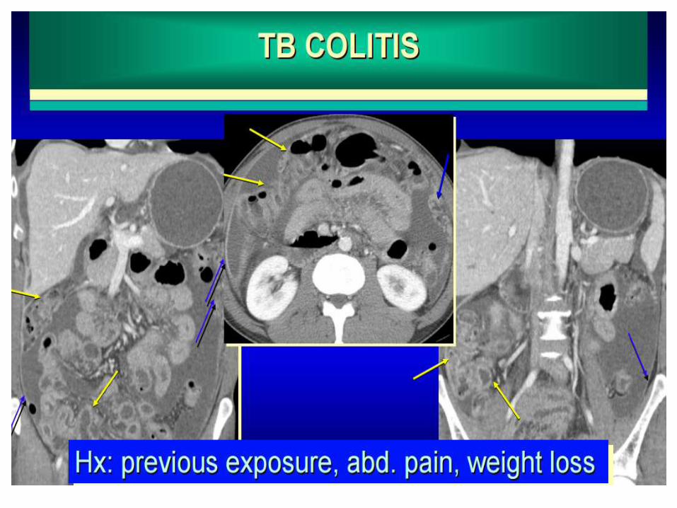
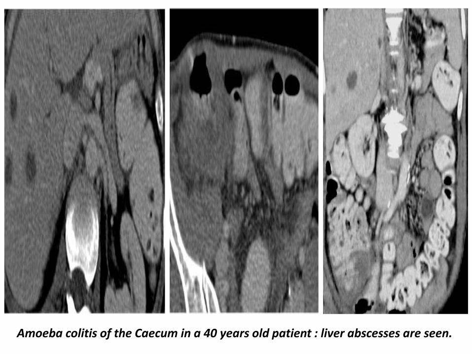
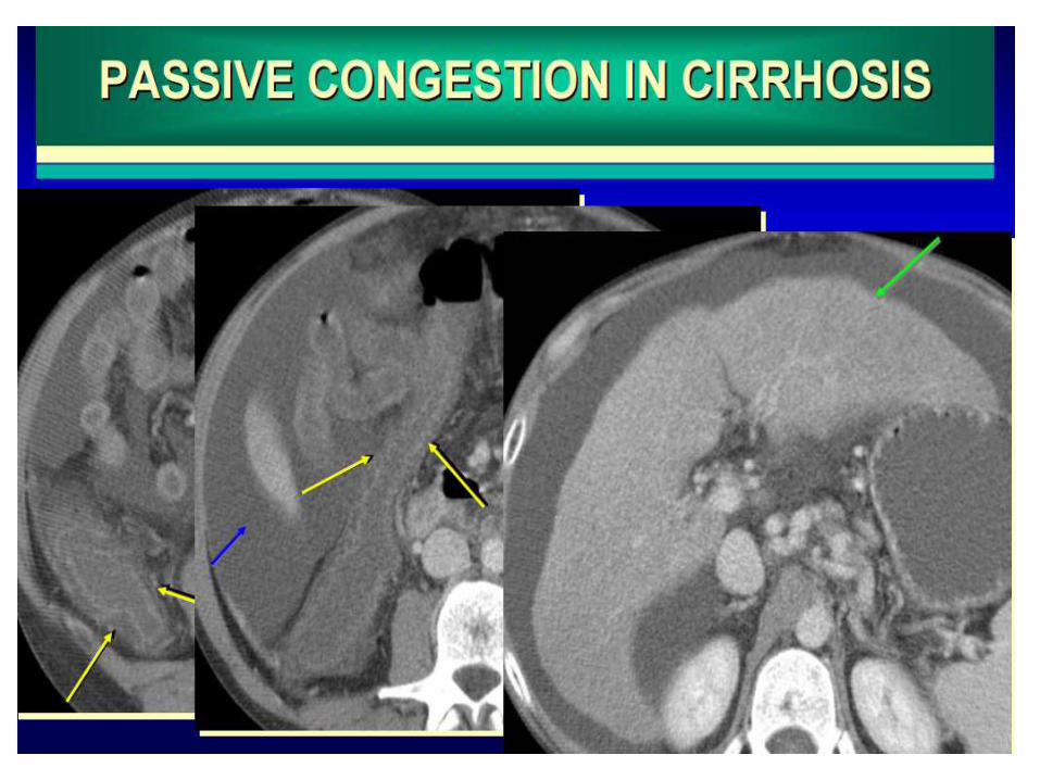
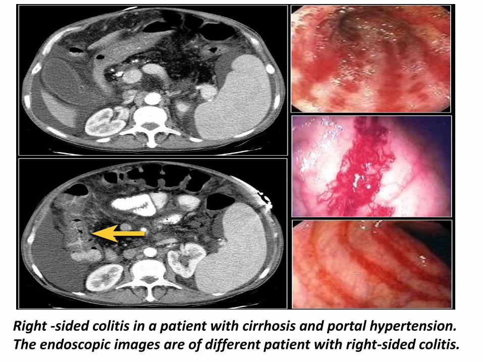
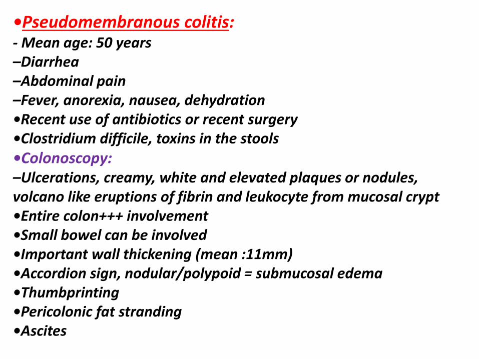
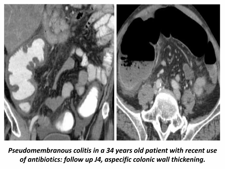
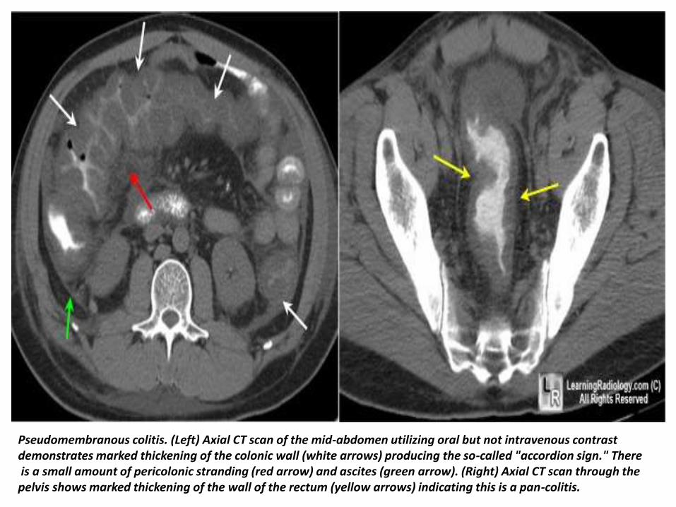
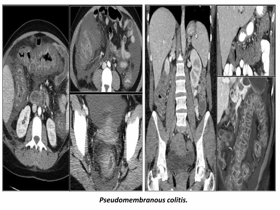
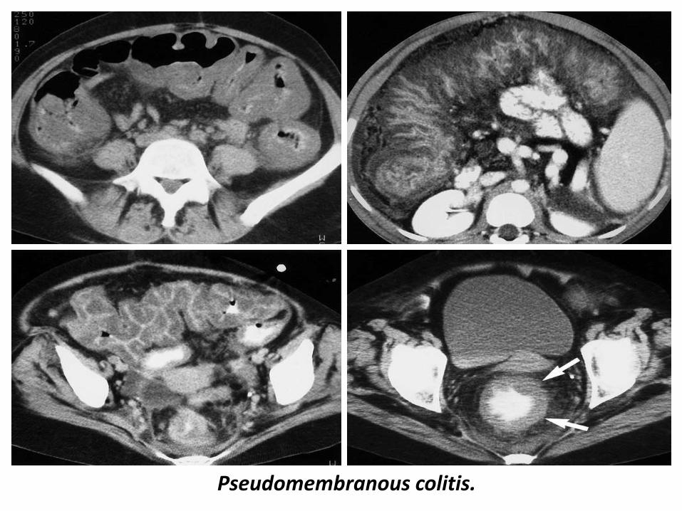
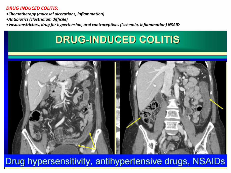
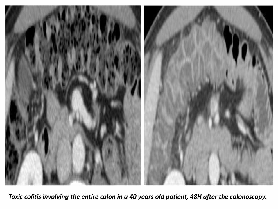
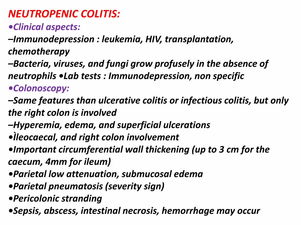
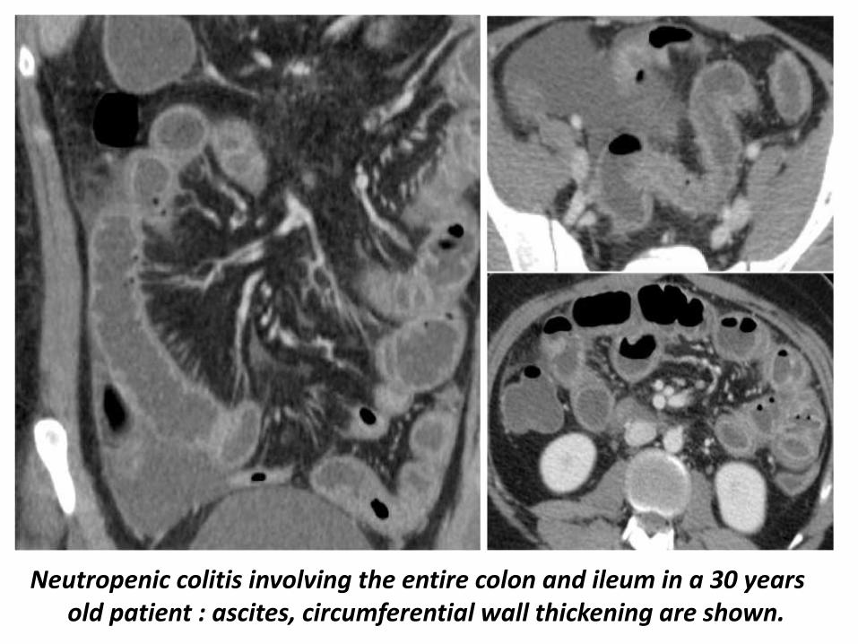
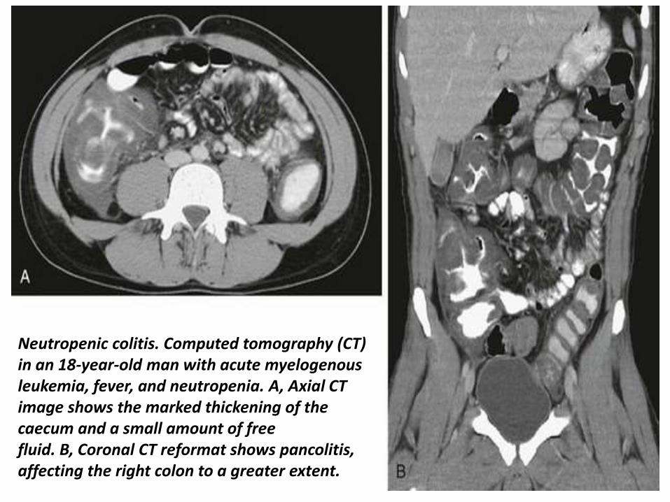
CT Images for Ischemic colitis
CT Images for Ischemic colitis
CT Images for Ischemic colitis
CT Images for Ischemic colitis
Magnetic resonance imaging follow-up of a patients with ischemic colitis resolved promptly Ischemic colitis (IC) of left side colon in a 57 year old woman with a recent history of acute hypertensive crisis who presented with left lower quadrant pain and massive rectal bleeding A Endoscopic procedure showed multiple necrotic area B and C Contrast-enhanced computed tomography (CT) and axial T2 fast-recovery fast-spin echo sequence (FRFSE) magnetic resonance imaging (MRI) after 32 h from CT showed acute IC (Type I CT and MRI) with wall thickening three layer sandwich sign and a mild amount of free fluid in the parabolic gutter D and E 2D coronal reformat CT and coronal T2 FRFSE MRI at the same time showed the entire involved tract F Ischemia resolved without complications with conservative therapy as shown in the follow-up MRI
Magnetic resonance imaging follow-up of a patient with ischemic colitis and worsening of clinical symptoms Ischemic colitis (IC) of sigmoid colon in a 62-year-old man with left lower quadrant pain and elevate lactate dehydrogenase levels who presented with melena and a recent history of stenting procedures for ischemic cardiopathy A Endoscopic procedure showed multiple necrotic area B 2D (two dimensional) coronal reformat contrast-enhanced computed tomography (CT) showed acute IC (Type I CT) C-E The patient had 2 magnetic resonance examinations (C and D-E) with an interval of 48 h due to worsening of clinical symptoms with an increase of the length and thickness of the involved tract (D-E) F The ischemic process resolved without complication after parenteral nutrition as showed in the follow-up magnetic resonance imaging performed after 384 h from the date of CT examination
7T magnetic resonance imaging investigation A Image of a 7T magnetic resonance imaging (MRI) abdominal scan before inferior mesenteric artery (IMA) ligation B A 7T MRI abdominal scan 1 h after IMA ligation C At 4 h after IMA ligation D At 6 h after IMA ligation E At 8 h after IMA ligation F Image of 7T MRI colon enema
Thank You

CT Images for Ischemic colitis
CT Images for Ischemic colitis
CT Images for Ischemic colitis
Magnetic resonance imaging follow-up of a patients with ischemic colitis resolved promptly Ischemic colitis (IC) of left side colon in a 57 year old woman with a recent history of acute hypertensive crisis who presented with left lower quadrant pain and massive rectal bleeding A Endoscopic procedure showed multiple necrotic area B and C Contrast-enhanced computed tomography (CT) and axial T2 fast-recovery fast-spin echo sequence (FRFSE) magnetic resonance imaging (MRI) after 32 h from CT showed acute IC (Type I CT and MRI) with wall thickening three layer sandwich sign and a mild amount of free fluid in the parabolic gutter D and E 2D coronal reformat CT and coronal T2 FRFSE MRI at the same time showed the entire involved tract F Ischemia resolved without complications with conservative therapy as shown in the follow-up MRI
Magnetic resonance imaging follow-up of a patient with ischemic colitis and worsening of clinical symptoms Ischemic colitis (IC) of sigmoid colon in a 62-year-old man with left lower quadrant pain and elevate lactate dehydrogenase levels who presented with melena and a recent history of stenting procedures for ischemic cardiopathy A Endoscopic procedure showed multiple necrotic area B 2D (two dimensional) coronal reformat contrast-enhanced computed tomography (CT) showed acute IC (Type I CT) C-E The patient had 2 magnetic resonance examinations (C and D-E) with an interval of 48 h due to worsening of clinical symptoms with an increase of the length and thickness of the involved tract (D-E) F The ischemic process resolved without complication after parenteral nutrition as showed in the follow-up magnetic resonance imaging performed after 384 h from the date of CT examination
7T magnetic resonance imaging investigation A Image of a 7T magnetic resonance imaging (MRI) abdominal scan before inferior mesenteric artery (IMA) ligation B A 7T MRI abdominal scan 1 h after IMA ligation C At 4 h after IMA ligation D At 6 h after IMA ligation E At 8 h after IMA ligation F Image of 7T MRI colon enema
Thank You
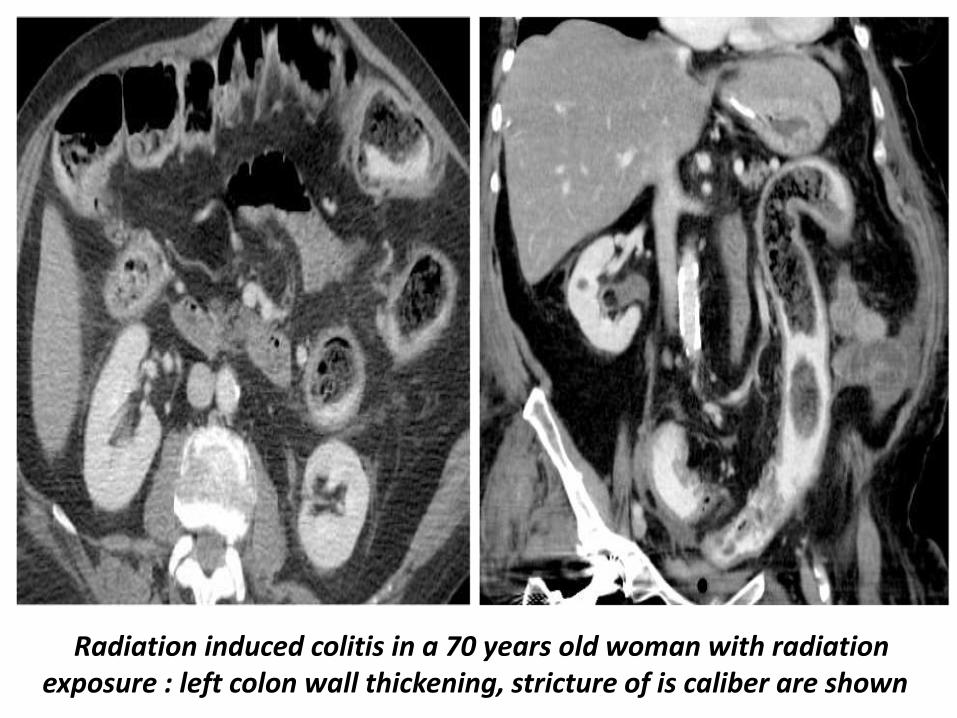
CT Images for Ischemic colitis
CT Images for Ischemic colitis
Magnetic resonance imaging follow-up of a patients with ischemic colitis resolved promptly Ischemic colitis (IC) of left side colon in a 57 year old woman with a recent history of acute hypertensive crisis who presented with left lower quadrant pain and massive rectal bleeding A Endoscopic procedure showed multiple necrotic area B and C Contrast-enhanced computed tomography (CT) and axial T2 fast-recovery fast-spin echo sequence (FRFSE) magnetic resonance imaging (MRI) after 32 h from CT showed acute IC (Type I CT and MRI) with wall thickening three layer sandwich sign and a mild amount of free fluid in the parabolic gutter D and E 2D coronal reformat CT and coronal T2 FRFSE MRI at the same time showed the entire involved tract F Ischemia resolved without complications with conservative therapy as shown in the follow-up MRI
Magnetic resonance imaging follow-up of a patient with ischemic colitis and worsening of clinical symptoms Ischemic colitis (IC) of sigmoid colon in a 62-year-old man with left lower quadrant pain and elevate lactate dehydrogenase levels who presented with melena and a recent history of stenting procedures for ischemic cardiopathy A Endoscopic procedure showed multiple necrotic area B 2D (two dimensional) coronal reformat contrast-enhanced computed tomography (CT) showed acute IC (Type I CT) C-E The patient had 2 magnetic resonance examinations (C and D-E) with an interval of 48 h due to worsening of clinical symptoms with an increase of the length and thickness of the involved tract (D-E) F The ischemic process resolved without complication after parenteral nutrition as showed in the follow-up magnetic resonance imaging performed after 384 h from the date of CT examination
7T magnetic resonance imaging investigation A Image of a 7T magnetic resonance imaging (MRI) abdominal scan before inferior mesenteric artery (IMA) ligation B A 7T MRI abdominal scan 1 h after IMA ligation C At 4 h after IMA ligation D At 6 h after IMA ligation E At 8 h after IMA ligation F Image of 7T MRI colon enema
Thank You
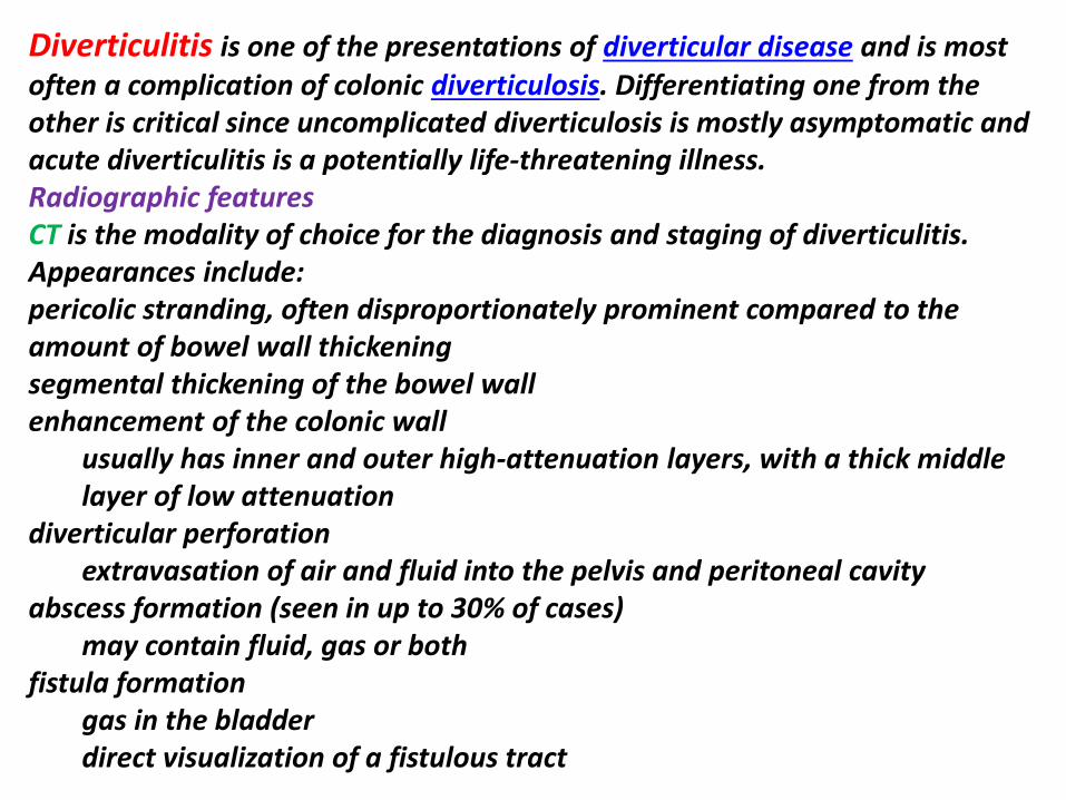
CT Images for Ischemic colitis
Magnetic resonance imaging follow-up of a patients with ischemic colitis resolved promptly Ischemic colitis (IC) of left side colon in a 57 year old woman with a recent history of acute hypertensive crisis who presented with left lower quadrant pain and massive rectal bleeding A Endoscopic procedure showed multiple necrotic area B and C Contrast-enhanced computed tomography (CT) and axial T2 fast-recovery fast-spin echo sequence (FRFSE) magnetic resonance imaging (MRI) after 32 h from CT showed acute IC (Type I CT and MRI) with wall thickening three layer sandwich sign and a mild amount of free fluid in the parabolic gutter D and E 2D coronal reformat CT and coronal T2 FRFSE MRI at the same time showed the entire involved tract F Ischemia resolved without complications with conservative therapy as shown in the follow-up MRI
Magnetic resonance imaging follow-up of a patient with ischemic colitis and worsening of clinical symptoms Ischemic colitis (IC) of sigmoid colon in a 62-year-old man with left lower quadrant pain and elevate lactate dehydrogenase levels who presented with melena and a recent history of stenting procedures for ischemic cardiopathy A Endoscopic procedure showed multiple necrotic area B 2D (two dimensional) coronal reformat contrast-enhanced computed tomography (CT) showed acute IC (Type I CT) C-E The patient had 2 magnetic resonance examinations (C and D-E) with an interval of 48 h due to worsening of clinical symptoms with an increase of the length and thickness of the involved tract (D-E) F The ischemic process resolved without complication after parenteral nutrition as showed in the follow-up magnetic resonance imaging performed after 384 h from the date of CT examination
7T magnetic resonance imaging investigation A Image of a 7T magnetic resonance imaging (MRI) abdominal scan before inferior mesenteric artery (IMA) ligation B A 7T MRI abdominal scan 1 h after IMA ligation C At 4 h after IMA ligation D At 6 h after IMA ligation E At 8 h after IMA ligation F Image of 7T MRI colon enema
Thank You
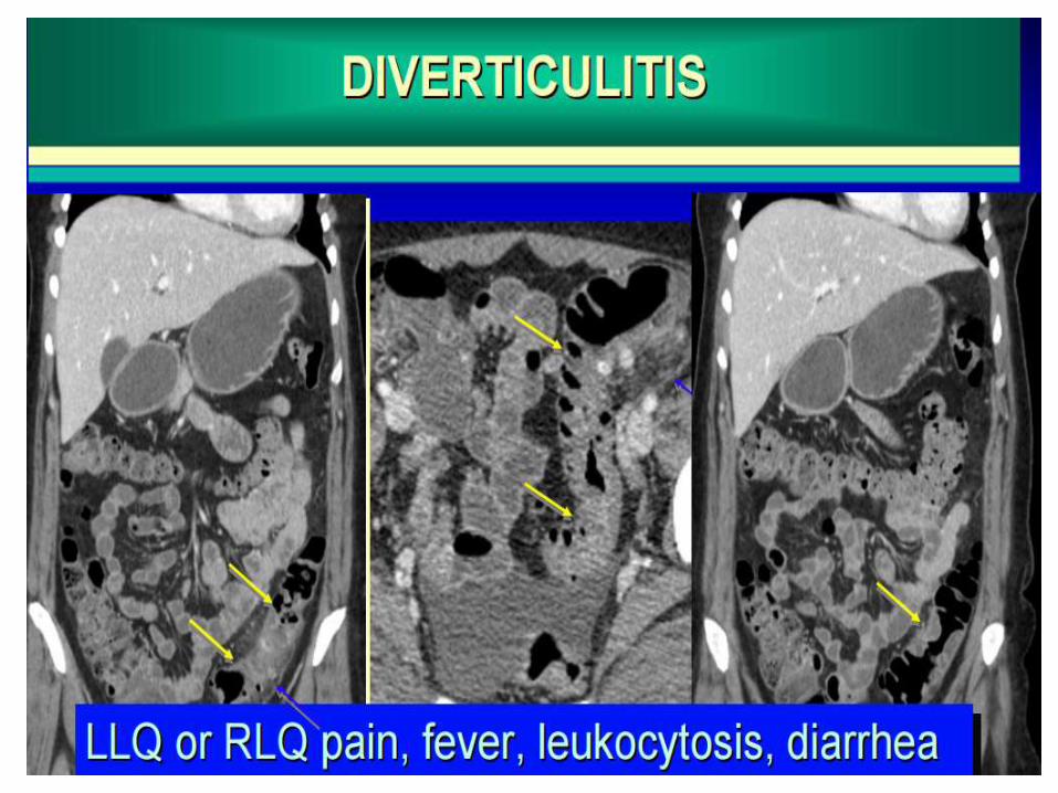
Thank You