Change of Soil Bacteria Diversity between Desertification ...

Annelies Smedts, Ruis Amery, Nico Moons, Etienne Jooken and
Boudewijn Meesschaert
Preparation of steviol by soil bacteria
in: Proceedings of the 2nd Stevia symposium: “Steviol glycosides: technical and pharmacological aspects”, June 27th 2008, KULeuven, Belgium pp. 5-27, ISBN: 9789074253-031, EAN 9789074253031, NUR 882 – 893
Euprint ed., Parkbosstraat 3, 3001 Heverlee www.euprint.be

CHAPTER 1
Preparation of steviol by soil bacteria
Annelies Smedts*, Ruis Amery*, Nico Moons**, Etienne Jooken* and
Boudewijn Meesschaert*/**
*: Department of Industrial Sciences and Technology Katholieke Hogeschool Brugge-Oostende
Zeedijk 101, 8400 Oostende, Belgium Associated to the Katholieke Universiteit Leuven as Faculty of Industrial Sciences
Tel.&Fax. +32 59 56 90 53 [email protected]
& **: Molecular Design and Synthesis
Department of Chemistry Katholieke Universiteit Leuven
Celestijnenlaan 200F, 3001 Leuven, Belgium Tel. +32 16 32 74 39 - Fax. +32 16 32 79 90
***: Centre for Surface Chemistry and Catalysis and Leuven Food Science and Nutrition Research Centre (LFoRCe)
Department of Molecular and Microbial Sciences Katholieke Universiteit Leuven
Arenberg Park 20 3001 Leuven, Belgium
[email protected] ABSTRACT
Stevioside was hydrolysed to steviol using soil samples of a Stevia plantation in
Paraguay. The best results were obtained with pasteurized soil samples that were
incubated at 37 °C with a 0.2% stevioside solution. The responsible bacteria could
be cultivated in and on a simple culture medium supplemented with stevioside. In
the liquid cultures, full hydrolysis of stevioside to steviol was observed by TLC
and HPLC analysis. All available evidence is in agreement with a two-step degra-
dation by different micro-organisms via steviolbioside. Acidification of the culture

6
filtrate resulted in the formation of a white precipitate and after isolation by cen-
trifugation and washing with water, steviol was obtained in a nearly quantitative
way. The steviol isolated was almost 100 % pure as shown by HPLC. The mass
spectrum and NMR spectra of the isolated product were in full agreement with
those of authentic steviol.
KEYWORDS
Stevioside, steviol, HPLC, bio-organic synthesis
INTRODUCTION
Stevioside (Figure 1a), a diterpenoid glycoside, is a natural sweetener extracted
from the leaves of Stevia rebaudiana (Geuns, 2004). Stevioside tastes 250 to 300
times sweeter than sugar (Geuns, 2003). Stevia is native in some parts of South
America (Paraguay, Brazil) and is nowadays cultivated in Paraguay, USA, Mexico,
Central America, Japan, China, Russia, Malaysia, South Korea, Spain, Italy, UK
and Belgium (Geuns, 2000).
Stevioside contains 3 different glucosidic bonds (Figure 1): it contains a β-linked
sophorose (β1-2 D-glucopyranosyl D-glucose) on C-13 and an ester β-glucosidic
linkage on the C-19 carboxyl group. Chemical hydrolysis of glycosidic bonds is
normally carried out under acidic conditions. However, under these conditions
steviol is converted to isosteviol (Figure 1b) and thus acidic hydrolysis of ste-
vioside to steviol is impossible. However, this method was successfully used for
the preparation of sophorose from stevioside (Kusakabe et al., 1987).
The chemical preparation of steviol is normally performed by reacting stevioside
with NaIO4 and then refluxing the reaction mixture with KOH. The steviol is then
obtained after repeated crystallisation. Total yield is often as little as 5 to maxi-
mum 10% (Ogawa. et al., 1980).
By 1972, the soil bacterial hydrolysis of stevioside to steviolbioside and steviol
was described (Yosioka et al., 1972). To this propose, an unknown bacterium des-

Preparation of steviol by soil bacteria 7
ignated as YSB-9 was used. Steviol which was the minor hydrolytic component,
was extracted from the culture medium with diethyl ether. Steviolbioside was the
major compound formed and it was extracted with water saturated n-butanol.
CH2
O
C
R2
OOR1
123 4 5 6 7
8910
1112
1314
15
16 17
18 19
20
a.
O
H3C COOH
CH3
CH3
b.
R1 R2 steviol H H steviolbioside H β-Glc-β-Glc(2→1) stevioside β-Glc β-Glc-β-Glc(2→1) rebaudioside A β-Glc β-Glc-β-Glc(2→1) │ β-Glc(3→1) rebaudioside B H β-Glc-β-Glc(2→1) │ β-Glc(3→1) rebaudioside C β-Glc β-Glc-α-Rha(2→1) (dulcoside) │ β-Glc(3→1) rebaudioside D β-Glc-β-Glc(2→1) β-Glc-β-Glc(2→1) │ β-Glc(3→1) rebaudioside E β-Glc-β-Glc(2→1) β-Glc-β-Glc(2→1) rebaudioside F β-Glc β-Glc-β-Xyl(2→1) │ β-Glc(3→1) dulcoside A β-Glc β-Glc-α-Rha(2→1)
Figure 1: Chemical structure of steviol (a; Wood et al., 1955) and isosteviol (b; Mossetig et al., 1963)

8
Nakano et al. (1998) isolated a Clavibacter michiganense with an active β-
glucosidase for the hydrolysis of ester linkages. The enzyme hydrolysed glucosyl
ester linkages at site 19 of rebaudioside A, stevioside, rubusoside and steviol
monoglucosyl ester, although it did not cleave the 13-O-linked glusosyl residue of
rubusoside and steviol monoside. This enzyme also had some transglucosidase
activity. The same research group further reported (Okamoto et al., 2000) the puri-
fication and characterization of a β-glucosidase from Flavobacterium johnsonae
that also hydrolysed the glucosyl ester linkages at C-19. In contrast to the enzyme
of C. michiganense, the enzyme of F. johnsonae also demonstrated β-glucosidic
activity towards ether glycosides since it also hydrolysed sophorose. However, the
enzyme could not convert steviolbioside into steviol monoside, nor could it hydro-
lyse stevioside into rubusoside. In contrast, it hydrolysed steviol monoside effi-
ciently to steviol and hydrolysed rubusoside to steviol monoglucosyl ester. Thus
the enzyme could not convert stevioside to steviol since it lacked the glucosidic
activity for the conversion of steviol to rubusoside as well as that for the hydrolysis
of steviolbioside to steviolmonoside.
Our primary idea was that a soil which is regularly contaminated with stevioside,
can be a reservoir for bacteria able tot hydrolyse or even metabolise stevioside.
Soils, on which Stevia had been already cultivated for centuries, were regularly
contaminated with stevioside. Such a soil sample was obtained from Paraguay. The
isolation of a microbial culture from soil samples from Paraguay that efficiently
hydrolyse stevioside to steviol is described herein.
MATERIALS and METHODS
Chemicals
Pure standards of rebaudioside A, stevioside, steviolbioside and steviol were ob-
tained from Struyf et al. (2008). They were also used for the construction of the
calibration lines. Different samples of stevioside (with different purity) were used
in the incubation experiments. The first stevioside (Ste1) was 60% pure and con-

Preparation of steviol by soil bacteria 9
tained rebaudioside A as most important contaminant (28 %), besides some re-
baudioside C (9 %); it originated from Paraguay and was a gift of Peter Grosser
(Medherbs). The second stevioside preparation (Ste2) used in the incubation ex-
periments, was an almost pure stevioside, obtained by the recrystallisation of a
95% pure stevioside sample that was obtained from DIC in Japan.
The solvents used for HPLC were acetonitrile and 25 mM H3PO4. The acetonitrile
(HPLC grade) and the H3PO4 (pure) were from Acros Organics (Geel, Belgium).
The chemicals for the cultivation of the bacteria were yeast-extract (Biolife Italiana
S.r.l., Milano, Italy), KH2PO4 pure, K2HPO4 very pure, MgSO4.7aqua pro analysis,
agar (all from Acros Organics), NH4NO3 pro analysis (Riedel-de Haën AG, Seelze,
Germany) and NaCl pure (Fisher Scientific, Leicester, UK).
Soil samples
The first soil sample (S1) was obtained from a Stevia plantation in Paraguay. The
second soil sample (S2) was obtained from soil contaminated Stevia leaves of Para-
guay. The leaves were sifted and collected. The soil without the leaves was used as
soil sample (S2).
Culture media
The fluid medium we used for the cultivation of the soil bacteria consisted of de-
mineralised water with 0.2% NH4NO3, 0.2 or 0.02% yeast-extracts as indicated in
the text, 0.1% KH2PO4 , 0.1% K2HPO4, 0.05% NaCl and 0.05% MgSO4.7aqua. The
pH was adjusted to 7.00 with 0.1 N NaOH. The medium was divided in 100 ml
portions in 300 ml Erlenmeyer flasks. Soil and stevioside were always added after
sterilisation (20 min at 121 °C). Solid media, with or without stevioside (added
before sterilisation) for slant cultures and for Petri dishes had the same composition
as the fluid medium, except that 1.5 % agar was added. Incubation was at 28°C or
at 37°C and for the fluid media with or without shaking on a gyratory shaker (200
rpm). HPLC control of fluid medium supplemented with stevioside showed that the

10
concentration of the latter was not influenced by the sterilization. Incubation of a
fluid culture medium with only the stevioside did not result in any bacterial growth.
Thin Layer Chromatography
Silica gel plates of Merck (without indicator) were used with n-butanol:acetic
acid:water (4:1:5; upper layer) as eluents. The cultures were applied as such after
centrifugation (Heraeus Christ UJ15; 5000 rpm). Sometimes, the pellet of the cen-
trifugation was extracted with diethyl ether. This extract was also applied on the
TLC plate. As a further control for the presence of steviol, an etheral extract of the
culture medium was also spotted. Detection was done by spraying with concen-
trated sulphuric acid and heating in an oven (150 °C for 10 min). Rf values (ap-
proximate values) were 0.41 for rebaudioside A, 0.44 for stevioside, 0.55 for ste-
violbioside and 0.93 for steviol.
High Performance liquid Chromatography
References and samples were analysed with a HPLC apparatus from Thermo Sci-
entific, consisting of an SCM1000 vacuum degasser, a P4000 pump, an AS1000
auto sampler with a fixed injection volume of 20 µl and an UV6000 diode array
detector. Separations were done on a ODS Hypersil Column (20 x 0.3 cm; 5 µm).
All samples were eluted by a linear gradient using 25 mM H3PO4 and acetonitrile
(ACN) as eluents, as follows: 0-10 min, 30-40% ACN; 10-20 min, 40-80% ACN;
20-30 min, 80% ACN. UV Spectra were recorded between 195 and 360 nm for
identification purposes, and the compounds were quantified at 200 nm. To prepare
stock solutions, stevioside, rebaudioside A and steviolbioside were dissolved in
20% acetonitrile and steviol was dissolved in pure acetonitrile. These solutions
were diluted to appropriate concentrations to make up calibration curves between 0
and 100 ppm. Retention times were 5.2 min for rebaudioside A; 5.4 min for ste-
vioside; 10.0 min for steviolbioside, 20.3 min for steviol and 22.5 min. for isoste-
viol. To follow stevioside hydrolysis of the different incubation mixtures, 2 ml

Preparation of steviol by soil bacteria 11
samples of the culture medium were withdrawn at regular intervals. The samples
were centrifuged (Hereaus Christ UJ15; 12.000 rpm) and the supernatant was
passed trough a 0.45 µm filter before injection in the HPLC.
Mass Spectrometry and NMR
The equipment used for the measurements was a LCQ Advantage (Thermo Scien-
tific) for the ESI-mass spectra and a Bruker Avance 300 for the NMR spectra. The
Bruker Avance is working on 300 MHz and the internal standard was TMS
(tetramethylsilaan, d = 0 ppm), solvent: CDCl3.
Isolation of micro-organisms
Several experiments were carried out in which soil samples were incubated in fluid
media with stevioside. In the three-ciphers-code used to designate the different
cultures the first cipher of the code (1xx, 2xx, 3xx, ) refers to the experiment.
Within each experiment, soil samples (5.0 g) and stevioside (0.2 g), after steriliza-
tion and cooling of the media to 80 °C, were added to 2 of the Erlenmeyer flasks
with culture medium. Incubation was then continued at 28°C or at 37 °C. This was
done in an attempt to reduce the amount of viable bacteria. In the three-ciphers-
code used these type of incubation is designated x1x (incubation at 28 °C) and x2x
(incubation at 37 °C). To 2 other Erlenmeyer flasks, the soil samples (5.0 g) and
the stevioside were added when the media had further cooled down to 37 °C and to
28°C; these mixtures were then further incubated at 37 °C and 28°C and are desig-
nated as x3x (incubation at 28 °C) and x4x (incubation at 37 °C). These primary
incubations are further designated as xx1. Table 1 summarizes the experimental
setup of the different experiments.
12
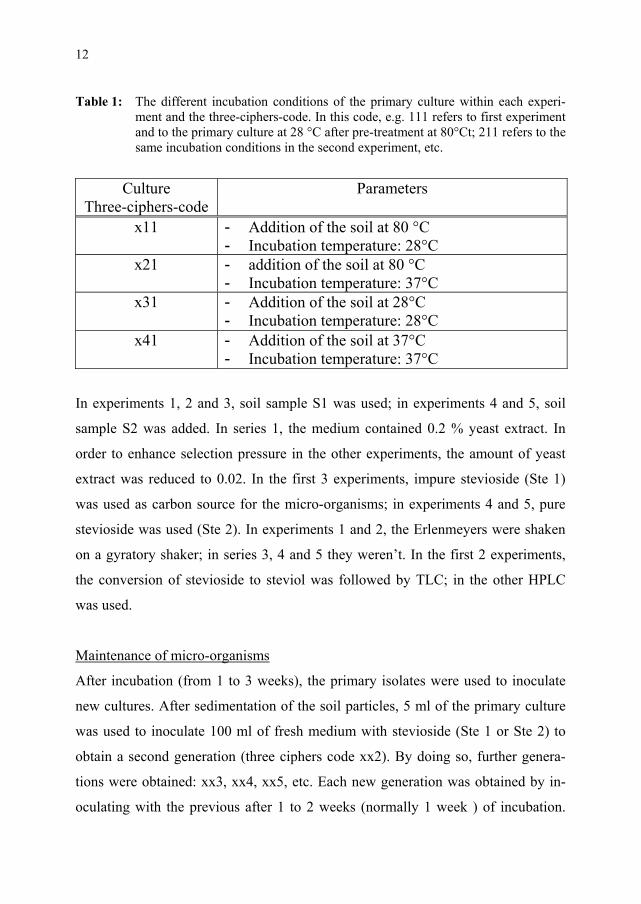
Table 1: The different incubation conditions of the primary culture within each experi-ment and the three-ciphers-code. In this code, e.g. 111 refers to first experiment and to the primary culture at 28 °C after pre-treatment at 80°Ct; 211 refers to the same incubation conditions in the second experiment, etc.
In experiments 1, 2 and 3, soil sample S1 was used; in experiments 4 and 5, soil
sample S2 was added. In series 1, the medium contained 0.2 % yeast extract. In
order to enhance selection pressure in the other experiments, the amount of yeast
extract was reduced to 0.02. In the first 3 experiments, impure stevioside (Ste 1)
was used as carbon source for the micro-organisms; in experiments 4 and 5, pure
stevioside was used (Ste 2). In experiments 1 and 2, the Erlenmeyers were shaken
on a gyratory shaker; in series 3, 4 and 5 they weren’t. In the first 2 experiments,
the conversion of stevioside to steviol was followed by TLC; in the other HPLC
was used.
Maintenance of micro-organisms
After incubation (from 1 to 3 weeks), the primary isolates were used to inoculate
new cultures. After sedimentation of the soil particles, 5 ml of the primary culture
was used to inoculate 100 ml of fresh medium with stevioside (Ste 1 or Ste 2) to
obtain a second generation (three ciphers code xx2). By doing so, further genera-
tions were obtained: xx3, xx4, xx5, etc. Each new generation was obtained by in-
oculating with the previous after 1 to 2 weeks (normally 1 week ) of incubation.
Culture Three-ciphers-code
Parameters
x11 - Addition of the soil at 80 °C - Incubation temperature: 28°C
x21 - addition of the soil at 80 °C - Incubation temperature: 37°C
x31 - Addition of the soil at 28°C - Incubation temperature: 28°C
x41 - Addition of the soil at 37°C - Incubation temperature: 37°C
Preparation of steviol by soil bacteria 13
Using stevioside as the carbon source, the final goal was to accumulate the micro-
organism(s) that is/are responsible for steviol formation. Steviol formation was
followed using either TLC or HPLC.
For macroscopic evaluation of the colonies formed, several of the cultures were
inoculated on the solid medium (with or without 0.2 % stevioside) using either the
pour-plate method or the spread-plate method. Samples of the different microbial
populations were also inoculated on slants; after incubation, the slants were stored
at 4°C.
Isolation and characterisation of steviol
For the isolation of steviol, we started from 2 of the cultures of experiment 5: incu-
bations 522 and 524. After augmenting the pH from 7 to 8 with 0.01 N NaOH, the
cultures were centrifuged (ALO 4233R; 4000 rpm) and the supernatant was filtered
over a 0.45 µm filter. Upon acidification to pH 4, a white precipitate formed. The
precipitate was isolated and the pellet was washed trice with ultra pure water. The
resulting powder was dried to constant weight in vacuo over P2O5. The product
obtained was analysed by HPLC and subjected to a classic identification program
(melting point; mass spectrum; proton- and carbon-13-NMR)
RESULTS AND DISCUSSION
Isolation and maintenance of micro-organisms
After 8 days of incubation of the primary cultures in experiment 1, the pH was
7.92, 8.98, 7.89 and 9.04 for the cultures 111, 121, 131 and 141, respectively, indi-
cating that no acidification occurs. This property minimizes a possible precipitation
of eventually formed steviol. Gram stain and microscopic examination of cultures
112, 122, 132 and 142 showed in each case a mixture of Gram positive and Gram
negative bacteria, both bacilli and cocci. Microscopic examination of the following
generations showed the same result.

14
The idea was that a micro organism that hydrolyses steviol glycosides to steviol
and sugars would benefit from these sugars and hence grow better on a medium
supplemented with stevioside than on a medium without it. Cultures 1x3, 1x6 and
1x7 were therefore inoculated on a solid media (with stevioside) in Petri dishes and
incubated at 28 °C (11x, 13x) or at 37 °C (12x, 14x). Fifteen colonies were selected
for further investigation. These micro organisms could be maintained on slant cul-
tures but grew equally well with or without stevioside in the medium.
In the first experiment the culture media were analysed by TLC. After centrifuga-
tion the clear supernatant was applied, as well as an ethereal extract of the pellet
and the supernatant. Differences in these chromatograms help to differentiate be-
tween the polar glycosides and the non-polar aglycon. In the beginning the TLC-
analysis failed, most probably because of insufficient application of the samples.
From generation six our analysis were successful and the results for generation 6, 7
and 8 were alike, indicating that the bacterial population probably did not change
during subsequent inoculations. The sixth bacterial generation was also inoculated
on agar slants and was reinvestigated after 2 months storage at 5°C. We used the
descendants of culture 126 in new cultures and followed stevioside hydrolysis by
HPLC. We concluded that the population didn’t lost its high potential for steviol
formation from stevioside and that it thus is possible to isolate stable bacterial
populations from a soil sample that hydrolyse stevioside to steviol.
Figure 2 gives the results for the TLC analysis of generation 8. After 1 d of incuba-
tion, TLC analysis of the culture filtrates showed traces of steviol and some other
degradation products of steviol glycosides. After 4 d, cultures 128 and 148 con-
tained steviol and some steviolbioside; culture 138 contained, besides some ste-
violbioside, an unknown component. This unknown was the only product formed
in culture 118 after 5 d of incubation and thus probably is a direct derivative of
stevioside or of one of the contaminants and hence e.g. may correspond to rubu-
soside or rebaudioside C. In contrast to steviol, steviolbioside and the unknown did
not accumulate in the ethereal extract, which is in agreement with their physico-

Preparation of steviol by soil bacteria 15
chemical properties. After 8 d of incubation, the unknown disappeared from culture
138 and this without apparent formation of steviol. In culture 118, the unknown
remained as most important new compound. After incubation for 11 d, the steviol
disappeared from the cultures 128 and 148. The unknown compound disappeared
from culture 118, however, but steviol could not be detected.
Steviol was not detected in the extracts made from the pellets obtained by centrifu-
gation of the different culture media, indicating that steviol did not precipitate un-
der the conditions of incubation.
The pattern of hydrolysis of stevioside to steviol in cultures 128 and 148 (at 37 °C)
is in agreement with a two-step conversion in which steviolbioside is formed as an
intermediate. It is unclear if these two steps are carried out by the same organisms
or if one or more species are responsible for the enzymatic conversion. It is prob-
able that in cultures 118 and 138 (at 28 °C) an alternative conversion occurred. The
disappearance of steviol from the culture, most likely, is due to further metabolism
and not to precipitation, since no steviol was found in the pellets obtained by cen-
trifugation of the cultures.
Cultures 118 and 138 were used for the inoculation of fluid medium with or with-
out stevioside: 119+, 119-, 139+ and 139-. After incubation at 28°C for 5 d, the pH
of the stevioside supplemented cultures (7.2 for 119+ and 8.3 for 139+) was higher
than for the non-supplemented cultures (6.9 for 119- and 6.9 for 139-). TLC analy-
sis of the stevioside supplemented cultures only showed steviol and no remaining
glycosides. Apparently, the latter were all hydrolysed to the aglycon and sugars.
Since no acidification of the culture media was observed, we can conclude that the
sugars were not fermented by the micro-organisms.

16
Figure 2: Results of experiment 1: TLC-analysis (silica gel plates, n-butanol:acetic acid:water–4:1:5; upper layer) of the cultures of generation 8 (118, 128, 138 & 148; for details of the primary incubation see table 1) after 1(A), 4 (B), 5(C) and 8 (D) days of incubation. Samples were applied after centrifugation (a); an ethereal extract of the broths was also applied (b);t0: culture medium with Ste 1 at the start of the experiment; S: steviol, Ste: stevioside Ste1 (impure stevioside used in the incubations.(Rf rebaudioside A = 0.41 ; Rf stevioside= 0.44; Rf steviolbioside = 0.55 and Rf steviol = 0.93 (approximate values); an unknown compound is found with an Rf of about 0.68).
All of the above facts indicate that the micro-organisms that are responsible for the
conversion of stevioside to steviol form a stable population. Apparently, the micro-
organisms do not ferment the sugars that they liberate from stevioside, or at least,
do not ferment them under the conditions used. Therefore, we repeated the experi-
ment under more stringent conditions for the micro-organisms by reducing the
amount of yeast extract from 0.2 % to 0.02 %.
The experiment was repeated using fluid medium containing, besides some salts
and stevioside (Ste 1), only 0.02 % yeast extract. Incubations with soil S1 were
done as in Table 1. Subculture of this second primary culture was done in a fluid
Preparation of steviol by soil bacteria 17
medium with a reduced amount of yeast extract (cultures 2x2) and the hydrolysis
of the stevioside was followed with TLC. Analysis of generation 2 to 5 gave simi-
lar results. Figure 3 gives the results for generation 4.
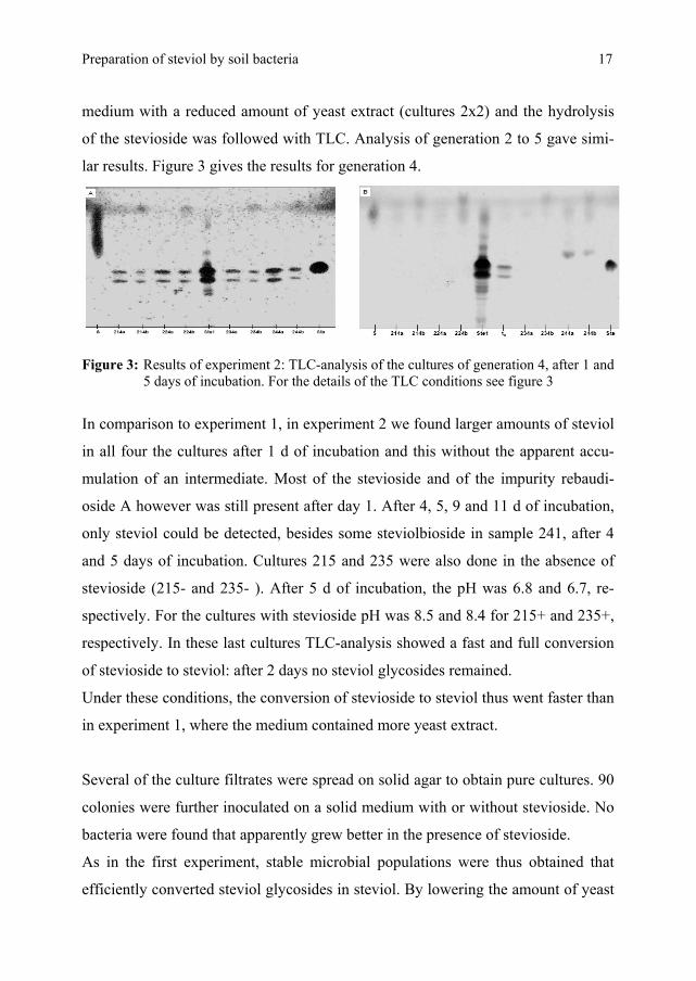
Figure 3: Results of experiment 2: TLC-analysis of the cultures of generation 4, after 1 and
5 days of incubation. For the details of the TLC conditions see figure 3
In comparison to experiment 1, in experiment 2 we found larger amounts of steviol
in all four the cultures after 1 d of incubation and this without the apparent accu-
mulation of an intermediate. Most of the stevioside and of the impurity rebaudi-
oside A however was still present after day 1. After 4, 5, 9 and 11 d of incubation,
only steviol could be detected, besides some steviolbioside in sample 241, after 4
and 5 days of incubation. Cultures 215 and 235 were also done in the absence of
stevioside (215- and 235- ). After 5 d of incubation, the pH was 6.8 and 6.7, re-
spectively. For the cultures with stevioside pH was 8.5 and 8.4 for 215+ and 235+,
respectively. In these last cultures TLC-analysis showed a fast and full conversion
of stevioside to steviol: after 2 days no steviol glycosides remained.
Under these conditions, the conversion of stevioside to steviol thus went faster than
in experiment 1, where the medium contained more yeast extract.
Several of the culture filtrates were spread on solid agar to obtain pure cultures. 90
colonies were further inoculated on a solid medium with or without stevioside. No
bacteria were found that apparently grew better in the presence of stevioside.
As in the first experiment, stable microbial populations were thus obtained that
efficiently converted steviol glycosides in steviol. By lowering the amount of yeast

18
extract in the culture medium, the conversion went faster and in one culture (241)
steviolbioside was found as an intermediate. Although the conversion went faster,
we found no indications for the use of the liberated sugars by the micro-organisms:
they did not grow better nor did they ferment the liberated sugars to acid.
In a third experiment, we tried to better quantify the concentration of stevioside,
and steviol. The experimental set up was as in Table 1 and the hydrolysis of the
steviol glycosides was followed by HPLC. Figure 4 summarizes the results.
Figure 4: Results of experiment 3: Conversion of stevioside (▲) to steviol (■) by a soil
sample from Paraguay. Table 1 summarizes pre-treatment and incubation condi-tions. The results were obtained after HPLC analysis as described in material and methods.
The small amount of stevioside detected reflects the fact that the preparation was
only 60 % pure. When one takes into account that the impurities of the stevioside
are other steviol glycosides such as rebaudioside A (28 %) and rebaudioside C (9
%), the high amounts of steviol obtained can be explained.

Preparation of steviol by soil bacteria 19
In the four incubations, apparently full conversion of the steviol glycosides into
steviol is reached within 50 to 100 h. Incubating at 37 °C (321 and 341) resulted in
faster steviol formation than incubation at 28°C (311 and 331). In none of the incu-
bations, at the different incubations times tested (after 1,2,3,4,5,7 and 8 d), steviol-
bioside or other components were detected. However, in sample 331 and 341, the
steviol rapidly disappeared, which may reflect that steviol is degraded further by
the microbial population. In contrast, steviol remained better in cultures 311 and
321. These cultures were pre-incubated at 80 °C and this probably eliminated the
bacteria that can metabolise the steviol further.
Experiment 3 was somehow hampered by the impurity of the substrate and in a
fourth experiment we used almost pure stevioside. A new soil sample, obtained
from soil contaminated Stevia leaves was used also. Since pre-treatment at 80°C
seemed to eliminate the bacteria that further metabolise the steviol formed, we
repeated only the incubations with the pre-treatment step. Incubation was at either
28 °C (411) or 37 °C (421). The results are summarized in Figure 5.
Figure 5: Results of experiment 4 after HPLC analysis: conversion of stevioside (▲) to
steviol (■) by soil sample S2. The cultures were pretreated at 80°C and incu-bated at 28 °C (left) or at 37 °C (right).
In both incubations, full and fast conversion of stevioside to steviol was observed.
At 37 °C, the conversion (90 % after 48 h) went faster than at 28 °C (20 % after 48
h). No intermediates of the hydrolysis of stevioside to steviol were detected during

20
chromatographic analysis. In contrast to experiment 3, in which prolonged incuba-
tion at 28 °C resulted in a decrease of the steviol concentration, it was now found
that prolonged incubation at 37 °C caused a decrease of the steviol concentration.
Since pre-treatment at 80 °C could be important to eliminate steviol degrading
micro-organisms, we standardized this heat treatment. Instead of adding the soil at
80 °C and further incubating at 28 °C (x11) or at 37 °C (x21), while slowly cooling
to the incubation temperature, in this fifth experiment the culture (with soil sample
S2) was kept at 80 °C for 20 min and was then quickly cooled to 37 °C for further
incubation (521). Figure 6 shows the results. Full conversion of stevioside to
steviol was obtained. The conversion
went somewhat slower than in ex-
periment 4, but still 75 % of the ste-
vioside was hydrolysed within 48
hours. The steviol remained in the
medium for at least 11 days.
Figure 6: Results of experiment 5: conver-sion of stevioside (▲) to steviol (■) by soil sample S2 that was first pretreated at 80°C for 20 min; incubations were at 37 °C.
As in experiments 1 and 2, the primary
culture was used to inoculate 100 ml
fresh medium with the same composi-
tion (culture 522). In the same way,
cultures 523 to 527 were inoculated:
after 1 to 3 weeks, 5 ml of the previous generation was used to inoculate the next
generation. The conversion of stevioside to steviol was followed by HPLC in all
cultures (see Figure 7).
The results, in this case, show that the resulting microbial population slowly lost its
capacity for steviol formation. The primary culture converted 75 % of the ste-
vioside to steviol in 48 h and this conversion decreased to 34 %, 31 %, 18 %, 22 %
and 13 % for cultures 522, 524, 525, 526 and 527, respectively.

Preparation of steviol by soil bacteria 21
Figure 7: Results of experiment 5 after HPLC analysis: conversion of stevioside (▲) to
steviol (■) by the succeeding generations (A – F, indicated by the last cipher in the 3-ciphers-code) derived from soil sample S2 that was first pre-treated at 80°C for 20 min; incubations were at 37 °C.
From the third generation on (culture 523), steviolbioside was found in the culture
filtrate indicating that the conversion of steviolbioside to steviol became rate limit-
ing in the conversion of stevioside to steviol. This also may indicate that the micro-
organism that is responsible for the conversion of steviol to steviolbioside is disap-
pearing from the microbial population and probably is different from the one that
converts steviolbioside to steviol. In cultures 522 and 524, which were inoculated

22
with rather old cultures (respectively 3 and 2 weeks) full conversion of stevioside
to steviol was obtained.
The conversion of stevioside to steviolbioside also decreased. In the primary cul-
ture, after 48 h all stevioside disappeared. In the following generations, this degra-
dation was limited to 10 to 30 % (except for culture 527), indicating that the micro-
organism responsible for the fist step also disappears from the bacterial population. In cultures with low steviol concentrations, the pH was always lesser than in those
with a good activity (6.5 - 7 vs 7.5- 8). This may have hampered the hydrolytic
reactions, and may have caused some steviol precipitation too. In further experi-
ments, the buffering capacity of the medium will be enhanced by doubling the
amount of phosphate.
To investigate the influence of the age of the cultures used, generation 7 was inocu-
lated 3 times, once with 5 ml of culture 526 after 3, 6 and 9 d: cultures 527/3, 527/6
and 527/9. The results are given in Figure 7F (inoculated with a 5 d old culture)
and in Figure 8 (cultures inoculated with a 3 and a 6 d old culture).
Figure 8: Results of experiment 5 after HPLC analysis: conversion of stevioside (▲) to
steviol (■) by the generation 7 derived from soil sample S2 that was first pre-treated at 80°C for 20 min; incubations were at 37. Inoculation was with genera-tion 6 was after 3 (left) and 9 (right) days of incubation (compare with 527/6 in figure 7 which was inoculated after 6 d of incubation.
The net formation of steviol is not influenced by the age of the inoculums. On the
contrary, an inoculum of 5 d seems optimal for the conversion of stevioside to ste-
violbioside and this resulted in high concentrations of steviolbioside (Figure 9a).
Using younger or older cultures for the inoculation resulted in a slower conversion

Preparation of steviol by soil bacteria 23
of stevioside into steviolbioside. But even in these conditions, the steviolbioside
accumulated (Figure 9b), showing that the latter conversion is the overall limiting
step.
Figure 9: HPLC analysis of the cultures 527/6 (a) and 527/9 (b; for details see the text) after 7 days of incubation.
A dilution series of culture 529 was used to inoculate pour-plates with or without
stevioside. There were no indications for the use of stevioside by the bacteria.
It remains possible that, in fluid culture, the bacteria using stevioside and those
using steviolbioside live in an association that metabolises stevioside. If so, the
bacteria use the sugars from the glycosides in a non-fermentative way. By inoculat-
ing on solid media, the bacteria are physically separated and apparently don’t use
the steviol glycoside. If so, this behaviour strongly hampers the isolation of the
responsible micro-organisms.
All available experimental evidence agreed with the fact that soil samples of Para-
guay contain a bacterial population that hydrolysis steviol glycosides such as ste-
vioside. Stevioside is converted to steviol via steviolbioside. The bacterial popula-
tion responsible for the first step is likely different from the one responsible for the
second step. This way, this microbial conversion is different from the one reported

24
by Yosioka et al. (1972). In their work, an unknown bacterium designated as YSB-
9, converted stevioside to steviol. However, the intermediate steviolbioside accu-
mulated in the medium, showing that the conversion of steviolbioside to steviol is
rate limiting. In the conversion of stevioside to steviol by the bacterial community
from Paraguayan soil samples, the conversion of steviolbioside to steviol is also
rate limiting. The first step in the hydrolysis of stevioside by the soil samples is
probably carried out by micro-organisms such as those isolated by Nakano et al.
(1998) and Okamoto et al. (2000). These authors isolated a Clavibacter michi-
ganense strain and a Flavobacterium johnsonae strain, respectively, that effectively
hydrolysed glycosidic ester linkages and e.g. converted stevioside to steviolbioside
The isolation of the responsible micro-organisms from soil samples on solid media
is hampered by the fact that the micro-organisms apparently don’t use the steviol
glycosides or the sugars derived from them for growth.
Isolation and characterisation of steviol
The remaining part of the culture filtrate of incubations 522 (55 ml) and 524 (65
ml) in which full hydrolysis of stevioside tot steviol occurred, were used as starting
material for the preparation of steviol. With the method described, respectively 40
mg (St 522) and 46 mg (St 524) of product were obtained. HPLC analysis showed
St 522 to be impure; it contained 91 % steviol. St 524 was 100 % pure. Taken into
account the purity, the recovery from the culture filtrates respectively was 88 and
94 % . The melting point of St 522 (after recrystallization from water) and St 524
was 211 °C and 210-211 °C respectively; Pezzuto et al. (1985) reported a mp of
211-213 °C. The mass spectrum of both St 522R and 524 was in agreement with
that of steviol: 658.1 (2M + Na), 998(3M + 2Na) and 1338.3 (4M + 2 Na). The
products were also analysed by 13C NMR and 1H NMR. Chemical shifts in 13C-
NMR of Ste 524 (in ppm; for the assignment of the carbon atoms see figure 1a ):
183.0 (C19), 115.7 (C16), 103.0 (C17), 80.3 (C13), 56.8 (C9), 53.8 (C5), 47.4 (C15),
46.9 (C14), 43.6 (C8), 41.7 (C4), 41.2 (C7), 40.5 (C1), 39.5 (C10), 39.3 (C12), 37.7

Preparation of steviol by soil bacteria 25
(C3), 29.0 (C18), 21.8 (C6), 20,4 (C11), 19.0 (C2), 15.4 (C20). Chemical shifts in 1H-
NMR of St 524 (in ppm): 4.82 – 4.99 (doublet of doublets; C17); 0.96 (s; C20); 1.24
(s; C18) 0.8 – 2.0 (ring H’s). The 300 MHz apparatus that was used for obtaining
the spectra did allow for further differentiation among the aliphatic hydrogen’s.
Both spectra were in agreement with the literature (Yang, 2007).
An easy and very efficient method was thus developed for the synthesis of steviol
from stevioside. The usual chemical conversion of stevioside to steviol under alka-
line conditions results in yields of 5 to maximum 10 % (Ogawa et al., 1980). In the
present bio-organic method, yields of 90 % were obtained. We hope that this
method can contribute to further work on steviol and its metabolites by the scien-
tific community.
CONCLUSIONS
1. Stevioside and other steviol glycosides degrading micro-organisms were
isolated from 2 soil samples originating from Paraguay.
2. The most active bacterial populations were obtained after pasteurization of
the soil samples by a pre-treatment at 80 °C.
3. Hydrolysis of steviol glycosides such as stevioside by the micro-organisms
surviving the pre-treatment went faster at 37 °C than at 28°C.
4. Enhancing selection pressure by reducing the amount of yeast extract from
02. % to 0.02 % stimulated the hydrolysis of steviol glycosides.
5. The hydrolysis of stevioside to steviol is a two-step process with steviol-
bioside as intermediate.
6. Two different micro-organisms (or populations of micro-organisms) are re-
sponsible for the process.
7. The conversion of steviolbioside to steviol is rate limiting in the conver-
sion of stevioside to steviol.

26
8. In cultures prepared from pre-treated soil samples, steviol remains longer
and, apparently, pre-treatment of the soil samples eliminates the bacteria
that further metabolise steviol.
9. Apparently, the micro-organisms that catalyse the hydrolysis of steviol
glycosides do not use the sugars that they split off: there are no signs for an
enhanced growth, neither for a fermentation of the sugars. This property of
the micro-organisms hampers the isolation of pure cultures.
10. The microbial population isolated from the soil samples retains its hydro-
lytic activity towards steviol glycosides upon further inoculation in fluid
media. This process can be repeated several times. The microbial popula-
tion responsible for the hydrolysis of steviol glycosides can be stored on
slant cultures for several months.
11. An easy method for the preparation of steviol from stevioside was devel-
oped. Steviol can be isolated from the culture filtrates of the microbial
populations in an almost quantitative way by simple acidification and cen-
trifugation. The product isolated must be washed several times with water
in order to obtain a pure product.
REFERENCES Geuns, J.M.C. Safety of Stevia and stevioside. Recent Res. Devel. Phytochem. 2000, 4, 75-
88. Geuns, J.M.C.; Buyse, J.; Vankeirsbilck, A.; Temme, L. Safety of stevioside used as a
sweetener, pp. 75-83 in Geuns, J.M.C. and Buyse, J. Eds. “Proceedings of the first symposium on the Safety of Stevioside”. KULeuven, 2004. Euprint Editions ISBN 9074253024.
Geuns, J.M.C. Molecules of interest: Stevioside. Phytochemistry 2003, 64, 913-921. Kusakabe, I.; Kusama, S.; Murazami, K. An easy method for the preparation of sophorose
from stevioside. Agric. Biol. Chem., 1987, 51 (8) 2255-2256.
Mossetig, E.; Beglinger, U.; Adder, F.; Lichti,H.; Quilt,, P.; Waters, J.A. Absolute configu- ration of steviol and isosteviol. J. Am. Chem. Soc.,1963, 85, 2305.

Preparation of steviol by soil bacteria 27
Nakano, H.; Okamoto, K.; Yatake, T.; Kiso, T.; Kitahata, S. Purification and characteriza- tion of a novel β-glucosidase from Clavibacter michiganense that hydrolyzes glu-cosyl ester linkage in steviol glycosides. J. Ferm &Bioeng., 1998, 85, 162-168.
Ogawa.T.; Nozaki, N.; Matsui, M.Total synthesis of stevioside. Tetrahedron, 1980, 36, 2641-2648.
Okamoto, K.; Nakano, H.; Yatake, T.; Kios, T.; Kitahata, S. Purification and some proper- ties of a β- glucosidase from Flavobacterium johnsonae; Biosc. Biotechnol. Bio-chem, 2000, 64(2), 333-340.
Pezzuto, J. M.; Compadre, C.M.; Swanson, S.M.; Nanayaklawa, N.P.D.; Kuighorn A.D. Metabolically activated steviol, the aglycone of stevioside, is mutagenic.Proc. Natl. Acad. Eci USA, 1985, 82, 2478-2482.
Struyf, T.; Chandia, N.P.; De Borggraeve, W.M.; Dehaen, W.; Geuns, J. M.C. Preparation
of pure standards of steviol glycosides. Identification of steviol glycosides by LC-MS and NMR in Geuns, J.M.C. Ed. “Proceedings of 2nd Stevia Symposium organ-ised by Eustas – Steviol glycosides: technical and pharmacological aspects”. KU-Leuven, Leuven, Belgium 2008, pp. 29-44, ISBN 9789074253-031
Yang, L.-M.; Hsu, F-L.; Chang, S.-F.; Cheng, J.-T.; Hsu, J.-Y.; Hsu, C.-Y.; Liu, P.-C.; Lin, S.-J. Microbial metabolism of steviol and seviol-16α,17-epoxide.Phtochemistry, 2007, 68, 562-570.
Yosioka, I.; Saijoh, S.; Waters J. A.; Kitagawa, I. Soil bacterial hydrolysis leading tot genu- ine aglycone VI: on stevioside. Chem. Pharm. Bull., 1972, 20, 2500-2502. Wood H.B.; Allerton, R.; Diehl, H.W.; Fletcher, H.G.Stevioside I: The structure of glucose
moieties. J. Org. Chem., 1955, 20, 875-883.
Acknowledgement: This research was only possible thanks to an IOF grant
(IOFHB/06/022) from the University of Leuven and the financial support
by Medherbs, Wiesbaden, Germany.