Preparation of a three-dimensional extracellular matrix...
6
ABSTRACT Introduction: the availability of transplantable livers is not suf- ficient to fulfill the current demand for grafts, with the search for therapeutic alternatives having generated different lines of research, one of which is the use of decellularized three-dimensional biological matrices and subsequent cell seeding to obtain a functional organ. Objective: to produce a decellularization protocol from rabbit liver to generate a three-dimensional matrix. Methods: a combination of physical, chemical (Triton X-100 and SDS) and enzymatic agents to decellularize rabbit livers was used. After 68 h of retrograde perfusion, a decellularized translucent matrix was generated. To evaluate if the decellularization protocol was successful, with the extracellular matrix being preserved, we carried out histological (light microscopy and scanning electron microscopy) and biochemical (DNA quantification) studies. Results: the decellularization process was verified by macroscopic observation of the organ using macroscopic staining, which revealed a correct conservation of bile and vascular trees. A microscopic observation corroborated these macroscopic results, with the hema- toxylin-eosin staining showing no cells or nuclear material and the presence of a portal triad. Wilde’s staining demonstrated the con- servation of reticulin fibers in the decellularized matrix. In addition, scanning electron microscopy revealed a preserved Glisson’s capsule and a decellularized matrix, with the DNA quantification being less than 10 % in the decellularized liver compared to control. Finally, the time taken to develop the decellularization protocol was less than 96 hours. Conclusions: the proposed decellularization protocol was cor- rect, and was verified by an absence of cells. The hepatic matrix had preserved vascular and bile ducts with a suitable three-dimen- sional architecture permitting further cell seeding. Key words: Decellularized liver. Biological scaffolds. Transplant. INTRODUCTION Liver transplantation is currently the only therapeutic alternative for acute liver failure, terminal liver disease or metabolic disorders originating in the liver. However, due to a shortage of donors, many patients who are on the wait- ing list never receive liver transplantation and others with indication of the graft have no access to waiting lists. More- over, the complexity of the function of the liver makes it impossible to use artificial systems to provide temporary hepatic support as for example in patients with renal insuf- ficiency. Alternatives to transplantation, such as the non-biological support systems proposed in the literature, have precise and limited indications (1,2). In other way, hepatocyte seeding, via injection into the splenic or portal veins, does not even offer a temporary solution as a bridge to transplantation, due to early death of hepatocytes with the remaining number being incapable of correcting any abnormalities or rescuing patients from liver failure. In facts, hepatocyte primary cul- tures lose their typical morphologies and function within a few days via dedifferentiation or epithelial-mesenchymal transition. Furthermore, other complications inherent to this technique, such as portal thrombosis, hypertension and pul- monary embolism, have also been reported (1,3,4). Three-dimensional biological scaffolds are commonly used in reconstructive surgery. Vracko (5) describes the importance of the extracellular matrix (ECM) in maintain- ing the liver structure and regeneration and recent devel- opments show the ability of the ECM to maintain the hepa- tocyte phenotype and function (1,4,6). Related to this, another line of research is the creation of three-dimensional Preparation of a three-dimensional extracellular matrix by decellularization of rabbit livers Gustavo A. Nari 1,2 , Mariana Cid 3,4 , Romina Comín 3 , Laura Reyna 5 , Gustavo Juri 6 , Ricardo Taborda 5 and Nancy A. Salvatierra 3,4 1 Department of Surgery. Florencio Díaz Hospital. Argentina. 2 Surgery UHC 4. UNC. Argentina. 3 Department of Chemistry. School of Biomedical Engineering. Faculty of Exact, Physical and Natural Sciences. National University of Córdoba. Argentina. 4 IIBYT-CONICET. Argentina. 5 LIADE (Laboratory of Applied Research, and Development). School of Biomedical Engineering. Faculty of Exact, Physical and Natural Sciences. National University of Córdoba. Argentina. 6 School of Biomedical Engineering, Faculty of Medical Sciences. UNC. Argentina 1130-0108/2013/105/3/138-143 REVISTA ESPAÑOLA DE ENFERMEDADES DIGESTIVAS Copyright © 2013 ARÁN EDICIONES, S. L. REV ESP ENFERM DIG (Madrid) Vol. 105. N.° 3, pp. 138-143, 2013 Received: 10-10-2012 Accepted: 26-12-2012 Correspondence: Gustavo Adrián Nari. Department of Surgery. Hospital Flo- rencio Díaz. Argentina e-mail: [email protected] ORIGINAL PAPERS Nari GA, Cid M, Comín R, Reyna L, Juri G, Taborda R, Sal- vatierra NA. Preparation of a three-dimensional extracellular matrix by decellularization of rabbit livers. Rev Esp Enferm Dig 2013;105:138-143.
Transcript of Preparation of a three-dimensional extracellular matrix...

ABSTRACT
Introduction: the availability of transplantable livers is not suf-ficient to fulfill the current demand for grafts, with the search fortherapeutic alternatives having generated different lines of research,one of which is the use of decellularized three-dimensional biologicalmatrices and subsequent cell seeding to obtain a functional organ.
Objective: to produce a decellularization protocol from rabbitliver to generate a three-dimensional matrix.
Methods: a combination of physical, chemical (Triton X-100and SDS) and enzymatic agents to decellularize rabbit livers wasused. After 68 h of retrograde perfusion, a decellularized translucentmatrix was generated. To evaluate if the decellularization protocolwas successful, with the extracellular matrix being preserved, wecarried out histological (light microscopy and scanning electronmicroscopy) and biochemical (DNA quantification) studies.
Results: the decellularization process was verified by macroscopicobservation of the organ using macroscopic staining, which revealeda correct conservation of bile and vascular trees. A microscopicobservation corroborated these macroscopic results, with the hema-toxylin-eosin staining showing no cells or nuclear material and thepresence of a portal triad. Wilde’s staining demonstrated the con-servation of reticulin fibers in the decellularized matrix. In addition,scanning electron microscopy revealed a preserved Glisson’s capsuleand a decellularized matrix, with the DNA quantification being lessthan 10 % in the decellularized liver compared to control. Finally,the time taken to develop the decellularization protocol was lessthan 96 hours.
Conclusions: the proposed decellularization protocol was cor-rect, and was verified by an absence of cells. The hepatic matrixhad preserved vascular and bile ducts with a suitable three-dimen-sional architecture permitting further cell seeding.
Key words: Decellularized liver. Biological scaffolds. Transplant.
INTRODUCTION
Liver transplantation is currently the only therapeuticalternative for acute liver failure, terminal liver disease ormetabolic disorders originating in the liver. However, dueto a shortage of donors, many patients who are on the wait-ing list never receive liver transplantation and others withindication of the graft have no access to waiting lists. More-over, the complexity of the function of the liver makes itimpossible to use artificial systems to provide temporaryhepatic support as for example in patients with renal insuf-ficiency.
Alternatives to transplantation, such as the non-biologicalsupport systems proposed in the literature, have precise andlimited indications (1,2). In other way, hepatocyte seeding,via injection into the splenic or portal veins, does not evenoffer a temporary solution as a bridge to transplantation, dueto early death of hepatocytes with the remaining numberbeing incapable of correcting any abnormalities or rescuingpatients from liver failure. In facts, hepatocyte primary cul-tures lose their typical morphologies and function within afew days via dedifferentiation or epithelial-mesenchymaltransition. Furthermore, other complications inherent to thistechnique, such as portal thrombosis, hypertension and pul-monary embolism, have also been reported(1,3,4).
Three-dimensional biological scaffolds are commonlyused in reconstructive surgery. Vracko (5) describes theimportance of the extracellular matrix (ECM) in maintain-ing the liver structure and regeneration and recent devel-opments show the ability of the ECM to maintain the hepa-tocyte phenotype and function (1,4,6). Related to this,another line of research is the creation of three-dimensional
Preparation of a three-dimensional extracellular matrix by decellularization of rabbit livers
Gustavo A. Nari1,2, Mariana Cid3,4, Romina Comín3, Laura Reyna5, Gustavo Juri6, Ricardo Taborda5
and Nancy A. Salvatierra3,4
1Department of Surgery. Florencio Díaz Hospital. Argentina. 2Surgery UHC 4. UNC. Argentina. 3Department ofChemistry. School of Biomedical Engineering. Faculty of Exact, Physical and Natural Sciences. National University of Córdoba. Argentina. 4IIBYT-CONICET. Argentina. 5LIADE (Laboratory of Applied Research, and Development).School of Biomedical Engineering. Faculty of Exact, Physical and Natural Sciences. National University of Córdoba. Argentina. 6School of Biomedical Engineering, Faculty of Medical Sciences. UNC. Argentina
1130-0108/2013/105/3/138-143REVISTA ESPAÑOLA DE ENFERMEDADES DIGESTIVASCopyright © 2013 ARÁN EDICIONES, S. L.
REV ESP ENFERM DIG (Madrid)Vol. 105. N.° 3, pp. 138-143, 2013
Received: 10-10-2012Accepted: 26-12-2012
Correspondence: Gustavo Adrián Nari. Department of Surgery. Hospital Flo-rencio Díaz. Argentinae-mail: [email protected]
ORIGINAL PAPERS
Nari GA, Cid M, Comín R, Reyna L, Juri G, Taborda R, Sal-vatierra NA. Preparation of a three-dimensional extracellularmatrix by decellularization of rabbit livers. Rev Esp Enferm Dig2013;105:138-143.

Vol. 105. N.° 3, 2013 PREPARATION OF A THREE-DIMENSIONAL EXTRACELLULAR MATRIX 139 BY DECELLULARIZATION OF RABBIT LIVERS
REV ESP ENFERM DIG 2013; 105 (3): 138-143
scaffolds of ECM through a decellularization process ofthe liver, where hepatocytes, stem cells or other cells canbe seeded to differentiate into hepatocytes in order toachieve a transplantable human liver. Researchers have pro-posed different models of decellularization to obtain a bio-logical scaffold to allow subsequent cell seeding (1,3,4,7-11). The decellularization is obtained through the use ofdifferent physical and/or chemical mechanisms to breakdown organ cells and leave only the ECM, which then pro-vides support to posterior cell seeding (12).
The aim of this paper is to provide a liver decellulariza-tion technique in rabbit liver and to evaluate the outcomeof this method.
MATERIAL AND METHODS
For this study, six New Zealand male rabbits of 2,100 ±120 g were subjected to a total hepatectomy with preser-vation of the vasculature. One rabbit was assigned for astudy of anatomy of the region, another being used for liverhistological control and the quantification of the DNA. Thelivers of the four remaining rabbits were incorporated intothe decellularization protocol. All procedures were con-ducted in accordance with the NIH Guide for the Care andUse of Laboratory Animals, as approved by the AnimalCare and Use Committee of the Universidad Nacional deCórdoba, Argentina, and efforts were made to minimizeanimal suffering and the number of animals used.
Surgical technique
Anesthesia was performed by injecting with 35 mg/kgketamine and 5 mg/kg xylazine intramuscularly (IM) as asingle dose. The rabbits were placed in the supine positionwith their legs fixed.
Surgery: By median incision which was later extendedto the right hemithorax. Identification was made of themesenteric vein and an intravenous injection was carriedout using 1,000 IU heparin.
Identification of the portal triad was performed and attachedby a ligature, identification and dissection of the infra-hepaticinferior caval vein was carried out. The adhesions of the liverwere removed and the incision was extending into the thorax.
Identification, dissection, ligature and cutting of the supra-hepatic inferior caval vein were performed along with a totalhepatectomy (Figs. 1 and 2)
Insertion of a catheter or teflon cannula in the supra-hepatic or infra-hepatic inferior caval vein respectively wasmade, which was fixed with a prolene 4/0 stitch and per-fusion was started with PBS solution for washing.
The organ was placed in PBS solution with crushed icefor transport and subsequent freezing at -80 °C.
Decellularization protocol
The liver was frozen in 500 ml PBS for 24 h at -80 °Cand later thawed at room temperature to aid cell lysis.
The catheter or teflon cannula inserted into the inferiorcaval vein was connected to a peristaltic pump and retro-grade perfusion cycles was started at a rate of between 6and 10 ml/min as follows: first, the liver was washed for 4hours with 0.02 % trypsin and 0.05 % EDTA in deionizedwater, before being perfused with 3 % Triton X-100 and0.05 % EGTA in PBS. Three washes of two hours eachwere performed, followed by a wash of 16 hours and later3 washes of 2 hours each. Next, we used the ionic detergentSDS (0.1 % in deionized water), which was perfused for4 hours, and then the organ was perfused with Triton X-100 and EGTA (3 % and 0.05 % in PBS) for 14 hours. Sub-sequent successive washes were performed with PBS anddeionized water to remove cell remains and detergents (1hour of deionized water, 2 washes with PBS for 1 hour, 30minutes of deionized water, and two washes for 30 minwith PBS). Finally, the organ was perfused for 1 hour with4 % ethanol in PBS to disinfect the organ.
Microscopic evaluation
– Light microscopy. For evaluation of the effectivenessof the decellularization, the two histological stainingtechniques, hematoxylin-eosin (HE), and the latterWilde silver staining was used to evaluate reticularfibers (collagen type III). These stainings were per-formed on the decellularized and control livers.
– Scanning electron microscopy. A complete analysisof Glisson’s capsule and the liver parenchyma was
Fig. 1. A. Mesenteric vein (white arrow filled). B. Portal triad and inferiorinfra-hepatic caval vein (small white arrows).
Fig. 2. A. Inferior supra-hepatic caval vein (white arrow). B. Total hepa-tectomy with catheter in perfusion stage prior to freezing.

140 G.A. NARI ET AL. REV ESP ENFERM DIG (Madrid)
REV ESP ENFERM DIG 2013; 105 (3): 138-143
performed by scanning electron microscopy (SEM)in LASEM, INIQUI-CONICET, UNSa using a scan-ning electron microscope model of JEOL JSM 6480Brand LV.
DNA quantification
The amount of DNA was quantified using a methoddescribed by Laird et al. (13). Briefly, 25 mg of tissue fromthe control and decellularized livers were homogenized ina solution containing 0.25 % trypsin and 1 mM EDTA indeionized water. The homogenate was incubated at 37 °Cwith constant stirring for 4 hours. Then, the cell lysis wascontinued with a solution containing 2 % SDS, 5 mMEDTA, 200 mM NaCl and 100 mM TRIS-HCl, pH 8.5 for24 h at 55 °C. The DNA extraction was carried out in iso-propanol and later dissolved in a solution of 10 mM Tris-HCl, 0.1 mM EDTA, pH 7.5. The amount of DNA wasdetermined spectrophotometrically at 260 nm.
RESULTS
The surgical technique was carried out without difficul-ties, and it was straight-forward to identify the anatomicalelements. Cannulation of the inferior caval vein was per-formed twice in a supra-hepatic way and twice in an infra-hepatic manner, the first with a K33 catheter and the secondwith a teflon cannula No. 16. The amount of PBS perfusedfor cleaning the liver prior to freezing was 210 ml.
The decellularization technique was performed asdescribed previously on four rabbit livers and the total timetaken was 94.30 hours, including 24 hours of freezing and5 hours of thawing at room temperature (Figs. 3 and 4).
Staining of the biliary tree on two rabbits was performedwith methylene blue through the gallbladder puncture,which revealed the existence of the bile ducts shown in Fig.5. A mixture of methylene blue with agarose was perfusedthrough the inferior caval vein in two other rabbits and agood vascular staining was observed (Fig. 5).
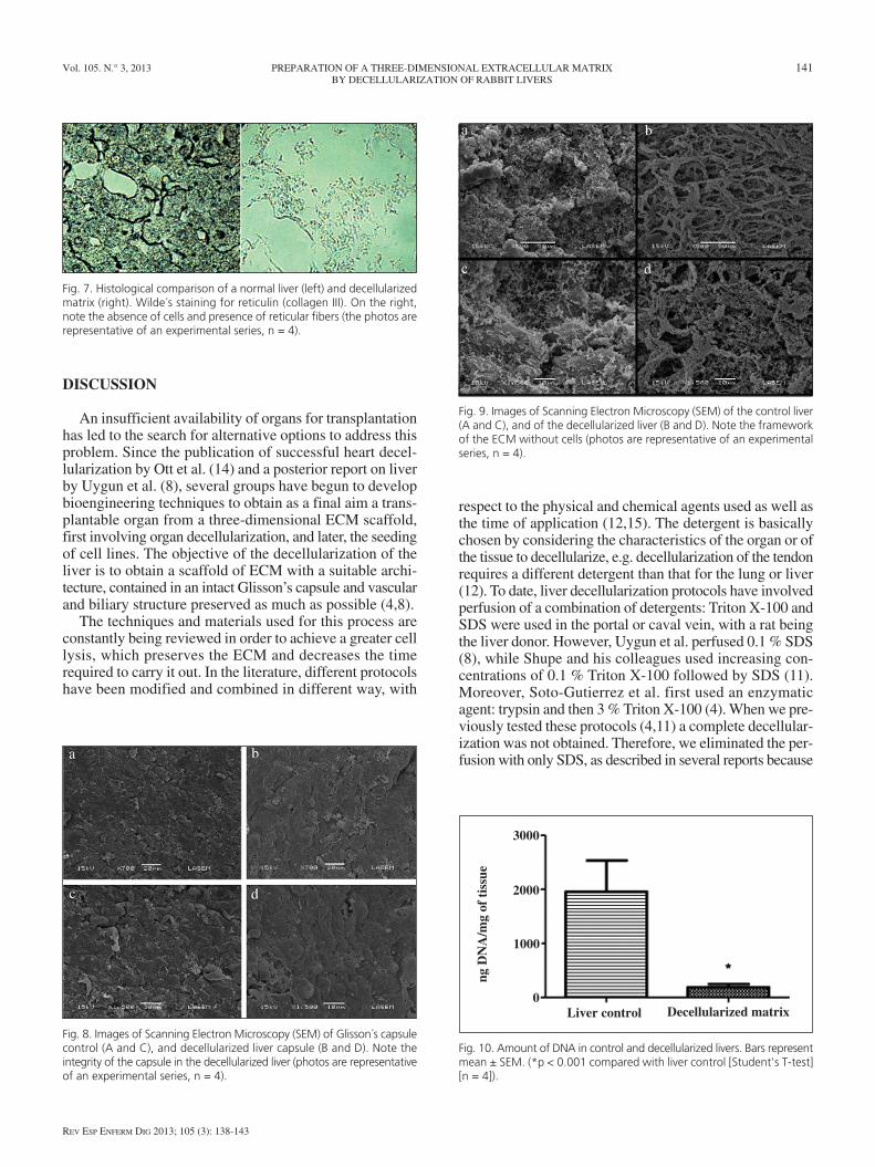
From the histological viewpoint, when the controls stainedwith HE and those with reticulin were compared using decel-lularized preparations, a complete disappearance of the cellpattern and the total absence of nuclei could be observed,with reticulin staining showing the presence of reticular fiberswithout cellular remains in the decellularized liver (Figs. 6and 7, respectively). Furthermore, HE staining revealed thepresence of a portal triad in the decellularized liver.
The SEM showed that the morphology of Glisson’s cap-sule was similar to that of the liver control (Fig. 8), andrevealed a complete decellularization of the liver parenchy-ma compared to control (Fig. 9). The Student’s t-test forunpaired samples showed that the DNA quantification wassignificantly different (t = 4.151, p < 0.001) between thedecellularized matrix values (1958 ± 579 ng/mg of tissue)and the control ones (192 ± 53 ng/mg) (Fig. 10).
Fig. 3. Liver at different stages of process of decellularization. Note theappearance of the vasculature.
Fig. 4. A. Completely decellularized lobe. B. Zoom in. Vascular pattern pre-served.
Fig. 5. A. Staining of bile ducts by vesicular puncture. B. Vascular stainingby injection of methylene blue with agarose.
Fig. 6. Histological comparison of a normal liver (left) and a decellularizedmatrix (right). Hematoxylin-Eosin staining. Note the presence of portaltriad in both preparations and the absence of cells and nuclei in thedecellularized matrix (photos are representative of an experimental series,n = 4).
Vol. 105. N.° 3, 2013 PREPARATION OF A THREE-DIMENSIONAL EXTRACELLULAR MATRIX 141 BY DECELLULARIZATION OF RABBIT LIVERS
REV ESP ENFERM DIG 2013; 105 (3): 138-143
DISCUSSION
An insufficient availability of organs for transplantationhas led to the search for alternative options to address thisproblem. Since the publication of successful heart decel-lularization by Ott et al. (14) and a posterior report on liverby Uygun et al. (8), several groups have begun to developbioengineering techniques to obtain as a final aim a trans-plantable organ from a three-dimensional ECM scaffold,first involving organ decellularization, and later, the seedingof cell lines. The objective of the decellularization of theliver is to obtain a scaffold of ECM with a suitable archi-tecture, contained in an intact Glisson’s capsule and vascularand biliary structure preserved as much as possible (4,8).
The techniques and materials used for this process areconstantly being reviewed in order to achieve a greater celllysis, which preserves the ECM and decreases the timerequired to carry it out. In the literature, different protocolshave been modified and combined in different way, with
respect to the physical and chemical agents used as well asthe time of application (12,15). The detergent is basicallychosen by considering the characteristics of the organ or ofthe tissue to decellularize, e.g. decellularization of the tendonrequires a different detergent than that for the lung or liver(12). To date, liver decellularization protocols have involvedperfusion of a combination of detergents: Triton X-100 andSDS were used in the portal or caval vein, with a rat beingthe liver donor. However, Uygun et al. perfused 0.1 % SDS(8), while Shupe and his colleagues used increasing con-centrations of 0.1 % Triton X-100 followed by SDS (11).Moreover, Soto-Gutierrez et al. first used an enzymaticagent: trypsin and then 3 % Triton X-100 (4). When we pre-viously tested these protocols (4,11) a complete decellular-ization was not obtained. Therefore, we eliminated the per-fusion with only SDS, as described in several reports because
Fig. 7. Histological comparison of a normal liver (left) and decellularizedmatrix (right). Wilde´s staining for reticulin (collagen III). On the right,note the absence of cells and presence of reticular fibers (the photos arerepresentative of an experimental series, n = 4).
Fig. 8. Images of Scanning Electron Microscopy (SEM) of Glisson´s capsulecontrol (A and C), and decellularized liver capsule (B and D). Note theintegrity of the capsule in the decellularized liver (photos are representativeof an experimental series, n = 4).
Fig. 9. Images of Scanning Electron Microscopy (SEM) of the control liver(A and C), and of the decellularized liver (B and D). Note the frameworkof the ECM without cells (photos are representative of an experimentalseries, n = 4).
Fig. 10. Amount of DNA in control and decellularized livers. Bars representmean ± SEM. (*p < 0.001 compared with liver control [Student's T-test][n = 4]).
Liver control Decellularized matrix
3000
2000
1000
0
ng D
NA
/mg
of t
issu
e

142 G.A. NARI ET AL. REV ESP ENFERM DIG (Madrid)
REV ESP ENFERM DIG 2013; 105 (3): 138-143
this technique results in a good decellularization by suc-cessfully removing cellular remains, but implied a higherloss of growth factors and of liver architecture (12) requiredfor the process of recellularization.
The new protocol described in the present investigationinvolved an initial treatment with trypsin, which alloweda better transfer from the detergents to the tissue, then per-fusion was carried out with 1% SDS and finally with TritonX-100. Thus, the decellularization was performed using acombination of enzymatic treatment (trypsin), followed byan ionic detergent (SDS), and finally a nonionic one (TritonX-100). This strategy of using three different agents in thedecellularization process minimized the contact time ofeach of these agents with the ECM, and also removed cellsand their remains by three different mechanisms, thus facil-iting a greater decellularization (12). Nevertheless, we wishto make it clear that changing the animal species implies achange in the size of the organ to be decellularized, whichin turn means developing a new protocol as it complicatesthe entry of detergents into the tissue as well as the removalof cell remains, due to the increased size of the organ.
The evaluation of the results of decellularization of thelivers is carried out by light microscopy (LM) with HEstaining, scanning electron microscopy (SEM), and thepresence of cell nuclei using the DAPI technique and/orDNA quantification by the determination of base pairs(bp). A successful decellularization is considered to havetaken place when fragments are less than 300 bp accordingto some authors, while others have proposed that theseshould be less than 200 bp. In this way, this would not gen-erate any inflammatory response in the host (4,16,17). Thesearch for DNA traces in commercial scaffolds revealedcellular remains and DNA content, although these wouldnot be able to trigger an inflammatory response. Moreover,the amount of Gal alpha 1,3 was also insufficient to triggeracute rejection, although it is known that Gal alpha 1,3cause rejection in xenotransplantation (16,18). Addition-ally, various studies, some of them on apoptotic porcinecells, have eliminated the possibility of the presence ofDNA traces of retroviruses in biological scaffolds for xeno-transplantation being able to transmit a disease to the host(19-21).
Our results have shown an encouraging decellularizationby observing the HE staining in the light microscopy to cor-roborate the presence of a preserved ECM. Likewise, thestaining for reticulin fibers (collagen type III) demonstratedtheir presence in the decellularized liver. We also observedthe presence of a complete portal triad, an important fact asthe preservation of bile ducts is a desired feature and is dif-ficult to achieve in this process. The presence of a preservedbile tract enables posterior reconstruction with bile epithelialcells (4), in the same way that a preserved vascular networkpermits subsequent seeding with endothelial cells.
The microscopic visualization of the bile ducts was ver-ified by macroscopic observation of the bile tree throughthe puncture and staining of the gallbladder. Furthermore,the vascular tree was preserved after the decellularization
process, as evidenced by the perfusion of methylene bluein 2 % agarose via the caval vein.
As noted here and in agreement with other authors, weconsider that the use of combinations containing trypsin,SDS, Triton X-100 associated with EGTA, after freezingat -80 °C is the best alternative to date to achieve an accept-able decellularization in a liver of about 100 g, althoughthe appearance of new products or the incorporation ofother techniques, such as sonication or electroporation, maybe included in the protocol in the future (15,22). Most decel-lularized livers reported by other researchers came fromrats, when involved decellularize sizes of up to 10 timeslower than those of rabbits. Therefore, if a liver of the sizeof rabbit can be recellularized, it provides a transplantablecell mass with a subsequent better chance of recovery ofthe liver functions in patients (7). A surgical technique pre-serving the vascular and bile elements and Glisson’s capsuleas well as the insertion of the teflon cannula or cathetercarefully permits a suitable perfusion in different lobes ofthe rabbit liver. In agreement with other authors, we per-formed a retrograde perfusion (caval-portal flow), due toit seeming to be better than anterograde perfusion (portal-caval flow), which was proposed by other researchers (3,4).Similarly, a peristaltic pump with controlled flows rangingfrom 6 to 10 ml/min ensured a ECM with a conserved archi-tecture. The decellularization protocol proposed here pro-duced successful results with HE staining, a technique forcollagen III, SEM and DNA quantification. In addition, thetime required for decellularization, including freezing time,was less than 96 hours.
In conclusion, although the results are positive, there arestill some points to be investigated. One of these is to verifythe presence of growth factors in our ECM, because theposterior recellularization process requires their presence(23). These factors are essential to determine the behaviour,adhesion, migration, differentiation, proliferation and sur-vival of seeded cells (1,6,24). In a decellularization protocolwith features similar to those described here, the remainingamount of growth factors was around 30-50 %, therebyallowing ECM recellularization with hepatocytes (4). Final-ly, and focusing on the recellularization, the efficient deliv-ery of oxygen and nutrients to the culture is necessary inorder to obtain a useful and functional organ.
ACKNOWLEDGEMENT
We thank Dr. Paul Hobson, native speaker, for the transla-tion of the text and Silvia Blanco for the SEM technical assis-tance. N.A.S is a scientific career researcher of CONICET.
REFERENCES
1. Zhou P, Lessa N, Estrada D, Severson EB, Lingala S, Zern M, et al.Decellularized liver matrix as a carrier for the transplantation of humanfetal and primary hepatocytes in Mice. Liver Transplantation 2011;17:418-27.

Vol. 105. N.° 3, 2013 PREPARATION OF A THREE-DIMENSIONAL EXTRACELLULAR MATRIX 143 BY DECELLULARIZATION OF RABBIT LIVERS
REV ESP ENFERM DIG 2013; 105 (3): 138-143
2. Rosa-Diez G, Gadano A. Sistemas no biológicos de soporte hepáticoartificial: ¿en qué consisten y que rol ocupan en la actualidad? ActaGastroenterol Latinoam 2012;42:135-44.
3. Bao J, Shi Y, Sun H, Yin X, Yang R, Li L, et al. Construction of aportal implantable functional tissue-engineered liver using Perfusion-decellularized matrix and heptocytes in rats. Cell Transplantation2011;20:753-66.
4. Soto-Gutierrez A, Zhang L, Medberry C, Fukumitsu K, Faulk D, JiangH, et al. A whole-Organ regenerative medicine approach for liverreplacement. Tissue Engineering 2011;17:677-86.
5. Vracko R. Basal lamina Scaffold-anatomy and significance for main-tenance of orderly tissue structure. Am J Pathol 1974;77:314-9.
6. Stellaro TR, Ranade A, Faulk DM, Mc Cabe GP, Dorko K, BadylakSF, et al. Maintenance of human hepatocyte function in vitro by liverderived extracelular matrix gels. Tissue Eng 2010(Part A);16:1075.
7. Baptista P, Siddiqui M, Lozier G, Rodriguez S, Atala A, Soker S. Theuse of a whole organ decellularization for the generation of a vascu-larized liver organoid. Hepatology 2011;53:604-17.
8. Uygun BE, Soto-Gutierrez A, Yagi H, Izamis Ml, Guzzardi MA, Shul-man C, et al. Organ reengineering through development of a trans-plantable recellularized liver graft using decellularized liver matrix .Nat Med 2010;16:814-20.
9. Badylak SF,Taylor D, Uygun K. Whole-organ tissue engineering:Decellularization and recellularization of three-dimensional matrixscaffolds. Annu Rev Biomed Eng 2011;13:27-53.
10. Park KM, Woo HM. Systemic decellularization for multi-organ scaf-folds in rats. Transplant Proc 2012;44:1151-4.
11. Shupe T, Williams M, Brown A, Willenberg B, Petersen BE. Methodfor decellularization of intact rat liver. Organogenesis 2010;6:134-36.
12. Crapo P, Gilbert T, Badylak S. An overview of tissue and whole organdecellularization processes. Biomaterials 2011;32:3233-43.
13. Laird P, Zijderveld A, Linders K, Rudnicki M jaenisch R et al. Sim-plified mammalian DNA isolation procedure. Nucleic Acids Research1991;19:4293-5.
14. Ott H, Matthiesen T, Goh SK Black LD, Kren SM, Netoff T, et al. Per-fusion-decellularized matrix: Using nature s platform to engineer abioartificial heart. Nat Med 2008;14:213-21.
15. Azhim A, Yamagami K, Muramatsu K, Morimoto Y, Tanaka M. Theuse of sonication treatment to completely decellularize blood arteries:A pilot study. Conf Proc IEEE Eng Med Biol Soc 2011;2011:2468-71.
16. Gilbert TW, Freund J, Badylak SF. Quantification of DNA in biologicscaffold materials. J Surg Res 2009;152:135-9.
17. Nagata S, Hanayama R, Kawane K. Autoimmunity and the clearanceof dead cells. Cell 2010;140:619-30.
18. Daly KA, Stewart-Akers AM, Hara H, Ezzelarab M, Long C, CorderoK, et al. Effect of alphaGal epitope on the response to small intestinalsubmucosa extracellular matrix in a nonhumanun primate model. TissueEng Part A 2009;15:3877-88.
19. Bisset LR, Boni J, Lutz H, Schupbach J. Lack of evidence for PERVexpression after apoptosis-mediated horizontal gene transfer betweenPorcine and Human cells. Xenotransplantation 2007;14:13-24.
20. Di Nicuolo G, Van der Kerkhove MP, Hoekstra R, Beld MG, AmorosoP, Battisti S, et al. No evidence of in vitro and in vivo porcine endoge-nous retrovirus infection after plasmapheresis through the AMC-bioar-tificial liver. Xenotransplantation 2005;12:286-92.
21. Hermida-Prieto M, Domenech N, Moscoso I, Diaz T, Ishi J, SalomonDR, et al. Lack of cross-species transmission of porcine endogenousretrovirus (PERV) to transplant recipients and Abbatoir workers in con-tact with pigs. Transplantation 2007;84:548-50.
22. Sano MB, Neal RE, Garcia PA, Gerber D, Robertson J, Davalos RV,et al. Towards the creation of decellularized organ constructs usingirreversible electroporation and active mechanical perfusion. BiomedEng Online 2010;9:83-6.
23. Hammond J, Gilbert T, Howard D, Zaitoun A, Michalopoulus G,Shakesheff K, et al. Scaffolds containing growth factors and extracel-lular matrix induce hepatocyte proliferation and cell migration in normaland regenerative rat liver. J Hepatol 2011;54:279-87.
24. Burra P, Tomat S, Conconi MT, Macchi C, Russo FP, Parnigotto PP,et al. Acellular liver matrix improves the survival and functions of iso-lated rat hepatocytes cultured in vitro. Int J Mol Med 2004;14:511-5.