Preparation, Characterization and Properties of Core-shell Cobalt ...
7
Sains Malaysiana 42(2)(2013): 167–173 Preparation, Characterization and Properties of Core-shell Cobalt Ferrite/ Polycaprolactone Nanomagnetic Biomaterials (Penyediaan, Pencirian dan Sifat-sifat Rangka-teras Nanomagnetik Biobahan Kobalt Ferit/Polikaprolakton) KHOO KOK SIONG*, NUR FARHANA AMARI, TAN CHUN YUAN, SHAHIDAN RADIMAN, REDZUWAN YAHAYA & MUHAMAD SAMUDI YASIR ABSTRACT Combination of magnetic and biocompatible materials to form core-shell nanomaterials has been widely used in medical fields. These core-shell magnetic biomaterials have a great potential for magnetic fluid hyperthermia (MFH) treatment to remedy cancer. The aims of this study were to investigate the production of core-shell cobalt ferrite/polycaprolactone (CoFe 2 O 4 /PCL) nanomaterials with different ratios of cobalt ferrite to caprolactone, to study the effects of using polymer in reducing the agglomerations between particles and to determine the structure, morphology, thermal and magnetic properties of these core-shell nanomaterials. The core-shell nanomaterials were produced by in situ polymerization method. The formation of the CoFe 2 O 4 /PCL was investigated by means of Fourier transform infrared spectroscopy (FTIR), x-ray diffractometer (XRD) and transmission electron microscopy (TEM). Its thermal properties were determined by using thermogravimetric analyzer (TGA). The vibrating sample magnetometer (VSM) was used to reveal the magnetic properties. The results for the XRD and FTIR spectra demonstrated the formation of cobalt ferrite and polycaprolactone in core-shell nanomaterials. From the TEM results, it was seen that the core-shell CoFe 2 O 4 /PCL nanomaterials were best formed at a ratio of CoFe 2 O 4 to monomer caprolactone mixtures of 1:4. Keywords: Cobalt ferrite; core-shell nanomaterials; polycaprolactone; TEM image ABSTRAK Gabungan penggunaan bahan bersifat magnetik dan bioserasi bagi menghasilkan rangka-teras nanobahan telah digunakan secara meluas dalam bidang perubatan. Rangka teras biobahan magnet ini mempunyai potensi yang besar sebagai hipertemia cecair magnet (MFH) bagi merawat barah. Tujuan kajian ini adalah untuk mengkaji penghasilan rangka-teras nanobahan CoFe 2 O 4 /PCL dalam kadar nisbah kobalt ferit dan kaprolakton yang berbeza, mengenal pasti kesan penggunaan polimer bagi mengurangkan penggumpalan zarah-zarah kobalt ferit dan melakukan pencirian struktur, morfologi, haba dan magnetik terhadap nanobahan ini. Rangka-teras nanobahan ini dihasilkan melalui proses pempolimeran in situ. Pembentukannya ditentukan menggunakan teknik pembelauan sinar-x (XRD), spektrometri transformasi Fourier inframerah (FTIR) dan mikroskop transmisi elektron (TEM). Sifat haba nanobahan ini dicirikan menggunakan penganalisis termogravimetri (TGA). Magnetometer sampel bergetar (VSM) pula digunakan bagi mengetahui sifat magnet bahan. Hasil daripada XRD dan spektrum FTIR menunjukkan kehadiran kobalt ferit dan polikaprolakton dalam sampel rangka-teras nanobahan. Morfologi daripada imej TEM menunjukkan rangka-teras nanobahan hanya terbentuk pada nisbah campuran 1:4 kobalt ferit ke kaprolakton. Kata kunci: Imej TEM; kobalt ferit; polikaprolakton; rangka-teras nanobahan INTRODUCTION Cancer is one of the world community’s major killers, and this is a cause of concern for us all. There has been extensive and wide-ranging research to remedy cancer using methods such as chemotherapy and radiotherapy to kill or reduce cancer cells. The chemotherapy method, which uses chemical substances, may have many bad implications (Alexiou et al. 2005) for patients. Therefore, various studies have been carried out to help find alternative methods of cancer treatment such as magnetic fluid hyperthermia treatment. Previously, many studies focused on magnetic or nanomagnetic material being applied in a biosensor and electronic materials (Sanjeev et al. 2010; Zhang et al. 2008), because of their unique properties (Hartono et al. 2009). Over time, scientists have found out that nanomagnetic materials have great potential in the bioengineering and medical fields (Xu et al. 2007). In the combination of inorganic materials such as nanomagnetic iron oxide with organic material, polymers can offer the benefit of improving the properties. From the study of iron oxide (Fe 2 O 3 ) nanoparticles, it is well-known that the material exhibits superparamagnetic properties, biocompatibilities, biodegradabilities (Ahmet et al. 2011) and low toxicity. Therefore, they are attractive nanocarriers for use in biomedical applications (Sun et al. 2008). This iron oxide
Transcript of Preparation, Characterization and Properties of Core-shell Cobalt ...

Sains Malaysiana 42(2)(2013): 167–173
Preparation, Characterization and Properties of Core-shell Cobalt Ferrite/Polycaprolactone Nanomagnetic Biomaterials
(Penyediaan, Pencirian dan Sifat-sifat Rangka-teras Nanomagnetik Biobahan Kobalt Ferit/Polikaprolakton)
Khoo KoK SioNg*, NuR FaRhaNa aMaRi, TaN ChuN YuaN, ShahidaN RadiMaN, RedzuwaN YahaYa & MuhaMad SaMudi YaSiR
aBSTRaCT
Combination of magnetic and biocompatible materials to form core-shell nanomaterials has been widely used in medical fields. These core-shell magnetic biomaterials have a great potential for magnetic fluid hyperthermia (MFH) treatment to remedy cancer. The aims of this study were to investigate the production of core-shell cobalt ferrite/polycaprolactone (CoFe2O4/PCL) nanomaterials with different ratios of cobalt ferrite to caprolactone, to study the effects of using polymer in reducing the agglomerations between particles and to determine the structure, morphology, thermal and magnetic properties of these core-shell nanomaterials. The core-shell nanomaterials were produced by in situ polymerization method. The formation of the CoFe2O4/PCL was investigated by means of Fourier transform infrared spectroscopy (FTIR), x-ray diffractometer (XRD) and transmission electron microscopy (TEM). Its thermal properties were determined by using thermogravimetric analyzer (TGA). The vibrating sample magnetometer (VSM) was used to reveal the magnetic properties. The results for the XRD and FTIR spectra demonstrated the formation of cobalt ferrite and polycaprolactone in core-shell nanomaterials. From the TEM results, it was seen that the core-shell CoFe2O4/PCL nanomaterials were best formed at a ratio of CoFe2O4 to monomer caprolactone mixtures of 1:4.
Keywords: Cobalt ferrite; core-shell nanomaterials; polycaprolactone; TEM image
aBSTRaK
Gabungan penggunaan bahan bersifat magnetik dan bioserasi bagi menghasilkan rangka-teras nanobahan telah digunakan secara meluas dalam bidang perubatan. Rangka teras biobahan magnet ini mempunyai potensi yang besar sebagai hipertemia cecair magnet (MFH) bagi merawat barah. Tujuan kajian ini adalah untuk mengkaji penghasilan rangka-teras nanobahan CoFe2O4/PCL dalam kadar nisbah kobalt ferit dan kaprolakton yang berbeza, mengenal pasti kesan penggunaan polimer bagi mengurangkan penggumpalan zarah-zarah kobalt ferit dan melakukan pencirian struktur, morfologi, haba dan magnetik terhadap nanobahan ini. Rangka-teras nanobahan ini dihasilkan melalui proses pempolimeran in situ. Pembentukannya ditentukan menggunakan teknik pembelauan sinar-x (XRD), spektrometri transformasi Fourier inframerah (FTIR) dan mikroskop transmisi elektron (TEM). Sifat haba nanobahan ini dicirikan menggunakan penganalisis termogravimetri (TGA). Magnetometer sampel bergetar (VSM) pula digunakan bagi mengetahui sifat magnet bahan. Hasil daripada XRD dan spektrum FTIR menunjukkan kehadiran kobalt ferit dan polikaprolakton dalam sampel rangka-teras nanobahan. Morfologi daripada imej TEM menunjukkan rangka-teras nanobahan hanya terbentuk pada nisbah campuran 1:4 kobalt ferit ke kaprolakton.
Kata kunci: Imej TEM; kobalt ferit; polikaprolakton; rangka-teras nanobahan
iNTRoduCTioN
Cancer is one of the world community’s major killers, and this is a cause of concern for us all. There has been extensive and wide-ranging research to remedy cancer using methods such as chemotherapy and radiotherapy to kill or reduce cancer cells. The chemotherapy method, which uses chemical substances, may have many bad implications (alexiou et al. 2005) for patients. Therefore, various studies have been carried out to help find alternative methods of cancer treatment such as magnetic fluid hyperthermia treatment. Previously, many studies focused on magnetic or nanomagnetic material being applied in a biosensor and
electronic materials (Sanjeev et al. 2010; zhang et al. 2008), because of their unique properties (hartono et al. 2009). over time, scientists have found out that nanomagnetic materials have great potential in the bioengineering and medical fields (Xu et al. 2007). In the combination of inorganic materials such as nanomagnetic iron oxide with organic material, polymers can offer the benefit of improving the properties. From the study of iron oxide (Fe2o3) nanoparticles, it is well-known that the material exhibits superparamagnetic properties, biocompatibilities, biodegradabilities (ahmet et al. 2011) and low toxicity. Therefore, they are attractive nanocarriers for use in biomedical applications (Sun et al. 2008). This iron oxide
168
has being widely used compared to cobalt ferrite, CoFe2o4 which has the same potential and benefits as iron oxide. This study used cobalt ferrite, which is a material with a reasonable saturation magnetization (Jiang & ai 2010), a biocompatible material, an antioxidant and stable in a physiological environment (Pita et al. 2008). all the properties of this magnetic material show that it is suitable for use in the field of medicine. Magnetic nanoparticles are not harmful to biological tissues and using them does not change their magnetic properties. To increase the functionality of cobalt ferrite, a polymer such as polycaprolactone, a well-known biocompatible and biodegradable material (Tadros 2009) was used to coat CoFe2o4 nanoparticles. This type of polymer has been widely used as a matrix for encapsulating active substances inside its microsphere structure. as proven, core-shell nanomaterials help to improve the properties (Kumar & Mohammad 2011; Yu et al. 2003) of the other materials used. The literature mentions several practical methods of chain polymerization, such as bulk polymerization, solution polymerization, suspension polymerization and emulsion polymerization (Nicholson 1991). in this study, we used an in situ polymerization method to create a polycaprolactone shell on Co-ferrite structure. Following this technique, a small amount of monomer was dispersed in acidic solution and initiated using a water soluble initiator such as potassium persulphate and ammonium persulphate. This initiator helps the formation of free radicals in solution and hence initializing the growth of polymer chain. The idea of using a polymer to coat a nanomagnetic material was developed because a polymer material like polycaprolactone possesses the ability to separate impurities that are presence in solution. Therefore, the usage is important for minimizing the level of substance toxicity in a biological system or in our body (Kang et al. 2007). The synthesis and characterization of different ratios of CoFe2o4/PCL will help in understanding the properties of these core-shell nanomaterials thus provide valuable information to access its potential for magnetic fluid hyperthermia treatment (Baldi et al. 2007).
MaTeRiaLS aNd MeThodS
Cobalt ferrite (CoFe2o4) with 25 nm-50 nm diameter was purchased from advanced Materials, 99% pure monomer caprolactone, C6h12o2 and 97% sulphuric acid, h2So4 from Sigma aldrich and ammonium persulphate (Nh4)2S2o8), was purchased from Fluka. The core-shell nanoparticles were produced by a simple in situ polymerization method of monomer caprolactone in the presence of cobalt ferrite, with a size of 25 - 50 nm as its core. The amount of 0.2 g:0.8 g (1:4), 0.5 g:0.5 g (1:1) and 0.8 g:0.2 g (4:1) in a ratio of cobalt ferrite: monomer caprolactone was added to a highly purity 97% of sulphuric acid solution. The mixture was stirred for a couple of hours before 0.2 g, 0.125 g and 0.05 g of ammonium persulphate, (Nh4)2S2o8) was slowly added, respectively, to a 1:4, 1:1 and 4:1 of cobalt ferrite: monomer caprolactone mixtures. The ammonium
persulphate (Nh4)2S2o8), which acts as initiator, was used to help polymerize the caprolactone (Chuang & Yang 2008). Then, the mixture was stirred for a day at room temperature with a mechanical stirrer. after completing the stirring process, the mixture was dried for a couple of days in a vacuum oven at 60oC. The structural properties of core-shell nanomaterials were checked by an x-ray diffractometer (XRD) and Fourier transform infrared spectroscopy (FTiR) while the morphology of each sample was examined using transmission electron microscopy (TeM). The thermal properties were recorded through thermogravimetric analysis (Tga) and the magnetic properties were checked using a vibrating sample magnetometer (VSM) at room temperature. For x-ray diffractometer (XRD), a gram of each sample was placed on a sample holder and examined using a Bruker, d8- advance diffractometer with Cu Kα radiation (λ = 0.154 nm). The results were analyzed by comparing the data with the standard database maintained by international Center diffraction data, iCdd, formerly known as the Joint Committee on Powder diffraction Standards, JCPdS. while the Fourier transform the infrared spectroscopy (FTiR), the samples were coated on transparent KBr pellets. The infrared radiation was passed through the samples and the amounts of radiation transmitted were measured using a FTiR spectrometer. The analysis took 5 min–10 min. The morphology of each sample can be checked and prepared by injecting small liquid droplets of each sample onto a copper grid and then drying them for several minutes at room temperature. The images were examined using a Philips CM12 transmission electron microscope. Tga is very useful in obtaining the weight composition in core-shell materials. in this study, the thermal properties were recorded using a STga/SdTa851 Mettler Toledo, thermogravimetric analyzer. The magnetic studies were examined using a VSM: model 7407 Lakeshore and the vibration of the samples in these VSM causes a sample movement over a finite distance. The results of the hysteresis loops, saturation magnetization, value of retentivity and coercivity can be derived through the software provided.
ReSuLTS aNd diSCuSSioN
The structures of cobalt ferrite/polycaprolactone nanoparticles were investigated using XRD and FTiR. Figure 1 shows the XRD patterns for (a) pure CoFe2o4 nanoparticles and the core-shell nanomaterials at ratio of CoFe2o4 to monomer caprolactone mixtures of (b) 1:4, (c) 1:1 and (d) 4:1. as shown in Figure 1(a), the diffraction peaks at 2θ = 18.5o, 30.2o, 35.5o, 37.2o, 43.0o, 53.5o, 57.0o, 62.6o were marked by their corresponding indices (1 1 1), (2 2 0), (3 1 1), (2 2 2), (4 0 0), (4 2 2), (5 1 1) and (4 4 0) were similarly indexed to the cubic spinel structure (JCP 2.2Ca:00-022-1086) with nearly no impurities, respective to cobalt ferrite (Jing et al. 2010; Liu et al. 2009). while in Figures 1(b), 1(c) and 1(d) show the same peak with

169
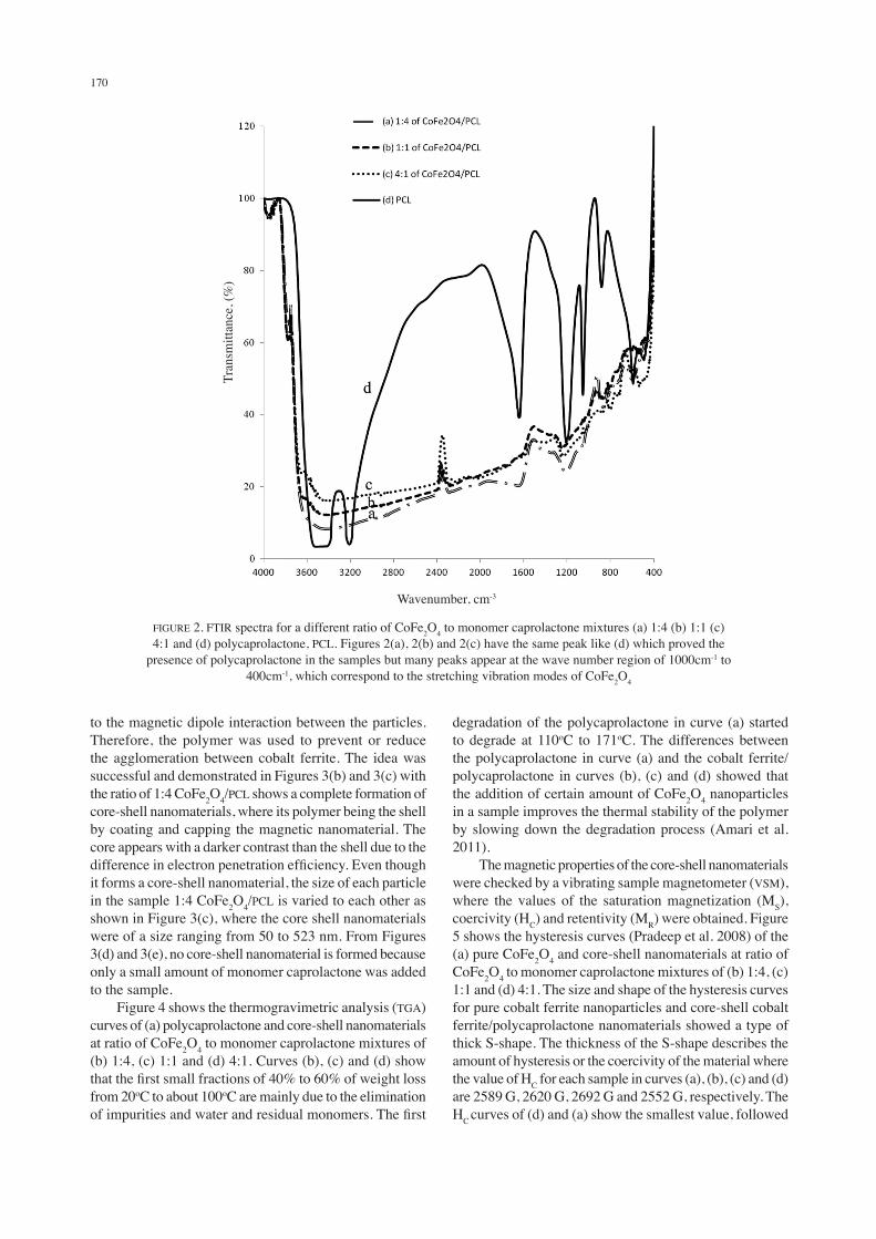
Figure 1(a) and also a broad amorphous diffraction peak appears at 2θ = 10o-29o and can be described as being nearly perpendicular to the semicrystalline polymer (Saikia & Kumar 2004). however, the addition of a small amount of polycaprolactone in samples Figures 1(c) and 1(d) prevents the formation of core-shell nanomaterials, but it helped to prevent the magnetic material, cobalt ferrite from agglomerating with itself and increased the intensity level of the samples. From the XRD patterns of Figure 1, the low intensity level of CoFe2o4 (as shown in Figure 1(b) indicates that the core-shell nanomaterial was best form in the ratio of CoFe2o4: monomer caprolactone mixtures of 1:4. The FTiR spectra of core-shell nanomaterials at the ratio of CoFe2o4 to monomer caprolactone mixtures of 1:4, 1:1 and 4:1 and polycaprolactone are shown in Figure 2. The FTiR spectra of polycaprolactone (Cristescu et al. 2007) in Figure 2(d) exist at the peak of 3208.37 cm-1 and 3523.71 cm-1 (C-h2 stretching of symmetry and asymmetry), 2095.19 cm-1and 1644.85 cm-1 (C=O) and also 1210.80 cm-1(C-C stretching) and 1054.27 cm-1 (C-o-C), while the chain of (C-h) exists at the value of 882.60 cm-1 and 591.06 cm-1. Figures 2(a), 2(b) and 2(c) show the FTiR spectra of 1:4, 1:1 and 4:1 of cobalt
ferrite/polycaprolactone nanomaterials, which have the same peak like Figure 2(d) proved the presence of polycaprolactone in the samples. From the curves, it is seen that many peaks appear at the wave number region of 1000 to 400 cm-1, which correspond to the stretching vibration modes of metal oxides. Those vibration modes could be contributed by the existence and overlapping of CoFe2o4 in the core-shell nanomaterials (gandhi et al. 2011; Qin et al. 2010). The existence of spinel ferrite, CoFe2o4 in a sample which had the structural properties of tetrahedron, attributed the high frequency band (600 to 580 cm-1) and octahedron, low frequency sites (440 to 400 cm-1). The vibrating spectra occurred at a frequency of 600.75 cm-1 and 416.05 cm-1 which corresponds to the tetrahedral and octahedral sites of CoFe2o4. The figures also indicate that no chemical interaction between CoFe2o4 and polycaprolactone, thus, CoFe2o4 only serves as the nucleation sites for the polymerization of caprolactone. The morphology or image of CoFe2o4 and 1:4, 1:1 and 4:1 of CoFe2o4/PCL was determined by transmission electron microscopy (TeM). Figure 3(a) shows that the CoFe2o4 nanoparticles were agglomerated. These magnetic nanoparticles incline to merge towards each other due
FiguRe 1. XRD patterns for (a) pure CoFe2o4 nanoparticles and core-shell nanomaterials at a different ratio of CoFe2o4 to monomer caprolactone mixtures of (b) 1:4 (c) 1:1 and (d) 4:1. All figures showed
the same peak except a broad amorphous diffraction peak appears at 2θ = 10o-29o in Figures 1(b), 1(c) and 1(d) that can be described as semicrystalline polymer
2-Theta - Scale
inte
nsity
(a.u
)
170
to the magnetic dipole interaction between the particles. Therefore, the polymer was used to prevent or reduce the agglomeration between cobalt ferrite. The idea was successful and demonstrated in Figures 3(b) and 3(c) with the ratio of 1:4 CoFe2o4/PCL shows a complete formation of core-shell nanomaterials, where its polymer being the shell by coating and capping the magnetic nanomaterial. The core appears with a darker contrast than the shell due to the difference in electron penetration efficiency. Even though it forms a core-shell nanomaterial, the size of each particle in the sample 1:4 CoFe2o4/PCL is varied to each other as shown in Figure 3(c), where the core shell nanomaterials were of a size ranging from 50 to 523 nm. From Figures 3(d) and 3(e), no core-shell nanomaterial is formed because only a small amount of monomer caprolactone was added to the sample. Figure 4 shows the thermogravimetric analysis (Tga) curves of (a) polycaprolactone and core-shell nanomaterials at ratio of CoFe2o4 to monomer caprolactone mixtures of (b) 1:4, (c) 1:1 and (d) 4:1. Curves (b), (c) and (d) show that the first small fractions of 40% to 60% of weight loss from 20oC to about 100oC are mainly due to the elimination of impurities and water and residual monomers. The first
degradation of the polycaprolactone in curve (a) started to degrade at 110oC to 171oC. The differences between the polycaprolactone in curve (a) and the cobalt ferrite/polycaprolactone in curves (b), (c) and (d) showed that the addition of certain amount of CoFe2o4 nanoparticles in a sample improves the thermal stability of the polymer by slowing down the degradation process (amari et al. 2011). The magnetic properties of the core-shell nanomaterials were checked by a vibrating sample magnetometer (VSM), where the values of the saturation magnetization (MS), coercivity (hC) and retentivity (MR) were obtained. Figure 5 shows the hysteresis curves (Pradeep et al. 2008) of the (a) pure CoFe2o4 and core-shell nanomaterials at ratio of CoFe2o4 to monomer caprolactone mixtures of (b) 1:4, (c) 1:1 and (d) 4:1. The size and shape of the hysteresis curves for pure cobalt ferrite nanoparticles and core-shell cobalt ferrite/polycaprolactone nanomaterials showed a type of thick S-shape. The thickness of the S-shape describes the amount of hysteresis or the coercivity of the material where the value of hC for each sample in curves (a), (b), (c) and (d) are 2589 g, 2620 g, 2692 g and 2552 g, respectively. The hC curves of (d) and (a) show the smallest value, followed
FiguRe 2. FTiR spectra for a different ratio of CoFe2o4 to monomer caprolactone mixtures (a) 1:4 (b) 1:1 (c) 4:1 and (d) polycaprolactone, PCL. Figures 2(a), 2(b) and 2(c) have the same peak like (d) which proved the
presence of polycaprolactone in the samples but many peaks appear at the wave number region of 1000cm-1 to 400cm-1, which correspond to the stretching vibration modes of CoFe2o4
wavenumber, cm-3
Tran
smitt
ance
, (%
)

171
by samples (b) and (c). in the absence of polycaprolactone that serves as nonmagnetic outer shells, pure CoFe2o4 nanoparticles may interact magnetically among each other (Kakarla et al. 2007). The magnetic interactions between the CoFe2o4 nanoparticles may lead to a decrease in the coercivity. The saturation magnetization, MS for pure CoFe2o4, 1:4 CoFe2o4/PCL, 4:1 CoFe2o4/PCL and 1:1 CoFe2o4/PCL are 0.39 emu/g, 0.12 emu/g, 0.15 emu/g and 0.31 emu/g, respectively. The retentivity values, MR, for each sample are 0.24 emu/g, 0.07 emu/g, 0.09 emu/g and 0.19 emu/g, respectively. The values of MS and MR in (b) and (c) show a large decreasing value compared with pure cobalt ferrite due to the presence of nonmagnetic polymer material coated magnetic nanoparticles.
CoNCLuSioN
The core-shell nanomaterials CoFe2o4/PCL were successfully synthesized through in situ polymerization of monomer caprolactone in the presence of magnetic nanoparticles, cobalt ferrite. The formation of CoFe2o4/PCL nanomaterials was confirmed using XRD, FTiR and TeM. The results showed that the core-shell nanomaterials were successfully formed at the ratio of CoFe2o4 to monomer caprolactone mixtures of 1:4. Thermal properties of core-shell nanomaterials were checked by thermogravimetric analysis where the existence of cobalt ferrite in samples helped to slow down the degradation process of the polycaprolactone. The magnetic properties show that the
(a) (b) (c) (d) (e)
FiguRe 3(a). TeM micrograph of CoFe2o4, (b) TeM micrograph of 1:4 CoFe2o4/PCL, (c) TeM micrograph in different size of 1:4 core-shell CoFe2o4/PCL, (d) TeM micrograph of 1:1 CoFe2o4/PCL and (e) TeM micrograph of 4:1 CoFe2o4/PCL
FiguRe 4. Thermogravimetric analysis (Tga) curves of (a) polycaprolactone and core-shell nanomaterials at ratio of CoFe2o4 to monomer caprolactone mixtures of (b) 1:4, (c) 1:1 and (d) 4:1. in curves (b), (c) and (d) showed that the addition of certain amount of CoFe2o4 nanoparticles in a sample improves the thermal stability of the
polymer by slowing down the degradation process
Temperature, °C
wei
ght,
mg

172
addition of polycaprolactone in samples decreased the magnetic saturation of CoFe2o4.
aCKNowLedgeMeNTS
The authors would like to acknowledge the Ministry of Higher Education, Malaysia for the financial support from the Fundamental Research grant Scheme, FRgS (uKM-ST-01-FRgS0063-2006).
ReFeReNCeS
ahmet, N.a., deniz, K. & Birgul, z.K. 2011. Magnetic nanocomposites with drug-intercalated layered double hydroxide shell supported on commercial magnetite and laboratory-made magnesium ferrite core materials. Materials Science and Engineering C 31(5): 851-857.
alexiou, C., Jurgons, R., Schmid, R., hilpert, a., Bergemann, C., Parak, F. & iro, h. 2005. In vitro and in vivo investigations of targeted chemotherapy with magnetic nanoparticles. Journal of Magnetism and Magnetic Materials 293: 389-393.
amari, N.F., Khoo, K.S., Radiman, S., Yasir, M.S. & Yahaya, R. 2011. Pencirian struktur spektrum, morfologi, haba dan optikal rangka teras nanobahan kobalt ferrit/polikaprolakton (CoFe2o4/PCL). Prosiding Kolokium Siswazah ke-11, pp. 107-109.
Baldi, g., Bonacchi, d., innocenti, d.C., Lorenzi, g. & Sangregorio, C. 2007. Cobalt ferrite nanoparticles: The control of the particle size and surface state and their effects on magnetic properties Journal of Magnetism and Magnetic Materials 311(1): 10-16.
Chuang, F-Y. & Yang, S-M. 2008. Cerium dioxide/polyaniline core shell nanocomposite. Journal of Colloid and Interface Science 320: 194-201.
Cristescu, R., doraiswamy, a., Socol, g., grigorescu, S., axente, e., Mihaiescu, d., Moldovan, a., Narayan, R.J., Stamatin, i., Mihailescu, i.N., Chisholm, B.J. & Chrisey, d.B. 2007. Polycaprolactone biopolymer thin films obtained by matrix assisted pulsed laser evaporation. Applied Surface Science 253(15): 6476-6479.
gandhi, N., Singh, K., ohlan, a., Singh, d.P. & dhawan, S.K. 2011. Thermal, dielectric and microwave absorption
FiguRe 5. Magnetic properties (a) pure CoFe2o4 and core-shell nanomaterials at ratio of CoFe2o4 to monomer caprolactone mixtures of (b) 1:4, (c) 1:1 and (d) 4:1. The values of MS and MR in (b) and (c) show a large decreasing value
compared with pure cobalt ferrite due to the presence of nonmagnetic polymer material coated magnetic nanoparticles
Mag
netiz
atio
n, e
mu/
g
Field, g

173
properties of polyaniline–CoFe2o4 Nanocomposites. Composites Science and Technology 71: 1754-176.
hartono, d., Qin, J.w., Yang, J.K.L. & Yung, L.Y.L. 2009. imaging the disruption of phospholipid monolayer by protein-coated nanoparticles using ordering transitions of liquid crystals. Biomaterials 30(5): 843-849.
Jiang, J. & Ai, L. 2010. Influence of annealing temperature on the formation, microstructure and magnetic properties of spinel nanocrystalline cobalt ferrites. Current Applied Physics 10(1): 284-288.
Jing, J., Lun, h.a. & ai, h.L. 2010. a novel poly(o-anisidine)/CoFe2o4 multifunctional nanocomposite: Preparation, characterization and properties. Synthetic Metals 160(5): 333-336.
Kakarla, R.R., Lee, K-P., anantha, i.g. & Kang, h-d. 2007. Organosilane modified magnetite nanoparticles/poly(aniline-co-o/m-aminobenzenesulfionic acid) composites: Synthesis and characterization. Reactive and Functional Polymers 67(10): 943-954.
Kang, B., Chang, S., dai, Y. & Chen, d. 2007. Radiation synthesis and magnetic properties of novel Co0.7Fe0.3/Chitosan compound nanoparticles for targeted drug carrier. Radiation Physics and Chemistry 76(6): 968-973.
Kumar, C.S.R. & Mohammad, F. 2011. Magnetic nanomaterials for hyperthermia-based therapy and controlled drug delivery. Advanced Drug Delivery Reviews 63(9): 789-808.
Liu, H., Xu, F., Li, L., Wang, Y. & Qiu, H. 2009. A novel CoFe2o4/polyacrylate nanocomposite prepared via an in situ polymerization in emulsion system. Reactive and Functional Polymer 69(1): 43-47.
Nicholson, J.w. 1991. The Chemistry of Polymers. Cambridge.Pradeep, a., Priyadharsini, P. & Chandrasekaran, g. 2008. Sol-
gel route of synthesis of nanoparticles of MgFe2o4 and XRD, FTiR and VSM study. Journal of Magnetism and Magnetic Materials 320(21): 2774-2779.
Pita, M., abad, J.M., dominguez, C.V., Briones, C., Marti, e.M., gago, J.a.M., Morales, M.P. & Fernandez, V.M. 2008. Synthesis of cobalt ferrite core/metallic shell nanoparticles for the development of a specific PNA/DNA biosensor. Journal of Colloid and Interface Science 321(2): 484-492.
Qin, R., Li, F., Jiang, w. & Chen, M. 2010. Synthesis and characterization of diethylenetriaminepentaacetic acid-
chitosan-coated cobalt ferrite core/shell nanostructures. Materials Chemistry and Physics 122(2): 498-501.
Saikia, d. & Kumar, a. 2004. ionic conduction in P(VdF-hFP)/PVdF–(PC + deC)–LiClo4 polymer gel electrolyte. Electrochimica Acta 49(16): 2581-2589.
Sanjeev, K., Vaishali, S., Saroj, a., uttam, K.M. & Ravinder, K.K. 2010. Bimodal Co0.5zn0.5Fe2o4/PaNi nanocomposites: Synthesis, formation mechanism and magnetic properties. Composites Science and Technology 70(2): 249-254.
Sun, C., Lee, J.S.h. & zhang, M. 2008. Magnetic nanoparticles in MR imaging and drug delivery. Advanced Drug Delivery Reviews 60(11): 1252-1265.
Tadros, T.F. 2009. Emulsion Science and Technology. weinheim: wiley-VCh.
Xu, T., Zhang, N., Nichols, H.L., Shi, D. & Wen, X. 2007. Modification of nanostructured materials for biomedical applications. Materials Science and Engineering C 27(3): 579-594.
Yu, M.h., devi, P.S., Lewis, L.h., oouma, P., Parise, J.B. & gambino, R.J. 2003. Towards a magnetic core-shell nanostructure: a novel composite made by a citrate-nitrate auto-ignition process. Materials Science and Engineering B 103(3): 262-270.
Zhang, Z., He, Q., Xiong, J., Xiong, Y. & Xiao, H. 2008. A novel biomaterial - Fe3o4:Tio2 core-shell nano particle with magnetic performance and high visible light photocatalytic activity. Optical Materials 31: 380-384.
School of applied PhysicsFaculty of Science and Technologyuniversiti Kebangsaan Malaysia43600 Bangi, Selangor darul ehsan Malaysia
*Corresponding author; email: [email protected]
Received: 8 March 2012accepted: 23 June 2012