Preparation and characterization of gelatin- tamarind gum ...
94
Preparation and characterization of gelatin- tamarind gum / carboxymethyl tamarind gum based phase separated hydrogels and films for tissue engineering applications Department of Biotechnology & Medical Engineering National Institute of Technology Rourkela Gauri Shankar Shaw
Transcript of Preparation and characterization of gelatin- tamarind gum ...
Preparation and characterization of gelatin-
tamarind gum / carboxymethyl tamarind gum based
phase separated hydrogels and films for tissue
engineering applications
Department of Biotechnology & Medical
Engineering National Institute of Technology Rourkela
Gauri Shankar Shaw
PREPARATION AND CHARACTERIZATION OF GELATIN-TAMARIND GUM /
CARBOXYMETHYL TAMARIND GUM BASED PHASE-SEPARATED
HYDROGELS AND FILMS FOR TISSUE ENGINEERING APPLICATIONS
Dissertation submitted to the
National Institute of Technology Rourkela
in partial fulfillment of the requirements
of the degree of
Master of Technology
(by Research)
in
Biotechnology & Medical Engineering
By
GAURI SHANKAR SHAW
(613BM6012)
under the supervision of
Prof. Kunal Pal
and
Prof. Krishna Pramanik
April, 2016
Department of Biotechnology & Medical Engineering
National Institute of Technology Rourkela

Biotechnology & Medical Engineering National Institute of Technology Rourkela
iii
April 12, 2015
Certificate of Examination
Roll Number: 613BM6012 Name: Gauri Shankar Shaw
Title of Dissertation: preparation and characterization of gelatin-tamarind gum/
carboxymethyl tamarind gum based phase-separated hydrogels and films for tissue
engineering applications
We the below signed, after checking the dissertation mentioned above and the official
record book (s) of the student, hereby state our approval of the dissertation submitted
in partial fulfillment of the requirements of the degree of Master in Technology (by
research) in Biotechnology and Medical Engineering at National Institute of
Technology Rourkela. We are satisfied with the volume, quality, correctness, and
originality of the work.
Prof. Krishna Pramanik Prof. Kunal Pal
Co-Supervisor Principal Supervisor
Prof. Amit Biswas Prof. Sujit Bhutia
Member (DSC) Member (DSC)
Prof. Samit Ari
Member (DSC) Examiner
Prof. Mukesh Kuma Gupta
Chairman (DSC)
Biotechnology & Medical Engineering National Institute of Technology Rourkela
iv
Prof. /Dr. Kunal Pal
Assistant Professor
April 12, 2016
Supervisor's Certificate
This is to certify that the work presented in this dissertation entitled “Preparation
and characterization of gelatin-tamarind gum / carboxymethyl tamarind gum based
phase-separated hydrogels and films for tissue engineering applications'' by ''Gauri
Shankar Shaw'', Roll Number 613BM6012, is a record of original research carried out by
him/her under my supervision and guidance in partial fulfillment of the requirements of
the degree of Master in Technology (by research) in Biotechnology and Medical
Engineering. Neither this dissertation nor any part of it has been submitted for any degree
or diploma to any institute or university in India or abroad.
Kunal Pal
Biotechnology & Medical Engineering National Institute of Technology Rourkela
v
April 12, 2015
Supervisors' Certificate
This is to certify that the work presented in this dissertation entitled '' PREPARATION
AND CHARACTERIZATION OF GELATIN-TAMARIND GUM / CARBOXYMETHYL TAMARIND GUM
BASED PHASE-SEPARATED HYDROGELS AND FILMS FOR TISSUE ENGINEERING APPLICATIONS''
by ''Gauri Shankar Shaw'', Roll Number 613BM6012, is a record of original research
carried out by him/her under our supervision and guidance in partial fulfillment of the
requirements of the degree of M. Tech (R) in Department of Biotechnology and Medical
Engineering. Neither this dissertation nor any part of it has been submitted for any degree
or diploma to any institute or university in India or abroad.
Krishna Pramanik Kunal Pal
Co-Supervisor Principal Supervisor
vi
Declaration of Originality
I, Gauri Shankar Shaw, Roll Number 613BM6012 hereby declare that this
dissertation entitled '' Preparation and characterization of gelatin-tamarind gum /
carboxymethyl tamarind gum based phase-separated hydrogels and films for tissue
engineering applications'' represents my original work carried out as a postgraduate
student of NIT Rourkela and, to the best of my knowledge, it contains no material
previously published or written by another person, nor any material presented for the
award of any other degree or diploma of NIT Rourkela or any other institution. Any
contribution made to this research by others, with whom I have worked at NIT Rourkela or
elsewhere, is explicitly acknowledged in the dissertation. Works of other authors cited in
this dissertation have been duly acknowledged under the section ''Bibliography''. I have
also submitted my original research records to the scrutiny committee for evaluation of
my dissertation.
I am fully aware that in case of any non-compliance detected in future, the Senate of
NIT Rourkela may withdraw the degree awarded to me on the basis of the present
dissertation.
April 12, 2016
NIT Rourkela
Gauri Shankar Shaw

vii
Acknowledgment
Successful completion of this project is the outcome of consistent guidance and
assistance from many people, faculty and friends and I am extremely fortunate to have got
this all along the completion of the project.
I owe my profound gratitude and respect to my project guide Dr. Kunal Pal and Dr.
Krishna Pramanik, Department of Biotechnology and Medical Engineering, NIT Rourkela
for their invaluable academic support and professional guidance, regular encouragement
and motivation at various stages of this project. Special thanks to Dr. Indranil Banerjee
for giving beautiful ideas and co-operation for the work. I am very much grateful to them
for allowing me to follow my own ideas.
I would like to extend my heartfelt gratitude to research scholars Mr. Biswajeet
Champaty, Mr Vinay Singh, Mr. Sai Satish, Ms. Beauty Behera, Ms. Dibyajyoti Biswal,
Ms. Preeti Madhuri Pandey, Ms. Indu Yadav and Mr. Suraj kumar Nayak whose ever
helping nature and suggestions have made my work easier by many folds. I would like to
thank all my friends and classmates for their constant moral support, suggestions, advices
and ideas. I have enjoyed their presence so much during my stay at NIT, Rourkela.
I will never forget the support provided by Mr. Haldhar Behera for providing valuable
help.
April 12, 2016
NIT Rourkela
Gauri Shankar Shaw
Roll Number: 613BM6012
viii
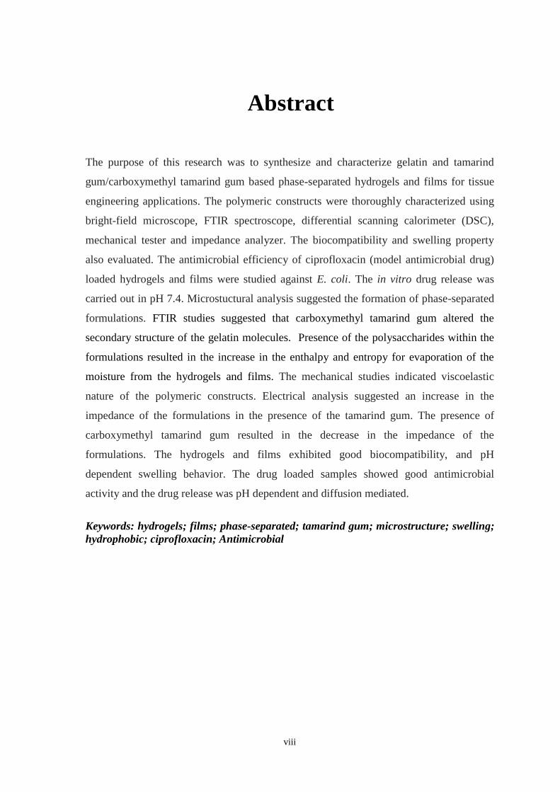
Abstract
The purpose of this research was to synthesize and characterize gelatin and tamarind
gum/carboxymethyl tamarind gum based phase-separated hydrogels and films for tissue
engineering applications. The polymeric constructs were thoroughly characterized using
bright-field microscope, FTIR spectroscope, differential scanning calorimeter (DSC),
mechanical tester and impedance analyzer. The biocompatibility and swelling property
also evaluated. The antimicrobial efficiency of ciprofloxacin (model antimicrobial drug)
loaded hydrogels and films were studied against E. coli. The in vitro drug release was
carried out in pH 7.4. Microstuctural analysis suggested the formation of phase-separated
formulations. FTIR studies suggested that carboxymethyl tamarind gum altered the
secondary structure of the gelatin molecules. Presence of the polysaccharides within the
formulations resulted in the increase in the enthalpy and entropy for evaporation of the
moisture from the hydrogels and films. The mechanical studies indicated viscoelastic
nature of the polymeric constructs. Electrical analysis suggested an increase in the
impedance of the formulations in the presence of the tamarind gum. The presence of
carboxymethyl tamarind gum resulted in the decrease in the impedance of the
formulations. The hydrogels and films exhibited good biocompatibility, and pH
dependent swelling behavior. The drug loaded samples showed good antimicrobial
activity and the drug release was pH dependent and diffusion mediated.
Keywords: hydrogels; films; phase-separated; tamarind gum; microstructure; swelling;
hydrophobic; ciprofloxacin; Antimicrobial
ix
Contents
Certificate of Examination iii
Supervisor's Certificate iv
Supervisors' Certificate v
Declaration of Originality vi
Acknowledgment vii
Abstract viii
List of Figures xii
List of Tables xiv
1 Introduction
1
2 Review of literature 5
2.1 Animal Derived Natural Polymers . . . . . . . . . . . . . . . . . . . . . . . . . . . . . . . 5
2.1.1 Collagen . . . . . . . . . . . . . . . . . . . . . . . . . . . . . . . . . . . . . . . . . . . . . 5
2.1.2 Gelatin . . . . . . . . . . . . . . . . . . . . . . . . . . . . . . . . . . . . . . . . . . . . . . 6
2.1.3 Hyaluronic acid . . . . . . . . . . . . . . . . . . . . . . . . . . . . . . . . . . . . . . . . 7
2.1.4 Elastin . . . . . . . . . . . . . . . . . . . . . . . . . . . . . . . . . . . . . . . . . . . . . 8
2.1.5 Chondroitin sulphate . . . . . . . . . . . . . . . . . . . . . . . . . . . . . . . . . . . . 8
2.1.6 Fibrin . . . . . . . . . . . . . . . . . . . . . . . . . . . . . . . . . . . . . . . . . . . . . . . 9
2.2 Plant Derived Natural Polymers . . . . . . . . . . . . . . . . . . . . . . . . . . . . . . . . . 10
2.2.1 Agarose . . . . . . . . . . . . . . . . . . . . . . . . . . . . . . . . . . . . . . . . . . . . . 10
2.2.2 Alginate . . . . . . . . . . . . . . . . . . . . . . . . . . . . . . . . . . . . . . . . . . . . . 10
2.2.3 Chitosan . . . . . . . . . . . . . . . . . . . . . . . . . . . . . . . . . . . . . . . . . . . . . 11
2.2.4 Tamarind gum (TG) . . . . . . . . . . . . . . . . . . . . . . . . . . . . . . . . . . . . . 12
2.2.5 Carboxymethyl tamarind gum (CMT) . . . . . . . . . . . . . . . . . . . . . . . . 13
2.3 Objectives . . . . . . . . . . . . . . . . . . . . . . . . . . . . . . . . . . . . . . . . . . . . . . . . 13
3 Development and Characterization of Gelatin-Tamarind Gum/
Carboxymethyl Tamarind Gum Based Phase-Separated Hydrogels: A
Comparative Study 14
3.1 Introduction . . . . . . . . . . . . . . . . . . . . . . . . . . . . . . . . . . . . . . . . . . . . . . . 14
x
3.2 Materials and Methods . . . . . . . . . . . . . . . . . . . . . . . . . . . . . . . . . . . . . . . . 16
3.2.1 Materials . . . . . . . . . . . . . . . . . . . . . . . . . . . . . . . . . . . . . . . . . . . . . . 16
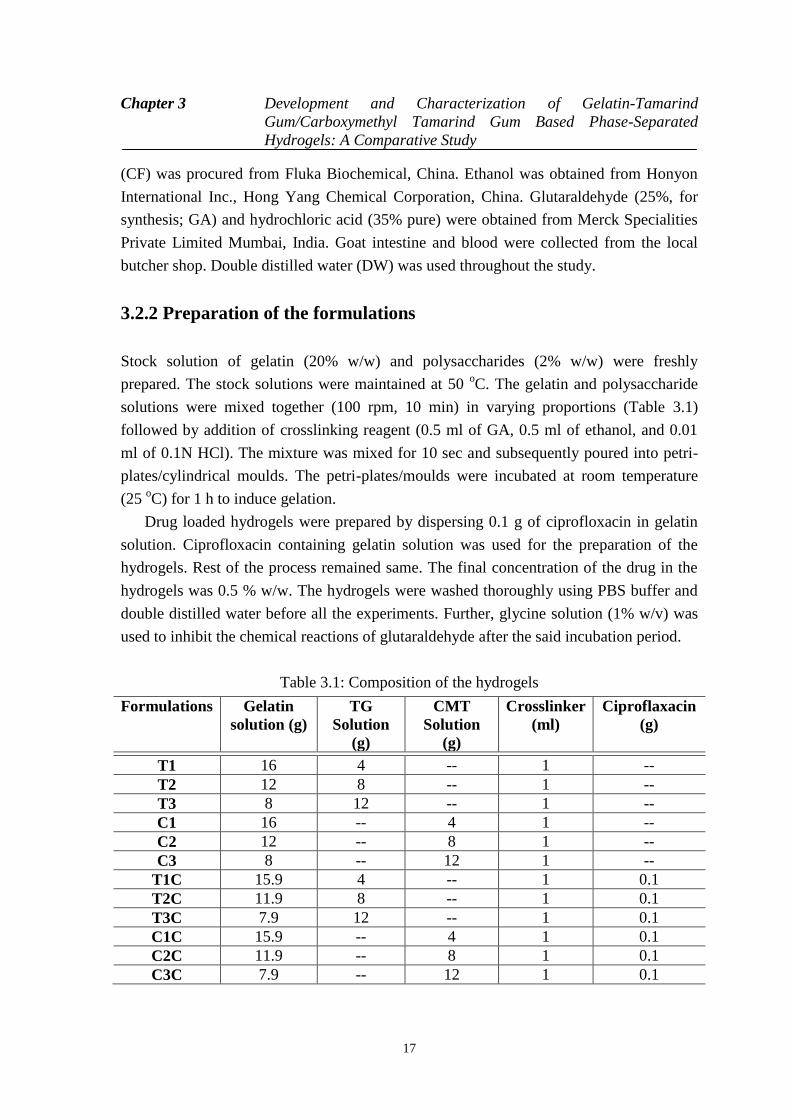
3.2.2 Preparation of the formulations . . . . . . . . . . . . . . . . . . . . . . . . . . . . . . 16
3.2.3 Microscopy studies . . . . . . . . . . . . . . . . . . . . . . . . . . . . . . . . . . . . . . 17
3.2.4 Infrared spectroscopy . . . . . . . . . . . . . . . . . . . . . . . . . . . . . . . . . . . . . 17
3.2.5 Thermal analysis. . . . . . . . . . . . . . . . . . . . . . . . . . . . . . . . . . . . . . . . . 17
3.2.6 Mechanical Analysis . . . . . . . . . . . . . . . . . . . . . . . . . . . . . . . . . . . . . 18
3.2.7 Impedance analysis . . . . . . . . . . . . . . . . . . . . . . . . . . . . . . . . . . . . . . 18
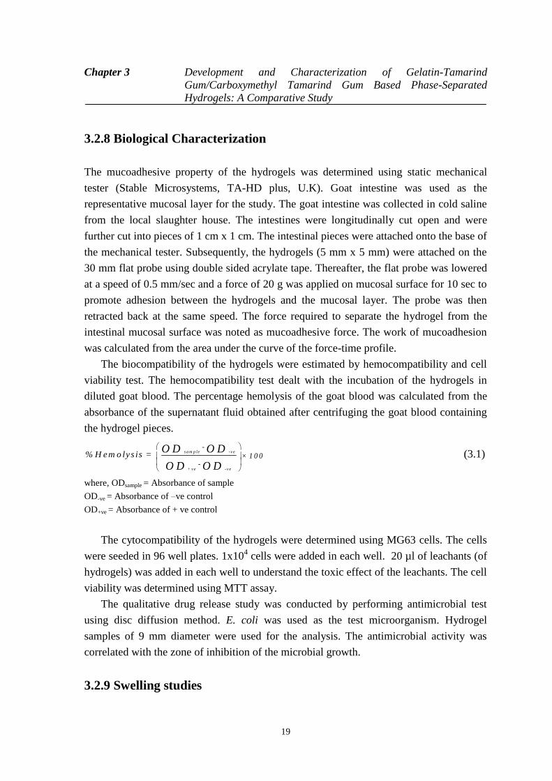
3.2.8 Biological Characterization . . . . . . . . . . . . . . . . . . . . . . . . . . . . . . . . . 18
3.2.9 Swelling studies . . . . . . . . . . . . . . . . . . . . . . . . . . . . . . . . . . . . . . . . . 19
3.2.10 Drug release studies . . . . . . . . . . . . . . . . . . . . . . . . . . . . . . . . . . . . . 19
3.3 Result and Discussion . . . . . . . . . . . . . . . . . . . . . . . . . . . . . . . . . . . . . . . . . 20
3.3.1 Preparation of hydrogels . . . . . . . . . . . . . . . . . . . . . . . . . . . . . . . . . . . 20
3.3.2 Microscopy . . . . . . . . . . . . . . . . . . . . . . . . . . . . . . . . . . . . . . . . . . . . 21
3.3.3 Infrared spectroscopy . . . . . . . . . . . . . . . . . . . . . . . . . . . . . . . . . . . . . 22
3.3.4 Thermal analysis . . . . . . . . . . . . . . . . . . . . . . . . . . . . . . . . . . . . . . . . 24
3.3.5 Mechanical Analysis. . . . . . . . . . . . . . . . . . . . . . . . . . . . . . . . . . . . . . 25
3.3.6 Impedance Analysis . . . . . . . . . . . . . . . . . . . . . . . . . . . . . . . . . . . . . 31
3.3.7 Biological Characterization . . . . . . . . . . . . . . . . . . . . . . . . . . . . . . . . . 33
3.3.8 Swelling studies . . . . . . . . . . . . . . . . . . . . . . . . . . . . . . . . . . . . . . . . . 34
3.3.9 Drug release study . . . . . . . . . . . . . . . . . . . . . . . . . . . . . . . . . . . . . . . 38
3.4 Conclusion . . . . . . . . . . . . . . . . . . . . . . . . . . . . . . . . . . . . . . . . . . . . . . . . . 41
4 Preparation, characterization and assessment of the novel gelatin-tamarind
gum/ carboxymethyl tamarind gum based phase-separated films for skin
tissue engineering applications 42
4.1 Introduction . . . . . . . . . . . . . . . . . . . . . . . . . . . . . . . . . . . . . . . . . . . . . . . . 42
4.2 Materials and method . . . . . . . . . . . . . . . . . . . . . . . . . . . . . . . . . . . . . . . . . 43
4.2.1 Materials . . . . . . . . . . . . . . . . . . . . . . . . . . . . . . . . . . . . . . . . . . . . . . 43
4.2.2 Preparation of polymeric solutions . . . . . . . . . . . . . . . . . . . . . . . . . . . . 44
4.2.3 Preparation of films . . . . . . . . . . . . . . . . . . . . . . . . . . . . . . . . . . . . . . . 54
4.2.4 Microscopy studies . . . . . . . . . . . . . . . . . . . . . . . . . . . . . . . . . . . . . . . 45

xi
4.2.5 Infrared spectroscopy. . . . . . . . . . . . . . . . . . . . . . . . . . . . . . . . . . . . . . 45
4.2.6 Thermal analysis . . . . . . . . . . . . . . . . . . . . . . . . . . . . . . . . . . . . . . . . . 45
4.2.7 Mechanical analysis . . . . . . . . . . . . . . . . . . . . . . . . . . . . . . . . . . . . . . 47
4.2.8 Impedance analysis . . . . . . . . . . . . . . . . . . . . . . . . . . . . . . . . . . . . . . . 46
4.2.9 Biological characterizations . . . . . . . . . . . . . . . . . . . . . . . . . . . . . . . . . 46
4.2.10 Swelling studies. . . . . . . . . . . . . . . . . . . . . . . . . . . . . . . . . . . . . . . . . 47
4.2.11 Drug release studies . . . . . . . . . . . . . . . . . . . . . . . . . . . . . . . . . . . . . 47
4.3 Result and discussion. . . . . . . . . . . . . . . . . . . . . . . . . . . . . . . . . . . . . . . . . . 48
4.3.1 Preparation of the films . . . . . . . . . . . . . . . . . . . . . . . . . . . . . . . . . . . . 48
4.3.2 Microscopic analysis . . . . . . . . . . . . . . . . . . . . . . . . . . . . . . . . . . . . . . 49
4.3.3 Infrared spectroscopy. . . . . . . . . . . . . . . . . . . . . . . . . . . . . . . . . . . . . . 50
4.3.4 Thermal analysis . . . . . . . . . . . . . . . . . . . . . . . . . . . . . . . . . . . . . . . 51
4.3.5 Mechanical studies . . . . . . . . . . . . . . . . . . . . . . . . . . . . . . . . . . . . . . . 52
4.3.6 Impedance analysis . . . . . . . . . . . . . . . . . . . . . . . . . . . . . . . . . . . . . . . 59
4.3.7 Biological characterizations . . . . . . . . . . . . . . . . . . . . . . . . . . . . . . . . . 60
4.3.8 Swelling studies . . . . . . . . . . . . . . . . . . . . . . . . . . . . . . . . . . . . . . . . . 61
4.3.9 Drug release studies. . . . . . . . . . . . . . . . . . . . . . . . . . . . . . . . . . . . . . . 64
4.4 Conclusion . . . . . . . . . . . . . . . . . . . . . . . . . . . . . . . . . . . . . . . . . . . . . . . . . 67
5 Summary 68
Bibliography 70
Dissemination 79
xii
List of Figures
3.1 Chemical structures of TG and CMT . . . . . . . . . . . . . . . . . 15
3.2 Pictographs of the hydrogels. (a) T1, (b) T2, (c) T3, (d) C1,
(e) C2, and (f) C3. . . . . . . . . . . . . . . . . . . . . . . . . . . . . . .
20
3.3 Light micrographs of the hydrogels. (a) T1, (b) T2, (c) T3,
(d) C1, (e) C2, and (F) C3. . . . . . . . . . . . . . . . . . . . . . . . .
21
3.4 FTIR spectra of the hydrogels. (a) TG hydrogels, and (b)
CMT hydrogels . . . . . . . . . . . . . . . . . . . . . . . . . . . . . . . .
22
3.5 Thermal analysis of the hydrogels. (a) T1, (b) T3, (c) C1,
and (d) C3 . . . . . . . . . . . . . . . . . . . . . . . . . . . . . . . . . . .
24
3.6 Mechanical studies of the hydrogels. (a) Resilience of TG
and CMT hydrogels, (b) Peak forces of TG and CMT
hydrogels, (c) % Stress relaxation of TG and CMT
hydrogels, and (d) D20 values of TG and CMT hydrogels. .
26
3.7 Analysis of SR data: (a) Stress relaxation profiles, (b) SR
data for modelling, (c) Kohlrausch model fitting of the
hydrogels, and (d) Weichart model fitting of the hydrogels. .
28
3.8 Impedance profiles: (a) TG hydrogels, and (b) CMT
hydrogels; and V-I characteristics; (c) TG hydrogels, and (d)
CMT hydrogels . . . . . . . . . . . . . . . . . . . . . . . . . . . . . . . .
32
3.9 Biological characterizations of the hydrogels. (a) Area under
the curve of mucoadhesive profiles, (b) % hemolysis of goat
blood, (c) Cell proliferation study, and (d) Antimicrobial
study . . . . . . . . . . . . . . . . . . . . . . . . . . . . . . . . . . . . . . .
33
3.10 Swelling study of the hydrogels. (a) Swelling profiles of the
hydrogels (pH 7.4), (b) Weibull model fitting for hydrogels ,
(c) Korsmeyer-Peppas model fitting for the hydrogels . . . . .
35
3.11 Drug release study of the hydrogels at pH 7.4 (a) Drug
release profiles of TG and CMT hydrogels, (b) Weibull
model fitting for hydrogels, (c) Korsmeyer-Peppas model
xiii
fitting for hydrogels, (d) Peppas-sahlin model fitting for
hydrogels, and (e) R/F ratio from Peppas-sahlin model . . . .
39
4.1 Photographs of the films. (a) C, (b) T1, (c) T2, (d) C1, and
(e) C2 . . . . . . . . . . . . . . . . . . . . . . . . . . . . . . . . . . . . . . .
48
4.2 Light micrographs of the films. (a) C, (b) T1, (c) T2, (d) C1,
and (e) C2 . . . . . . . . . . . . . . . . . . . . . . . . . . . . . . . . . . . .
49
4.3 FTIR spectra of the films. . . . . . . . . . . . . . . . . . . . . . . . . 50
4.4 Thermal profiles of the films. (a) C, (b) T1, (c) T2, (d) C1,
and (e) C2 . . . . . . . . . . . . . . . . . . . . . . . . . . . . . . . . . . . .
52
4.5 Tensile and bursting strength results of the films. (a) Tensile
strengths of the films, and (b) Bursting strengths of the films.
53
4.6 Stress relaxation results of the films. (a) % Stress relaxation
of the films, and (b) D20 values of the films . . . . . . . . . . . . .
54
4.7 Analysis of SR data: (a) Stress relaxation profiles, (b) SR
data for modelling, (c) Kohlrausch model fitting of the films,
and (d) Weichart model fitting of the films . . . . . . . .
57
4.8 Impedance profiles: (a) TG films, and (b) CMT films; and
V-I profiles: (c) TG films, and (b) CMT films . . . . . . . . . . .
58
4.9 Biological characterizations of the films. (a)
Hemocompatibility, (b) Antimicrobial study, and (c) Cell
proliferation study using osteoblast cells . . . . . . . . . . . . . .
60
4.10 Swelling study of the films. (a) Swelling profiles of TGand
CMT films, (b) Weibull model fitting, and (c) Korsmeyer-
Peppas model fitting . . . . . . . . . . . . . . . . . . . . . . . . . . . . .
61
4.11 Drug release study of the films at pH 7.4 (a) Drug release
profiles of the TG and CMT films, (b) Weibull model fitting
for films, (c) Korsmeyer-Peppas model fitting for films, (d)
Peppas-sahlin model fitting for films, and (e) R/F ratio from
Peppas-sahlin model. . . . . . . . . . . . . . . . . . . . . . . . . . . . .
65
xiv
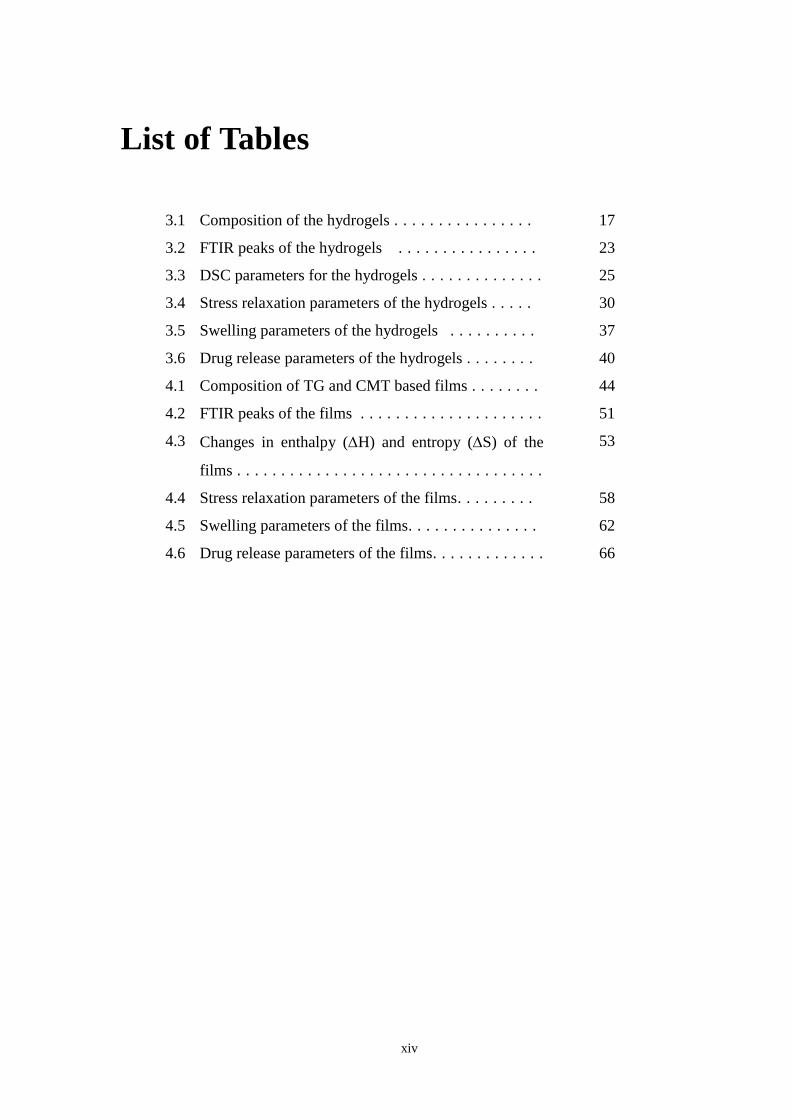
List of Tables
3.1 Composition of the hydrogels . . . . . . . . . . . . . . . . 17
3.2 FTIR peaks of the hydrogels . . . . . . . . . . . . . . . . 23
3.3 DSC parameters for the hydrogels . . . . . . . . . . . . . . 25
3.4 Stress relaxation parameters of the hydrogels . . . . . 30
3.5 Swelling parameters of the hydrogels . . . . . . . . . . 37
3.6 Drug release parameters of the hydrogels . . . . . . . . 40
4.1 Composition of TG and CMT based films . . . . . . . . 44
4.2 FTIR peaks of the films . . . . . . . . . . . . . . . . . . . . . 51
4.3 Changes in enthalpy (H) and entropy (S) of the
films . . . . . . . . . . . . . . . . . . . . . . . . . . . . . . . . . . .
53
4.4 Stress relaxation parameters of the films. . . . . . . . . 58
4.5 Swelling parameters of the films. . . . . . . . . . . . . . . 62
4.6 Drug release parameters of the films. . . . . . . . . . . . . 66
Chapter 1 Introduction
1
Chapter 1
Introduction Organ transplantation is still the main medical procedure to cure a patient with damaged
tissues and organs [1]. In the recent past, tissue engineering has attracted the attention of
the researchers and the surgeons. Tissue engineering is a field of science, which involves
fabricating of tissues and organs for replacing damaged parts of the human body [2]. This
field has opened up a new area in medicine and has provided new treatment modalities for
many disease conditions, where conventional treatment has failed. In the last two decades,
the field of tissue engineering has gained tremendous importance in the field of medicine
due to the enormous advantageous potentialities it has offered to the surgeons [3]. The
advances in tissue engineering have allowed the scientists in regenerating organs and
tissues [4]. This has allowed the reducing demand for organs and tissues to a great extent,
thereby, resulting in overcoming the shortage of organ donors to a certain extent. The
field of tissue engineering is multi-disciplinary in nature requiring the expertise of cell
biology, materials science and medicine (diseased organ and biomolecule delivery) [5]. In
recent days, the advances in imaging modalities (e.g. fluorescent microscopy, confocal
microscopy, environmental scanning electron microscopy, field emission scanning
electron microscopy) have played an important role in understanding the interaction
between the cells and the materials [6]. The major challenge in tissue engineering is the
designing of the artificial extracellular matrix (ECM) component, which can promote cell
proliferation onto itself [7]. The architectures used as artificial ECM are often regarded as
scaffolds. A scaffold is defined as the porous architecture which has the capability to
support cell growth and allow deposition of the natural ECM proteins over it during the
initial stages [8]. The deposition of the ECM proteins over the scaffolds elicits specific
cellular activity, which promotes functional integration of the cell-scaffold constructs
Chapter 1 Introduction
2
with the body tissues [9]. The deposition of the proteins over the scaffolds is mainly due
to the non-specific adsorption [10]. The scaffolds may be designed using materials which
undergo biodegradation/ bioresorption during the integration process [11]. Such scaffolds
lose their existence once they have completed their tasks. The desired properties (physical
or chemical) of the scaffolds are different for different tissues and are mainly dependent
on the functionality of the organ and the specific application which is expected to be met
by the scaffold [12]. Scaffolds may be designed to induce the regeneration of the tissues
and the organs, which do not possess regeneration capability [13]. Such scaffolds have
been regarded as regeneration templates. In short, the process of regeneration using the
tissue engineering protocol includes initial isolation of specific cells from the biopsies of
the patients [14]. The isolated cells are then cultured over the scaffolds in vitro and
subsequently transplanted into the patient. The transplanted cell-scaffold constructs help
in the regeneration of the tissues or organs in vivo [15]. If the isolated cells are stem cells,
the cells have to be differentiated into the specific cells before the cell-scaffold constructs
are transplanted.
The properties of the materials used for the fabrication of the scaffold play a
significant role in the success of the tissue engineering procedure [16]. The scaffolds are
expected to be highly biocompatible with negligible antigenicity and excellent
thromboresistant behavior [17]. Scaffolds can be designed using biomaterials, namely,
polymers, ceramics and metals [18]. Of the different types of biomaterials, polymeric
biomaterials have gained much importance [19]. This is due to the fact that the polymers
are more versatile materials than the other types of biomaterials. Polymers are available
with different chemistries. This allows easy modification of the surface properties of the
polymeric architectures, which might be necessary to improve the cell proliferation [20].
Additionally, the physical properties of the scaffolds may be easily altered by designing
the polymeric architectures using polymer blends and composites [21]. This can allow the
scientists to develop scaffolds using materials which can promote biomolecular
recognition based interactions of the scaffolds and the surrounding tissues [22]. Further,
modulating the properties of the polymeric architectures allow scientists in studying the
interactions between the cells and the developed constructs under in vitro conditions [23].
The research on the biomaterials has provided information on a group of polymeric
materials, which can help restoring the functionality of the diseased/ traumatized tissues
or organs in a relatively quick time. Polymeric materials have been successfully used to
design sutures, bone plates and screws, acetabular cup, vascular grafts, heart valves,
intraocular lens, ligaments, skin grafts, wound dressings and so on [24].
The polymeric biomaterials used for scaffold fabrication are broadly categorized into
two groups, namely, synthetic polymers and natural polymers [25]. Though many of the
Chapter 1 Introduction
3
synthetic polymers are known to have better mechanical properties, controlled
biodegradability and biocompatibility, the high cost of these polymers restricts their use
to design commercially viable scaffolds [26]. On the other hand, natural polymers are
much cheaper due to their abundance in nature [27]. The natural polymers are further
categorized into two broad categories as per their source of origin, namely, animal
derived natural polymers and plant derived natural polymers [28]. The commonly used
animal derived natural polymers include collagen, gelatin, hyaluronic acid, elastin,
chondroitin sulphate, and fibrin [29]. On the other hand, plant derived natural polymers
include agarose, alginate, chitosan and tamarind gum [30]. These polymers are
crosslinked to form hydrogels [31]. The crosslinking may either be due to covalent
bonding, physical entanglements, associative interactions due to hydrogen bonding and
van der Waals interactions and crystallite interactions [32]. Hydrogels are 3-D polymeric
constructs, which can hold large amount of water into their architecture [33]. They are
reported to be highly biocompatible in nature. The inherent biocompatibility of the
hydrogels has been explained by the presence of water in high quantity. In addition to the
presence of water in high quantity, hydrogels are soft and flexible, thereby, mimicking the
properties of the tissues. The afore-mentioned properties along with the ability of the
hydrogels to deliver drugs at controlled rate make them suitable candidates for tissue
engineering applications, where there is a need to deliver growth factors and stem cell
differentiation factors for proper regeneration of the tissues and the organs [34].
In recent years, phase-separated hydrogels have received special attention of the
researchers. Phase-separated hydrogels are the hydrogels having two distinct phases
where a polysaccharide-rich phase is homogeneously dispersed in a protein-rich phase
[35]. These are also regarded as water-in-water emulsions due to the existence of two
separate aqueous phases having distinct interfaces [36]. The phase separation occurs due
to thermodynamic instability of the molecules in the hydrogel [37]. Phase-separated
hydrogels have been proposed by various researchers for various tissue engineering and
drug delivery applications [38]. Usually, gelatin has been used as continuous polymeric
phase. On the other hand, different polysaccharides have been experimented as the
dispersed aqueous phase. A thorough literature survey suggested that though tamarind
gum and its carboxymethylated derivatives have been used for animal cell culture and
tissue engineering applications, no reports on applications on tamarind gum/
carboxymethyl tamarind gum based phase-separated polymeric constructs could be
located [39].
Taking a note of the afore-mentioned facts, the current study proposes the
development of gelatin and tamarind gum/carboxymethyl tamarind gum based phase-
separated hydrogels and films for bone and skin tissue engineering applications. The
Chapter 1 Introduction
4
hydrogels and films were characterized thoroughly using bright-field microscope, FTIR
spectroscope, differential scanning calorimeter (DSC), mechanical tester and impedance
analyzer. The swelling and the biocompatibility properties were also evaluated under in
vitro conditions. The antimicrobial efficiency of ciprofloxacin (model antimicrobial drug)
loaded hydrogels and films were studied against E. coli. The in vitro drug release was
carried out in both gastric and intestinal pHs.
5
Chapter 2
Review of literature In this section, a thorough review of the literature on various natural biopolymers and
their applications in the field of tissue engineering has been done. As previously
discussed, the natural polymers can be categorized into two broad categories as per their
source of origin, namely, animal derived natural polymers and plant derived natural
polymers.
2.1 Animal Derived Natural Polymers
In this section, different applications of animal derived natural polymers in the field of
tissue engineering in the last five years have been discussed. The commonly used animal
derived natural polymers include collagen, gelatin, hyaluronic acid, elastin, chondroitin
sulphate, and fibrin.
2.1.1 Collagen
Collagen is the most abundant protein available in the extracellular matrices (ECMs) of
the living tissues [40]. It provides structural support and strength to the tissues along with
a degree of elasticity. Collagen is extracted from different animal sources like skin, bones
and connective tissues of cow, pig, horse, chicken and fish [41]. The purification of
commercial collagen meshwork or sponge is done through enzymatic processes and salt/
acid extraction. Collagen requires proper processing before use to reduce its antigenicity
[42]. It has found numerous applications in tissue engineering due to its permeability, in
vivo stability, porosity and hydrophilic nature.
Chapter 2 Review of literature
6
In the recent years, researchers have also investigated the applicability of collagen based
composite polymeric scaffolds (e.g. demineralized bone power homogeneously mixed
with type I collagen) for bone tissue engineering applications along with the pure collagen
scaffolds. It is found that the collagen based composite polymeric scaffolds exhibit better
osteoinductive potential than the pure collagen scaffolds [43]. Lomas et al. (2013)
reported the use of PHBHHx (poly (3-hydroxybutyrate-co-3-hydroxyhexanoate))/
collagen composite scaffolds in combination with human embryonic stem cells (hESCs)
and messenchymal stem cells (MSCs) as a biocompatible approach for replacement of
damaged tissues. The PHBHHx/collagen composite scaffolds were prepared through
syringe injection of collagen/cell mixtures into PHBHHx porous tubes (generated using a
dipping method followed by salt leaching) [44]. Although various properties and
structures of natural ECMs are mimicked by collagen, collagen hydrogels often don‘t
exhibit the suitable three-dimensional (3D) and mechanical properties essential for
various types of tissues [45]. Han et al. (2013) have reported a new microribbon-like
scaffold made of type I collagen having adjustable stiffness, 3D structure and
microporous nature desirable for cell migration [46]. Cao et al. (2015) have reported the
development of fish collagen-based scaffolds containing PLGA microspheres for
controlled growth factor delivery in skin tissue engineering, where the fish collagen-based
scaffolds were prepared by freeze dying method and integrated with bFGF-loaded PLGA
microspheres (MPs). These scaffolds exhibited a very good biocompatible nature and the
ability to stimulate skin tissue regeneration and fibroblast cell growth [47].
2.1.2 Gelatin
Gelatin is a partial hydrolysis derivative of collagen. It preserves several signaling
sequences of collagen like the Arg-Gly-Asp (RGD) sequence that encourages the
adhesion, differentiation and growth of cells. Gelatin exhibits much lesser antigenicity
than collagen. However, the poor mechanical strength of pure gelatin reduces its direct
use in various tissue engineering applications like in cartilage tissue engineering. In
general, gelatin based formulations are mechanically stable and function as suitable space
filling materials in bone tissue engineering applications [22].
In the recent years, gelatin has received much attention of the researchers because of
its natural origin and its capability to suspend cells in a gel at low temperatures [48].
Various researchers have proposed the manual fabrication of liver tissue constructs
prepared from gelatin and chitosan mixed with hepatocytes prior to fixation of
glutaraldehyde [49].

Chapter 2 Review of literature
7
Billiet et al. (2014) have suggested the 3D printing of gelatin methacrylamide cell-laden
tissue-engineered constructs for liver tissue engineering, which have high cell viability
[50]. Yazdimamaghani et al. (2014) have synthesized hybrid microporous gelatin/
bioactive-glass/ nanosilver scaffolds having controlled degradation and antimicrobial
properties for bone tissue engineering applications. These macroporous scaffolds were
prepared from an aqueous solution of gelatin using freeze-drying method and crosslinking
was achieved using genipin at ambient temperature. These scaffolds can be used as
antibacterial scaffolds as evident from the viability of the hESCs on these scaffolds [51].
Surface topography of the scaffolds has been found to affect the stem cells and is
considered as a physical stimulus to alter the cellular activities (e.g. adhesion, growth and
differentiation) on two-dimensional (2D) surfaces. Therefore, the incorporation of
suitable topography to 3D scaffolds can be helpful to direct the cell fate for various tissue
engineering applications. Nadeem et al. (2015) have reported a new fabrication method,
based on computer controlled machining and lamination, to produce 3D calcium
phosphate/ gelatin scaffolds having surface micropatterns (created by embossing before
machining) to promote bone tissue regeneration [52]. Bareil et al. (2010) has reported that
collagen based biomaterials are the better materials for tissue engineering applications
and regenerative medicine due to their superior biocompatibility and low immunogenicity
[53]. Gelatin is the denatured form of collagen protein where the natural triple-helix
structure of collagen breaks in to single-stand molecules by hydrolysis process. Zhu et al.
has proposed that gelatin is less immunogenic than collagen and it retains the signals like
Arg-Gly-Asp (RGD) sequence. Due its less immunogenic nature, it promotes the cell
adhesion, differentiation, migration and proliferation [54]. Chen et al. (2013) has reported
that gelatin is potential for in situ applications as it is non-immunogenic in nature [55].
Tan et al. (2010) has also proposed that gelatin is a superior material for tissue
engineering applications as it is less immunogenic in nature [56].
2.1.3 Hyaluronic acid
Hyaluronic acid is a linear polysaccharide found in all types of connective, epithelial and
neural tissues of animals [57]. It is energetically stable and has high molecular weight
[58]. Hyaluronic acid is a major component in animal extracellular matrix. It consists of
repeated disaccharides made of N-acetylglucosamine and glucuronic acid [59]. It is
synthesized by the class of proteins named as hyaluronan synthases.
Collins et al. (2013) have reported the fabrication of hyaluronic acid based chemically
crosslinked hydrogels for use as space filling and bulking materials to cure urinary
incontinence and to maintain alveolar spaces [60]. Nath et al. (2015) have synthesized
hyaluronic acid/chitosan based scaffolds (crosslinked with genipin), for use in bone tissue
engineering, that promotes the regeneration of defective and damaged bones. This can be
attributed to the immobilization and controlled release of bone morphogenic protein-2
Chapter 2 Review of literature
8
(BMP-2) from the scaffolds [61]. Hyaluronic acid/tyramine based covalently crosslinked
hydrogels are capable of regenerating of cartilage tissues. Skop et al. (2014) have
reported the application of hyaluronic acid/gelatin based scaffolds in nervous tissues (as
cell delivery systems in translational therapy) for stroke recovery [62]. Hyaluronic acid
based hydrogels are used as drug delivery vehicles due to their controlled degradation
process. High biocompatibility nature of hyaluronic acid doesn‘t allow scar formation.
Park et al. (2012) have suggested that hyaluronic acid based scaffolds can induce
angiogenesis [63]. Due to its high viscoelastic nature, it has great potential in the field of
dermal filling. Liu et al. (2013) have reported the development of collagen-gelatin-
hyaluronic acid based biomimetic films for cornea tissue engineering applications using
1-ethyl-3-(3-dimethyl aminopropyl) carbodiimide (EDC) and N-hydroxysuccinimide
(NHS) as the crosslinkers. These films are highly biocompatible in nature and promote
adhesion and proliferation of human corneal epithelial cells [64]. Ivan et al. (2014) have
reported the synthesis of calcium phosphate-chitosan-hyaluronic acid based biodegradable
scaffolds using a biomimetic co-precipitation method for application in the field of bone
tissue engineering. These scaffolds exhibit a slow degeneration and limited swelling in
simulated body fluids [65].
2.1.4 Elastin
Elastin is a protein based biopolymer, present in various connective tissues. It has an
amorphous structure, wavy appearance (when viewed under the light microscope) and
highly refractive nature [66]. Although, it constitutes a small fraction of a tissue, its role is
highly important. It provides elasticity to tissues and organs [66]. Elastin plays an
important role for the flow of blood in the arteries by acting as a medium for pressure
wave transmission.
Rnjak et al. (2013) have reported the importance of elastin in the healing of wounds
and in the designing of dermal substitute [67]. Girrotti et al. (2015) have reported the
development of recombinant protein-based biomaterials obtained from elastin and their
applications for the repairing of soft tissue [68]. Grover et al. (2012) have investigated
various properties (e.g. structural, mechanical and degradation) of scaffolds synthesized
from collagen, gelatin and elastin and have suggested that the use of gelatin (instead of
collagen) with incorporation of elastin can be considered as a low cost design strategy of
scaffolds for potential applications in soft tissue engineering [69]. Machado et al. (2012)
have reported the synthesis of elastin based nanoparticles for the delivery of bone
morphogenic proteins [70]. Dunphy et al. (2014) have proposed that elastin-collagen
based hydrogels are suitable materials for application in lung tissue engineering [71].
Chapter 2 Review of literature
9
2.1.5 Chondroitin sulphate
Chondroitin sulfate is an abundant biopolymer, which is commonly derived from the
cartilages of shark, pig and cow. It is a sulfated glycosaminoglycan (GAG), which
consists of sugars of N-acetylgalactosamine and glucuronic acid. It is the major structural
constituent of animal cartilage and provides resistance to the tissues and the organs during
compression. Chondroitin sulphate based drugs are commonly used for heart diseases,
heart attacks, breast cancer and several bone diseases. It is also used to prepare veterinary
medicines to cure wounds, burns and scrapes in animals.
Chondroitin sulphate has useful applications in the cartilage tissues as it the major
structural component of cartilage. Silva et al. (2013) have reported the fabrication of
chitosan- chondroitin sulphate based nanostructured 3D scaffolds, which supported the
adhesion and growth of bovine chondrocytes [72]. These scaffolds were highly porous
and viscoelastic in nature, which made them a better asset in the area of cartilage tissue
engineering. Levett et al. (2014) have also reported the preparation of gelatin-chondroitin
sulphate based hydrogels for application in field of cartilage tissue engineering. The
prepared hydrogels behaved as an extracellular matrix and enhanced the chondrogenesis
process [73]. In general, mechanical strength of many polymeric constructs is increased
by adding some ceramics for utilizing them as load bearing scaffolds. Venkatesan et al.
(2012) have fabricated chitosan-hydroxyapatite-chondroitin sulphate based freeze dried
scaffolds for bone tissue engineering applications. Due to addition of hydroxyapatite, the
mechanical strength of the scaffolds was enhanced. The proliferation of MG-63 cells was
improved on the surface of the scaffold and no cytotoxicity was found. So the fabricated
scaffolds can be considered as a suitable component in the area of bone tissue engineering
[74]. As chondroitin sulphate is highly biodegradable and biocompatible, it has no toxic
effect to the living body systems. Deepthi et al. (2014) have developed chitin-poly
(butylenes succinate) - chondroitin sulphate based hydrogels for skin tissue applications.
The presence of chondroitin sulphate in the devloped hydrogels enhanced the cell
adhesion process. Proliferation of fibroblasts was better on the hydrogel surface. The
above results demonstrated the capability of chondroitin sulphate to be used in skin tissue
engineering [75]. Yan et al. (2013) have prepared silk fibroin-chondroitin sulphate-
hyaluronic acid based scaffolds for the reconstruction of the dermal tissues. In their study,
dermis regeneration and collagen deposition was achieved on the scaffold surface [76].
Chapter 2 Review of literature
10
2.1.6 Fibrin
Fibrin is a biopolymer, composed of blood proteins like fibrinogen and thrombin. It is a
major ECM component. Fibrin based scaffolds are one of the most useful assets in the
field of tissue engineering due to their high biocompatibility, non-toxicity and
degradability nature. The physical and chemical properties of the fibrin based scaffolds
can be altered as per the requirement. Fibrin is commonly used to prepare scaffolds for
skin tissue engineering applications (e.g. wound healing). Fibrin is capable of inducing
angiogenesis and can promote the proliferation of cells in an appropriate manner. Martin
et al. (2013) have reported the influence of fibrin and fibrin-agarose on the ECM profile
of bioengineered oral mucosa [77]. Puente et al. (2014) have reported the possible cell
culture applications of autologous fibrin scaffolds [78].
2.2 Plant Derived Natural Polymers
As discussed in the previous section, the commonly used plant derived natural polymers
include agarose, alginate, chitosan, tamarind gum and carboxymethyl tamarind gum. In
this section, different applications of plant derived natural polymers in the field of tissue
engineering in the last five years have been discussed.
2.2.1 Agarose
Agarose is a linear biopolymer, commonly derived from seaweed. It is a white powder
which gets dissolved in hot water and forms gel after cooling. Agarobiose disaccharide is
the main structural component of agarose. Agarose is commonly used for gel
electrophoresis. Due to low mechanical strength, it is added with other polymers to
fabricate polymeric constructs for tissue engineering applications. It is highly
biocompatible and degradable in nature.
Miguel et al. (2014) have developed chitosan–agarose based hydrogels for skin tissue
engineering application. In their study, a better attachment and viability of cells on the
hydrogel surface was observed during in vitro cell study. The in vivo study showed
complete healing of the wounds after 21 days. So, the agarose based biomaterials have
great potential in the field of skin tissue engineering [79]. Bhatt et al. (2012) have
fabricated chitosan-gelatin-agarose based cryogels [80]. In their study, different cell lines
(cardiac and fibroblast) were cultured on the gel surface and the proliferation was found
to be very good. This study also suggested the promising nature of agarose based
materials in skin tissue engineering applications. Jebahi et al. (2014) have developed
Chapter 2 Review of literature
11
agarose-chitosan based scaffolds as bone grafts for bone tissue engineering applications
[81]. The graft was implanted for 30 days in a rabbit. It was found that angiogenesis was
increased and formation of new tissue occurred on the site. These results suggest that
agarose-chitosan based biomaterials can be used for the regeneration of bones.
2.2.2 Alginate
Alginate is an anionic polymer derived from the cell walls of brown algae. This is a linear
polymer with high molecular mass. It has high water absorption property, which makes it
useful for thickening of foods in different food industries. It is used as a gelling agent in
pharmaceutical industries. Due to high biocompatibility, it acts as an excellent biomaterial
for numerous applications.
Venkatesan et al. (2015) have developed alginate-chitosan-gelatin based hydrogels
which can be used as skin substitutes for skin tissue engineering applications [82]. In
recent years, alginate based injectable hydrogels have been prepared to induce tissue
regeneration. Kirdponpattara et al. (2015) have fabricated freeze dried cellulose-alginate
scaffolds for tissue engineering applications. These scaffolds were analyzed by cell study
using fibroblast cells. The proliferation and the attachment of the fibroblast cells was
quite good on the scaffold surface, which suggested the potential of alginate based
scaffolds to be used for tissue engineering applications [83]. Castilho et al. (2015) have
developed alginate-tri calcium phosphate (TCP) based scaffolds for regeneration of bone
tissue. The mechanical strength of the developed hydrogels was high and they promoted
proliferation of osteoblast cells. These results suggested the suitability of the alginate-
TCP based scaffolds for bone tissue engineering applications [84]. Sowjanya et al. (2013)
have prepared alginate-chitosan-nano silica based scaffolds for bone tissue engineering
applications. These scaffolds showed better proliferation of osteoblasts (during cell study)
and no toxic effect was found. These results suggested that alginate can be used for bone
tissue applications [85].
2.2.3 Chitosan
Chitosan is a semi-crystalline biopolymer, commonly found in the exoskeleton of marine
animals (e.g. shrimps, crabs and lobsters). It is commercially produced by deacetylation
of chitin. Shalumon et al. (2012) have developed poly(lactic acid)-Chitosan based
nanofibers using electrospun method for skin tissue engineering applications [86]. The
cell study of the developed nano-fibres with human dermal fibroblasts suggested the
orientation of cells along the direction of fiber alignments. These nanofibers have been
Chapter 2 Review of literature
12
proposed for potential use as skin tissue substitutes. Han et al. (2014) have fabricated
gelatin-chitosan based sponges for potential application as skin substitutes [87]. All the
characterizations of the fabricated sponges were done thoroughly and biocompatibility
was tested by MTT assay method. Proliferation and adhesion of the cells on the sponge
surface was found to be better. Based on these results, the fabricated sponges have been
proposed as suitable material for skin tissue engineering applications like wound healing
[87]. Rahman et al. (2013) have reported the fabrication of gelatin-chitosan porous
scaffold films [88]. The microscopic analysis of these films indicated a smooth and
homogeneous surface. In vivo cell study on a rat model suggested very good healing
process. Therefore, gelatin-chitosan porous scaffold films have been proposed as a
promising biomaterial for skin tissue applications. Frohbergh et al. (2012) have prepared
hydroxyapatite-chitosan based nanofibers prepared by electrospinning method [89]. The
osteoblast cell proliferation on these nanofibers was found to be very good and they
showed expression of mRNA. These results suggested that hydroxyapatite-chitosan based
nanofibers can be considered as a potential material for bone tissue engineering. Niranjan
et al. (2013) have reported the fabrication of zinc doped chitosan/-glycerophosphate
hydrogels. The differentiation and proliferation of osteoblasts were found to be enhanced
by these hydrogels which suggested that these hydrogels can be used as materials for bone
tissue engineering applications.
2.2.4 Tamarind gum (TG)
Tamarind gum is extracted from the endosperm of the seeds of the tamarind tree
(Tamarindus indica) [90]. It is also termed as tamarind kernel powder. The seeds are
collected initially and put in dry places. Several steps are properly followed to prepare the
tamarind gum powder like seed collection, seed coat removal, milling, grinding and
sieving. Chemical structure of tamarind gum consists of -(1,4)-d-glucan back bone
substituted with side chains of -(1,4)-d-xylopyranosy and (1,6) linked(-d-
galactopyranosyl-(1,2)--d-xylopyranosyl) to glucose residues [91]. In its chemical
composition, glucose, xylose and galactose units are available in the proportions of
2.8:2.25:1.0 as the monomer units. Tamarind gum has common applications as
stabilizing, thickening, emulsifying and gelling agent in different food and pharmaceutical
industries. It is highly biocompatible, non-toxic, non-carcinogenic, biodegradable and
hydrophobic in nature and has high drug loading capacity. These basic characteristics of
tamarind gum make it a promising material in the field of tissue engineering. Generally, it
forms gel at high temperature, which having viscoelastic nature.
Chapter 2 Review of literature
13
In recent years, researchers have developed tamarind gum based tablets that exhibit
very good drug release property [92]. So, tamarind gum has the potential to be used as
drug delivery vehicle for tissue engineering applications. Nayak et al. (2014) have studied
the release of metfomin HCl from tamarind gum polysaccharide-gellan gum based beads
[93]. The release was excellent and pH dependent. But, no reports have been found where
tamarind gum is used for application of skin and bone tissue engineering. Manchanda et
al. (2014) has reported that tamarind gum polysaccharide has several applications in the
field of pharmaceuticals as it is non-toxic and non-irritant in nature. Due to its high drug
holding capacity, it has several applications as controlled drug delivery systems [94].
Sahoo et al. (2010) has also proposed that due to high muco-adhesive and non-toxic
nature, TG has several applications as biomaterials in tissue engineering applications [95].
2.2.5 Carboxymethyl tamarind gum (CMT)
In some cases, quick degradation and unpleasant odour of tamarind gum reduce its
application. So, some chemical modification of tamarind gum has been proposed to make
it a better material for tissue engineering application. Carboxymethyl tamarind gum
(CMT) is the modification of tamarind gum [96]. The CMT powder solution is more
viscous than tamarind gum solution. CMT is hydrophilic in nature and capable of
absorbing more water. The carboxymethyl group enhances the viscosity and makes the
molecule resistant toward enzymatic attack. Due to above characteristic phenomena,
both TG and CMT are commonly used as drug delivery systems and emulsifying agents
[97]. CMT has been used to develop novel drug delivery systems for pharmaceutical
applications. Sanyasi et al. (2014) have developed CMT-HEMA (2-
Hydroxyethylmethcrylate) hydrogels, which were capable of inducing osteogenesis [39].
The proliferation and attachment of bone precursor cells were better on their surface. So,
CMT can be a useful asset for bone tissue engineering applications.
2.3 Objectives
Taking a note from the literature review, gelatin-tamarind gum and gelatin-carboxymethyl
tamarind gum based phase-separated hydrogels and films were developed and analyzed
for tissue engineering applications. The following objectives were set:
i. Development of gelatin-tamarind gum and gelatin-carboxymethyl tamarind gum
based hydrogels and films for tissue engineering applications.
Chapter 2 Review of literature
14
ii. To study the physicochemical and mechanical properties of the polymeric
structures.
iii. Mathematical modelling of the experimental data obtained from the physical and
experiments.
14
Chapter 3
Development and Characterization of
Gelatin-Tamarind Gum/Carboxymethyl
Tamarind Gum Based Phase-Separated
Hydrogels: A Comparative Study
3.1 Introduction
Polysaccharides are generally obtained from plant sources and are usually biocompatible
[98]. Due to their inherent biocompatibility, polysaccharides have been explored to design
polymeric constructs of biomedical importance (pharmaceutical, cosmetic and tissue
engineering applications) [99]. The mechanical properties of the polysaccharide based
polymeric constructs are usually poor. Scientists have applied various methodologies to
improve the mechanical properties of the polysaccharide constructs [100]. Among the
various methodologies, the commonly used techniques include blending the
polysaccharides with other polymers (e.g. gelatin, PVA etc.) and crosslinking (chemical
and physical) of the polymeric constructs [101]. The commonly studied polysaccharides
include starch, carboxymethyl cellulose, methyl cellulose, chitosan, alginate,
carboxymethyl chitosan and carboxymethyl starch. In recent years, tamarind gum (TG),
due to its thickening property, has been explored as a natural polysaccharide for the
development of pharmaceutical formulations [102] and food products [103]. The
thickening property of TG helps stabilizing emulsions and induce gelation of the aqueous
phase [104]. TG is extracted from the seeds of the plant, Tamarindus indica [94]. The
backbone of TG consists of β -(1,4)-D-glucan substituted with side chains of α-(1,4)-D-
xylopyranose and (1,6) linked [β-D-galactopyranosyl-(1,2)-α-D-xylopyranosyl] to
Chapter 3 Development and Characterization of Gelatin-Tamarind
Gum/Carboxymethyl Tamarind Gum Based Phase-Separated
Hydrogels: A Comparative Study
15
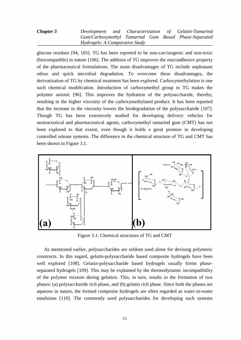
glucose residues [94, 105]. TG has been reported to be non-carcinogenic and non-toxic
(biocompatible) in nature [106]. The addition of TG improves the mucoadhesive property
of the pharmaceutical formulations. The main disadvantages of TG include unpleasant
odour and quick microbial degradation. To overcome these disadvantages, the
derivatization of TG by chemical treatment has been explored. Carboxymethylation is one
such chemical modification. Introduction of carboxymethyl group in TG makes the
polymer anionic [96]. This improves the hydration of the polysaccharide, thereby,
resulting in the higher viscosity of the carboxymethylated product. It has been reported
that the increase in the viscosity lowers the biodegradation of the polysaccharide [107].
Though TG has been extensively studied for developing delivery vehicles for
neutraceutical and pharmaceutical agents, carboxymethyl tamarind gum (CMT) has not
been explored to that extent, even though it holds a great promise in developing
controlled release systems. The difference in the chemical structure of TG and CMT has
been shown in Figure 3.1.
Figure 3.1: Chemical structures of TG and CMT
As mentioned earlier, polysaccharides are seldom used alone for devising polymeric
constructs. In this regard, gelatin-polysaccharide based composite hydrogels have been
well explored [108]. Gelatin-polysaccharide based hydrogels usually forms phase-
separated hydrogels [109]. This may be explained by the thermodynamic incompatibility
of the polymer mixture during gelation. This, in turn, results in the formation of two
phases: (a) polysaccharide rich phase, and (b) gelatin rich phase. Since both the phases are
aqueous in nature, the formed composite hydrogels are often regarded as water-in-water
emulsions [110]. The commonly used polysaccharides for developing such systems
Chapter 3 Development and Characterization of Gelatin-Tamarind
Gum/Carboxymethyl Tamarind Gum Based Phase-Separated
Hydrogels: A Comparative Study
16
include (but not limited to) starch, soluble starch, hydrated starch, carboxymethyl starch,
carboxymethyl cellulose dextran and maltodextan. No reports were found to study the
properties of gelatin- TG and gelatin- CMT phase-separated hydrogels.
In the current study, an in-depth analysis was done to optimize the gelatin
concentration and it was found that gelatin concentration more than 20% is difficult to
handle due to higher viscosity [111]. So, the gelatin concentration was fixed to 20% for
all the formulations. As per the literature study, 3 % carboxymethyl tamarind gum was
used (Sanyasi et al. 2013) to develop the hydrogels, which showed a better proliferation
of osteo-precursor cells [39]. Mishra et al. (2011) has reported that tamarind gum is a
suitable material for grafting process. In their study, concentration of TG was fixed to 1%
and a greater thermal stability was observed in the developed formulations [112]. So, the
polysaccharide solution was prepared with two different concentrations (1% and 2 %) to
develop the hydrogels. Further, different proportions of gelatin and TG/CMT (5:0, 4:1,
3:2, 2:3 and 1:4) were taken to develop the hydrogels and films. From all the
compositions, the best compositions were selected out as per their stability. The
formulation 1:4 was not formed due to lower proportion of gelatin. So, all the hydrogels
and films were selected by verifying their mechanical strength and stability. Optimization
process facilitated choosing the best compositions for the hydrogels and films for their
tissue engineering application. Taking a note from the above, we have developed the
hydrogels and films by altering both the concentrations and proportions of the polymer
and polysaccharides.
Taking inspiration from the above, we have tried to develop gelatin-TG and gelatin-
CMT based phase-separated hydrogels. The physicochemical, thermal and
electrochemical properties of the hydrogels were thoroughly characterized using FTIR
spectroscopy, differential scanning calorimetry, static mechanical tester and impedance
analyzer. The biological activity of the hydrogels were studied by mucoadhesive and
biocompatibility (hemocompatibility and cell viability assay) studies. To understand the
ability of the developed hydrogels as vehicles for controlled release, the hydrogels were
loaded with ciprofloxacin (fluroquinolone antibiotic). The drug release kinetics and the
antimicrobial activity of the drug loaded hydrogels were also studied in-depth.
3.2 Materials and Methods
3.2.1 Materials
TG and CMT (degree of carboxylation of CMT is 0.372) were procured from Maruti
hydrocolloids, India. Gelatin was procured from Himedia, Mumbai, India. Ciprofloxacin
Chapter 3 Development and Characterization of Gelatin-Tamarind
Gum/Carboxymethyl Tamarind Gum Based Phase-Separated
Hydrogels: A Comparative Study
17
(CF) was procured from Fluka Biochemical, China. Ethanol was obtained from Honyon
International Inc., Hong Yang Chemical Corporation, China. Glutaraldehyde (25%, for
synthesis; GA) and hydrochloric acid (35% pure) were obtained from Merck Specialities
Private Limited Mumbai, India. Goat intestine and blood were collected from the local
butcher shop. Double distilled water (DW) was used throughout the study.
3.2.2 Preparation of the formulations
Stock solution of gelatin (20% w/w) and polysaccharides (2% w/w) were freshly
prepared. The stock solutions were maintained at 50 oC. The gelatin and polysaccharide
solutions were mixed together (100 rpm, 10 min) in varying proportions (Table 3.1)
followed by addition of crosslinking reagent (0.5 ml of GA, 0.5 ml of ethanol, and 0.01
ml of 0.1N HCl). The mixture was mixed for 10 sec and subsequently poured into petri-
plates/cylindrical moulds. The petri-plates/moulds were incubated at room temperature
(25 oC) for 1 h to induce gelation.
Drug loaded hydrogels were prepared by dispersing 0.1 g of ciprofloxacin in gelatin
solution. Ciprofloxacin containing gelatin solution was used for the preparation of the
hydrogels. Rest of the process remained same. The final concentration of the drug in the
hydrogels was 0.5 % w/w. The hydrogels were washed thoroughly using PBS buffer and
double distilled water before all the experiments. Further, glycine solution (1% w/v) was
used to inhibit the chemical reactions of glutaraldehyde after the said incubation period.
Table 3.1: Composition of the hydrogels
Formulations Gelatin
solution (g)
TG
Solution
(g)
CMT
Solution
(g)
Crosslinker
(ml)
Ciproflaxacin
(g)
T1 16 4 -- 1 --
T2 12 8 -- 1 --
T3 8 12 -- 1 --
C1 16 -- 4 1 --
C2 12 -- 8 1 --
C3 8 -- 12 1 --
T1C 15.9 4 -- 1 0.1
T2C 11.9 8 -- 1 0.1
T3C 7.9 12 -- 1 0.1
C1C 15.9 -- 4 1 0.1
C2C 11.9 -- 8 1 0.1
C3C 7.9 -- 12 1 0.1
Chapter 3 Development and Characterization of Gelatin-Tamarind
Gum/Carboxymethyl Tamarind Gum Based Phase-Separated
Hydrogels: A Comparative Study
18
3.2.3 Microscopy studies
The microstructures of the uncrosslinked physical formulations were visualized under
bright field microscope (LEICA-DM 750 equipped with ICC 50-HD camera, Germany).
The formulations were converted into thin smears over glass slides before visualization.
3.2.4 Infrared spectroscopy
The raw materials and the hydrogels were analyzed using FTIR spectrophotometer
((Alpha-E, Bruker, USA). The analysis was done in the wavenumber range of 4500 cm-1
to 450 cm-1
. The spectrophotometer was being operated in the ATR mode.
3.2.5 Thermal analysis
The thermal profiles of the raw materials and the dried hydrogels were tested using
differential scanning calorimeter (DSC 200 F3 Maia, Netzsch, Germany) in the
temperature range of 40 o
C to 400 oC under nitrogen atmosphere. The rate of thermal
scanning was 5 oC/min.
3.2.6 Mechanical Analysis
The mechanical properties of the hydrogels were tested using a static mechanical tester
(Stable Microsystems, TA-HD plus, U.K). The hydrogels were prepared in cylindrical
moulds. The height and diameter of the hydrogels was 20 mm and 15 mm, respectively.
This resulted in the L/D ratio of 1.33. The hydrogels were subjected to cyclic compression
and cyclic stress relaxation studies to understand the viscoelastic properties of the
hydrogels [113].
3.2.7 Impedance analysis
The electrical properties of the hydrogels were tested using an in-house built impedance
analyzer in the frequency range of 200 Hz - 20 KHz. The setup was used to determine the
V-I characteristic by altering the amplitude of the sinusoidal voltage signals. The
frequency of the sinusoidal signal was kept constant at 10 KHz.
Chapter 3 Development and Characterization of Gelatin-Tamarind
Gum/Carboxymethyl Tamarind Gum Based Phase-Separated
Hydrogels: A Comparative Study
19
3.2.8 Biological Characterization
The mucoadhesive property of the hydrogels was determined using static mechanical
tester (Stable Microsystems, TA-HD plus, U.K). Goat intestine was used as the
representative mucosal layer for the study. The goat intestine was collected in cold saline
from the local slaughter house. The intestines were longitudinally cut open and were
further cut into pieces of 1 cm x 1 cm. The intestinal pieces were attached onto the base of
the mechanical tester. Subsequently, the hydrogels (5 mm x 5 mm) were attached on the
30 mm flat probe using double sided acrylate tape. Thereafter, the flat probe was lowered
at a speed of 0.5 mm/sec and a force of 20 g was applied on mucosal surface for 10 sec to
promote adhesion between the hydrogels and the mucosal layer. The probe was then
retracted back at the same speed. The force required to separate the hydrogel from the
intestinal mucosal surface was noted as mucoadhesive force. The work of mucoadhesion
was calculated from the area under the curve of the force-time profile.
The biocompatibility of the hydrogels were estimated by hemocompatibility and cell
viability test. The hemocompatibility test dealt with the incubation of the hydrogels in
diluted goat blood. The percentage hemolysis of the goat blood was calculated from the
absorbance of the supernatant fluid obtained after centrifuging the goat blood containing
the hydrogel pieces.
=% H em o lys iss a m p le -v e
+ v e -v e
-
× 1 0 0-
O D O D
O D O D
(3.1)
where, ODsample = Absorbance of sample
OD-ve = Absorbance of –ve control
OD+ve = Absorbance of + ve control
The cytocompatibility of the hydrogels were determined using MG63 cells. The cells
were seeded in 96 well plates. 1x10⁴ cells were added in each well. 20 µl of leachants (of
hydrogels) was added in each well to understand the toxic effect of the leachants. The cell
viability was determined using MTT assay.
The qualitative drug release study was conducted by performing antimicrobial test
using disc diffusion method. E. coli was used as the test microorganism. Hydrogel
samples of 9 mm diameter were used for the analysis. The antimicrobial activity was
correlated with the zone of inhibition of the microbial growth.
3.2.9 Swelling studies
Chapter 3 Development and Characterization of Gelatin-Tamarind
Gum/Carboxymethyl Tamarind Gum Based Phase-Separated
Hydrogels: A Comparative Study
20
The swelling profile of the hydrogel was determined at pH 7.4 (phosphate buffer). The
weights of the hydrogels, immersed in the swelling media, were determined after an
interval of 15 min for the first 1 h and 30 min for the next 9 h. The study was conducted at
room temperature. The swelling index was calculated as per the following equation:
Swelling Index (SI) = T 0
T
W - W
W (3.2)
where, WT = Weight of sample at time T, and W0 = Dry weight of the sample before the start of the study.
3.2.10 Drug release studies
The drug release studies were carried out using accurately weighed hydrogel samples
(~350 mg). The hydrogels were put in dialysis tube containing 1 ml of dissolution media.
Both ends of the dialysis membrane were sealed using dialysis tube clips. The setup was
lowered in a beaker containing 50 ml of dissolution media, kept under stirring at 100 rpm.
The temperature of the dissolution media was maintained at 37 oC. At regular intervals of
time, the dissolution media was replaced with fresh dissolution media for 12 h. The
replaced media was analyzed for the drug content using UV-visible spectrophotometer
(Systronics, Double beam spectrophotometer (2203), India). The study was conducted
using phosphate buffer (pH 7.4).
3.3 Result and Discussion
3.3.1 Preparation of hydrogels
Phase-separated hydrogels are a special class of mix polymer systems. In these hydrogels,
the polymers separate out (concentrate) as individual polymeric phases. The phase-
separation may happen in different ways due to inter- and intra- polymeric interactions.
Based on the interactions, the mix biopolymer system may form three types of molecular
architectures, namely, segregative phase-separation, associative phase-separation and
bicontinuous phase separation [105, 114]. Segregative phase-separation happens when the
two polymers have negative associative interactions. The affinity of the polymer towards
the solvent may also alter the molecular dynamics of the segregative phase-separation
process. This results in the formation of two phases which are enriched with either of the
polymers. The associative phase-separation has been reported to occur when the
interactions among the two polymers are very strong. This result in the separation of the
Chapter 3 Development and Characterization of Gelatin-Tamarind
Gum/Carboxymethyl Tamarind Gum Based Phase-Separated
Hydrogels: A Comparative Study
21
polymer-polymer composite (dispersed phase) and the solvent forms the continuous
phase. The biocontinous phase separated hydrogels are formed when both the polymer
phases appear as continuum phase. Quite often, many scientists have regarded this class
of phase-separated hydrogels as a specific category of segregative phase-separation [115].
Gelatin-polysaccharides based phase-separated systems have been reported to form
hydrogels by segregative phase-separation mechanism. These hydrogels are usually
chemically crosslinked to improve the physical stability. This is done because the
previous reports suggest that water-in-water type of emulsions have stability issues,
similar to the one confronted by the oil-water emulsions [116].
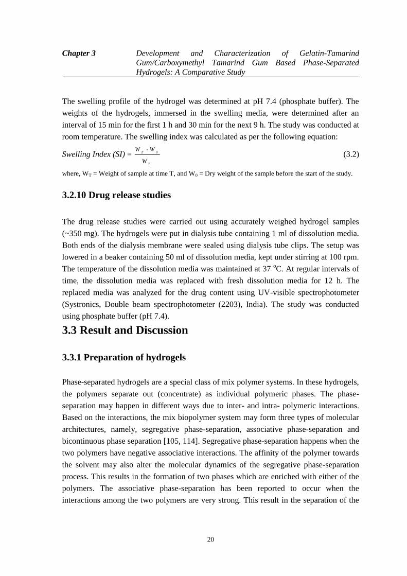
Figure 3.2: Pictographs of the hydrogels. (a) T1, (b) T2, (c) T3, (d) C1, (e) C2, and (f) C3
In this study, it was observed that an increase in the proportion of TG was associated
with the increase in the whiteness of the formulation (Figure 3.2). It has been previously
reported that the emulsions appears as white in color due to the diffraction of the light
from the interface of the internal and the continuum phases. This gives the indication that
there was a probability of formation of water-in-water emulsions. Similar observation was
also made in the gelatin-CMT hydrogels. The whiteness of the CMT hydrogels was lower
than the TG hydrogels. This observation may be explained by the fact that the
carboxymethylation of TG resulted in the increased hydrophilicity of the TG backbone.
The increase in the hydrophilicity might have improved the interaction amongst gelatin
and CMT. Hence, it may be expected that the degree of phase-separation will be lower as
compared to the TG hydrogels. Hydrogels were smooth to touch and had a soothing
effect.
Chapter 3 Development and Characterization of Gelatin-Tamarind
Gum/Carboxymethyl Tamarind Gum Based Phase-Separated
Hydrogels: A Comparative Study
22
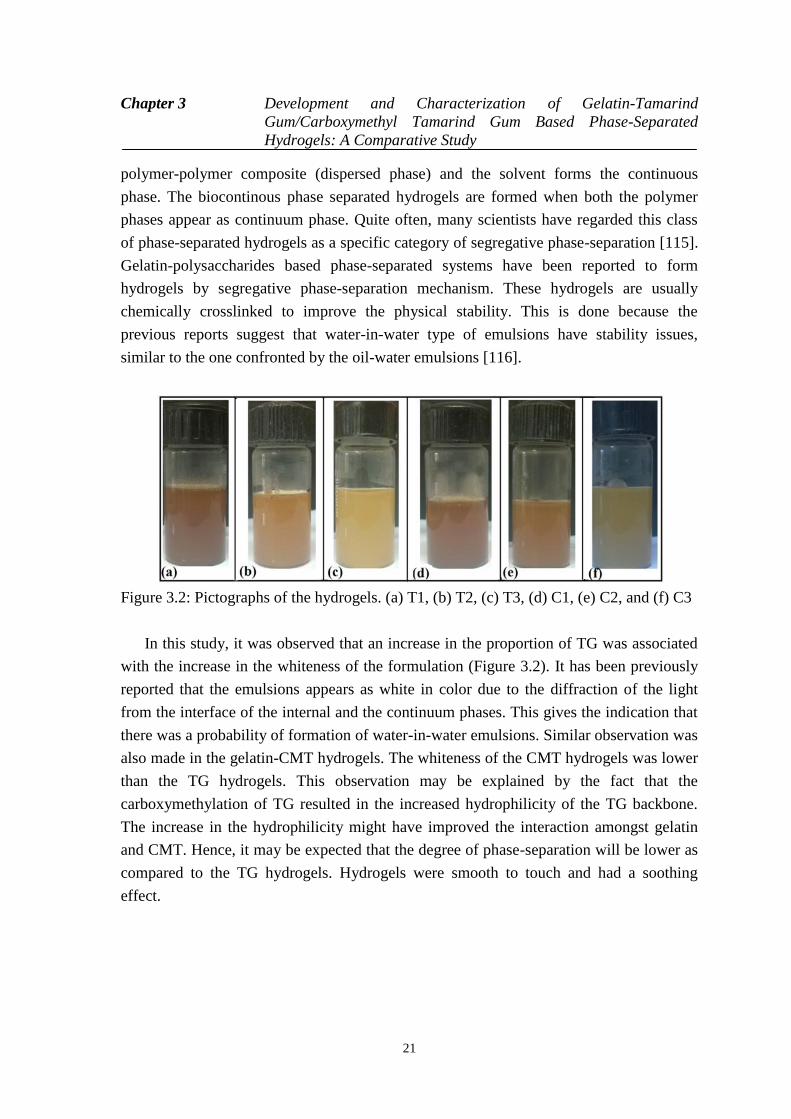
3.3.2 Microscopy
Figure 3.3: Light micrographs of the hydrogels. (a) T1, (b) T2, (c) T3, (d) C1, (e) C2, and
(F) C3
The gelatin polysaccharide mixture (50 oC) was converted into thin smears over a
glass slide. The smear was observed under the bright field microscope (Figure 3.3). The
micrographs of the formulation show homogeneous distribution of the globular
microstructure of polysaccharides within the gelatin continuum phase. The size of the
globular microstructures was found to increase as the concentration of the
polysaccharides was increased in the formulations. This may be due to the higher intra-
polysaccharide interactions during the gelation process. The globular size of the dispersed
phase was found to be higher in TG hydrogels. Similar results were expected from the
visual observation of the hydrogel matrices. The large globular size of the dispersed phase
in the TG hydrogels may be accounted to the higher intra-polysaccharide interactions.
Carboxymethylation of tamarind gum resulted in the formation of anionic polyelectrolyte,
which in turn, resulted in the increased hydration of the polysaccharides. This resulted in
the decrease in the intra-polysaccharide interactions with a subsequent increase in the
inter-polymer interactions. The decrease in the intra-polysaccharide interactions may be
explained by the ionization of the carboxylic groups of CMT. The ionization of the
carboxylic groups resulted in the steric hindrance, which in turn, hindered the process of
self-aggregation of CMT [117].
Chapter 3 Development and Characterization of Gelatin-Tamarind
Gum/Carboxymethyl Tamarind Gum Based Phase-Separated
Hydrogels: A Comparative Study
23
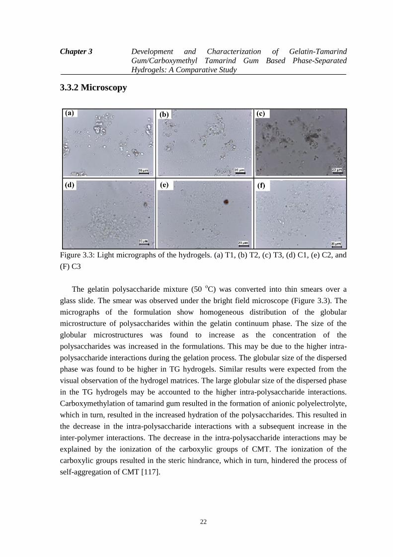
3.3.3 Infrared spectroscopy
Figure 3.4: FTIR spectra of the hydrogels. (a) TG hydrogels, and (b) CMT hydrogels
The FTIR spectra of hydrogels were acquired in the ATR mode (Figure 3.4, Table
3.2). The spectra of all the hydrogels were found to be similar with that of gelatin alone
hydrogel as reported in the literature [118]. Gelatin is a protein molecule. The gelatin
hydrogel showed a broad peak at ~3320 cm-1
. This peak may be associated with the O-H
and N-H stretching vibrations. The peak at ~1650 cm-1
can be explained by C-O and C-N
stretching of the amide bonds. The peak at ~1550 cm1 may be associated with amide –II
bonds, whereas, the peak at ~1250 cm-1
may be attributed to amide III bonds. It has been
reported by various groups that the peak in the region of 1600-1700 cm-1
is an important
peak for the analysis of the secondary protein structures. Addition of TG to the gelatin
hydrogels did not shift the peak position of the gelatin in the hydrogels. This suggested
that there were no significant changes in the secondary structure of the gelatin. CMT in
lower proportions did not alter the peak position at ~1629 cm-1
but at higher concentration
of CMT, there was a shift in the amide-I peak towards higher wavenumber. Such shift in
the amide-I peak have been previously explained by the interaction of the COO- groups
(present in polysaccharides) with the amide-I group of the gelatin [118]. Additionally, the
Chapter 3 Development and Characterization of Gelatin-Tamarind
Gum/Carboxymethyl Tamarind Gum Based Phase-Separated
Hydrogels: A Comparative Study
24
extent of hydrogen bonding among the polysaccharide containing hydrogels were
estimated by determining the area under the curve of the peak present in the region of
3700-2900 cm-1
. The area under the curve of the TG hydrogels were found to be in the
order of T1>>T2>>T3. On the other hand, the area under the curve for the CMT
containing hydrogels was found to be higher for the formulations containing higher
amount of CMT. In general, the area under the curve of the CMT hydrogels was higher
than the TG hydrogels. The above result may be explained by the relatively hydrophobic
nature of TG. An increase in the TG concentration within the formulation resulted in the
predominant hydrophobic interaction among the TG molecules. This, in turn, affected the
inter-molecular interactions amongst the TG and the gelatin molecules. On the other hand,
CMT molecules interacted with the gelatin molecules due to the availability of the free
carboxylic groups.
Table 3.2: FTIR peaks of the hydrogels
Formulations Amide (I)
(cm-1
)
Amide (II)
(cm-1
)
Amide (III)
(cm-1
)
AUC (Absorbance)
T1 1657 1562 1260 250.23
T2 1645 1560 1257 223.56
T3 1648 1560 1230 180.65
C1 1656 1563 1264 220.52
C2 1646 1539 1236 232.56
C3 1629 1535 1224 255.85
3.3.4 Thermal analysis
T1, T3, C1 and C3 were taken as the representative hydrogels for the thermal analysis.
The thermal profiles and the thermal parameters of the hydrogels have been given in
Figure 3.5 and Table 3.3, respectively. A broad peak was observed in the initial part of the
thermal profiles. This peak may be explained by the evaporation of the water molecules
from the hydrogels. The peak position was at ~100 oC for T1, whereas, it was at ~78
oC
for T3. The lowering of the endothermic peak temperature suggested that there might be a
reduction in the hydrogel-water interactions. This can be explained by the presence of the
hydrophobic TG in higher concentration in T3. The enthalpy and entropy of the TG
hydrogels were found to be higher when the concentration of the TG was more. This
suggested that the energy required to evaporate the water molecules present in the
hydrogels was higher when the concentration of polysaccharides was increased.
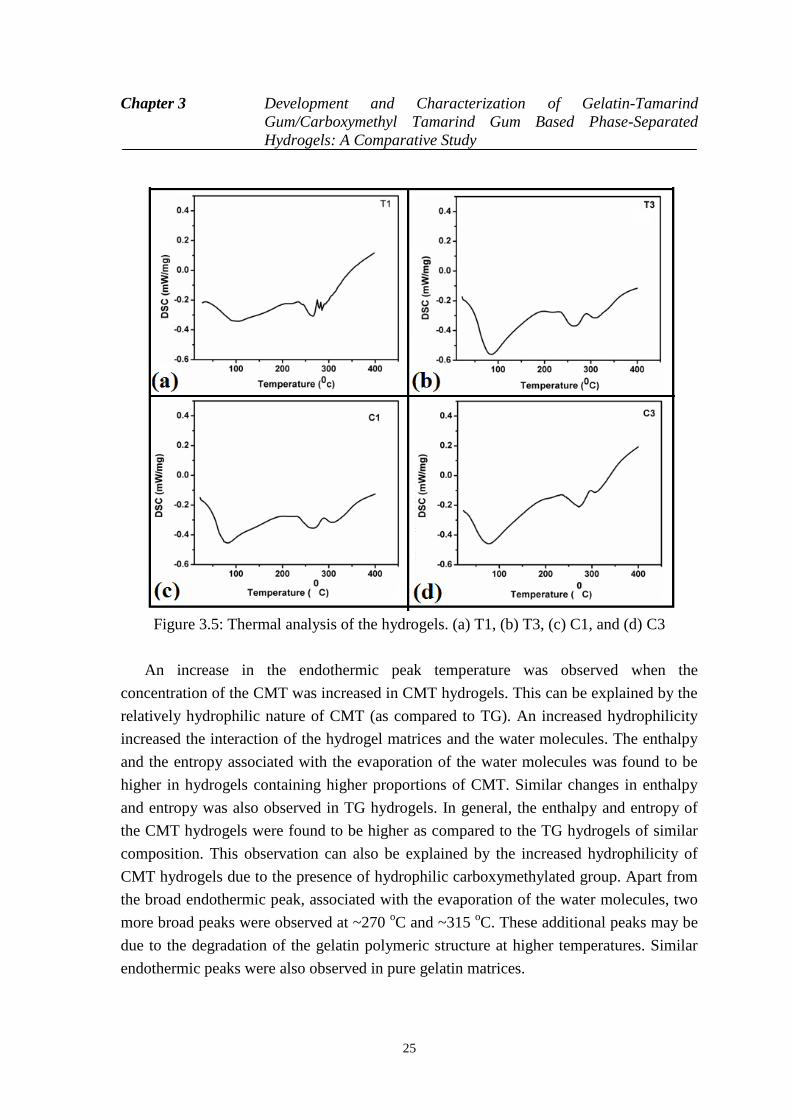
Chapter 3 Development and Characterization of Gelatin-Tamarind
Gum/Carboxymethyl Tamarind Gum Based Phase-Separated
Hydrogels: A Comparative Study
25
Figure 3.5: Thermal analysis of the hydrogels. (a) T1, (b) T3, (c) C1, and (d) C3
An increase in the endothermic peak temperature was observed when the
concentration of the CMT was increased in CMT hydrogels. This can be explained by the
relatively hydrophilic nature of CMT (as compared to TG). An increased hydrophilicity
increased the interaction of the hydrogel matrices and the water molecules. The enthalpy
and the entropy associated with the evaporation of the water molecules was found to be
higher in hydrogels containing higher proportions of CMT. Similar changes in enthalpy
and entropy was also observed in TG hydrogels. In general, the enthalpy and entropy of
the CMT hydrogels were found to be higher as compared to the TG hydrogels of similar
composition. This observation can also be explained by the increased hydrophilicity of
CMT hydrogels due to the presence of hydrophilic carboxymethylated group. Apart from
the broad endothermic peak, associated with the evaporation of the water molecules, two
more broad peaks were observed at ~270 oC and ~315
oC. These additional peaks may be
due to the degradation of the gelatin polymeric structure at higher temperatures. Similar
endothermic peaks were also observed in pure gelatin matrices.
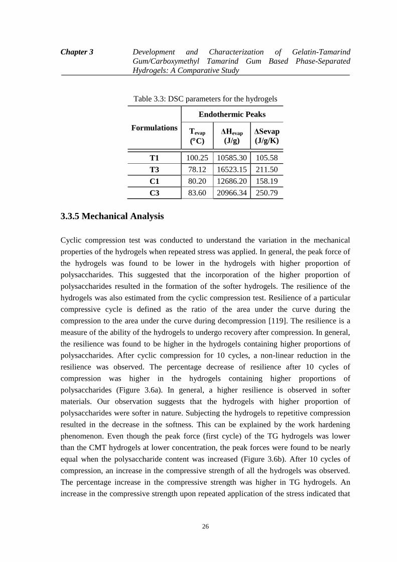
Chapter 3 Development and Characterization of Gelatin-Tamarind
Gum/Carboxymethyl Tamarind Gum Based Phase-Separated
Hydrogels: A Comparative Study
26
Table 3.3: DSC parameters for the hydrogels
Formulations
Endothermic Peaks
Tevap
(C)
ΔHevap
(J/g)
ΔSevap
(J/g/K)
T1 100.25 10585.30 105.58
T3 78.12 16523.15 211.50
C1 80.20 12686.20 158.19
C3 83.60 20966.34 250.79
3.3.5 Mechanical Analysis
Cyclic compression test was conducted to understand the variation in the mechanical
properties of the hydrogels when repeated stress was applied. In general, the peak force of
the hydrogels was found to be lower in the hydrogels with higher proportion of
polysaccharides. This suggested that the incorporation of the higher proportion of
polysaccharides resulted in the formation of the softer hydrogels. The resilience of the
hydrogels was also estimated from the cyclic compression test. Resilience of a particular
compressive cycle is defined as the ratio of the area under the curve during the
compression to the area under the curve during decompression [119]. The resilience is a
measure of the ability of the hydrogels to undergo recovery after compression. In general,
the resilience was found to be higher in the hydrogels containing higher proportions of
polysaccharides. After cyclic compression for 10 cycles, a non-linear reduction in the
resilience was observed. The percentage decrease of resilience after 10 cycles of
compression was higher in the hydrogels containing higher proportions of
polysaccharides (Figure 3.6a). In general, a higher resilience is observed in softer
materials. Our observation suggests that the hydrogels with higher proportion of
polysaccharides were softer in nature. Subjecting the hydrogels to repetitive compression
resulted in the decrease in the softness. This can be explained by the work hardening
phenomenon. Even though the peak force (first cycle) of the TG hydrogels was lower
than the CMT hydrogels at lower concentration, the peak forces were found to be nearly
equal when the polysaccharide content was increased (Figure 3.6b). After 10 cycles of
compression, an increase in the compressive strength of all the hydrogels was observed.
The percentage increase in the compressive strength was higher in TG hydrogels. An
increase in the compressive strength upon repeated application of the stress indicated that

Chapter 3 Development and Characterization of Gelatin-Tamarind
Gum/Carboxymethyl Tamarind Gum Based Phase-Separated
Hydrogels: A Comparative Study
27
there was an increase in the elastic component with a corresponding decrease in the
viscous component. This kind of phenomenon is often regarded as work hardening.
Figure 3.6: Mechanical studies of the hydrogels. (a) Resilience of TG and CMT
hydrogels, (b) Peak forces of TG and CMT hydrogels, (c) % Stress relaxation of TG and
CMT hydrogels, and (d) D20 values of TG and CMT hydrogels
The hydrogels were further subjected to cyclic stress relaxation (SR). The SR was
found to be higher in the first cycle. As the hydrogels were subjected to repeated SR
studies, a decrease in the SR was observed (Figure 3.6c). This indicated that during the
cyclic SR studies, there was an increase in the viscoelastic solid property of the material
with the subsequent decrease in the viscoelastic fluid nature. These results are concurrent
with the cyclic compression studies which also suggested an increase in the viscoelastic
solid component of the hydrogels as they were subjected to cyclic compression.
The firmness of the hydrogels was predicted by calculating the D20 values. D20 is regarded
as the distance travelled by the probe to attain a force of 20g. In general, the D20 values
are inversely proportional to the firmness of the hydrogels. This is due to the fact that the
firm hydrogels will resist the movement of the probe as compared to the softer hydrogels.
An increase in the D20 values was observed with an increase in the polysaccharide content
(Figure 3.6d). Amongst the 2 types of the hydrogels, CMT hydrogels showed a lower D20
values as compared to TG hydrogels. This suggested that the CMT hydrogels were
Chapter 3 Development and Characterization of Gelatin-Tamarind
Gum/Carboxymethyl Tamarind Gum Based Phase-Separated
Hydrogels: A Comparative Study
28
relatively more firm as compared to the TG hydrogels. This result may be correlated to
the extent of the H- bonding predicted from the area under the curve of the FTIR peak in
the region 3700 cm-1
- 2900 cm-1
. The FTIR results showed that the AUC were
comparatively higher in CMT hydrogels, suggesting a higher degree of hydrogen bonding
in CMT hydrogels.
To have a depth analysis on the relaxation processes of the hydrogels, the relaxation
profiles obtained from SR studies were fitted with Kohlrausch and Weichart model.
Kohlrausch model is a basic mathematical model which is used for the analysis of the
relaxation process of the polymeric constructs (Figure 3.7c). The ζ∞/ ζ0 (limiting stress)
values are denoted as residual elastic component at the end of the relaxation process
(Equation 3.3). The limiting stress values of the TG based hydrogels showed a decrease in
limiting stress values with an increase of the polysaccharides contents where CMT based
hydrogels showed no significant changes (Table 3.4). In the other hand, ζ1/ ζ0 (transient
stress) values of the hydrogels increased with the increase of TG content. The transient
stress of CMT based hydrogels has no significant changes like limiting stress values. The
relaxation time was same for all the samples.
t- β
ττ 1
0 0 0
σ σ σ= + × e
σ σ σ
(3.3)
where,
0
=limiting stress, 1
0
=transient stress, = relaxation time, =stretching parameter, t= time.
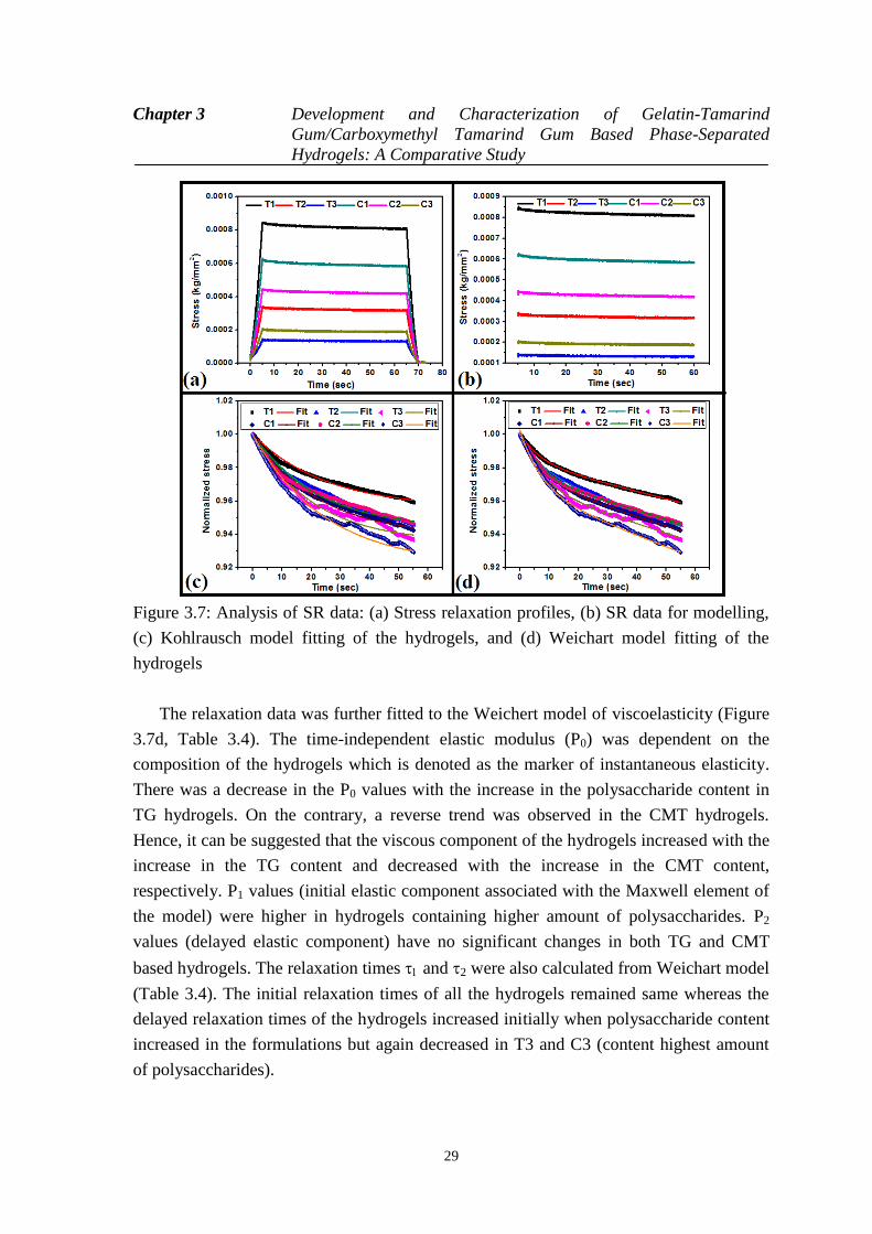
Chapter 3 Development and Characterization of Gelatin-Tamarind
Gum/Carboxymethyl Tamarind Gum Based Phase-Separated
Hydrogels: A Comparative Study
29
Figure 3.7: Analysis of SR data: (a) Stress relaxation profiles, (b) SR data for modelling,
(c) Kohlrausch model fitting of the hydrogels, and (d) Weichart model fitting of the
hydrogels
The relaxation data was further fitted to the Weichert model of viscoelasticity (Figure
3.7d, Table 3.4). The time-independent elastic modulus (P0) was dependent on the
composition of the hydrogels which is denoted as the marker of instantaneous elasticity.
There was a decrease in the P0 values with the increase in the polysaccharide content in
TG hydrogels. On the contrary, a reverse trend was observed in the CMT hydrogels.
Hence, it can be suggested that the viscous component of the hydrogels increased with the
increase in the TG content and decreased with the increase in the CMT content,
respectively. P1 values (initial elastic component associated with the Maxwell element of
the model) were higher in hydrogels containing higher amount of polysaccharides. P2
values (delayed elastic component) have no significant changes in both TG and CMT
based hydrogels. The relaxation times and 2 were also calculated from Weichart model
(Table 3.4). The initial relaxation times of all the hydrogels remained same whereas the
delayed relaxation times of the hydrogels increased initially when polysaccharide content
increased in the formulations but again decreased in T3 and C3 (content highest amount
of polysaccharides).
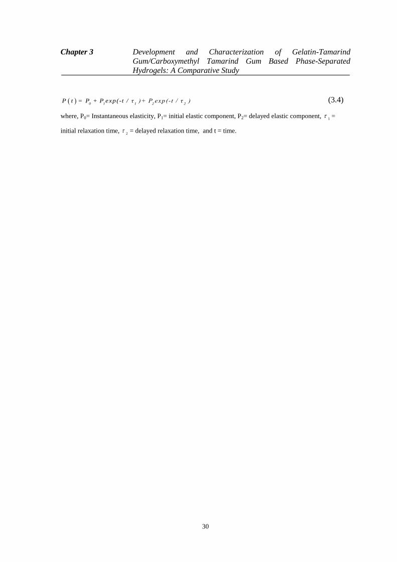
Chapter 3 Development and Characterization of Gelatin-Tamarind
Gum/Carboxymethyl Tamarind Gum Based Phase-Separated
Hydrogels: A Comparative Study
30
0 1 1 2 2P t = P + P exp(-t / τ )+ P exp(-t / τ ) (3.4)
where, P0= Instantaneous elasticity, P1= initial elastic component, P2= delayed elastic component, 1
=
initial relaxation time, 2
= delayed relaxation time, and t = time.
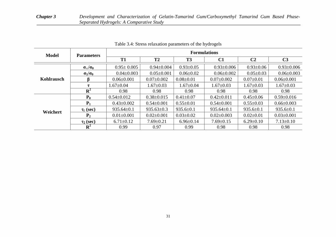
Chapter 3 Development and Characterization of Gelatin-Tamarind Gum/Carboxymethyl Tamarind Gum Based Phase-
Separated Hydrogels: A Comparative Study
31
Table 3.4: Stress relaxation parameters of the hydrogels
Model Parameters Formulations
T1 T2 T3 C1 C2 C3
Kohlrausch
σ∞/σ0 0.95 0.005 0.940.004 0.930.05 0.930.006 0.930.06 0.930.006
σ1/σ0 0.040.003 0.050.001 0.060.02 0.060.002 0.050.03 0.060.003
β 0.060.001 0.070.002 0.080.01 0.070.002 0.070.01 0.060.001
τ 1.670.04 1.670.03 1.670.04 1.670.03 1.670.03 1.670.03
R2 0.98 0.98 0.98 0.98 0.98 0.98
Weichert
P0 0.540.012 0.380.015 0.410.07 0.420.011 0.450.06 0.590.016
P1 0.430.002 0.540.001 0.550.01 0.540.001 0.550.03 0.660.003
(sec) 935.640.1 935.630.3 935.60.1 935.640.1 935.60.1 935.60.1
P2 0.010.001 0.020.001 0.030.02 0.020.003 0.020.01 0.030.001
(sec) 6.710.12 7.690.21 6.960.14 7.690.15 6.290.10 7.130.10
R2 0.99 0.97 0.99 0.98 0.98 0.98
Chapter 3 Development and Characterization of Gelatin-Tamarind
Gum/Carboxymethyl Tamarind Gum Based Phase-Separated
Hydrogels: A Comparative Study
32
3.3.6 Impedance Analysis
The impedance profiles of both the types of hydrogels were found to be similar. An
increase in TG content resulted in the increase in the impedance of the hydrogels (Figure
3.8a). This can be explained by the hydrophobic nature of the TG molecules, which
promoted the formation of predominant biphasic systems. The TG molecules in the
hydrogel system behaved as dielectric material. As the proportion of TG was increased,
there was a corresponding increase in the size of the dispersed phase molecules. This, in
turn, resulted in the increase in the dielectric behavior of the dispersed phase. In general,
higher the proportion of dielectric material, higher is the impedance. An increase in the
CMT concentration resulted in the decrease in the impedance of the CMT hydrogels
(Figure 3.8b). This observation may be explained by the polyelectrolyte nature of CMT.
An increase in the polysaccharide content resulted in the increase in the CMT
(polyelectrolyte) content in the hydrogels, which in turn, promoted the flow of current
through the hydrogel matrices. The impedance profile of the hydrogels showed higher
impedance in lower frequencies. The impedance exponentially degraded to a base level at
higher frequencies. This kind of impedance profile is usually associated with the
capacitive nature of the materials. The higher impedance at lower frequency may be
explained by the electrode polarization effect at lower frequencies. The polarization effect
is minimized when the frequency of the injecting current is increased. This is due to the
quick reversal of the electrode polarity at higher frequencies. This phenomenon prevents
the polarization effect at the electrode-sample interface [120].
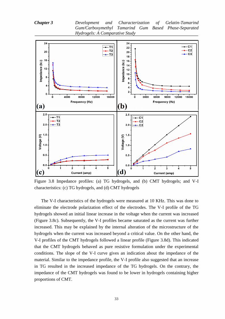
Chapter 3 Development and Characterization of Gelatin-Tamarind
Gum/Carboxymethyl Tamarind Gum Based Phase-Separated
Hydrogels: A Comparative Study
33
Figure 3.8 Impedance profiles: (a) TG hydrogels, and (b) CMT hydrogels; and V-I
characteristics: (c) TG hydrogels, and (d) CMT hydrogels
The V-I characteristics of the hydrogels were measured at 10 KHz. This was done to
eliminate the electrode polarization effect of the electrodes. The V-I profile of the TG
hydrogels showed an initial linear increase in the voltage when the current was increased
(Figure 3.8c). Subsequently, the V-I profiles became saturated as the current was further
increased. This may be explained by the internal alteration of the microstructure of the
hydrogels when the current was increased beyond a critical value. On the other hand, the
V-I profiles of the CMT hydrogels followed a linear profile (Figure 3.8d). This indicated
that the CMT hydrogels behaved as pure resistive formulation under the experimental
conditions. The slope of the V-I curve gives an indication about the impedance of the
material. Similar to the impedance profile, the V-I profile also suggested that an increase
in TG resulted in the increased impedance of the TG hydrogels. On the contrary, the
impedance of the CMT hydrogels was found to be lower in hydrogels containing higher
proportions of CMT.
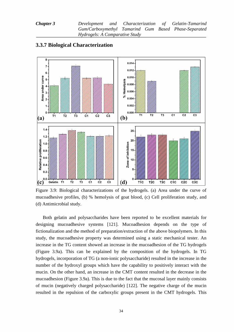
Chapter 3 Development and Characterization of Gelatin-Tamarind
Gum/Carboxymethyl Tamarind Gum Based Phase-Separated
Hydrogels: A Comparative Study
34
3.3.7 Biological Characterization
Figure 3.9: Biological characterizations of the hydrogels. (a) Area under the curve of
mucoadhesive profiles, (b) % hemolysis of goat blood, (c) Cell proliferation study, and
(d) Antimicrobial study.
Both gelatin and polysaccharides have been reported to be excellent materials for
designing mucoadhesive systems [121]. Mucoadhesion depends on the type of
fictionalization and the method of preparation/extraction of the above biopolymers. In this
study, the mucoadhesive property was determined using a static mechanical tester. An
increase in the TG content showed an increase in the mucoadhesion of the TG hydrogels
(Figure 3.9a). This can be explained by the composition of the hydrogels. In TG
hydrogels, incorporation of TG (a non-ionic polysaccharide) resulted in the increase in the
number of the hydroxyl groups which have the capability to positively interact with the
mucin. On the other hand, an increase in the CMT content resulted in the decrease in the
mucoadhesion (Figure 3.9a). This is due to the fact that the mucosal layer mainly consists
of mucin (negatively charged polysaccharide) [122]. The negative charge of the mucin
resulted in the repulsion of the carboxylic groups present in the CMT hydrogels. This

Chapter 3 Development and Characterization of Gelatin-Tamarind
Gum/Carboxymethyl Tamarind Gum Based Phase-Separated
Hydrogels: A Comparative Study
35
resulted in a negative interaction between the mucosal layer and the CMT hydrogels.
Amongst the hydrogels of similar composition, T1 showed lower mucoadhesive
properties than C1, whereas, T2 and C2 showed similar mucoadhesive properties. This
observation may be explained by the interaction of the polysaccharides with the gelatin
matrix.
The biocompatibility of the hydrogels was determined by hemocompatibility and
cytocompatibility tests. The hemocompatibility study is based on the determination of the
lysis of RBC in the presence of the hydrogels. The lysis of RBC results in the release of
the hemoglobin in the external fluid. The hemoglobin gets solubilized in the external fluid
and gives a yellowish color. The suspended unwanted components of the blood are
pelletized by centrifugation. The yellowish color of the supernatant fluid is measured
using a UV- visible spectrophotometer and is compared with the positive and negative
controls to determine the percentage hemolysis of the RBCs in the presence of the
hydrogels. All the hydrogel formulations were found to be highly hemocompatible as the
percentage hemolysis in the presence of hydrogels was <<5% (Figure 3.9b).
The cytocompatibility of the hydrogels was checked using MG63 cell line (osteoblast
cells). Extract of the hydrogels were prepared and were used for the analysis. The
experiment was done to test whether the leachants of the hydrogels have any adverse
effect on the proliferation of the cells. The relative proliferation of all the hydrogels was
> 1, which indicated the cytocompatible nature of the hydrogels (Figure 3.9c). It may also
be concluded that the prepared hydrogels supported the growth of MG63 cells to a certain
extent as compared to the control. In general, TG hydrogels showed better cell growth as
compared to CMT hydrogels.
The antimicrobial activity of the drug loaded hydrogels was estimated against E.
coli. The blank hydrogels did not show any zone of inhibition but samples loaded with the
antimicrobial drug showed a marked zone of inhibitions (Figure 3.9d). This suggested that
the blank hydrogels did not contain any antimicrobial activity, whereas, a marked
antimicrobial activity of the drug loaded hydrogels was evident. The result suggested that
the drug was released from the hydrogels matrices in active form.
3.3.8 Swelling studies
The swelling study was conducted at pH 7.4 (Figure 3.10). The swelling of the all the
hydrogels was found to be higher in the formulations containing higher proportion of
polysaccharides (TG/CMT). At pH 7.4, the carboxylic and amino groups existed as
zwitter ions. This resulted in the existence of the gelatin network as a neutral polymeric
architecture. Hence, the swelling of the matrices was dependent mainly on the absorption
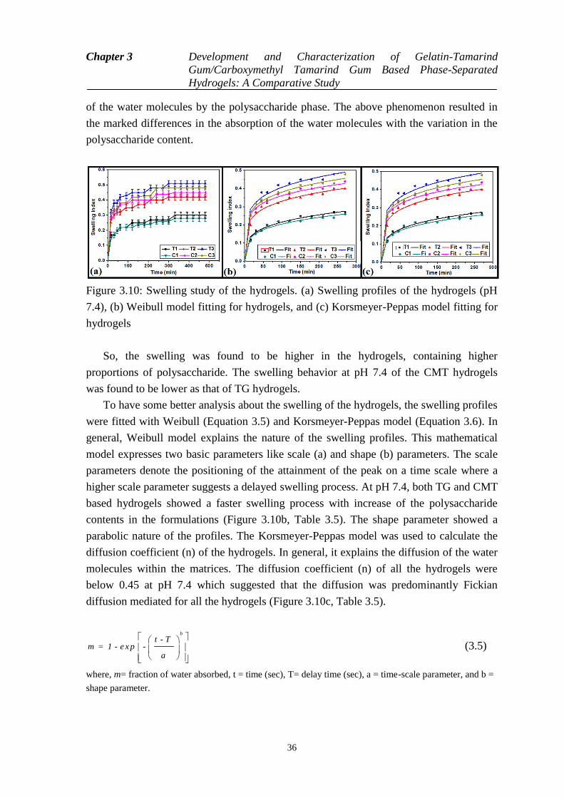
Chapter 3 Development and Characterization of Gelatin-Tamarind
Gum/Carboxymethyl Tamarind Gum Based Phase-Separated
Hydrogels: A Comparative Study
36
of the water molecules by the polysaccharide phase. The above phenomenon resulted in
the marked differences in the absorption of the water molecules with the variation in the
polysaccharide content.
Figure 3.10: Swelling study of the hydrogels. (a) Swelling profiles of the hydrogels (pH
7.4), (b) Weibull model fitting for hydrogels, and (c) Korsmeyer-Peppas model fitting for
hydrogels
So, the swelling was found to be higher in the hydrogels, containing higher
proportions of polysaccharide. The swelling behavior at pH 7.4 of the CMT hydrogels
was found to be lower as that of TG hydrogels.
To have some better analysis about the swelling of the hydrogels, the swelling profiles
were fitted with Weibull (Equation 3.5) and Korsmeyer-Peppas model (Equation 3.6). In
general, Weibull model explains the nature of the swelling profiles. This mathematical
model expresses two basic parameters like scale (a) and shape (b) parameters. The scale
parameters denote the positioning of the attainment of the peak on a time scale where a
higher scale parameter suggests a delayed swelling process. At pH 7.4, both TG and CMT
based hydrogels showed a faster swelling process with increase of the polysaccharide
contents in the formulations (Figure 3.10b, Table 3.5). The shape parameter showed a
parabolic nature of the profiles. The Korsmeyer-Peppas model was used to calculate the
diffusion coefficient (n) of the hydrogels. In general, it explains the diffusion of the water
molecules within the matrices. The diffusion coefficient (n) of all the hydrogels were
below 0.45 at pH 7.4 which suggested that the diffusion was predominantly Fickian
diffusion mediated for all the hydrogels (Figure 3.10c, Table 3.5).
b
t - Tm = 1 - e x p -
a
(3.5)
where, m= fraction of water absorbed, t = time (sec), T= delay time (sec), a = time-scale parameter, and b =
shape parameter.
Chapter 3 Development and Characterization of Gelatin-Tamarind
Gum/Carboxymethyl Tamarind Gum Based Phase-Separated
Hydrogels: A Comparative Study
37
nm k t (3.6)
where, k= rate constant, n= diffusion exponent
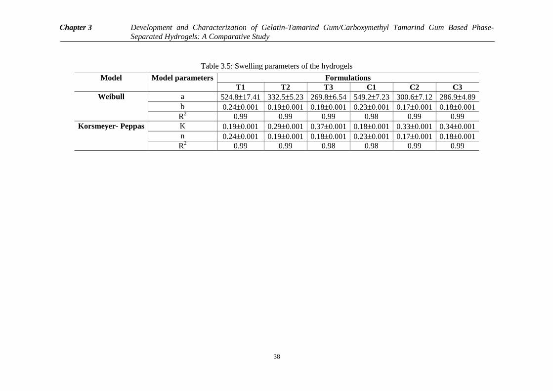
Chapter 3 Development and Characterization of Gelatin-Tamarind Gum/Carboxymethyl Tamarind Gum Based Phase-
Separated Hydrogels: A Comparative Study
38
Table 3.5: Swelling parameters of the hydrogels
Model
Model parameters Formulations
T1 T2 T3 C1 C2 C3
Weibull a 524.817.41 332.55.23 269.86.54 549.27.23 300.67.12 286.94.89
b 0.240.001 0.190.001 0.180.001 0.230.001 0.170.001 0.180.001
R2 0.99 0.99 0.99 0.98 0.99 0.99
Korsmeyer- Peppas
K 0.190.001 0.290.001 0.370.001 0.180.001 0.330.001 0.340.001
n 0.240.001 0.190.001 0.180.001 0.230.001 0.170.001 0.180.001
R2
0.99 0.99 0.98 0.98 0.99 0.99

Chapter 3 Development and Characterization of Gelatin-Tamarind
Gum/Carboxymethyl Tamarind Gum Based Phase-Separated
Hydrogels: A Comparative Study
39
3.3.9 Drug release study
The swelling studies of the hydrogels indicated a possibility of pH dependent drug release
from the phase-separated hydrogels. In general, the release of the drug from the hydrogels
is dependent on the swelling property of the hydrogels. Hence, to understand the pH
sensitive drug release behavior from the hydrogels, the drug release studies were
conducted at 7.4 (average intestinal pH) (Figure 3.11a). The release of the drug from the
TG hydrogels was higher from the hydrogels with higher TG content, i.e, cumulative
percentage of drug release (CPDR) was in the order of T1<T2<T3. The CPDR of the
CMT at pH 7.4 was also found to be in the order of C1<C2<C3. The observed result from
the phase-separated hydrogels can be correlated and well explained by the swelling
studies. In general, the release of the drugs from the hydrogel matrices depends on the
rate of diffusion of the dissolution media into the matrices. Higher flow of dissolution
media into the hydrogels resulted in the increased dissolution of the drug and
consequently increased diffusion of the drug from the polymer matrices.
To make a depth analysis of the drug release process, the drug releases were fitted
with Weibull (Equation 3.7), Korsmeyer-Peppas (Equation 3.8) and Peppas-sahlin
(Equation 3.9) models. The scale (a) and shape (b) parameters of the Weibull model
explain the nature of the release profiles. At ph 7.4, the release was slowest in T1 and C1.
But with an increase in the polysaccharide contents, the release became faster in the
hydrogels. The shape parameter showed a parabolic nature of the profiles. Subsequently,
the diffusion mechanism of the drug from the hydrogels was estimated by calculating the
diffusion value (n) using Korsmeyar-Peppas diffusion model (Figure 3.11c). The ‗n‘
values for the TG hydrogels (except T2) were found to be 0.45 at pH 7.4. This suggested
that the diffusion was predominantly Fickian Mediated for all the TG hydrogels (except
T2) at pH 7.4. Whereas the release of the drugs from the CMT based hydrogels was
Fickian diffusion mediated for all the samples (except C1) at pH 7.4. Pappas- Sahlin
model was used to predict the contribution of the polymer chain relaxation in the release
profile [123]. The constant K1 is associated with the Fickian diffusion, whereas, the
constant K2 is associated with the diffusion due to the polymer relaxation (Figure 3.11d).
At pH 7.4, constant K1 and K (from Korsmeyer-Peppas model) were of the similar trend.
This was quite expected because the Korsmeyer-Peppas model indicated that the diffusion
of the drug was predominantly Fickian diffusion mediated. The K2 values of all the films
were much lower. This suggested a least effect of polymer relaxation during the drug
release process (Table 3.6).
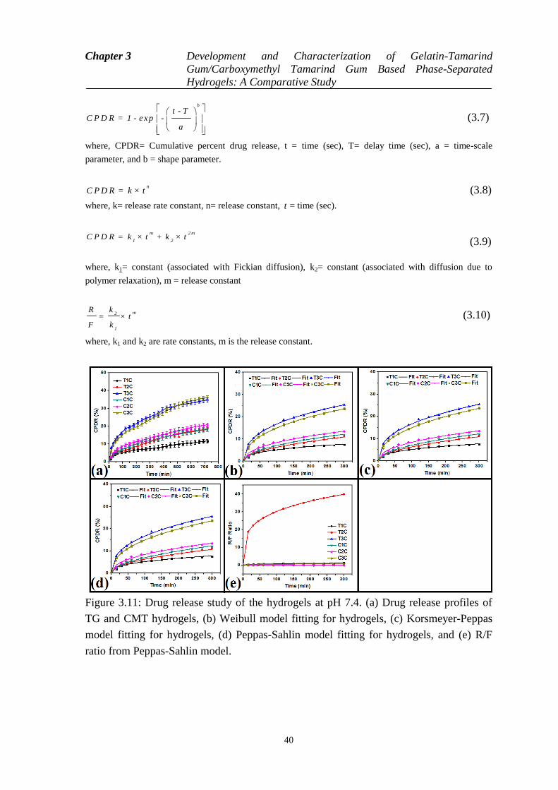
Chapter 3 Development and Characterization of Gelatin-Tamarind
Gum/Carboxymethyl Tamarind Gum Based Phase-Separated
Hydrogels: A Comparative Study
40
b
t - TC P D R = 1 - e x p -
a
(3.7)
where, CPDR= Cumulative percent drug release, t = time (sec), T= delay time (sec), a = time-scale
parameter, and b = shape parameter.
nC P D R = k × t (3.8)
where, k= release rate constant, n= release constant, t = time (sec).
m 2 m
1 2C P D R = k × t + k × t
(3.9)
where, k1= constant (associated with Fickian diffusion), k2= constant (associated with diffusion due to
polymer relaxation), m = release constant
m2
1
kR= × t
F k (3.10)
where, k1 and k2 are rate constants, m is the release constant.
Figure 3.11: Drug release study of the hydrogels at pH 7.4. (a) Drug release profiles of
TG and CMT hydrogels, (b) Weibull model fitting for hydrogels, (c) Korsmeyer-Peppas
model fitting for hydrogels, (d) Peppas-Sahlin model fitting for hydrogels, and (e) R/F
ratio from Peppas-Sahlin model.

Chapter 3 Development and Characterization of Gelatin-Tamarind Gum/Carboxymethyl Tamarind Gum Based Phase-
Separated Hydrogels: A Comparative Study
41
Table 3.6: Drug release parameters of the hydrogels
Model
Parameters Formulations
T1 T2 T3 C1 C2 C3
Weibull a 105.252.15 62.321.01 38.671.45 146.323.25 92.312.35 49.232.13
b 0.370.001 0.510.01 0.420.001 0.510.01 0.450.01 0.450.01
R2 0.99 0.98 0.99 0.99 0.99 0.98
Korsmeyer-
Peppas
K 0.960.03 0.640.02 2.840.06 0.720.01 1.130.03 2.200.04
n 0.360.001 0.490.01 0.380.001 0.490.01 0.430.001 0.410.001
R2 0.99 0.98 0.99 0.98 0.99 0.99
Peppas- Sahlin K1 0.950.02 0.680.01 2.280.05 0.830.03 1.140.04 2.220.05
K2 0.01 0.01 0.01 0.04 0.01 0.01
m 0.360.001 0.240.001 0.380.001 0.390.001 0.430.001 0.410.001
R2 0.99 0.99 0.98 0.98 0.99 0.99
Chapter 3 Development and Characterization of Gelatin-Tamarind
Gum/Carboxymethyl Tamarind Gum Based Phase-Separated
Hydrogels: A Comparative Study
42
3.4 Conclusion
The current study delineates the development of TG and CMT based phase-separated
hydrogels. In this study, gelatin was used as the second polymeric phase. An increase in
the hydrophilicity of TG by carboxymethylation resulted in the increase in the
compatibility of the polysaccharide and the gelatin phase. This resulted in the better
mechanical properties of the CMT based hydrogels. A higher proliferation of the MG63
cells was observed in the presence of the extracts of TG hydrogels. The CMT based
hydrogels also showed higher proliferation as compared to the controls. Both types of the
hydrogels also showed good mucoadhesive properties. Due the presence of the free
carboxylic groups in CMT, a pH sensitive swelling and drug release behavior from the
CMT hydrogels were observed.
43
Chapter 4
Preparation, characterization and
assessment of the novel gelatin-tamarind
gum/ carboxymethyl tamarind gum based
phase-separated films for skin tissue
engineering applications
4.1 Introduction
Protein and polysaccharide based formulations have been studied extensively for food and
pharmaceutical applications [124]. Depending upon the composition of the formulations,
there is a drastic change in the structural properties of the protein-polysaccharide
formulations. When the aqueous solution of the proteins and the polysaccharides are
mixed together, there is a possibility of formation of a liquid water-in-water emulsion,
complex coacervates (where both the polymers appears in a single concentrated phase)
and soluble complexes due to formation of self-organised formulations at different length
scales [125]. The formation of any of the above-mentioned formulations is governed by
the thermodynamic compatibility amongst the proteins and polysaccharides [126].
Recently, there has been an increased research on the development of protein-
polysaccharide based formulations. Various polysaccharides (starch, cellulose, pectin,
chitin, and glycogen) have been used either alone or in different combinations [127]. This
has resulted in the development of formulations of different properties.
Gelatin is an animal protein and is used for developing hydrogels for biomedical
applications (tissue engineering, drug delivery, wound healing) [128]. It is usually
Chapter 4 Preparation, characterization and assessment of the novel gelatin-
tamarind gum/ carboxymethyl tamarind gum based phase-
separated films for skin tissue engineering applications
44
obtained by controlled hydrolysis of collagen, obtained from the bovine bones (gelatin-B)
and porcine skins (gelatin-A) [129]. Gelatin is highly biocompatible, biodegradable and
has good mechanical properties which makes it suitable to be used for tissue engineering
and wound healing applications [130]. Due to its film-forming ability, gelatin has been
used to develop films. Gelatin films have been explored as wound dressing materials [29].
Tamarind gum (TG) is obtained from the outer layer of tamarind seed kernels
(Tamarindus indica) [131]. TG is a non-anionic branched polysaccharide and is soluble in
water. Solubilization of TG in water results in the formulation of a viscous solution [132].
Due to this reason, aqueous solution of TG is commonly used for thickening and gelling
purposes [133]. This gum has been approached for food applications in Japan [134]. The
main advantage of TG is its easy availability and low cost. The main drawback of TG is
its fast biodegradability. Hence, chemical modifications of TG have been proposed.
Amongst the various proposed chemical modifications, carboxymethylation of TG is most
common [96]. Unfortunately, due to very poor mechanical properties of TG, it has to be
blended with another polymer, which will provide better mechanical property.
Taking a note from the above, gelatin- TG/ CMT phase-separated films were
prepared. Polyethylene glycol (PEG, 400 molecular weight) was used as the plasticizer
[135]. The gelatin-polysaccharide based films were prepared in different concentrations
[136]. The films were thoroughly characterized by bright field microscopy, FTIR
spectroscopy, differential scanning calorimetry (DSC), mechanical analyzer and
impedance analyzer. The swelling properties of the films were tested in different pH
conditions (1.2 and 7.4). The biocompatibility of the films was established by
hemocompatibility (using goat blood) and cell viability (using HaCaT cells) studies. To
study the ability of the films as controlled delivery vehicles, the films were loaded with
ciprofloxacin. The release profiles of the drug-loaded films were determined at three
different pHs (1.2 and 7.4). The antimicrobial activities of the drug-loaded films were
tested against the E. coli as the model microorganism [137].
4.2 Materials and method
4.2.1 Materials
TG and CMT (degree of carboxylation is 0.372) were purchased from Maruti
Hydrocolloids, India. Gelatin was procured from Himedia, Mumbai, India. Ciprofloxacin
(CF) was purchased from Fluka Biochemical, China. Ethanol was obtained from Honyon
International Inc., Hong Yang Chemical Corporation, China. Glutaraldehyde (25%, for
synthesis; GA) and hydrochloric acid (35% pure) were obtained from Merck Specialities
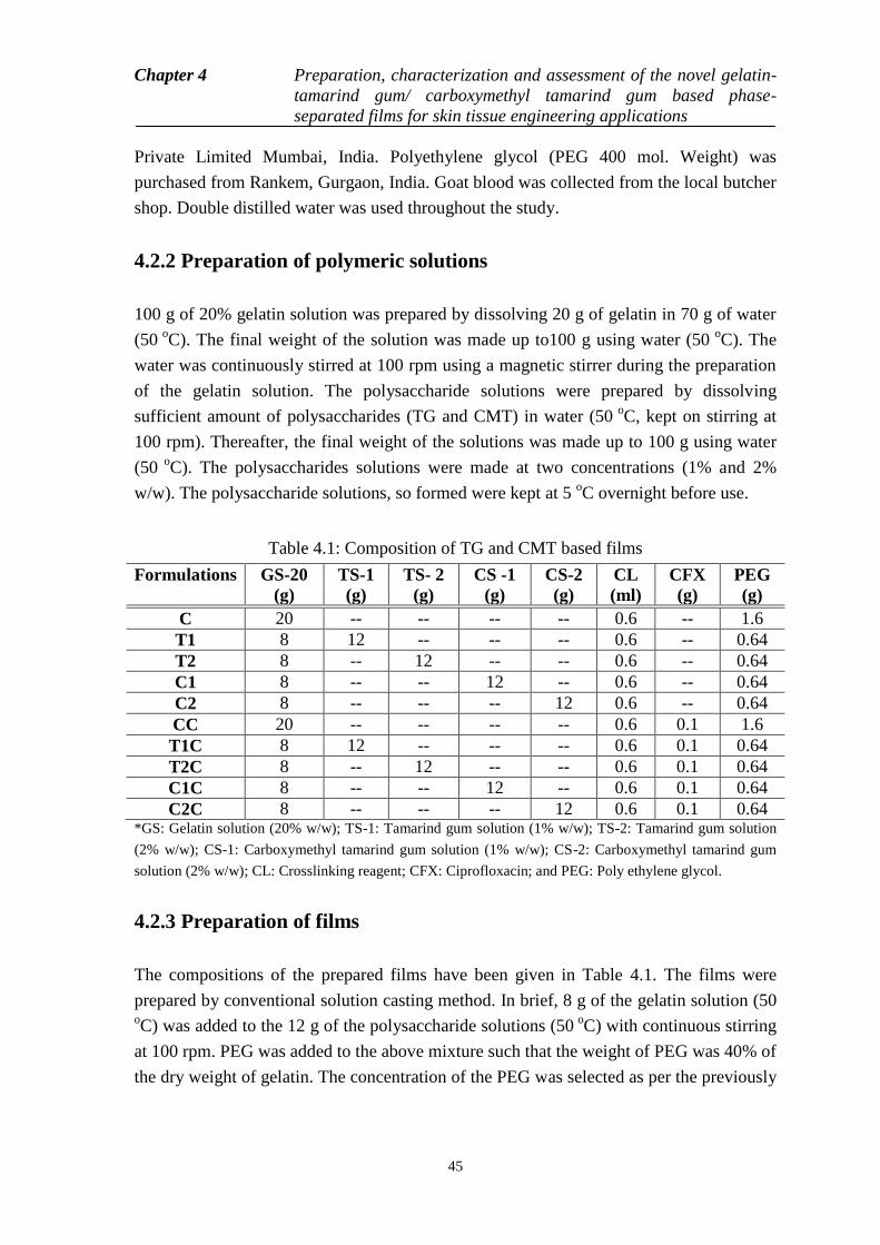
Chapter 4 Preparation, characterization and assessment of the novel gelatin-
tamarind gum/ carboxymethyl tamarind gum based phase-
separated films for skin tissue engineering applications
45
Private Limited Mumbai, India. Polyethylene glycol (PEG 400 mol. Weight) was
purchased from Rankem, Gurgaon, India. Goat blood was collected from the local butcher
shop. Double distilled water was used throughout the study.
4.2.2 Preparation of polymeric solutions
100 g of 20% gelatin solution was prepared by dissolving 20 g of gelatin in 70 g of water
(50 o
C). The final weight of the solution was made up to100 g using water (50 o
C). The
water was continuously stirred at 100 rpm using a magnetic stirrer during the preparation
of the gelatin solution. The polysaccharide solutions were prepared by dissolving
sufficient amount of polysaccharides (TG and CMT) in water (50 o
C, kept on stirring at
100 rpm). Thereafter, the final weight of the solutions was made up to 100 g using water
(50 o
C). The polysaccharides solutions were made at two concentrations (1% and 2%
w/w). The polysaccharide solutions, so formed were kept at 5 oC overnight before use.
Table 4.1: Composition of TG and CMT based films
Formulations GS-20
(g)
TS-1
(g)
TS- 2
(g)
CS -1
(g)
CS-2
(g)
CL
(ml)
CFX
(g)
PEG
(g)
C 20 -- -- -- -- 0.6 -- 1.6
T1 8 12 -- -- -- 0.6 -- 0.64
T2 8 -- 12 -- -- 0.6 -- 0.64
C1 8 -- -- 12 -- 0.6 -- 0.64
C2 8 -- -- -- 12 0.6 -- 0.64
CC 20 -- -- -- -- 0.6 0.1 1.6
T1C 8 12 -- -- -- 0.6 0.1 0.64
T2C 8 -- 12 -- -- 0.6 0.1 0.64
C1C 8 -- -- 12 -- 0.6 0.1 0.64
C2C 8 -- -- -- 12 0.6 0.1 0.64 *GS: Gelatin solution (20% w/w); TS-1: Tamarind gum solution (1% w/w); TS-2: Tamarind gum solution
(2% w/w); CS-1: Carboxymethyl tamarind gum solution (1% w/w); CS-2: Carboxymethyl tamarind gum
solution (2% w/w); CL: Crosslinking reagent; CFX: Ciprofloxacin; and PEG: Poly ethylene glycol.
4.2.3 Preparation of films
The compositions of the prepared films have been given in Table 4.1. The films were
prepared by conventional solution casting method. In brief, 8 g of the gelatin solution (50
oC) was added to the 12 g of the polysaccharide solutions (50
oC) with continuous stirring
at 100 rpm. PEG was added to the above mixture such that the weight of PEG was 40% of
the dry weight of gelatin. The concentration of the PEG was selected as per the previously
Chapter 4 Preparation, characterization and assessment of the novel gelatin-
tamarind gum/ carboxymethyl tamarind gum based phase-
separated films for skin tissue engineering applications
46
reported literature. Thereafter, the crosslinking agent (0.4 ml of ethyl alcohol, 0.1 ml of
glutaraldehyde, 0.01 ml of HCl) was added to the above mixture and stirred for 30 secs.
The petri plates were then put in an environmental chamber for 48 h at 40 o
C. The dried
films were collected from the environmental chamber at the end of 48 h and were kept at
5 oC in sealed polyethylene packets for the further studies. The drug loaded films were
prepared in a similar manner. Ciprofloxacin was added to the mixture of gelatin-
polysaccharide and PEG. Rest of the method remained same. The films were washed
thoroughly using PBS buffer and double distilled water before all the experiments.
Further, glycine solution (1% w/v) was used to inhibit the chemical reactions of
glutaraldehyde after the said incubation period.
4.2.4 Microscopy studies
The bright field micrographs of the films were observed under bright field microscope
(LEICA-DM 750 equipped with ICC 50-HD camera, Germany). The formulations were
converted into thin smears over glass slides before visualization.
4.2.5 Infrared spectroscopy
The IR spectra of the prepared films were measured using a FTIR spectrometer (AlpHa-E,
Bruker, USA) working in the ATR mode. The analysis was done in the wavenumber
range of 4500 cm-1
to 450 cm-1
. The ATR module was fitted with the ZnSe.
4.2.6 Thermal analysis
The thermal profiles of the films were analysed using differential scanning calorimeter
(DSC 200 F3 Maia, Netzsch, Germany) in the temperature range of 40 oC to 400
oC at a
scanning rate of 5 oC/min under inert atmosphere (nitrogen gas). The calibration was done
using indium. Nearly 10 mg of the sample was used for the analysis.
4.2.7 Mechanical analysis
The mechanical properties of the films were tested using a static mechanical tester (Stable
Microsystems, TA-HD plus, U.K). The samples were cut in rectangular pieces having
dimensions of 5 mm x 60 mm. The films were attached to the sample holder, such that the
sample length during the study was 50 mm. The tensile strength of the films was
determined by stretching the films at a rate of 1 mm/sec till the breaking point was
Chapter 4 Preparation, characterization and assessment of the novel gelatin-
tamarind gum/ carboxymethyl tamarind gum based phase-
separated films for skin tissue engineering applications
47
achieved. The bursting strength of the films was measured using a 5 mm spherical probe.
For the test, the samples were cut into 30 mm x 30 mm square pieces and attached to the
sample holder. Thereafter, the spherical probe was allowed to penetrate the sample at a
rate of 1 mm/sec till the bursting of the films was achieved. Stress relaxation (SR) was
done in the tensile mode. For Stress relaxation, the samples were stretched to 10 mm at
rate of 1 mm/sec and were subsequently allowed to relax for sixty seconds.
4.2.8 Impedance analysis
The electrical properties of the films were measured using an in-house built impedance
analyzer in the frequency range of 100 Hz and 20 KHz. The current-voltage relationship
(I-V characteristic) was measured at frequency of 1 KHz.
4.2.9 Biological characterizations
The hemolysis of the RBCs (goat blood) in the presence of the prepared films were
studied as per the reported literature [138]. The study provides information about the
hemocompatibility of the samples. For this study, fresh goat blood was collected in a
round bottom flask which contained tri-sodium citrate. The round bottom flask was
immediately transferred to an ice-bath. The collected blood was used within 1 h of
collection [139]. 8 ml of the blood was diluted with 10 ml of normal saline. The +ve
control of the test was made by adding 0.1 ml of HCl in a 15 ml falcon tube containing
0.5 ml of diluted blood. The volume was made up to 10 ml with normal saline. The -ve
control was prepared by diluting 0.5 ml of the dilute blood to 10 ml of normal saline. The
test sample was prepared by diluting 0.5 ml of blood to 10 ml using normal saline and
adding the pieces of the films (3 mm x 3 mm). Thereafter, all the falcon tubes were
incubated for 2 h in an incubator (37 oC). After the incubation, the falcon tubes were
centrifuged at 4000 rpm for 10 min, so as to allow the settling of the blood components.
The supernatant was spectrophotometrically analyzed using UV-visible
spectrophotometer. Percentage hemolysis was calculated as per equation 4.1.
% 1 0 0sa m p le v e
v e v e
O D O DH e m o ly s is
O D O D
(4.1)
where, ODsample = Absorbance of sample
OD –ve = Absorbance of –ve control
OD+ve = Absorbance of + ve control

Chapter 4 Preparation, characterization and assessment of the novel gelatin-
tamarind gum/ carboxymethyl tamarind gum based phase-
separated films for skin tissue engineering applications
48
For antimicrobial study, the films were cut as 9 mm discs. The discs were placed over
nutrient agar plates, previously inoculated with 100 l of E. coli suspension [139]. The
petri-plates were put in an incubator (37o C) for a period of 12 h. At the end of the
incubation, the zone of inhibition was measured.
The proliferative index of the HaCaT cells in the presence of test samples was studied
via MTT assay. The cells were maintained in complete DMEM media (10% FBS and 1%
antimycotic-antibiotic solution) at 37°C, 5% CO2. Wells of a sterile 96-well plate that
were pre-treated with solutions were let to set before inoculation of cells. Cells culture
flasks upon attaining 80% confluence were trypsinized for cell harvesting and 1 x 10⁴
cells were added to each well of the 96 well plate containing either TG or CMT films and
incubated for 24 hrs to ensure proper cell adhesion. MTT assay was carried out by adding
100µl of MTT reagent (MTT reagent and DMEM complete media in the ratio 1:10) to
each well for 4 h of incubation. After completion of incubation, the formazan crystals
formed were dissolved in 100 μl of DMSO. The absorbance of DMSO solution was then
measured at 595nm and the cell proliferation was represented in terms of cell proliferation
index (CPI).
4.2.10 Swelling studies
The water absorption profile of the films was measured at pH 7.4. For this study, the films
were cut into rectangular pieces of 3 mm x 5 mm size (initial weight: Wi) and the films
were put into the swelling medium. The films were taken out from the swelling media at
regular intervals, wiped using tissue paper to get rid of the free water and weighed
accurately (Wf). The swelling index was determined using the formula given in equation
4.2.
Swelling index (SI) = f i
i
-W W
W
(4.2)
where, Wf = Initial weight of the films
Wi = Final weight of the films
4.2.11 Drug release studies
The drug loaded films were cut into rectangular pieces (3mm x 5mm). The films were put
into dissolution media. At predefined intervals of time, the films were taken out from the
dissolution media. The previous dissolution media was spectrophotometrically analyzed
at 271 nm to determine the amount of the drug released. The experiment was conducted at
37 oC for 12 h.
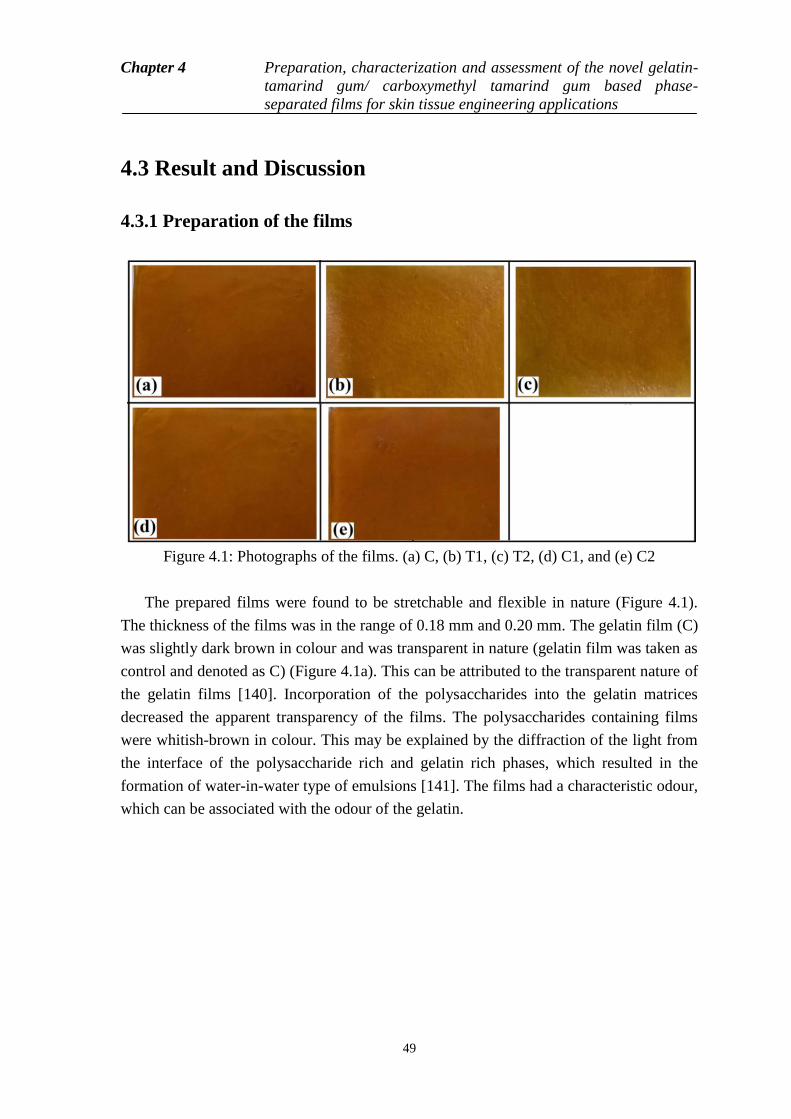
Chapter 4 Preparation, characterization and assessment of the novel gelatin-
tamarind gum/ carboxymethyl tamarind gum based phase-
separated films for skin tissue engineering applications
49
4.3 Result and Discussion
4.3.1 Preparation of the films
Figure 4.1: Photographs of the films. (a) C, (b) T1, (c) T2, (d) C1, and (e) C2
The prepared films were found to be stretchable and flexible in nature (Figure 4.1).
The thickness of the films was in the range of 0.18 mm and 0.20 mm. The gelatin film (C)
was slightly dark brown in colour and was transparent in nature (gelatin film was taken as
control and denoted as C) (Figure 4.1a). This can be attributed to the transparent nature of
the gelatin films [140]. Incorporation of the polysaccharides into the gelatin matrices
decreased the apparent transparency of the films. The polysaccharides containing films
were whitish-brown in colour. This may be explained by the diffraction of the light from
the interface of the polysaccharide rich and gelatin rich phases, which resulted in the
formation of water-in-water type of emulsions [141]. The films had a characteristic odour,
which can be associated with the odour of the gelatin.
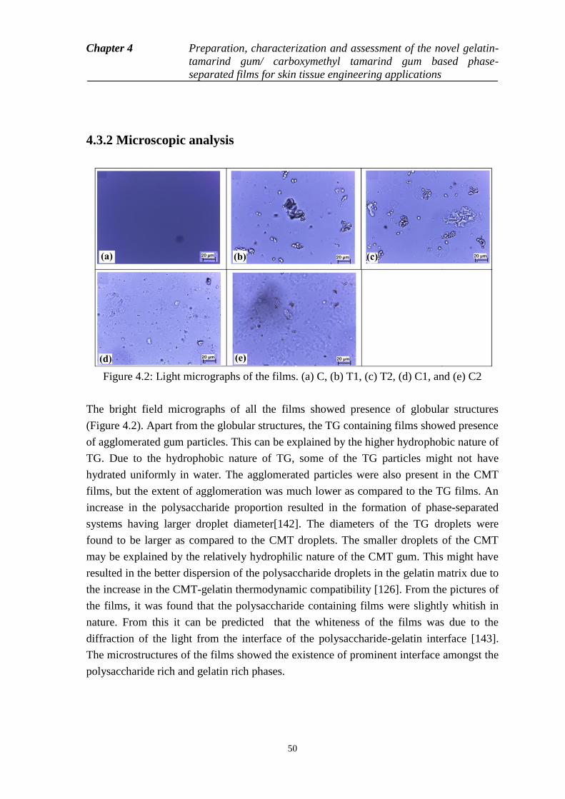
Chapter 4 Preparation, characterization and assessment of the novel gelatin-
tamarind gum/ carboxymethyl tamarind gum based phase-
separated films for skin tissue engineering applications
50
4.3.2 Microscopic analysis
Figure 4.2: Light micrographs of the films. (a) C, (b) T1, (c) T2, (d) C1, and (e) C2
The bright field micrographs of all the films showed presence of globular structures
(Figure 4.2). Apart from the globular structures, the TG containing films showed presence
of agglomerated gum particles. This can be explained by the higher hydrophobic nature of
TG. Due to the hydrophobic nature of TG, some of the TG particles might not have
hydrated uniformly in water. The agglomerated particles were also present in the CMT
films, but the extent of agglomeration was much lower as compared to the TG films. An
increase in the polysaccharide proportion resulted in the formation of phase-separated
systems having larger droplet diameter[142]. The diameters of the TG droplets were
found to be larger as compared to the CMT droplets. The smaller droplets of the CMT
may be explained by the relatively hydrophilic nature of the CMT gum. This might have
resulted in the better dispersion of the polysaccharide droplets in the gelatin matrix due to
the increase in the CMT-gelatin thermodynamic compatibility [126]. From the pictures of
the films, it was found that the polysaccharide containing films were slightly whitish in
nature. From this it can be predicted that the whiteness of the films was due to the
diffraction of the light from the interface of the polysaccharide-gelatin interface [143].
The microstructures of the films showed the existence of prominent interface amongst the
polysaccharide rich and gelatin rich phases.
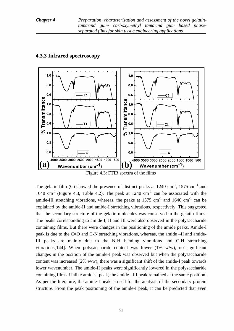
Chapter 4 Preparation, characterization and assessment of the novel gelatin-
tamarind gum/ carboxymethyl tamarind gum based phase-
separated films for skin tissue engineering applications
51
4.3.3 Infrared spectroscopy
Figure 4.3: FTIR spectra of the films
The gelatin film (C) showed the presence of distinct peaks at 1240 cm-1
, 1575 cm-1
and
1640 cm-1
(Figure 4.3, Table 4.2). The peak at 1240 cm-1
can be associated with the
amide-III stretching vibrations, whereas, the peaks at 1575 cm-1
and 1640 cm-1
can be
explained by the amide-II and amide-I stretching vibrations, respectively. This suggested
that the secondary structure of the gelatin molecules was conserved in the gelatin films.
The peaks corresponding to amide-I, II and III were also observed in the polysaccharide
containing films. But there were changes in the positioning of the amide peaks. Amide-I
peak is due to the C=O and C-N stretching vibrations, whereas, the amide –II and amide-
III peaks are mainly due to the N-H bending vibrations and C-H stretching
vibrations[144]. When polysaccharide content was lower (1% w/w), no significant
changes in the position of the amide-I peak was observed but when the polysaccharide
content was increased (2% w/w), there was a significant shift of the amide-I peak towards
lower wavenumber. The amide-II peaks were significantly lowered in the polysaccharide
containing films. Unlike amide-I peak, the amide –III peak remained at the same position.
As per the literature, the amide-I peak is used for the analysis of the secondary protein
structure. From the peak positioning of the amide-I peak, it can be predicted that even
Chapter 4 Preparation, characterization and assessment of the novel gelatin-
tamarind gum/ carboxymethyl tamarind gum based phase-
separated films for skin tissue engineering applications
52
though, there were no significant changes in the secondary protein structure of the gelatin
protein when polysaccharide content was lower. An increase in the polysaccharide
content to 2% has significantly affected the secondary protein structures of the gelatin.
The broad peak at around 3400 cm-1
can be associated with the stretching vibration of O-
H groups bonded with N-H groups [145]. The area under the curve provides information
about the extent of interactions. An increase in the TG content resulted in the decrease in
the area under the curve of the peak. This was due to the hydrophobic nature of TG which
resulted in the decrease in the interaction of the polar groups of the TG and the gelatin
molecules. On the contrary, due to presence of the carboxymethyl groups, an increased
interaction amongst the CMT and the gelatin molecules were observed (increase in the
area under curve with increase in the CMT content). The peak at 3400 cm-1
was also
observed in gelatin film (C). This peak can be explained by the intermolecular
interactions of the hydroxyl groups and the amino groups of the gelatin molecules and
intermolecular interaction amongst the hydroxyl groups of the PEG and the amino groups
of the gelatin molecules [146].
Table 4.2: FTIR peaks of the films
Formulations Amide (I)
(cm-1
)
Amide (II)
(cm-1
)
Amide (III)
(cm-1
)
AUC (Absorbance)
C 1640 1575 1240 167.2323
T1 1644 1548 1446 252.226
T2 1624 1531 1244 179.334
C1 1646 1536 1240 201.4654
C2 1626 1546 1242 211.9797
4.3.4 Thermal analysis
All the films showed a broad endothermic peak in the region of 75 oC and 90
oC. A
careful examination of the peaks suggested that the broad peak is a combination of two
endothermic peaks (Figure 4.4). In all the films, the second endothermic peak appeared as
a shoulder peak (demarcated by arrow marks). The presence of the dual peaks can be
explained by the evaporation of the water molecules from the films. In general, there are
two types of water molecules which are present in the biopolymeric films, namely free
water and bound water. The first peak is associated with the evaporation of the free water
from the films, whereas, the second peak is due to the evaporation of the bound water
molecules.

Chapter 4 Preparation, characterization and assessment of the novel gelatin-
tamarind gum/ carboxymethyl tamarind gum based phase-
separated films for skin tissue engineering applications
53
Figure 4.4: Thermal profiles of the films. (a) C, (b) T1, (c) T2, (d) C1, and (e) C2
The enthalpy of water evaporation (free water + bound water) was calculated from the
Netzsch proteus software. The enthalpies of the polysaccharide containing films were
much higher as compared to the control (Table 4.3). This can be explained by the better
water holding capacity of the polysaccharides. An increase in the polysaccharide content
correspondingly increased the enthalpy of water evaporation [147]. This indicated that the
water holding capacity of the films which contained higher amount of polysaccharides
were better as compared to the films, which contained lower amount of polysaccharides.
The enthalpy of evaporation of water molecule was slightly higher in C1 as compared to
T1 but when polysaccharide content was increased in T2 and C2, the enthalpy of T2 was
higher as compared to C2. This may be due to the fact that once the hydrophobic chains
of the TG were exposed; there was an interaction between the water molecules and the
hydrophobic domains present in the polysaccharide. This interaction resulted in the
absorption of more amount of water. The water which is absorbed due to the hydrophobic
domains is regarded as secondary bound water. On the contrary, the presence of the
carboxylic groups in CMT resulted in relative hydrophilic nature of the polysaccharide.
Hence, the interactions of the water molecules with the hydrophobic domains of the
polysaccharides were negligible. The above observation may be explained by the
significant increase in the enthalpy of the TG films and a minor increase in the CMT
films.
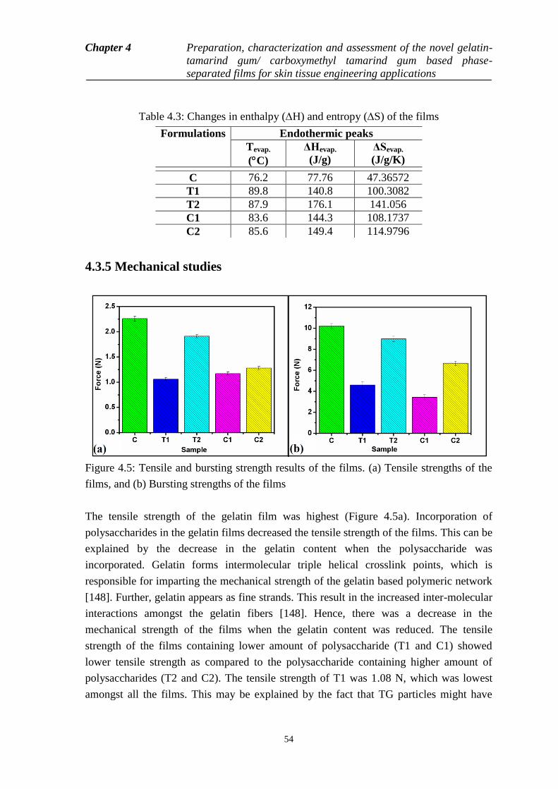
Chapter 4 Preparation, characterization and assessment of the novel gelatin-
tamarind gum/ carboxymethyl tamarind gum based phase-
separated films for skin tissue engineering applications
54
Table 4.3: Changes in enthalpy (H) and entropy (S) of the films
4.3.5 Mechanical studies
Figure 4.5: Tensile and bursting strength results of the films. (a) Tensile strengths of the
films, and (b) Bursting strengths of the films
The tensile strength of the gelatin film was highest (Figure 4.5a). Incorporation of
polysaccharides in the gelatin films decreased the tensile strength of the films. This can be
explained by the decrease in the gelatin content when the polysaccharide was
incorporated. Gelatin forms intermolecular triple helical crosslink points, which is
responsible for imparting the mechanical strength of the gelatin based polymeric network
[148]. Further, gelatin appears as fine strands. This result in the increased inter-molecular
interactions amongst the gelatin fibers [148]. Hence, there was a decrease in the
mechanical strength of the films when the gelatin content was reduced. The tensile
strength of the films containing lower amount of polysaccharide (T1 and C1) showed
lower tensile strength as compared to the polysaccharide containing higher amount of
polysaccharides (T2 and C2). The tensile strength of T1 was 1.08 N, which was lowest
amongst all the films. This may be explained by the fact that TG particles might have
Formulations Endothermic peaks
Tevap.
(C)
ΔHevap.
(J/g)
ΔSevap.
(J/g/K)
C 76.2 77.76 47.36572
T1 89.8 140.8 100.3082
T2 87.9 176.1 141.056
C1 83.6 144.3 108.1737
C2 85.6 149.4 114.9796
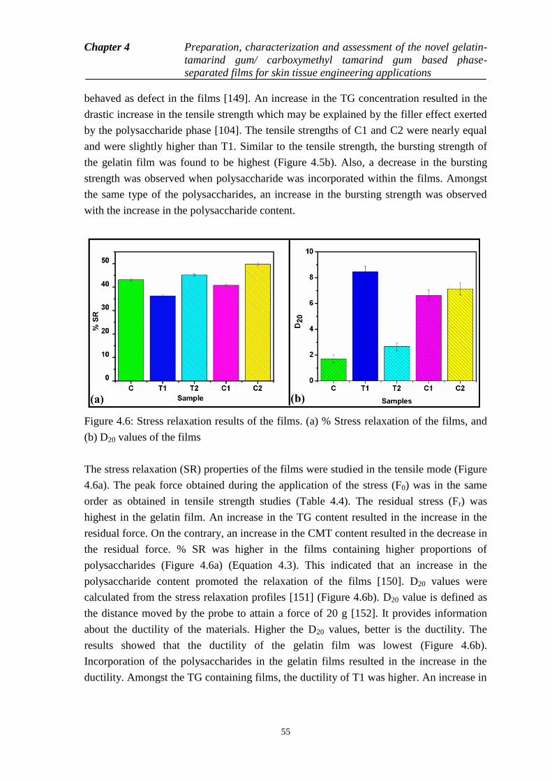
Chapter 4 Preparation, characterization and assessment of the novel gelatin-
tamarind gum/ carboxymethyl tamarind gum based phase-
separated films for skin tissue engineering applications
55
behaved as defect in the films [149]. An increase in the TG concentration resulted in the
drastic increase in the tensile strength which may be explained by the filler effect exerted
by the polysaccharide phase [104]. The tensile strengths of C1 and C2 were nearly equal
and were slightly higher than T1. Similar to the tensile strength, the bursting strength of
the gelatin film was found to be highest (Figure 4.5b). Also, a decrease in the bursting
strength was observed when polysaccharide was incorporated within the films. Amongst
the same type of the polysaccharides, an increase in the bursting strength was observed
with the increase in the polysaccharide content.
Figure 4.6: Stress relaxation results of the films. (a) % Stress relaxation of the films, and
(b) D20 values of the films
The stress relaxation (SR) properties of the films were studied in the tensile mode (Figure
4.6a). The peak force obtained during the application of the stress (F0) was in the same
order as obtained in tensile strength studies (Table 4.4). The residual stress (Fr) was
highest in the gelatin film. An increase in the TG content resulted in the increase in the
residual force. On the contrary, an increase in the CMT content resulted in the decrease in
the residual force. % SR was higher in the films containing higher proportions of
polysaccharides (Figure 4.6a) (Equation 4.3). This indicated that an increase in the
polysaccharide content promoted the relaxation of the films [150]. D20 values were
calculated from the stress relaxation profiles [151] (Figure 4.6b). D20 value is defined as
the distance moved by the probe to attain a force of 20 g [152]. It provides information
about the ductility of the materials. Higher the D20 values, better is the ductility. The
results showed that the ductility of the gelatin film was lowest (Figure 4.6b).
Incorporation of the polysaccharides in the gelatin films resulted in the increase in the
ductility. Amongst the TG containing films, the ductility of T1 was higher. An increase in
Chapter 4 Preparation, characterization and assessment of the novel gelatin-
tamarind gum/ carboxymethyl tamarind gum based phase-
separated films for skin tissue engineering applications
56
the TG content in T2 resulted in a drastic reduction in the ductility of the films. The
ductility of the CMT containing films was nearly equal and may be explained by the fact
that the compatibility amongst the CMT and the gelatin molecules were much better due
to introduction of the carboxymethyl groups in CMT.
0 r
0
F - F% S R = × 1 0 0
F (4.3)
where, F0= applied stress, Fr= residual stress.
The relaxation profiles (from SR studies) were fitted with Kohlrausch model (Figure 4.7c)
(Equation 4.4), the simplest model used for predicting the relaxation process of the
polymeric constructs [153]. It is an empirical relaxation function and is useful in
justifying the occurrence of common relaxation processes [154]. The ζ∞/ ζ0 (limiting
stress) values are an indicator the residual elastic component at the end of the relaxation
process. The limiting stress values suggested that the residual elastic component of gelatin
film (control), except C2, was lower than the polysaccharide containing films (Table 4.4).
The tamarind gum films showed higher limiting stress values as compared to
carboxymethyl tamarind gum films. An increase in the polysaccharide content lowered
the limiting stress of the films. This confirmed our prediction that the filler effect was
predominant in these films. An increase in the polysaccharide content (in T2 and C2)
drastically decreased the limiting stress values thereby confirming our prediction that at
higher polysaccharide content defects were incorporated within the films. Additionally, it
was observed that the limiting stress was better in the tamarind gum containing films as
compared to the carboxymethyl tamarind gum containing films. This observation may be
related to the hydrophilic nature of the carboxymethyl tamarind gum. The ζ1/ ζ0 (transient
stress) values of the gelatin film was higher (except C2) than the polysaccharide films. In
general, the relaxation of the gelatin film was faster as compared to the polysaccharide
containing films (except C2). The relaxation time was fastest in C2. This suggested a
predominant polymer relaxation process as compared to the polymer chain breakage
mediated relaxation process [153]. The stretching parameter (β) had values in the range of
0 and 1 suggesting a stretched decay processed.
t- β
ττ 1
0 0 0
σ σ σ= + × e
σ σ σ
(4.4)
where,
0
= limiting stress, 1
0
= transient stress, = relaxation time, = stretching parameter, t= time.
Chapter 4 Preparation, characterization and assessment of the novel gelatin-
tamarind gum/ carboxymethyl tamarind gum based phase-
separated films for skin tissue engineering applications
57
The normalized relaxation profiles (from SR studies) were further fitted to the Weichert
model of viscoelasticity (Figure 4.7d) (Equation 4.5) [153]. P0 values are a marker of the
instantaneous elasticity. The P0 values were in same order as that of the limiting stress
values (calculated from the Kohlrausch model). In general, the P0 values were higher than
P1 and P2 values. This suggested predominant elastic nature of the films. P1 values (initial
elastic component associated with the Maxwell element of the model) were higher in the
films with higher proportions of polysaccharides. The P2 (delayed elastic component)
values were higher than the P1 values in gelatin and films containing lower proportions of
polysaccharide (T1 and C1). At higher proportions of polysaccharides (T2 and C2), a
reverse trend was observed. The relaxation times were also calculated from the Weichert
model (Table 3.4). The initial relaxation time (1) was lowest in C2 followed by gelatin
film, T1, C1 and T2, respectively. An increase in the tamarind gum content delayed the
initial relaxation process. On the contrary, an increase in the carboxylmethyl tamarind
gum promoted initial relaxation process. This suggested that tamarind gum in higher
proportions delayed molecular rearrangement whereas carboxymethyl tamarind gum
promoted molecular rearrangement. Similar trend was also seen in the 2 (delayed
relaxation time) values for the polysaccharide containing films. Interestingly, the delayed
relaxation time value of gelatin film was highest and comparable with T2. The delayed
relaxation time provide information about the polymer-polymer interactions and the
breakage of the polymer chains, respectively [155]. The result suggested an increased
polymer-polymer interaction with a corresponding lower chance of polymer chain
breakage when tamarind gum content was increased (in T2). The gelatin film had similar
delayed relaxation time as that of T2. The delayed relaxation time of carboxylmethyl
tamarind gum films was not significantly different, suggesting that the variation in the
proportion of the carboxymethyl tamarind gum (within the experimental compositions)
did not affect the polymer-polymer interactions and chances of polymer chain breakage.
0 1 1 2 2exp( / ) exp( / )P t P P t P t (4.5)
where, P0= Instantaneous elasticity, P1= initial elastic component, P2= delayed elastic component, 1= initial
relaxation time, 2= delayed relaxation time, and t = time

Chapter 4 Preparation, characterization and assessment of the novel gelatin-
tamarind gum/ carboxymethyl tamarind gum based phase-
separated films for skin tissue engineering applications
58
Figure 4.7: Analysis of SR data: (a) Stress relaxation profiles, (b) SR data for modeling,
(c), Kohlrausch model fitting of the films, and (d) Weichart model fitting of the films
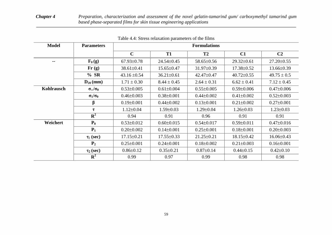
Chapter 4 Preparation, characterization and assessment of the novel gelatin-tamarind gum/ carboxymethyl tamarind gum
based phase-separated films for skin tissue engineering applications
59
Table 4.4: Stress relaxation parameters of the films
Model Parameters Formulations
C T1 T2 C1 C2
-- F0 (g) 67.930.78 24.540.45 58.650.56 29.320.61 27.200.55
Fr (g) 38.610.41 15.650.47 31.970.39 17.380.52 13.660.39
% SR 43.16 0.54 36.210.61 42.470.47 40.720.55 49.75 0.5
D20 (mm) 1.71 0.30 8.44 0.45 2.64 0.31 6.62 0.41 7.12 0.45
Kohlrausch σ∞/σ0 0.530.005 0.610.004 0.550.005 0.590.006 0.470.006
σ1/σ0 0.460.003 0.380.001 0.440.002 0.410.002 0.520.003
β 0.190.001 0.440.002 0.130.001 0.210.002 0.270.001
τ 1.120.04 1.590.03 1.290.04 1.260.03 1.230.03
R2 0.94 0.91 0.96 0.91 0.91
Weichert P0 0.530.012 0.600.015 0.540.017 0.590.011 0.470.016
P1 0.200.002 0.140.001 0.250.001 0.180.001 0.200.003
(sec) 17.150.21 17.550.33 21.250.21 18.150.42 16.060.43
P2 0.250.001 0.240.001 0.180.002 0.210.003 0.160.001
(sec) 0.860.12 0.350.21 0.870.14 0.440.15 0.420.10
R2 0.99 0.97 0.99 0.98 0.98
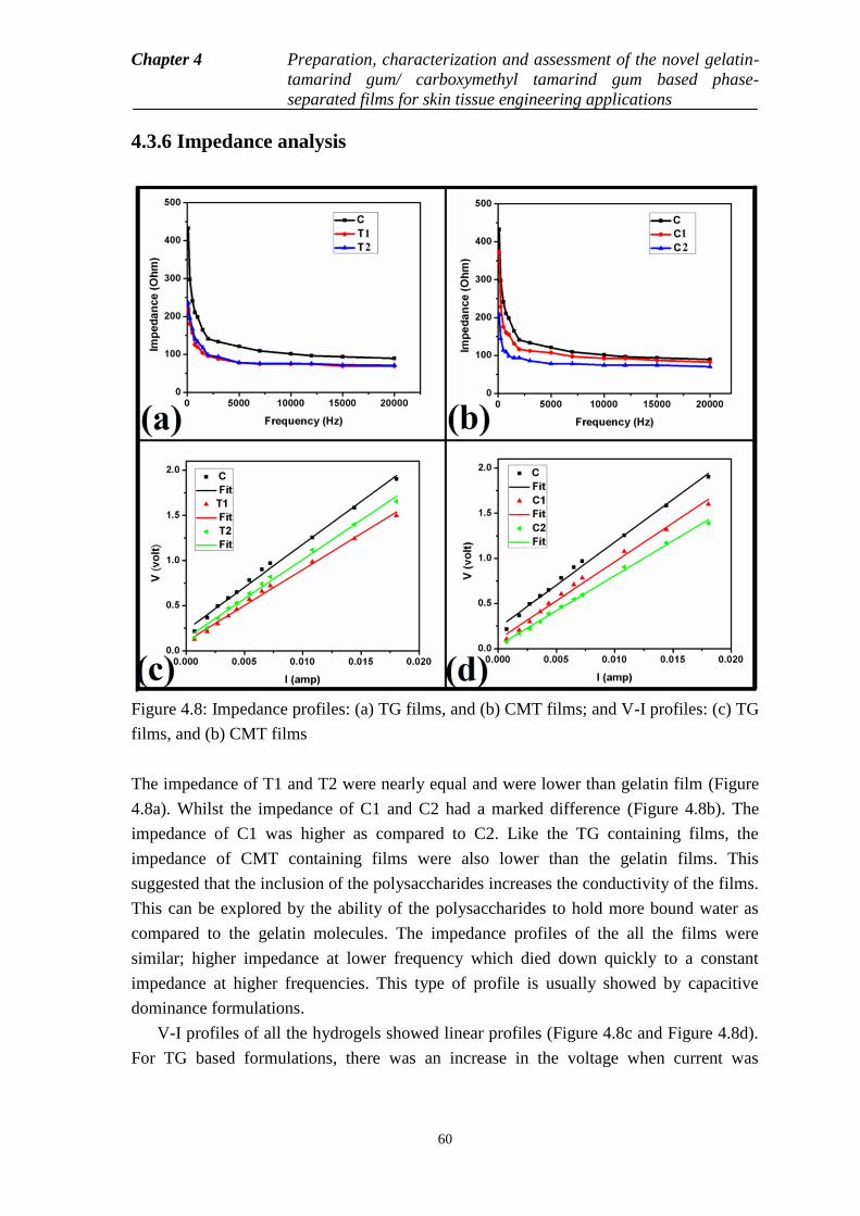
Chapter 4 Preparation, characterization and assessment of the novel gelatin-
tamarind gum/ carboxymethyl tamarind gum based phase-
separated films for skin tissue engineering applications
60
4.3.6 Impedance analysis
Figure 4.8: Impedance profiles: (a) TG films, and (b) CMT films; and V-I profiles: (c) TG
films, and (b) CMT films
The impedance of T1 and T2 were nearly equal and were lower than gelatin film (Figure
4.8a). Whilst the impedance of C1 and C2 had a marked difference (Figure 4.8b). The
impedance of C1 was higher as compared to C2. Like the TG containing films, the
impedance of CMT containing films were also lower than the gelatin films. This
suggested that the inclusion of the polysaccharides increases the conductivity of the films.
This can be explored by the ability of the polysaccharides to hold more bound water as
compared to the gelatin molecules. The impedance profiles of the all the films were
similar; higher impedance at lower frequency which died down quickly to a constant
impedance at higher frequencies. This type of profile is usually showed by capacitive
dominance formulations.
V-I profiles of all the hydrogels showed linear profiles (Figure 4.8c and Figure 4.8d).
For TG based formulations, there was an increase in the voltage when current was

Chapter 4 Preparation, characterization and assessment of the novel gelatin-
tamarind gum/ carboxymethyl tamarind gum based phase-
separated films for skin tissue engineering applications
61
increased with the simultaneous increase of the TG content in the formulations (T2>T1).
But both the films have lower values than the gelatin film. The linear profiles suggested
the formation of pure resistive formulations under the experimental condition. In case of
CMT based formulations, a reverse trend was followed. C1 showed a higher profile than
C2. But gelatin film has higher V-I values as compared to the all polysaccharide content
films.
4.3.7 Biological characterizations
Figure 4.9: Biological characterizations of the films. (a) Hemocompatibility, (b)
Antimicrobial study, and (c) Cell proliferation study using osteoblast cells
The study of the hemocompatibility of the wound dressing materials is an important study
as the materials are expected to be in contact with the blood cells at wound-site [156]. In
this study, the films were incubated in the presence of the goat blood cells and the degree
of disruption of the RBCs was determined. All the prepared films showed a hemolysis
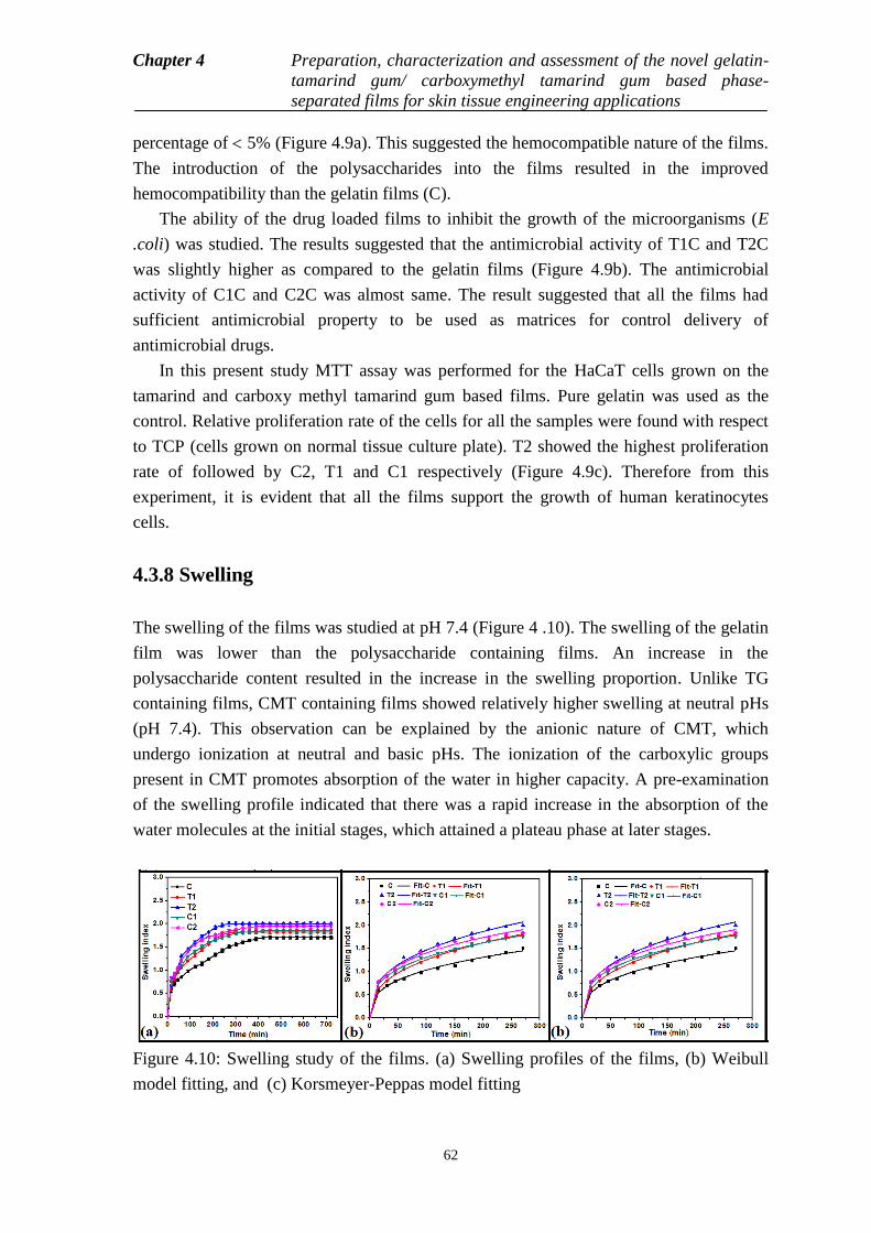
Chapter 4 Preparation, characterization and assessment of the novel gelatin-
tamarind gum/ carboxymethyl tamarind gum based phase-
separated films for skin tissue engineering applications
62
percentage of 5% (Figure 4.9a). This suggested the hemocompatible nature of the films.
The introduction of the polysaccharides into the films resulted in the improved
hemocompatibility than the gelatin films (C).
The ability of the drug loaded films to inhibit the growth of the microorganisms (E
.coli) was studied. The results suggested that the antimicrobial activity of T1C and T2C
was slightly higher as compared to the gelatin films (Figure 4.9b). The antimicrobial
activity of C1C and C2C was almost same. The result suggested that all the films had
sufficient antimicrobial property to be used as matrices for control delivery of
antimicrobial drugs.
In this present study MTT assay was performed for the HaCaT cells grown on the
tamarind and carboxy methyl tamarind gum based films. Pure gelatin was used as the
control. Relative proliferation rate of the cells for all the samples were found with respect
to TCP (cells grown on normal tissue culture plate). T2 showed the highest proliferation
rate of followed by C2, T1 and C1 respectively (Figure 4.9c). Therefore from this
experiment, it is evident that all the films support the growth of human keratinocytes
cells.
4.3.8 Swelling
The swelling of the films was studied at pH 7.4 (Figure 4 .10). The swelling of the gelatin
film was lower than the polysaccharide containing films. An increase in the
polysaccharide content resulted in the increase in the swelling proportion. Unlike TG
containing films, CMT containing films showed relatively higher swelling at neutral pHs
(pH 7.4). This observation can be explained by the anionic nature of CMT, which
undergo ionization at neutral and basic pHs. The ionization of the carboxylic groups
present in CMT promotes absorption of the water in higher capacity. A pre-examination
of the swelling profile indicated that there was a rapid increase in the absorption of the
water molecules at the initial stages, which attained a plateau phase at later stages.
Figure 4.10: Swelling study of the films. (a) Swelling profiles of the films, (b) Weibull
model fitting, and (c) Korsmeyer-Peppas model fitting
Chapter 4 Preparation, characterization and assessment of the novel gelatin-
tamarind gum/ carboxymethyl tamarind gum based phase-
separated films for skin tissue engineering applications
63
To have a depth analysis of the swelling kinetics of the films, the experimental data was
fitted to Weibull model (Equation 4.6) and Korsmeyer-Peppas (KP) model (Equation 4.7).
Weibulll model is a general model which helps in the analysis of the nature of the profiles
[157]. Scale (a) and shape (b) parameters of the swelling profiles were calculated from the
Weibull model (Table 4.6). The scale parameters indicate the positioning of the
attainment of the peak on a time scale. A higher scale parameter suggests a delayed
process. From the results, it was observed that the swelling was faster in the films with
higher amount of polysaccharides. The scale value of the gelatin film was highest (C)
which showed that the swelling was slowest in gelatin films. For both TG and CMT films,
the swelling was faster in higher polysaccharide content formulations (T2 and C2).
Amongst the polysaccharides containing films, an increase in the polysaccharide content
resulted in the increase in the diffusion coefficient (k values from KP model) of the water
molecules within the films. This, in turn, resulted in the faster absorption of the water
molecules within the film matrices. The shape parameter (b) calculated from the Weibull
model indicated a parabolic nature of the profiles. Diffusion exponent (n) value was
calculated from the Korsmeyer-Peppas model. The n-value gives an indication about the
nature of the diffusion of the water molecules within the matrices. At pH 7.4, the n-value
suggested that the diffusion of the water molecules in all the films except gelatin film (C)
was governed by anomalous diffusion (n value in the range of 0.45 to 0.89). This
suggested that the absorption of the water by films was due to the simultaneous
occurrence of diffusion of water molecules and relaxation of the polymeric chains during
the swelling process. The n value for C was lower than 0.45, this suggested the swelling
process was predominantly Fickian diffusion mediated.
b
t - Tm = 1 - e x p -
a
(4.6)
where, m= fraction of water absorbed, t= time (sec), T= delay time (sec), a= time-scale parameter, and b=
shape parameter.
nm = K × t (4.7)
where, k= rate constant, n= diffusion exponent.
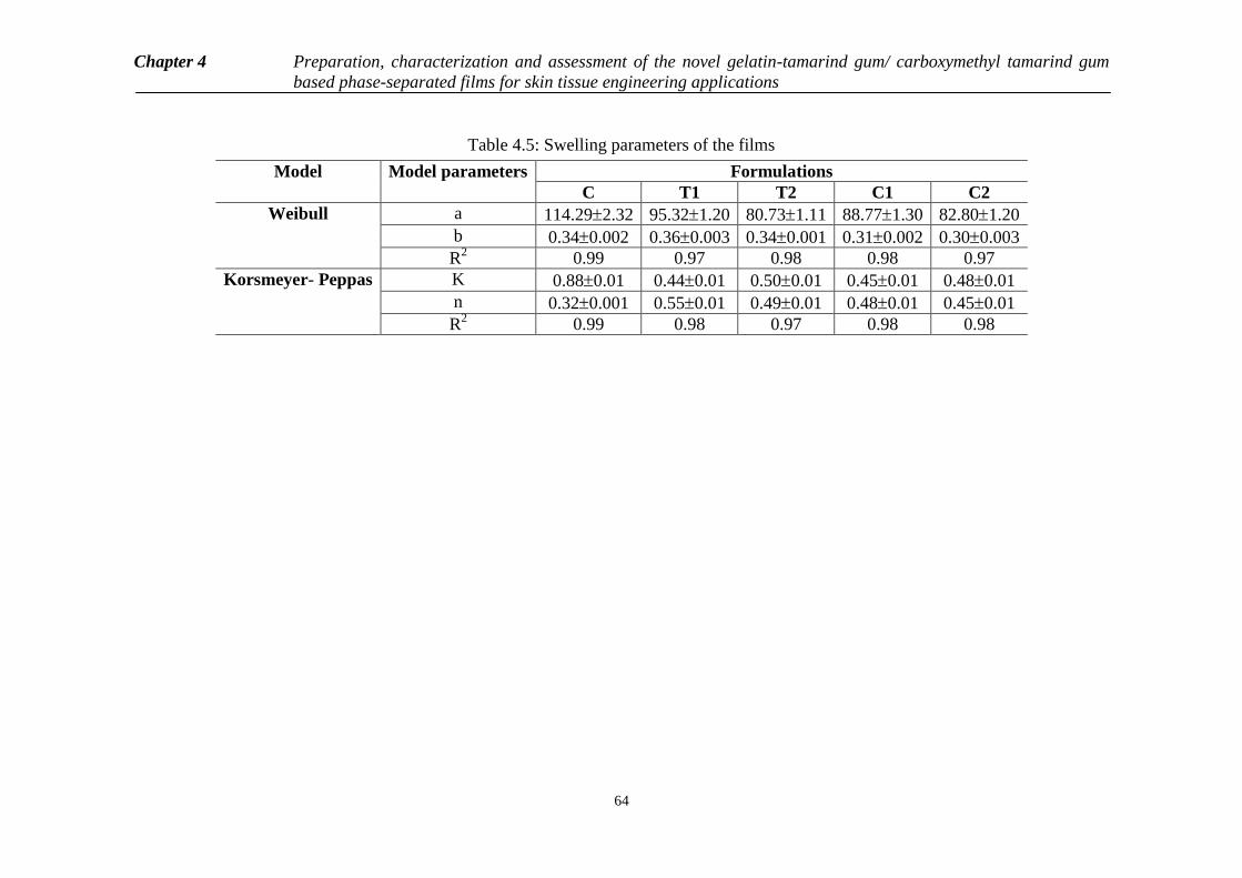
Chapter 4 Preparation, characterization and assessment of the novel gelatin-tamarind gum/ carboxymethyl tamarind gum
based phase-separated films for skin tissue engineering applications
64
Table 4.5: Swelling parameters of the films
Model
Model parameters Formulations
C T1 T2 C1 C2
Weibull a 114.292.32 95.321.20 80.731.11 88.771.30 82.801.20
b 0.340.002 0.360.003 0.340.001 0.310.002 0.300.003
R2 0.99 0.97 0.98 0.98 0.97
Korsmeyer- Peppas
K 0.880.01 0.440.01 0.500.01 0.450.01 0.480.01
n 0.320.001 0.550.01 0.490.01 0.480.01 0.450.01
R2
0.99 0.98 0.97 0.98 0.98
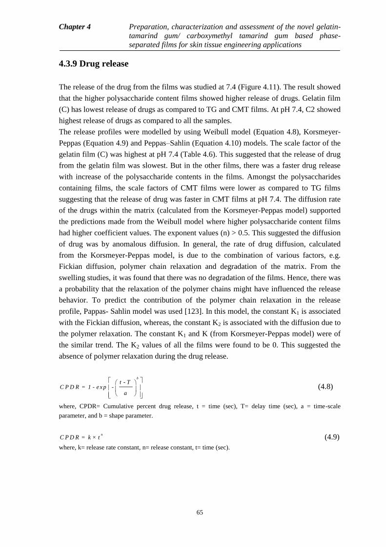
Chapter 4 Preparation, characterization and assessment of the novel gelatin-
tamarind gum/ carboxymethyl tamarind gum based phase-
separated films for skin tissue engineering applications
65
4.3.9 Drug release
The release of the drug from the films was studied at 7.4 (Figure 4.11). The result showed
that the higher polysaccharide content films showed higher release of drugs. Gelatin film
(C) has lowest release of drugs as compared to TG and CMT films. At pH 7.4, C2 showed
highest release of drugs as compared to all the samples.
The release profiles were modelled by using Weibull model (Equation 4.8), Korsmeyer-
Peppas (Equation 4.9) and Peppas–Sahlin (Equation 4.10) models. The scale factor of the
gelatin film (C) was highest at pH 7.4 (Table 4.6). This suggested that the release of drug
from the gelatin film was slowest. But in the other films, there was a faster drug release
with increase of the polysaccharide contents in the films. Amongst the polysaccharides
containing films, the scale factors of CMT films were lower as compared to TG films
suggesting that the release of drug was faster in CMT films at pH 7.4. The diffusion rate
of the drugs within the matrix (calculated from the Korsmeyer-Peppas model) supported
the predictions made from the Weibull model where higher polysaccharide content films
had higher coefficient values. The exponent values (n) > 0.5. This suggested the diffusion
of drug was by anomalous diffusion. In general, the rate of drug diffusion, calculated
from the Korsmeyer-Peppas model, is due to the combination of various factors, e.g.
Fickian diffusion, polymer chain relaxation and degradation of the matrix. From the
swelling studies, it was found that there was no degradation of the films. Hence, there was
a probability that the relaxation of the polymer chains might have influenced the release
behavior. To predict the contribution of the polymer chain relaxation in the release
profile, Pappas- Sahlin model was used [123]. In this model, the constant K1 is associated
with the Fickian diffusion, whereas, the constant K2 is associated with the diffusion due to
the polymer relaxation. The constant K1 and K (from Korsmeyer-Peppas model) were of
the similar trend. The K2 values of all the films were found to be 0. This suggested the
absence of polymer relaxation during the drug release.
b
t - TC P D R = 1 - e x p -
a
(4.8)
where, CPDR= Cumulative percent drug release, t = time (sec), T= delay time (sec), a = time-scale
parameter, and b = shape parameter.
nC P D R = k × t (4.9)
where, k= release rate constant, n= release constant, t= time (sec).
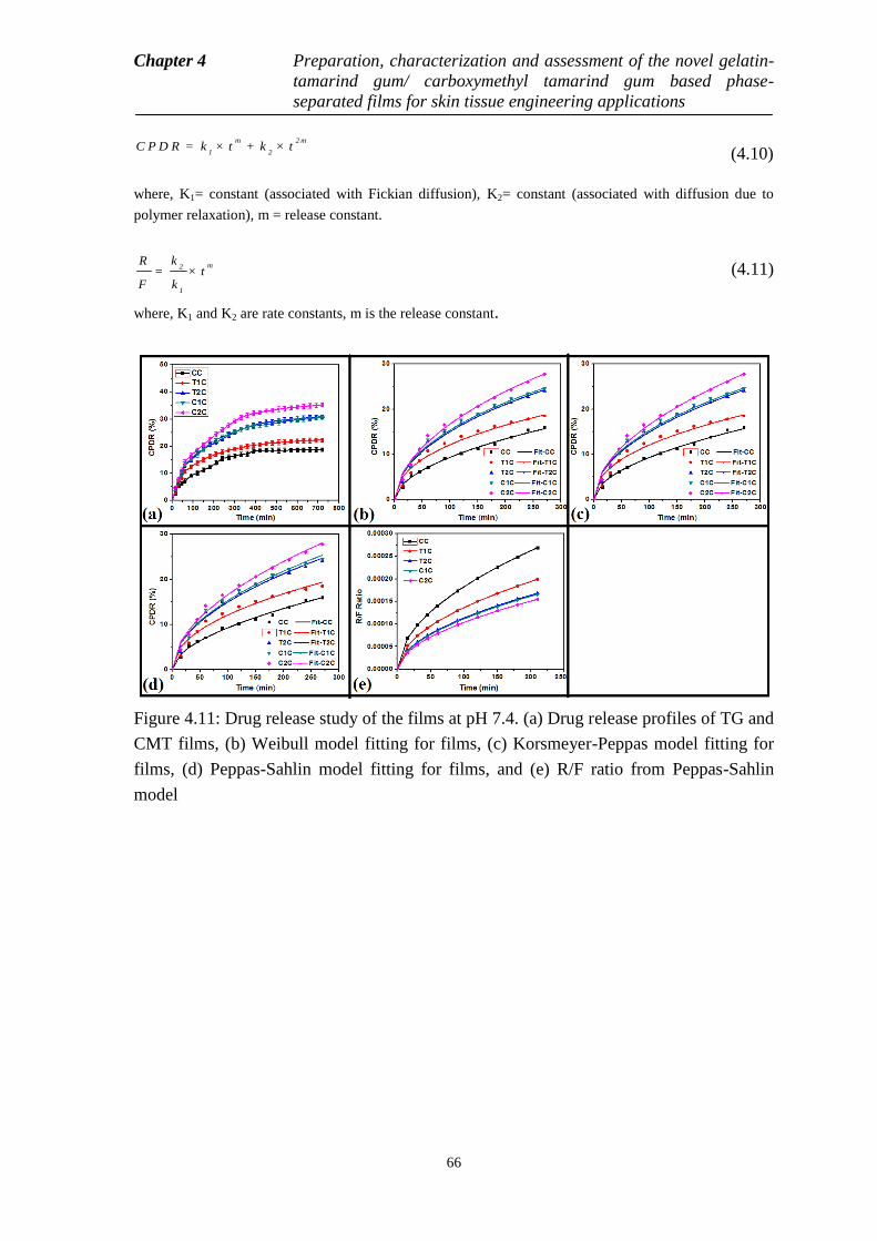
Chapter 4 Preparation, characterization and assessment of the novel gelatin-
tamarind gum/ carboxymethyl tamarind gum based phase-
separated films for skin tissue engineering applications
66
m 2 m
1 2C P D R = k × t + k × t
(4.10)
where, K1= constant (associated with Fickian diffusion), K2= constant (associated with diffusion due to
polymer relaxation), m = release constant.
m2
1
kR= × t
F k (4.11)
where, K1 and K2 are rate constants, m is the release constant.
Figure 4.11: Drug release study of the films at pH 7.4. (a) Drug release profiles of TG and
CMT films, (b) Weibull model fitting for films, (c) Korsmeyer-Peppas model fitting for
films, (d) Peppas-Sahlin model fitting for films, and (e) R/F ratio from Peppas-Sahlin
model
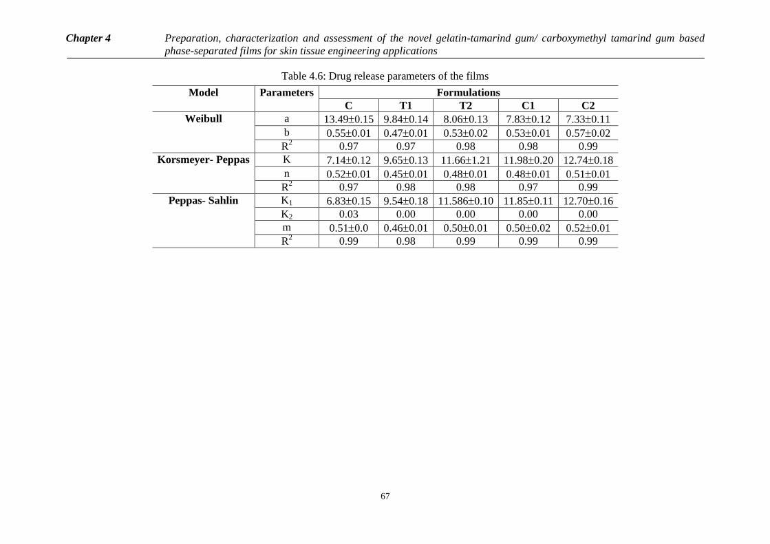
Chapter 4 Preparation, characterization and assessment of the novel gelatin-tamarind gum/ carboxymethyl tamarind gum based
phase-separated films for skin tissue engineering applications
67
Table 4.6: Drug release parameters of the films
Model
Parameters Formulations
C T1 T2 C1 C2
Weibull
a 13.490.15 9.840.14 8.060.13 7.830.12 7.330.11
b 0.550.01 0.470.01 0.530.02 0.530.01 0.570.02
R2 0.97 0.97 0.98 0.98 0.99
Korsmeyer- Peppas
K 7.140.12 9.650.13 11.661.21 11.980.20 12.740.18
n 0.520.01 0.450.01 0.480.01 0.480.01 0.510.01
R2
0.97 0.98 0.98 0.97 0.99
Peppas- Sahlin K1 6.830.15 9.540.18 11.5860.10 11.850.11 12.700.16
K2 0.03 0.00 0.00 0.00 0.00
m 0.510.0 0.460.01 0.500.01 0.500.02 0.520.01
R2 0.99 0.98 0.99 0.99 0.99
Chapter 4 Preparation, characterization and assessment of the novel gelatin-
tamarind gum/ carboxymethyl tamarind gum based phase-
separated films for skin tissue engineering applications
68
4.4 Conclusion
The current chapter discusses about the development of the gelatin-tamarind gum
(TG)/carboxymethyl tamarind gum (CMT) based phase separated films for skin tissue
engineering and drug delivery applications. The agglomeration of the polysaccharide
phase was higher in the TG containing films as compared to the CMT films. This can be
explored by the derivatization of TG molecules with carboxymethyl group which
increased the hydrophilicity of the CMT molecules. The FTIR studies suggested that
when the polysaccharide was in corporate in higher proportions, the helical structure of
the gelatin molecules might have been altered significantly. The thermal profiles of the
films suggested that the incorporation of the polysaccharides in the films increased the
water containing capacity of the films. The extent of water holding capacity was higher in
CMT containing films as compared to the TG films at similar composition. The
mechanical properties indicated better mechanical properties of the TG containing films
which was attributed to the filler effect exerted by the TG molecules. The stress relaxation
studies suggested that CMT containing films showed better relaxation properties as
compared to TG containing films. All the films were found to be hemocompatible when
tested with goat blood. The drug loaded films (Ciprofloxacin) showed good antimicrobial
properties against E. coli. The ability of the films to support HaCaT (human
keratinocytes) was examined. Incorporation of polysaccharides within the gelatin films
improved the proliferation of the HaCaT cells. Amongst the polysaccharide containing
films an increase in the polysaccharide content improved the cell proliferation. Amongst
TG and CMT based films, TG films showed better cell viability. The result suggested that
the TG and CMT containing films may be tried as matrices for skin tissue engineering.
The swelling profiles of the films showed a slight pH dependent behavior. Drug release
profiles indicated a pH dependent release profile. From the above result it can be
concluded that the development has sufficient properties for exploration as matrices for
skin tissue engineering and controlled drug delivery applications.
69
Chapter 5
Summary
The current research work was focused on the fabrication and characterization of gelatin-
tamarind gum (TG)/carboxymethyl tamarind gum based phase-separated architectures for
tissue engineering applications. The microscopic analysis of the hydrogels clearly
indicated the formation of phase-separated systems by forming both gelatin rich phase
and polysaccharide rich phase in a distinct manner. The FTIR spectroscopy of the
hydrogels showed an alteration in the secondary structure of the gelatin molecules due to
the addition of the carboxymethyl tamarind gum in to the formulations. The swelling and
drug release were pH dependent for all the hydrogels. The hydrogels were
hemocompatible in nature and had good mucoadhesive property. The in vitro cell culture
of the hydrogels suggested a better proliferation of osteoblast cells (MG 63) on
polysaccharide content formulations. So the hydrogels can be used as non-load bearing
substitutes in bone tissue engineering. The films were prepared by conventional solution
casting method. Polyethylene glycol was used as the plasticizer to induce a better
elasticity in the films. The microscopy showed the formation of phase separated systems.
Tamarind gum based films showed the presence of agglomerated gum particles due the
hydrophobic nature of the tamarind gum. FTIR spectroscopy suggested the presence of
both gelatin and polysaccharides within the films. The water holding capacity and
mechanical properties were better in the polysaccharide containing films as was indicated
by the thermal and mechanical studies, respectively. Impedance analysis suggested the
better dielectric properties of the TG films as compared to the CMT films. The films were
hemocompatible in nature and supported the proliferation of human keratinocytes. The
drug containing films showed good antimicrobial properties against E. coli. The films
showed pH dependent swelling and drug release profiles. The analysis of the afore-
mentioned results indicated that the pre prepared films may be tried as matrices for skin
Chapter 5 Summary
70
tissue engineering and drug release applications. So, the above developed polymeric
architectures are can be used for tissue engineering applications.
Future aspects
The toxicity profiles of the prepared polymeric architectures need to be assessed using
suitable mammalian models. The prepared polymeric structures need to be assessed for
tissue engineering applications in suitable animal models. The in vivo studies are further
required to analyze the biocompatibility of the developed biomaterials. To have some
better understanding about the drug release profiles, different drugs are needed. After all,
it‘s not the end of this research. It‘s a long path to cover.
70
Bibliography
[1] G. Rigotti, A. Marchi, M. Galie, G. Baroni, D. Benati, M. Krampera, A. Pasini, and A. Sbarbati,
"Clinical treatment of radiotherapy tissue damage by lipoaspirate transplant: a healing process
mediated by adipose-derived adult stem cells," Plastic and reconstructive surgery, vol. 119, pp.
1409-1422, 2007.
[2] L. G. Griffith and G. Naughton, "Tissue engineering--current challenges and expanding
opportunities," Science, vol. 295, pp. 1009-1014, 2002.
[3] D. F. Stamatialis, B. J. Papenburg, M. Gironés, S. Saiful, S. N. Bettahalli, S. Schmitmeier, and M.
Wessling, "Medical applications of membranes: drug delivery, artificial organs and tissue
engineering," Journal of Membrane Science, vol. 308, pp. 1-34, 2008.
[4] J. W. Nichol and A. Khademhosseini, "Modular tissue engineering: engineering biological tissues
from the bottom up," Soft matter, vol. 5, pp. 1312-1319, 2009.
[5] H. F. Tibbals, Medical nanotechnology and nanomedicine: CRC Press, 2010.
[6] J. Bergsma, F. Rozema, R. Bos, G. Boering, W. De Bruijn, and A. Pennings, "In vivo degradation
and biocompatibility study of in vitro pre-degraded as-polymerized polylactide particles,"
Biomaterials, vol. 16, pp. 267-274, 1995.
[7] B.-S. Kim and D. J. Mooney, "Development of biocompatible synthetic extracellular matrices for
tissue engineering," Trends in biotechnology, vol. 16, pp. 224-230, 1998.
[8] C. Liu, Z. Xia, and J. Czernuszka, "Design and development of three-dimensional scaffolds for
tissue engineering," Chemical Engineering Research and Design, vol. 85, pp. 1051-1064, 2007.
[9] H. Shin, S. Jo, and A. G. Mikos, "Biomimetic materials for tissue engineering," Biomaterials, vol.
24, pp. 4353-4364, 2003.
[10] E. Brynda, J. Pacherník, M. Houska, Z. Pientka, and P. Dvorák, "Surface immobilized protein
multilayers for cell seeding," Langmuir, vol. 21, pp. 7877-7883, 2005.
[11] Y. Onuma and P. W. Serruys, "Bioresorbable scaffold the advent of a new era in percutaneous
coronary and peripheral revascularization?," Circulation, vol. 123, pp. 779-797, 2011.
[12] S. J. Hollister, "Porous scaffold design for tissue engineering," Nature materials, vol. 4, pp. 518-
524, 2005.
[13] K. Leong, C. Cheah, and C. Chua, "Solid freeform fabrication of three-dimensional scaffolds for
engineering replacement tissues and organs," Biomaterials, vol. 24, pp. 2363-2378, 2003.
[14] F. Oberpenning, J. Meng, J. J. Yoo, and A. Atala, "De novo reconstitution of a functional
mammalian urinary bladder by tissue engineering," Nature biotechnology, vol. 17, pp. 149-155,
1999.
[15] T. Shin'oka, Y. Imai, and Y. Ikada, "Transplantation of a tissue-engineered pulmonary artery,"
New England Journal of Medicine, vol. 344, pp. 532-533, 2001.
[16] S. Zhang, "Fabrication of novel biomaterials through molecular self-assembly," Nature
biotechnology, vol. 21, pp. 1171-1178, 2003.
[17] D. W. Hutmacher, "Scaffold design and fabrication technologies for engineering tissues—state of
the art and future perspectives," Journal of Biomaterials Science, Polymer Edition, vol. 12, pp.
107-124, 2001.
[18] S. I. Stupp and P. V. Braun, "Molecular manipulation of microstructures: biomaterials, ceramics,
and semiconductors," Science, vol. 277, pp. 1242-1248, 1997.
[19] C. Agrawal and R. B. Ray, "Biodegradable polymeric scaffolds for musculoskeletal tissue
engineering," Journal of biomedical materials research, vol. 55, pp. 141-150, 2001.
[20] B. K. Mann, A. S. Gobin, A. T. Tsai, R. H. Schmedlen, and J. L. West, "Smooth muscle cell
growth in photopolymerized hydrogels with cell adhesive and proteolytically degradable domains:
synthetic ECM analogs for tissue engineering," Biomaterials, vol. 22, pp. 3045-3051, 2001.
[21] S. Yang, K.-F. Leong, Z. Du, and C.-K. Chua, "The design of scaffolds for use in tissue
engineering. Part I. Traditional factors," Tissue engineering, vol. 7, pp. 679-689, 2001.
71
[22] D. W. Hutmacher, J. T. Schantz, C. X. F. Lam, K. C. Tan, and T. C. Lim, "State of the art and
future directions of scaffold‐based bone engineering from a biomaterials perspective," Journal of
tissue engineering and regenerative medicine, vol. 1, pp. 245-260, 2007.
[23] S. H. Cartmell, B. D. Porter, A. J. García, and R. E. Guldberg, "Effects of medium perfusion rate
on cell-seeded three-dimensional bone constructs in vitro," Tissue engineering, vol. 9, pp. 1197-
1203, 2003.
[24] J. B. Park and J. D. Bronzino, Biomaterials: principles and applications: crc press, 2002.
[25] A. R. Shrivats, M. C. McDermott, and J. O. Hollinger, "Bone tissue engineering: state of the
union," Drug discovery today, vol. 19, pp. 781-786, 2014.
[26] J. C. Middleton and A. J. Tipton, "Synthetic biodegradable polymers as orthopedic devices,"
Biomaterials, vol. 21, pp. 2335-2346, 2000.
[27] L. Yu, K. Dean, and L. Li, "Polymer blends and composites from renewable resources," Progress
in Polymer Science, vol. 31, pp. 576-602, 2006.
[28] D. L. Kaplan, Introduction to biopolymers from renewable resources: Springer, 1998.
[29] S. Huang and X. Fu, "Naturally derived materials-based cell and drug delivery systems in skin
regeneration," Journal of Controlled Release, vol. 142, pp. 149-159, 2010.
[30] R. A. Gross and B. Kalra, "Biodegradable polymers for the environment," Science, vol. 297, pp.
803-807, 2002.
[31] A. S. Hoffman, "Hydrogels for biomedical applications," Advanced drug delivery reviews, vol. 64,
pp. 18-23, 2012.
[32] W. Hennink and C. F. Van Nostrum, "Novel crosslinking methods to design hydrogels," Advanced
drug delivery reviews, vol. 64, pp. 223-236, 2012.
[33] Q. Wang, J. L. Mynar, M. Yoshida, E. Lee, M. Lee, K. Okuro, K. Kinbara, and T. Aida, "High-
water-content mouldable hydrogels by mixing clay and a dendritic molecular binder," Nature, vol.
463, pp. 339-343, 2010.
[34] Y. Qiu and K. Park, "Environment-sensitive hydrogels for drug delivery," Advanced drug delivery
reviews, vol. 64, pp. 49-60, 2012.
[35] V. K. Singh, S. S. Sagiri, K. Pal, S. M. Khade, D. K. Pradhan, and M. K. Bhattacharya,
"Gelatin‐carbohydrate phase‐separated hydrogels as bioactive carriers in vaginal delivery:
preparation and physical characterizations," Journal of Applied Polymer Science, vol. 131, 2014.
[36] E. Dickinson, "Hydrocolloids at interfaces and the influence on the properties of dispersed
systems," Food Hydrocolloids, vol. 17, pp. 25-39, 2003.
[37] R. G. Weiss and P. Terech, "Molecular gels," Materials with Self-Assembled Fibrillar Networks,
2006.
[38] A. S. Hoffman, "The origins and evolution of ―controlled‖ drug delivery systems," Journal of
controlled release, vol. 132, pp. 153-163, 2008.
[39] S. Sanyasi, A. Kumar, C. Goswami, A. Bandyopadhyay, and L. Goswami, "A carboxy methyl
tamarind polysaccharide matrix for adhesion and growth of osteoclast-precursor cells,"
Carbohydrate Polymers, vol. 101, pp. 1033-1042, 2014.
[40] L. L. Hench and J. M. Polak, "Third-generation biomedical materials," Science, vol. 295, pp. 1014-
1017, 2002.
[41] K. Jayathilakan, K. Sultana, K. Radhakrishna, and A. Bawa, "Utilization of byproducts and waste
materials from meat, poultry and fish processing industries: a review," Journal of food science and
technology, vol. 49, pp. 278-293, 2012.
[42] W. Zhao, X. Jin, Y. Cong, Y. Liu, and J. Fu, "Degradable natural polymer hydrogels for articular
cartilage tissue engineering," Journal of Chemical Technology and Biotechnology, vol. 88, pp.
327-339, 2013.
[43] T. Thitiset, S. Damrongsakkul, T. Bunaprasert, W. Leeanansaksiri, and S. Honsawek,
"Development of collagen/demineralized bone powder scaffolds and periosteum-derived cells for
bone tissue engineering application," International journal of molecular sciences, vol. 14, pp.
2056-2071, 2013.
[44] A. J. Lomas, W. R. Webb, J. Han, G.-Q. Chen, X. Sun, Z. Zhang, A. J. El Haj, and N. R. Forsyth,
"Poly (3-hydroxybutyrate-co-3-hydroxyhexanoate)/collagen hybrid scaffolds for tissue engineering
applications," Tissue Engineering Part C: Methods, vol. 19, pp. 577-585, 2013.
[45] S. Ahn, H. Yoon, G. Kim, Y. Kim, S. Lee, and W. Chun, "Designed three-dimensional collagen
scaffolds for skin tissue regeneration," Tissue Engineering Part C: Methods, vol. 16, pp. 813-820,
2009.
72
[46] L. H. Han, S. Yu, T. Wang, A. W. Behn, and F. Yang, "Microribbon‐Like Elastomers for
Fabricating Macroporous and Highly Flexible Scaffolds that Support Cell Proliferation in 3D,"
Advanced Functional Materials, vol. 23, pp. 346-358, 2013.
[47] H. Cao, M.-M. Chen, Y. Liu, Y.-Y. Liu, Y.-Q. Huang, J.-H. Wang, J.-D. Chen, and Q.-Q. Zhang,
"Fish collagen-based scaffold containing PLGA microspheres for controlled growth factor delivery
in skin tissue engineering," Colloids and Surfaces B: Biointerfaces, vol. 136, pp. 1098-1106, 2015.
[48] D. J. Richards, Y. Tan, J. Jia, H. Yao, and Y. Mei, "3D printing for tissue engineering," Israel
Journal of Chemistry, vol. 53, pp. 805-814, 2013.
[49] Y. Yan, X. Wang, Y. Pan, H. Liu, J. Cheng, Z. Xiong, F. Lin, R. Wu, R. Zhang, and Q. Lu,
"Fabrication of viable tissue-engineered constructs with 3D cell-assembly technique,"
Biomaterials, vol. 26, pp. 5864-5871, 2005.
[50] T. Billiet, E. Gevaert, T. De Schryver, M. Cornelissen, and P. Dubruel, "The 3D printing of gelatin
methacrylamide cell-laden tissue-engineered constructs with high cell viability," Biomaterials, vol.
35, pp. 49-62, 2014.
[51] M. Yazdimamaghani, D. Vashaee, S. Assefa, K. Walker, S. Madihally, G. Köhler, and L. Tayebi,
"Hybrid macroporous gelatin/bioactive-glass/nanosilver scaffolds with controlled degradation
behavior and antimicrobial activity for bone tissue engineering," Journal of biomedical
nanotechnology, vol. 10, pp. 911-931, 2014.
[52] D. Nadeem, C.-A. Smith, M. J. Dalby, R. D. Meek, S. Lin, G. Li, and B. Su, "Three-dimensional
CaP/gelatin lattice scaffolds with integrated osteoinductive surface topographies for bone tissue
engineering," Biofabrication, vol. 7, p. 015005, 2015.
[53] R. Parenteau-Bareil, R. Gauvin, and F. Berthod, "Collagen-based biomaterials for tissue
engineering applications," Materials, vol. 3, pp. 1863-1887, 2010.
[54] J. Zhu and R. E. Marchant, "Design properties of hydrogel tissue-engineering scaffolds," Expert
review of medical devices, vol. 8, pp. 607-626, 2011.
[55] T. Chen, H. D. Embree, E. M. Brown, M. M. Taylor, and G. F. Payne, "Enzyme-catalyzed gel
formation of gelatin and chitosan: potential for in situ applications," Biomaterials, vol. 24, pp.
2831-2841, 2003.
[56] H. Tan and K. G. Marra, "Injectable, biodegradable hydrogels for tissue engineering applications,"
Materials, vol. 3, pp. 1746-1767, 2010.
[57] G. Kogan, L. Šoltés, R. Stern, and P. Gemeiner, "Hyaluronic acid: a natural biopolymer with a
broad range of biomedical and industrial applications," Biotechnology letters, vol. 29, pp. 17-25,
2007.
[58] J. Necas, L. Bartosikova, P. Brauner, and J. Kolar, "Hyaluronic acid (hyaluronan): a review,"
Veterinarni medicina, vol. 53, pp. 397-411, 2008.
[59] A. Fakhari and C. Berkland, "Applications and emerging trends of hyaluronic acid in tissue
engineering, as a dermal filler and in osteoarthritis treatment," Acta biomaterialia, vol. 9, pp. 7081-
7092, 2013.
[60] M. N. Collins and C. Birkinshaw, "Hyaluronic acid based scaffolds for tissue engineering—A
review," Carbohydrate Polymers, vol. 92, pp. 1262-1279, 2013.
[61] S. D. Nath, C. Abueva, B. Kim, and B. T. Lee, "Chitosan–hyaluronic acid polyelectrolyte complex
scaffold crosslinked with genipin for immobilization and controlled release of BMP-2,"
Carbohydrate Polymers, vol. 115, pp. 160-169, 2015.
[62] N. B. Skop, F. Calderon, C. H. Cho, C. D. Gandhi, and S. W. Levison, "Improvements in
biomaterial matrices for neural precursor cell transplantation," Molecular and cellular therapies,
vol. 2, p. 1, 2014.
[63] D. Park, Y. Kim, H. Kim, K. Kim, Y.-S. Lee, J. Choe, J.-H. Hahn, H. Lee, J. Jeon, and C. Choi,
"Hyaluronic acid promotes angiogenesis by inducing RHAMM-TGFβ receptor interaction via
CD44-PKCδ," Molecules and cells, vol. 33, pp. 563-574, 2012.
[64] Y. Liu, L. Ren, and Y. Wang, "Crosslinked collagen–gelatin–hyaluronic acid biomimetic film for
cornea tissue engineering applications," Materials Science and Engineering: C, vol. 33, pp. 196-
201, 2013.
[65] F. D. Ivan, A. Marian, C. E. Tanase, M. Butnaru, and L. Vereştiuc, "Biomimetic composites based
on calcium phosphates and chitosan-hyaluronic acid with potential application in bone tissue
engineering," in Key Engineering Materials, 2014, pp. 191-196.
[66] L. B. Sandberg, N. T. Soskel, and J. G. Leslie, "Elastin structure, biosynthesis, and relation to
disease states," New England Journal of Medicine, vol. 304, pp. 566-579, 1981.

73
[67] J. Rnjak-Kovacina and A. S. Weiss, "The role of elastin in wound healing and dermal substitute
design," in Dermal Replacements in General, Burn, and Plastic Surgery, ed: Springer, 2013, pp.
57-66.
[68] A. Girotti, D. Orbanic, A. Ibáñez‐Fonseca, C. Gonzalez‐Obeso, and J. C. Rodríguez‐Cabello,
"Recombinant Technology in the Development of Materials and Systems for Soft‐Tissue Repair,"
Advanced healthcare materials, vol. 4, pp. 2423-2455, 2015.
[69] C. N. Grover, R. E. Cameron, and S. M. Best, "Investigating the morphological, mechanical and
degradation properties of scaffolds comprising collagen, gelatin and elastin for use in soft tissue
engineering," Journal of the mechanical behavior of biomedical materials, vol. 10, pp. 62-74,
2012.
[70] R. Machado, P. C. Bessa, R. L. Reis, J. C. Rodriguez-Cabello, and M. Casal, "Elastin-based
nanoparticles for delivery of bone morphogenetic proteins," Nanoparticles in Biology and
Medicine: Methods and Protocols, pp. 353-363, 2012.
[71] S. E. Dunphy, J. A. Bratt, K. M. Akram, N. R. Forsyth, and A. J. El Haj, "Hydrogels for lung tissue
engineering: Biomechanical properties of thin collagen–elastin constructs," Journal of the
mechanical behavior of biomedical materials, vol. 38, pp. 251-259, 2014.
[72] J. M. Silva, N. Georgi, R. Costa, P. Sher, R. L. Reis, C. A. Van Blitterswijk, M. Karperien, and J.
F. Mano, "Nanostructured 3D constructs based on chitosan and chondroitin sulphate multilayers
for cartilage tissue engineering," PloS one, vol. 8, p. e55451, 2013.
[73] P. A. Levett, F. P. Melchels, K. Schrobback, D. W. Hutmacher, J. Malda, and T. J. Klein, "A
biomimetic extracellular matrix for cartilage tissue engineering centered on photocurable gelatin,
hyaluronic acid and chondroitin sulfate," Acta biomaterialia, vol. 10, pp. 214-223, 2014.
[74] J. Venkatesan, R. Pallela, I. Bhatnagar, and S.-K. Kim, "Chitosan–amylopectin/hydroxyapatite and
chitosan–chondroitin sulphate/hydroxyapatite composite scaffolds for bone tissue engineering,"
International journal of biological macromolecules, vol. 51, pp. 1033-1042, 2012.
[75] S. Deepthi, C. Viha, C. Thitirat, T. Furuike, H. Tamura, and R. Jayakumar, "Fabrication of
chitin/poly (butylene succinate)/chondroitin sulfate nanoparticles ternary composite hydrogel
scaffold for skin tissue engineering," Polymers, vol. 6, pp. 2974-2984, 2014.
[76] S. Yan, Q. Zhang, J. Wang, Y. Liu, S. Lu, M. Li, and D. L. Kaplan, "Silk fibroin/chondroitin
sulfate/hyaluronic acid ternary scaffolds for dermal tissue reconstruction," Acta biomaterialia, vol.
9, pp. 6771-6782, 2013.
[77] S. S. Martin, M. Alaminos, T. Zorn, M. Sánchez‐Quevedo, I. Garzon, I. Rodriguez, and A.
Campos, "The effects of fibrin and fibrin‐agarose on the extracellular matrix profile of
bioengineered oral mucosa," Journal of tissue engineering and regenerative medicine, vol. 7, pp.
10-19, 2013.
[78] P. de la Puente and D. Ludeña, "Cell culture in autologous fibrin scaffolds for applications in tissue
engineering," Experimental cell research, vol. 322, pp. 1-11, 2014.
[79] S. P. Miguel, M. P. Ribeiro, H. Brancal, P. Coutinho, and I. J. Correia, "Thermoresponsive
chitosan–agarose hydrogel for skin regeneration," Carbohydrate Polymers, vol. 111, pp. 366-373,
2014.
[80] S. Bhat and A. Kumar, "Cell proliferation on three-dimensional chitosan–agarose–gelatin cryogel
scaffolds for tissue engineering applications," Journal of bioscience and bioengineering, vol. 114,
pp. 663-670, 2012.
[81] S. Jebahi, G. B. Saleh, M. Saoudi, S. Besaleh, H. Oudadesse, M. Mhadbi, T. Rebai, H. Keskes, and
A. El Feki, "Genotoxicity effect, antioxidant and biomechanical correlation: experimental study of
agarose–chitosan bone graft substitute in New Zealand white rabbit model," Proceedings of the
Institution of Mechanical Engineers, Part H: Journal of Engineering in Medicine, vol. 228, pp.
800-809, 2014.
[82] J. Venkatesan, R. Jayakumar, S. Anil, E. P. Chalisserry, R. Pallela, and S.-K. Kim, "Development
of Alginate-Chitosan-Collagen Based Hydrogels for Tissue Engineering," Journal of Biomaterials
and Tissue Engineering, vol. 5, pp. 458-464, 2015.
[83] S. Kirdponpattara, A. Khamkeaw, N. Sanchavanakit, P. Pavasant, and M. Phisalaphong,
"Structural modification and characterization of bacterial cellulose–alginate composite scaffolds
for tissue engineering," Carbohydrate Polymers, vol. 132, pp. 146-155, 2015.
[84] M. Castilho, J. Rodrigues, I. Pires, B. Gouveia, M. Pereira, C. Moseke, J. Groll, A. Ewald, and E.
Vorndran, "Fabrication of individual alginate-TCP scaffolds for bone tissue engineering by means
of powder printing," Biofabrication, vol. 7, p. 015004, 2015.
74
[85] J. Sowjanya, J. Singh, T. Mohita, S. Sarvanan, A. Moorthi, N. Srinivasan, and N. Selvamurugan,
"Biocomposite scaffolds containing chitosan/alginate/nano-silica for bone tissue engineering,"
Colloids and Surfaces B: Biointerfaces, vol. 109, pp. 294-300, 2013.
[86] F. Croisier and C. Jérôme, "Chitosan-based biomaterials for tissue engineering," European
Polymer Journal, vol. 49, pp. 780-792, 2013.
[87] F. Han, Y. Dong, Z. Su, R. Yin, A. Song, and S. Li, "Preparation, characteristics and assessment of
a novel gelatin–chitosan sponge scaffold as skin tissue engineering material," International journal
of pharmaceutics, vol. 476, pp. 124-133, 2014.
[88] M. M. Rahman, S. Pervez, B. Nesa, and M. A. Khan, "Preparation and characterization of porous
scaffold composite films by blending chitosan and gelatin solutions for skin tissue engineering,"
Polymer International, vol. 62, pp. 79-86, 2013.
[89] M. E. Frohbergh, A. Katsman, G. P. Botta, P. Lazarovici, C. L. Schauer, U. G. Wegst, and P. I.
Lelkes, "Electrospun hydroxyapatite-containing chitosan nanofibers crosslinked with genipin for
bone tissue engineering," Biomaterials, vol. 33, pp. 9167-9178, 2012.
[90] T. Gerard, "Tamarind Gum in Hand book of water soluble gums and resins," USA: McGraw-Hill
Book Co, vol. 12, pp. 1-23, 1980.
[91] H. Mirhosseini and B. T. Amid, "A review study on chemical composition and molecular structure
of newly plant gum exudates and seed gums," Food Research International, vol. 46, pp. 387-398,
2012.
[92] G. Phani Kumar, G. Battu, and L. R. N. Kota, "Preparation and evaluation of sustained release
matrix tablets of lornoxicam using tamarind seed polysaccharide," Int J pharm Res & Dev, vol. 2,
pp. 90-98, 2011.
[93] A. K. Nayak, D. Pal, and K. Santra, "Tamarind seed polysaccharide–gellan mucoadhesive beads
for controlled release of metformin HCl," Carbohydrate Polymers, vol. 103, pp. 154-163, 2014.
[94] M. Sharma, D. Mondal, C. Mukesh, and K. Prasad, "Preparation of tamarind gum based soft ion
gels having thixotropic properties," Carbohydrate polymers, vol. 102, pp. 467-471, 2014.
[95] S. Sahoo, R. Sahoo, and P. L. Nayak, "Tamarind Seed Polysachharide: A Versatile Biopolymer
For Mucoadhesive Applications," J. Pharm. Biomed. Sci, vol. 8, pp. 1-12, 2011.
[96] P. Goyal, V. Kumar, and P. Sharma, "Carboxymethylation of tamarind kernel powder,"
Carbohydrate polymers, vol. 69, pp. 251-255, 2007.
[97] R. Manchanda, S. Arora, and R. Manchanda, "Tamarind seed polysaccharide and its modification-
versatile pharmaceutical excipients-a review," Int J PharmTech Res, vol. 6, pp. 412-420, 2014.
[98] V. F. Sechriest, Y. J. Miao, C. Niyibizi, A. Westerhausen–Larson, H. W. Matthew, C. H. Evans, F.
H. Fu, and J. K. Suh, "GAG‐augmented polysaccharide hydrogel: A novel biocompatible and
biodegradable material to support chondrogenesis," Journal of biomedical materials research, vol.
49, pp. 534-541, 2000.
[99] R. Gref, J. Rodrigues, and P. Couvreur, "Polysaccharides grafted with polyesters: novel
amphiphilic copolymers for biomedical applications," Macromolecules, vol. 35, pp. 9861-9867,
2002.
[100] A. Dufresne, "Comparing the mechanical properties of high performances polymer
nanocomposites from biological sources," Journal of Nanoscience and Nanotechnology, vol. 6, pp.
322-330, 2006.
[101] G. Ciardelli, V. Chiono, G. Vozzi, M. Pracella, A. Ahluwalia, N. Barbani, C. Cristallini, and P.
Giusti, "Blends of poly-(ε-caprolactone) and polysaccharides in tissue engineering applications,"
Biomacromolecules, vol. 6, pp. 1961-1976, 2005.
[102] G. K. Jani, D. P. Shah, V. D. Prajapati, and V. C. Jain, "Gums and mucilages: versatile excipients
for pharmaceutical formulations," Asian J Pharm Sci, vol. 4, pp. 309-323, 2009.
[103] R. R. Leakey, "Potential for novel food products from agroforestry trees: a review," Food
chemistry, vol. 66, pp. 1-14, 1999.
[104] D. R. Picout, S. B. Ross-Murphy, N. Errington, and S. E. Harding, "Pressure cell assisted
solubilization of xyloglucans: Tamarind seed polysaccharide and detarium gum,"
Biomacromolecules, vol. 4, pp. 799-807, 2003.
[105] R. V. Kulkarni, S. Mutalik, B. S. Mangond, and U. Y. Nayak, "Novel interpenetrated polymer
network microbeads of natural polysaccharides for modified release of water soluble drug: in‐vitro
and in‐vivo evaluation," Journal of Pharmacy and Pharmacology, vol. 64, pp. 530-540, 2012.

75
[106] M. Sano, E. Miyata, S. Tamano, A. Hagiwara, N. Ito, and T. Shirai, "Lack of carcinogenicity of
tamarind seed polysaccharide in B6C3F< sub> 1</sub> mice," Food and chemical toxicology, vol.
34, pp. 463-467, 1996.
[107] J. Tomlin and N. Read, "The relation between bacterial degradation of viscous polysaccharides and
stool output in human beings," British journal of nutrition, vol. 60, pp. 467-475, 1988.
[108] K. Ross, L. Pyrak-Nolte, and O. Campanella, "The effect of mixing conditions on the material
properties of an agar gel—microstructural and macrostructural considerations," Food
Hydrocolloids, vol. 20, pp. 79-87, 2006.
[109] J.-L. Doublier, C. Garnier, D. Renard, and C. Sanchez, "Protein–polysaccharide interactions,"
Current Opinion in Colloid & Interface Science, vol. 5, pp. 202-214, 2000.
[110] V. Tolstoguzov, "Compositions and phase diagrams for aqueous systems based on proteins and
polysaccharides," International review of cytology, vol. 192, pp. 3-31, 1999.
[111] N. J. Einerson, K. R. Stevens, and W. J. Kao, "Synthesis and physicochemical analysis of gelatin-
based hydrogels for drug carrier matrices," Biomaterials, vol. 24, pp. 509-523, 2003.
[112] A. Mishra and A. V. Malhotra, "Graft copolymers of xyloglucan and methyl methacrylate,"
Carbohydrate polymers, vol. 87, pp. 1899-1904, 2012.
[113] J. M. Cloyd, N. R. Malhotra, L. Weng, W. Chen, R. L. Mauck, and D. M. Elliott, "Material
properties in unconfined compression of human nucleus pulposus, injectable hyaluronic acid-based
hydrogels and tissue engineering scaffolds," European Spine Journal, vol. 16, pp. 1892-1898,
2007.
[114] N. Lorén, M. Langton, and A.-M. Hermansson, "Confocal laser scanning microscopy and image
analysis of kinetically trapped phase-separated gelatin/maltodextrin gels," Food Hydrocolloids,
vol. 13, pp. 185-198, 1999.
[115] F. Jara, O. E. Pérez, and A. M. Pilosof, "Impact of phase separation of whey
proteins/hydroxypropylmethylcellulose mixtures on gelation dynamics and gels properties," Food
Hydrocolloids, vol. 24, pp. 641-651, 2010.
[116] D. Oakenfull, J. Pearce, and R. Burley, "Protein gelation," FOOD SCIENCE AND
TECHNOLOGY-NEW YORK-MARCEL DEKKER-, pp. 111-142, 1997.
[117] L. Huang, G. Reekmans, D. Saerens, J.-M. Friedt, F. Frederix, L. Francis, S. Muyldermans, A.
Campitelli, and C. V. Hoof, "Prostate-specific antigen immunosensing based on mixed self-
assembled monolayers, camel antibodies and colloidal gold enhanced sandwich assays,"
Biosensors and bioelectronics, vol. 21, pp. 483-490, 2005.
[118] Y. Pranoto, C. M. Lee, and H. J. Park, "Characterizations of fish gelatin films added with gellan
and< i> κ</i>-carrageenan," LWT-Food Science and Technology, vol. 40, pp. 766-774, 2007.
[119] M. De La Cochetiere, T. Durand, P. Lepage, A. Bourreille, J. Galmiche, and J. Dore, "Resilience
of the dominant human fecal microbiota upon short-course antibiotic challenge," Journal of
clinical microbiology, vol. 43, pp. 5588-5592, 2005.
[120] L. Bammou, M. Mihit, R. Salghi, A. Bouyanzer, S. Al-Deyab, L. Bazzi, and B. Hammouti,
"Inhibition effect of natural Artemisia oils towards tinplate corrosion in HCL solution: chemical
characterization and electrochemical study," Int. J. Electrochem. Sci, vol. 6, pp. 1454-1467, 2011.
[121] J. Smart, I. Kellaway, and H. Worthington, "An in‐vitro investigation of mucosa‐adhesive
materials for use in controlled drug delivery," Journal of pharmacy and pharmacology, vol. 36, pp.
295-299, 1984.
[122] A. Smirnov, E. Tako, P. Ferket, and Z. Uni, "Mucin gene expression and mucin content in the
chicken intestinal goblet cells are affected by in ovo feeding of carbohydrates," Poultry science,
vol. 85, pp. 669-673, 2006.
[123] H. Mohammadi and W. Herzog, "A novel model for diffusion based release kinetics using an
inverse numerical method," Medical engineering & physics, vol. 33, pp. 893-899, 2011.
[124] L. Chen, G. E. Remondetto, and M. Subirade, "Food protein-based materials as nutraceutical
delivery systems," Trends in Food Science & Technology, vol. 17, pp. 272-283, 2006.
[125] C. F. Rediguieri, O. de Freitas, M. P. Lettinga, and R. Tuinier, "Thermodynamic incompatibility
and complex formation in pectin/caseinate mixtures," Biomacromolecules, vol. 8, pp. 3345-3354,
2007.
[126] S. Turgeon, M. Beaulieu, C. Schmitt, and C. Sanchez, "Protein–polysaccharide interactions: phase-
ordering kinetics, thermodynamic and structural aspects," Current opinion in colloid & interface
science, vol. 8, pp. 401-414, 2003.
76
[127] T. F. Vandamme, A. Lenourry, C. Charrueau, and J. Chaumeil, "The use of polysaccharides to
target drugs to the colon," Carbohydrate polymers, vol. 48, pp. 219-231, 2002.
[128] J. L. Drury and D. J. Mooney, "Hydrogels for tissue engineering: scaffold design variables and
applications," Biomaterials, vol. 24, pp. 4337-4351, 2003.
[129] G. Zhang, T. Liu, Q. Wang, L. Chen, J. Lei, J. Luo, G. Ma, and Z. Su, "Mass spectrometric
detection of marker peptides in tryptic digests of gelatin: A new method to differentiate between
bovine and porcine gelatin," Food Hydrocolloids, vol. 23, pp. 2001-2007, 2009.
[130] Y. Ikada and H. Tsuji, "Biodegradable polyesters for medical and ecological applications,"
Macromolecular rapid communications, vol. 21, pp. 117-132, 2000.
[131] M. S. Jangdey, A. Gupta, and A. K. Sah, "Development and Evaluation of Mucoadhesive
Sustained Release Tablet using Tamarindus indica Gum," Asian Journal of Research in
Pharmaceutical Science, vol. 4, pp. 77-82, 2014.
[132] M. F. Saettone, S. Burgalassi, B. Giannaccini, E. Boldrini, P. Bianchini, and G. Luciani,
"Ophthalmic solutions viscosified with tamarind seed polysaccharide," ed: Google Patents, 2000.
[133] A. Mishra and A. V. Malhotra, "Tamarind xyloglucan: a polysaccharide with versatile application
potential," Journal of Materials Chemistry, vol. 19, pp. 8528-8536, 2009.
[134] C. S. Kumar and S. Bhattacharya, "Tamarind seed: properties, processing and utilization," Critical
reviews in food science and nutrition, vol. 48, pp. 1-20, 2008.
[135] R. Sothornvit and J. M. Krochta, "Plasticizer effect on mechanical properties of β-lactoglobulin
films," Journal of Food Engineering, vol. 50, pp. 149-155, 2001.
[136] A. Al-Hassan and M. Norziah, "Starch–gelatin edible films: water vapor permeability and
mechanical properties as affected by plasticizers," Food Hydrocolloids, vol. 26, pp. 108-117, 2012.
[137] K. Vimala, M. M. Yallapu, K. Varaprasad, N. N. Reddy, S. Ravindra, N. S. Naidu, and K. M.
Raju, "Fabrication of curcumin encapsulated chitosan-PVA silver nanocomposite films for
improved antimicrobial activity," Journal of Biomaterials and Nanobiotechnology, vol. 2, p. 55,
2011.
[138] K. Pal, A. K. Banthia, and D. K. Majumdar, "Preparation and characterization of polyvinyl
alcohol-gelatin hydrogel membranes for biomedical applications," Aaps Pharmscitech, vol. 8, pp.
E142-E146, 2007.
[139] S. I. Park, M. Daeschel, and Y. Zhao, "Functional properties of antimicrobial lysozyme‐chitosan
composite films," Journal of Food Science, vol. 69, pp. M215-M221, 2004.
[140] M. Ahmad, S. Benjakul, T. Prodpran, and T. W. Agustini, "Physico-mechanical and antimicrobial
properties of gelatin film from the skin of unicorn leatherjacket incorporated with essential oils,"
Food Hydrocolloids, vol. 28, pp. 189-199, 2012.
[141] J. M. Manski, A. J. van der Goot, and R. M. Boom, "Advances in structure formation of
anisotropic protein-rich foods through novel processing concepts," Trends in Food Science &
Technology, vol. 18, pp. 546-557, 2007.
[142] T. Moschakis, B. S. Murray, and C. G. Biliaderis, "Modifications in stability and structure of whey
protein-coated o/w emulsions by interacting chitosan and gum arabic mixed dispersions," Food
Hydrocolloids, vol. 24, pp. 8-17, 2010.
[143] K. Varaprasad, E. R. Sadiku, K. Ramam, J. Jayaramudu, and G. S. M. Reddy, "Significances of
Nanostructured Hydrogels for Valuable Applications," 2014.
[144] T. Miyazawa and E. Blout, "The infrared spectra of polypeptides in various conformations: amide I
and II bands1," Journal of the American Chemical Society, vol. 83, pp. 712-719, 1961.
[145] A. Sionkowska, M. Wisniewski, J. Skopinska, C. Kennedy, and T. Wess, "Molecular interactions
in collagen and chitosan blends," Biomaterials, vol. 25, pp. 795-801, 2004.
[146] S. Mao, C. Guo, Y. Shi, and L. C. Li, "Recent advances in polymeric microspheres for parenteral
drug delivery-part 1," Expert opinion on drug delivery, vol. 9, pp. 1161-1176, 2012.
[147] M. N. Angles and A. Dufresne, "Plasticized starch/tunicin whiskers nanocomposites. 1. Structural
analysis," Macromolecules, vol. 33, pp. 8344-8353, 2000.
[148] N. M. Sarbon, F. Badii, and N. K. Howell, "The effect of chicken skin gelatin and whey protein
interactions on rheological and thermal properties," Food Hydrocolloids, vol. 45, pp. 83-92, 2015.
[149] J. Zhang, S. Xu, S. Zhang, and Z. Du, "Preparation and characterization of tamarind gum/sodium
alginate composite gel beads," Iranian Polymer Journal, vol. 17, pp. 899-906, 2008.
[150] C. M. Yoshida, E. Oliveira, and T. T. Franco, "Chitosan tailor-made films: the effects of additives
on barrier and mechanical properties," Packaging Technology and Science, vol. 22, pp. 161-170,
2009.
77
[151] P. Goyal, R. Dhar, S. S. Sagiri, K. Uvanesh, K. Senthilguru, G. Shankar, A. Samal, K. Pramanik, I.
Banerjee, and S. S. Ray, "Synthesis and characterization of novel dual environment-responsive
hydrogels of Hydroxyethyl methacrylate and Methyl cellulose," Designed Monomers and
Polymers, vol. 18, pp. 367-377, 2015.
[152] M. James, D. Hughes, Z. Chen, H. Lombard, D. Hattingh, D. Asquith, J. Yates, and P. Webster,
"Residual stresses and fatigue performance," Engineering Failure Analysis, vol. 14, pp. 384-395,
2007.
[153] H. J. Maria, N. Lyczko, A. Nzihou, K. Joseph, C. Mathew, and S. Thomas, "Stress relaxation
behavior of organically modified montmorillonite filled natural rubber/nitrile rubber
nanocomposites," Applied Clay Science, vol. 87, pp. 120-128, 2014.
[154] M. Berberan-Santos, E. Bodunov, and B. Valeur, "History of the Kohlrausch (stretched
exponential) function: Focus on uncited pioneering work in luminescence," arXiv preprint
arXiv:0804.1814, 2008.
[155] S. L. Shenoy, W. D. Bates, H. L. Frisch, and G. E. Wnek, "Role of chain entanglements on fiber
formation during electrospinning of polymer solutions: good solvent, non-specific polymer–
polymer interaction limit," Polymer, vol. 46, pp. 3372-3384, 2005.
[156] K. Pal, A. Banthia, and D. Majumdar, "Biomedical evaluation of polyvinyl alcohol–gelatin
esterified hydrogel for wound dressing," Journal of Materials Science: Materials in Medicine, vol.
18, pp. 1889-1894, 2007.
[157] J. G. Ibrahim, M. H. Chen, and D. Sinha, Bayesian survival analysis: Wiley Online Library, 2005.
Dissemination Journal Articles (From Thesis)
1. G. S. Shaw, K. Uvanesh, S.N. Gautham, Vinay Singh, Krishna Pramanik, Indranil
78
Banerjee, Naresh Kumar & Kunal Pal. Development and characterization of gelatin-
tamarindgum/carboxymethyl tamarind gum based phase-separated hydrogels: a
comparative study. Designed Monomers and Polymers, 2015. Published online.
DOI: 10.1080/15685551.2015.1041075. (SCI, Impact factor: 2.78)
2. G. S. Shaw, Dibyajyoti Biswal, Anupriya B., Indranil Banerjee, Krishna Pramanik,
Arfat Anis and Kunal Pal. Preparation, characterization and assessment of the novel
gelatin-tamarind gum/carboxymethyl tamarind gum based phase-separated films for
skin tissue engineering applications. Polymer-Plastics Technology and Engineering
(In press). (SCI, Impact factor: 1.48).
Journal Articles (From other works)
1. Priyanka Goyal, Rik Dhar, Sai S. Sagiri, K. Uvanesh, K. Senthilguru, G. S. Shaw,
Ajit Samal, Krishna Pramanik, Indranil Banerjee, Sirsendu Sekhar Ray & Kunal
Pal. Synthesis and characterization of novel dual environment-responsive
hydrogels of Hydroxyethyl methacrylate and Methyl cellulose. Designed
Monomers and Polymers, 2015. Published online. DOI:
10.1080/15685551.2015.1012626. (SCI, Impact factor: 2.78)
2. Sai Sateesh Sagiri, Uvanesh Kasiviswanathan, G. S. Shaw, Meenakshi Singh,
Arfat Anis, and Kunal Pal. Effect of sorbitan monostearate concentration on the
thermal, mechanical and drug release properties of oleogels. Korean Journal of
Chemical Engineering. Published online. DOI: 10.1007/s11814-015-0295-4.
(SCI, Impact factor: 1.166).
3. S. P. Mallick, G. S. Shaw, Uvanesh K., D. Biswal, Suraj Nayak, S. S. Sagiri, V.
K. Singh1, M. K. Bhattacharya, A. Anis and K. Pal. An in-depth analysis of the
mechanical, electrical and drug release properties of gelatin-starch phase-
separated hydrogels. Polymer-Plastics Technology and Engineering.published
online. SCI, Impact factor: 1.48).