
Preparation and Characterisation of ZnO - SiO2 and Bi2O3 ... · Nanochem Res 3(1): 79-84, Winter...
6
Nanochem Res 3(1): 79-84, Winter and Spring 2018 RESEARCH PAPER Preparaon and Characterisaon of ZnO - SiO 2 and Bi 2 O 3 – CuO Nanocomposites Alagappan Subramaniyan 1 *, Velajagadessa Visweswaran 2 , Chandrasekar Saravana Kumar 2 , Thambu Sornakumar 2 1 Department of Physics, Thiagarajar College of Engineering, Madurai 625006, T.N., India 2 Department of Mechanical Engineering, Thiagarajar College of Engineering Madurai 625006, T.N., India * Corresponding Author Email: [email protected] In the present work, ZnO - SiO 2 and Bi 2 O 3 – CuO nanocomposites have been prepared by sol gel with alternate precursors exisng in literature. They are characterised by XRD, SEM,UV, FTIR and photoluminescence spectra .The XRD results indicate a crystallite size of approximately 80 nm for both nanocomposites. SEM image shows a heterogeneous parcle size for both samples. The band gap of ZnO - SiO 2 and Bi 2 O 3 – CuO obtained from Taucs plot is 4.1 eV and 2.85 eV, respecvely. PL spectra show high intensity absorpon for ZnO-SiO 2 in comparison to Bi 2 O 3 – CuO. The composites of Bi 2 O 3 –CuO is recommended for efficient opcal coang against the coang made from Bi 2 O 3 ARTICLE INFO Arcle History: Received 9 December 2017 Accepted 16 February 2018 Published 1 March 2018 Keywords: Bi 2 O 3 –CuO Nanocomposites Sol gel ZnO-SiO 2 ABSTRACT is work is licensed under the Creative Commons Attribution 4.0 International License. To view a copy of this license, visit http://creativecommons.org/licenses/by/4.0/. How to cite this article Subramaniyan A, Visweswaran V, Saravana Kumar C, Sornakumar T. Preparation and Characterisation of ZnO - SiO 2 and Bi 2 O 3 – CuO Nanocomposites. Nanochem Res, 2018; 3(1): 79-84. DOI: 10.22036/ncr.2018.01.008 INTRODUCTION Nanocomposites are the 21 st century materials which find application in aerospace, biomedical and automobile industries[1]. An appreciable property change is observed between the micro composite and nanocomposite of the same chemical composition. e size property correlation can further contribute to the property of nanocomposite. Among the various class of nanocomposites, oxide based composites have been investigated extensively due to their ease of preparation. Oxide composites are alternatives to silicon carbide composites owing to their low cost and improving thermal stability at high temperature [2]. Oxide–Oxide ceramic matrix composites have enabled wide spread industry adoption because of low cost fibre and development in fabric architectures [3]. A recent research demonstrates the major application of metal oxide composites as gas sensors [4]. ZnO - SiO 2 nanocomposites were investigated for their sensing properties by Mossad et al. [5]. ZnO - SiO 2 nanocomposites have also been studied and characterised by Panthohan et al. [6]. Mossad synthesised ZnO - SiO 2 nanocomposites from zinc acetate, TEOS, and SiO 2 . Panthohan used rice husk as a source of silica source. ZnO- SiO 2 nanocomposites show UV, Visible and white light emissions with relatively small limiting threshold [7].In the present work, ZnO- SiO 2 nanocomposites have been synthesised from Zinc Sulphate and SiO 2 powder. Bi 2 O 3 nanoparticles were synthesised and investigated for their catalysis, optical coatings and gas sensors [8]. Bi 2 O 3 nanoparticles of 50 nm were synthesised previously with bismuth nitrate, and urea [9].Nanocomposites of Bismuth oxide- zirconia, bismuth oxide- multi walled carbon nanotube, bismuth oxide-barium titanate, and bismuth oxide-polyaniline have been reported earlier[10-11]. Bismuth oxide nanocomposites
Transcript of Preparation and Characterisation of ZnO - SiO2 and Bi2O3 ... · Nanochem Res 3(1): 79-84, Winter...
Nanochem Res 3(1): 79-84, Winter and Spring 2018
RESEARCH PAPER
Preparation and Characterisation of ZnO - SiO2 and Bi2O3 – CuO NanocompositesAlagappan Subramaniyan1*, Velajagadessa Visweswaran2, Chandrasekar Saravana Kumar2,Thambu Sornakumar2
1 Department of Physics, Thiagarajar College of Engineering, Madurai 625006, T.N., India2 Department of Mechanical Engineering, Thiagarajar College of Engineering Madurai 625006, T.N., India
* Corresponding Author Email: [email protected]
In the present work, ZnO - SiO2 and Bi2O3 – CuO nanocomposites have been prepared by sol gel with alternate precursors existing in literature. They are characterised by XRD, SEM,UV, FTIR and photoluminescence spectra .The XRD results indicate a crystallite size of approximately 80 nm for both nanocomposites. SEM image shows a heterogeneous particle size for both samples. The band gap of ZnO - SiO2 and Bi2O3– CuO obtained from Taucs plot is 4.1 eV and 2.85 eV, respectively. PL spectra show high intensity absorption for ZnO-SiO2 in comparison to Bi2O3–CuO. The composites of Bi2O3–CuO is recommended for efficient optical coating against the coating made from Bi2O3
ARTICLE INFO
Article History:Received 9 December 2017Accepted 16 February 2018Published 1 March 2018
Keywords:Bi2O3–CuONanocompositesSol gelZnO-SiO2
ABSTRAC T
This work is licensed under the Creative Commons Attribution 4.0 International License.To view a copy of this license, visit http://creativecommons.org/licenses/by/4.0/.
How to cite this articleSubramaniyan A, Visweswaran V, Saravana Kumar C, Sornakumar T. Preparation and Characterisation of ZnO - SiO2 and Bi2O3 – CuO Nanocomposites. Nanochem Res, 2018; 3(1): 79-84. DOI: 10.22036/ncr.2018.01.008
INTRODUCTIONNanocomposites are the 21st century materials
which find application in aerospace, biomedical and automobile industries[1]. An appreciable property change is observed between the micro composite and nanocomposite of the same chemical composition. The size property correlation can further contribute to the property of nanocomposite. Among the various class of nanocomposites, oxide based composites have been investigated extensively due to their ease of preparation. Oxide composites are alternatives to silicon carbide composites owing to their low cost and improving thermal stability at high temperature [2]. Oxide–Oxide ceramic matrix composites have enabled wide spread industry adoption because of low cost fibre and development in fabric architectures [3]. A recent research demonstrates the major application of metal oxide composites as gas sensors [4]. ZnO - SiO2 nanocomposites were
investigated for their sensing properties by Mossad et al. [5]. ZnO - SiO2 nanocomposites have also been studied and characterised by Panthohan et al. [6]. Mossad synthesised ZnO - SiO2 nanocomposites from zinc acetate, TEOS, and SiO2. Panthohan used rice husk as a source of silica source. ZnO- SiO2 nanocomposites show UV, Visible and white light emissions with relatively small limiting threshold [7].In the present work, ZnO- SiO2 nanocomposites have been synthesised from Zinc Sulphate and SiO2 powder.
Bi2O3 nanoparticles were synthesised and investigated for their catalysis, optical coatings and gas sensors [8]. Bi2O3 nanoparticles of 50 nm were synthesised previously with bismuth nitrate, and urea [9].Nanocomposites of Bismuth oxide-zirconia, bismuth oxide- multi walled carbon nanotube, bismuth oxide-barium titanate, and bismuth oxide-polyaniline have been reported earlier[10-11]. Bismuth oxide nanocomposites

80
A. Subramaniyan / Nanocomposites
Nanochem Res 3(1): 79-84, Winter and Spring 2018
have been used as electrolyte for low temperature fuel cells [10] and H2O2biosensors [11].CuO has a narrow band gap of 1.2eV and is used for sensors, solar cells and optoelectronic industries [12]. Synthesising nanocomposites of Bi2O3–CuO can contribute to the increase in thermal and electrical properties of Bi2O3.
EXPERIMENTALSPreparation of ZnO - SiO2 nanocomposites
3g of Zinc sulphate is dissolved in 50 ml distilled water along with 2 cc of ammonium hydroxide .The solution is stirred for 10 min. 1 g of SiO2 powder is added to the solution while stirring for 30 min at room temperature. This is followed by simultaneously heating and stirring at 70 °C for four h. A paste of the precursors obtained is then heated at 500 °C for 2 h to get the desired nanocomposite.
Preparation of Bi2O3– CuO composites3g of copper sulphate is added to 50 ml distilled
water and stirred for 20 min. Similarly, 3g of bismuth sulphate is added to 50 ml distilled water and stirred for 20 min. Then, both solutions are mixed and 0.5 g NaOH is added to the solution during stirring. The solution is heated at 200 °C for 3 h and 600 °C for 2 h to get the desired
nanocomposite. Colour changes were observed with addition of NaOH.
RESULT AND DISCUSSIONCharacterization of the prepared nanocomposites
X-Ray diffraction: Powder diffraction pattern of the samples are collected from PANalytical X’Pert PRO powder X-ray Diffractometer with a step size of 0.05 and diffraction angles 20 ̊ to 80°. The scan step time is 10 sec and K alpha radiation is 1.54060A. A mixture of amorphous and crystalline morphology is observed from the XRD pattern of ZnO - SiO2 nano composite as indicated in Fig 1. During preparation, the sample was calcined at 500 °C for two h. This could be the reason for the presence of crystalline phases of wurtzite ZnO observed at 2θ values of 30, 46 and 73. Similar results have been also reported for ZnO - SiO2nano composite heated at 600 °C [6]. The peak obtained at 22 and 26 are indicative of SiO2 phase. Since Zinc sulphate is taken in larger amount (3g) in comparison to SiO2 (1g), there are more peaks for ZnO .This also indicates incomplete dispersion of the individual phases of composites.
The crystallite size is calculated by Debye Scherrer’s equation i.e. D=0.91λ/β.cosθ where,D– Crystallite size in nm.
Fig. 1. XRD image of ZnO-SiO2 nanocomposite
Fig. 2. XRD of Bi2O3– CuO nanocomposite
Fig 1. XRD image of ZnO - SiO2 nano composite.
20 30 40 50 60 70 800
50
100
150
Fig 2 . XRD of Bi2O3– CuO nano composite
Position [°2Theta] (Copper (Cu))20 30 40 50 60 70 80
0
200
400
600
800
Position [°2Theta] (Copper (Cu))

81Nanochem Res 3(1): 79-84, Winter and Spring 2018
A. Subramaniyan / Nanocomposites
λ – Wave length of the X-ray radiation in nm-1,β – Corrected full width at half maximum height, θ – Diffraction angle in degrees.
The crystallie size for ZnO - SiO2 nanocomposite is approximately 80 nm.
A crystalline morphology is observed from the XRD pattern of Bi2O3– CuO nanocomposite, as shown in Fig 2. The peaks observed at diffraction angles of 35-40 and 55, 63, and 75 confirm the presence of CuO, and peaks at 26 and 45 confirm the presence of Bi2O3. CuO is identified in its monoclinic tenorite phase and the results are in accordance with JCPDS 89-2531. Bi2O3 is identified in its monoclinic alpha phase [11].The crystallite size of the prepared Bi2O3– CuO nano composite was found to be 80 nm using the Debye Scherrer formulae.
SEM CharacterisationFig. 3 shows the SEM image of ZnO-SiO2
nanocomposite with inhomogenous grain size and high agglomeration. Fig. 4 shows the SEM image of Bi2O3– CuO with irregular shaped particles. A large
volume fraction is seen in nano size varying from 100-250 nm.
UV Characterization UV-Vis absorption spectrum of dispersed
powders for both composites were recorded with UV-Vis-NIR spectrometer, Oceanoptics from 200-800 nm. Band gap is found by constructing Taucs plot for different energy values from transmission spectrum for both nanocomposites as shown in Figs. 5 and 6. The band gap for ZnO - SiO2 and Bi2O3– CuO are approximately 4.05 eV and 2.85 eV. Amorphous silicon thin films have a band gap of 9.3 eV as reported by Weinberg [12]. Celabrese reported the band gap of α quartz as 6.3 eV [13]. For 3 nm particles the band gap of SiO2 is 2.6 eV [14]. The band gap properties of semiconductors largely depend on the nanosize and percentage of crystallinity. The present work identifies the band gap for ZnO - SiO2 as 4.05 eV for a size of 80 nm.The results depend on the nanosize, shape
Fig 3.
Fig. 3. SEM image of ZnO-SiO2 nanocomposite
Fig 4.
Fig. 4. SEM image of Bi2O3– CuO nanocomposite

82
A. Subramaniyan / Nanocomposites
Nanochem Res 3(1): 79-84, Winter and Spring 2018
and also volume fraction of individual phase and method of measurement of band gap. The band gap of monoclinic alpha phase of Bi2O3 is 2.85 eV [15] and monoclinic tenorite phase of CuO is 2.1eV .In the present work, the band gap of Bi2O3– CuO is the same as that of monclinc Bi2O3– CuO. Thus, the
same optical properties of Bi2O3 can be obtained, however, the thermal conductivity of the composite can increase due to presence of CuO. Bi2O3–CuO can produce optical coatings with better heat transfer and cut down additional expenditure in cooling for optoelectronic devices.
Fig 5.
Fig 5. Taucs plot of ZnO - SiO2
Fig. 6. Taucs plot of Bi2O3– CuO
Fig 6 .
Fig 7.
Fig. 7. FTIR Image of ZnO -SiO2 nanocomposite
83Nanochem Res 3(1): 79-84, Winter and Spring 2018
A. Subramaniyan / Nanocomposites
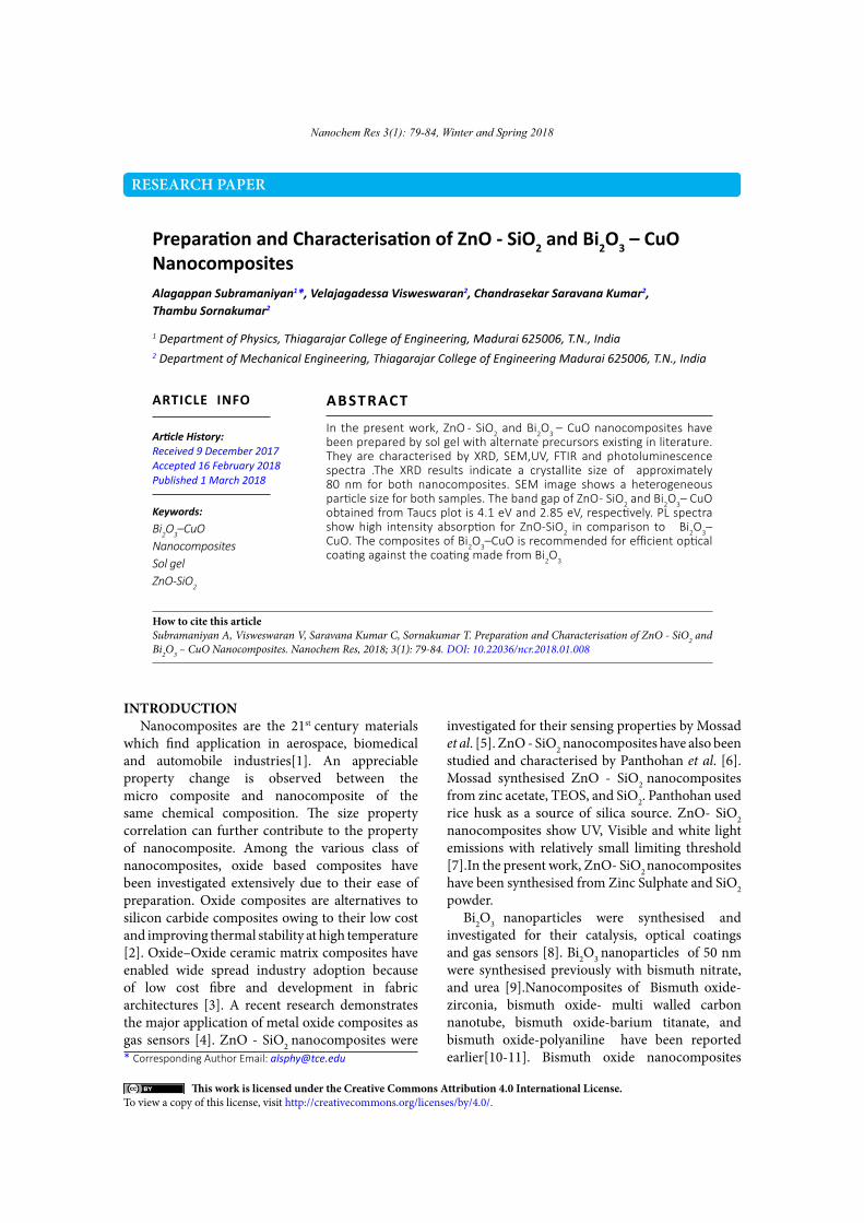
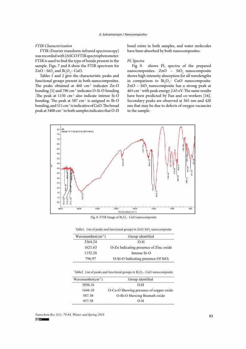
FTIR Characterization FTIR (Fourier transform infrared spectroscopy)
was recorded with JASCO FTIR spectrophotometer. FTIR is used to find the type of bonds present in the sample. Figs. 7 and 8 show the FTIR spectrum for ZnO - SiO2 and Bi2O3– CuO.
Tables 1 and 2 give the characteristic peaks and functional groups present in both nanocomposites. The peaks obtained at 460 cm-1 indicates Zn-O bonding [5] and 796 cm-1 indicates O-Si-O bonding .The peak at 1150 cm-1 also indicate intense Si-O bonding. The peak at 587 cm-1 is assigned to Bi-O bonding, and 511 cm-1 is indicative of CuO. The broad peak at 3400 cm-1 in both samples indicates that O-H
bond exists in both samples, and water molecules have been absorbed by both nanocomposites.
PL SpectraFig 9. shows PL spectra of the prepared
nanocomposites. ZnO – SiO2 nanocomposite shows high intensity absorption for all wavelengths in comparison to Bi2O3– CuO nanocomposite. ZnO – SiO2 nanocomposite has a strong peak at 465 cm-1 with peak energy 2.65 eV. The same results have been predicted by Fan and co-workers [16]. Secondary peaks are observed at 365 nm and 420 nm that may be due to defects of oxygen vacancies in the sample.
Table1.
Wavenumber(cm-1) Group identified3564.24 O-H1621.63 O-Zn Indicating presence of Zinc oxide1152.28 Intense Si-O796.97 O-Si-O Indicating presence Of SiO2
Table2.
Wavenumber(cm-1) Group identified3850.16 O-H1644.10 O-Cu-O Showing presence of copper oxide587.38 O-Bi-O Showing Bismuth oxide457.38 O-H
Table1. List of peaks and functional groups in ZnO-SiO2 nanocomposite
Table2. List of peaks and functional groups in Bi2O3– CuO nanocomposite
Fig 8.
Fig. 8. FTIR Image of Bi2O3– CuO nanocomposite

84 Nanochem Res 3(1): 79-84, Winter and Spring 2018
Fig. 9. PL spectra of the prepared nanocomposites
Fig. 9.
CONCLUSIONZnO-SiO2 and Bi2O3-CuO nanocomposites
were prepared by sol gel with alternate precursors existing in the literature, and characterised by XRD, SEM, UV, FTIR and PL. Both nanocomposites have a crystallize of 80 nm, and have compressive microstrain in the sample. The band gap of ZnO-SiO2 and Bi2O3– CuO obtained from Taucs plot is 4.1 eV and 2.85 eV, respectively. Based on the results, we suggest Bi2O3– CuO for efficient optical coatings with thermal management and good cathode material for solid oxide fuel cells. ZnO-SiO2 nanoocmposite is recommended as a better catalyst for photo degradation in comparison to Bi2O3-CuO. The PL spectra show high intensity absorption for ZnO-SiO2 in comparison to Bi2O3-CuO.
CONFLICT OF INTEREST The authors declare that there is no conflict of
interests regarding the publication of this paper.
REFERENCES1. Camargo PHC, Satyanarayana KG, Wypych F.
Nanocomposites: synthesis, structure, properties and new application opportunities. Materials Research. 2009;12(1):1-39.
2. Parlier M, Ritti MH, Jankowiak A. Potential and Perspectives for Oxide/Oxide Composites. AerospaceLab. 2011(3):p. 1-12.
3. Lincoln J, Jackson B, Barnes A, Beaber AR, Visser L. Oxide-Oxide Ceramic Matrix Composites - Enabling Widespread Industry Adoption. Advances in High Temperature Ceramic Matrix Compo sites and Materials for Sustainable Development; Ceramic Transactions, Volume CCLXIII: John Wiley & Sons, Inc.; 2017. p. 401-12.
4. Korotcenkov G, Cho BK. Metal oxide composites in conductometric gas sensors: Achievements and challenges.
Sensors and Actuators B: Chemical. 2017;244:182-210.5. Ali AM, Harraz FA, Ismail AA, Al-Sayari SA, Algarni H,
Al-Sehemi AG. Synthesis of amorphous ZnO–SiO 2 nanocomposite with enhanced chemical sensing properties. Thin Solid Films. 2016;605:277-82.
6. Pantohan EG, Candidato RT, Vequizo RM. Surface characteristics and structural properties of sol-gel prepared ZnO-SiO2 nanocomposite powders. IOP Conference Series: Materials Science and Engineering. 2015;79:012024.
7. Irimpan L, Krishnan B, Nampoori VPN, Radhakrishnan P. Linear and nonlinear optical characteristics of ZnO-SiO_2 nanocomposites. Applied Optics. 2008;47(24):4345.
8. Mallahi M, Shokuhfar A, Vaezi M, Esmaeilirad A, Mazinani V. Synthesis and characterization of bismuth oxide nanoparticles via sol-gel method. AJER. 2014;3:162-5.
9. Jha RK, Pasricha R, Ravi V. Synthesis of bismuth oxide nanoparticles using bismuth nitrate and urea. Ceramics International. 2005;31(3):495-7.
10. Joh DW, Park JH, Kim DY, Yun B-H, Lee KT. High performance zirconia-bismuth oxide nanocomposite electrolytes for lower temperature solid oxide fuel cells. Journal of Power Sources. 2016;320:267-73.
11. Periasamy AP, Yang S, Chen S-M. Preparation and characterization of bismuth oxide nanoparticles-multiwalled carbon nanotube composite for the development of horseradish peroxidase based H2O2 biosensor. Talanta. 2011;87:15-23.
12. Weinberg ZA, Rubloff GW, Bassous E. Transmission, photoconductivity, and the experimental band gap of thermally grown SiO2films. Physical Review B. 1979;19(6):3107-17.
13. Calabrese E, Fowler WB. Electronic energy-band structure of α quartz. Physical Review B. 1978;18(6):2888-96.
14. Nikitin T, Khriachtchev L. Optical and Structural Properties of Si Nanocrystals in SiO2 Films. Nanomaterials. 2015;5(2):614-55.
15. Leontie L, Caraman M, Alexe M, Harnagea C. Structural and optical characteristics of bismuth oxide thin films. Surface Science. 2002;507-510:480-5.
16. Fan XM, Lian JS, Zhao L, Liu YH. Single violet luminescence emitted from ZnO films obtained by oxidation of Zn film on quartz glass. Applied Surface Science. 2005;252(2):420-4.


![Synthesis and Characterisation of Lanthanum added ZnO ...joics.org/gallery/ics-1925.pdf · ZnO [26-30]. It clearly shows that the prepared ZnO and La doped ZnO samples revelation](https://static.fdocuments.net/doc/165x107/5ea23502b68dcf2dd872f588/synthesis-and-characterisation-of-lanthanum-added-zno-joicsorggalleryics-1925pdf.jpg)