Preliminary Study on the Assessment of the Marginal Fit of ...
6
Article Preliminary Study on the Assessment of the Marginal Fit of Three-Dimensional Methacrylate Oligomer Phosphine Oxide Provisional Fixed Dental Prostheses Made by Digital Light Processing Pedro Molinero-Mourelle 1 , Miguel Gómez-Polo 1, * , Cristina Gómez-Polo 2 , Rocio Ortega 3 , Jaime del Río Highsmith 1 and Alicia Celemín-Viñuela 1 1 Department of Conservative Dentistry and Orofacial Prosthetics, Faculty of Dentistry, Complutense University of Madrid, 28040 Madrid, Spain; [email protected] (P.M.-M.); [email protected] (J.d.R.H.); [email protected] (A.C.-V.) 2 Department of Surgery, University of Salamanca, 37007 Salamanca, Spain; [email protected] 3 Department of Prosthesis, School of Dentistry, European University of Madrid, 28045 Madrid, Spain; [email protected] * Correspondence: [email protected] Received: 10 August 2020; Accepted: 1 September 2020; Published: 7 September 2020 Abstract: This article aimed to assess the marginal fit of methacrylate-oligomer-phosphine-oxide curable-resin provisional-fixed dental prostheses made by digital-light-processing (DLP) three-dimensional (3D) printing. A stainless-steel master model with two abutments was scanned, and five three-unit provisional bridges were designed and printed in VITA shade A3.5 curable resin in 50 μm-thick layers. The marginal fit of each abutment was measured at six points using a profile projector. A descriptive data analysis of the fit measurements was performed by descriptive and explorative processes with the SPSS software. The curable-resin provisional restorations made by DLP 3D printing reached values of 46.37 μm (SD: 29.58 μm), which were considered clinically acceptable, with values similar to polyethylene-methacrylate and polyether-ether-ketone provisional restorations. Keywords: dental prosthesis; provisional restoration; CAD/CAM; marginal fit; 3D printing 1. Introduction Background Partially edentulous rehabilitation is currently a standard treatment option in daily clinical practice [1,2]. Although implant-supported restorations have grown in importance over the last 10 years due to patient and clinician demands, conventional tooth-borne restorations are still indicated [3]. These restorations have proven to be a reliable option over the years; however, their preparation in many cases requires several appointments to ensure their correct seating and performance [2–4]. Following dental preparation, provisional restorations ensure pulpal protection, periodontal health, interocclusal and intra-arch tooth relationships, occlusal function, and aesthetics during the confection of definitive fixed partial dentures (FPDs), aspects related to marginal fit [5]. One of the most important factors in relation to clinical behavior and survival in tooth-borne dental prostheses is marginal fit [6]. Marginal fit is related to the materials and manufacturing techniques of choice. Manufactured imperfections may result from a poor manufacturing process and the materials used for their confection. Lack of fit can create gaps leading to technical or biological complications [6,7]. Prosthesis 2020, 2, 240–245; doi:10.3390/prosthesis2030021 www.mdpi.com/journal/prosthesis
Transcript of Preliminary Study on the Assessment of the Marginal Fit of ...

Article
Preliminary Study on the Assessment of the MarginalFit of Three-Dimensional Methacrylate OligomerPhosphine Oxide Provisional Fixed Dental ProsthesesMade by Digital Light Processing
Pedro Molinero-Mourelle 1 , Miguel Gómez-Polo 1,* , Cristina Gómez-Polo 2, Rocio Ortega 3,Jaime del Río Highsmith 1 and Alicia Celemín-Viñuela 1
1 Department of Conservative Dentistry and Orofacial Prosthetics, Faculty of Dentistry, ComplutenseUniversity of Madrid, 28040 Madrid, Spain; [email protected] (P.M.-M.); [email protected] (J.d.R.H.);[email protected] (A.C.-V.)
2 Department of Surgery, University of Salamanca, 37007 Salamanca, Spain; [email protected] Department of Prosthesis, School of Dentistry, European University of Madrid, 28045 Madrid, Spain;
[email protected]* Correspondence: [email protected]
Received: 10 August 2020; Accepted: 1 September 2020; Published: 7 September 2020�����������������
Abstract: This article aimed to assess the marginal fit of methacrylate-oligomer-phosphine-oxidecurable-resin provisional-fixed dental prostheses made by digital-light-processing (DLP)three-dimensional (3D) printing. A stainless-steel master model with two abutments was scanned,and five three-unit provisional bridges were designed and printed in VITA shade A3.5 curable resinin 50 µm-thick layers. The marginal fit of each abutment was measured at six points using a profileprojector. A descriptive data analysis of the fit measurements was performed by descriptive andexplorative processes with the SPSS software. The curable-resin provisional restorations made by DLP3D printing reached values of 46.37 µm (SD: 29.58 µm), which were considered clinically acceptable,with values similar to polyethylene-methacrylate and polyether-ether-ketone provisional restorations.
Keywords: dental prosthesis; provisional restoration; CAD/CAM; marginal fit; 3D printing
1. Introduction
Background
Partially edentulous rehabilitation is currently a standard treatment option in daily clinicalpractice [1,2]. Although implant-supported restorations have grown in importance over the last 10 yearsdue to patient and clinician demands, conventional tooth-borne restorations are still indicated [3].These restorations have proven to be a reliable option over the years; however, their preparation inmany cases requires several appointments to ensure their correct seating and performance [2–4].
Following dental preparation, provisional restorations ensure pulpal protection, periodontalhealth, interocclusal and intra-arch tooth relationships, occlusal function, and aesthetics during theconfection of definitive fixed partial dentures (FPDs), aspects related to marginal fit [5].
One of the most important factors in relation to clinical behavior and survival in tooth-bornedental prostheses is marginal fit [6]. Marginal fit is related to the materials and manufacturingtechniques of choice. Manufactured imperfections may result from a poor manufacturing process andthe materials used for their confection. Lack of fit can create gaps leading to technical or biologicalcomplications [6,7].
Prosthesis 2020, 2, 240–245; doi:10.3390/prosthesis2030021 www.mdpi.com/journal/prosthesis

Prosthesis 2020, 2 241
With the establishment of computer-aided design/computer-aided manufacturing (CAD/CAM)technology in dental medicine, there has been an increase in the development of new materials,designs, and techniques in the manufacturing of dental prosthesis [8]. One of these recent methods hasbeen the use of three-dimensional (3D) additive printers in dental laboratories and dental clinics [9].Three-dimensional additive technology allows the obtaining of prostheses, radiological and/or surgicalsplints, digital models, etc. made with different materials at a competitive cost and without the loss ofmaterials linked to the milling process [9,10]. In the case of resin polymers, these systems allow the3D impression of provisional restorations as part of a chairside concept, within the same clinic andappointment [10]. Digital light processing (DLP) additive technology can use several monomers andresin systems, such as a UV-curable hybrid resin, cationic-initiated epoxy monomers, or photocuringmulti-phase polymers [11].
One of these systems is digital light processing (DLP), a 3D printing system based on the useof a digital light projection source (high-power LED). The layers are illuminated using a light maskcreated by a digital micromirror device. Each mirror corresponds to a pixel of the projected image,polymerizing the entire resin layer at once [11,12].
Additive technology is relatively new in dental medicine, and there are few studies evaluating thepreclinical behavior of systems and materials. Due to the increasing demand for CAD/CAM systemsand the great offer of 3D printers by the industry, its evaluation is necessary.
This preliminary study aimed to assess the marginal fit of a curable resin as a material forprovisional three-unit FPDs made from three-dimensional printing and to compare the fit valuesagainst those described in the literature, in order to determine whether these provisional restorationscould be clinically acceptable.
2. Results
Five provisional three-unit FDPs were obtained for vertical-marginal-discrepancy assessmentmeasured at six points, three in buccal and three in palatal/lingual areas.
A total of 60 measurements (30 per abutment) at 4×magnification were performed. The resultsof the analysis of marginal fit reported a maximal discrepancy of 152.52 µm, minimum of 7.5 µm,and mean of 46.37 µm. A descriptive analysis of the quantitative variables (DESCRIPTIVE andEXPLORE processes) was performed, as shown in Table 1.
Table 1. Frequency table of the marginal fit of provisional FDPs (µm).
Measurements N Mean Median SD Minimum Maximum
Distal–buccal 5 76.36 76.35 76.35 60.05 152.52Distal–buccal 5 41.05 22.5 41.51 7.5 105.03Distal–buccal 5 31.96 31.96 26.90 8.75 75.17Distal–lingual 5 62.49 62.49 26.06 37.83 100.12Distal–lingual 5 61.13 61.13 26.84 37.58 105.03Distal–lingual 5 29.5 29.5 15.61 13.46 52.74Mesial–Buccal 5 30.03 30.03 12.74 13.75 40.08Mesial–Buccal 5 22.65 22.65 7.47 15.21 35.09Mesial–Buccal 5 33.86 33.86 1.73 32.52 36.59Mesial–lingual 5 43.47 43.47 18.29 18.2 60.21Mesial–lingual 5 76.89 76.89 21.66 40.49 95.03Mesial–lingual 5 47.05 52.74 21.21 16.68 70.04
Total 60 46.37 38.65 29.58 7.5 152.52
Data expressed in µm.

Prosthesis 2020, 2 242
3. Discussion
This study evaluated the marginal fit of curable resin as a three-unit provisional FPD materialmade from 3D printing. The proposed null hypothesis was rejected since the marginal adjustment ofthese restorations was within the accepted margins in the literature.
To date, the literature has provided a range of marginal fit values for fixed prostheses between 10and 110 µm, although what is considered clinically acceptable is 120 µm [13,14]. Despite the fact thatseveral methods have been reported to assess the gap between a restoration and its margin, the presentstudy was based on the use of a profile projector following the assessments performed by Holmes et al.and the reported marginal gap was measured for the marginal fit [15].
A study published by Park et al. in 2019 evaluated the internal and the marginal fit of resin interimFDPs that were 3D printed using DLP technology with two different thickness layers and five buildorientations. In this study, in addition to an internal-fit evaluation, the authors assessed the marginaldiscrepancy according to the method described by Holmes et al. [14]. The results for the marginaldiscrepancy of the restorations confectioned at an orientation of 60◦ and made of 100 µm-thick layersprovided the best mean discrepancy values (50.0 ± 14.7 µm). Nevertheless, it must be noted that mostof the 3D printed groups showed worse results than the milled ones [16].
DLP technology is based on an ultraviolet (UV) light source that cures a photosensitive liquidpolymer in layers following a CAD design [11]. A methodological limitation can be noted, consideringthat a direct comparison could not be obtained, as the thickness of the cement was different fromthat in the present study (0.24 mm) and the inner side of the prosthesis was not observed. However,considering the obtained fit ranges, the mean results can be considered clinically acceptable and arelower than those reported in the aforementioned study. A previous study of 3D provisional crownsmade using the same methodology reported a mean marginal fit value of 122.89 µm in the marginal-fitdiscrepancy of the restorations made with polylactic acid (PLA) using a fused-deposition-modeling(FDM) 3D system. FDM uses a preformed polymer as a building material that uses the input ofprocessing energy in the pre-deposition stage to obtain a polymer melt that can be applied using a fineprint head or nozzle [11,17]. Although this study analyzed singe interim restorations by extrapolatingthe results, the FDPs of the present study obtained minor discrepancy results [16].
Comparing the misfit results of CAD/CAM 3D printing techniques with those of conventionalmanufacturing methods, Givens et al. assessed the marginal fit of provisional restorations made bydirect manufacturing methods using polyethylene methacrylate (PEMA), self-polymerizing bisacrylic,and dual-curing bisacrylic, showing misfit results similar to those obtained in the present study,between 177 and 319 µm [5].
Alharbi et al. studied the marginal and internal fit of printed and milled restorations (additiveand subtractive methods), in preparations with different finishing lines with a micro-CT; the meandiscrepancy in 3D printing went from 28 to 41 µm, while in the milling group, it was between 32 and56 µm [18].
Regarding CAD/CAM techniques, we found bigger discrepancies; Abdullah et al. also assessed thefit of direct-technique and CAD/CAM provisional materials. The average marginal discrepancy of thePEMA restorations was 193.07 µm, while the polyether ether ketone (PEEK) restorations manufacturedby CAD/CAM ranged between 46.75 and 60.61 µm [19].
Since this study was designed as a descriptive preliminary study, its main limitation was thesmall sample size and the lack of a control group and direct comparisons against previously publishedstudies. Nevertheless, this article intended to be the starting point for future clinical studies in orderto assess the feasibility of this 3D printing system using the aforementioned material. Taking intoaccount the sample size limitation and considering that the minimum values of 7.5 µm and maximumof 152.52 µm are very broad, alongside the fact that standard deviation (29.58) is more than 50% ofthe mean (46.37), these results should be carefully interpreted, as this SD shows that the data areless reliable.
Prosthesis 2020, 2 243
Additive technology is relatively new for temporary restorations; material selection can influencemarginal fit. Although there is no material for provisional restorations that can be considered as the“gold standard”, the literature on methacrylate oligomer phosphine oxide is scarce. Although theresults are within clinically acceptable limits, the ability of this material to be printed with 50 µm-thicklayers can influence the marginal fit, since in milled or injected materials, the thickness can be influencedby the prosthetic design space [5].
4. Materials and Methods
A comparative preliminary in vitro study of the vertical marginal fit of provisional three-unitFPDs made of 3D-printed methacrylate oligomer phosphine oxide curable resin was carried out at theDepartment of Conservative Dentistry and Orofacial Prosthetics, Complutense University of Madrid.
A master model was designed from two abutment teeth for a tooth-supported three-unit resindental prosthesis. A stainless-steel machined master model with two standardized abutments asa master die was confectioned to simulate a first premolar and first molar prepared for three-unitFPDs. The technical specifications of the abutments were a 5 mm height, an occlusal diameter of5 mm, a 1 mm-wide chamfer finishing line, a 6◦ convergence angle of the axial walls, and roundedangles. The cast was scanned using the EVO Ceratomic Protechno scanner (PROTECHNO, FamadentS.L.U. Vilamalla, Girona, Spain); subsequently, five posterior three-unit FDPs were designed using theEXOCAD software (Exocad GmbH, Darmstadt, Germany) (Figure 1a,b).
Prosthesis 2020, 1, FOR PEER REVIEW 4
Since this study was designed as a descriptive preliminary study, its main limitation was the
small sample size and the lack of a control group and direct comparisons against previously
published studies. Nevertheless, this article intended to be the starting point for future clinical studies
in order to assess the feasibility of this 3D printing system using the aforementioned material. Taking
into account the sample size limitation and considering that the minimum values of 7.5 μm and
maximum of 152.52 μm are very broad, alongside the fact that standard deviation (29.58) is more
than 50% of the mean (46.37), these results should be carefully interpreted, as this SD shows that the
data are less reliable.
Additive technology is relatively new for temporary restorations; material selection can
influence marginal fit. Although there is no material for provisional restorations that can be
considered as the “gold standard”, the literature on methacrylate oligomer phosphine oxide is scarce.
Although the results are within clinically acceptable limits, the ability of this material to be printed
with 50 μm-thick layers can influence the marginal fit, since in milled or injected materials, the
thickness can be influenced by the prosthetic design space [5].
4. Materials and Methods
A comparative preliminary in vitro study of the vertical marginal fit of provisional three-unit
FPDs made of 3D-printed methacrylate oligomer phosphine oxide curable resin was carried out at the
Department of Conservative Dentistry and Orofacial Prosthetics, Complutense University of Madrid.
A master model was designed from two abutment teeth for a tooth-supported three-unit resin
dental prosthesis. A stainless-steel machined master model with two standardized abutments as a
master die was confectioned to simulate a first premolar and first molar prepared for three-unit FPDs.
The technical specifications of the abutments were a 5 mm height, an occlusal diameter of 5 mm, a 1
mm-wide chamfer finishing line, a 6° convergence angle of the axial walls, and rounded angles. The
cast was scanned using the EVO Ceratomic Protechno scanner (PROTECHNO, Famadent S.L.U.
Vilamalla, Girona, Spain); subsequently, five posterior three-unit FDPs were designed using the
EXOCAD software (Exocad GmbH, Darmstadt, Germany) (Figure 1a,b).
Figure 1. (a) Master die STL file. (b) Provisional fixed dental prostheses; STL file design.
The FPD abutments were designed with an insertion angle of 6°, using a cementation line of 0
mm at the cervical area and 0.08 mm at the rest of the prosthetic surface. The bridge connectors were
9 mm2 in surface area. Once the restorations were designed, the STL file was obtained and transferred
to the Rapidshape-SHERAprint 30 DLP system printer (SHERA Werkstoff-Technologie GmbH & Co.
KG, Lemförde, Germany). As a preliminary study to test the feasibility of a possible clinical study,
five three-unit tooth-supported provisional restorations were printed in curable resin (methacrylate
oligomer phosphine oxide, SHERA Werkstoff-Technologie GmbH & Co. KG, Germany) with 50 μm-
thick layers in the A3.5 VITA shade (VITA Zahnfabrik H, Bad Säckingen, Germany). The
manufacturing process was carried out at the faculty dental laboratory in a room with controlled
light that does not affect the 3D printer and with a constant controlled temperature between 25 and
30 °C (Table 2).
(a) (b)
Figure 1. (a) Master die STL file. (b) Provisional fixed dental prostheses; STL file design.
The FPD abutments were designed with an insertion angle of 6◦, using a cementation line of0 mm at the cervical area and 0.08 mm at the rest of the prosthetic surface. The bridge connectorswere 9 mm2 in surface area. Once the restorations were designed, the STL file was obtained andtransferred to the Rapidshape-SHERAprint 30 DLP system printer (SHERA Werkstoff-TechnologieGmbH & Co. KG, Lemförde, Germany). As a preliminary study to test the feasibility of a possibleclinical study, five three-unit tooth-supported provisional restorations were printed in curable resin(methacrylate oligomer phosphine oxide, SHERA Werkstoff-Technologie GmbH & Co. KG, Germany)with 50 µm-thick layers in the A3.5 VITA shade (VITA Zahnfabrik H, Bad Säckingen, Germany).The manufacturing process was carried out at the faculty dental laboratory in a room with controlledlight that does not affect the 3D printer and with a constant controlled temperature between 25 and30 ◦C (Table 2).
Table 2. Technical specifications of the curable resin (methacrylate oligomer phosphine oxide) providedby the manufacturer.
Characteristics Value
Viscosity at 23 ◦C 0.9–1.4 Pa sBending strength =85 MPa
Bending e-module =2100 MPaShore hardness D 80–90
Absorption of water <30 µg/mm2
Water solubility <5 µg/mm2
Prosthesis 2020, 2 244
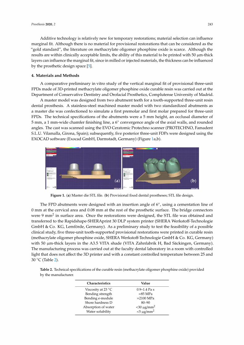
The restorations were manufactured using the SHERAflash-light plus curing unit (SHERAWerkstoff-Technologie GmbH & Co. KG, Germany). After 3D printing, all the FPDs were examinedboth on the inside and on the finish line prior to measurement in order to identify printing imperfectionsand cleaned with 95% ethanol for 2 min, and subsequently, the bridges were trimmed at their baseand polished. The vertical marginal fit of the restorations was assessed by measuring the externaland marginal vertical gap. The measures were the vertical distance between the crown margin andthe prepared cavosurface angle following previous studies at six points for each abutment, makingthree marks at the buccal and at the lingual surface of the die, using a profile projector with a 4×magnification (Toupview V.x643.7.6701, Photonics Co., Ltd., Suzhou, China) [20]. A total of sixtymeasurements were recorded for the FDPs (Figure 2a,b).
Prosthesis 2020, 1, FOR PEER REVIEW 5
Table 2. Technical specifications of the curable resin (methacrylate oligomer phosphine oxide)
provided by the manufacturer.
Characteristics Value
Viscosity at 23 °C 0.9–1.4 Pa s
Bending strength =85 MPa
Bending e-module =2100 MPa
Shore hardness D 80–90
Absorption of water <30 μg/mm2
Water solubility <5 μg/mm2
The restorations were manufactured using the SHERAflash-light plus curing unit (SHERA
Werkstoff-Technologie GmbH & Co. KG, Germany). After 3D printing, all the FPDs were examined
both on the inside and on the finish line prior to measurement in order to identify printing
imperfections and cleaned with 95% ethanol for 2 min, and subsequently, the bridges were trimmed
at their base and polished. The vertical marginal fit of the restorations was assessed by measuring
the external and marginal vertical gap. The measures were the vertical distance between the crown
margin and the prepared cavosurface angle following previous studies at six points for each
abutment, making three marks at the buccal and at the lingual surface of the die, using a profile
projector with a 4× magnification (Toupview V.x643.7.6701, Photonics Co., Ltd., Suzhou, China) [20].
A total of sixty measurements were recorded for the FDPs (Figure 2a,b).
Figure 2. (a) Provisional fixed dental prostheses. (b) Vertical marginal fit profile projector (4x).
The statistical data analysis was performed by the descriptive analysis of the quantitative
variables (DESCRIPTIVE and EXPLORE processes) using the SPSS software (SPSS 23.0, Chicago, IL,
USA) and Microsoft Excel for Mac 2011 (Excel Version 14.4.6, Microsoft, WA, USA).
5. Conclusions
Within the limitations of this preliminary in vitro study, methacrylate oligomer phosphine oxide
curable resin provisional restorations made by DLP 3D seems to provide marginal fit values within
the clinically acceptable limits. Further well-designed comparative studies are needed to obtain more
reliable conclusions.
Author Contributions: Conceptualization, P.M.-M. and M.G.-P.; methodology, P.M.-M., M.G.-P. and A.C.-V.;
software, P.M.-M.; validation, P.M.-M., M.G.-P. and A.C.-V.; formal analysis, P.M.-M., M.G.-P. and C.G.-P.;
research, P.M.-M. and M.G.-P.; writing—original draft preparation, P.M.-M., R.O. and M.G.-P.; writing—review
and editing, P.M.-M., M.G.-P., R.O. and A.C.-V.; visualization, C.G.-P.; supervision, J.d.R.H. and A.C.-V.; project
(a) (b)
Figure 2. (a) Provisional fixed dental prostheses. (b) Vertical marginal fit profile projector (4x).
The statistical data analysis was performed by the descriptive analysis of the quantitative variables(DESCRIPTIVE and EXPLORE processes) using the SPSS software (SPSS 23.0, Chicago, IL, USA) andMicrosoft Excel for Mac 2011 (Excel Version 14.4.6, Microsoft, WA, USA).
5. Conclusions
Within the limitations of this preliminary in vitro study, methacrylate oligomer phosphine oxidecurable resin provisional restorations made by DLP 3D seems to provide marginal fit values withinthe clinically acceptable limits. Further well-designed comparative studies are needed to obtain morereliable conclusions.
Author Contributions: Conceptualization, P.M.-M. and M.G.-P.; methodology, P.M.-M., M.G.-P. and A.C.-V.;software, P.M.-M.; validation, P.M.-M., M.G.-P. and A.C.-V.; formal analysis, P.M.-M., M.G.-P. and C.G.-P.; research,P.M.-M. and M.G.-P.; writing—original draft preparation, P.M.-M., R.O. and M.G.-P.; writing—review and editing,P.M.-M., M.G.-P., R.O. and A.C.-V.; visualization, C.G.-P.; supervision, J.d.R.H. and A.C.-V.; project administration,A.C.-V.; funding acquisition, J.d.R.H. All authors have read and agreed to the published version of the manuscript.
Funding: This study received no external funding.
Acknowledgments: The authors would like to express their gratitude to Alberto Cervera for providing the testingand analysis methods and Alexandra Helm for the linguistic revision.
Conflicts of Interest: The authors declare no conflict of interest.
References
1. Abt, E.; Carr, A.B.; Worthington, H.V. Interventions for replacing missing teeth: Partially absent dentition.Cochrane Database Syst. Rev. 2012, 2, CD003814. [CrossRef] [PubMed]
2. Salinas, T.; Block, M.S.; Sadan, A. Fixed partial denture or single-tooth implant restoration?Statistical considerations for sequencing and treatment. J. Oral Maxillofac. Surg. 2004, 62, 2–16. [PubMed]
3. Reitemeier, B.; Hänsel, K.; Kästner, C.; Weber, A.; Walter, M.H. A prospective 10-year study of metal ceramicsingle crowns and fixed dental prosthesis retainers in private practice set tings. J. Prosthet. Dent. 2013, 109,149–155. [CrossRef]

Prosthesis 2020, 2 245
4. Limones, A.; Molinero-Mourelle, P.; Azevedo, L.; Romeo-Rubio, M.; Correia, A.; Gómez-Polo, M.Zirconia-ceramic vs. metal-ceramic multi-unit tooth-supported posterior fixed dental prosthesis: A systematicreview and meta-analysis. J. Am. Dent. Assoc. 2020, 151, 230–238. [CrossRef] [PubMed]
5. Givens, E.J., Jr.; Neiva, G.; Yaman, P.; Dennison, J.B. Marginal adaptation and color stability of four provisionalmate-rials. J. Prosthodont. 2008, 17, 97–101. [CrossRef] [PubMed]
6. Svanborg, P. A systematic review on the accuracy of zirconia crowns and fixed dental prosthe-ses.Biomater. Investig. Dent. 2020, 7, 9–15. [PubMed]
7. Rinke, S.; Fornefett, D.; Gersdorff, N.; Lange, K.; Roediger, M. Multifactorial analysis of the impact ofdiffer-ent manufacturing processes on the marginal fit of zirconia copings. Dent. Mater. J. 2012, 31, 601–609.[CrossRef] [PubMed]
8. Joda, T.; Ferrari, M.; Gallucci, G.O.; Wittneben, J.G.; Brägger, U. Digital technology in fixed implantprostho-dontics. Periodontology 2017, 73, 178–192. [CrossRef] [PubMed]
9. Revilla-León, M.; Meyer, M.J.; Özcan, M. Metal additive manufacturing technologies: Literature review ofcurrent status and prosthodontic applications. Int. J. Comput. Dent. 2019, 22, 55–67. [PubMed]
10. Revilla-León, M.; Meyers, M.J.; Zandinejad, A.; Özcan, M. A review on chemical composition, mechanicalproperties, and manufacturing work flow of additively manufactured current polymers for interim dentalrestorations. J. Esthet. Restor. Dent. 2019, 31, 51–57. [CrossRef] [PubMed]
11. Stansbury, J.W.; Idacavage, M.J. 3D printing with polymers: Challenges among expanding options andopportunities. Dent. Mater. 2016, 32, 54–64. [CrossRef] [PubMed]
12. Alharbi, N.; Wismeijer, D.; Osman, R. Additive Manufacturing Techniques in Prosthodontics: Where Do WeCurrently Stand? A Critical Review. Int. J. Prosthodont. 2017, 30, 474–484. [CrossRef] [PubMed]
13. McLean, J.W.; Von Fraunhofer, J.A. The estimation of cement film thickness by an in vivo technique.Br. Dent. J. 1971, 131, 107–111. [CrossRef] [PubMed]
14. Nawafleh, N.; Mack, F.; Evans, J.; Mackay, J.; Hatamleh, M.M. Accuracy and Reliability of Methods toMeasure Marginal Adaptation of Crowns and FDPs: A Literature Review. J. Prosthodont. 2013, 22, 419–428.[CrossRef] [PubMed]
15. Holmes, J.R.; Bayne, S.C.; Holland, G.A.; Sulik, W.D. Considerations in measurement of marginalfit. J. Prosthet. Dent. 1989, 62, 405–408. [CrossRef]
16. Park, G.-S.; Kim, S.-K.; Heo, S.-J.; Koak, J.-Y.; Seo, D.-G. Effects of Printing Parameters on the Fit ofImplant-Supported 3D Printing Resin Prosthetics. Matererials 2019, 12, 2533. [CrossRef] [PubMed]
17. Molinero-Mourelle, P.; Canals, S.; Gomez-Polo, M.; Sola-Ruiz, M.; Highsmith, J.D.R.; Viñuela, A. PolylacticAcid as a Material for Three-Dimensional Printing of Provisional Restorations. Int. J. Prosthodont. 2018, 31,349–350. [CrossRef] [PubMed]
18. Alharbi, N.; Alharbi, S.; Cuijpers, V.M.; Osman, R.B.; Wismeijer, D. Three-dimensional evaluation of marginaland internal fit of 3D-printed interim restorations fabricated on different finish line designs. J. Prosthodont. Res.2018, 62, 218–226. [CrossRef] [PubMed]
19. Abdullah, A.O.; Tsitrou, E.A.; Pollington, S. Comparative in vitro evaluation of CAD/CAM vs. conven-tionalprovisional crowns. J. Appl. Oral Sci. 2016, 24, 258–263. [CrossRef] [PubMed]
20. Ortega, R.; Gonzalo, E.; Gomez-Polo, M.; Lopez-Suarez, C.; Suarez, M.J. SEM evaluation of the precisionof fit of CAD/CAM zirconia and metal-ceramic posterior crowns. Dent. Mater. J. 2017, 36, 387–393.[CrossRef] [PubMed]
© 2020 by the authors. Licensee MDPI, Basel, Switzerland. This article is an open accessarticle distributed under the terms and conditions of the Creative Commons Attribution(CC BY) license (http://creativecommons.org/licenses/by/4.0/).