Predisposing Factors for Adrenal Insufficiency · more, secondary adrenal insufficiency has been...
12
review article The new england journal of medicine n engl j med 360;22 nejm.org may 28, 2009 2328 CURRENT CONCEPTS Predisposing Factors for Adrenal Insufficiency Stefan R. Bornstein, M.D. From the Department of Medicine, Tech- nical University of Dresden, Dresden, Ger- many. Address reprint requests to Dr. Bornstein at the Department of Medicine, Technical University of Dresden, Fetscher- str. 74, 01307 Dresden, Germany, or at [email protected]. N Engl J Med 2009;360:2328-39. Copyright © 2009 Massachusetts Medical Society. A drenal insufficiency — the clinical manifestation of deficient production or action of glucocorticoids — is a life-threatening disorder that may result from either primary adrenal failure or secondary adrenal disease due to impairment of the hypothalamic–pituitary axis. 1,2 This article focuses on pro- viding the practicing clinician with new insights into predisposing factors for adre- nal insufficiency. When and during what situations should a clinician suspect ad- renal insufficiency? What genetic disorders, infections, and medications should be considered? What are the current views on the underlying mechanisms? The cardinal clinical symptoms of adrenocortical insufficiency, as first described by Thomas Addison in 1855, 3 include weakness, fatigue, anorexia, and abdominal pain, with orthostatic hypotension, salt craving, and characteristic hyperpigmenta- tion of the skin occurring with primary adrenal failure. The acute syndrome con- stitutes a medical emergency since it may result in a severe hypotensive crisis and clouded sensorium, together with pain in the muscles, joints, or abdomen and fever. 1,2 In the diagnostic workup for the disorder, the capacity of the adrenal cortex to respond to corticotropin is tested with the use of the standard short corticotropin test, which measures the serum cortisol level before and 30 or 60 minutes after an intravenous or intramuscular injection of 250 μg of corticotropin. 4 An increase in the serum cortisol level to peak concentrations above 500 nmol per liter (18 μg per deciliter) indicates a normal response. The adrenal responsiveness to an exogenous corticotropin challenge is impaired in most cases of secondary adrenal disease. With mild secondary adrenal insufficiency, however, the hypothalamic–pituitary– adrenal axis may appear intact, with a normal response to a corticotropin chal- lenge. Recent evidence suggests that the 1-μg corticotropin stimulation test is more sensitive than the 250-μg corticotropin test for establishing the diagnosis of sec- ondary adrenal insufficiency. 5 Once adrenal insufficiency is diagnosed, glucocorticoid replacement is initiated in two or three daily doses; one half to two thirds of the daily dose (15 to 25 mg of hydrocortisone) is given in the morning, in line with the physiologic cortisol-secre- tion pattern. Mineralocorticoid replacement (0.05 to 0.2 mg of fludrocortisone daily as a morning dose) is required only in the case of primary adrenal insufficiency, and dehydroepiandrosterone replacement (25 to 50 mg) remains an optional treatment. 1,2 Management of an acute adrenal crisis consists of immediate intravenous ad- ministration of 100 mg of hydrocortisone, followed by 100 to 200 mg of hydrocor- tisone every 24 hours and a continuous infusion of larger volumes of physiologic saline solution (initially 1 liter per hour) under continuous cardiac monitoring. Timely diagnosis and clinical management of this condition are critical, and physicians in all areas of medicine should be aware of the causes, signs, and symptoms that her- ald adrenal insufficiency. The New England Journal of Medicine Downloaded from nejm.org at UNIVERSITY OF CHICAGO LIBRARIES on May 8, 2013. For personal use only. No other uses without permission. Copyright © 2009 Massachusetts Medical Society. All rights reserved.
Transcript of Predisposing Factors for Adrenal Insufficiency · more, secondary adrenal insufficiency has been...
review article
T h e n e w e ngl a nd j o u r na l o f m e dic i n e
n engl j med 360;22 nejm.org may 28, 20092328
CURRENT CONCEPTS
Predisposing Factors for Adrenal Insufficiency
Stefan R. Bornstein, M.D.
From the Department of Medicine, Tech-nical University of Dresden, Dresden, Ger-many. Address reprint requests to Dr. Bornstein at the Department of Medicine, Technical University of Dresden, Fetscher-str. 74, 01307 Dresden, Germany, or at [email protected].
N Engl J Med 2009;360:2328-39.Copyright © 2009 Massachusetts Medical Society.
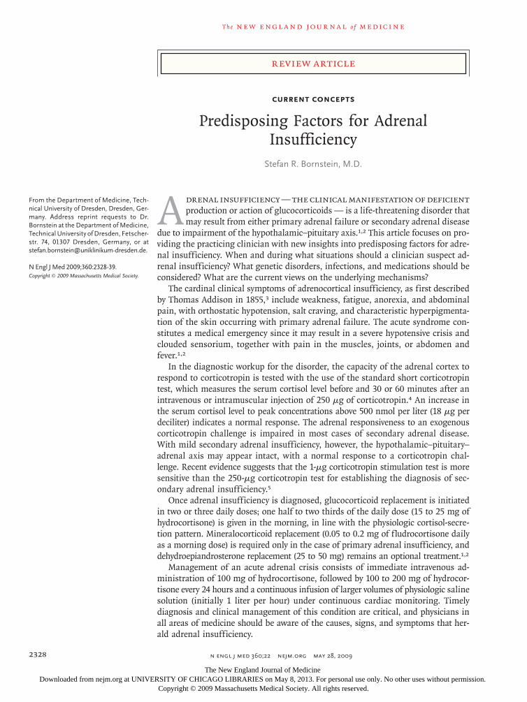
A drenal insufficiency — the clinical manifestation of deficient production or action of glucocorticoids — is a life-threatening disorder that may result from either primary adrenal failure or secondary adrenal disease
due to impairment of the hypothalamic–pituitary axis.1,2 This article focuses on pro-viding the practicing clinician with new insights into predisposing factors for adre-nal insufficiency. When and during what situations should a clinician suspect ad-renal insufficiency? What genetic disorders, infections, and medications should be considered? What are the current views on the underlying mechanisms?
The cardinal clinical symptoms of adrenocortical insufficiency, as first described by Thomas Addison in 1855,3 include weakness, fatigue, anorexia, and abdominal pain, with orthostatic hypotension, salt craving, and characteristic hyperpigmenta-tion of the skin occurring with primary adrenal failure. The acute syndrome con-stitutes a medical emergency since it may result in a severe hypotensive crisis and clouded sensorium, together with pain in the muscles, joints, or abdomen and fever.1,2
In the diagnostic workup for the disorder, the capacity of the adrenal cortex to respond to corticotropin is tested with the use of the standard short corticotropin test, which measures the serum cortisol level before and 30 or 60 minutes after an intravenous or intramuscular injection of 250 μg of corticotropin.4 An increase in the serum cortisol level to peak concentrations above 500 nmol per liter (18 μg per deciliter) indicates a normal response. The adrenal responsiveness to an exogenous corticotropin challenge is impaired in most cases of secondary adrenal disease. With mild secondary adrenal insufficiency, however, the hypothalamic–pituitary–adrenal axis may appear intact, with a normal response to a corticotropin chal-lenge. Recent evidence suggests that the 1-μg corticotropin stimulation test is more sensitive than the 250-μg corticotropin test for establishing the diagnosis of sec-ondary adrenal insufficiency.5
Once adrenal insufficiency is diagnosed, glucocorticoid replacement is initiated in two or three daily doses; one half to two thirds of the daily dose (15 to 25 mg of hydrocortisone) is given in the morning, in line with the physiologic cortisol-secre-tion pattern. Mineralocorticoid replacement (0.05 to 0.2 mg of fludrocortisone daily as a morning dose) is required only in the case of primary adrenal insufficiency, and dehydroepiandrosterone replacement (25 to 50 mg) remains an optional treatment.1,2
Management of an acute adrenal crisis consists of immediate intravenous ad-ministration of 100 mg of hydrocortisone, followed by 100 to 200 mg of hydrocor-tisone every 24 hours and a continuous infusion of larger volumes of physiologic saline solution (initially 1 liter per hour) under continuous cardiac monitoring. Timely diagnosis and clinical management of this condition are critical, and physicians in all areas of medicine should be aware of the causes, signs, and symptoms that her-ald adrenal insufficiency.
The New England Journal of Medicine Downloaded from nejm.org at UNIVERSITY OF CHICAGO LIBRARIES on May 8, 2013. For personal use only. No other uses without permission.
Copyright © 2009 Massachusetts Medical Society. All rights reserved.
current concepts
n engl j med 360;22 nejm.org may 28, 2009 2329
Her edi ta r y Disor der s A sso ci ated w i th A dr ena l
Insufficienc y
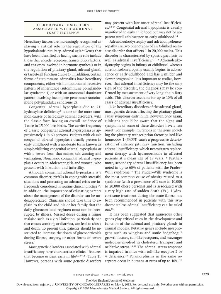
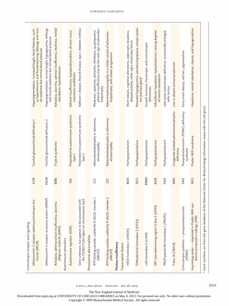
Hereditary factors are increasingly recognized as playing a critical role in the regulation of the hypothalamic–pituitary–adrenal axis.6 Genes that have been identified as having such a role include those that encode receptors, transcription factors, and enzymes involved in hormone synthesis or in the regulation of pituitary-gland, adrenal-gland, or target-cell function (Table 1). In addition, certain forms of autoimmune adrenalitis have hereditary components, either with an autosomal recessive pattern of inheritance (autoimmune polyglandu-lar syndrome 1) or with an autosomal dominant pattern involving incomplete penetrance (autoim-mune polyglandular syndrome 2).
Congenital adrenal hyperplasia due to 21- hydroxylase deficiency7 is one of the more com-mon causes of hereditary adrenal disorders, with the classic form having an overall incidence of 1 case in 15,000 live births. The carrier frequency of classic congenital adrenal hyperplasia is ap-proximately 1 in 60 persons. Patients with classic congenital adrenal hyperplasia usually present in early childhood with a moderate form known as simple-virilizing congenital adrenal hyperplasia or with a severe form that causes salt wasting and virilization. Nonclassic congenital adrenal hyper-plasia occurs in adolescent girls and women, who present with hirsutism and infertility.
Although congenital adrenal hyperplasia is a common disorder, pitfalls in coping with stressful situations and preventing an adrenal crisis are in-frequently considered in routine clinical practice7,8; in addition, the importance of educating parents about the management of the disorder can be un-derappreciated. Clinicians should take time to ex-plain to the child and his or her family that the daily glucocorticoid regimen must not be inter-rupted by illness. Missed doses during a minor malaise such as a viral infection, particularly one that causes vomiting or diarrhea, can lead to shock and death. To prevent this, patients should be in-structed to increase the doses of glucocorticoids during illness, surgery, or other forms of severe stress.
Most genetic disorders associated with adrenal insufficiency have characteristic clinical features that become evident early in life1,2,9-11 (Table 1). However, persons with some genetic disorders
may present with late-onset adrenal insufficien-cy.12-14 Congenital adrenal hypoplasia is usually manifested in early childhood but may not be ap-parent until adolescence or early adulthood.14
Adrenoleukodystrophy and adrenomyeloneu-ropathy are two phenotypes of an X-linked reces-sive disorder that affects 1 in 20,000 males. This disorder is characterized by spastic paralysis as well as adrenal insufficiency.1,2,15 Adrenoleuko-dystrophy begins in infancy or childhood, whereas adrenomyeloneuropathy usually begins in adoles-cence or early adulthood and has a milder and slower progression. It is important to realize, how-ever, that adrenal insufficiency may be the only sign of the disorder; the diagnosis may be con-firmed by measurement of very-long-chain fatty acids. This disorder accounts for up to 10% of all cases of adrenal insufficiency.
Like hereditary disorders of the adrenal gland, most genetic defects affecting the pituitary gland cause symptoms early in life; however, once again, clinicians should be aware that the signs and symptoms of some of these disorders have a late onset. For example, mutations in the gene encod-ing the pituitary transcription factor paired-like homeobox 1 (PROP1) cause a progressive deterio-ration of anterior pituitary function, including adrenal insufficiency, which necessitates replace-ment therapy with hydrocortisone in affected patients at a mean age of 18 years.13 Further-more, secondary adrenal insufficiency has been noted in up to 60% of patients with the Prader–Willi syndrome.16 The Prader–Willi syndrome is the most common cause of obesity related to a syndrome (with a prevalence of 1 case in 10,000 to 20,000 obese persons) and is associated with a very high rate of sudden death (3%). Hydro-cortisone treatment during the acute illness has been recommended in patients with this syn-drome unless adrenal insufficiency can be ruled out.16
It has been suggested that numerous other genes play critical roles in the development and function of the adrenal and pituitary glands in animal models. Putative genes include morpho-gens such as wingless and sonic hedgehog,17 growth factors, toll-like receptors, and scavenger molecules involved in cholesterol transport and oxidative stress.18,19 The adrenal stress response is impaired in mice with toll-like receptor 2 or 4 deficiency.19 Polymorphisms in the same re-ceptors occur in humans at rates of up to 10%.20
The New England Journal of Medicine Downloaded from nejm.org at UNIVERSITY OF CHICAGO LIBRARIES on May 8, 2013. For personal use only. No other uses without permission.
Copyright © 2009 Massachusetts Medical Society. All rights reserved.
T h e n e w e ngl a nd j o u r na l o f m e dic i n e
n engl j med 360;22 nejm.org may 28, 20092330
Tabl
e 1.
Gen
etic
Def
ects
Ass
ocia
ted
with
Adr
enal
Insu
ffic
ienc
y.
Prim
ary
Adr
enal
Insu
ffic
ienc
yG
ene
No.
*D
isor
der
Clin
ical
Cha
ract
eris
tics
Enzy
mes
in s
tero
idog
enes
is a
nd c
hole
ster
ol m
etab
olis
m
21-H
ydro
xyla
se (
CYP
21A
2)15
89C
onge
nita
l adr
enal
hyp
erpl
asia
Am
bigu
ous
geni
talia
, hir
sutis
m, p
rese
nce
or a
bsen
ce o
f sal
t w
astin
g
3 B
eta-
hydr
oxys
tero
id d
ehyd
roge
nase
type
II
(HSD
3B2)
3284
Con
geni
tal a
dren
al h
yper
plas
iaA
mbi
guou
s ge
nita
lia, p
rem
atur
e pu
barc
he, h
irsu
tism
, pre
s-en
ce o
r ab
senc
e of
sal
t was
ting
Ster
oid
11-b
eta-
hydr
oxyl
ase
(CYP
11B
1)15
84C
onge
nita
l adr
enal
hyp
erpl
asia
Vir
iliza
tion,
impa
ired
cor
tisol
syn
thes
is, h
yper
tens
ion
due
to h
igh
deox
ycor
ticos
tero
ne le
vel
Ster
oid
17-a
lpha
-hyd
roxy
lase
(C
YP17
A1)
1586
Con
geni
tal a
dren
al h
yper
plas
iaH
yper
tens
ion,
pri
mar
y am
enor
rhea
, sex
ual i
nfan
tilis
m
P-45
0 (c
ytoc
hrom
e) o
xido
redu
ctas
e (P
OR
)54
47C
onge
nita
l adr
enal
hyp
erpl
asia
Abn
orm
al g
enita
lia, s
kele
tal m
alfo
rmat
ion
(the
Ant
ley–
Bix
ler
synd
rom
e), i
mpa
ired
ste
roid
ogen
esis
Ster
oido
geni
c ac
ute
regu
lato
ry p
rote
in (
STA
R)
6770
Con
geni
tal l
ipoi
d ad
rena
l hyp
erpl
asia
Seve
re g
luco
cort
icoi
d an
d m
iner
aloc
ortic
oid
defic
ienc
y,
grow
th fa
ilure
P-45
0 (c
ytoc
hrom
e) s
ide-
chai
n cl
eava
ge (
CYP
11A
1)15
83P4
50 s
ide-
chai
n–cl
eava
ge d
efic
ienc
yC
litor
omeg
aly,
ear
ly-o
nset
or
late
-ons
et a
dren
al in
suffi
cien
-cy
with
out a
dren
al h
yper
plas
ia
7-D
ehyd
roch
oles
tero
l red
ucta
se (
DH
CR
7)17
17Sm
ith–L
emli–
Opi
tz s
yndr
ome
Hyp
onat
rem
ia, h
yper
kale
mia
, cho
lest
erol
def
icie
ncy
Tran
scri
ptio
n fa
ctor
s
Nuc
lear
rec
epto
r su
bfam
ily 0
, gro
up B
, mem
ber
1 (N
R0B
1)19
0C
onge
nita
l adr
enal
hyp
opla
sia
Hyp
ogon
adot
ropi
c hy
pogo
nadi
sm in
mal
es
Nuc
lear
rec
epto
r su
bfam
ily 5
, gro
up A
, mem
ber
1 (s
tero
idog
enic
fact
or 1
) (N
R5A
1)25
16C
onge
nita
l adr
enal
hyp
opla
sia
46, X
Y ka
ryot
ype
in fe
mal
es, w
ith g
onad
al d
ysge
nesi
s
Gen
e un
know
n, b
ut lo
cate
d on
chr
omos
ome
X64
589
Intr
aute
rine
gro
wth
ret
arda
tion,
met
a-ph
ysea
l dys
plas
ia, a
dren
al h
ypop
lasi
a co
ngen
ita, a
nd g
enita
l abn
orm
aliti
es
(IM
AG
E) s
yndr
ome
Intr
aute
rine
gro
wth
ret
arda
tion,
met
aphy
seal
dys
plas
ia, a
d-re
nal i
nsuf
ficie
ncy,
gon
adal
ano
mal
ies
Mito
chon
dria
l abn
orm
ality
(ge
ne u
nkno
wn)
Kea
rns–
Sayr
e sy
ndro
me
Exte
rnal
oph
thal
mop
legi
a, r
etin
al d
egen
erat
ion,
and
car
diac
co
nduc
tion
defe
cts;
oth
er e
ndoc
rine
dis
orde
rs
Stor
age
dise
ase
— li
pase
A, l
ysos
omal
aci
d, c
hole
ster
ol
este
rase
(LI
PA)
3988
Wol
man
’s d
isea
seB
ilate
ral a
dren
al c
alci
ficat
ion,
hep
atos
plen
omeg
aly
Ster
ol s
ecre
tion
ATP
-bin
ding
cas
sett
e, s
ubfa
mily
G (
WH
ITE)
, mem
-be
r 5
(AB
CG
5)64
240
Sito
ster
olem
ia (
also
kno
wn
as p
hyto
ster
-ol
emia
)X
anth
omat
a, p
rem
atur
e co
rona
ry a
rter
y di
seas
e, a
rthr
itis,
sh
ort s
tatu
re, g
onad
al a
nd a
dren
al fa
ilure
ATP
-bin
ding
cas
sett
e, s
ubfa
mily
G (
WH
ITE)
, mem
-be
r 8
(AB
CG
8)64
241
Sito
ster
olem
ia (
also
kno
wn
as p
hyto
ster
-ol
emia
)X
anth
omat
a, p
rem
atur
e co
rona
ry a
rter
y di
seas
e, a
rthr
itis,
sh
ort s
tatu
re, g
onad
al a
nd a
dren
al fa
ilure
The New England Journal of Medicine Downloaded from nejm.org at UNIVERSITY OF CHICAGO LIBRARIES on May 8, 2013. For personal use only. No other uses without permission.
Copyright © 2009 Massachusetts Medical Society. All rights reserved.
current concepts
n engl j med 360;22 nejm.org may 28, 2009 2331
Cor
ticot
ropi
n-re
cept
or a
nd s
igna
ling
Mel
anoc
ortin
2 r
ecep
tor
(adr
enoc
ortic
otro
pic
hor-
mon
e) (
MC
2R)
4158
Fam
ilial
glu
coco
rtic
oid
defic
ienc
y 1
Hyp
erpi
gmen
tatio
n, in
crea
sed
heig
ht, f
acia
l fea
ture
s, s
uch
as h
yper
telo
rism
and
fron
tal b
ossi
ng, l
etha
rgy
and
mus
-cl
e w
eakn
ess
but n
orm
al b
lood
pre
ssur
e
Mel
anoc
ortin
2 r
ecep
tor
acce
ssor
y pr
otei
n (M
RA
P)56
246
Fam
ilial
glu
coco
rtic
oid
defic
ienc
y 2
Hyp
erpi
gmen
tatio
n, n
orm
al h
eigh
t, hy
pogl
ycem
ia, l
etha
rgy
and
mus
cle
wea
knes
s bu
t nor
mal
blo
od p
ress
ure
Ach
alas
ia, a
dren
ocor
tical
insu
ffici
ency
, ala
crim
a (A
llgro
ve, t
ripl
e-A
) (A
AA
S)80
86Tr
iple
-A s
yndr
ome
Ach
alas
ia, a
lacr
ima,
adr
enal
insu
ffici
ency
, dea
fnes
s, m
enta
l re
tard
atio
n, h
yper
kera
tosi
s
Aut
oim
mun
e ad
rena
litis
Aut
oim
mun
e re
gula
tor
(AIR
E)32
6Po
lyen
docr
ine
auto
imm
une
synd
rom
e ty
pe 1
Adr
enal
insu
ffici
ency
, hyp
opar
athy
roid
ism
, chr
onic
muc
o-cu
tane
ous
cand
idia
sis
Gen
e un
know
n, b
ut a
ppea
rs to
be
asso
ciat
ed w
ith
the
CD
28/C
TLA
4 re
gion
on
chro
mos
ome
2q33
Poly
endo
crin
e au
toim
mun
e sy
ndro
me
type
2A
ddis
on’s
dis
ease
, thy
roid
dis
ease
, typ
e 1
diab
etes
mel
litus
Pero
xiso
mal
abn
orm
aliti
es
ATP
-bin
ding
cas
sett
e, s
ubfa
mily
D (
ALD
), m
embe
r 1
(AB
CD
1)21
5A
dren
oleu
kody
stro
phy
or a
dren
omy-
elon
euro
path
yW
eakn
ess,
spa
stic
ity, d
emen
tia, b
lindn
ess,
qua
drip
ares
is;
adre
nal i
nsuf
ficie
ncy
may
be
the
only
sig
n of
adr
enol
eu-
kody
stro
phy
ATP
-bin
ding
cas
sett
e, s
ubfa
mily
D (
ALD
), m
embe
r 2
(AB
CD
2)22
5A
dren
oleu
kody
stro
phy
or a
dren
omy-
elon
euro
path
yA
dren
omye
lone
urop
athy
is a
mild
er v
aria
nt o
f adr
enol
eu-
kody
stro
phy,
with
slo
wer
pro
gres
sion
Pitu
itary
insu
ffic
ienc
y
Tran
scri
ptio
n fa
ctor
s
HES
X h
omeo
box
1 (H
ESX
1)88
20Pa
nhyp
opitu
itari
smSh
ort s
tatu
re, c
ogni
tive
alte
ratio
ns, s
epto
-opt
ic d
yspl
asia
, de
laye
d pu
bert
y, o
ther
sig
ns o
f pitu
itary
failu
re
Ort
hode
ntic
le h
omeo
box
2 (O
TX2)
5015
Panh
ypop
ituita
rism
Neo
nata
l hyp
ogly
cem
ia, p
ituita
ry h
ypop
lasi
a, e
ctop
ic p
oste
-ri
or p
ituita
ry g
land
LIM
hom
eobo
x 4
(LH
X4)
8988
4Pa
nhyp
opitu
itari
smG
row
th h
orm
one,
thyr
otro
pin,
and
cor
ticot
ropi
n
defic
ienc
ies
SRY
(sex
-det
erm
inin
g re
gion
Y)-
box
3 (S
OX
3)66
58Pa
nhyp
opitu
itari
smIn
fund
ibul
ar h
ypop
lasi
a, h
ypop
ituita
rism
, var
ying
deg
rees
of
men
tal r
etar
datio
n
PRO
P pa
ired
-like
hom
eobo
x 1
(PR
OP1
)56
26Pa
nhyp
opitu
itari
smLa
te-o
nset
cor
ticot
ropi
n de
ficie
ncy,
occ
asio
nally
enl
arge
d se
lla tu
rcic
a
T-bo
x 19
(TB
X19
)90
95C
onge
nita
l iso
late
d ad
reno
cort
icot
roph
ic
defic
ienc
yLo
w o
r ab
sent
cor
tisol
pro
duct
ion
Cor
ticot
ropi
n sy
nthe
sis
— p
roop
iom
elan
ocor
tin
(PO
MC
)54
43Pr
oopi
omel
anoc
ortin
(PO
MC
) de
ficie
ncy
synd
rom
eEa
rly-
onse
t obe
sity
, red
hai
r, p
igm
enta
tion
Impr
intin
g ce
nter
— im
prin
ted
in P
rade
r–W
illi s
yn-
drom
e (n
onpr
otei
n co
ding
) (I
PW)
3653
Prad
er–W
illi s
yndr
ome
Hyp
oton
ia, m
enta
l ret
arda
tion,
obe
sity
, and
hyp
ogon
adis
m
* G
ene
num
bers
are
from
the
gen
e da
taba
se o
f the
Nat
iona
l Cen
ter
for
Bio
tech
nolo
gy I
nfor
mat
ion
(ww
w.n
cbi.n
lm.n
ih.g
ov/)
.
The New England Journal of Medicine Downloaded from nejm.org at UNIVERSITY OF CHICAGO LIBRARIES on May 8, 2013. For personal use only. No other uses without permission.
Copyright © 2009 Massachusetts Medical Society. All rights reserved.
T h e n e w e ngl a nd j o u r na l o f m e dic i n e
n engl j med 360;22 nejm.org may 28, 20092332
Whether mutations and gene polymorphisms in-volving these factors predispose affected persons to adrenal insufficiency requires clarification.
Drugs a s Pr edisposing Fac t or s for Gluco corticoid Deficienc y
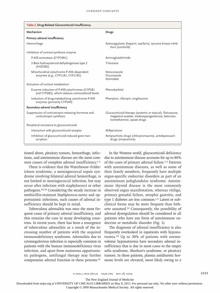
Drugs may cause glucocorticoid deficiency at hy-pothalamic, pituitary, and adrenal levels as well as at the sites of the glucocorticoid receptor, its signaling pathway, and peripheral glucocorticoid metabolism (Table 2). Suppression of the hypo-thalamic–pituitary–adrenal axis by exogenous glu-cocorticoid treatment is the most common cause of an impaired adrenal response. According to current estimates, nearly 1% of people in the gen-eral population (2.5% of those who are more than 70 years of age) are treated with long-term regi-mens of glucocorticoids for inflammation related to chronic disease.21 Since the proportion of elderly persons in the population is increasing, this fig-ure is likely to rise.
To avoid an unexpected adrenal crisis in per-sons admitted to the hospital on an emergency basis, physicians should not only ask whether the patient has been taking glucocorticoids but should also be aware of the many obscure situations in-volving the use of glucocorticoids. Patients may be unaware of or reluctant to report exposure to glucocorticoids. These may include athletes, pa-tients with cancer, patients with orthopedic con-ditions, and persons receiving adrenal extracts from sites on the Internet for what has been termed the “adrenal fatigue syndrome.” A lack of awareness that continuous use of topical gluco-corticoids can suppress adrenal function is another widespread problem in daily practice. Concomitant use of glucocorticoids with inhibitors (e.g., itra-conazole, diltiazem, mibefradil, and even grape-fruit juice) of CYP3A4, the most abundant drug-metabolizing cytochrome P450 enzyme, prolongs the biologic half-life of the glucocorticoid, there-by markedly enhancing its effect in suppressing adrenal function.22 In addition to glucocorticoids, other steroid compounds such as megestrol ac-etate and medroxyprogesterone inhibit the hypo-thalamic–pituitary–adrenal axis. Consideration of the integrity of the hypothalamic–pituitary–adre-nal axis will also be important when the new se-lective glucocorticoid-receptor activators come into common use.23
There has been an increased incidence of adre-nal dysfunction, particularly among patients who are receiving antifungal therapies. Such therapies (e.g., ketoconazole) are known to interfere with glucocorticoid synthesis and are therefore also used, in doses of 400 to 800 mg per day, to treat hypercortisolism. Although some of the newer antifungal compounds (e.g., itraconazole and flu-conazole) have fewer adrenostatic effects, adrenal insufficiency that occurs after treatment with high doses has been reported.24 These compounds may therefore confer a predisposition to adrenal insuf-ficiency during states of increased glucocorticoid requirement, such as severe stress in any kind of critical illness.
Etomidate, a commonly used, potent hypnotic agent, can also lower cortisol levels, even after a single injection of the drug.25 Therefore, in the case of any critically ill patient, the clinician should specifically ask about the use of etomidate; if the patient is receiving etomidate, the clinician should consider adding glucocorticoid therapy.26
It is prudent to monitor adrenal function dur-ing severe stress in patients who are receiving novel tyrosine kinase–targeting drugs, since some of these compounds (e.g., sunitinib) have been shown in studies in animals to cause adrenal dys-function and hemorrhage.27 The underlying mech-anism may be related to the fact that vascular endothelial growth factor–receptor antagonists impair endothelial integrity, which may then lead to hemorrhage in the highly vascularized adrenal gland during stress.
Finally, since growing numbers of chronically ill patients take multiple drugs, clinicians must consider the additive effect of a combination of drugs with antiglucocorticoid effects. Recent com-prehensive toxicologic in vitro assays have shown that increasing numbers of environmental com-pounds have the capability to impair adrenal ste-roidogenesis. Such compounds range from endo-crine disruptors (e.g., phytoestrogen flavonoids) to widely used insecticides (e.g., lindane). The ef-fect on adrenal function in humans, however, has yet to be determined.28
Dise a ses the Clinici a n Should Consider
Diseases that cause outright adrenal insufficiency are rare. In addition to the genetic defects men-
The New England Journal of Medicine Downloaded from nejm.org at UNIVERSITY OF CHICAGO LIBRARIES on May 8, 2013. For personal use only. No other uses without permission.
Copyright © 2009 Massachusetts Medical Society. All rights reserved.
current concepts
n engl j med 360;22 nejm.org may 28, 2009 2333
tioned above, pituitary tumors, hemorrhage, infec-tions, and autoimmune disease are the most com-mon causes of complete adrenal insufficiency.1,2
There is evidence that the Waterhouse–Frider-ichsen syndrome, a meningococcal sepsis syn-drome involving bilateral adrenal hemorrhage, is not limited to meningococcal infection, but may occur after infection with staphylococci or other pathogens.29,30 Considering the steady increase in methicillin-resistant Staphylococcus aureus and op-portunistic infections, such causes of adrenal in-sufficiency should be kept in mind.
Tuberculous adrenalitis was once the most fre-quent cause of primary adrenal insufficiency, and this remains the case in many developing coun-tries. In recent years, there has been a resurgence of tuberculous adrenalitis as a result of the in-creasing number of patients with the acquired immunodeficiency syndrome. Adrenalitis due to cytomegalovirus infection is especially common in patients with the human immunodeficiency virus infection, and apart from numerous opportunis-tic pathogens, antifungal therapy may further compromise adrenal function in these persons.31
In the Western world, glucocorticoid deficiency due to autoimmune disease accounts for up to 80% of the cases of primary adrenal failure.1,2 Patients with autoimmune diseases, as well as some of their family members, frequently have multiple organ-specific endocrine disorders as part of an autoimmune polyglandular syndrome. Autoim-mune thyroid disease is the most commonly observed organ manifestation, whereas vitiligo, primary gonadal failure, atrophic gastritis, and type 1 diabetes are less common.1,2 Latent or sub-clinical forms may be more frequent than hith-erto assumed.32 Consequently, the possibility of adrenal dysregulation should be considered in all patients who have any form of autoimmune en-docrine or metabolic disorder (Fig. 1).
The diagnosis of adrenal insufficiency is also frequently overlooked in inpatients with hypona-tremia.33 Up to 20% of patients with normo-volemic hyponatremia have secondary adrenal in-sufficiency that is due in most cases to the empty sella syndrome, Sheehan’s syndrome, or pituitary tumors. In these patients, plasma antidiuretic hor-mone levels are elevated, most likely owing to a
Table 2. Drug-Related Glucocorticoid Insufficiency.
Mechanism Drugs
Primary adrenal insufficiency
Hemorrhage Anticoagulants (heparin, warfarin), tyrosine kinase inhib-itors (sunitinib)
Inhibition of cortisol-synthesis enzyme
P-450 aromatase (CYP19A1) Aminoglutethimide
3 Beta hydroxysteroid-dehydrogenase type 2 (HSD3B2)
Trilostane
Mitochondrial cytochrome P-450–dependent enzymes (e.g., CYP11A1, CYP11B1)
KetoconazoleFluconazoleEtomidate
Activation of cortisol metabolism
Enzyme induction of P-450 cytochromes (CYP2B1 and CYP2B2), which reduces corticosteroid levels
Phenobarbital
Induction of drug-metabolizing cytochrome P-450 enzymes (primarily CYP3A4)
Phenytoin, rifampin, troglitazone
Secondary adrenal insufficiency
Suppression of corticotropin-releasing hormone and corticotropin synthesis
Glucocorticoid therapy (systemic or topical), fluticasone, megestrol acetate, medroxyprogesterone, ketorolac tromethamine, opiate drugs
Peripheral resistance to glucocorticoids
Interaction with glucocorticoid receptor Mifepristone
Inhibition of glucocorticoid-induced gene tran-scription
Antipsychotic drugs (chlorpromazine), antidepressant drugs (imipramine)
The New England Journal of Medicine Downloaded from nejm.org at UNIVERSITY OF CHICAGO LIBRARIES on May 8, 2013. For personal use only. No other uses without permission.
Copyright © 2009 Massachusetts Medical Society. All rights reserved.
T h e n e w e ngl a nd j o u r na l o f m e dic i n e
n engl j med 360;22 nejm.org may 28, 20092334
failure of endogenous glucocorticoid to suppress the hormone. Hydrocortisone-replacement thera-py leads to rapid normalization of serum sodium levels.33
Another underdiagnosed clinical problem is hypopituitarism due to brain injury. Pituitary dys-function occurs in up to 30% of patients with trauma to the brain and may not appear until months or years after the traumatic incident.34
Gluco corticoid Insufficienc y R el ated t o Cr i tic a l Illness
Disease processes that cause a predisposition to adrenal failure during periods of increased stress appear to be more frequent than previously as-sumed. Terms such as “relative adrenal insufficien-cy” and, more accurately, “critical illness–related corticosteroid insufficiency” have been used to characterize these conditions.
Recently, expert panels and consensus confer-ences involving intensivists, pulmonologists, and endocrinologists have examined the clinical rel-evance of adrenal insufficiency and have provided recommendations for diagnosis and manage-ment.35 The syndrome has been defined as inad-equate glucocorticoid activity in relation to the se-verity of the patient’s illness and has been most prominently investigated in cases of sepsis and septic shock.36-39 The best test currently available for establishing the diagnosis is the 1-μg corti-cotropin stimulation test, in which cortisol levels are measured 30 minutes after stimulation, with a level of less than 25 μg per deciliter (690 nmol per liter) or an increment over baseline of less than 9 μg per deciliter (250 nmol per liter) representing an inadequate adrenal response. An inadequate response to corticotropin testing occurs in up to 60% of patients with sepsis38; however, declining cortisol-binding globulin levels in patients with sepsis may moderate the impairment of active free-cortisol production. To better define this syn-drome, endocrine testing for adrenal insufficiency in patients with sepsis or other critical illnesses must be improved. Confounding factors such as variability in sampling and cortisol assays, includ-ing interfering antibodies, need to be considered. Measurement of free cortisol or widespread im-plementation of more accurate mass spectrometry methods might help to overcome these analytic limitations.40,41
Mechanisms of adrenal suppression in sepsis remain largely unclear; however, cytokines such as tumor necrosis factor-α or other peptides de-rived from blood cells — known as corticostatins — that may compete with corticotropin on its re-ceptor 42 influence adrenal regulation during in-flammation, induce tissue resistance to glucocor-ticoids, or have both effects.43 In order for an adrenocortical cell to respond adequately to the severe stress of inflammation, intraadrenal cell–cell communication needs to be intact.42 This in-volves a close crosstalk of adrenocortical cells with chromaffin cells, as well as endothelial cells and intraadrenal immune cells.42 As summarized in Figure 2, it has been suggested that neuropeptides, neurotransmitters, oxidative stress, altered adrenal blood flow, and substrate deficiency due to low lipoprotein cholesterol levels and drug interactions affect adrenal integrity.42,44,45 Septicemia itself and medications used during its treatment (Table 2) may interfere with receptor signaling associated with the membrane microdomains, with the ma-chinery of cholesterol transport and storage, with enzymes involved in steroidogenesis, and with the mitochondrial function that is critical for steroido-genesis.46 Furthermore, impaired blood supply to the pars distalis may induce pituitary ischemia, necrosis, or both during septic shock, and an in-creased accumulation of nitric oxide, superoxide, or central neuropeptides or prostaglandins con-tributes to a decrease in hypothalamic–pituitary hormones in patients with sepsis (Fig. 2).
The clinical consequences of impaired adrenal function in patients with sepsis remain unclear. A recent large, multicenter, randomized, double-blind, placebo-controlled trial showed that al-though hydrocortisone does help to reverse septic shock, it does not improve survival.39 Therefore, general use of glucocorticoids in patients with sepsis does not appear to be warranted. A clearer understanding of the relevant causes of adrenal insufficiency in patients with sepsis and a more refined definition of subgroups that may benefit from glucocorticoid therapy are required. Addi-tional factors that may contribute to the conflict-ing results include the severity of the sepsis, the duration of therapy, and the use or nonuse of fludrocortisone39 to treat hypoaldosteronism. It is imperative that we gain a better understanding of both the true pattern of cortisol secretion during critical illness and the pharmacokinetics of vari-
The New England Journal of Medicine Downloaded from nejm.org at UNIVERSITY OF CHICAGO LIBRARIES on May 8, 2013. For personal use only. No other uses without permission.
Copyright © 2009 Massachusetts Medical Society. All rights reserved.
current concepts
n engl j med 360;22 nejm.org may 28, 2009 2335
TraumaInfectiousdiseases
Coagulationdisorders
TumorsEndocrine
autoimmunedisorders
Tissueinfiltration
Liverdisease
Head trauma
Bleeding
Burns
Pregnancy
Surgicaltrauma
Sports
Short stature
Disorders of sexual differentiation
Hirsutism
Obesity in congenital adrenal and pituitary disorders
Hashimoto’sdisease
Diabetes mellitus type 1
Perniciousanemia
Vitiligo in autoimmune polyglandular syndromes
Mentaldisorders,addictions
Chronicinflammatory
disorders
1
Campion
5/11/09
AUTHOR PLEASE NOTE:Figure has been redrawn and type has been reset
Please check carefully
Author
Fig #
Title
ME
DEArtist
Issue date
COLOR FIGURE
Draft 5Bornstein
Knoper
5/28/09
Table 3
Clinicalsigns andpatienthistory
Rheumatoidarthritis
Asthma
Crohn’sdisease
Allergies
HIV-1–cyto-megalovirusinfection
Tuberculosis
Candidiasis
Histo-plasmosis
Waterhouse–Friderichsen syndrome in sepsis
Thrombo-cytopenia
Drug-relatedadrenalhemorrhage(e.g., with heparin)
Antiphospho-lipidsyndrome causing adrenal hemorrhage
Hepatitis
Liver failure
Liver trans-plantation with low lipoproteinsand adrenal exhaustion syndrome
Pituitary andadrenal tumors
Metastases (bilateral lymphoma, renal cancer)
History of hypophy-sectomy
History of bilateral adrenal-ectomy orremoval of incidentaloma
History of irradiation
Adrenal
AmyloidosisHemo-chromatosisSarcoidosis
Pituitary
HistiocytosisWegener’sgranulomatosisSarcoidosis
Anxietydisorders
Atypicaldepression
Anti-depressants
Alcohol
Be aware of late onset of adrenalinsufficiency
Be aware ofthyroidhormone triggering adrenal crisis
Check for steroid use in elite athletes
Consider pituitary apoplexy (Sheehan’s syndrome)in pregnancy
Check foruse of perioperativeetomidate
Check for intranasal, intraarticular, and topical use ofsteroids andnovelselectivegluco-corticoid-receptoragonists
Check forantifungaltherapies or rifampin or steroids
Be aware ofnon-meningo-coccal infectionscausing Waterhouse–Friderichsen syndrome
Considermasking byimmuno-suppressive steroidtherapy
Check for craniospinalirradiation inleukemia andother braintumors
Considerpalliation ofbreast and endometrial cancer with megestrol acetate or use of othersteroid in tumor therapy
Additional consider-ations
Consideradditionalsuppression by steroid therapy
Be awareof HPA-axisdys-regulation
Rule out Addison’s disease inpatients withanorexianervosa
Consider nonspecific symptoms: fatigue, hypotension, malaise, vomiting, abdominal pain, fever of unknown origin, failure to thrive,hemodynamic complications in patients in critical condition (fluid resistance in shock, poor response to vasopressors)
Suspect impaired HPA-axis; initiate diagnostic workup and hydrocortisone treatment
Congenitalabnormalities
Riskgroups
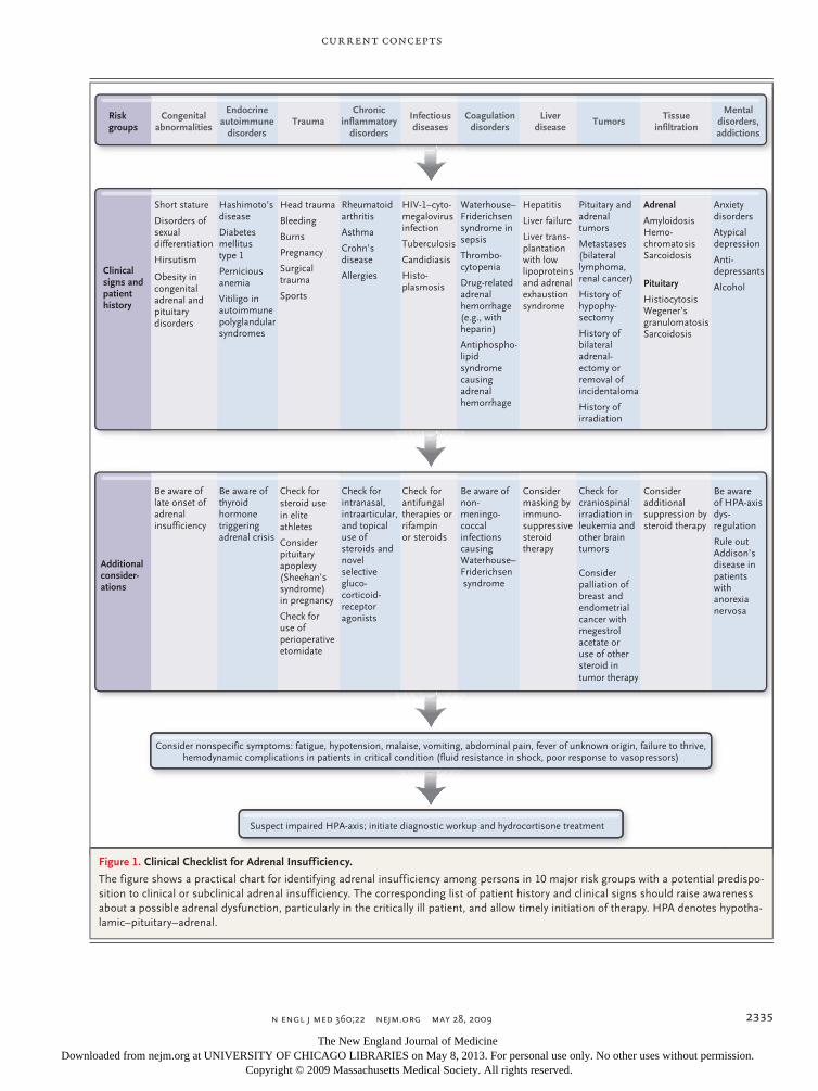
Figure 1. Clinical Checklist for Adrenal Insufficiency.
The figure shows a practical chart for identifying adrenal insufficiency among persons in 10 major risk groups with a potential predispo-sition to clinical or subclinical adrenal insufficiency. The corresponding list of patient history and clinical signs should raise awareness about a possible adrenal dysfunction, particularly in the critically ill patient, and allow timely initiation of therapy. HPA denotes hypotha-lamic–pituitary–adrenal.
The New England Journal of Medicine Downloaded from nejm.org at UNIVERSITY OF CHICAGO LIBRARIES on May 8, 2013. For personal use only. No other uses without permission.
Copyright © 2009 Massachusetts Medical Society. All rights reserved.
T h e n e w e ngl a nd j o u r na l o f m e dic i n e
n engl j med 360;22 nejm.org may 28, 20092336
ous hydrocortisone-replacement therapies. Finally, the adverse effects of glucocorticoid replacement on insulin resistance, protein catabolism, and im-munosuppression may be aggravated by high-fat parenteral nutrition in critically ill patients, since lipids have recently been shown to increase the action of glucocorticoids.47
On the basis of available evidence, current rec-ommendations, and good clinical practice, and irrespective of the results of adrenal testing, mod-erate doses of hydrocortisone (200 to 300 mg per day) should be given soon after the onset of septic shock in patients who remain hypotensive despite adequate administration of fluids and vasopres-sor agents.35,36,39 Current evidence is insufficient to recommend the replacement of other steroids that are suppressed in patients with sepsis, in-cluding mineralocorticoids and adrenal andro-gens.1,48,49
In addition to impairment of adrenal glucocor-ticoid regulation, hypoaldosteronism occurs fre-quently in critically ill patients. This condition probably does not result from a selective effect on the adrenal zona glomerulosa or aldosterone syn-thase; rather, it seems likely that the same mech-anisms that lead to glucocorticoid insufficiency account for the hypoaldosteronism. Future stud-ies will need to address these mechanisms. The role of mineralocorticoid supplementation in the treatment of critically ill patients is already being investigated in ongoing multicenter trials.
Several randomized studies have assessed the role of glucocorticoid treatment in patients with acute lung injury or the acute respiratory distress syndrome.50,51 A consistent finding in these stud-ies was that such treatment resulted in an acceler-ated resolution of the disorders.35 In addition, preliminary data suggest that glucocorticoids have a beneficial effect in patients with severe pancrea-titis52 and in those who have undergone trauma with hemorrhagic shock, as well as in patients who have just undergone cardiac surgery53 and those who are being weaned from mechanical venti-lation.54
It has become evident that patients with liver diseases have adrenal disturbances. Signs of adre-nal insufficiency are present in 33% of patients with acute liver failure, 65% of patients with chronic liver disease and sepsis, and 92% of pa-tients who have undergone a liver transplanta-tion.45 Consequently, the term “hepato–adrenal syndrome” has been introduced. It has been sug-gested that immunosuppression with glucocorti-
coids in liver-transplant recipients has masked the syndrome. Patients with liver diseases have very low lipoprotein levels, and a substrate shortage may therefore lead to an adrenal exhaustion syn-drome. However, a reduction in total cortisol may reflect a decrease in cortisol-binding globulin rather than a decrease in free cortisol. Never-theless, patients with liver diseases should be carefully monitored for symptoms and signs of adrenal insufficiency and may benefit from glu-cocorticoid-replacement therapy.55
Conclusion
In 1855, Thomas Addison concluded that “my ex-perience, though necessarily limited, leads to a belief that [adrenal insufficiency] is by no means
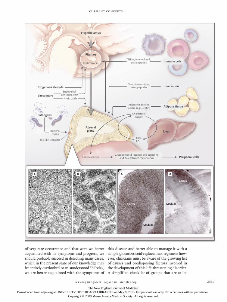
Figure 2 (facing page). Current Concepts of Immune–Endocrine and Metabolic Factors Involved in Glucocor-ticoid Dysregulation in Critical Illness.
Cytokines, chemokines, and adipokines derived from immune cells and fat cells and bacterial and viral tox-ins mediated by toll-like receptors modify pituitary– adrenal hormone synthesis and glucocorticoid tissue sensitivity and the activation of peripheral cortisol me-tabolism. Similarly, the function of the hypothalamic–pituitary–adrenal axis is influenced by blood flow, endothelial factors, neurotransmitters, and neuropep-tides. Furthermore, synthesis of pituitary and adrenal hormones requires a large supply of vitamins, antioxi-dants, and cholesterol. Impairment and dysregulation of any of these pathways will disrupt the integrity of the hypothalamic–pituitary–adrenal axis and may lead to a disturbed adrenal stress response. An electron micro-graph shows the vesicular mitochondria of an adreno-cortical cell in a normal mouse and in a mouse with suppressed corticosterone responses due to the ad-ministration of dexamethasone (Panels A and B, re-spectively; uranyl acetate and lead citrate staining). The number and conformational structure of internal mitochondrial membranes correlate with the steroido-genic capacity of an adrenocortical cell. Animal models with a defect in steroid production frequently show al-terations in vesicular mitochondria, with reduction and tubular transformation of internal membranes.19,42 Staining of histologic sections of adrenal glands with Sudan black shows the storage of lipid droplets in the adrenal cortex of rats in an unstressed state and 2 hours after stimulation with corticotropin-releasing hormone (Panels C and D, respectively). Since choles-terol constitutes the substrate for steroidogenesis, there is a rapid disappearance of cholesterol-storing liposomes after activation of the hypothalamic–pitu-itary–adrenal axis (Panel D), illustrating the require-ment of an external cholesterol supply for the adrenal gland. CRH denotes corticotropin-releasing hormone, HDL denotes high-density lipoprotein, LDL low-density lipoprotein, and TNF tumor necrosis factor.
The New England Journal of Medicine Downloaded from nejm.org at UNIVERSITY OF CHICAGO LIBRARIES on May 8, 2013. For personal use only. No other uses without permission.
Copyright © 2009 Massachusetts Medical Society. All rights reserved.
current concepts
n engl j med 360;22 nejm.org may 28, 2009 2337
of very rare occurrence and that were we better acquainted with its symptoms and progress, we should probably succeed in detecting many cases, which in the present state of our knowledge may be entirely overlooked or misunderstood.”3 Today, we are better acquainted with the symptoms of
this disease and better able to manage it with a simple glucocorticoid-replacement regimen; how-ever, clinicians must be aware of the growing list of causes and predisposing factors involved in the development of this life-threatening disorder. A simplified checklist of groups that are at in-
Exogenous steroids
Pathogens
Adipose tissue
Hypothalamus
Pituitary
Vasculature
Innervation
LiverAdrenalgland
TNF-α, interleukin-6, corticostatins
Toll-like receptors
Bacterialtoxins
Adipocyte-derivedfactors (e.g., leptin)
Corticotropin
CRH
GlucocorticoidGlucocorticoid receptor and signaling
and downstream metabolism Peripheral cells
Endothelial-derived factors
Nitric oxide
Neurotransmitters,neuropeptides
Cholesterolsupply
HDLLDL
Immune cells
A B C D
Cortex
Medulla
Medulla
Cortex
2
Campion
5/11/09
AUTHOR PLEASE NOTE:Figure has been redrawn and type has been reset
Please check carefully
Author
Fig #
Title
ME
DEArtist
Issue date
COLOR FIGURE
Draft 8Bornstein
Knoper
5/28/09
Predisposing Factors for Adrenal Insufficiency
The New England Journal of Medicine Downloaded from nejm.org at UNIVERSITY OF CHICAGO LIBRARIES on May 8, 2013. For personal use only. No other uses without permission.
Copyright © 2009 Massachusetts Medical Society. All rights reserved.
T h e n e w e ngl a nd j o u r na l o f m e dic i n e
n engl j med 360;22 nejm.org may 28, 20092338
creased risk for adrenal impairment may help to raise awareness among clinicians (Fig. 1). This in-formation is important, since timely and adequate hydrocortisone replacement in patients with acute adrenal insufficiency represents a lifesaving and effective solution in medical emergencies.
Supported by grants from Deutsche Forschungsgemeinschaft (BO 1141/8-1 and SFB 655 – TP A6), from the Sander Foundation, and from the Center for Regenerative Therapies Dresden.
No potential conflict of interest relevant to this article was reported.
I thank Dr. Graeme Eisenhofer, Kathy Eisenhofer, Dr. Wiebke Arlt, and Dr. Monika Ehrhart-Bornstein for their careful reading of the manuscript.
References
Arlt W, Allolio B. Adrenal insufficien-1. cy. Lancet 2003;361:1881-93.
Oelkers W. Adrenal insufficiency. 2. N Engl J Med 1996;335:1206-12.
Addison T. On the constitutional and 3. local effects of disease of the supra-renal capsules. London: Samuel Highley, 1855.
Grinspoon SK, Biller BM. Clinical re-4. view 62: laboratory assessment of adrenal insufficiency. J Clin Endocrinol Metab 1994;79:923-31.
Magnotti M, Shimshi M. Diagnosing 5. adrenal insufficiency: which test is best — the 1-microg or the 250-microg cosyn-tropin stimulation test? Endocr Pract 2008; 14:233-8.
Lin L, Ferraz-de-Souza B, Achermann 6. JC. Genetic disorders involving adrenal development. Endocr Dev 2007;11:36-46.
Merke DP, Bornstein SR. Congenital 7. adrenal hyperplasia. Lancet 2005;365: 2125-36.
Merke DP, Chrousos GP, Eisenhofer 8. G, et al. Adrenomedullary dysplasia and hypofunction in patients with classic 21-hydroxylase deficiency. N Engl J Med 2000;343:1362-8.
Perry R, Kecha O, Paquette J, Huot C, 9. Van VG, Deal C. Primary adrenal insuffi-ciency in children: twenty years experi-ence at the Sainte-Justine Hospital, Mon-treal. J Clin Endocrinol Metab 2005;90: 3243-50.
Rajab A, Kelberman D, de Castro SC, 10. et al. Novel mutations in LHX3 are associ-ated with hypopituitarism and sensorineu-ral hearing loss. Hum Mol Genet 2008; 17:2150-9.
Sandrini F, Farmakidis C, Kirschner 11. LS, et al. Spectrum of mutations of the AAAS gene in Allgrove syndrome: lack of mutations in six kindreds with isolated resistance to corticotropin. J Clin Endo-crinol Metab 2001;86:5433-7.
Kim CJ, Lin L, Huang N, et al. Severe 12. combined adrenal and gonadal deficiency caused by novel mutations in the choles-terol side chain cleavage enzyme, P450scc. J Clin Endocrinol Metab 2008;93:696-702.
Böttner A, Keller E, Kratzsch J, et al. 13. PROP1 mutations cause progressive dete-rioration of anterior pituitary function in-cluding adrenal insufficiency: a longitu-dinal analysis. J Clin Endocrinol Metab 2004;89:5256-65.
Lee YW, Won JC, Ki CS, et al. Clinical 14.
and genetic analysis of a Korean patient with late-onset X-linked adrenal hypopla-sia congenita and hypogonadotropic hy-pogonadism: identification of a novel mu-tation in the NR0B1 gene. J Int Med Res 2008;36:357-61.
Sadeghi-Nejad A, Senior B. Adrenomy-15. eloneuropathy presenting as Addison’s disease in childhood. N Engl J Med 1990; 322:13-6.
de Lind van Wijngaarden RF, Otten BJ, 16. Festen DA, et al. High prevalence of cen-tral adrenal insufficiency in patients with Prader-Willi syndrome. J Clin Endocrinol Metab 2008;93:1649-54.
Kempná P, Flück CE. Adrenal gland 17. development and defects. Best Pract Res Clin Endocrinol Metab 2008;22:77-93.
Cai L, Ji A, de Beer FC, Tannock LR, 18. van der Westhuyzen DR. SR-BI protects against endotoxemia in mice through its roles in glucocorticoid production and he-patic clearance. J Clin Invest 2008;118:364-75.
Bornstein SR, Zacharowski P, Schu-19. mann RR, et al. Impaired adrenal stress response in Toll-like receptor 2-deficient mice. Proc Natl Acad Sci U S A 2004; 101:16695-700.
Schröder NW, Schumann RR. Single 20. nucleotide polymorphisms of Toll-like re-ceptors and susceptibility to infectious disease. Lancet Infect Dis 2005;5:156-64.
van Staa TP, Leufkens HG, Abenhaim 21. L, Begaud B, Zhang B, Cooper C. Use of oral corticosteroids in the United King-dom. QJM 2000;93:105-11.
Varis T, Kivisto KT, Backman JT, Neu-22. vonen PJ. The cytochrome P450 3A4 in-hibitor itraconazole markedly increases the plasma concentrations of dexamethasone and enhances its adrenal-suppressant ef-fect. Clin Pharmacol Ther 2000;68:487-94.
Schäcke H, Berger M, Rehwinkel H, 23. Asadullah K. Selective glucocorticoid re-ceptor agonists (SEGRAs): novel ligands with an improved therapeutic index. Mol Cell Endocrinol 2007;275:109-17.
Shibata S, Kami M, Kanda Y, et al. 24. Acute adrenal failure associated with flu-conazole after administration of high-dose cyclophosphamide. Am J Hematol 2001;66:303-5.
Hildreth AN, Mejia VA, Maxwell RA, 25. Smith PW, Dart BW, Barker DE. Adrenal suppression following a single dose of
etomidate for rapid sequence induction: a prospective randomized study. J Trauma 2008;65:573-9.
den Brinker M, Joosten KF, Liem O, et 26. al. Adrenal insufficiency in meningococ-cal sepsis: bioavailable cortisol levels and impact of interleukin-6 levels and intuba-tion with etomidate on adrenal function and mortality. J Clin Endocrinol Metab 2005;90:5110-7.
Rock EP, Goodman V, Jiang JX, et al. 27. Food and Drug Administration drug ap-proval summary: sunitinib malate for the treatment of gastrointestinal stromal tu-mor and advanced renal cell carcinoma. Oncologist 2007;12:107-13.
Harvey PW, Everett DJ, Springall CJ. 28. Adrenal toxicology: a strategy for assess-ment of functional toxicity to the adrenal cortex and steroidogenesis. J Appl Toxicol 2007;27:103-15.
Adem PV, Montgomery CP, Husain AN, 29. et al. Staphylococcus aureus sepsis and the Waterhouse–Friderichsen syndrome in chil-dren. N Engl J Med 2005;353:1245-51.
Hamilton D, Harris MD, Foweraker J, 30. Gresham GA. Waterhouse-Friderichsen syndrome as a result of non-meningococ-cal infection. J Clin Pathol 2004;57:208-9.
Marik PE, Kiminyo K, Zaloga GP. Ad-31. renal insufficiency in critically ill patients with human immunodeficiency virus. Crit Care Med 2002;30:1267-73.
Betterle C, Lazzarotto F, Presotto F. 32. Autoimmune polyglandular syndrome Type 2: the tip of an iceberg? Clin Exp Im-munol 2004;137:225-33.
Diederich S, Franzen NF, Bahr V, 33. Oelkers W. Severe hyponatremia due to hypopituitarism with adrenal insufficien-cy: report on 28 cases. Eur J Endocrinol 2003;148:609-17.
Schneider HJ, Kreitschmann-Ander-34. mahr I, Ghigo E, Stalla GK, Agha A. Hy-pothalamopituitary dysfunction follow-ing traumatic brain injury and aneurysmal subarachnoid hemorrhage: a systematic review. JAMA 2007;298:1429-38.
Marik PE, Pastores SM, Annane D, et 35. al. Recommendations for the diagnosis and management of corticosteroid insuf-ficiency in critically ill adult patients: con-sensus statements from an international task force by the American College of Critical Care Medicine. Crit Care Med 2008;36:1937-49.
The New England Journal of Medicine Downloaded from nejm.org at UNIVERSITY OF CHICAGO LIBRARIES on May 8, 2013. For personal use only. No other uses without permission.
Copyright © 2009 Massachusetts Medical Society. All rights reserved.
current concepts
n engl j med 360;22 nejm.org may 28, 2009 2339
Annane D, Sébille V, Charpentier C, et 36. al. Effect of treatment with low doses of hydrocortisone and fludrocortisone on mortality in patients with septic shock. JAMA 2002;288:862-71.
Dellinger RP, Levy MM, Carlet JM, et 37. al. Surviving Sepsis Campaign: interna-tional guidelines for management of se-vere sepsis and septic shock: 2008. Crit Care Med 2008;36:296-327. [Erratum, Crit Care Med 2008;36:1394-6.]
Cooper MS, Stewart PM. Corticoste-38. roid insufficiency in acutely ill patients. N Engl J Med 2003;348:727-34.
Sprung CL, Annane D, Keh D, et al. 39. Hydrocortisone therapy for patients with septic shock. N Engl J Med 2008;358:111-24.
Hamrahian AH, Oseni TS, Arafah 40. BM. Measurements of serum free cortisol in critically ill patients. N Engl J Med 2004;350:1629-38.
Vogeser M, Briegel J, Jacob K. Determi-41. nation of serum cortisol by isotope-dilu-tion liquid-chromatography electrospray ionization tandem mass spectrometry with on-line extraction. Clin Chem Lab Med 2001;39:944-7.
Bornstein SR, Engeland WC, Ehrhart-42. Bornstein M, Herman JP. Dissociation of ACTH and glucocorticoids. Trends Endo-crinol Metab 2008;19:175-80.
Charmandari E, Kino T, Ichijo T, 43. Chrousos GP. Generalized glucocorticoid resistance: clinical aspects, molecular mechanisms, and implications of a rare genetic disorder. J Clin Endocrinol Metab 2008;93:1563-72.
Bornstein SR, Briegel J. A new role for 44. glucocorticoids in septic shock: balanc-ing the immune response. Am J Respir Crit Care Med 2003;167:485-6.
O’Beirne J, Holmes M, Agarwal B, et 45. al. Adrenal insufficiency in liver disease — what is the evidence? J Hepatol 2007; 47:418-23.
Bornstein SR, Ehrhart-Bornstein M, 46. Güse-Behling H, Scherbaum WA. Structure and dynamics of adrenal mitochondria following stimulation with corticotropin releasing hormone. Anat Rec 1992;234: 255-62.
Sivabalan S, Renuka S, Menon VP. Fat 47. feeding potentiates the diabetogenic ef-fect of dexamethasone in Wistar rats. Int Arch Med 2008;1:7.
Arlt W, Hammer F, Sanning P, et al. 48. Dissociation of serum dehydroepiandros-terone and dehydroepiandrosterone sul-fate in septic shock. J Clin Endocrinol Metab 2006;91:2548-54.
Marx C, Petros S, Bornstein SR, et al. 49. Adrenocortical hormones in survivors and nonsurvivors of severe sepsis: diverse
time course of dehydroepiandrosterone, dehydroepiandrosterone-sulfate, and cor-tisol. Crit Care Med 2003;31:1382-8.
Confalonieri M, Urbino R, Potena A, 50. et al. Hydrocortisone infusion for severe community-acquired pneumonia: a pre-liminary randomized study. Am J Respir Crit Care Med 2005;171:242-8.
Meduri GU, Marik PE, Pastores SM, 51. Annane D. Corticosteroids in ARDS: a counterpoint. Chest 2007;132:1093-4.
Eklund A, Leppäniemi A, Kemppain-52. en E, Pettilä V. Vasodilatory shock in se-vere acute pancreatitis without sepsis: is there any place for hydrocortisone treat-ment? Acta Anaesthesiol Scand 2005;49: 379-84.
Halonen J, Halonen P, Järvinen O, et 53. al. Corticosteroids for the prevention of atrial fibrillation after cardiac surgery: a randomized controlled trial. JAMA 2007; 297:1562-7.
Huang CJ, Lin HC. Association be-54. tween adrenal insufficiency and ventila-tor weaning. Am J Respir Crit Care Med 2006;173:276-80.
Fernández J, Escorsell A, Zabalza M, 55. et al. Adrenal insufficiency in patients with cirrhosis and septic shock: effect of treatment with hydrocortisone on surviv-al. Hepatology 2006;44:1288-95.Copyright © 2009 Massachusetts Medical Society.
posting presentations at medical meetings on the internet
Posting an audio recording of an oral presentation at a medical meeting on the Internet, with selected slides from the presentation, will not be considered prior publication. This will allow students and physicians who are unable to attend the meeting to hear the presentation and view the slides. If there are any questions about this policy, authors should feel free to call the Journal’s Editorial Offices.
The New England Journal of Medicine Downloaded from nejm.org at UNIVERSITY OF CHICAGO LIBRARIES on May 8, 2013. For personal use only. No other uses without permission.
Copyright © 2009 Massachusetts Medical Society. All rights reserved.