Prediction of Endometrial Hyperplasia and Cancer among...
7
Research Article Prediction of Endometrial Hyperplasia and Cancer among Premenopausal Women with Abnormal Uterine Bleeding Luca Giannella , 1 Lillo Bruno Cerami, 1 Tiziano Setti, 1 Ezio Bergamini, 1 and Fausto Boselli 2 Local Health Authority of Reggio Emilia-IRCCS, Obstetrics and Gynecology Unit, Cesare Magati Hospital, Scandiano, Italy Mother-Infant Department, Institute of Obstetrics and Gynecology, University of Modena and Reggio Emilia, Modena, Italy Correspondence should be addressed to Luca Giannella; [email protected] Received 26 November 2018; Accepted 28 February 2019; Published 18 March 2019 Academic Editor: Jose Guilherme Cecatti Copyright © 2019 Luca Giannella et al. is is an open access article distributed under the Creative Commons Attribution License, which permits unrestricted use, distribution, and reproduction in any medium, provided the original work is properly cited. Objective. To create a prediction model including clinical variables for the prediction of premalignant/malignant endometrial pathology in premenopausal women with abnormal uterine bleeding (AUB). Methods. is is an observational retrospective study including 240 premenopausal women with AUB referred to diagnostic hysteroscopy. Based on the presence of endometrial hyperplasia (EH) or cancer (EC), the women were divided into cases (EH/EC) and controls (no EH/EC). Univariate, stepwise logistic regression and ROC curve analysis were performed. Results. 12 women had EH/EC (5%). Stepwise logistic regression analysis showed that EH/EC associated significantly with BMI ≥ 30 (OR=7.70, 95% CI 1.90 to 31.17), diabetes (OR=9.71, 95% CI 1.63 to 57.81), and a thickened endometrium (OR=1.20, 95% CI 1.08 to 1.34, criterion > 11 mm). e AUC was 0.854 (95% confidence intervals 0.803 to 0.896, p<0.0001). Considering the pretest probability for EH/EC of 5%, the prediction model with a positive likelihood ratio of 8.14 showed a posttest probability of 30%. e simultaneous presence of two or three risk factors was significantly more common in women with EH/EC than controls (50% vs. 6.6 and 25% vs. 0%, respectively, p<0.0001). Conclusion. When premenopausal vaginal bleeding occurs in diabetic obese women with ET > 11 mm, the percentage of premalignant/malignant endometrial pathology increases by 25%. It is likely that the simultaneous presence of several risk factors is necessary to significantly increase the probability of endometrial pathology. 1. Introduction Abnormal uterine bleeding (AUB) is one of the most frequent reasons for a gynecological evaluation [1]. It can be caused by structural and nonstructural uterine disorders. According to FIGO system PALM-COEIN, the causes can be the following: polyp, adenomyosis, leiomyoma, malignancy, coagulopathy, ovulatory disfunction, endometrial, iatrogenic, or not yet classified [2]. Although in most cases AUB is not linked to a malignant or premalignant lesion, it should not be underestimated. We know that, in postmenopausal women with AUB, there is a risk of endometrial cancer of 10% [3, 4]. en, with transvaginal ultrasonography showing an endometrial thickness (ET) < 4 mm, this risk falls below 1% [5]. In premenopausal women (PW) with AUB this risk strat- ification is not possible because the predictive performance of ET showed conflicting results [6–9]. In this group of women, other clinical variables are taken into account for the risk of EH/EC: obesity, nulliparity, age, infertility, intermen- strual bleeding, anovulation, and diabetes [10]. Based on the presence of these risk factors, some guidelines recommend endometrial biopsy as mandatory in women over the age of 40, or under the age of 40 in the presence of comorbidities [11, 12]. e UK NICE guidance recommends endometrial biopsy in PW with persistent intermenstrual bleeding (IB), or for women over the age of 45 with heavy menstrual bleeding (HMB), aſter the failure of medical treatment [13, 14]. Despite the presence of these guidelines, many studies in the literature have not given decisive results on the weight of the abovementioned risk factors [15–18]. erefore, the correct management of PW with AUB is not entirely clear. In a recent systematic literature review the risk of atypical endometrial hyperplasia (AEH) or cancer in PW with AUB Hindawi BioMed Research International Volume 2019, Article ID 8598152, 6 pages https://doi.org/10.1155/2019/8598152
Transcript of Prediction of Endometrial Hyperplasia and Cancer among...

Research ArticlePrediction of Endometrial Hyperplasia and Cancer amongPremenopausal Women with Abnormal Uterine Bleeding
Luca Giannella ,1 Lillo Bruno Cerami,1 Tiziano Setti,1
Ezio Bergamini,1 and Fausto Boselli2
1Local Health Authority of Reggio Emilia-IRCCS, Obstetrics and Gynecology Unit, Cesare Magati Hospital, Scandiano, Italy2Mother-Infant Department, Institute of Obstetrics and Gynecology, University of Modena and Reggio Emilia, Modena, Italy
Correspondence should be addressed to Luca Giannella; [email protected]
Received 26 November 2018; Accepted 28 February 2019; Published 18 March 2019
Academic Editor: Jose Guilherme Cecatti
Copyright © 2019 Luca Giannella et al.This is an open access article distributed under the Creative Commons Attribution License,which permits unrestricted use, distribution, and reproduction in any medium, provided the original work is properly cited.
Objective. To create a prediction model including clinical variables for the prediction of premalignant/malignant endometrialpathology in premenopausal women with abnormal uterine bleeding (AUB). Methods. This is an observational retrospectivestudy including 240 premenopausal women with AUB referred to diagnostic hysteroscopy. Based on the presence of endometrialhyperplasia (EH) or cancer (EC), the women were divided into cases (EH/EC) and controls (no EH/EC). Univariate, stepwiselogistic regression and ROC curve analysis were performed. Results. 12 women had EH/EC (5%). Stepwise logistic regressionanalysis showed that EH/EC associated significantly with BMI ≥ 30 (OR=7.70, 95% CI 1.90 to 31.17), diabetes (OR=9.71, 95%CI 1.63 to 57.81), and a thickened endometrium (OR=1.20, 95% CI 1.08 to 1.34, criterion > 11mm). The AUC was 0.854 (95%confidence intervals 0.803 to 0.896, p<0.0001). Considering the pretest probability for EH/EC of 5%, the prediction model witha positive likelihood ratio of 8.14 showed a posttest probability of 30%. The simultaneous presence of two or three risk factors wassignificantly more common in women with EH/EC than controls (50% vs. 6.6 and 25% vs. 0%, respectively, p<0.0001).Conclusion.Whenpremenopausal vaginal bleeding occurs in diabetic obesewomenwithET> 11 mm, the percentage of premalignant/malignantendometrial pathology increases by 25%. It is likely that the simultaneous presence of several risk factors is necessary to significantlyincrease the probability of endometrial pathology.
1. Introduction
Abnormal uterine bleeding (AUB) is one of the most frequentreasons for a gynecological evaluation [1]. It can be caused bystructural and nonstructural uterine disorders. According toFIGO system PALM-COEIN, the causes can be the following:polyp, adenomyosis, leiomyoma, malignancy, coagulopathy,ovulatory disfunction, endometrial, iatrogenic, or not yetclassified [2]. Although in most cases AUB is not linkedto a malignant or premalignant lesion, it should not beunderestimated. We know that, in postmenopausal womenwith AUB, there is a risk of endometrial cancer of 10%[3, 4]. Then, with transvaginal ultrasonography showing anendometrial thickness (ET) < 4mm, this risk falls below 1%[5].
In premenopausal women (PW) with AUB this risk strat-ification is not possible because the predictive performance
of ET showed conflicting results [6–9]. In this group ofwomen, other clinical variables are taken into account for therisk of EH/EC: obesity, nulliparity, age, infertility, intermen-strual bleeding, anovulation, and diabetes [10]. Based on thepresence of these risk factors, some guidelines recommendendometrial biopsy as mandatory in women over the age of40, or under the age of 40 in the presence of comorbidities[11, 12]. The UK NICE guidance recommends endometrialbiopsy in PWwith persistent intermenstrual bleeding (IB), orfor women over the age of 45 with heavy menstrual bleeding(HMB), after the failure of medical treatment [13, 14].
Despite the presence of these guidelines, many studies inthe literature have not given decisive results on the weightof the abovementioned risk factors [15–18]. Therefore, thecorrect management of PW with AUB is not entirely clear.
In a recent systematic literature review the risk of atypicalendometrial hyperplasia (AEH) or cancer in PW with AUB
HindawiBioMed Research InternationalVolume 2019, Article ID 8598152, 6 pageshttps://doi.org/10.1155/2019/8598152

2 BioMed Research International
was low: 1.31% [19]. These results raise the question aboutthe appropriateness of endometrial biopsy in this population.In order to decrease false-positive cases, it is clear thatendometrial biopsy should only be recommended in selectedcases.
In this regard, in order to select a population mostat risk of EH/EC, the objective of the present study wasto create a predictive model including clinical variablesfor the prediction of premalignant/malignant endometrialpathology in PW with AUB.
2. Materials and Methods
This is an observational retrospective study including 240premenopausal women with AUB referred to diagnostichysteroscopy at nonuniversity (Scandiano) and university(Modena) hospital, Italy, fromMarch 2010 toNovember 2014.As the present study was merely observational and includedonly analysis of data from routine measurements, with noadditional or experimental interventions, institutional reviewboard approval was not required. All patients providedwritten informed consent for the use of their data for researchpurposes prior to hysteroscopy.
AUB was defined by the presence of bleeding from theuterine corpus that was abnormal in volume, regularity,and/or timing, according to what was reported by women[2]. We excluded women with menopausal status (absence ofmenstruation for at least 12 months after the age of 40 years)[4]. When it was impossible to perform a hysteroscopy, thatcase was excluded from the study.
We included all those women who had a definitivehistological diagnosis which we considered our referencestandard. Vabra endometrial sampling for women withoutany intrauterine structural lesion; targeted biopsy in womenwith a suspected premalignant ormalignant lesion; intrauter-ine lesion resection in women with polyps or myomas; allwomen with an atypical EH (AEH), as well as all women withan intrauterinemalignancy, underwent a hysterectomywhichrepresented our reference standard as definitive histologicalfinding.
Based on the presence of endometrial hyperplasia (EH) orcancer (EC), thewomenwere divided into cases (EH/EC) andcontrols (no EH/EC). Histological diagnosis of endometrialhyperplasia refers to the WHO 2014 classification: atypicaland nonatypical [20].
All diagnostic hysteroscopies were performed withoutanesthesia in an outpatient setting and vaginoscopy witha saline solution as distension medium and using a 5mmcontinuous-flow sheath with a viewing angle of 30∘.
All data were collected from medical records. Patientcharacteristics taken into account were age (years), age atmenarche (years), parity, body mass index (BMI = weight(kg)/height2 (m2)), presence of hypertension or diabetes,menstrual cycle phase, family history of breast and col-orectal cancer, current hormonal therapy (progestogen only,combined oral contraceptives, and vaginal ring), smokinghabit, endometrial thickness (mm), infertility, IB, tamoxifenusers, and duration of AUB (expressed in months from itsbeginning).
The Kolmogorov-Smirnov test was used as the test fornormal distribution. Continuous variables were expressed asmedian and interquartile range. Qualitative variables wereexpressed as numbers and percentages. Univariate logisticregression analysis was used to test all studied independentvariables and the results were expressed as an odds ratio(OR)with 95% confidence intervals (CI).Multivariate logisticregression analysis was used to identify variables that asso-ciated significantly with EH/EC. For this, we included asexplanatory variables all the variables that showed a p value≤ 0.25 in the univariate model [21]. Multivariate analysis wasperformed using a stepwisemethod with an entrance and exitp value of 0.05/0.1.The predicted probabilities of the stepwiselogistic regression analysis were then used to create a fullROC curve and to estimate the sensitivity, specificity, positivepredictive value (PPV), negative predictive value (NPV),positive likelihood ratio (LR+), and negative likelihood ratio(LR-) of the prediction model. Receiver operating character-istic (ROC) curve analysis was also used to determine theoptimal cut-off value of endometrial thickness for the pre-diction of EH/EC. After considering our disease prevalence(all cases of EH/EC) as the pretest probability for premalig-nant/malignant endometrial pathology, the likelihood ratiowas used to calculate the posttest odds from the pretest oddsof disease: posttest odds = pretest odds x likelihood ratio.The relation between odds and probability is as follows: odds= P/(1-P) and P = odds/(1+odds). Using these equations,we could calculate the posttest probability of disease fromthe pretest probability of disease [22]. Comparisons betweencategorical variables were performed by the chi-square testin order to assess if the presence of one or more risk factorsassociated with EH/EC.
All statistical analyses were performed using MedCalcStatistical Software version 18.10.2 (MedCalc Software bvba,Ostend, Belgium; http://www.medcalc.org; 2018). A 𝑝 valueof less than 0.05 was considered to be statistically significant.
3. Results
Thedata of 240 consecutive patients were analyzed retrospec-tively. Characteristics of the cohort are shown in Table 1.
Endometrial samples showed 3 women with EC (1.3%), 4women with AEH (1.7%), 5 women with non-AEH (2%), 75womenwith polyps (31.2%), 33women withmyomas (13.8%),and 120 women with negative results (50%). The prevalenceof EH/EC was 5%.
Univariate logistic regression analysis showed that a BMI≥ 30 (OR=8.13, 95%CI 2.34 to 28.21), the presence of diabetes(OR=12.33, 95%CI 2.64 to 57.4), or a thickened endometrium(expressed as a continuous variable inmm) (OR=1.15, 95%CI1.05 to 1.26) associated with EH/EC (Table 2).
Multivariate analysis with the stepwise method showedthat EH/EC associated significantly with BMI ≥ 30 (OR=7.70,95% CI 1.90 to 31.17), diabetes (OR=9.71, 95% CI 1.63 to57.81), and a thickened endometrium (OR=1.20, 95% CI 1.08to 1.34) (Table 3). The variables that were not included in themodel were menstruation cycle phase, menarche, nulliparity,tamoxifen users, and duration of AUB.
BioMed Research International 3
Table 1: Patient characteristics.
VariablesStudy participants
(n=240)n (%)
Age (years)Median (interquartile range) 44.0 (40.0 - 48.5)
Menstrual cycle phaseProliferative 135 (56.2)Secretive 105 (43.8)
Nulliparity 63 (26.2)Menarche age (years)Median (interquartile range) 12 (11.5 – 13.0)
Current hormonal therapy 38 (15.8)Body mass index≥ 30 53 (22.1)< 30 187 (77.9)
Hypertension 41 (17.1)Diabetes 9 (3.7)Smoking habit 71 (29.6)Endometrial thickness (mm)Median (interquartile range) 10.0 (8.0 – 13.0)
Infertility 36 (15.0)Intermenstrual bleeding 45 (18.8)Breast cancer family history 9 (3.75)Colorectal cancer family history 7 (2.9)Duration of AUB (months)Median (interquartile range) 9 (8 – 23)
Tamoxifen users 2 (0.83)Histology
EC 3 (1.3)AEH 4 (1.7)Non-AEH 5 (2.0)Polyp 75 (31.2)Myoma 33 (13.8)Negative 120 (50)
EC: endometrial cancer; AEH: atypical endometrial hyperplasia; AUB:abnormal uterine bleeding.
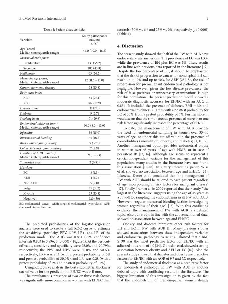
The predicted probabilities of the logistic regressionanalysis were used to create a full ROC curve to estimatethe sensitivity, specificity, PPV, NPV, LR+, and LR- of theprediction model. The AUC was 0.854 (95% confidenceintervals 0.803 to 0.896, p<0.0001) (Figure 1). At the best cut-off value, sensitivity and specificity were 75.0% and 90.79%,respectively; the PPV and NPV were 30.0% and 98.6%,respectively; LR+ was 8.14 (with a pretest probability of 5%and posttest probability of 30.0%), and LR- was 0.28 (with apretest probability of 5% and posttest probability of 1.5%).
Using ROC curve analysis, the best endometrial thicknesscut-off value for the prediction of EH/EC was > 11mm.
The simultaneous presence of two or three risk factorswas significantly more common in women with EH/EC than
controls (50% vs. 6.6 and 25% vs. 0%, respectively, p<0.0001)(Table 4).
4. Discussion
Thepresent study showed that half of the PWwith AUB haveendocavitary uterine lesions. The prevalence of EC was 1.3%,while the prevalence of EH plus EC was 5%. These resultsare in line with previous data reported in the literature [19].Despite the low percentage of EC, it should be emphasizedthat the risk of progression to cancer for nonatypical EH canreach up to 10% and up to 40% for AEH [23]. So, the risk ofprogression for premalignant endometrial pathology is notnegligible. However, given the low disease prevalence, therisk of false positives or unnecessary examinations is highfor this population. The present prediction model showed amoderate diagnostic accuracy for EH/EC with an AUC of0.854. It included the presence of diabetes, BMI ≥ 30, andendometrial thickness > 11mmwith a posttest probability forEC of 30%, from a pretest probability of 5%. Furthermore, itwould seem that the simultaneous presence of more than onerisk factor significantly increases the percentage of EH/EC.
To date, the management of PW with AUB providesthe need for endometrial sampling in women over 35–40years of age, or under this cut-off value in the presence ofcomorbidities (anovulation, obesity, and diabetes) [1, 11, 12].Another management option provides endometrial biopsyin women over 45 years of age with HMB, or in case ofpersistent IB [13, 14]. Although age seems to represent acrucial independent variable for the management of thispopulation, many studies in the literature have not foundthis association [15–18]. In a very interesting paper, Wiseet al. showed no association between age and EH/EC [24].Likewise, Esmer et al. concluded that “the management ofPW with AUB should be tailored to each patient regardlessof age, incorporating all risk factors for malignant disease”[17]. Finally, Iram et al. in 2009 reported that their study, “thelargest in the literature, suggests using the age of 45 years asa cut-off for sampling the endometrium in all PW with AUB.However, irregular menstrual bleeding justifies investigatingwomen regardless of their age” [15]. With this conflictingevidence, the management of PW with AUB is a debatedtopic. Also our study, in line with the abovementioned data,showed no association between age and EH/EC.
Obesity and diabetes represent other risk factors forEH end EC in PW with AUB [1]. Many previous studiesshowed associations between these independent variablesand endometrial pathology. Wise et al. showed that a BMI≥ 30 was the most predictive factor for EH/EC with anadjusted odds ratio of 4.0 [24]. Guraslan et al. showed a strongassociation between obesity and AEH or EC [16]. Also thepresent study showed that diabetes and obesity are predictivefactors for EH/EC with an AOR of 9.7 and 7.7, respectively.
The study of endometrial thickness as a predictive factorfor endometrial pathology in PW with AUB is anotherdebated topic with conflicting results in the literature. Thebiggest limitation of this investigation is given by the factthat the endometrium of premenopausal women already

4 BioMed Research International
Table 2: Univariate logistic regression analysis showing associations with EH/EC.
Variables OR 95% CI p valueAge (years) 1.00 0.90 to 1.10 0.981Menstrual cycle phase
Secretive 0.41 0.10 to 1.56 0.191Nulliparity
Yes 3.00 0.93 to 9.67 0.065Menarche age (years) 0.69 0.45 to 1.06 0.092Current hormonal therapy
Yes 1.83 0.47 to 7.12 0.378BMI≥ 30 8.13 2.34 to 28.21 0.001
HypertensionYes 0.97 0.20 to 4.59 0.968
DiabetesYes 12.33 2.64 to 57.40 0.001
Smoking habitYes 0.46 0.09 to 2.15 0.325
Endometrial thickness (mm) 1.15 1.05 to 1.26 0.002Infertility
Yes 1.96 0.50 to 7.65 0.327Intermenstrual bleeding
Yes 1.47 0.38 to 5.68 0.571Breast cancer family history
Yes 2.50 0.28 to 21.79 0.406Colorectal cancer family history
Yes 3.36 0.37 to 30.41 0.280Duration of AUB (months) 0.92 0.84 to 1.01 0.105Tamoxifen users
Yes 6.81 0.65 to 70.97 0.108EH/EC: endometrial hyperplasia/endometrial cancer; OR: odds ratio; CI: confidence intervals; BMI: body mass index; AUB: abnormal uterine bleeding.
Table 3: Multivariate logistic regression analysis for the prediction of EH/EC.
Variables OR 95% CI p value∗
BMI ≥ 30 7.70 1.90 to 31.17 0.004Diabetes = yes 9.71 1.63 to 57.81 0.012Endometrial thicknesscriterion: > 11 mm 1.20 1.08 to 1.34 <0.001
∗Using stepwise method, variable not included in the model: nulliparity=yes, menarche, menstrual cycle phase=secretive, tamoxifen users=yes, and durationof AUB. EH/EC: endometrial hyperplasia/endometrial cancer; OR: odds ratio; CI: confidence intervals; BMI: body mass index.
Table 4: Presence of risk factors in women with and without EH/EC.
Risk factors(BMI>30; diabetes, ET>11 mm)
Women without EH/ECn (%)
Women with EH/ECn (%) p value
<0.0001None 105 (46.1) 0 (0)1 risk factor 108 (47.3) 3 (25.0)2 risk factors 15 (6.6) 6 (50.0)3 risk factors 0 (0) 3 (25.0)EH/EC: endometrial hyperplasia/endometrial cancer; BMI: body mass index.
BioMed Research International 5
Sens
itivi
ty
100 − specificity
100
80
60
40
20
0
100806040200
Figure 1: ROC curve associated with the prediction model. The area under the curve was 0.854 (95% CI 0.803 to 0.896, p < 0.0001).
undergoes periodic changes of its thickness depending on themenstrual cycle phase. Therefore, its predictive performanceis affected by this physiological occurrence. Several authorsin their studies showed that endometrial thickness was oflittle value for the prediction of EH/EC [8, 9]. However, otherstudies showed that the presence of an ET> 8mm in PWwithAUB should provide an endometrial biopsy since the risk ofendometrial pathology increases [6, 7]. AlsoWise et al., whenthey included ET in the multivariate model, showed a strongassociation between EH/EC and ET > 11mm (AOR=4.20,95% CI 1.58–11.15) [24]. The same ET cut-off value (>11mm)was associated with EH/EC in the present study.
Our prediction model including diabetes, ET > 11mm,and BMI ≥ 30 showed a moderate diagnostic accuracy(AUC= 0.854) [25], with a LR+ of 8.14 and LR- of 0.28 atthe best cut-off value. Considering our disease prevalencefor EH/EC of 5%, the presence of the abovementionedvariables increases the risk of endometrial pathology to 30%.Furthermore, when diabetes, ET > 11mm, and BMI ≥ 30 areabsent, the risk of EH/EC falls to 1.5%. This means that falsepositives and false negatives decrease. Far from saying thatonly women with these characteristics should perform anendometrial biopsy, the results of the present study seem toemphasize that women significantly at risk of EH/EC usuallyhave the simultaneous presence of multiple risk factors andthat, probably, different management based on the presenceof only one risk factor does not significantly improve thediagnostic performance. Future studies with the objective ofmeasuring this outcome will be able to assess the reliability ofthis hypothesis.
The present study has the great limitation of beingretrospective. Given the low prevalence of EH/EC, a furtherweakness of the studywas represented by the small number ofsubjects with premalignant ormalignant endometrial pathol-ogy. A strength of the study is represented by the fact that
each woman had a histological examination as a referencestandard. Furthermore, all clinical variables included in thestudyweremeasurable in eachwoman (there were nomissingdata).
5. Conclusions
Limited to the study population, when premenopausal vagi-nal bleeding occurs in diabetic obese women with ET> 11mm, the risk of premalignant/malignant endometrialpathology increases by 25%. It is likely that the simultaneouspresence of several risk factors is necessary to significantlyincrease the probability of endometrial pathology.
Data Availability
The data used to support the findings of this study areavailable from the corresponding author upon request.
Conflicts of Interest
The authors have no conflicts of interest regarding thepublication of this paper.
References
[1] M. G. Sweet, T. A. Schmidt-Dalton, P. M.Weiss, and K. P. Mad-sen, “Evaluation andmanagement of abnormal uterine bleedingin premenopausal women,” American Family Physician, vol. 85,no. 1, pp. 35–43, 2012.
[2] M. G. Munro, H. O. D. Critchley, M. S. Broder, and I. S. Fraser,“FIGO classification system (PALM-COEIN) for causes ofabnormal uterine bleeding in nongravidwomen of reproductiveage,” International Journal of Gynecology and Obstetrics, vol. 113,no. 1, pp. 3–13, 2011.

6 BioMed Research International
[3] T. Bignardi, T. Van den Bosch, and G. Condous, “Abnormaluterine andpost-menopausal bleeding in the acute gynaecologyunit,” Best Practice & Research Clinical Obstetrics & Gynaecol-ogy, vol. 23, no. 5, pp. 595–607, 2009.
[4] L. Giannella, K. Mfuta, T. Setti, L. B. Cerami, E. Bergamini, andF. Boselli, “A risk-scoring model for the prediction of endome-trial cancer among symptomatic postmenopausal women withendometrial thickness> 4mm,” BioMed Research International,vol. 2014, Article ID 130569, 7 pages, 2014.
[5] N. Van Hanegem, M. C. Breijer, K. S. Khan et al., “Diagnosticevaluation of the endometrium in postmenopausal bleeding: anevidence-based approach,”Maturitas, vol. 68, no. 2, pp. 155–164,2011.
[6] S. Ozdemir, C. Celik, K. Gezginc, D. KIresi, and H. Esen,“Evaluation of endometrial thickness with transvaginal ultra-sonography and histopathology in premenopausal women withabnormal vaginal bleeding,” Archives of Gynecology and Obstet-rics, vol. 282, no. 4, pp. 395–399, 2010.
[7] C.Getpook andS.Wattanakumtornkul, “Endometrial thicknessscreening in premenopausal women with abnormal uterinebleeding,” Journal of Obstetrics and Gynaecology Research, vol.32, no. 6, pp. 588–592, 2006.
[8] M. Kim, J. Kim, and S. M. Kim, “Endometrial evaluation withtransvaginal ultrasonography for the screening of endometrialhyperplasia or cancer in premenopausal and perimenopausalwomen,” Obstetrics & Gynecology Science, vol. 59, no. 3, p. 192,2016.
[9] T. Van den Bosch, L. Ameye, D. Van Schoubroeck, T. Bourne,and D. Timmerman, “Intra-cavitary uterine pathology inwomen with abnormal uterine bleeding: a prospective study of1220women,” Facts ViewsVisObgyn, vol. 7, no. 1, pp. 17–24, 2015.
[10] C. M. Farquhar, A. Lethaby, M. Sowter, J. Verry, and J. Baranyai,“An evaluation of risk factors for endometrial hyperplasia inpremenopausal women with abnormal menstrual bleeding,”American Journal of Obstetrics & Gynecology, vol. 181, no. 3, pp.525–529, 1999.
[11] Royal College of Obstetricians and Gynaecologists. Standardsfor gynaecology: report of e working party. London: RCOG,2008.
[12] S. Singh, C. Best, S. Dunn et al., “Abnormal Uterine Bleeding inPre-Menopausal Women,” Journal of Obstetrics and Gynaecol-ogy Canada, vol. 35, no. 5, pp. 473–475, 2013.
[13] National Institutefor health and clinical excellence CG44.Heavy menstrual bleeding. NICE, 2007.
[14] Committee on Practice Bulletins—Gynecology. Practice bul-letin no. 128: diagnosis of abnormal uterine bleeding inreproductive-aged women. Obstet Gynecol. 2012 Jul;120(1):197-206.
[15] S. Iram, P.Musonda, andA.A. Ewies, “Premenopausal bleeding:When should the endometrium be investigated?—A retrospec-tive non-comparative study of 3006 women,” European Journalof Obstetrics & Gynecology and Reproductive Biology, vol. 148,no. 1, pp. 86–89, 2010.
[16] H. Guraslan, K. Dogan, C. Kaya et al., “Could body massindex be an indicator for endometrial biopsy in premenopausalwomenwith heavymenstrual bleeding?”Archives of Gynecologyand Obstetrics, vol. 294, no. 2, pp. 395–402, 2016.
[17] A. Corbacioglu Esmer, O. Akbayir, B. P. C. Goksedef et al., “Isthere an appropriate cutoff age for sampling the endometriumin premenopausal bleeding?” Gynecologic and Obstetric Investi-gation, vol. 77, no. 1, pp. 40–44, 2014.
[18] I. Gawron, M. Loboda, D. Babczyk et al., “Endometrial can-cer and hyperplasia rate in women before menopause withabnormal uterine bleeding undergoing endometrial sampling,”Przeglad Lekarski, vol. 74, no. 4, pp. 139-43, 2017.
[19] M. E. Pennant, R. Mehta, P. Moody et al., “Premenopausalabnormal uterine bleeding and risk of endometrial cancer,”BJOG: An International Journal ofObstetrics&Gynaecology, vol.124, no. 3, pp. 404–411, 2017.
[20] G. Emons, M. W. Beckmann, D. Schmidt, and P. Mall-mann, “New WHO classification of endometrial hyperplasias,”Geburtshilfe und Frauenheilkunde, vol. 75, no. 2, pp. 135-136,2015.
[21] D. W. Hosmer and S. Lemeshow, Applied Logistic Regression,John Wiley & Sons, Inc, Hoboken, NJ, USA, 2000.
[22] I. A. Gardner and M. Greiner, “Receiver-operating characteris-tic curves and likelihood ratios: Improvements over traditionalmethods for the evaluation and application of veterinary clinicalpathology tests,”Veterinary Clinical Pathology, vol. 35, no. 1, pp.8–17, 2006.
[23] J. V. Lacey Jr., O. B. Ioffe, B. M. Ronnett et al., “Endometrialcarcinoma risk among women diagnosed with endometrialhyperplasia: the 34-year experience in a large health plan,”British Journal of Cancer, vol. 98, no. 1, pp. 45–53, 2008.
[24] M. R. Wise, P. Gill, S. Lensen, J. M. D. Thompson, andC. M. Farquhar, “Body mass index trumps age in decisionfor endometrial biopsy: cohort study of symptomatic pre-menopausal women,” American Journal of Obstetrics & Gyne-cology, vol. 215, no. 5, pp. 598–598.e8, 2016.
[25] J. A. Swets, “Measuring the accuracy of diagnostic systems,”Science, vol. 240, no. 4857, pp. 1285–1293, 1988.

Stem Cells International
Hindawiwww.hindawi.com Volume 2018
Hindawiwww.hindawi.com Volume 2018
MEDIATORSINFLAMMATION
of
EndocrinologyInternational Journal of
Hindawiwww.hindawi.com Volume 2018
Hindawiwww.hindawi.com Volume 2018
Disease Markers
Hindawiwww.hindawi.com Volume 2018
BioMed Research International
OncologyJournal of
Hindawiwww.hindawi.com Volume 2013
Hindawiwww.hindawi.com Volume 2018
Oxidative Medicine and Cellular Longevity
Hindawiwww.hindawi.com Volume 2018
PPAR Research
Hindawi Publishing Corporation http://www.hindawi.com Volume 2013Hindawiwww.hindawi.com
The Scientific World Journal
Volume 2018
Immunology ResearchHindawiwww.hindawi.com Volume 2018
Journal of
ObesityJournal of
Hindawiwww.hindawi.com Volume 2018
Hindawiwww.hindawi.com Volume 2018
Computational and Mathematical Methods in Medicine
Hindawiwww.hindawi.com Volume 2018
Behavioural Neurology
OphthalmologyJournal of
Hindawiwww.hindawi.com Volume 2018
Diabetes ResearchJournal of
Hindawiwww.hindawi.com Volume 2018
Hindawiwww.hindawi.com Volume 2018
Research and TreatmentAIDS
Hindawiwww.hindawi.com Volume 2018
Gastroenterology Research and Practice
Hindawiwww.hindawi.com Volume 2018
Parkinson’s Disease
Evidence-Based Complementary andAlternative Medicine
Volume 2018Hindawiwww.hindawi.com
Submit your manuscripts atwww.hindawi.com