Alterations of the human gut microbiome in multiple sclerosis
REVIEW ARTICLE
Predicting Drug Extraction in the Human Gut Wall: AssessingContributions from Drug Metabolizing Enzymes and TransporterProteins using Preclinical Models
Sheila Annie Peters1 • Christopher R. Jones2 • Anna-Lena Ungell3 •
Oliver J. D. Hatley4
Published online: 19 February 2016
� The Author(s) 2016. This article is published with open access at Springerlink.com
Abstract Intestinal metabolism can limit oral bioavail-
ability of drugs and increase the risk of drug interactions. It is
therefore important to be able to predict and quantify it in drug
discovery and early development. In recent years, a plethora
of models—in vivo, in situ and in vitro—have been discussed
in the literature. The primary objective of this review is to
summarize the current knowledge in the quantitative predic-
tion of gut-wall metabolism. As well as discussing the suc-
cesses of current models for intestinal metabolism, the
challenges in the establishment of good preclinical models are
highlighted, including species differences in the isoforms;
regional abundances and activities of drug metabolizing
enzymes; the interplay of enzyme-transporter proteins; and
lack of knowledge on enzyme abundances and availability of
empirical scaling factors. Due to its broad specificity and high
abundance in the intestine, CYP3A is the enzyme that is
frequently implicated in human gut metabolism and is
therefore the major focus of this review. A strategy to assess
the impact of gut wall metabolism on oral bioavailability
during drug discovery and early development phases is pre-
sented. Current gaps in the mechanistic understanding and the
prediction of gut metabolism are highlighted, with sugges-
tions on how they can be overcome in the future.
Key Points
A summary of current knowledge for the prediction
of intestinal metabolism using in vivo, in situ,
in vitro and mathematical models is provided.
A strategy for the prediction of intestinal extraction
that can be applied in drug discovery and early
development is outlined.
Gaps in current knowledge and technology that
hamper the prediction of intestinal metabolism have
been identified and a future direction proposed.
1 Introduction
Oral dosing is the preferred route of administration as it is
cheap, convenient and safe for patients [1, 2]. However,
oral drug bioavailability is often limited by first-pass
extraction in the gut and liver requiring higher doses
compared with intravenous administration. Poor oral
bioavailability has led to the failure of many drugs. As
such, pharmaceutical companies aim to minimize hepatic
and intestinal metabolism through drug design during lead
optimization (LO). Oral bioavailability (F) is defined as:
F ¼ FaFGFH
where Fa is the fraction of orally administered drug that is
absorbed into the enterocytes, FG is the fraction of drug
& Sheila Annie Peters
1 Present Address: Translational Quantitative Pharmacology,
BioPharma, R&D Global Early Development, Merck KGaA,
Frankfurter Str. 250, F130/005, 64293 Darmstadt, Germany
2 Oncology Innovative Medicines DMPK, AstraZeneca,
Alderley Park, UK
3 Investigative ADME, Non-Clinical Development, UCB New
Medicines, BioPharma SPRL, Braine l’Alleud, Belgium
4 Simcyp Limited (A Certara Company), Blades Enterprise
Centre, Sheffield, UK
Clin Pharmacokinet (2016) 55:673–696
DOI 10.1007/s40262-015-0351-6
escaping first-pass metabolism in the enterocytes, and FH is
the fraction of drug escaping first-pass hepatic metabolism
and biliary secretion. Absolute oral bioavailability
(Fa 9 FG 9 FH) is determined by comparing the drug
exposure (area under the plasma concentration–time pro-
file) following oral administration with that after intra-
venous administration, assuming that first-pass metabolism
from organs other than liver and gut can be neglected.
Intestinal metabolism can occur in the gut lumen as well
as in enterocytes. In the gut lumen, microflora-mediated
reduction [3] and hydrolysis [4–6] can be important but are
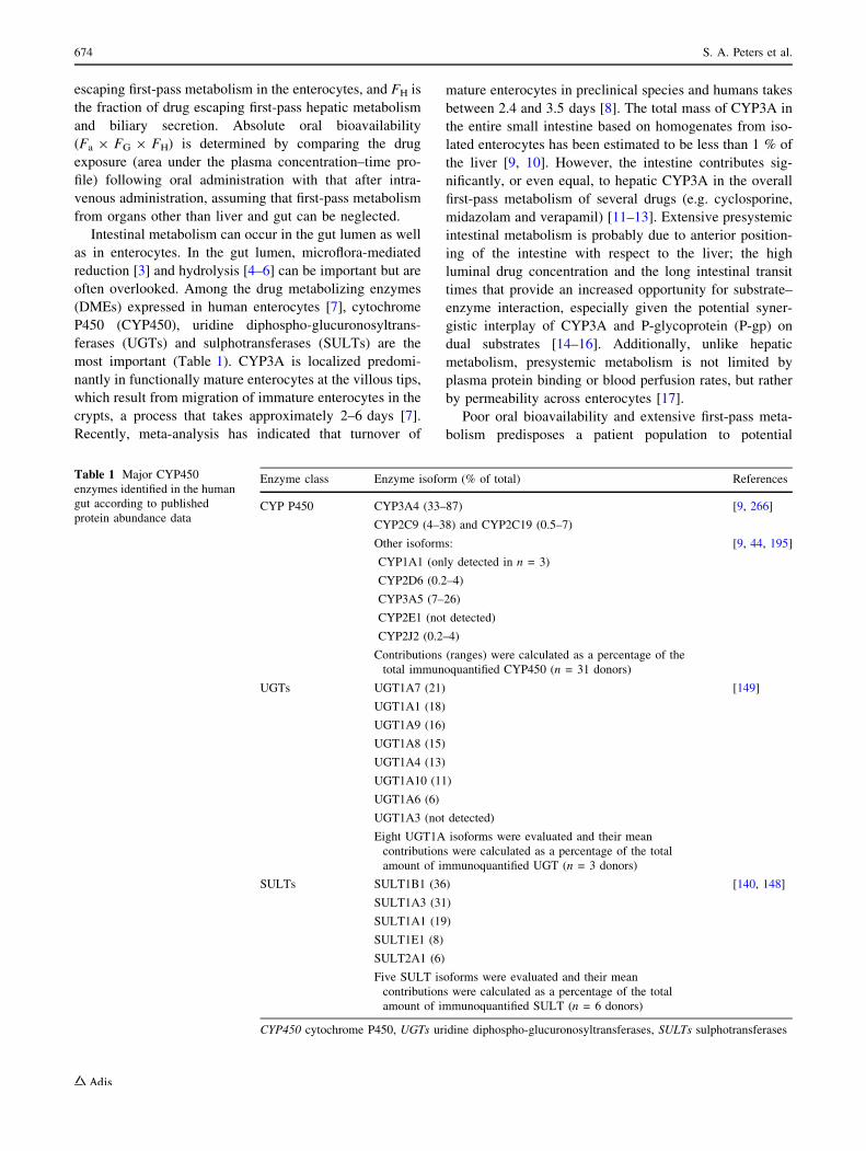
often overlooked. Among the drug metabolizing enzymes
(DMEs) expressed in human enterocytes [7], cytochrome
P450 (CYP450), uridine diphospho-glucuronosyltrans-
ferases (UGTs) and sulphotransferases (SULTs) are the
most important (Table 1). CYP3A is localized predomi-
nantly in functionally mature enterocytes at the villous tips,
which result from migration of immature enterocytes in the
crypts, a process that takes approximately 2–6 days [7].
Recently, meta-analysis has indicated that turnover of
mature enterocytes in preclinical species and humans takes
between 2.4 and 3.5 days [8]. The total mass of CYP3A in
the entire small intestine based on homogenates from iso-
lated enterocytes has been estimated to be less than 1 % of
the liver [9, 10]. However, the intestine contributes sig-
nificantly, or even equal, to hepatic CYP3A in the overall
first-pass metabolism of several drugs (e.g. cyclosporine,
midazolam and verapamil) [11–13]. Extensive presystemic
intestinal metabolism is probably due to anterior position-
ing of the intestine with respect to the liver; the high
luminal drug concentration and the long intestinal transit
times that provide an increased opportunity for substrate–
enzyme interaction, especially given the potential syner-
gistic interplay of CYP3A and P-glycoprotein (P-gp) on
dual substrates [14–16]. Additionally, unlike hepatic
metabolism, presystemic metabolism is not limited by
plasma protein binding or blood perfusion rates, but rather
by permeability across enterocytes [17].
Poor oral bioavailability and extensive first-pass meta-
bolism predisposes a patient population to potential
Table 1 Major CYP450
enzymes identified in the human
gut according to published
protein abundance data
Enzyme class Enzyme isoform (% of total) References
CYP P450 CYP3A4 (33–87)
CYP2C9 (4–38) and CYP2C19 (0.5–7)
[9, 266]
Other isoforms:
CYP1A1 (only detected in n = 3)
CYP2D6 (0.2–4)
CYP3A5 (7–26)
CYP2E1 (not detected)
CYP2J2 (0.2–4)
Contributions (ranges) were calculated as a percentage of the
total immunoquantified CYP450 (n = 31 donors)
[9, 44, 195]
UGTs UGT1A7 (21)
UGT1A1 (18)
UGT1A9 (16)
UGT1A8 (15)
UGT1A4 (13)
UGT1A10 (11)
UGT1A6 (6)
UGT1A3 (not detected)
Eight UGT1A isoforms were evaluated and their mean
contributions were calculated as a percentage of the total
amount of immunoquantified UGT (n = 3 donors)
[149]
SULTs SULT1B1 (36)
SULT1A3 (31)
SULT1A1 (19)
SULT1E1 (8)
SULT2A1 (6)
Five SULT isoforms were evaluated and their mean
contributions were calculated as a percentage of the total
amount of immunoquantified SULT (n = 6 donors)
[140, 148]
CYP450 cytochrome P450, UGTs uridine diphospho-glucuronosyltransferases, SULTs sulphotransferases
674 S. A. Peters et al.
toxicity arising from high doses as well as large
interindividual variability [18] in exposures. In addition,
extensive first-pass metabolism increases the risk of drug–
drug interactions (DDIs) [19–23], the magnitude of which
depends on the fraction escaping metabolism of both the
substrate and the inhibitor [24] (if both the substrate and
inhibitor are metabolized by the inhibited enzyme). The
DDI risk arising from first-pass extraction can also be
transporter-mediated [25–28]. These risks highlight the
need for robust, quantitative models for predicting drug
extraction through the gut wall and liver, backed by a
sound understanding of the underlying mechanisms.
After decades of research, hepatic drug metabolism is
well-understood. Only in recent years has there been an
increased effort to understand intestinal first-pass extrac-
tion. Among the factors limiting intestinal availability
(Fa 9 FG), intestinal metabolism can be a major determi-
nant [29] but is poorly understood. This is due to a number
of confounding factors affecting it; namely, drug transit
through the gastrointestinal tract, drug permeability, solu-
bility and intestinal blood flow. Species differences in the
isoforms, regional abundances and activities of DMEs [9,
30, 31] and transporters [32, 33], as well as the interplay of
enzyme-transporter proteins and overlap of substrate and
inhibitor specificity [34], make it difficult to establish good
preclinical models for this process. Knowledge gaps on
enzyme abundances and lack of empirical enzyme activity
scaling factors are the main limitations of in vitro methods
for the evaluation of intestinal metabolism. The large
interindividual variability and inaccurate estimation of
human intestinal extraction, even when intravenous phar-
macokinetic (PK) data are available, pose additional chal-
lenges to the validation of both in vitro and in vivo models
[35, 36]. Due to its broad specificity and high abundance in
the intestine, CYP3A is the enzyme most frequently
implicated in human gut metabolism, and therefore the
most studied. Focusing on CYP-mediated processes, this
review will summarize current knowledge on the advan-
tages and limitations of preclinical models (in vivo, in situ,
in vitro and mathematical) available for quantitative pre-
diction of intestinal availability. This review will also
outline a strategy applicable in drug discovery and early
development, identify gaps in current understanding and
propose future directions for the prediction of human gut-
wall metabolism.
2 Human In Vivo FG
Seminal work from clinical studies with CYP3A drug
substrates such as cyclosporine A [11, 37, 38] and mida-
zolam [12, 39, 40] established the role of gut-wall meta-
bolism in limiting human oral bioavailability. Substantial
intestinal extraction was demonstrated in patients after
sampling portal vein blood following intraduodenal drug
administration during the anhepatic phase of liver trans-
plantations. However, interpretation of this PK data is
challenging given confounding factors attributable to the
use of anaesthetics, surgery and the often poor condition of
patients. These studies, as with in situ regional perfusions
[41, 42], are rarely undertaken in humans for ethical,
technical and cost reasons [21, 43].
Alternative clinical approaches rely on indirect estima-
tion of human FG from intravenous/oral area under the
curve (AUC) data or drug–food interaction data. Compar-
ison of AUCs following intravenous and oral drug
administration is relatively straightforward, although
assumptions often hinder interpretation [44]. For example,
the extent of metabolic extraction in the intestine can be
over-emphasized if (1) notable extrahepatic systemic
clearance is left unaccounted [45]; (2) the blood:plasma
ratio deviates significantly from unity [46]; or (3) first-
order elimination is not conserved at each site of drug
administration [47, 48]. Additionally, calculated FG can be
sensitive and biased according to the average value used
for liver blood flow [49, 50]. When factors limiting oral
absorption (efflux, low Fa) are minimal and transporter-
mediated uptake is negligible [46, 51], the drug–food
interaction method offers an attractive, pragmatic model
for estimating the extent of FG for CYP3A drug substrates.
This is because certain fruit juices offer complete and
exclusive presystemic inhibition of CYP3A in the small
intestine, providing means for FG to be more readily
identifiable and separated from hepatic first-pass elimina-
tion [52–58]. Advantageously, the interaction approach
requires only measurement of oral AUC in the presence
and absence of inhibitor, avoids many of the confounding
factors associated with the intravenous/oral method, and
allows researchers to benefit from the abundance of clinical
data available [46, 51]. Clinical studies helped to delineate
the metabolic component in liver and intestine for drugs
such as alfentanil, cyclosporine, felodipine, midazolam,
nifedipine and tacrolimus [46, 52, 54–56, 59, 60]. Grape-
fruit juice (GFJ) has been the most popular CYP3A inhi-
bitor in food–drug interactions trialled thus far. The extent
of clinical drug interactions with GFJ can be variable,
depending on the GFJ strength and duration of adminis-
tration [51, 61, 62]. Understanding underlying mechanisms
and kinetics should aid study design and control exposure
to inhibitory agents (furanocoumarins) mediating the
interactions [57, 58, 61, 63–65]. In turn, this should address
interstudy reproducibility and erroneous estimation of FG
[21]. Detailed discussion of the advantages, limitations and
underlying assumptions of these in vivo models are elab-
orated elsewhere [21, 46, 51]. Uncoupling FG and Fa
remains a significant challenge to any clinical approach, as
Predicting Drug Extraction in the Human Gut Wall 675
does factoring out potential contributions from nonhepatic
tissues [43].
3 Regional Differences in Intestinal Metabolismand Transport in the Human Gut
Compounds that have their maximum absorption in dif-
ferent regions of the gut are likely to have very different
FG, even if they have comparable metabolic liabilities. This
is probably due to regional differences in drug uptake into
enterocytes, efflux and intestinal metabolism arising from
regional variation in the luminal environment (e.g. pH,
composition of intestinal fluids) [66–68] and epithelial
membrane (e.g. surface area, expression of enzymes and
transporters) [67]. Information on regional expression of
enzymes and transporters is important for understanding
the in vivo plasma PK profile and possible DDIs with co-
medications.
It is well known that passive diffusion varies in humans
and preclinical models according to the physicochemical
properties of the drug in question and the region of intes-
tine [69, 70]. Lipophilic compounds have the same or
higher permeability coefficients in the lower bowel com-
pared with the upper small intestine. With hydrophilic
molecules the trend is reversed [69, 70], partly due to
differences in the membrane lipid composition and the
tightness of the tight junctional area [70]. Regional dif-
ferences in drug uptake into the enterocytes can also occur
due to differences in intracellular metabolism along the
gut. Higher rates of intracellular metabolism can cause sink
conditions inside the cells. A concentration gradient
maintained across the membrane favors increased uptake,
as has been suggested for indinavir [71].
Efflux proteins impact gut metabolism by reducing the
intracellular drug concentrations exposed to DMEs in the
enterocytes. At high doses, efflux proteins may become
saturated, allowing a larger proportion of drug to pass the
membrane efficiently. Expression of the main ABC trans-
porters is heterogeneous along the gastrointestinal tract
(Table 2) [72–74]. It is well known that multidrug resis-
tance protein 1 (MDR1) [ABCB1, P-gp] is preferentially
expressed toward the lower parts of the human small
intestine (ileum) [74–76], while multidrug resistance-as-
sociated protein 3 (MRP3) [ABCC3] is expressed at higher
levels than MDR1 along the small and large intestines [74].
In contrast, the efflux protein MRP2 (ABCC2) is expressed
at relatively high levels in the small intestine but at
extremely low levels in the colonic regions [74]. The other
important efflux protein, breast cancer resistance protein
(BCRP) (ABCG2), has higher levels than both MRP2 and
MRP3 in the small intestine, but very low levels in the
colonic regions [77, 78].
The solute carrier (SLC) proteins, including PepT1
(SLC15A), MCT1 (SLC16A), OATPs (OATP2B1; SLC0),
OATs (SLC22A), OCT/OCTN (SLC22A) and the recently
described PMAT (SLC29), may add significantly to uptake
of the drug into enterocytes for many compounds with
lower lipophilicities ([72], and refs. therein). These are also
affected by food constituents as well as genetic polymor-
phisms and disease states [79, 80]. OATP2B1 has been
found to have an unexpected influence on the absorption of
the drug aliskiren [81]. The compound has affinity for
MDR1 as well as CYP3A4, and inhibition of these mech-
anisms was expected to increase its bioavailability. How-
ever, in a study involving 11 healthy volunteers, a
reduction of the absorption of aliskiren was found, when
coadministered with GFJ, due to additional inhibition of
the uptake transporter OATP2B1 by GFJ. Thus, prediction
of drug absorption and DDI in the clinic gets further
complicated for substrates of uptake transporters.
The regional distribution of these SLC proteins in the
intestine, as well as species differences, are largely
unknown. However, PepT1 is reported to be highly dis-
tributed in the proximal intestine of humans and many
animal models [72, 82], and MCT1 is well known to be
highly abundant along the whole gastrointestinal tract. The
abundances of other SLC proteins tend not to be significant
[78, 83, 155]. Further details can be found in the excellent
review by Estudante et al., and references therein [72].
The abundance and catalytic activity of the main human
CYP450 enzymes is generally highest in the proximal
regions (i.e. duodenum and proximal jejunum), declining
towards the lower ileum after a slight increase from the
duodenum to the jejunum (Table 2) ([84–87], and refs.
therein]). Data on regional gene expression and enzymatic
activity are readily available for CYP3A4 and members of
the CYP2C family, but are less well-characterized for other
CYP450s. Paine et al., measured CYP450 protein levels
along the gastrointestinal tract in 31 human donors and found
that after CYP3A4, the most abundant enzyme was CYP2C9,
then CYP2C19 with low levels of CYP2J2 and CYP2D6 [9].
Western blot data indicated that concentrations of CYP3A4
and CYP2C isoforms decreased dramatically towards the
distal small intestine, with CYP2C levels falling faster
compared with CYP3A4 [9, 86]. Information available on
the expression of CYP450s in the human colonic enterocyte
is limited, and is contradictory depending on the technique
used [88–90]. Using messenger RNA (mRNA) and protein
analysis, Bergheim et al., [91] found expression levels of
CYP2C, CYP2E1 and CYP3A5 significantly differed
between different regions of the large intestine, with CYP2C
significantly higher in the ascending colon, and CYP2E1 and
CYP3A5 significantly lower. In contrast, others failed to
detect any significant levels of CYP4502C8–10 and
CYP2E1 protein in colonic tissue [63, 64].
676 S. A. Peters et al.
The most obvious difference between the small intestine
and colonic tissue is the content of CYP3A4 and CYP3A5.
In the small intestine, CYP3A5 is detected only at low
levels [89] and may be absent in some individuals [9],
while in colonic tissues, very low levels of CYP3A4 and
higher relative expression of CYP3A5 were reported. Thus,
CYP3A5 constituted the major CYP3A isoform in this
tissue [88]. The overall lower rate of hydroxylation in the
colonic region compared with the proximal jejunum was
confirmed by van de Kerkhof et al., using a mixture of
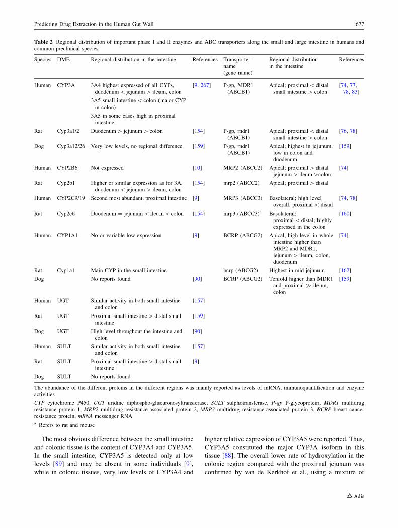
Table 2 Regional distribution of important phase I and II enzymes and ABC transporters along the small and large intestine in humans and
common preclinical species
Species DME Regional distribution in the intestine References Transporter
name
(gene name)
Regional distribution
in the intestine
References
Human CYP3A 3A4 highest expressed of all CYPs,
duodenum\ jejunum[ ileum, colon
3A5 small intestine\ colon (major CYP
in colon)
3A5 in some cases high in proximal
intestine
[9, 267] P-gp, MDR1
(ABCB1)
Apical; proximal\ distal
small intestine[ colon
[74, 77,
78, 83]
Rat Cyp3a1/2 Duodenum[ jejunum[ colon [154] P-gp, mdr1
(ABCB1)
Apical; proximal\ distal
small intestine[ colon
[76, 78]
Dog Cyp3a12/26 Very low levels, no regional difference [159] P-gp, mdr1
(ABCB1)
Apical; highest in jejunum,
low in colon and
duodenum
[159]
Human CYP2B6 Not expressed [10] MRP2 (ABCC2) Apical; proximal[ distal
jejunum[ ileum[colon
[74]
Rat Cyp2b1 Higher or similar expression as for 3A,
duodenum\ jejunum[ ileum, colon
[154] mrp2 (ABCC2) Apical; proximal[ distal
Human CYP2C9/19 Second most abundant, proximal intestine [9] MRP3 (ABCC3) Basolateral; high level
overall, proximal\ distal
[74, 78]
Rat Cyp2c6 Duodenum = jejunum\ ileum\ colon [154] mrp3 (ABCC3)a Basolateral;
proximal\ distal; highly
expressed in the colon
[160]
Human CYP1A1 No or variable low expression [9] BCRP (ABCG2) Apical; high level in whole
intestine higher than
MRP2 and MDR1,
jejunum[ ileum, colon,
duodenum
[74]
Rat Cyp1a1 Main CYP in the small intestine bcrp (ABCG2) Highest in mid jejunum [162]
Dog No reports found [90] BCRP (ABCG2) Tenfold higher than MDR1
and proximal � ileum,
colon
[159]
Human UGT Similar activity in both small intestine
and colon
[157]
Rat UGT Proximal small intestine[ distal small
intestine
[159]
Dog UGT High level throughout the intestine and
colon
[90]
Human SULT Similar activity in both small intestine
and colon
[157]
Rat SULT Proximal small intestine[ distal small
intestine
[9]
Dog SULT No reports found
The abundance of the different proteins in the different regions was mainly reported as levels of mRNA, immunoquantification and enzyme
activities
CYP cytochrome P450, UGT uridine diphospho-glucuronosyltransferase, SULT sulphotransferase, P-gp P-glycoprotein, MDR1 multidrug
resistance protein 1, MRP2 multidrug resistance-associated protein 2, MRP3 multidrug resistance-associated protein 3, BCRP breast cancer
resistance protein, mRNA messenger RNAa Refers to rat and mouse
Predicting Drug Extraction in the Human Gut Wall 677
CYP450 substrates [CYP3A4/5 substrate midazolam
(CYP3A4/5), followed by CYP2C9 (diclofenac) and
CYP2D6 (bufuralol)] [90].
Regional differences in the abundance of phase II
enzymes in the gut are not well-understood. van de Ker-
khof et al. have reported similar activity for UGT and
SULT enzymes [based on 7-hydroxy-coumarin (7-HC)
conjugation] in both the proximal jejunum and the colon.
They also report that glucuronidation efficiency in the gut
was approximately sixfold higher than sulphation in both
regions [90]. As with all quantitative approaches to mea-
suring transporter and DME protein abundances, compar-
ison of expression levels of different phase I and II
enzymes between regions of the small intestine and colonic
tissue, and between different studies, may be difficult given
the different techniques used for quantification, i.e. protein
quantitation using Western blot or liquid chromatography–
tandem mass spectrometry (LC–MS/MS), as well as
immunohistochemistry, mRNA and enzymatic activity
using selected probes.
4 Combined Action of Drug MetabolizingEnzymes (DMEs) and Transporters in the Gut
Co-localization of CYP3A and MDR1 in the enterocyte
along the crypt villus axis, overlapping substrate speci-
ficities and poor oral bioavailability of their joint substrates
[15], have lead scientists to suspect an interplay between
ABC transporters and members of the CYP450 enzyme
families in the intestinal membrane affecting intestinal
absorption and metabolism. It may also explain some DDIs
which cannot be rationalized through either protein acting
alone [15, 72].
P-gp is situated in the apical membrane of the enterocyte
and efflux substrates from inside the cell towards the
intestinal lumen. The CYP3A enzymes are located within
the endoplasmic reticulum. It is therefore suggested that
P-gp may regulate the intracellular concentration of dual
substrates of P-gp and CYP3A. One might speculate that
increasing P-gp levels in the proximal to distal direction
serves to recycle its substrate and aid the efficient elimi-
nation of harmful molecules by reducing the intracellular
drug concentration to levels below DME saturation in the
proximal region, and increase the residence time of its
substrate for metabolism in the distal gastrointestinal tract.
Although demonstrated in vitro [90], some authors have
pointed out that the extent of such synergistic effects
in vivo is minimal [92]. The extent of activity of DMEs and
P-gp are determined by the drug concentration at the site of
the absorption, which in turn is related to solubility (bio-
pharmaceutical classification) [73] and regional stability of
the substrate in the gut lumen. Murakami and Takano
suggested that the biopharmaceutics classification system
(BCS) Class I compounds that are readily water-soluble
and have high permeability will be rapidly absorbed in the
upper part of the small intestine by passive diffusion [73].
Consequently, these compounds can be extensively
metabolized due to higher CYP450 expression in the
proximal small intestine and lower dependence on P-gp
and/or BCRP [29]. However, BCS class II and III, as well
as intravenous compounds with poor solubility and/or
permeability, are likely to be absorbed in the lower parts of
the intestine where metabolism can be substantial if they
are also substrates of P-gp/BCRP [73]. In conclusion,
compounds with high permeability that are typically
absorbed in the upper region of the small intestine can
escape the combined action and potentiation of metabolism
and efflux, while moderate and low-soluble compounds
have an increased potential to be involved in the recycling
action of transporters.
Metabolites formed by CYP450s can be efficiently
transported to either the mucosal or blood side of the
enterocyte by the action of either MDR1, BCRP or the
MRPs. The metabolite of ropivacain was secreted to a
larger extent on the luminal side of the human jejunum
compared with the ileum [93]. Although the transporters
involved were not identified, examples such as this clearly
highlight the need for a greater understanding of the
interplay between enzymes and transporters in the intesti-
nal tract.
5 Preclinical Models for the Prediction of FG
Various preclinical models of the intestine have been
described (see Table 3). If appropriately integrated into
DMPK strategies for optimization of drug absorption,
distribution, metabolism and excretion (ADME) properties,
it may provide more reliable prediction of human first-pass
oral clearance, bioavailability and PK profile. Models
should be considered in terms of their complexity, the
mechanistic understanding they provide, and their clinical
translation, e.g. quantitative prediction of the fraction of
drug escaping metabolism in the human gut wall.
Often there is a trade-off between the different in vitro,
in situ or in vivo models. On the one hand, in vivo models
retain the native architecture of the small intestine and
physiologically relevant expression profiles of DMEs, co-
factors and transporter proteins. They offer integration of
dynamic processes such as the mesenteric blood circulation
and mucous layer coupled with function to study com-
plexities arising from simultaneous metabolism–transporter
interplay [15, 94, 95]. Notable attractions with in situ
techniques are they closely mimic the in vivo situation yet
provide a unique opportunity to study intestinal events in
678 S. A. Peters et al.
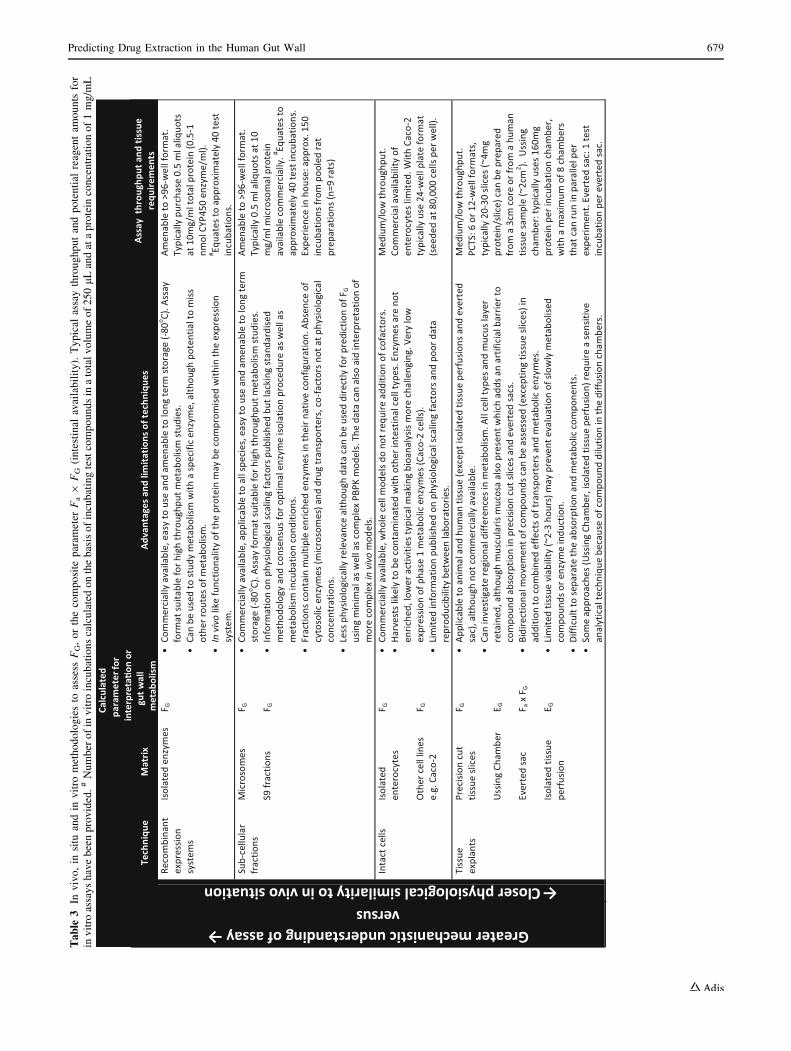
Table
3In
viv
o,
insi
tuan
din
vit
rom
eth
od
olo
gie
sto
asse
ssF
G,
or
the
com
po
site
par
amet
erF
a9
FG
(in
test
inal
avai
lab
ilit
y).
Ty
pic
alas
say
thro
ug
hp
ut
and
po
ten
tial
reag
ent
amo
un
tsfo
r
inv
itro
assa
ys
hav
eb
een
pro
vid
ed.
#N
um
ber
of
inv
itro
incu
bat
ion
sca
lcu
late
do
nth
eb
asis
of
incu
bat
ing
test
com
po
un
ds
ina
tota
lv
olu
me
of
25
0l
Lan
dat
ap
rote
inco
nce
ntr
atio
no
f1
mg
/mL
Predicting Drug Extraction in the Human Gut Wall 679
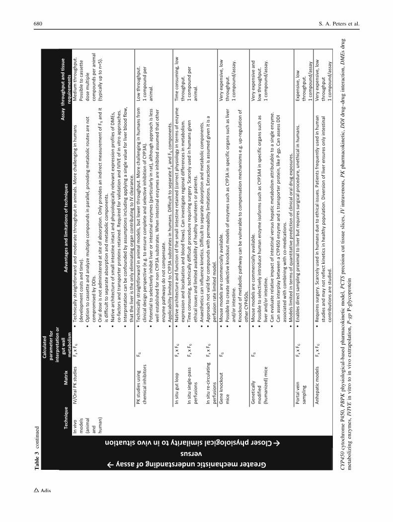
Table
3co
nti
nu
ed
CYP450
cyto
chro
me
P4
50
,PBPK
ph
ysi
olo
gic
al-b
ased
ph
arm
aco
kin
etic
mo
del
,PCTS
pre
cisi
on
cut
tiss
ue
slic
es,IV
intr
aven
ou
s,PK
ph
arm
aco
kin
etic
,DDI
dru
g–
dru
gin
tera
ctio
n,DME
sd
rug
met
abo
lizi
ng
enzy
mes
,IVIVE
inv
itro
toin
viv
oex
trap
ola
tio
n,P-gp
P-g
lyco
pro
tein
680 S. A. Peters et al.
isolation, e.g. absence of biliary excretion and enterohep-
atic recirculation; however, this isolation could compro-
mise the interpretability of the results. Furthermore, species
differences in enzymes and transporters (Table 2) can
make them unreliable for human FG prediction. In vitro
models employing human-specific systems lack native
architecture, but, in combination with mathematical mod-
els, hold the promise of robust prediction, provided there is
a correlation between in vitro and human in vivo.
5.1 In Vivo and In Situ Animal Models
Gut-wall metabolism has been studied in several animal
models [96–102]. By varying sites of drug administration
(oral, intraportal and intravenous routes are common but
intraperitoneal is also used) and PK sampling, the available
fractions in liver and intestine can be calculated from
comparison of AUCs under first-order conditions [103–
107]; however, comparison of intravenous and oral AUCs
after sampling at one site is more straightforward and
routinely applied [108–110]. As such, similar issues
described with the indirect approaches are to be expected.
Despite differences in the CYP450 isoforms expressed in
the rat compared with human (see the following para-
graphs), a good correlation [root mean square error
(RMSE) = 0.19] between rat and human FG has been
reported using a set of 11 CYP3A-metabolized compounds
that had both intravenous and oral PK data [111]. Ten of
these 11 compounds studied had human Fa of 0.8 or higher.
As rat is a good model for human oral drug absorption, it
follows that any differences in the intestinal availability
should arise from intestinal efflux or metabolism. For BCS
class I compounds, good solubility ensures sufficient con-
centrations for the saturation of efflux transporters. A high
permeability rate ensures a low residence time within
enterocytes, resulting in a rate of metabolic extraction that
is limited by the rate of drug permeation through the
enterocytes, rather than by the intrinsic ability of the
intestinal DMEs. With permeability-limited intestinal
extraction, any differences in enzyme isoform, abundance
or activity have limited impact on the metabolic extraction,
especially since CYP450s with broad substrate specificity
abound in both human and rat, and their regional distri-
butions are similar in both species. However, for hepatic
extraction, lower hepatic concentrations compared with the
gut lumen implies a greater role for transporters, while
species differences in plasma protein binding and blood
flow rate contribute to varying exposures to uptake and
efflux transporters, as well as to DMEs, heightening the
impact of differences in transporter and enzyme isoform,
abundance and activities on hepatic extraction. This may
explain why the rat is a good model for the prediction of
human FG but is a poor predictor of human oral
bioavailability [112]. A strong correlation was also repor-
ted between cynomolgus monkey and human FG for human
CYP3A [113]. However, the cost and ethical concerns limit
the availability of monkey PK studies. Animal models may
be useful in the prediction of human FG for CYP3A-me-
tabolized compounds, but not much is known about their
utility for other intestinal DMEs.
In situ approaches include the perfused gut loop, single-
pass and recirculating intestinal perfusions, portal vein
cannulations, portacaval shunts and transpositions [114–
121]. These techniques open the possibility of studying
route-dependent intestinal metabolism following systemic
and luminal drug presentation. This has been shown with
acetaminophen, morphine and enalapril [96, 98, 106, 118,
122, 123]. Naturally-occurring tissue structure and physi-
ology are retained, notwithstanding surgical manipulation
and effect of anaesthetics [114, 124, 125]. Minimal inter-
ference of intestinal function and architecture means opti-
mal tissue viability is maintained [106]. A full complement
of endogenous DMEs and transporter proteins are present.
Discrete segments of the small intestine can be evaluated to
assess the impact of regional differences in DMEs or
transporter expression and gut physiology. Intestinal
metabolism can be more rigorously evaluated if drug
concentrations are measured after sampling from mesen-
teric or portal veins [126]. Detailed methodologies are
provided elsewhere [114, 127–130]. In situ models have
several advantages over in vivo models. For instance,
bypassing the stomach means acidic compounds are unli-
kely to precipitate, and therefore dissolution rates do not
confound intestinal drug concentrations and resultant
plasma levels [94]. These models can be exploited to
investigate metabolism–transporter interplay, as exempli-
fied with midazolam, indinavir and UK-343,664 [48, 131,
132]. Various aspects need to be controlled [133], includ-
ing luminal flow rate and thickness of the unstirred water
layer (UWL), which can be rate-limiting for rapidly
absorbed compounds [106]. In spite of their utility, iso-
lating perfused organs from, or within, the laboratory ani-
mals typically requires specialized surgical procedures. As
such, these approaches are arguably more labour-intensive,
time-consuming and costly compared with other in vivo
and in vitro approaches.
Whereas certain processes, such as passive diffusion,
can be relatively well-predicted from animals [94, 106,
134–139], pronounced species differences in expression of
DME isoforms, substrate selectivity and abundance along
the gastrointestinal tract [30, 31, 113, 140–142] implies
that human FG prediction from preclinical species is not
always feasible. The differences in relative protein
expression of individual CYP450 isoforms in rat, dog,
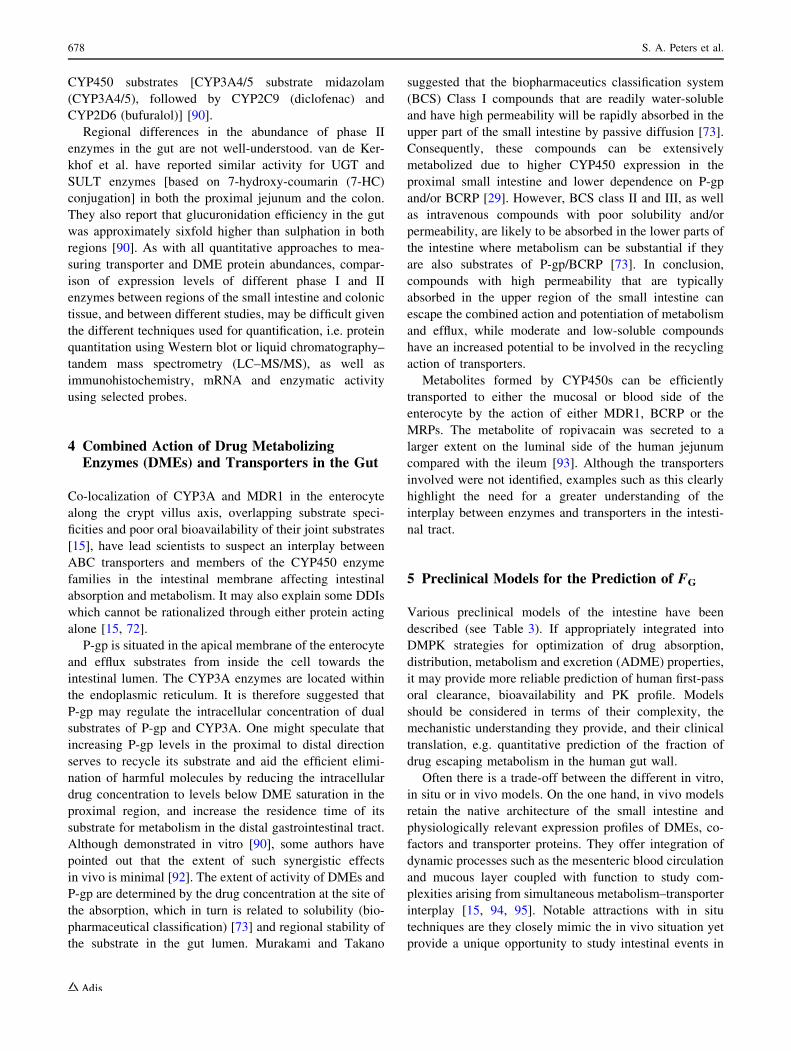
monkey and human small intestines are presented in Fig. 1.
These species differences have led to substantial
Predicting Drug Extraction in the Human Gut Wall 681

differences in apparent Fa 9 FG as well as FG [30, 143–
145]. Metabolism studies, using drug substrates for human
CYP450s [141, 143, 146] and UGT enzymes [142, 147],
have generally reported poor correlation between human
and animal FG. Certain DMEs appear to be selectively
expressed in human intestines, including UGT1A8,
UGT1A10 and SULT1A3 [140, 148, 149]. To the best of
our knowledge, there is no known animal orthologue of
SULT1A3 or UGT1A10 [147]. Additionally, there are
known species differences for other enzyme classes
expressed in the gut, such as the carboxylesterases [3, 4,
150, 151]. If a drug is shown to be a substrate for one of
these enzymes then predicting human FG using animal data
would be questionable.
Species differences in regional enzymatic profiles exist
within the intestine (Table 2). The regional decrease in
CYP3A in humans, as a function of distance along the
small intestine from the duodenum to the ileum [9, 85], is
similar to the rat small intestine [152–154]. Interestingly, in
the rat intestine Cyp2b1 is highly expressed, whereas the
equivalent isoform in humans (CYP2B6) is not expressed
[9]. Rat Cyp2b1 is present at much higher protein levels in
the upper parts of the intestine, whereas in humans the
equivalent isoform (CYP2D6) is not [154]. The trend in the
regional expression of rat Cyp2c isoforms (such as
Cyp2c6) is opposite to that of Cyp3a, with higher expres-
sion towards the lower bowel [154] (Table 2). In contrast
to being absent in the human small intestine, the extra-
hepatic enzyme Cyp1a1 has been reported by some authors
to be the predominant CYP450 isoform expressed in rat
small intestine, together with Cyp3a [9, 154–156]. The
glucuronidation/sulphation ratio can differ between regions
according to individual species. In rat colon and proximal
jejunum, the ratio was 16 and 23, respectively [157],
showing a clear species difference in conjugation activity
and regional difference compared with humans (see Sect. 3
and van de Kerkhof et al. [90]).
Regional expression of Mdr1b in rat intestine is similar
to regional expression of MDR1 in the human small
intestine and colon [74–76, 78, 158] (Table 2). Contrary to
this, canine intestinal Mdr1 expression is highest in the
jejunum and very low in the lower parts of the ileum and
colon [159]. For the MRP2 transporter, humans are similar
to rat, with high expression in the proximal small intestine
and very low levels in the lower bowel [74, 78, 83]. Abcc3
(Mrp3) is highly expressed in the colon of rats [160], and
mice, at both the mRNA and protein levels [161] (Table 2).
Interestingly, MRP3 (the corresponding transporter gene in
humans) has the highest level of all the efflux proteins
expressed along the intestinal tract [74]. In mouse, the
expression of bcrp is higher in the mid jejunum compared
with the rest of the intestinal tract [162]. A similar cellular
location (basolateral in enterocytes) found in mice, rats and
humans further supports a high degree of conservation for
ABC transporters amongst eukaryotes.
5.2 Knockout and Transgenic Mouse Models
In vivo rodent models with intestinal or hepatic enzyme
gene knockdown or replacement have demonstrated the
impact of intestinal versus hepatic elimination [163–168].
These knockout (KO) and/or genetically modified (GM),
transgenic (TG) mouse models have been established to
create more reliable in vivo systems to study and predict
human response to novel chemical entities (NCEs) [163,
169–173]. For example, the importance of CYP3A meta-
bolism in the intestine and liver has been illustrated with
docetaxel in KO mice lacking all Cyp3a genes [Cyp3a
(-/-)] [174]. When the CYP3A anticancer drug was
administered intravenously to Cyp3a (-/-) mice, a sev-
enfold increase in systemic exposure was observed com-
pared with wild-type. After oral dosing, an 18-fold higher
systemic exposure was reported. Similar findings were
reported for lopinavir and triazolam (CYP3A) [175, 176],
debrisoquine (CYP2D6) [177] and tolbutamide (CYP2C9)
[178]. This highlights the critical role intestinal CYP3A
plays in human first-pass oral clearance and bioavailability.
Unfortunately, compensatory mechanisms arising from
expression of host (murine) DMEs may confound data
interpretation from KO models. For instance, clearance of
Han Wistar rat, male (n=3)
Cyp2b1 (61%)
Cyp2d1 (12.3%)
Cyp2c6 (1.5%)
Cyp3a (25.1%)
Beagle dog (n=35)
Cyp2d15 (~10-30%)
Cyp3a12 (~70-90%)
Cynomolgus Monkey (n=35)
Cyp3a4 (17%)
Cyp3a5 (25%)
Cyp4f2/3(32%)
Cyp2j2 (8.5%)
Cyp2c9/19 (8.7%)
Cyp1a1/2 (2.1%)
Cyp2d17/44(0.2%)
Cyp4f12(10%)
Cyp4f11(1.9%)
CYP3A4/5 (84%)
CYP2C9 (12%) CYP2C19
(1.4%)
CYP2D6(0.7%)
CYP2J2(1.3%)
Human (n=31)
CYP1A2 not shown, detected in n=3
Fig. 1 Small intestine CYP450 pie charts for rat, dog, monkey and
humans. The mean percentage contributions of individual CYP450
enzymes were calculated from total immunoquantified CYP450
protein (dog, monkey, human) or total spectrally determined
CYP450 (rat). Analysis was based on published data: rat [154], dog
[254, 255], monkey [268] and humans [9]. Additionally, CYP4F2
protein was detected (*7 pmol/mg protein) in human small intestine
microsomal fractions [269]. This has been excluded from the human
CYP450 pie chart because the total intestinal CYP450 content has not
been reported, precluding comparison of relative abundances of
individual enzyme isoforms between studies. CYP450 cytochrome
P450
682 S. A. Peters et al.
midazolam was expected to be severely reduced in Cyp3a
KO mice. In spite of this, metabolism was only marginally
altered versus wild-type [179]. The revelation that several
murine Cyp2c isoforms were significantly upregulated in
Cyp3a KO mice, and could catalyze formation of mida-
zolam 10- and 40-hydroxymidazolam, helped rationalize
these results [179]. In contrast, no such effect was seen
with triazolam, apparently a more selective Cyp3a sub-
strate in mouse [176]. These compensatory mechanisms are
likely to be drug- and species-dependent. In vitro studies
characterizing background metabolism may prevent
assessment of drugs susceptible to elevated host DMEs.
Alternatively, exciting development of a viable mouse
model in which all murine CYP450 genes have been
deleted could avoid this issue altogether [180]. Interest-
ingly, TG mice have been generated that are capable of
expressing human CYP3A4 in the intestine and/or the liver
on top of a mouse Cyp3a KO background [174]. As
demonstrated with docetaxel and lopinavir, these human-
ized mice can provide mechanistic insight into the separate
and combined roles of intestinal and hepatic human
CYP3A, and the interplay with transporters such as P-gp
in vivo [163, 175].
5.3 In Vitro and Mathematical Models
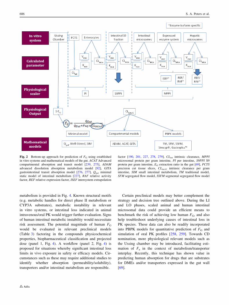
‘Bottom-up’ quantitative predictions that use in vitro data
in conjunction with mathematical models describing pro-
cesses within the gastrointestinal tract have been imple-
mented [181–185]. Several of the in vitro assays
highlighted in Table 3 are simplistic in nature, take less
time to complete, and are a fraction of the cost of animal
experiments. This makes them eminently suited for
screening NCEs and designing out presystemic metabolic
liabilities. Models vary in complexity, from simple mem-
brane preparations of individually recombinantly expressed
enzymes [186–190] to subcellular fractions [12, 43, 45, 85,
153, 191–195], intact cells [196–201] to tissue explants.
The latter retain their in vivo tissue architecture, albeit
lacking physiological surroundings. Although relatively
labour-intensive and lower throughput (see Table 3),
working with whole tissue preparations such as precision
cut tissue slices (PCTS) [157, 202–206], Ussing chamber
[43, 69, 90, 93, 207], everted sac [126, 208–212] and iso-
lated tissue perfusions [94, 197, 213–216] brings with it
several advantages. The cell–cell contacts remain intact, all
cell types are present and the DME systems, co-factors and
transporters are available at physiologically relevant con-
centrations that more closely mimic the in vivo situation.
With the isolated perfusion technique, variables such as
temperature, pH, osmolality, blood pressure and flow can
be controlled [217]. Metabolism on both sides of the
intestine (luminal and vascular) can be studied. However,
maintaining tissue viability is the major issue with this
approach [94]. As such, its application is generally limited
to animal tissue, and short-term incubations (approximately
2 h post-excision) impeding assessment of slowly metab-
olized drugs or enzyme induction [43]. This can be cir-
cumvented in situ but is not without complication. Loss of
tissue viability caused by insufficient oxygenation is also
problematic for everted sacs (1–2 h) and Ussing chambers
preparations (2–4 h) [218–220]. Nevertheless, everted sacs
provide a fast and relatively inexpensive model for mea-
suring regional differences in metabolism [106, 126]. The
small volume inside the sacs offers analytical advantages
over the isolated tissue perfusion and Ussing chamber,
which due to sample dilution require sensitive bioanalysis
to detect drug and/or metabolites [119, 126].
The Ussing chamber can be used with animal or human
tissue providing a good model of drug absorption, trans-
porter interactions, as well as metabolism, during passage
across the gastrointestinal membrane [42, 69, 90, 219, 221–
223]. Drug can be added to luminal or serosal sides,
allowing bidirectional transport and metabolism kinetics to
be studied in different sections of the intestine [106].
Detailed mechanistic interpretation from studies such as
these may require additional insight from experimentation
with enzyme inhibitors, radiolabelled drug, or separate
consideration of metabolism and permeability, e.g. using
intestinal microsomes and Caco-2 monolayers. Indeed the
extent of drug extraction in human intestine (Eg) has been
successfully predicted for testosterone, midazolam and
ropivacaine using human in vitro Ussing experiments [69,
93]. PCTS have received growing attention now that
reproducible production of very thin slices (between 250
and 450 lm) is possible. These maintain better viability
[203] and retain a high drug biotransformation capacity
[43]. PCTS are also suitable for studying regional differ-
ences in intestinal metabolism, as well as regulation of
enzymes and transporters involved in drug disposition
[206, 224]. Applicable to all species, investigations have
been reported in mouse [225], rat [157] and human [90].
Additionally, other tissues can be examined, allowing the
extent of metabolism in different organs to be compared
[204]. Potential drawbacks include poor penetration of
highly metabolized drugs into the slices inner cell layers,
lag time in phase II metabolism, and nonspecific binding to
the slices [202, 226].
Intestinal subcellular fractions (S9 homogenates or
microsomes) are one of the more established in vitro
approaches used in drug discovery. More information has
been published on the physiological scalars (Table 4) and
several investigators have explored quantitative prediction
of FG [49, 111, 227–231]. Commercial availability of
animal and human samples, ease of storage and automa-
tion, make them an attractive option in terms of assay
Predicting Drug Extraction in the Human Gut Wall 683
speed, capacity and cost. The fractions contain multiple
enriched enzymes in their native configuration for assess-
ment of phase I and selective phase II metabolism [45, 113,
195, 231, 232]; however, they do not contain a full com-
plement of DMEs and lack potentially important interac-
tions with uptake and efflux transporters. Incubations
require the addition of expensive co-factors for optimal
DME activity, often at higher nonphysiological concen-
trations. Others have suggested metabolic rates in S9 and
microsomal fractions can be much lower compared with
matrix such as PCTS [43]. This may be attributed to poor
recovery of enzymes through suboptimal preparation pro-
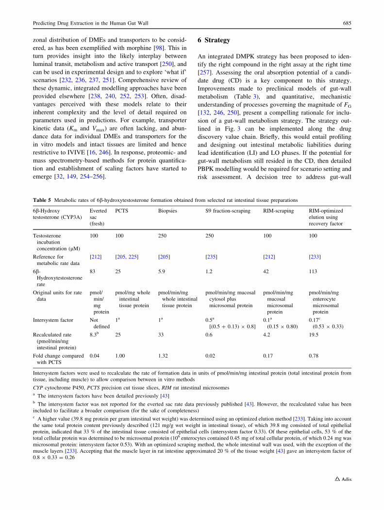
cedures [195]. Presented in Table 5 are 6b-hydroxy
testosterone rat data (normalized to units of pmol/min/mg
rat intestinal protein) including intrinsic clearance (CLint)
from in-house intestinal microsomes prepared under opti-
mal conditions [233, 234]. Interestingly, in-house micro-
somes achieved broadly similar rates compared with PCTS
and biopsies [43, 205, 225], and were much higher than
previous microsomal preparations [212, 235]. This high-
lights progress made with DME extraction procedures,
which, until recently, have limited the scalability of
intestinal microsomes [192, 195]. Given the profound
effect enzyme extraction procedure and incubation condi-
tions can have on enzyme activity, consensus is needed on
best practise before we can expect significant improve-
ments to the accuracy and reproducibility of predictions.
In vitro models allow the function of metabolic enzymes,
transporters and absorption processes to be studied in the gut
wall. However, to apply this data to retrospectively explain,
or prospectively predict, human oral PK ultimately requires
insight into the mechanisms influencing drug behaviour
in vivo [132, 236–238]. As such, several mechanistic
approaches of varying complexity have been described for
in vitro to in vivo extrapolation (IVIVE), some of which are
available commercially, e.g. GastroPlusTM and Simcyp�
[17, 92, 132, 238–241]. These mathematical translations
(Fig. 2) can be relatively straightforward and ‘minimal’
models, such as QGut, require only in vitro metabolic CLint
and cell permeability data to estimate FG. Several groups
have reported successful prediction of FG in animals [242,
243] and humans [49, 227, 228, 244, 245] with this
approach. Drugs with high in vivo extractions (FG values
\0.5) were less accurately predicted and may reflect
inability of the QGut model to account for changes in ente-
rocyte drug concentration and therefore saturation of DME
and efflux transporter processes [246]. However, to the
authors’ knowledge, no critical assessment of possible sys-
tematic underprediction of IVIVE similar to that reported for
hepatic metabolism [247–249] is available in the literature.
Improved FG and oral clearance prediction was noted when
the same set of drugs were evaluated using a physiological-
based PK (PBPK) model. This was partly attributed to the
model’s ability to account for saturation of intestinal meta-
bolism by using maximum velocity (Vmax) and Michaelis-
Menten constant Km, the substrate concentration at which
the reaction rate is half of Vmax rather than CLint [246].
Sophisticated PBPK models have been published, such
as the segmental segregated flow model (SSFM), which
encompass all salient variables, e.g. absorption, gastroin-
testinal transit, metabolism, transport and efflux [95, 98].
This allows route-dependent intestinal metabolism and
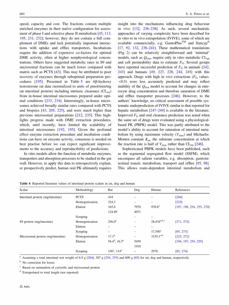
Table 4 Reported literature values of intestinal protein scalars in rat, dog and human
Scalar Methodology Rat Dog Human References
Intestinal protein (mg/intestine) PCTS 434 – – [204]
Homogenization 324.3 – – [225]
Elution 165.6
124.89
7970
4071
970.8a [197, 198, 254, 255, 270]
Scraping – –
S9 protein (mg/intestine) Homogenization 256.0a – 28,476a,b,c [271, 272]
Elution – – –
Scraping – – 17,540c [85, 273]
Microsomal protein (mg/intestine) Homogenization 17.1b – 3155.1a,b [225, 272]
Elution 54.4d, 16.5a 5459
1918
[194, 197, 254, 255]
Scraping 156a, 114a – 2978 [85, 274]
a Assuming a total intestinal wet weight of 6.9 g [204], 307 g [254, 255] and 809 g [85] for rat, dog and human, respectivelyb No correction for lossesc Based on summation of cytosolic and microsomal proteind Extrapolated to total length (not reported)
684 S. A. Peters et al.
zonal distribution of DMEs and transporters to be consid-
ered, as has been exemplified with morphine [98]. This in
turn provides insight into the likely interplay between
luminal transit, metabolism and active transport [250], and
can be used in experimental design and to explore ‘what if’
scenarios [232, 236, 237, 251]. Comprehensive review of
these dynamic, integrated modelling approaches have been
provided elsewhere [238, 240, 252, 253]. Often, disad-
vantages perceived with these models relate to their
inherent complexity and the level of detail required on
parameters used in predictions. For example, transporter
kinetic data (Km and Vmax) are often lacking, and abun-
dance data for individual DMEs and transporters for the
in vitro models and intact tissues are limited and hence
restrictive to IVIVE [16, 246]. In response, proteomic- and
mass spectrometry-based methods for protein quantifica-
tion and establishment of scaling factors have started to
emerge [32, 149, 254–256].
6 Strategy
An integrated DMPK strategy has been proposed to iden-
tify the right compound in the right assay at the right time
[257]. Assessing the oral absorption potential of a candi-
date drug (CD) is a key component to this strategy.
Improvements made to preclinical models of gut-wall
metabolism (Table 3), and quantitative, mechanistic
understanding of processes governing the magnitude of FG
[132, 246, 250], present a compelling rationale for inclu-
sion of a gut-wall metabolism strategy. The strategy out-
lined in Fig. 3 can be implemented along the drug
discovery value chain. Briefly, this would entail profiling
and designing out intestinal metabolic liabilities during
lead identification (LI) and LO phases. If the potential for
gut-wall metabolism still resided in the CD, then detailed
PBPK modelling would be required for scenario setting and
risk assessment. A decision tree to address gut-wall
Table 5 Metabolic rates of 6b-hydroxytestosterone formation obtained from selected rat intestinal tissue preparations
6b-Hydroxy
testosterone (CYP3A)
Everted
sac
(fresh)
PCTS Biopsies S9 fraction-scraping RIM-scraping RIM-optimized
elution using
recovery factor
Testosterone
incubation
concentration (lM)
100 100 250 250 100 100
Reference for
metabolic rate data
[212] [205, 225] [205] [235] [212] [233]
6b-
Hydroxytestosterone
rate
83 25 5.9 1.2 42 113
Original units for rate
data
pmol/
min/
mg
protein
pmol/mg whole
intestinal
tissue protein
pmol/min/mg
whole intestinal
tissue protein
pmol/min/mg mucosal
cytosol plus
microsomal protein
pmol/min/mg
mucosal
microsomal
protein
pmol/min/mg
enterocyte
microsomal
protein
Intersystem factor Not
defined
1a 1a 0.5a
[(0.5 ? 0.13) 9 0.8]
0.1a
(0.15 9 0.80)
0.17c
(0.53 9 0.33)
Recalculated rate
(pmol/min/mg
intestinal protein)
8.3b 25 33 0.6 4.2 19.5
Fold change compared
with PCTS
0.04 1.00 1.32 0.02 0.17 0.78
Intersystem factors were used to recalculate the rate of formation data in units of pmol/min/mg intestinal protein (total intestinal protein from
tissue, including muscle) to allow comparison between in vitro methods
CYP cytochrome P450, PCTS precision cut tissue slices, RIM rat intestinal microsomesa The intersystem factors have been detailed previously [43]b The intersystem factor was not reported for the everted sac rate data previously published [43]. However, the recalculated value has been
included to facilitate a broader comparison (for the sake of completeness)c A higher value (39.8 mg protein per gram intestinal wet weight) was determined using an optimized elution method [233]. Taking into account
the same total protein content previously described (121 mg/g wet weight in intestinal tissue), of which 39.8 mg consisted of total epithelial
protein, indicated that 33 % of the intestinal tissue consisted of epithelial cells (intersystem factor 0.33). Of these epithelial cells, 53 % of the
total cellular protein was determined to be microsomal protein (106 enterocytes contained 0.45 mg of total cellular protein, of which 0.24 mg was
microsomal protein: intersystem factor 0.53). With an optimized scraping method, the whole intestinal wall was used, with the exception of the
muscle layers [233]. Accepting that the muscle layer in rat intestine approximated 20 % of the tissue weight [43] gave an intersystem factor of
0.8 9 0.33 = 0.26
Predicting Drug Extraction in the Human Gut Wall 685
metabolism is provided in Fig. 4. Known structural motifs
(e.g. metabolic handles for direct phase II metabolism or
CYP3A substrates), metabolic instability in relevant
in vitro systems, or intestinal loss indicated in animal
intravenous/oral PK would trigger further evaluation. Signs
of human intestinal metabolic instability would necessitate
risk assessment. The potential magnitude of human FG
would be evaluated in relevant preclinical models
(Table 3) factoring in the compounds physicochemical
properties, biopharmaceutical classification and projected
dose (panel 1, Fig. 4). A workflow (panel 2, Fig. 4) is
proposed for situations whereby significant intestinal loss
limits in vivo exposure in safety or efficacy models. Cir-
cumstances such as these may require additional studies to
identify whether absorption (permeability/solubility),
transporters and/or intestinal metabolism are responsible.
Certain preclinical models may better complement the
strategy and decision tree outlined above. During the LI
and LO phases, scaled animal and human intestinal
microsomal data could provide an efficient means to
benchmark the risk of achieving low human FG, and also
help troubleshoot underlying causes of intestinal loss in
PK species. These data can also be readily incorporated
into PBPK models for quantitative prediction of FG and
simulation of oral PK profiles [258, 259]. Towards CD
nomination, more physiological relevant models such as
the Ussing chamber may be introduced, facilitating esti-
mation of Fa in the context of metabolism/transporter
interplay. Recently, this technique has shown value in
predicting human absorption for drugs that are substrates
for DMEs and/or transporters expressed in the gut wall
[69].
Fig. 2 Bottom-up approach for prediction of FG using established
in vitro systems and mathematical models of the gut. ACAT Advanced
compartmental absorption and transit model [239, 275], ADAM
advanced dissolution absorption metabolism model [92], GITA
gastrointestinal transit absorption model [276, 277], Qgut minimal
static model of intestinal metabolism [227], RAF relative activity
factor, REF relative expression factor, ISEF intersystem extrapolation
factor [190, 201, 227, 278, 279], CLint intrinsic clearance, MPPI
microsomal protein per gram intestine, PI per intestine, S9PPI S9
protein per gram intestine, EG extraction ratio in the gut [69], PCTS
precision cut tissue slices, CLint,G intrinsic clearance per gram
intestine, SIM small intestinal metabolism, TM traditional model,
SFM segregated flow model, SSFM segmental segregated flow model
686 S. A. Peters et al.
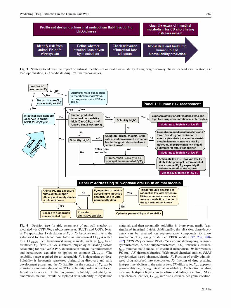
Fig. 4 Decision tree for risk assessment of gut-wall metabolism
mediated via CYP450s, carboxylesterases, SULTs and UGTs. Note,
as FH approaches 1 calculation of Fa 9 FG becomes sensitive to the
value used for liver blood flow. Intestinal microsomal CLint is scaled
to a CLint,u,G then transformed using a model such as Qgut to an
estimated FG. #For CYP3A substrates, physiological scaling factors
accounting for relative CYP3A abundance in human liver microsomes
and hepatocytes can also be applied to estimate CLint,u,G. *The
solubility range required for an acceptable Fa is dependent on dose.
Solubility is frequently reassessed during drug discovery and early
development phases and the FG liability, in the context of Fa, can be
revisited as understanding of an NCEs’ solubility profile is developed.
Initial measurement of thermodynamic solubility, potentially on
amorphous material, would be replaced with solubility of crystalline
material, and then potentially solubility in biorelevant media (e.g.,
simulated intestinal fluids). Additionally, the pKa (ion class-depen-
dent) can be assessed on representative compounds to allow
simulation of Fa using established PBPK models [92, 239, 280–
282]. CYP450 cytochrome P450, UGTs uridine diphospho-glucurono-
syltransferases, SULTs sulphotransferases, CLint intrinsic clearance,
Qgut minimal static model of intestinal metabolism, IV intravenous,
PO oral, PK pharmacokinetics, NCEs novel chemical entities, PBPK
physiological-based pharmacokinetic, Fa Fraction of orally adminis-
tered drug absorbed into enterocytes, FG fraction of drug escaping
first-pass metabolism in the enterocytes, ER efflux ratio, Papp apparent
permeability, Fa 9 FG intestinal availability, FH fraction of drug
escaping first-pass hepatic metabolism and biliary secretion, NCEs
new chemical entities, CLint,G intrinsic clearance per gram intestine
Fig. 3 Strategy to address the impact of gut-wall metabolism on oral bioavailability during drug discovery phases. LI lead identification, LO
lead optimization, CD candidate drug, PK pharmacokinetics
Predicting Drug Extraction in the Human Gut Wall 687
7 Challenges and Future Perspectives
When two or more drugs are coadministered, the effects
could be additive, synergistic or antagonistic due to DDIs
affecting the absorption and/or therapeutic profile of the
victim drug. Overlapping substrate specificities for multi-
ple enzymes and transporters might also enhance the
complexity of the absorption profile along the gastroin-
testinal tract. Thus, overall understanding is a result of
complex interplay between physiological (e.g. enzymes
and transporters, blood flow, region of the intestine, lumi-
nal fluid composition) and physicochemical factors (e.g.
pKa, solubility, dissolution, lipophilicity, substrate to
enzymes and/or transporters) characterizing the drug
molecule. In addition, genetic polymorphisms in drug
transporter and DMEs, as well as disease states, may be
responsible for variability in the profile and adverse events
arising from co-medication among patients, which may be
different from healthy volunteers [260] and is difficult to
predict from preclinical tools.
In this review, we have illustrated the PK complexity
associated with oral administration of drugs linked to
intestinal regional variation in DMEs/transporters, as well
as species and model differences. Assessing whether clin-
ical candidates have the right risk/benefit balance for
patients can be challenging given the inherent complexities
and difficulties in the early screening phase and translation
into clinical use. Because of the complexity, PBPK mod-
elling will be a crucial tool as it enables efficient integra-
tion of knowledge on compound behaviour with the
dynamics of intestinal physiology in the preclinical models
and humans [236]. However, for successful modelling,
high-quality data from in vitro and in vivo preclinical tools
needs to be generated (see above strategy). New bioana-
lytical tools for quantitatively analyzing DME [149, 255,
261, 262] and transporter [33, 256, 263, 264] isoform
abundances are already available to improve the quantita-
tive translation between preclinical animals and humans,
and will benefit understanding [185, 256, 265]. Knowledge
of the impact of pharmacogenomics and disease on regio-
nal intestinal availability and variability in underlying
mechanisms is scarce. Alongside this, reports focused on
back translation of clinical outcome that enable evaluation
of the successes or failures of predictions, made from
preclinical data, will be crucial to advancing understanding
and selection of the best tools for future development
activities.
Compliance with Ethical Standards
Funding No funding was received for the preparation of this
manuscript.
Conflicts of interest Sheila Annie Peters, Christopher R. Jones,
Anna-Lena Ungell and Oliver Hatley have no conflicts of interest to
declare.
Open Access This article is distributed under the terms of the
Creative Commons Attribution-NonCommercial 4.0 International
License (http://creativecommons.org/licenses/by-nc/4.0/), which per-
mits any noncommercial use, distribution, and reproduction in any
medium, provided you give appropriate credit to the original
author(s) and the source, provide a link to the Creative Commons
license, and indicate if changes were made.
References
1. Bartholow M. Top 200 drugs of 2011. Pharmacy times. Avail-
able at: http://www.pharmacytimes.com/publications/issue/2012/
July2012/Top-200-Drugs-of-2011. Accessed Jan 2013.
2. Liu G, Franssen E, Fitch MI, Warner E. Patient preferences for
oral versus intravenous palliative chemotherapy. J Clin Oncol.
1997;15(1):110–5.
3. Rafil F, Franklin W, Heflich RH, Cerniglia CE. Reduction of
nitroaromatic compounds by anaerobic bacteria isolated from
the human gastrointestinal tract. Appl Environ Microbiol.
1991;57(4):962–8.
4. Nishimuta H, Houston JB, Galetin A. Hepatic, intestinal, renal,
and plasma hydrolysis of prodrugs in human, cynomolgus mon-
key, dog, and rat: implications for in vitro–in vivo extrapolation of
clearance of prodrugs. Drug Metab Dispos. 2014;42(9):1522–31.
5. Goldin BR. Intestinal microflora: metabolism of drugs and
carcinogens. Ann Med. 1990;22(1):43–8.
6. McCabe M, Sane RS, Keith-Luzzi M, Xu J, King I, Whitcher-
Johnstone A, et al. Defining the role of gut bacteria in the
metabolism of deleobuvir: in vitro and in vivo studies. Drug
Metab Dispos. 2015;43(10):1612–8.
7. Kaminsky LS, Zhang Q-Y. The small intestine as a xenobiotic-
metabolizing organ. Drug Metab Dispos. 2003;31(12):1520–5.
8. Darwich AS, Aslam U, Ashcroft DM, Rostami-Hodjegan A.
Meta-analysis of the turnover of intestinal epithelia in preclini-
cal animal species and humans. Drug Metab Dispos. 2014;
42(12):2016–22.
9. Paine MF, Hart HL, Ludington SS, Haining RL, Rettie AE,
Zeldin DC. The human intestinal cytochrome P450 ‘‘pie’’. Drug
Metab Dispos. 2006;34(5):880–6.
10. Yang J, Tucker GT, Rostami Hodjegan A. Cytochrome P450 3A
expression and activity in the human small intestine. Clin
Pharmacol Ther. 2004;76(4):391.
11. Kolars JC, Watkins P, Merion RM, Awni W. First-pass meta-
bolism of cyclosporin by the gut. Lancet. 1991;338(8781):
1488–90.
12. Paine MF, Shen DD, Kunze KL, Perkins JD, Marsh CL,
McVicar JP, et al. First-pass metabolism of midazolam by the
human intestine. Clin Pharmacol Ther. 1996;60(1):14–24.
13. von Richter O, Greiner B, Fromm MF, Fraser R, Omari T,
Barclay ML, et al. Determination of in vivo absorption, meta-
bolism, and transport of drugs by the human intestinal wall and
liver with a novel perfusion technique. Clin Pharmacol Ther.
2001;70(3):217–27.
14. Benet L, Cummins C, Wu C. Unmasking the dynamic interplay
between efflux transporters and metabolic enzymes. Int J Pharm.
2004;277(1):3–9.
15. Benet LZ. The drug transporter-metabolism alliance: uncover-
ing and defining the interplay. Mol Pharm. 2009;6(6):1631–43.
688 S. A. Peters et al.
16. Siissalo S, Heikkinen AT. In vitro methods to study the interplay
of drug metabolism and efflux in the intestine. Curr Drug Metab.
2013;14(1):102–11.
17. Fagerholm U. Prediction of human pharmacokinetics: gut-wall
metabolism. J Pharm Pharmacol. 2007;59(10):1335–43.
18. Hellriegel ET, Bjornsson TD, Hauck WW. Interpatient vari-
ability in bioavailability is related to the extent of absorption:
implications for bioavailability and bioequivalence studies. Clin
Pharmacol Ther. 1996;60(6):601–7.
19. Fahmi OA, Hurst S, Plowchalk D, Cook J, Guo F, Youdim K,
et al. Comparison of different algorithms for predicting clinical
drug–drug interactions, based on the use of CYP3A4 in vitro
data: predictions of compounds as precipitants of interaction.
Drug Metab Dispos. 2009;37(8):1658–66.
20. Fahmi OA, Maurer TS, Kish M, Cardenas E, Boldt S, Nettleton
D. A combined model for predicting CYP3A4 clinical net drug–
drug interaction based on CYP3A4 inhibition, inactivation, and
induction determined in vitro. Drug Metab Dispos. 2008;36(8):
1698–708.
21. Galetin A, Gertz M, Houston JB. Potential role of intestinal first-
pass metabolism in the prediction of drug–drug interactions.
Expert Opin Drug Metab Toxicol. 2008;4(7):909–22.
22. Galetin A, Gertz M, Houston JB. Contribution of intestinal
cytochrome P450-mediated metabolism to drug–drug inhibition
and induction interactions. Drug Metab Pharmacokinet. 2010;
25(1):28–47.
23. Vieira ML, Kirby B, Ragueneau-Majlessi I, Galetin A, Chien J,
Einolf H, et al. Evaluation of various static in vitro–in vivo
extrapolation models for risk assessment of the CYP3A inhibi-
tion potential of an investigational drug. Clin Pharmacol Ther.
2014;95(2):189–98.
24. Peters SA, Schroeder PE, Giri N, Dolgos H. Evaluation of the
use of static and dynamic models to predict drug–drug interac-
tion and its associated variability: impact on drug discovery and
early development. Drug Metab Dispos. 2012;40(8):1495–507.
25. Konig J, Muller F, Fromm MF. Transporters and drug–drug
interactions: important determinants of drug disposition and
effects. Pharmacol Rev. 2013;65(3):944–66.
26. Muller F, Fromm MF. Transporter-mediated drug–drug inter-
actions. Pharmacogenomics. 2011;12(7):1017–37.
27. Yoshida K, Maeda K, Sugiyama Y. Hepatic and intestinal drug
transporters: prediction of pharmacokinetic effects caused by
drug–drug interactions and genetic polymorphisms. Ann Rev
Pharmacol Toxicol. 2013;53:581–612.
28. Zakeri-Milani P, Valizadeh H. Intestinal transporters: enhanced
absorption through P-glycoprotein-related drug interactions.
Expert Opin Drug Metab Toxicol. 2014;10(6):859–71.
29. Wu C-Y, Benet LZ. Predicting drug disposition via application
of BCS: transport/absorption/elimination interplay and devel-
opment of a biopharmaceutics drug disposition classification
system. Pharm Res. 2005;22(1):11–23.
30. Komura H, Iwaki M. In vitro and in vivo small intestinal
metabolism of CYP3A and UGT substrates in preclinical ani-
mals species and humans: species differences. Drug Metab Rev.
2011;43(4):476–98.
31. Martignoni M, Groothuis GM, de Kanter R. Species differences
between mouse, rat, dog, monkey and human CYP-mediated
drug metabolism, inhibition and induction. Expert Opin Drug
Metab Toxicol. 2006;2(6):875–94.
32. Tucker TG, Milne AM, Fournel-Gigleux S, Fenner KS, Cough-
trie MW. Absolute immunoquantification of the expression of
ABC transporters P-glycoprotein, breast cancer resistance pro-
tein and multidrug resistance-associated protein 2 in human liver
and duodenum. Biochem Pharmacol. 2012;83(2):279–85.
33. Groer C, Bruck S, Lai Y, Paulick A, Busemann A, Heidecke C,
et al. LC–MS/MS-based quantification of clinically relevant
intestinal uptake and efflux transporter proteins. J Pharm
Biomed Anal. 2013;85:253–61.
34. Shi S, Li Y. Interplay of drug-metabolizing enzymes and
transporters in drug absorption and disposition. Curr Drug
Metab. 2015;15(10):915–41.
35. Mohri K, Uesawa Y. Enzymatic activities in the microsomes
prepared from rat small intestinal epithelial cells by differential
procedures. Pharm Res. 2001;18(8):1232–6.
36. Sharer JE, Shipley LA, Vandenbranden MR, Binkley SN,
Wrighton SA. Comparisons of phase I and phase II in vitro hepatic
enzyme activities of human, dog, rhesus monkey, and cynomolgus
monkey. Drug Metab Dispos. 1995;23(11):1231–41.
37. Kolars JC, Lown KS, Schmiedlin-Ren P, Ghosh M, Fang C,
Wrighton SA, et al. CYP3A gene expression in human gut
epithelium. Pharmacogenetics. 1994;4(5):247–59.
38. Hebert MF, Roberts JP, Prueksaritanont T, Benet LZ.
Bioavailability of cyclosporine with concomitant rifampin
administration is markedly less than predicted by hepatic
enzyme induction. Clin Pharmacol Ther. 1992;52(5):453–7.
39. Tsunoda SM, Velez RL, Moltke LL, Greenblatt DJ. Differenti-
ation of intestinal and hepatic cytochrome P450 3A activity with
use of midazolam as an in vivo probe: effect of ketoconazole.
Clin Pharmacol Ther. 1999;66(5):461–71.
40. Thummel KE, O’Shea D, Paine MF, Shen DD, Kunze KL,
Perkins JD, et al. Oral first-pass elimination of midazolam
involves both gastrointestinal and hepatic CYP3A-mediated
metabolism. Clin Pharmacol Ther. 1996;59(5):491–502.
41. Lennernas H, Ahrenstedt O, Hallgren R, Knutson L, Ryde M,
Paalzow LK. Regional jejunal perfusion, a new in vivo approach
to study oral drug absorption in man. Pharm Res. 1992;9(10):
1243–51.
42. Lennernas H, Nylander S, Ungell A-L. Jejunal permeability: a
comparison between the Ussing Chamber technique and the
single-pass perfusion in humans. Pharm Res. 1997;14(5):
667–71.
43. van de Kerkhof EG, de Graaf IA, Groothuis GM. In vitro
methods to study intestinal drug metabolism. Curr Drug Metab.
2007;8(7):658–75.
44. Lin JH, Chiba M, Baillie TA. Is the role of the small intestine in
first-pass metabolism overemphasized? Pharmacol Rev. 1999;
51(2):135–58.
45. Cubitt HE, Houston JB, Galetin A. Relative importance of
intestinal and hepatic glucuronidation: impact on the prediction
of drug clearance. Pharm Res. 2009;26(5):1073–83.
46. Galetin A, Gertz M, Houston JB. Contribution of intestinal
cytochrome p450-mediated metabolism to drug–drug inhibition
and induction interactions. Drug Metab Pharmacokinet.
2010;25(1):28–47.
47. Eeckhoudt S, Horsmans Y, Verbeeck R-K. Differential induc-
tion of midazolam metabolism in the small intestine and liver by
oral and intravenous dexamethasone pretreatment in rat. Xeno-
biotica. 2002;32(11):975–84.
48. Kotegawa T, Laurijssens BE, von Moltke LL, Cotreau MM,
Perloff MD, Venkatakrishnan K, et al. In vitro, pharmacokinetic,
and pharmacodynamic interactions of ketoconazole and mida-
zolam in the rat. J Pharmacol Exper Ther. 2002;302(3):1228–37.
49. Kadono K, Akabane T, Tabata K, Gato K, Terashita S, Tera-
mura T. Quantitative prediction of intestinal metabolism in
humans from a simplified intestinal availability model and
empirical scaling factor. Drug Metab Dispos. 2010;38(7):
1230–7.
50. Kato M, Chiba K, Hisaka A, Ishigami M, Kayama M, Mizuno
N, et al. The intestinal first-pass metabolism of substrates of
CYP3A4 and P-glycoprotein-quantitative analysis based on
information from the literature. Drug Metab Pharmacokinet.
2003;18(6):365–72.
Predicting Drug Extraction in the Human Gut Wall 689
51. Gertz M, Davis JD, Harrison A, Houston JB, Galetin A.
Grapefruit juice-drug interaction studies as a method to assess
the extent of intestinal availability: utility and limitations. Curr
Drug Metab. 2008;9(8):785–95.
52. Ducharme MP, Warbasse LH, Edwards DJ. Disposition of
intravenous and oral cyclosporine after administration with
grapefruit juice. Clin Pharmacol Ther. 1995;57(5):485–91.
53. Farkas D, Oleson LE, Zhao Y, Harmatz JS, Zinny MA, Court
MH, et al. Pomegranate juice does not impair clearance of oral
or intravenous midazolam, a probe for cytochrome P450-3A
activity: comparison with grapefruit juice. J Clin Pharmacol.
2007;47(3):286–94.
54. Kharasch ED, Walker A, Hoffer C, Sheffels P. Intravenous and
oral alfentanil as in vivo probes for hepatic and first-pass
cytochrome P450 3A activity: Noninvasive assessment by use of
pupillary miosis. Clin Pharmacol Ther. 2004;76(5):452–66.
55. Kupferschmidt HH, Ha HR, Ziegler WH, Meier PJ, Krahenbuhl
S. Interaction between grapefruit juice and midazolam in
humans. Clin Pharmacol Ther. 1995;58(1):20–8.
56. Lundahl J, Regardh C, Edgar B, Johnsson G. Effects of grape-
fruit juice ingestion—pharmacokinetics and haemodynamics of
intravenously and orally administered felodipine in healthy men.
Eur J Clin Pharmacol. 1997;52(2):139–45.
57. Paine MF, Criss AB, Watkins PB. Two major grapefruit juice
components differ in intestinal CYP3A4 inhibition kinetic and
binding properties. Drug Metab Dispos. 2004;32(10):1146–53.
58. Lown KS, Bailey DG, Fontana RJ, Janardan SK, Adair CH,
Fortlage LA, et al. Grapefruit juice increases felodipine oral
availability in humans by decreasing intestinal CYP3A protein
expression. J Clin Invest. 1997;99(10):2545.
59. Rashid T, Martin U, Clarke H, Waller D, Renwick A, George C.
Factors affecting the absolute bioavailability of nifedipine. Br J
Clin Pharmacol. 1995;40(1):51–8.
60. Floren LC, Bekersky I, Benet LZ, Mekki Q, Dressler D, Lee JW,
et al. Tacrolimus oral bioavailability doubles with coadminis-
tration of ketoconazole. Clin Pharmacol Ther. 1997;62(1):41–9.
61. Guo L-Q, Fukuda K, Ohta T, Yamazoe Y. Role of furanocoumarin
derivatives on grapefruit juice-mediated inhibition of human
CYP3A activity. Drug Metab Dispos. 2000;28(7):766–71.
62. Ameer B, Weintraub RA. Drug interactions with grapefruit
juice. Clin Pharmacokinet. 1997;33(2):103–21.
63. Paine MF, Widmer WW, Hart HL, Pusek SN, Beavers KL, Criss
AB, et al. A furanocoumarin-free grapefruit juice establishes
furanocoumarins as the mediators of the grapefruit juice:
felodipine interaction. Am J Clinl Nutr. 2006;83(5):1097–105.
64. Paine MF, Oberlies NH. Clinical relevance of the small intestine
as an organ of drug elimination: drug-fruit juice interactions.
Expert Opin Drug Metab Toxicol. 2007;3(1):67–80.
65. Paine MF, Criss AB, Watkins PB. Two major grapefruit juice
components differ in time to onset of intestinal CYP3A4 inhi-
bition. J Pharmacol Exper Ther. 2005;312(3):1151–60.
66. Neuhoff S, Ungell A-L, Zamora I, Artursson P. pH-dependent
bidirectional transport of weakly basic drugs across Caco-2
monolayers: implications for drug–drug interactions. Pharm
Res. 2003;20(8):1141–8.
67. Ungell A-L. In vitro absorption studies and their relevance to
absorption from the GI tract. Drug Dev Ind Pharm.
1997;23(9):879–92.
68. Neuhoff S, Ungell A-L, Zamora I, Artursson P. pH-Dependent
passive and active transport of acidic drugs across Caco-2 cell
monolayers. Eur J Pharm Sci. 2005;25(2):211–20.
69. Sjoberg A, Lutz M, Tannergren C, Wingolf C, Borde A, Ungell
A-L. Comprehensive study on regional human intestinal per-
meability and prediction of fraction absorbed of drugs using the
Ussing chamber technique. Eur J Pharm Sci. 2013;48(1):
166–80.
70. Ungell AL, Nylander S, Bergstrand S, Sjoberg A, Lennernas H.
Membrane transport of drugs in different regions of the
intestinal tract of the rat. J Pharm Sci. 1998;87(3):360–6.
71. Li LY, Amidon GL, Kim JS, Heimbach T, Kesisoglou F, Topliss
JT, et al. Intestinal metabolism promotes regional differences in
apical uptake of indinavir: coupled effect of P-glycoprotein and
cytochrome P450 3A on indinavir membrane permeability in rat.
J Pharmacol Exp Ther. 2002;301(2):586–93.
72. Estudante M, Morais JG, Soveral G, Benet LZ. Intestinal drug
transporters: an overview. Adv Drug Deliv Rev. 2013;65(10):
1340–56.
73. Murakami T, Takano M. Intestinal efflux transporters and drug
absorption. Expert Opin Drug Metab Toxicol. 2008;4(7):
923–39.
74. Zimmermann C, Gutmann H, Hruz P, Gutzwiller J-P, Beglinger
C, Drewe J. Mapping of multidrug resistance gene 1 and mul-
tidrug resistance-associated protein isoform 1 to 5 mRNA
expression along the human intestinal tract. Drug Metab Dispos.
2005;33(2):219–24.
75. Makhey VD, Guo A, Norris DA, Hu P, Yan J, Sinko PJ.
Characterization of the regional intestinal kinetics of drug efflux
in rat and human intestine and in Caco-2 cells. Pharm Res.
1998;15(8):1160–7.
76. Stephens R, O’Neill C, Warhurst A, Carlson G, Rowland M,
Warhurst G. Kinetic profiling of P-glycoprotein-mediated drug
efflux in rat and human intestinal epithelia. J Pharmacol Exp
Ther. 2001;296(2):584–91.
77. Englund G, Rorsman F, Ronnblom A, Karlbom U, Lazorova L,
Grasjo J, et al. Regional levels of drug transporters along the
human intestinal tract: co-expression of ABC and SLC trans-
porters and comparison with Caco-2 cells. Eur J Pharm Sci.
2006;29(3):269–77.
78. Seithel A, Karlsson J, Hilgendorf C, Bjorquist A, Ungell A-L.
Variability in mRNA expression of ABC-and SLC-transportersin human intestinal cells: comparison between human segments
and Caco-2 cells. Eur J Pharm Sci. 2006;28(4):291–9.
79. Miao Q, Liu Q, Wang C, Meng Q, Guo X, Peng J, et al. Inhi-
bitory effect of zinc on the absorption of JBP485 via the gas-
trointestinal oligopeptide transporter (PEPT1) in rats. Drug
Metab Pharmacokinet. 2011;26(5):494–502.
80. Yamamoto-Furusho JK, Mendivil-Rangel EJ, Villeda-Ramirez
MA, Fonseca-Camarillo G, Barreto-Zuniga R. Gene expression
of carnitine organic cation transporters 1 and 2 (OCTN) is
downregulated in patients with ulcerative colitis. Inflamm
Bowel Dis. 2011;17(10):2205–6.
81. Tapaninen T, Neuvonen PJ, Niemi M. Grapefruit juice greatly
reduces the plasma concentrations of the OATP2B1 and
CYP3A4 substrate aliskiren. Clin Pharmacol Ther. 2010;88(3):
339–42.
82. Herrera-Ruiz D, Wang Q, Gudmundsson OS, Cook TJ, Smith
RL, Faria TN, et al. Spatial expression patterns of peptide
transporters in the human and rat gastrointestinal tracts, Caco-2
in vitro cell culture model, and multiple human tissues. AAPS
PharmSci. 2001;3(1):E9.
83. Hilgendorf C, Ahlin G, Seithel A, Artursson P, Ungell A-L,
Karlsson J. Expression of thirty-six drug transporter genes in
human intestine, liver, kidney, and organotypic cell lines. Drug
Metab Dispos. 2007;35(8):1333–40.
84. De Waziers I, Cugnenc P, Yang C, Leroux J, Beaune P. Cyto-
chrome P 450 isoenzymes, epoxide hydrolase and glutathione
transferases in rat and human hepatic and extrahepatic tissues.
J Pharmacol Exp Ther. 1990;253(1):387–94.
85. Paine MF, Khalighi M, Fisher JM, Shen DD, Kunze KL, Marsh
CL, et al. Characterization of interintestinal and intraintestinal
variations in human CYP3A-dependent metabolism. J Pharma-
col Exp Ther. 1997;283(3):1552–62.
690 S. A. Peters et al.
86. Zhang Q-Y, Dunbar D, Ostrowska A, Zeisloft S, Yang J,
Kaminsky LS. Characterization of human small intestinal
cytochromes P-450. Drug Metab Dispos. 1999;27(7):804–9.
87. Thummel KE, Kunze KL, Shen DD. Enzyme-catalyzed pro-
cesses of first-pass hepatic and intestinal drug extraction. Adv
Drug Deliv Rev. 1997;27(2):99–127.
88. Gervot L, Carriere V, Costet P, Cugnenc P-H, Berger A, Beaune
PH, et al. CYP3A5 is the major cytochrome P450 3A expressed
in human colon and colonic cell lines. Environ Toxicol Phar-
macol. 1996;2(4):381–8.
89. Kivisto KT, Griese E-U, Fritz P, Linder A, Hakkola J, Raunio H,
et al. Expression of cytochrome P 450 3A enzymes in human
lung: a combined RT-PCR and immunohistochemical analysis
of normal tissue and lung tumours. Naunyn Schmiedebergs Arch
Pharmacol. 1996;353(2):207–12.
90. van de Kerkhof EG, Ungell AL, Sjoberg AK, de Jager MH,
Hilgendorf C, de Graaf IA, et al. Innovative methods to study
human intestinal drug metabolism in vitro: precision-cut slices
compared with ussing chamber preparations. Drug Metab Dis-
pos. 2006;34(11):1893–902.
91. Bergheim I, Bode C, Parlesak A. Distribution of cytochrome
P450 2C, 2E1, 3A4, and 3A5 in human colon mucosa. BMC
Pharmacol Toxicol. 2005;5(1):4.
92. Darwich AS, Neuhoff S, Jamei M, Rostami-Hodjegan A.
Interplay of metabolism and transport in determining oral drug
absorption and gut wall metabolism: a simulation assessment
using the ‘‘Advanced Dissolution, Absorption, Metabolism
(ADAM)’’ model. Curr Drug Metab. 2010;11(9):716–29.
93. Berggren S, Lennernas P, Ekelund M, Westrom B, Hoogstraate
J, Lennernas H. Regional transport and metabolism of ropiva-
caine and its CYP3A4 metabolite PPX in human intestine.
J Pharm Pharmacol. 2003;55(7):963–72.
94. Le Ferrec E, Chesne C, Artusson P, Brayden D, Fabre G, Gires
P, et al. In vitro models of the intestinal barrier. Altern Lab
Anim. 2001;29:649–68.
95. Pang KS. Modeling of intestinal drug absorption: roles of
transporters and metabolic enzymes (for the Gillette Review
Series). Drug Metab Dispos. 2003;31(12):1507–19.
96. Koster AS, Hofman GA, Frankhuijzen-Sierevogel A, Noord-
hoek J. Presystemic and systemic intestinal metabolism of
fenoterol in the conscious rat. Drug Metab Dispos. 1985;13(4):
464–70.
97. Saitoh H, Saikachi Y, Kobayashi M, Yamaguchi M, Oda M,
Yuhki Y, et al. Limited interaction between tacrolimus and
P-glycoprotein in the rat small intestine. Eur J Pharm Sci.
2006;28(1):34–42.
98. Doherty MM, Pang KS. Route-dependent metabolism of mor-
phine in the vascularly perfused rat small intestine preparation.
Pharm Res. 2000;17(3):291–8.
99. Ilett KF, Tee LB, Reeves PT, Minchin RF. Metabolism of drugs
and other xenobiotics in the gut lumen and wall. Pharmacol
Ther. 1990;46(1):67–93.
100. Hirayama H, Morgado J, Gasinska I, Pang K. Estimations of
intestinal and liver extraction in the in vivo rat: studies on
gentisamide conjugation. Drug Metab Dispos. 1990;18:580–7.
101. Hirayama H, Pang K. First-pass metabolism of gentisamide:
influence of intestinal metabolism on hepatic formation of
conjugates. Studies in the once-through vascularly perfused rat
intestine-liver preparation. Drug Metab Dispos. 1990;18(5):
580–7.
102. Xu X, Hirayama H, Pang KS. First-pass metabolism of salicy-
lamide. Studies in the once-through vascularly perfused rat intes-
tine-liver preparation. Drug Metab Dispos. 1989;17(5):556–63.
103. Mistry M, Houston JB. Quantitation of extrahepatic metabolism.
Pulmonary and intestinal conjugation of naphthol. Drug Metab
Dispos. 1985;13(6):740–5.
104. Mistry M, Houston JB. Glucuronidation in vitro and in vivo.
Comparison of intestinal and hepatic conjugation of morphine,
naloxone, and buprenorphine. Drug Metab Dispos. 1987;15(5):
710–7.
105. Raoof AA, Augustijns PR, Verbeeck RK. In vivo assessment of
intestinal, hepatic, and pulmonary first pass metabolism of
propofol in the rat. Pharm Res. 1996;13(6):891–5.
106. Bohets H, Annaert P, Mannens G, Anciaux K, Verboven P,
Meuldermans W, et al. Strategies for absorption screening in
drug discovery and development. Curr Top Med Chem.
2001;1(5):367–83.
107. Lennernas H, Renberg L, Hoffmann K-J, Regardh C. Presys-
temic elimination of the beta-blocker pafenolol in the rat after
oral and intraperitoneal administration and identification of a
main metabolite in both rats and humans. Drug Metab Dispos.
1993;21(3):435–40.
108. Aoki M, Okudaira K, Haga M, Nishigaki R, Hayashi M. Con-
tribution of rat pulmonary metabolism to the elimination of
lidocaine, midazolam, and nifedipine. Drug Metab Dispos.
2010;38(7):1183–8.
109. Cui Z, He P, Luo M, Xia S, Wu M. Phenacetin-O-deethylation
in extrahepatic tissues of rats. Eur J Drug Metab Pharmacokinet.
2002;27(2):107–11.
110. Li X, Xia S, Lv Y, He P, Han J, Wu M. Conjugation metabolism
of acetaminophen and bilirubin in extrahepatic tissues of rats.
Life Sci. 2004;74(10):1307–15.
111. Karlsson FH, Bouchene S, Hilgendorf C, Dolgos H, Peters SA.
Utility of in vitro systems and preclinical data for the prediction
of human intestinal first-pass metabolism during drug discovery
and preclinical development. Drug Metab Dispos. 2013;41(12):
2033–46.
112. Musther H, Olivares-Morales A, Hatley OJ, Liu B, Hodjegan
AR. Animal versus human oral drug bioavailability: do they
correlate? Eur J Pharm Sci. 2014;57:280–91.
113. Nishimuta H, Sato K, Mizuki Y, Yabuki M, Komuro S. Species
differences in intestinal metabolic activities of cytochrome P450
isoforms between cynomolgus monkeys and humans. Drug
Metab Pharmacokinet. 2011;26(3):300–6.
114. Mudra DR, Desino KE, Desai PV. In silico, in vitro and in situ
models to assess interplay between CYP3A and P-gp. Curr Drug
Metab. 2011;12(8):750–73.
115. Barr WH, Riegelman S. Intestinal drug absorption and meta-
bolism I: comparison of methods and models to study physio-
logical factors of in vitro and in vivo intestinal absorption.
J Pharm Sci. 1970;59(2):154–63.
116. Gugler R, Lain P, Azarnoff DL. Effect of portacaval shunt on
the disposition of drugs with and without first-pass effect.
J Pharmacol Exp Ther. 1975;195(3):416–23.
117. Lo M-W, Pond SM, Effeney DJ, Silber BM, Riegelman S, Tozer
TN. Nonlinear formation of propranolol metabolites in dogs
after portacaval transpositions. J Pharmacokinet Biopharm.
1984;12(4):401–12.
118. Doherty MM, Pang KS. First-pass effect: significance of the
intestine for absorption and metabolism. Drug Chem Toxicol.
1997;20(4):329–44.
119. Castle S, Tucker G, Woods H, Underwood J, Nicholson C,
Havler M, et al. Assessment of an in situ rat intestine preparation
with perfused vascular bed for studying the absorption and first-
pass metabolism of drugs. J Pharmacol Methods. 1985;14(4):
255–74.
120. Matsuda Y, Konno Y, Satsukawa M, Kobayashi T, Takimoto Y,
Morisaki K, et al. Assessment of intestinal availability of vari-
ous drugs in the oral absorption process using portal vein-can-
nulated rats. Drug Metab Dispos. 2012;40(12):2231–8.
121. Kadono K, Koakutsu A, Naritomi Y, Terashita S, Tabata K,
Teramura T. Comparison of intestinal metabolism of CYP3A
Predicting Drug Extraction in the Human Gut Wall 691
substrates between rats and humans: application of portal-
systemic concentration difference method. Xenobiotica. 2013;
44(6):511–21.
122. Pang K, Yuen V, Fayz S, Te Koppele J, Mulder G. Absorption
and metabolism of acetaminophen by the in situ perfused rat
small intestine preparation. Drug Metab Dispos. 1986;14(1):
102–11.
123. Pang KS, Cherry W, Ulm E. Disposition of enalapril in the
perfused rat intestine-liver preparation: absorption, metabolism
and first-pass effect. J Pharmacol Exp Ther. 1985;233(3):788–95.
124. Uhing MR, Kimura RE. The effect of surgical bowel manipu-
lation and anesthesia on intestinal glucose absorption in rats.
J Clin Invest. 1995;95(6):2790.
125. Yuasa H, Matsuda K, Watanabe J. Influence of anesthetic reg-
imens on intestinal absorption in rats. Pharm Res. 1993;10(6):
884–8.
126. Barthe L, Woodley J, Houin G. Gastrointestinal absorption of
drugs: methods and studies. Fundam Clin Pharmacol. 1999;
13(2):154–68.
127. Stappaerts J, Brouwers J, Annaert P, Augustijns P. In situ per-
fusion in rodents to explore intestinal drug absorption: chal-
lenges and opportunities. Int J Pharm. 2015;478(2):665–81.
128. Griffiths R, Lewis A, Jeffrey P. Models of drug absorption
in situ and in conscious animals. In: Borchardt RT, Smith PL,
Wilson, editors. Models for assessing drug absorption and
metabolism. Springer: Berlin; 1996. p. 67–84.
129. Jeong EJ, Liu Y, Lin H, Hu M. In situ single-pass perfused rat
intestinal model for absorption and metabolism. In: Caldwell
GW, Zhengyin Y, editors. Optimization in drug discovery.
Springer: Berlin; 2004. p. 65–76.
130. Schurgers N, Bijdendijk J, Tukker JJ, Crommelin DJ. Compar-
ison of four experimental techniques for studying drug absorp-
tion kinetics in the anesthetized rat in situ. J Pharm Sci.
1986;75(2):117–9.
131. Cummins CL, Salphati L, Reid MJ, Benet LZ. In vivo modu-
lation of intestinal CYP3A metabolism by P-glycoprotein:
studies using the rat single-pass intestinal perfusion model.
J Pharmacol Exp Ther. 2003;305(1):306–14.
132. Abuasal BS, Bolger MB, Walker DK, Kaddoumi A. In silico
modeling for the nonlinear absorption kinetics of UK-343,664: a
P-gp and CYP3A4 substrate. Mol Pharm. 2012;9(3):492–504.
133. Hu M, Sinko P, Johnson D, Amidon G. Membrane permeability
parameters for some amino acids and b-lactam antibiotics:
application of the boundary layer approach. J Theor Biol.
1988;131(1):107–14.
134. Balimane PV, Chong S, Morrison RA. Current methodologies
used for evaluation of intestinal permeability and absorption.
J Pharmacol Toxicol Methods. 2000;44(1):301–12.
135. Chiou WL, Barve A. Linear correlation of the fraction of oral
dose absorbed of 64 drugs between humans and rats. Pharm Res.
1998;15(11):1792–5.
136. Chiou WL, Buehler PW. Comparison of oral absorption and
bioavailability of drugs between monkey and human. Pharm
Res. 2002;19(6):868–74.
137. Chiou WL, Jeong HY, Chung SM, Wu TC. Evaluation of using
dog as an animal model to study the fraction of oral dose
absorbed of 43 drugs in humans. Pharm Res. 2000;17(2):
135–40.
138. Ward K, Nagilla R, Jolivette L. Comparative evaluation of oral
systemic exposure of 56 xenobiotics in rat, dog, monkey and
human. Xenobiotica. 2005;35(2):191–210.
139. Zhao YH, Abraham MH, Le J, Hersey A, Luscombe CN, Beck
G, et al. Evaluation of rat intestinal absorption data and corre-
lation with human intestinal absorption. Eur J Med Chem.
2003;38(3):233–43.
140. Riches Z, Stanley EL, Bloomer JC, Coughtrie MW. Quantitative
evaluation of the expression and activity of five major sulfo-
transferases (SULTs) in human tissues: the SULT ‘‘pie’’. Drug
Metab Dispos. 2009;37(11):2255–61.
141. Nishimuta H, Nakagawa T, Nomura N, Yabuki M. Species
differences in hepatic and intestinal metabolic activities for 43
human cytochrome P450 substrates between humans and rats or
dogs. Xenobiotica. 2013;43(11):948–55.
142. Furukawa T, Naritomi Y, Tetsuka K, Nakamori F, Moriguchi H,
Yamano K, et al. Species differences in intestinal glucuronida-
tion activities between humans, rats, dogs and monkeys.
Xenobiotica. 2013;44(3):205–16.
143. Akabane T, Tabata K, Kadono K, Sakuda S, Terashita S, Ter-
amura T. A comparison of pharmacokinetics between humans
and monkeys. Drug Metab Dispos. 2010;38(2):308–16.
144. Takahashi M, Washio T, Suzuki N, Igeta K, Yamashita S.
Investigation of the intestinal permeability and first-pass meta-
bolism of drugs in cynomolgus monkeys using single-pass
intestinal perfusion. Biol Pharm Bull. 2009;33(1):111–6.
145. Bueters T, Juric S, Sohlenius-Sternbeck A-K, Hu Y, Bylund J.
Rat poorly predicts the combined non-absorbed and presys-
temically metabolized fractions in the human. Xenobiotica.
2012;43(7):607–16.
146. Komura H, Iwaki M. Species differences in in vitro and in vivo
small intestinal metabolism of CYP3A substrates. J Pharm Sci.
2008;97(5):1775–800.
147. Jeong EJ, Liu Y, Lin H, Hu M. Species-and disposition model-
dependent metabolism of raloxifene in gut and liver: role of
UGT1A10. Drug Metab Dispos. 2005;33(6):785–94.
148. Teubner W, Meinl W, Florian S, Kretzschmar M, Glatt H.
Identification and localization of soluble sulfotransferases in the
human gastrointestinal tract. Biochem J. 2007;404:207–15.
149. Harbourt DE, Fallon JK, Ito S, Baba T, Ritter JK, Glish GL,
et al. Quantification of human uridine-diphosphate glucuronosyl
transferase 1A isoforms in liver, intestine, and kidney using
nanobore liquid chromatography–tandem mass spectrometry.
Anal Chem. 2011;84(1):98–105.
150. Berry LM, Wollenberg L, Zhao Z. Esterase activities in the
blood, liver and intestine of several preclinical species and
humans. Drug Metab Lett. 2009;3(2):70–7.
151. Williams ET, Bacon JA, Bender DM, Lowinger JJ, Guo W-K,
Ehsani ME, et al. Characterization of the expression and activity
of carboxylesterases 1 and 2 from the beagle dog, cynomolgus
monkey, and human. Drug Metab Dispos. 2011;39(12):2305–13.
152. Takara K, Ohnishi N, Horibe S, Yokoyama T. Expression pro-
files of drug-metabolizing enzyme CYP3A and drug efflux
transporter multidrug resistance 1 subfamily mRNAs in rat small
intestine. Drug Metab Dispos. 2003;31(10):1235–9.
153. Fasco MJ, Silkworth J, Dunbar DA, Kaminsky LS. Rat small
intestinal cytochromes P450 probed by warfarin metabolism.
Mol Pharmacol. 1993;43(2):226–33.
154. Mitschke D, Reichel A, Fricker G, Moenning U. Characteriza-
tion of cytochrome P450 protein expression along the entire
length of the intestine of male and female rats. Drug Metab
Dispos. 2008;36(6):1039–45.
155. Windmill KF, McKinnon RA, Zhu X, Gaedigk A, Grant DM,
McManus ME. The role of xenobiotic metabolizing enzymes in
arylamine toxicity and carcinogenesis: functional and localiza-
tion studies. Mut Res. 1997;376(1):153–60.
156. Zhang Q-Y, Wikoff J, Dunbar D, Kaminsky L. Characterization
of rat small intestinal cytochrome P450 composition and
inducibility. Drug Metab Dispos. 1996;24(3):322–8.
157. van de Kerkhof EG, de Graaf IA, de Jager MH, Meijer DK,
Groothuis GM. Characterization of rat small intestinal and colon
precision-cut slices as an in vitro system for drug metabolism
692 S. A. Peters et al.
and induction studies. Drug Metab Dispos. 2005;33(11):
1613–20.
158. Mouly S, Paine MF. P-glycoprotein increases from proximal to
distal regions of human small intestine. Pharm Res.
2003;20(10):1595–9.
159. Haller S, Schuler F, Lazic SE, Bachir-Cherif D, Kramer SD,
Parrott NJ, et al. Expression profiles of metabolic enzymes and
drug transporters in the liver and along the intestine of beagle
dogs. Drug Metab Dispos. 2012;40(8):1603–11.
160. Rost D, Mahner S, Sugiyama Y, Stremmel W. Expression and
localization of the multidrug resistance-associated protein 3 in
rat small and large intestine. Am J Physiol Gastrointest Liver
Physiol. 2002;282(4):G720–6.
161. Mutch DM, Anderle P, Fiaux M, Mansourian R, Vidal K, Wahli
W, et al. Regional variations in ABC transporter expression
along the mouse intestinal tract. Physiol Genomics. 2004;17(1):
11–20.
162. Enokizono J, Kusuhara H, Sugiyama Y. Regional expression
and activity of breast cancer resistance protein (Bcrp/Abcg2) in
mouse intestine: overlapping distribution with sulfotransferases.
Drug Metab Dispos. 2007;35(6):922–8.
163. van Waterschoot RA, Schinkel AH. A critical analysis of the
interplay between cytochrome P450 3A and P-glycoprotein:
recent insights from knockout and transgenic mice. Pharma-
colog Rev. 2011;63(2):390–410.
164. Choo EF, Woolsey S, DeMent K, Ly J, Messick K, Qin A, et al.
Use of transgenic mouse models to understand the oral dispo-
sition and drug–drug interaction potential of cobimetinib, a
MEK inhibitor. Drug Metab Dispos. 2015;43(6):864–9.
165. Shen H-W, Jiang X-L, Gonzalez FJ, Yu A-M. Humanized
transgenic mouse models for drug metabolism and pharma-
cokinetic research. Curr Drug Metab. 2011;12(10):997–1006.
166. Tang SC, Hendrikx JJ, Beijnen JH, Schinkel AH. Genetically
modified mouse models for oral drug absorption and disposition.
Curr Opin Pharmacol. 2013;13(6):853–8.
167. Cheung C, Gonzalez FJ. Humanized mouse lines and their
application for prediction of human drug metabolism and toxi-
cological risk assessment. J Pharmacol Exp Ther. 2008;327(2):
288–99.
168. Scheer N, Wolf CR. Genetically humanized mouse models of
drug metabolizing enzymes and transporters and their applica-
tions. Xenobiotica. 2014;44(2):96–108.
169. Salyers KL, Xu Y. Animal models for studying drug metabo-
lizing enzymes and transporters. In: Zhang D, Surapaneni S,
editors. ADME-enabling technologies in drug design and
development. Wiley: London; 2012. p. 253.
170. Lin JH. Applications and limitations of genetically modified
mouse models in drug discovery and development. Curr Drug
Metab. 2008;9(5):419–38.
171. Mols R, Brouwers J, Schinkel AH, Annaert P, Augustijns P.
Intestinal perfusion with mesenteric blood sampling in wild-type
and knockout mice evaluation of a novel tool in biopharma-
ceutical drug profiling. Drug Metab Dispos. 2009;37(6):1334–7.
172. Muruganandan S, Sinal C. Mice as clinically relevant models for
the study of cytochrome P450-dependent metabolism. Clin
PharmacolTher. 2008;83(6):818–28.
173. Gonzalez FJ, Fang Z-Z, Ma X. Transgenic mice and metabo-
lomics for study of hepatic xenobiotic metabolism and toxicity.
Expert Opin Drug Metab Toxicol. 2015;11(6):869–81.
174. van Herwaarden AE, Wagenaar E, van der Kruijssen CM, van
Waterschoot RA, Smit JW, Song JY, et al. Knockout of cyto-
chrome P450 3A yields new mouse models for understanding
xenobiotic metabolism. J Clin Invest. 2007;117(11):3583–92.
175. van Waterschoot RA, ter Heine R, Wagenaar E, van der Krui-
jssen CM, Rooswinkel RW, Huitema AD, et al. Effects of
cytochrome P450 3A (CYP3A) and the drug transporters
P-glycoprotein (MDR1/ABCB1) and MRP2 (ABCC2) on the
pharmacokinetics of lopinavir. Br J Pharmacol. 2010;160(5):
1224–33.
176. van Waterschoot RA, Rooswinkel RW, Sparidans RW, van
Herwaarden AE, Beijnen JH, Schinkel AH. Inhibition and
stimulation of intestinal and hepatic CYP3A activity: studies in
humanized CYP3A4 transgenic mice using triazolam. Drug
Metab Dispos. 2009;37(12):2305–13.
177. Scheer N, Kapelyukh Y, McEwan J, Beuger V, Stanley LA,
Rode A, et al. Modeling human cytochrome P450 2D6 meta-
bolism and drug–drug interaction by a novel panel of knockout
and humanized mouse lines. Mol Pharmacol. 2012;81(1):63–72.
178. Scheer N, Kapelyukh Y, Chatham L, Rode A, Buechel S, Wolf
CR. Generation and characterization of novel cytochrome P450
Cyp2c gene cluster knockout and CYP2C9 humanized mouse
lines. Mol Pharmacol. 2012;82(6):1022–9.
179. van Waterschoot RA, van Herwaarden AE, Lagas JS, Sparidans
RW, Wagenaar E, van der Kruijssen CM, et al. Midazolam
metabolism in cytochrome P450 3A knockout mice can be
attributed to up-regulated CYP2C enzymes. Mol Pharmacol.
2008;73(3):1029–36.
180. Scheer N, McLaughlin LA, Rode A, MacLeod AK, Henderson
CJ, Wolf CR. Deletion of 30 murine cytochrome p450 genes
results in viable mice with compromised drug metabolism. Drug
Metab Dispos. 2014;42(6):1022–30.
181. Jamei M, Dickinson GL, Rostami-Hodjegan A. A framework for
assessing inter-individual variability in pharmacokinetics using
virtual human populations and integrating general knowledge of
physical chemistry, biology, anatomy, physiology and genetics:
a tale of’bottom-up’vs’ top-down’recognition of covariates.
Drug Metab Pharmacokinet. 2009;24(1):53–75.
182. Tsamandouras N, Rostami-Hodjegan A, Aarons L. Combining
the ‘bottom up’and ‘top down’approaches in pharmacokinetic
modelling: fitting PBPK models to observed clinical data. Br J
Clin Pharmacol. 2015;79(1):48–55.
183. Kostewicz ES, Abrahamsson B, Brewster M, Brouwers J, Butler
J, Carlert S, et al. In vitro models for the prediction of in vivo
performance of oral dosage forms. Eur J Pharm Sci.
2014;57:342–66.
184. Jones H, Chen Y, Gibson C, Heimbach T, Parrott N, Peters S,
et al. Physiologically based pharmacokinetic modeling in drug
discovery and development: a pharmaceutical industry per-
spective. Clin Pharmacol Ther. 2015;97(3):247–62.
185. Heikkinen AT, Fowler S, Gray L, Li J, Peng Y, Yadava P, et al.
In vitro to in vivo extrapolation and physiologically based
modeling of cytochrome P450 mediated metabolism in beagle
dog gut wall and liver. Mol Pharm. 2013;10(4):1388–99.
186. Klees TM, Sheffels P, Dale O, Kharasch ED. Metabolism of
alfentanil by cytochrome p4503a (cyp3a) enzymes. Drug Metab
Dispos. 2005;33(3):303–11.
187. Lalovic B, Phillips B, Risler LL, Howald W, Shen DD. Quan-
titative contribution of CYP2D6 and CYP3A to oxycodone
metabolism in human liver and intestinal microsomes. Drug
Metab Dispos. 2004;32(4):447–54.
188. Baranczewski P, Stanczak A, Sundberg K, Svensson R, Wallin A,
Jansson J, et al. Introduction to in vitro estimation of metabolic
stability and drug interactions of new chemical entities in drug
discovery and development. Pharmacol Rep. 2006;58(4):453–72.
189. Crespi CL. Xenobiotic-metabolizing Human Cells as Tools for
Pharmacological and Toxocological Research. Adv Drug Res.
1995;26:180–237.
190. Proctor N, Tucker G, Rostami-Hodjegan A. Predicting drug
clearance from recombinantly expressed CYPs: intersystem
extrapolation factors. Xenobiotica. 2004;34(2):151–78.
191. Bonkovsky HL, Hauri H-P, Marti U, Gasser R, Meyer UA.
Cytochrome P450 of small intestinal epithelial cells.
Predicting Drug Extraction in the Human Gut Wall 693
Immunochemical characterization of the increase in cytochrome
P450 caused by phenobarbital. Gastroenterology. 1985;88(2):
458–67.
192. Bruyere A, Decleves X, Bouzom F, Proust L, Martinet M,
Walther B, et al. Development of an optimized procedure for the
preparation of rat intestinal microsomes: comparison of hepatic
and intestinal microsomal cytochrome P450 enzyme activities in
two rat strains. Xenobiotica. 2009;39(1):22–32.
193. Cotreau MM, von Moltke LL, Beinfeld MC, Greenblatt DJ.
Methodologies to study the induction of rat hepatic and
intestinal cytochrome P450 3A at the mRNA, protein, and cat-
alytic activity level. J Pharmacol Toxicol Methods. 2000;
43(1):41–54.
194. Dawson JR, Bridges JW. Intestinal microsomal drug metabo-
lism: A comparison of rat and guinea-pig enzymes, and of rat
crypt and villous tip cell enzymes. Biochem Pharmacol.
1981;30(17):2415–20.
195. Galetin A, Houston JB. Intestinal and hepatic metabolic activity
of five cytochrome P450 enzymes: impact on prediction of first-
pass metabolism. J Pharmacol Exp Ther. 2006;318(3):1220–9.
196. Klippert P, Borm P, Noordhoek J. Prediction of intestinal first-
pass effect of phenacetin in the rat from enzyme kinetic data:
correlation with in in vivo data using mucosal blood flow.
Biochem Pharmacol. 1982;31(15):2545–8.
197. Koster AS, Noordhoek J. Glucuronidation in the rat intestinal wall:
Comparison of isolated mucosal cells, latent microsomes and
activated microsomes. Biochem Pharm. 1983;32(5):895–900.
198. Richter O, Burk O, Fromm MF, Thon KP, Eichelbaum M,
Kivisto KT. Cytochrome P450 3A4 and P-glycoprotein
expression in human small intestinal enterocytes and hepato-
cytes: a comparative analysis in paired tissue specimens. Clin
Pharmacol Ther. 2004;75(3):172–83.
199. Watkins PB, Wrighton S, Schuetz E, Molowa D, Guzelian P.
Identification of glucocorticoid-inducible cytochromes P-450 in
the intestinal mucosa of rats and man. J Clin Invest.
1987;80(4):1029.
200. Weiser MM. Intestinal epithelial cell surface membrane glyco-
protein synthesis I. An indicator of cellular differentiation.
J Biol Chem. 1973;248(7):2536–41.
201. Zhang Z, Li Y, Shou M, Zhang Y, Ngui J, Stearns R, et al.
Influence of different recombinant systems on the cooperativity
exhibited by cytochrome P4503A4. Xenobiotica. 2004;34(5):
473–86.
202. de Graaf IA, de Kanter R, de Jager MH, Camacho R, Lan-
genkamp E, van de Kerkhof EG, et al. Empirical validation of a
rat in vitro organ slice model as a tool for in vivo clearance
prediction. Drug Metab Dispos. 2006;34(4):591–9.
203. de Graaf IA, Olinga P, de Jager MH, Merema MT, de Kanter R,
van de Kerkhof EG, et al. Preparation and incubation of preci-
sion-cut liver and intestinal slices for application in drug
metabolism and toxicity studies. Nat Protoc. 2010;5(9):1540–51.
204. de Kanter R, Monshouwer M, Draaisma A, De Jager M, De
Graaf I, Proost J, et al. Prediction of whole-body metabolic
clearance of drugs through the combined use of slices from rat
liver, lung, kidney, small intestine and colon. Xenobiotica.
2004;34(3):229–41.
205. de Kanter R, Tuin A, van de Kerkhof E, Martignoni M,
Draaisma AL, de Jager MH, et al. A new technique for preparing
precision-cut slices from small intestine and colon for drug
biotransformation studies. J Pharmacol Toxicol Methods.
2005;51(1):65–72.
206. Groothuis GM, de Graaf IA. Precision-cut intestinal slices as
in vitro tool for studies on drug metabolism. Curr Drug Metab.
2013;14(1):112–9.
207. Nejdfors P, Ekelund M, Jeppsson B, Westrom B. Mucosal
in vitro permeability in the intestinal tract of the pig, the rat, and
man: species-and region-related differences. Scand J Gastroen-
terol. 2000;35(5):501–7.
208. Mariappan T, Singh S. Evidence of efflux-mediated and saturable
absorption of rifampicin in rat intestine using the ligated loop and
everted gut sac techniques. Mol Pharm. 2004;1(5):363–7.
209. Richter E, Strugala G. An all-glass perfusator for investigation
of the intestinal transport and metabolism of foreign compounds
in vitro. J Pharmacolog Methods. 1985;14(4):297–304.
210. Emoto C, Yamazaki H, Yamasaki S, Shimada N, Nakajima M,
Yokoi T. Use of everted sacs of mouse small intestine as enzyme
sources for the study of drug oxidation activities in vitro.
Xenobiotica. 2000;30(10):971–82.
211. Arellano C, Philibert C, Vachoux C, Woodley J, Houin G. The
metabolism of midazolam and comparison with other CYP
enzyme substrates during intestinal absorption: in vitro studies
with rat everted gut sacs. J Pharm Pharmaceut Sci. 2007;10(1):
26–36.
212. Takemoto K, Yamazaki H, Tanaka Y, Nakajima M, Yokoi T.
Catalytic activities of cytochrome P450 enzymes and UDP-glu-
curonosyltransferases involved in drug metabolism in rat everted
sacs and intestinal microsomes. Xenobiotica. 2003;33(1):43–55.
213. Fisher R, Parsons D. A preparation of surviving rat small
intestine for the study of absorption. The J Physiol. 1949;
110(1–2):36–46.
214. Andlauer W, Kolb J, Furst P. Isoflavones from tofu are absorbed
and metabolized in the isolated rat small intestine. J Nutr.
2000;130(12):3021–7.
215. Andlauer W, Kolb J, Stehle P, Furst P. Absorption and meta-
bolism of genistein in isolated rat small intestine. J Nutr.
2000;130(4):843–6.
216. De Vries M, Hofman G, Koster A, Noordhoek J. Systemic
intestinal metabolism of 1-naphthol. A study in the isolated
vascularly perfused rat small intestine. Drug Metab Dispos.
1989;17(5):573–8.
217. Kavin H, Levin NW, Stanley MM. Isolated perfused rat small
bowel: technic, studies of viability, glucose absorption. J Appl
Physiol. 1967;22(3):604–11.
218. Larsson J, Pantzar N, Permert J, Olaison G. Integrity and
metabolism of human ileal mucosa in vitro in the Ussing
chamber. Acta Physiol Scand. 1998;162:47–56.
219. Gotoh Y, Kamada N, Momose D. The advantages of the Ussing
Chamber in drug absorption studies. J Biomol Screen.
2005;10(5):517–23.
220. Polentarutti BI, Peterson AL, Sjoberg AK, Anderberg EKI, Utter
LM, Ungell A-LB. Evaluation of viability of excised rat
intestinal segments in the Ussing chamber: investigation of
morphology, electrical parameters, and permeability character-
istics. Pharm Res. 1999;16(3):446–54.
221. Lampen A, Christians U, Guengerich FP, Watkins PB, Kolars
JC, Bader A, et al. Metabolism of the immunosuppressant
tacrolimus in the small intestine: cytochrome P450, drug inter-
actions, and interindividual variability. Drug Metab Dispos.
1995;23(12):1315–24.
222. Rogers SM, Back D, Orme M. Intestinal metabolism of
ethinyloestradiol and paracetamol in vitro: studies using Ussing
chambers. Br J Clin Pharmacol. 1987;23(6):727–34.
223. Lennernas H. Animal data: the contributions of the Ussing
Chamber and perfusion systems to predicting human oral drug
delivery in vivo. Adv Drug Deliv Rev. 2007;59(11):1103–20.
224. Martignoni M, de Kanter R, Grossi P, Mahnke A, Saturno G,
Monshouwer M. An in vivo and in vitro comparison of CYP
induction in rat liver and intestine using slices and quantitative
RT-PCR. Chem Biol Interact. 2004;151(1):1–11.
225. Martignoni M, Groothuis G, de Kanter R. Comparison of mouse
and rat cytochrome P450-mediated metabolism in liver and
intestine. Drug Metab Dispos. 2006;34(6):1047–54.
694 S. A. Peters et al.
226. Worboys PD, Bradbury A, Houston JB. Kinetics of drug meta-
bolism in rat liver slices III. Relationship between metabolic
clearance and slice uptake rate. Drug Metab Dispos. 1997;25(4):
460–7.
227. Yang J, Jamei M, Yeo KR, Tucker GT, Rostami-Hodjegan A.
Prediction of intestinal first-pass drug metabolism. Curr Drug
Metab. 2007;8(7):676–84.
228. Gertz M, Harrison A, Houston JB, Galetin A. Prediction of
human intestinal first-pass metabolism of 25 CYP3A substrates
from in vitro clearance and permeability data. Drug Metab
Dispos. 2010;38(7):1147–58.
229. Bruyere A, Decleves X, Bouzom F, Ball K, Marques C, Treton
X, et al. Effect of variations in the amounts of P-glycoprotein
(ABCB1), BCRP (ABCG2) and CYP3A4 along the human
small intestine on PBPK models for predicting intestinal first
pass. Mol Pharm. 2010;7(5):1596–607.
230. Nishimuta H, Nakagawa T, Nomura N, Yabuki M. Significance
of reductive metabolism in human intestine and quantitative
prediction of intestinal first-pass metabolism by cytosolic
reductive enzymes. Drug Metab Dispos. 2013;41(5):1104–11.
231. Naritomi Y, Nakamori F, Furukawa T, Tabata K. Prediction of
hepatic and intestinal glucuronidation using in vitro–in vivo
extrapolation. Drug Metab Pharmacokinet. 2015;30(1):21–9.
232. Wu B, Dong D, Hu M, Zhang S. Quantitative prediction of
glucuronidation in humans using the in vitro-in vivo extrapo-
lation approach. Curr Top Med Chem. 2013;13(11):1343–52.
233. Hatley O, Jones C, Galetin A, Rostami-Hodjegan A. The rat as a
model for screening intestinal metabolism potential. Poster
presentation at the 2012 AAPS annual meeting and exposition;
october 14–17 2012; chicago. poster m1264; 2012.
234. Hatley OJD. Mechanistic prediction of intestinal first-pass
metabolism using in vitro data in preclinical species and in man
[PhD thesis]. Manchester: The University of Manchester; 2014.
235. Sohlenius-Sternbeck A-K, Orzechowski A. Characterization of
the rates of testosterone metabolism to various products and of
glutathione transferase and sulfotransferase activities in rat
intestine and comparison to the corresponding hepatic and renal
drug-metabolizing enzymes. Chem Biol Interact. 2004;148(1):
49–56.
236. Peters SA. Identification of intestinal loss of a drug through
physiologically based pharmacokinetic simulation of plasma
concentration-time profiles. Clin Pharmacokinet. 2008;47(4):
245–59.
237. Peters SA. Evaluation of a generic physiologically based phar-
macokinetic model for lineshape analysis. Clin Pharmacokinet.
2008;47(4):261–75.
238. Chow EC, Pang SK. Why we need proper PBPK models to
examine intestine and liver oral drug absorption. Curr Drug
Metab. 2013;14(1):57–79.
239. Agoram B, Woltosz WS, Bolger MB. Predicting the impact of
physiological and biochemical processes on oral drug bioavail-
ability. Adv Drug Deliv Rev. 2001;50:S41–67.
240. Dokoumetzidis A, Kalantzi L, Fotaki N. Predictive models for
oral drug absorption: from in silico methods to integrated
dynamical models. Expert Opin Drug Metab Toxicol.
2007;3(4):491–505.
241. Ando H, Hisaka A, Suzuki H. A new physiologically based
pharmacokinetic model for the prediction of gastrointestinal
drug absorption: translocation model. Drug Metab Dispos.
2015;43(4):590–602.
242. Furukawa T, Nakamori F, Tetsuka K, Naritomi Y, Moriguchi H,
Yamano K, et al. Quantitative prediction of intestinal glu-
curonidation of drugs in rats using in vitro metabolic clearance
data. Drug Metab Pharmacokinet. 2012;27(2):171–80.
243. Furukawa T, Yamano K, Naritomi Y, Tanaka K, Terashita S,
Teramura T. Method for predicting human intestinal first-pass
metabolism of UGT substrate compounds. Xenobiotica. 2012;
42(10):980–8.
244. Nakamori F, Naritomi Y, K-i Hosoya, Moriguchi H, Tetsuka K,
Furukawa T, et al. Quantitative prediction of human intestinal
glucuronidation effects on intestinal availability of UDP-glu-
curonosyltransferase substrates using in vitro data. Drug Metab
Dispos. 2012;40(9):1771–7.
245. Nishimuta H, Sato K, Yabuki M, Komuro S. Prediction of the
intestinal first-pass metabolism of CYP3A and UGT substrates
in humans from in vitro data. Drug Metab Pharmacokinet.
2011;26(6):592–601.
246. Gertz M, Houston JB, Galetin A. Physiologically based phar-
macokinetic modeling of intestinal first-pass metabolism of
CYP3A substrates with high intestinal extraction. Drug Metab
Dispos. 2011;39(9):1633–42.
247. Sohlenius-Sternbeck AK, Afzelius L, Prusis P, Neelissen J,
Hoogstraate J, Johansson J, et al. Evaluation of the human predic-
tion of clearance from hepatocyte and microsome intrinsic clear-
ance for 52 drug compounds. Xenobiotica. 2010;40(9):637–49.
248. Obach RS. Prediction of human clearance of twenty-nine drugs
from hepatic microsomal intrinsic clearance data: an examina-
tion of in vitro half-life approach and nonspecific binding to
microsomes. Drug Metab Dispos. 1999;27(11):1350–9.
249. Jones HM, Houston JB. Substrate depletion approach for
determining in vitro metabolic clearance: time dependencies in
hepatocyte and microsomal incubations. Drug Metab Dispos.
2004;32(9):973–82.
250. Tam D, Tirona RG, Pang SK. Segmental intestinal transporters
and metabolic enzymes on intestinal drug absorption. Drug
Metab Dispos. 2003;31(4):373–83.
251. Sun H, Pang SK. Physiological modeling to understand the
impact of enzymes and transporters on drug and metabolite data
and bioavailability estimates. Pharm Res. 2010;27(7):1237–54.
252. Pang KS, Chow EC. Commentary: theoretical predictions of
flow effects on intestinal and systemic availability in physio-
logically based pharmacokinetic intestine models: the traditional
model, segregated flow model, and QGut model. Drug Metab
Dispos. 2012;40(10):1869–77.
253. Fan J, Chen S, Chow EC, Pang SK. PBPK modeling of intestinal
and liver enzymes and transporters in drug absorption and
sequential metabolism. Curr Drug Metab. 2010;11(9):743–61.
254. Heikkinen AT, Friedlein A, Lamerz J, Jakob P, Cutler P, Fowler
S, et al. Mass spectrometry-based quantification of CYP
enzymes to establish in vitro/in vivo scaling factors for intestinal
and hepatic metabolism in beagle dog. Pharm Res. 2012;29(7):
1832–42.
255. Heikkinen AT, Friedlein A, Matondo M, Hatley OJ, Petsalo A,
Juvonen R, et al. Quantitative ADME proteomics–CYP and
UGT enzymes in the beagle dog liver and intestine. Pharm Res.
2015;32(1):74–90.
256. Uchida Y, Ohtsuki S, Kamiie J, Terasaki T. Blood-brain barrier
(BBB) pharmacoproteomics: reconstruction of in vivo brain
distribution of 11 P-glycoprotein substrates based on the BBB
transporter protein concentration, in vitro intrinsic transport
activity, and unbound fraction in plasma and brain in mice.
J Pharmacol Exp Ther. 2011;339(2):579–88.
257. Ballard P, Brassil P, Bui KH, Dolgos H, Petersson C, Tunek A,
et al. The right compound in the right assay at the right time: an
integrated discovery DMPK strategy. Drug Metab Rev.
2012;44(3):224–52.
258. Emoto C, Fukuda T, Cox S, Christians U, Vinks A. Develop-
ment of a physiologically-based pharmacokinetic model for
sirolimus: predicting bioavailability based on intestinal CYP3A
content. CPT Pharmacometrics Syst Pharmacol. 2013;2(7):1–9.
259. Heikkinen AT, Baneyx G, Caruso A, Parrott N. Application of
PBPK modeling to predict human intestinal metabolism of
Predicting Drug Extraction in the Human Gut Wall 695

CYP3A substrates: an evaluation and case study using Gas-
troPlusTM. Eur J Pharm Sci. 2012;47(2):375–86.
260. Pal D, Mitra AK. MDR-and CYP3A4-mediated drug–drug
interactions. J Neuroimmune Pharmacol. 2006;1(3):323–39.
261. Achour B, Russell MR, Barber J, Rostami-Hodjegan A.
Simultaneous quantification of the abundance of several cyto-
chrome P450 and uridine 50-diphospho-glucuronosyltransferase
enzymes in human liver microsomes using multiplexed targeted
proteomics. Drug Metab Dispos. 2014;42(4):500–10.
262. Sato Y, Nagata M, Tetsuka K, Tamura K, Miyashita A,
Kawamura A, et al. Optimized methods for targeted peptide-
based quantification of human uridine 50-diphosphate-glu-
curonosyltransferases in biological specimens using liquid
chromatography-tandem mass spectrometry. Drug Metab Dis-
pos. 2014;42(5):885–9.
263. Oswald S, Groer C, Drozdzik M, Siegmund W. Mass spec-
trometry-based targeted proteomics as a tool to elucidate the
expression and function of intestinal drug transporters. AAPS J.
2013;15(4):1128–40.
264. Wang L, Prasad B, Salphati L, Chu X, Gupta A, Hop CE, et al.
Interspecies variability in expression of hepatobiliary trans-
porters across human, dog, monkey, and rat as determined
by quantitative proteomics. Drug Metab Dispos. 2015;43(3):
367–74.
265. Harwood M, Neuhoff S, Carlson G, Warhurst G, Rostami-
Hodjegan A. Absolute abundance and function of intestinal drug
transporters: a prerequisite for fully mechanistic in vitro–in vivo
extrapolation of oral drug absorption. Biopharm Drug Dispos.
2013;34(1):2–28.
266. Thelen K, Dressman JB. Cytochrome P450-mediated metabo-
lism in the human gut wall. J Pharm Pharmacol. 2009;61(5):
541–58.
267. Thorn M, Finnstrom N, Lundgren S, Rane A, Loof L. Cyto-
chromes P450 and MDR1 mRNA expression along the human
gastrointestinal tract. Br J Clin Pharmacol. 2005;60(1):54–60.
268. Uehara S, Murayama N, Nakanishi Y, Nakamura C, Hashizume
T, Zeldin DC, et al. Immunochemical detection of cytochrome
P450 enzymes in small intestine microsomes of male and female
untreated juvenile cynomolgus monkeys. Xenobiotica. 2014;
44(9):769–74.
269. Wang MZ, Wu JQ, Bridges AS, Zeldin DC, Kornbluth S, Tid-
well RR, et al. Human enteric microsomal CYP4F enzymes O-
demethylate the antiparasitic prodrug pafuramidine. Drug Metab
Dispos. 2007;35(11):2067–75.
270. Borm PJ, Koster AS, Frankhuijzen-Sierevogel A, Noordhoek J.
Comparison of two cell isolation procedures to study in vitro
intestinal wall biotransformation in control and 3-methyl-
cholanthrene pretreated rats. Cell Biochem Funct. 1983;1(3):
161–7.
271. Behera D, Damre A, Varghese A, Addepalli V. In vitro evalua-
tion of hepatic and extra-hepatic metabolism of coumarins using
rat subcellular fractions: correlation of in vitro clearance with
in vivo data. Drug Metab Drug Interact. 2008;23(3–4):329–50.
272. Pacifici G, Franchi M, Bencini C, Repetti F, Di Lascio N,
Muraro G. Tissue distribution of drug-metabolizing enzymes in
humans. Xenobiotica. 1988;18(7):849–56.
273. Gibbs JP, Yang J-S, Slattery JT. Comparison of human liver and
small intestinal glutathione S-transferase-catalyzed busulfan
conjugation in vitro. Drug Metab Dispos. 1998;26(1):52–5.
274. Shirkey R, Chakraborty J, Bridges J. An improved method for
preparing rat small intestine microsomal fractions for studying
drug metabolism. Anal Biochem. 1979;93:73–81.
275. Lawrence XY. An integrated model for determining causes of
poor oral drug absorption. Pharm Res. 1999;16(12):1883–7.
276. Sawamoto T, Haruta S, Kurosaki Y, Higaki K, Kimura T. Pre-
diction of the plasma concentration profiles of orally adminis-
tered drugs in rats on the basis of gastrointestinal transit kinetics
and absorbability. J Pharm Pharmacol. 1997;49(4):450–7.
277. Kimura T, Iwasaki N, Yokoe J-I, Haruta S, Yokoo Y, Ogawara
K-I, et al. Analysis and prediction of absorption profile including
hepatic first-pass metabolism of N-methyltyramine, a potent
stimulant of gastrin release present in beer, after oral ingestion
in rats by gastrointestinal-transit-absorption model. Drug Metab
Dispos. 2000;28(5):577–81.
278. Remmel RP, Burchell B. Validation and use of cloned,
expressed human drug-metabolizing enzymes in heterologous
cells for analysis of drug metabolism and drug–drug interac-
tions. Biochem Pharmacol. 1993;46(4):559–66.
279. Venkatakrishnan K, von Moltke LL, Harmatz JS, Crespi CL,
Greenblatt DJ. Comparison between cytochrome P450 (CYP)
content and relative activity approaches to scaling from cDNA-
expressed CYPs to human liver microsomes: ratios of accessory
proteins as sources of discrepancies between the approaches.
Drug Metab Dispos. 2000;28(12):1493–504.
280. Sjogren E, Westergren J, Grant I, Hanisch G, Lindfors L, Len-
nernas H, et al. In silico predictions of gastrointestinal drug
absorption in pharmaceutical product development: application
of the mechanistic absorption model GI-Sim. Eur J Pharm Sci.
2013;49(4):679–98.
281. Thelen K, Coboeken K, Willmann S, Burghaus R, Dressman JB,
Lippert J. Evolution of a detailed physiological model to sim-
ulate the gastrointestinal transit and absorption process in
humans, part 1: oral solutions. J Pharm Sci. 2011;100(12):
5324–45.
282. Parrott N, Lave T. Applications of physiologically based
absorption models in drug discovery and development. Mol
Pharm. 2008;5(5):760–75.
696 S. A. Peters et al.