Precisely Timed Nicotinic Activation Drives SST … 18.pdf ·...
21
Article Precisely Timed Nicotinic Activation Drives SST Inhibition in Neocortical Circuits Highlights d Neocortical pyramidal-to-SST synaptic transmission is extremely weak d Endogenous acetylcholine release enhances Pyr-to-SST synaptic strength d Acetylcholine activates presynaptic nicotinic receptors and PKA signaling d Cholinergic boost of glutamatergic inputs is target-cell specific Authors Joanna Urban-Ciecko, Jean-Sebastien Jouhanneau, Stephanie E. Myal, James F.A. Poulet, Alison L. Barth Correspondence [email protected] In Brief Using in vitro and in vivo dual patch- clamp recordings and optogenetics, Urban-Ciecko et al. demonstrate endogenous acetylcholine release in the neocortex selectively enhances synaptic transmission between pyramidal neurons and SST interneurons through presynaptic nicotinic receptors Urban-Ciecko et al., 2018, Neuron 97, 611–625 February 7, 2018 ª 2018 Published by Elsevier Inc. https://doi.org/10.1016/j.neuron.2018.01.037
Transcript of Precisely Timed Nicotinic Activation Drives SST … 18.pdf ·...

Article
Precisely Timed Nicotinic
Activation Drives SSTInhibition in Neocortical CircuitsHighlights
d Neocortical pyramidal-to-SST synaptic transmission is
extremely weak
d Endogenous acetylcholine release enhances Pyr-to-SST
synaptic strength
d Acetylcholine activates presynaptic nicotinic receptors and
PKA signaling
d Cholinergic boost of glutamatergic inputs is target-cell
specific
Urban-Ciecko et al., 2018, Neuron 97, 611–625February 7, 2018 ª 2018 Published by Elsevier Inc.https://doi.org/10.1016/j.neuron.2018.01.037
Authors
Joanna Urban-Ciecko,
Jean-Sebastien Jouhanneau,
Stephanie E. Myal, James F.A. Poulet,
Alison L. Barth
In Brief
Using in vitro and in vivo dual patch-
clamp recordings and optogenetics,
Urban-Ciecko et al. demonstrate
endogenous acetylcholine release in the
neocortex selectively enhances synaptic
transmission between pyramidal neurons
and SST interneurons through
presynaptic nicotinic receptors
Neuron
Article
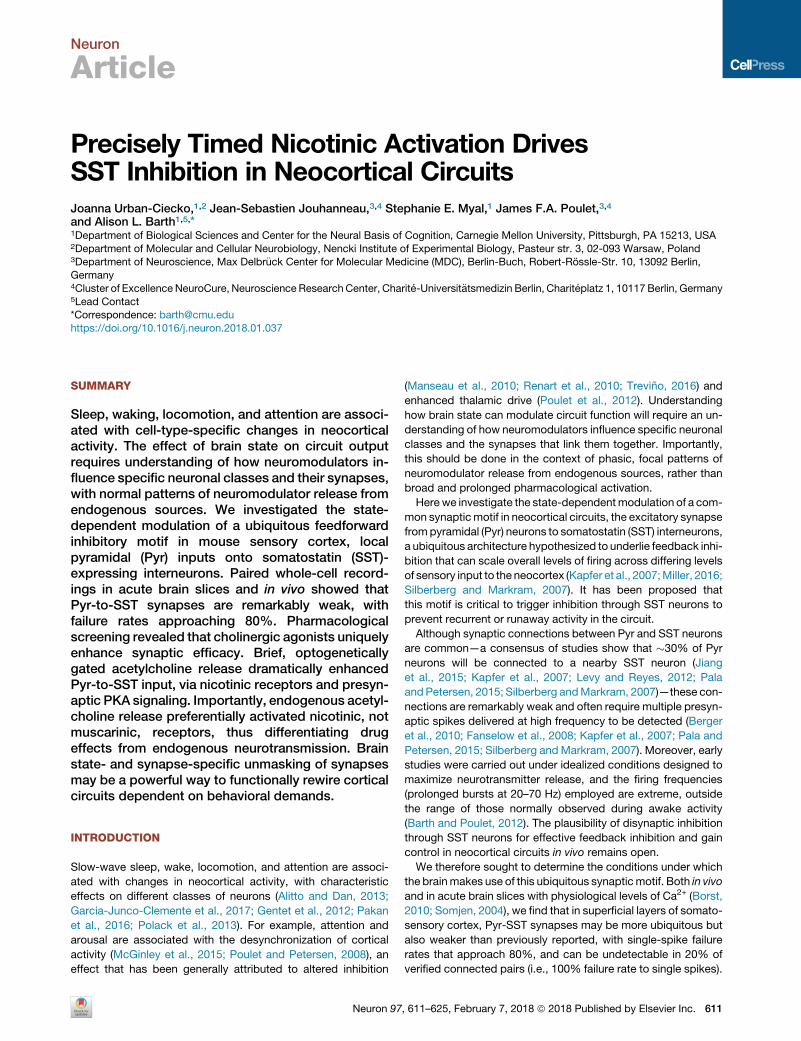
Precisely Timed Nicotinic Activation DrivesSST Inhibition in Neocortical CircuitsJoanna Urban-Ciecko,1,2 Jean-Sebastien Jouhanneau,3,4 Stephanie E. Myal,1 James F.A. Poulet,3,4
and Alison L. Barth1,5,*1Department of Biological Sciences and Center for the Neural Basis of Cognition, Carnegie Mellon University, Pittsburgh, PA 15213, USA2Department of Molecular and Cellular Neurobiology, Nencki Institute of Experimental Biology, Pasteur str. 3, 02-093 Warsaw, Poland3Department of Neuroscience, Max Delbr€uck Center for Molecular Medicine (MDC), Berlin-Buch, Robert-Rossle-Str. 10, 13092 Berlin,
Germany4Cluster of Excellence NeuroCure, Neuroscience Research Center, Charite-Universit€atsmedizin Berlin, Chariteplatz 1, 10117 Berlin, Germany5Lead Contact*Correspondence: [email protected]
https://doi.org/10.1016/j.neuron.2018.01.037
SUMMARY
Sleep, waking, locomotion, and attention are associ-ated with cell-type-specific changes in neocorticalactivity. The effect of brain state on circuit outputrequires understanding of how neuromodulators in-fluence specific neuronal classes and their synapses,with normal patterns of neuromodulator release fromendogenous sources. We investigated the state-dependent modulation of a ubiquitous feedforwardinhibitory motif in mouse sensory cortex, localpyramidal (Pyr) inputs onto somatostatin (SST)-expressing interneurons. Paired whole-cell record-ings in acute brain slices and in vivo showed thatPyr-to-SST synapses are remarkably weak, withfailure rates approaching 80%. Pharmacologicalscreening revealed that cholinergic agonists uniquelyenhance synaptic efficacy. Brief, optogeneticallygated acetylcholine release dramatically enhancedPyr-to-SST input, via nicotinic receptors and presyn-aptic PKA signaling. Importantly, endogenous acetyl-choline release preferentially activated nicotinic, notmuscarinic, receptors, thus differentiating drugeffects from endogenous neurotransmission. Brainstate- and synapse-specific unmasking of synapsesmay be a powerful way to functionally rewire corticalcircuits dependent on behavioral demands.
INTRODUCTION
Slow-wave sleep, wake, locomotion, and attention are associ-
ated with changes in neocortical activity, with characteristic
effects on different classes of neurons (Alitto and Dan, 2013;
Garcia-Junco-Clemente et al., 2017; Gentet et al., 2012; Pakan
et al., 2016; Polack et al., 2013). For example, attention and
arousal are associated with the desynchronization of cortical
activity (McGinley et al., 2015; Poulet and Petersen, 2008), an
effect that has been generally attributed to altered inhibition
Neuron 97
(Manseau et al., 2010; Renart et al., 2010; Trevino, 2016) and
enhanced thalamic drive (Poulet et al., 2012). Understanding
how brain state can modulate circuit function will require an un-
derstanding of how neuromodulators influence specific neuronal
classes and the synapses that link them together. Importantly,
this should be done in the context of phasic, focal patterns of
neuromodulator release from endogenous sources, rather than
broad and prolonged pharmacological activation.
Herewe investigate the state-dependentmodulation of a com-
mon synapticmotif in neocortical circuits, the excitatory synapse
frompyramidal (Pyr) neurons to somatostatin (SST) interneurons,
a ubiquitous architecture hypothesized to underlie feedback inhi-
bition that can scale overall levels of firing across differing levels
of sensory input to the neocortex (Kapfer et al., 2007;Miller, 2016;
Silberberg and Markram, 2007). It has been proposed that
this motif is critical to trigger inhibition through SST neurons to
prevent recurrent or runaway activity in the circuit.
Although synaptic connections between Pyr and SST neurons
are common—a consensus of studies show that �30% of Pyr
neurons will be connected to a nearby SST neuron (Jiang
et al., 2015; Kapfer et al., 2007; Levy and Reyes, 2012; Pala
and Petersen, 2015; Silberberg andMarkram, 2007)—these con-
nections are remarkably weak and often require multiple presyn-
aptic spikes delivered at high frequency to be detected (Berger
et al., 2010; Fanselow et al., 2008; Kapfer et al., 2007; Pala and
Petersen, 2015; Silberberg andMarkram, 2007). Moreover, early
studies were carried out under idealized conditions designed to
maximize neurotransmitter release, and the firing frequencies
(prolonged bursts at 20–70 Hz) employed are extreme, outside
the range of those normally observed during awake activity
(Barth and Poulet, 2012). The plausibility of disynaptic inhibition
through SST neurons for effective feedback inhibition and gain
control in neocortical circuits in vivo remains open.
We therefore sought to determine the conditions under which
the brainmakes use of this ubiquitous synapticmotif. Both in vivo
and in acute brain slices with physiological levels of Ca2+ (Borst,
2010; Somjen, 2004), we find that in superficial layers of somato-
sensory cortex, Pyr-SST synapses may be more ubiquitous but
also weaker than previously reported, with single-spike failure
rates that approach 80%, and can be undetectable in 20% of
verified connected pairs (i.e., 100% failure rate to single spikes).
, 611–625, February 7, 2018 ª 2018 Published by Elsevier Inc. 611

What are the conditions underwhich local excitatory inputs can
engage SST inhibition? We find that synaptic efficacy at Pyr-SST
connections is uniquely enhanced by cholinergic signaling, a
critical neuromodulatory pathway that has been implicated
in arousal, attention, and also cognitive disorders such as
Alzheimer’s disease. Increased synaptic strength at Pyr-SST
synapses can be observed both with pharmacological activation
of acetylcholine receptors (AChRs) and with the delivery of ACh
from endogenous sources matched to normal patterns of cholin-
ergic neural firing. These effects are mediated by presynaptic
nicotinic AChRs (nAChRs) and the activation of PKA signaling
pathways at Pyr-SST inputs, and require a delay between
cholinergic release and the enhancement of release probability.
Cholinergic enhancement of excitatory synaptic strength at
SST neurons is synapse specific, as it was absent at layer 2
(L2) Pyr-to-Pyr synapses and Pyr-to-parvalbumin (PV)-express-
ing interneurons. Thus, local excitatory input to SST neurons is
selectively enhanced during the cholinergic modulation of
network activity. Our findings indicate that brain state can
selectively alter network function through the input-specific
modulation of specific synaptic motifs, and provide a potential
mechanism by which attention can decorrelate neural activity
and control the gain of local circuits through increased inhibition.
RESULTS
Pyr Inputs to SST Neurons Are Weak and Have a HighFailure RateBrain slice and in vivo studies indicate that pyramidal cells in the
neocortex are reciprocally connected to nearby SST neurons
with high probability, with estimates typically ranging from
20% to 30% for Pyr-to-SST connections and >60% for SST-
to-Pyr connectivity (Urban-Ciecko and Barth, 2016), data that
suggest SST neurons could provide temporally precise feed-
back inhibition. Although the recruitment and impact of SST-
mediated inhibition have been extensively modeled (Krishna-
murthy et al., 2015; Markram et al., 2015; Potjans and Diesmann,
2014; Vierling-Claassen et al., 2010), the weak synaptic proper-
ties of Pyr-to-SST connections indicate that this motif might be
difficult to engage except for under very specialized regimes,
in which individual pyramidal neurons fire multiple spikes at
non-physiologically high frequencies (Berger et al., 2010;
Hilscher et al., 2017; Kapfer et al., 2007; Silberberg and
Markram, 2007). To quantify these limitations and better under-
stand when SST neurons can be engaged for temporally precise
inhibition, we examined Pyr-to-SST synapses under a variety of
stimulation conditions, both in acute brain slices and in vivo.
For slice recordings, we adjusted extracellular Ca2+ levels to
values observed in cerebrospinal fluid (CSF) (�1 mM; Somjen,
2004) and enabled some slow oscillatory network activity to be
present, conditions that approximate the quiet resting state
in vivo (Sanchez-Vives and McCormick, 2000). Using a 10 spike
train delivered at 20 Hz, we identified synaptically connected
Pyr-SST pairs in L2 of mouse somatosensory (barrel) cortex
and calculated the first-spike mean amplitude and failure rate
(Figure 1). Because neocortical neurons typically fire at very
low rates (Barth and Poulet, 2012), analysis of synaptic
responses to the initial spike is a reasonable way to assess
612 Neuron 97, 611–625, February 7, 2018
Pyr input strength to SST neurons under normal behavioral
conditions.
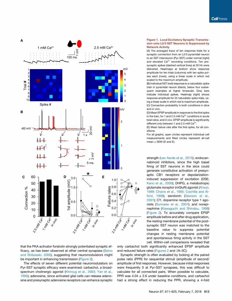
Synaptic strength was remarkably weak under these
conditions. Mean excitatory postsynaptic potential (EPSP)
amplitudes, calculated after the first spike (10-sweep average),
were nearly 5-fold lower than some previously reported values
(Jiang et al., 2015; Pala and Petersen, 2015): 0.18 ± 0.02 mV
(range 0–1.67, n = 90). Mean failure rates across all cells were
0.79 ± 0.02, indicating that EPSP responses to a single presyn-
aptic spike are the exception rather than the rule for these local
excitatory inputs. Indeed, we found that 20% of connected pairs
would have beenmissed by a single-spike analysis (for example,
see Jiang et al., 2015; Pala and Petersen, 2015) due to 100%
failure rates for the initial spike at these connections. Importantly,
increasing extracellular Ca2+ and suppressing background activ-
ity in the slice were sufficient to reveal connections that were
otherwise difficult to detect (Figures 1A, 1D, and 1E). Delivery
of a naturalistic spike train (previously recorded from L2 Pyr
neurons in vivo) that included spike bursts recorded did not alter
failure rates in acute brain slices (Figure 1B).
Connection probability was determined by assessments of re-
sponses throughout the 10 spike train. Direct synaptic connec-
tions from Pyr to SST neurons were found in 53% of tested pairs
(220/411), and this probability did not change when Ca2+ levels
were higher, since the strong facilitation observed enabled us
to accurately classify neurons as connected even if single-spike
failure rates were high. Connection probabilities for Pyr-SST
pairs were similar for in vivo recordings, using spike-train stimu-
lation to detect weak inputs. Using multiple trial averaging and
analysis of responses across a prolonged spike train, we found
that 41.2% (7/17) of tested Pyr cells were synaptically connected
to nearby SST neurons (Figure 1C).
Within-cell comparisons of Pyr-SST synaptic efficacy
recorded in both normal (physiological, 1 mM) and then elevated
(2.5 mM) extracellular Ca2+ levels showed that EPSP amplitudes
were significantly larger (normal 0.53 ± 0.34 mV versus elevated
0.24 ± 0.2 mV; n = 8, p = 0.02) and single-spike failure
rates tended to be lower (normal 0.58 ± 0.01 versus elevated
81% ± 6%; p = 0.08; Wilcoxon test; Figures 1D and 1E).
How do these values from acute brain slices compare to
synaptic performance of Pyr-to-SST inputs in vivo? Although
high levels of spontaneous subthreshold activity during in vivo
recordings made it challenging to assess synaptic strength
and response failures, we found that during hyperpolarized
periods of synaptic quiescence (i.e., downstates) isolated-spike
EPSP failures occurred in nearly three-quarters of trials
(73% ± 4%, n = 6). This value was similar to that recorded in
acute brain slices (Figures 1D and 1E). Thus, these data indicate
that the frequency of Pyr-to-SST connections is common and
failure rates are high, and that our recording conditions for acute
brain slices recapitulate what has been observed in vivo.
Cholinergic Enhancement of Pyr-SST InputsThe energetic costs of building and maintaining this ubiquitous
synaptic motif across the neocortex is at odds with the weak
and unreliable responses observed in our recordings. We hy-
pothesized that there might be specific conditions under which
local excitatory inputs might be revealed. Initially we observed
3
2
1
0
4
51
mV
100 ms
Tria
l#
Spike #
123456789
10
A
Con
nect
ion
%
C D E
1 mM Ca2+ 2.5 mM Ca2+
1 s 20 m
V-60 mV
5 ms
2 m
V
* *
2
1
0
B
0
10
20
30
40
50
60
1
0
3
2
4
5
mV
mV
0.0
0.5
1.5
3.0
1st a
mpl
itude
(mV
)
0.0
0.2
0.4
0.6
0.8
1.0
Failu
re ra
tes
*
Figure 1. Local Excitatory Synaptic Transmis-
sion onto L2/3 SST Neurons Is Suppressed by
Network Activity
(A) The averaged trace of ten response trials for a
synaptic connection from an L2/3 pyramidal neuron
to an SST interneuron (Pyr-SST) under normal (right)
and elevated Ca2+ recording conditions. Ten pre-
synaptic spikes (dashed vertical lines) at 20 Hz were
delivered. Heatmaps at bottom show response
amplitude for ten trials (columns) with ten spike pul-
ses each (rows), using a linear scale in which red
scaled to the maximum amplitude.
(B) Individual SST (red) response to a naturalistic spike
train in pyramidal neuron (black), below four subse-
quent examples at higher timescale. Gray bars
indicate individual spikes. Heatmap (right) shows
response amplitude for 25 naturalistic spike trials, us-
ing a linear scale in which red is maximum amplitude.
(C) Connection probability in both conditions in slice
and in vivo.
(D)MeanEPSPamplitude in response to the first spike
in the train, for 1 and 2.5 mMCa2+ conditions in acute
brain slice, and in vivo. EPSP amplitude is significantly
different only between 1 and 2.5 mM Ca2+.
(E) Mean failure rate after the first spike, for all con-
ditions.
For all graphs, open circles represent individual cell
measurements and filled circles represent all-cell
mean ± SEM (D and E).
that the PKA activator forskolin strongly potentiated synaptic ef-
ficacy, as has been observed at other central synapses (Seino
and Shibasaki, 2005), suggesting that neuromodulators might
be important in enhancing transmission (Figure 2).
The effects of seven different potential neuromodulators on
Pyr-SST synaptic efficacy were examined: carbachol, a broad-
spectrum cholinergic agonist (Khiroug et al., 2003; Yan et al.,
1995); adenosine, since activated glial cells can release adeno-
sine and presynaptic adenosine receptors can enhance synaptic
strength (van Aerde et al., 2015); endocan-
nabinoid inhibitors, since the high basal
firing of SST neurons in the slice could
generate constitutive activation of presyn-
aptic CB1 receptors or depolarization-
induced suppression of excitation (DSE;
Kano et al., 2009); DHPG, a metabotropic
glutamate receptor (mGluR) agonist (Anwyl,
1999; Chavis et al., 1995; Cochilla and Al-
ford, 1998); serotonin (Dawson et al.,
2001); CY, dopamine receptor type 1 ago-
nists (Surmeier et al., 2007); and norepi-
nephrine (Kawaguchi and Shindou, 1998)
(Figure 2). To accurately compare EPSP
amplitude before and after drug application,
the resting membrane potential of the post-
synaptic SST neuron was matched to the
baseline value to suppress potential
changes in resting membrane potential
and spontaneous firing activity in the SST
cell. Within-cell comparisons revealed that
only carbachol both significantly enhanced EPSP amplitude
and reduced failure rates (Figures 2 and 3A–3C).
Synaptic strength is often evaluated by looking at the paired
pulse ratio (PPR) for sequential stimuli (amplitude of second/
amplitude of first response). However, because initial responses
were frequently 0 at Pyr-SST synapses, this was difficult to
calculate for all connected pairs. When possible to calculate,
PPR was 4.04 ± 2.6 under baseline conditions, and carbachol
had a strong effect in reducing the PPR, showing a 4-fold
Neuron 97, 611–625, February 7, 2018 613

*
A
B
AM 2
51
aden
osin
e
DH
PG
CY
(DA
)
nore
pine
phrin
e
sero
toni
n
carb
acho
l
DS
E
*fo
rsko
lin
*
0.50.60.70.80.91.01.11.2
Failu
re ra
tes
(fold
cha
nge)
*
AM 2
51
aden
osin
e
DH
PG
CY
(DA
)
nore
pine
phrin
e
sero
toni
n
carb
acho
l
DSE
fors
kolin
0.6
0.8
1.0
1.2
1.4
1.62.0
1st a
mpl
itude
(fol
d ch
ange
)
Figure 2. Screening for Modulatory Systems Regulating Pyr-SST
Synaptic Transmission
(A) The fold change of the first EPSP amplitude in a baseline condition and the
following bath-applied agents: forskolin (cell-permeable activator of adenylyl
cyclase), carbachol (a broad-spectrum cholinergic agonist), adenosine,
AM-251 (endocannabinoid inhibitors) for depolarization-induced suppression
of excitation (DSE), DHPG (a metabotropic glutamate receptor antagonist),
serotonin, CY 208-243 (D1 receptor agonist), and norepinephrine.
(B) The fold change of the mean failure rate for the same conditions as in (A).
Open circles represent individual cell values; bars represent all-cell
mean ± SEM.
decrease (PPR = 0.9 ± 0.32) after drug application (n = 4). The
carbachol-dependent reduction in PPR, together with the
decreased failure rate, suggested a presynaptic locus for
modulation at the Pyr-to-SST synapse.
Which AChR subtype mediates the increase in Pyr-SST
synaptic efficacy? Cholinergic receptors can be nicotinic
(ligand-gated cation channels; nAChRs) or muscarinic (metabo-
tropic G protein-coupled receptors; mAChRs). ACh has been
hypothesized to selectively alter synaptic transmission at some
neocortical synapses (Arroyo et al., 2014; Gil et al., 1997; Gioanni
et al., 1999; McGehee et al., 1995) and presynaptic nAChRs can
enhance transmission at mossy fiber inputs to CA3 neurons in
the hippocampus (Cheng and Yakel, 2015a, 2015b). Pharmaco-
logical analysis to selectively block either nAChR or mAChRs
614 Neuron 97, 611–625, February 7, 2018
revealed that nicotinic, but not muscarinic, receptors were
required to enhance Pyr-to-SST connection strength (Figures
3D–3G). The carbachol effect of enhancing EPSP efficacy was
blocked in the presence of the nAChR antagonist mecamylamine
(amplitude baseline 0.20 ± 0.07 mV versus carbachol + meca-
mylamine 0.26 ± 0.10, failure rate baseline 0.72 ± 0.07 versus
carbachol + mecamylamine 0.72 ± 0.07; n = 6, p = 0.84 and
p = 0.99, respectively; Figures 3D and 3E). In contrast, the
carbachol-dependent increase in Pyr-SST input strength was
maintained with bath application of the mAChR-selective antag-
onist atropine, indicating mAChRs were not involved (amplitude
baseline 0.13 ± 0.06mV versus carbachol + atropine 0.28 ± 0.06,
failure rate baseline 0.80 ± 0.05 versus carbachol + atropine
0.63 ± 0.04; n = 8, p = 0.0007 and p = 0.03, respectively; Figures
3F and 3G).
Although we attempted to augment Pyr-to-SST synaptic
strength using bath application of nicotine, we did not observe
any effect under these conditions (data not shown). This is likely
due to the rapid desensitization of nAChRs, which has been
well documented in other systems (Auerbach and Akk, 1998;
Cachelin and Colquhoun, 1989; Ochoa et al., 1989).
Endogenous ACh Release Is Sufficient to AwakenPyr-SST SynapsesProlonged bath application of cholinergic agonists is markedly
different from themanner in which ACh is released during normal
brain function. Especially in an actively firing network, cholinergic
effects on diverse cell types across a circuit can make it difficult
to differentiate direct and indirect effects. In addition, bath
application of agonists results in broad spatial and temporal
activation that may not reflect the precision of endogenous
ACh release.
To determine whether endogenous release of ACh might be
sufficient to elicit the same increase in Pyr-to-SST synaptic
strength, we used optogenetic activation of cholinergic terminals
in the neocortex. In vivo recordings from cholinergic neurons in
the basal forebrain indicate that these neurons may fire at a
low frequency in quiet, awake animals (�4 Hz; Hangya et al.,
2015; Hassani et al., 2009; Lee et al., 2005) and may be synchro-
nously activated to fire a single, precisely timed spike when a
behaviorally relevant cue is present (Hangya et al., 2015). With
this in mind, we sought to determine what the temporal con-
straints of cholinergic modulation of Pyr-to-SST inputs were.
SST neurons were virally labeled in an SST-Cre3 ChAT-ChR2
transgenic mouse to enable light-evoked ACh release, and acute
brain slices were prepared for analysis. Simultaneous optoge-
netic stimulation of cholinergic afferents with Pyr stimulation
did not reveal an immediate enhancement of EPSP efficacy in
synaptically coupled SST neurons for the first few pulses in the
presynaptic spike train, inconsistent with a fast nAChR response
at either pre- or postsynaptic locations (Figure S1). Because PKA
signaling had been implicated in enhancing transmission at
Pyr-to-SST synapses, we hypothesized that the activation of
signal transduction cascades might require some delay from
nAChR activation to enhanced release (Cheng and Yakel,
2014, 2015a). Consistent with this, we observed a significant
change in amplitude and failure rates �200 ms after the onset
of the light flash (Figure S1), with a >3-fold increase in response

1stam
plitu
de (m
V)
Failu
re ra
tes
n.s
0.0
0.2
0.4
0.6
0.8n.s
0.00.20.40.60.81.0
1stam
plitu
de (m
V)
base. carb.
* *B
A
D
0.0
0.2
0.4
0.6
0.8
Failu
re ra
tes
C
0.00.20.40.60.81.0
base. carb.
E
base. carb.+mec.
Spike #
carbacholbaseline
Tria
l #
123456789
10
1 m
V
100 ms
3
2
1
0
4
5
mV
1stam
plitu
de (m
V)
base. carb.+atrop.
*
Failu
re ra
tes
GF
0.0
0.2
0.4
0.6
0.8
0.00.20.40.60.81.0 *
base. carb.+mec.
base. carb.+atrop.
Figure 3. Nicotinic Receptors Enhance EPSP
Efficacy onto Pyr-SST Connections
(A) The averaged trace of ten response trials for a
Pyr-to-SST connection under baseline conditions
(left) and in carbachol. Ten presynaptic spikes
(dashed vertical lines) at 20 Hz were delivered.
Heatmaps at bottom show response amplitude as
in Figure 1.
(B) Mean EPSP amplitude in response to the first
spike in the train, for baseline and in carbachol
conditions.
(C) Mean failure rate after the first spike, for both
conditions.
(D and E) The same as in (B) and (C) but with selective
nicotinic receptor activation (carbachol and atropine,
a muscarinic receptor antagonist).
(F andG) The same as in (B) and (C) but with selective
muscarinic receptor activation (carbachol and
mecamylamine, a nicotinic receptor antagonist).
For all graphs, open circles represent individual cell
measurements and filled circles represent all-cell
mean ± SEM (B–G).
Neuron 97, 611–625, February 7, 2018 615

1 m
V
100 ms
Tria
l #
123456789
10
Spike #
light ONlight OFF
1
0
2
3
B
-200 0 200 400 ms
A
-200 0 200 400 ms
* *
1stam
plitu
de (m
V)
OFF ON0.0
0.4
0.8
1.2
1stam
plitu
de (m
V)
OFF ON+mecamylamine
n.s
0.0
0.4
0.8
1.2 s
OFF ON+mecamylamine
Failu
re ra
tes
n.s
0.00.20.40.60.81.0 s
Failu
re ra
tes
0.00.20.40.60.81.0
OFF ON
C
E F
D
mV
Figure 4. Nicotinic Receptors Enhance
EPSP Efficacy at Pyr-SST Connections
(A) Schematic of the stimulation protocol. One
single blue light pulse (10 ms) was delivered
200 ms prior to the presynaptic spike train.
(B) The averaged trace of EPSP under baseline/
light OFF and light ON conditions. Heatmaps at
bottom show response amplitude for both condi-
tions as described in previous figures.
(C) Mean EPSP amplitude in response to the first
spike in the train, for both conditions.
(D) Mean failure rate after the first spike, for both
conditions.
(E and F) The same as in (C) and (D) but in the
presence of the nicotinic receptor antagonist
(mecamylamine).
For all graphs, open circles represent individual cell
measurements and filled circles represent all-cell
mean ± SEM (C–F). See also Figures S1 and S2.
amplitude (0.27 ± 0.11 mV to 0.81 ± 0.37 mV; n = 4 and
p = 0.0002) and a nearly 2-fold reduction in mean failure rates
(from 0.68 ± 0.11 to 0.35 ± 0.16; n = 4 and p = 0.03).
We thus examined the effect of ACh release on EPSP efficacy,
using a 200 ms interval between the activation of cholinergic
fibers and the stimulation of Pyr inputs. Remarkably, a single light
pulse (10 ms) was sufficient to generate a significant increase
616 Neuron 97, 611–625, February 7, 2018
in amplitude and a reduction in failure rates
that nearly matched what had been
observed with carbachol application (Fig-
ure 4; amplitude baseline 0.32 ± 0.13 mV
versus flash 0.62 ± 0.15 mV; n = 6 and p =
0.04; failure ratebaseline0.58±0.09versus
flash 0.38 ± 0.07; n = 6 and p = 0.0006).
To characterize the time course and
duration of this cholinergic enhancement,
we investigated two additional time inter-
vals between the activation of cholinergic
fibers and the stimulation of Pyr inputs:
50 and 500 ms (Figures S2A and S2B).
A 50 ms delay between optogenetically
released ACh and Pyr-SST synaptic acti-
vation did not change input strength,
although it was sufficient to drive a signifi-
cant reduction in failure rates. A 500 ms
delay between ACh release and Pyr-SST
synaptic activation did not alter either
amplitudeor failure rates.Finally,we tested
whether multiple flashes might augment
the observed increase in Pyr-SST input
strength. Increasing the number of flashes
did not influence synaptic efficacy (Fig-
ure S2C), likely due to pronounced synap-
tic depression at cholinergic terminals
induced by prolonged optogenetic stimu-
lation (data not shown).
To confirm that the ChAT-channelrho-
dopsin-mediated enhancement of Pyr-
SST synaptic strength was acting through the same pathways
as established using bath application of carbachol, we carried
out the same experiments in the presence of the nAChR antag-
onist mecamylamine. Similar to the effects described in
Figure 3, pharmacological blockade of nAChRs prevented
optogenetically gated cholinergic enhancement of Pyr-SST
connections (Figures 4E and 4F; amplitude baseline

carb. carb.+
atrop.
carb.+
mec.
ON
*
*
*
carbachol
carbachol + atropine
carbachol + mecamylamine
A
C
1 s 20 m
V
light ONB
0
1
2
3
4
5
9
Firin
g fre
quen
cy (f
old
chan
ge)
Figure 5. SSTNeuron Spontaneous Firingwith Pharmacological and
Optogenetic ACh Receptor Activation
(A) Firing frequency in baseline and in the presence of carbachol, carbachol
and atropine, and carbachol and mecamylamine.
(B) The example traces showing the spike (top) and depolarization (bottom) of
SST neurons in response to a single 10 ms blue light pulse.
(C) Mean fold change in spontaneous firing frequency for different conditions:
carbachol, carbachol and atropine, carbachol and mecamylamine, and light
ON. The bar graphs represent mean ± SEM.
0.18 ± 0.11 mV versus flash 0.20 ± 0.10 mV; n = 5 and p = 0.99;
failure rate baseline 0.72 ± 0.09 versus flash 0.62 ± 0.05; n = 5
and p = 0.38). Thus, we conclude that ACh release from endog-
enous cholinergic afferents acts through nAChRs to enhance
local excitatory transmission onto neocortical SST neurons.
Timed, Endogenous ACh Release Does Not IncreaseOverall SST Firing RatesBath application of AChR agonists, using either ACh or the
agonist carbachol, can induce a 3-fold increase in SST sponta-
neous firing activity (Chen et al., 2015; Fanselow et al., 2008)
(Figure 5A). This was not the case for Pyr neurons, in which
carbachol depolarized resting membrane potential (Table S1)
but was not sufficient to change Pyr spontaneous activity, which
was typically very low (<0.02 Hz). Consistent with other studies
(Chen et al., 2015; Fanselow et al., 2008), we found that mAChR,
but not nAChR, activation was primarily responsible for the
strong increase in spontaneous SST firing with carbachol.
Interestingly, brief optogenetically activated release of ACh
was not sufficient to influence overall firing rates of SST neurons
in acute brain slices. A subset of SST neurons (4/18) could be
driven to fire a single spike at short latency with a single
light pulse (Figure 5B). However, the majority of SST neurons
(14/18) responded to light-evoked ACh release with a brief depo-
larization followed by hyperpolarization (Figure 5B), suggestive
of ACh-activated inhibition elsewhere in the local network (Alitto
and Dan, 2013; Arroyo et al., 2012). This delayed synaptic
inhibition was eliminated in the presence of GABAa receptor
antagonists (data not shown).
The difference in results for light-evoked ACh release versus
the bath application of a cholinergic agonist underscores the
importance of investigating neuromodulation delivered from
endogenous sources: precisely timed and highly local ACh
release had specific effects that mapped onto only a subset of
phenomenon observed with pharmacological treatments.
Basal Forebrain Afferents Enhance Pyr-SSTConnections In Vivo
Acute brain slices represent a useful platform for efficiently
screening through drugs and conditions to characterize synaptic
function. To determine whether precisely timed cholinergic acti-
vationmight also influence synaptic connections in intact circuits
and compare the magnitude of these effects to acute brain slice
analysis, we carried out paired whole-cell recordings between
Pyr and SST neurons in somatosensory cortex of anesthetized
SST-Cre 3 Ai9 transgenic mice.
To examine how endogenous release of ACh affected Pyr-SST
inputs in vivo, we virally transduced neurons in the basal fore-
brain of SST-Cre3 Ai9 transgenic mice with a ChR2-expressing
virus and then prepared animals for targeted whole-cell record-
ings 2–4weeks later (Figure 6). Because basal forebrain afferents
to the neocortex are almost exclusively cholinergic (Do et al.,
2016), this ensured that ACh was the primary neurotransmitter
released. Indeed, we observed immunohistochemical co-locali-
zation of ChR2-expressing fibers with the vesicular ACh trans-
porter in superficial layers of S1 (Figure S3).
We evaluated whether optogenetically gated neurotransmitter
release could influence EPSP amplitude in vivo, using a single
light pulse 200ms prior to the test stimulus driving the presynap-
tic Pyr cell (Figures 6 and S4). Pyr and SST neurons were identi-
fied during the in vivo recording session and cell identity was
confirmed using evoked firing output. Brief spike doublets
were evoked in Pyr neurons and the EPSP amplitude of Pyr
inputs in synaptically connected SST neurons was evaluated,
in which the EPSP triggered by the first spike of a doublet was
compared before and after optogenetic activation of cholinergic
afferents (STAR Methods). Effects were remarkably similar to
those observed in acute brain slices: a single light pulse
significantly increased EPSP amplitude almost 2-fold (0.17 ±
0.07 mV versus 0.37 ± 0.09 mV; n = 6, p = 0.03) and reduced
Neuron 97, 611–625, February 7, 2018 617

OFF ON
*
0.0
0.2
0.4
0.6
0.8
1.0
OFF ON
*Fa
ilure
rate
s
0.0
0.2
0.4
0.6
0.8
1.0
1st a
mpl
itude
(mV
)
0.0
0.2
0.4
0.6
0.8
1.0
1st a
mpl
itude
(mV
)
0.0
0.2
0.4
0.6
0.8
1.0
Failu
re ra
tes
OFF ON+mecamylamine
OFF ON+mecamylamine
n.s n.s
B
G
C
I
F
A
light ONlight OFF
2-4 weeks
P10 P24-38
ChR2-EYFPN.Basalis
RecordingS1 cortex 485 nm
Pyr
SST
20 μm
10 m
V50
0 pA
250 ms
ED
1 m
V50
mV
20 ms
0.5
mV
2 ms
2 ms
0.1
mV
5 150-200 10 20 ms
SSTPyr
H
Js
Figure 6. Nicotinic Receptors Enhance EPSP Efficacy at Pyr-SST Connections In Vivo
(A) Schematic of the experimental procedure. Postnatal day (P)10 SST-Cre x Ai9 pups were injected in nucleus basalis (bregma, 0.02 mm; lat, 1.3 mm; depth,
4.5 mm). After an incubation period of 2–4 weeks, mice were anesthetized and electrophysiological recordings were performed in somatosensory cortex.
(B) Schematic of the recording setup.
(legend continued on next page)
618 Neuron 97, 611–625, February 7, 2018

failure rates to 0.51 ± 0.06 (versus 0.73 ± 0.04 without stimulus;
n = 6, p = 0.03). These results indicate that precisely time activa-
tion of fibers from the basal forebrain can enhance the efficacy of
Pyr-to-SST synapses in the intact brain.
To verify that the observed effects in vivo were due to the
activation of nAChRs, we carried out a subset of experiments
in which mecamylamine was included in the Ringer’s solution
above the brain and allowed to diffuse for at least 20 min before
recording commenced. Under these conditions, optogenetic
activation of basal forebrain afferents did not increase the
amplitude of Pyr-to-SST connections, consistent with a
cholinergic effect of stimulation (Figures 6I and 6J; amplitude
baseline 0.23 ± 0.07 mV versus flash 0.27 ± 0.08 mV;
p = 0.38; failure rate baseline 0.65 ± 0.04 versus flash
0.71 ± 0.04; n = 7, p = 0.19). Although ChR2 expression in
the nucleus basalis and associated basal forebrain structures
is not solely cholinergic, the mecamylamine blockade of
EPSP enhancement both in vivo and in acute brain slice
indicates a common, nAChR-dependent mechanism.
Presynaptic nAChRs Activate PKA Signaling Pathwaysto Enhance Release ProbabilityBoth anatomical and electrophysiological evidence indicate that
nAChRs can be presynaptic in the mammalian cerebral cortex
(Arroyo et al., 2012, 2014; Levy and Aoki, 2002; Lubin et al.,
1999; McGehee et al., 1995; Vidal and Changeux, 1993),
although some postsynaptic activity for these receptors has
also been observed (Halff et al., 2014; Puddifoot et al., 2015).
Because nAChR activation reduced the PPR and decreased
failure rates, we hypothesized that these receptors were presyn-
aptically localized.
We took advantage of the recording configuration afforded by
direct analysis of synaptically coupled neuron pairs to determine
whether nAChR-mediated PKA activation was required pre- or
postsynaptically. To confirm that this increasewasdue to the acti-
vation of presynaptic pathways, we included the PKA inhibitor
peptide PKI in the pipette internal solution for the presynaptic
Pyr neuron, and experiments were carried out in acute brain
slices where SST neurons had been virally labeled in an SST-
Cre 3 ChAT-ChR2 transgenic mouse. Blocking PKA signaling
in the presynaptic Pyr neuron was sufficient to prevent light-
activated ACh release from enhancing synaptic efficacy at
Pyr-SST connections (Figures 7 and S5; amplitude baseline
0.57 ± 0.21 mV versus flash 0.47 ± 0.17 mV; p = 0.19; failure rate
baseline0.52±0.11versusflash0.55±0.11; n=8,p=0.68).These
(C) In vivo two-photon image of a pyramidal neuron (green cell soma) connected
electrode outlines.
(D) Example recording of a Pyr (black trace) connected to SST (red trace). Botto
(E) Zoom of the gray rectangle in (D). In this example, two spikes were evoked
Vm line = �60.5 mV.
(F) Top: schematic of the experimental protocol. One blue light pulse (10 ms) w
Pyr doublet of spikes. Middle: example single traces showing the SST response to
(blue traces) conditions. Bottom: average responses from the above Pyr-to-SST c
Vm line (right) = �47 mV.
(G) Mean EPSP amplitude in response to the first spike of the doublet, for both c
(H) Mean failure rate after the first spike, for both conditions.
(I and J) The same as in (G) and (H) but in the presence of the nicotinic receptor
See also Figures S3 and S4.
results indicate that presynaptic nAChRs activate PKA signaling
pathways to enhance synaptic strength at Pyr-SST synapses.
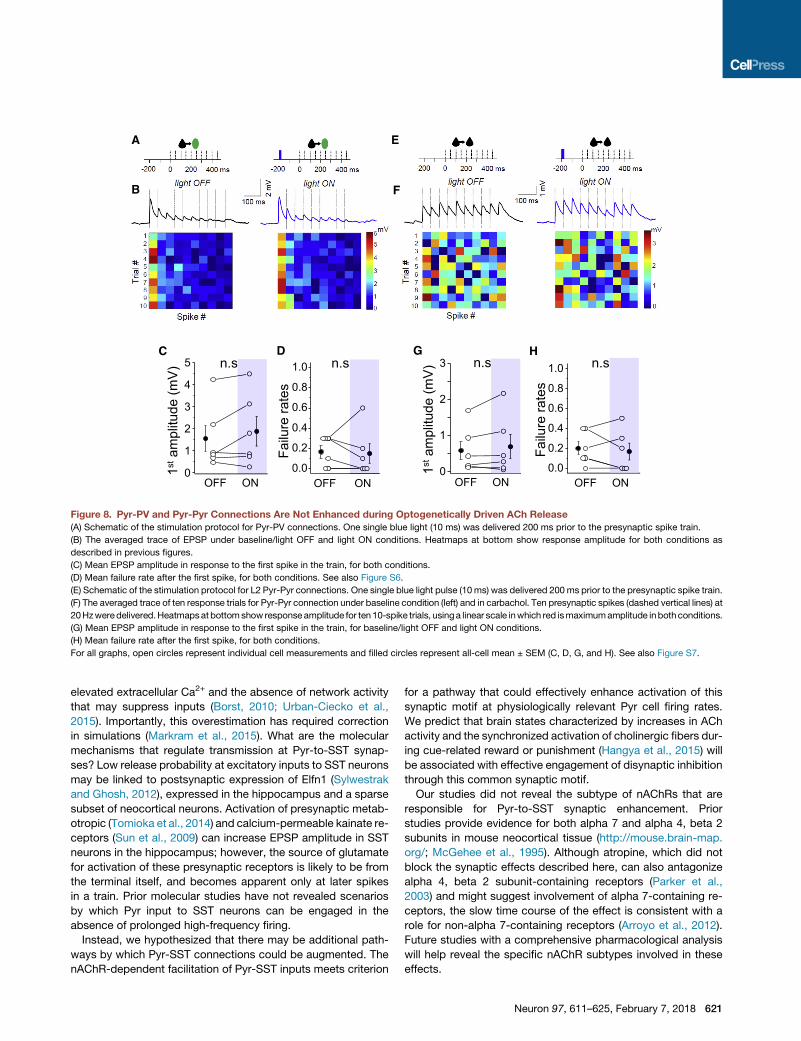
Pyr-to-PV Synapses Are Not Enhanced by EndogenousACh ReleaseTo assess whether the observed effects might facilitate input to
other inhibitory neuron subtypes, we examined the effect of both
pharmacological and optogenetic stimulation of cholinergic
pathways on Pyr-to-PV neuron synapses (Figure 8). Although
bath application of carbachol significantly increased EPSP
amplitude (Figure S6), unlike Pyr-to-SST connections, this effect
was not recapitulated by optogenetic stimulation (amplitude
baseline 1.55 ± 0.59 mV versus flash 1.88 ± 0.67 mV; p = 0.18;
failure rate baseline 0.17 ± 0.06 versus flash 0.15 ± 0.09; n = 6,
p = 0.82). Therefore, cholinergic enhancement of local excitatory
input is target specific, shifting the weight of excitatory drive
toward SST, but not PV, neuron activation.
ACh Does Not Influence Synaptic Strength atIntracortical Excitatory SynapsesOur data indicate that precisely timed ACh release can influence
local feedforward excitation onto SST neurons. To determine
whether this was synapse specific, we investigated the effect
of optogenetically evoked ACh release on direct synaptic con-
nections between L2 Pyr neurons in a ChAT-ChR2 transgenic
mouse (Figure 8). A single light flash 200 ms prior to the test train
did not show any alteration in synaptic efficacy from Pyr inputs
onto Pyr neurons (Figures 8 and S7).
To differentiate the effects of endogenous ACh release from
prolonged drug application, we examined the effects of carba-
chol at Pyr-Pyr synapses (Figures S7A and S7C). The net effect
of carbachol on Pyr-Pyr connections in L2 was amodest, but not
significant, synaptic depression, an effect that we hypothesized
might be indirect, mediated by the mAChR-dependent increase
in the firing activity of SST and other GABAergic neurons across
the cortical network. Spontaneous SST firing is an important
contributor to tonic, presynaptic GABAb activation that
suppresses excitatory transmission in the neocortex (Urban-
Ciecko et al., 2015). Consistent with this hypothesis, application
of the GABAb antagonist CGP reversed this carbachol-induced
synaptic depression between Pyr neurons, without showing any
further enhancement of synaptic efficacy compared to baseline
values (data not shown). As the mean amplitude of EPSPs was
relatively large for the subset of the connections analyzed, it
remains possible that endogenous ACh release might influence
to an SST interneuron (yellow cell soma). White dashed lines show recording
m trace shows injected current into Pyr to drive spiking. Vm line = �70 mV.
and only the second one led to an EPSP in the SST interneuron (red trace).
as delivered on the brain surface (5–15 mm) 200 ms prior to the presynaptic
the first evoked Pyr spike under baseline/light OFF (black traces) and light ON
onnection (light OFF, n = 27 trials; light ON, n = 30 trials). Vm line (left) =�45mV;
onditions.
antagonist (mecamylamine).
Neuron 97, 611–625, February 7, 2018 619

100 ms
0.5
mV
PKA inhibitor peptide: light OFF PKA inhibitor peptide: light ON
-200 0 200 400 -200 0 200 400 ms
Tria
l #
123456789
10
Spike #
1
0
2
3
B
A
C
n.s
OFF ON+PKA inhibitor peptide
0.0
0.5
1.0
1.5
1st a
mpl
itude
(mV
) .sD
n.s
0.00.20.40.60.81.0
Failu
re ra
tes
s
OFF ON+PKA inhibitor peptide
mV
Figure 7. Presynaptic Inhibition of PKA
Eliminates ACh Enhancement of Pyr-SST
Synapses
(A) Schematic of the stimulation protocol. One single
blue light (10 ms) was delivered 200 ms prior to the
presynaptic spike train in the presence of PKA
inhibitor peptide in the presynaptic Pyr cell.
(B) The averaged trace of EPSP under baseline/light
OFF and light ON conditions. Heatmaps at bottom
show response amplitude for both conditions as
described in previous figures.
(C) Mean EPSP amplitude in response to the first
spike in the train, for both conditions.
(D) Mean failure rate after the first spike, for both
conditions.
For all graphs, open circles represent individual cell
measurements and filled circles represent all-cell
mean ± SEM (C and D). See also Figure S5.
very small EPSPs between pyramidal neurons under some
conditions.
These results indicate that the effects of endogenous ACh
release on increasing excitatory drive are target-cell specific,
enhancing local glutamatergic input to SST neurons without
increasing recurrent excitation across the L2 network as awhole.
DISCUSSION
It has long been known that excitatory inputs onto SST neurons
are remarkably weak and strongly facilitating, even under opti-
mized conditions (Beierlein et al., 2003; Fanselow et al., 2008;
Levy et al., 2008; Pala and Petersen, 2015; Reyes et al., 1998).
Prior studies have hypothesized that high-frequency firing is
required to activate SST neurons (Berger et al., 2010; Kapfer
et al., 2007; Silberberg and Markram, 2007); however, these re-
gimes are not physiologically plausible for Pyr neurons in many
areas of the neocortex, especially in superficial layers where
firing rates can be low (Barth and Poulet, 2012). Here we show
that the weak synaptic efficacy of local excitatory inputs to
SST neurons can be significantly and uniquely enhanced by
620 Neuron 97, 611–625, February 7, 2018
spatially and temporally specific ACh
release. Remarkably, brief optogenetic
activation of endogenous ACh release fully
recapitulated the synaptic effects of bath-
applied cholinergic agonists, without influ-
encing resting membrane potential and
mean spontaneous firing activity of SST
neurons observed with drug application.
Functional versus AnatomicalConnectivity in NeocorticalNetworksAnatomical mapping of cell-type-specific
synaptic connections across the cor-
tical column has been an important goal
of many recent investigations, using both
serial blockface electron microscopy
(EM) reconstruction and rabies virus
approaches to provide quantitative detail about circuit function
(for example, see Bock et al., 2011; Wall et al., 2016). The weak
functional contribution of Pyr-SST connections is at odds with
their ubiquitous anatomical presence and underscores the
importance of electrophysiological analysis to infer circuit pro-
cessing capabilities. Our results show that state-dependent syn-
aptic modulation can effectively rewire cortical circuits, shifting
the balance of inhibition by engagingSST neuronswith local input
when ACh levels are transiently increased. On face, the fast cir-
cuit rewiring observed here is conceptually similar to previous
studies showing that GABAb signaling can silence recurrent
excitatory connections in the L2 network (Urban-Ciecko et al.,
2015). Defining the dynamic range for synaptic function is
required for proper estimation of how specific synaptic motifs
are recruited during different brain states and neuromodulatory
conditions.
Molecular Properties of Pyr-SST SynapsesThe weak and strongly facilitating properties of Pyr-SST synap-
ses have beenwell established. Prior studies in acute brain slices
have significantly overestimated synaptic strength due to
C D
0
1
2
3
4
5
1stam
plitu
de (m
V) n.s
OFF ON0.0
0.2
0.4
0.6
0.8
1.0
Failu
re ra
tes
n.s
OFF ON
HG
OFF ON0
1
2
3
OFF ON0.0
0.2
0.4
0.6
0.8
1.0
Failu
re ra
tes
n.s n.s
1stam
plitu
de (m
V)
B
A
F
E
Figure 8. Pyr-PV and Pyr-Pyr Connections Are Not Enhanced during Optogenetically Driven ACh Release
(A) Schematic of the stimulation protocol for Pyr-PV connections. One single blue light (10 ms) was delivered 200 ms prior to the presynaptic spike train.
(B) The averaged trace of EPSP under baseline/light OFF and light ON conditions. Heatmaps at bottom show response amplitude for both conditions as
described in previous figures.
(C) Mean EPSP amplitude in response to the first spike in the train, for both conditions.
(D) Mean failure rate after the first spike, for both conditions. See also Figure S6.
(E) Schematic of the stimulation protocol for L2 Pyr-Pyr connections. One single blue light pulse (10 ms) was delivered 200 ms prior to the presynaptic spike train.
(F) The averaged trace of ten response trials for Pyr-Pyr connection under baseline condition (left) and in carbachol. Ten presynaptic spikes (dashed vertical lines) at
20Hzweredelivered.Heatmapsat bottomshow responseamplitude for ten10-spike trials, usinga linear scale inwhich red ismaximumamplitude inbothconditions.
(G) Mean EPSP amplitude in response to the first spike in the train, for baseline/light OFF and light ON conditions.
(H) Mean failure rate after the first spike, for both conditions.
For all graphs, open circles represent individual cell measurements and filled circles represent all-cell mean ± SEM (C, D, G, and H). See also Figure S7.
elevated extracellular Ca2+ and the absence of network activity
that may suppress inputs (Borst, 2010; Urban-Ciecko et al.,
2015). Importantly, this overestimation has required correction
in simulations (Markram et al., 2015). What are the molecular
mechanisms that regulate transmission at Pyr-to-SST synap-
ses? Low release probability at excitatory inputs to SST neurons
may be linked to postsynaptic expression of Elfn1 (Sylwestrak
and Ghosh, 2012), expressed in the hippocampus and a sparse
subset of neocortical neurons. Activation of presynaptic metab-
otropic (Tomioka et al., 2014) and calcium-permeable kainate re-
ceptors (Sun et al., 2009) can increase EPSP amplitude in SST
neurons in the hippocampus; however, the source of glutamate
for activation of these presynaptic receptors is likely to be from
the terminal itself, and becomes apparent only at later spikes
in a train. Prior molecular studies have not revealed scenarios
by which Pyr input to SST neurons can be engaged in the
absence of prolonged high-frequency firing.
Instead, we hypothesized that there may be additional path-
ways by which Pyr-SST connections could be augmented. The
nAChR-dependent facilitation of Pyr-SST inputs meets criterion
for a pathway that could effectively enhance activation of this
synaptic motif at physiologically relevant Pyr cell firing rates.
We predict that brain states characterized by increases in ACh
activity and the synchronized activation of cholinergic fibers dur-
ing cue-related reward or punishment (Hangya et al., 2015) will
be associated with effective engagement of disynaptic inhibition
through this common synaptic motif.
Our studies did not reveal the subtype of nAChRs that are
responsible for Pyr-to-SST synaptic enhancement. Prior
studies provide evidence for both alpha 7 and alpha 4, beta 2
subunits in mouse neocortical tissue (http://mouse.brain-map.
org/; McGehee et al., 1995). Although atropine, which did not
block the synaptic effects described here, can also antagonize
alpha 4, beta 2 subunit-containing receptors (Parker et al.,
2003) and might suggest involvement of alpha 7-containing re-
ceptors, the slow time course of the effect is consistent with a
role for non-alpha 7-containing receptors (Arroyo et al., 2012).
Future studies with a comprehensive pharmacological analysis
will help reveal the specific nAChR subtypes involved in these
effects.
Neuron 97, 611–625, February 7, 2018 621

Functional Consequences of ACh Release Timing onCortical ProcessingPharmacological studies with persistent cholinergic application
over many minutes have provided abundant evidence for
cholinergic signaling in neocortical circuits, where effects have
been attributed to both muscarinic and nicotinic receptors.
Recent studies using temporally precise optogenetic stimulation
of cholinergic afferents (Alitto and Dan, 2013; Chen et al., 2015;
Hangya et al., 2015; Joshi et al., 2016) have refined the hypoth-
eses about how the effects of endogenous ACh release compare
to prior pharmacological studies, in which AChR activation can
be prolonged and direct and indirect effects can be difficult to
disentangle.
Prior studies have not been able to reveal whether the temporal
and spatial organization of cholinergic release can activate
different receptor subtypes, in different cellular compartments
and across different neuronal subtypes. In addition, efforts to
investigate howendogenousACh release functions have typically
employed non-physiological, high-frequency stimulation of affer-
ents (Chen et al., 2015; Joshi et al., 2016; Kuchibhotla et al., 2017)
that do not represent normal regimes of ACh fiber activation. Here
we show that brief and precisely timed optogenetic activation of
ACh release mimics the synaptic, nAChR-dependent effects of
cholinergic agonists without recapitulating the profound changes
in network activity that may be due to the activation of mAChRs.
The ability to dissociate synaptic and network effectswith precise
optical stimulation suggests that theremaybemultiple conditions
for subtype-specificAChsignalingdependingon the taskat hand.
It is tempting to speculate that cholinergic neurons of the basal
forebrain take advantage of these different regimes to achieve
different network functions.
nAChRs, Cognitive Disorders, and Sensory ProcessingACh signaling and the activity of cholinergic neurons in the basal
forebrain are biochemical correlates of attention and cue salience
(Picciotto et al., 2012), and cholinergic activity has been linked to
both muscarinic and nicotinic AChR activation (see, for example,
muscarinic [Herrero et al., 2008] and nicotinic [Guillem et al.,
2011]). nAChRs in particular have been linked to sensory process-
ing deficits in schizophrenia and attention disorders (Freedman,
2014; Guillem et al., 2011; Parikh et al., 2016). Our findings sug-
gest that selective engagement of feedback inhibition through
SST neuronsmight be essential for enhanced sensory processing
in the attentive, cued state. The high failure rate of Pyr-to-SST
inputs suggests that local drive to SST neurons is negligible under
normal conditions when Pyr cell firing rates are low. However,
during active sensation, cholinergic enhancement of local input
may be amplified by the sheer number of firing pyramidal neurons
with convergent input onto SST neurons. Under these conditions,
strong feedback inhibition through SST neurons is likely to occur,
effects that selectively enhance short-latency evoked responses
and reduce recurrent network activity.
The Asynchronous State and Synapse-SpecificCholinergic Enhancement of EPSP EfficacyACh neurons increase their firing rates during movement and
arousal in awake rodents and have been linked to a global
increase in cortical activity (Buzsaki et al., 1988; Eggermann
622 Neuron 97, 611–625, February 7, 2018
et al., 2014; Metherate et al., 1992) and a decrease in correlated
firing of neurons in the neocortex of primates as well as rodents
(Cohen and Maunsell, 2009; Herrero et al., 2008; Pafundo et al.,
2016; Renart et al., 2010).
The synapse-selective effects of ACh on enhancing excitatory
drive onto a subtype of inhibitory neurons without influencing
feedforward excitatory connections (Pyr-Pyr connections) are
consistent with a link between increased inhibition and the
desynchronized state (Doiron et al., 2016). The desynchronous
cortical state has been associated with attention, enhanced
sensory processing, and increased activity of cholinergic neu-
rons in the basal forebrain (Castro-Alamancos, 2004; Cohen
and Maunsell, 2009; Marguet and Harris, 2011; Poulet and
Petersen, 2008).
The synapse-selective nAChR-mediated effects identified
here are predicted to enhance inhibition without altering excita-
tion, and suggest a potential link between the selective engage-
ment of SST-mediated inhibition, network desynchronization,
and the behavioral correlates of attention. Transient cholinergic
activity may enable the cortical network to engage alternative
processing strategies depending upon brain state, a design
structure that has parallels in field-programmable gate arrays
and integrated circuits in computer chips, where hardware can
be dynamically reconfigured according to user demands.
It is tantalizing to imagine that the cholinergic enhancement of
SST-mediated feedback inhibition may be linked to specific al-
terations in cognitive function in awake and behaving animals.
Toward these ends, future experiments to determine how
specific microcircuits perform during different brain states and
behavioral demands will be critical.
STAR+METHODS
Detailed methods are provided in the online version of this paper
and include the following:
d KEY RESOURCES TABLE
d CONTACT FOR REAGENT AND RESOURCE SHARING
d EXPERIMENTAL MODEL AND SUBJECT DETAILS
B Animals
d METHOD DETAILS
B Virus injection
B Brain slice preparation
B Whole-cell recording in acute brain slices
B Whole-cell recording in vivo
B Neuron classification
B Connectivity analysis
B Optical stimulation
B Pharmacology
B Immunohistochemistry
d QUANTIFICATION AND STATISTICAL ANALYSIS
B For acute brain slice recording
B For in vivo recording
SUPPLEMENTAL INFORMATION
Supplemental Information includes seven figures and one table and can be
found with this article online at https://doi.org/10.1016/j.neuron.2018.01.037.
ACKNOWLEDGMENTS
Special thanks to Joanne Steinmiller for expert animal care, and Megumi
Matsushita and Rogan Grant for technical assistance. We would like to thank
Janett Konig and Charlene Memler for technical assistance and Dr. Phillip
Wisinski-Bokiniec for his imaging technical support. This work was supported
by the McKnight Foundation (A.L.B.) and NIH R01NS088958 (A.L.B.
and J.F.A.P.), the National Science Centre, Poland (2015/18/E/NZ4/00721;
J.U.-C.), the European Research Council (ERC-2015-CoG-682422; J.F.A.P.),
the DFG (DFG-FOR-2143-Interneuron; J.F.A.P.), the Berlin Institute of Health
(BIH, 1.2 TRG 4 TP 2; J.F.A.P.) and the European Union (FP7, 3x3Dimaging
323945; J.F.A.P.).
AUTHOR CONTRIBUTIONS
Conceptualization, J.U.-C., A.L.B., J.-S.J., and J.F.A.P.; Methodology, J.U.-
C., A.L.B., J.-S.J., and J.F.A.P.; Investigation, J.U.-C., S.E.M., and J.-S.J.;
Writing – Original Draft, J.U.-C. and A.L.B.; Writing – Review & Editing, J.U.-
C., A.L.B., J.-S.J., and J.F.A.P.; Funding Acquisition, A.L.B., J.U.-C., and
J.F.A.P.; Resources, A.L.B. and J.F.A.P.; Supervision, A.L.B. and J.F.A.P.
DECLARATION OF INTERESTS
The authors declare no competing interests.
Received: March 10, 2017
Revised: October 30, 2017
Accepted: January 12, 2018
Published: February 7, 2018
REFERENCES
Alitto, H.J., and Dan, Y. (2013). Cell-type-specific modulation of neocortical
activity by basal forebrain input. Front. Syst. Neurosci. 6, 79.
Anwyl, R. (1999). Metabotropic glutamate receptors: electrophysiological
properties and role in plasticity. Brain Res. Brain Res. Rev. 29, 83–120.
Arroyo, S., Bennett, C., Aziz, D., Brown, S.P., and Hestrin, S. (2012). Prolonged
disynaptic inhibition in the cortex mediated by slow, non-a7 nicotinic excita-
tion of a specific subset of cortical interneurons. J. Neurosci. 32, 3859–3864.
Arroyo, S., Bennett, C., and Hestrin, S. (2014). Nicotinic modulation of cortical
circuits. Front. Neural Circuits 8, 30.
Auerbach, A., and Akk, G. (1998). Desensitization of mouse nicotinic
acetylcholine receptor channels. A two-gate mechanism. J. Gen. Physiol.
112, 181–197.
Barth, A.L., and Poulet, J.F.A. (2012). Experimental evidence for sparse firing in
the neocortex. Trends Neurosci. 35, 345–355.
Beierlein, M., Gibson, J.R., and Connors, B.W. (2003). Two dynamically
distinct inhibitory networks in layer 4 of the neocortex. J. Neurophysiol. 90,
2987–3000.
Berger, T.K., Silberberg, G., Perin, R., and Markram, H. (2010). Brief bursts
self-inhibit and correlate the pyramidal network. PLoS Biol. 8, https://doi.
org/10.1371/journal.pbio.1000473.
Berndt, A., Schoenenberger, P., Mattis, J., Tye, K.M., Deisseroth, K.,
Hegemann, P., and Oertner, T.G. (2011). High-efficiency channelrhodopsins
for fast neuronal stimulation at low light levels. Proc. Natl. Acad. Sci. USA
108, 7595–7600.
Bock, D.D., Lee, W.C., Kerlin, A.M., Andermann, M.L., Hood, G., Wetzel, A.W.,
Yurgenson, S., Soucy, E.R., Kim, H.S., and Reid, R.C. (2011). Network anat-
omy and in vivo physiology of visual cortical neurons. Nature 471, 177–182.
Borst, J.G. (2010). The low synaptic release probability in vivo. Trends
Neurosci. 33, 259–266.
Buzsaki, G., Bickford, R.G., Ponomareff, G., Thal, L.J., Mandel, R., and Gage,
F.H. (1988). Nucleus basalis and thalamic control of neocortical activity in the
freely moving rat. J. Neurosci. 8, 4007–4026.
Cachelin, A.B., and Colquhoun, D. (1989). Desensitization of the acetylcholine
receptor of frog end-plates measured in a Vaseline-gap voltage clamp.
J. Physiol. 415, 159–188.
Castro-Alamancos, M.A. (2004). Absence of rapid sensory adaptation in
neocortex during information processing states. Neuron 41, 455–464.
Chavis, P., Nooney, J.M., Bockaert, J., Fagni, L., Feltz, A., and Bossu, J.L.
(1995). Facilitatory coupling between a glutamate metabotropic receptor
and dihydropyridine-sensitive calcium channels in cultured cerebellar granule
cells. J. Neurosci. 15, 135–143.
Chen, N., Sugihara, H., and Sur, M. (2015). An acetylcholine-activated
microcircuit drives temporal dynamics of cortical activity. Nat. Neurosci. 18,
892–902.
Cheng, Q., and Yakel, J.L. (2014). Presynaptic a7 nicotinic acetylcholine
receptors enhance hippocampal mossy fiber glutamatergic transmission via
PKA activation. J. Neurosci. 34, 124–133.
Cheng, Q., and Yakel, J.L. (2015a). Activation of a7 nicotinic acetylcholine
receptors increases intracellular cAMP levels via activation of AC1 in
hippocampal neurons. Neuropharmacology 95, 405–414.
Cheng, Q., and Yakel, J.L. (2015b). The effect of a7 nicotinic receptor activa-
tion on glutamatergic transmission in the hippocampus. Biochem. Pharmacol.
97, 439–444.
Cochilla, A.J., and Alford, S. (1998). Metabotropic glutamate receptor-
mediated control of neurotransmitter release. Neuron 20, 1007–1016.
Cohen, M.R., and Maunsell, J.H. (2009). Attention improves performance pri-
marily by reducing interneuronal correlations. Nat. Neurosci. 12, 1594–1600.
Dawson, L.A., Nguyen, H.Q., and Li, P. (2001). The 5-HT(6) receptor antagonist
SB-271046 selectively enhances excitatory neurotransmission in the rat frontal
cortex and hippocampus. Neuropsychopharmacology 25, 662–668.
Do, J.P., Xu, M., Lee, S.H., Chang,W.C., Zhang, S., Chung, S., Yung, T.J., Fan,
J.L., Miyamichi, K., Luo, L., et al. (2016). Cell type-specific long-range connec-
tions of basal forebrain circuit. eLife 5, https://doi.org/10.7554/eLife.13214.
Doiron, B., Litwin-Kumar, A., Rosenbaum, R., Ocker, G.K., and Josi�c, K. (2016).
The mechanics of state-dependent neural correlations. Nat. Neurosci. 19,
383–393.
Eggermann, E., Kremer, Y., Crochet, S., and Petersen, C.C. (2014). Cholinergic
signals in mouse barrel cortex during active whisker sensing. Cell Rep. 9,
1654–1660.
Fanselow, E.E., Richardson, K.A., and Connors, B.W. (2008). Selective,
state-dependent activation of somatostatin-expressing inhibitory interneurons
in mouse neocortex. J. Neurophysiol. 100, 2640–2652.
Finnerty, G.T., Roberts, L.S., and Connors, B.W. (1999). Sensory experience
modifies the short-term dynamics of neocortical synapses. Nature 400,
367–371.
Freedman, R. (2014). a7-nicotinic acetylcholine receptor agonists for cognitive
enhancement in schizophrenia. Annu. Rev. Med. 65, 245–261.
Garcia-Junco-Clemente, P., Ikrar, T., Tring, E., Xu, X., Ringach, D.L., and
Trachtenberg, J.T. (2017). An inhibitory pull-push circuit in frontal cortex.
Nat. Neurosci. 20, 389–392.
Gentet, L.J., Kremer, Y., Taniguchi, H., Huang, Z.J., Staiger, J.F., and
Petersen, C.C. (2012). Unique functional properties of somatostatin-express-
ing GABAergic neurons in mouse barrel cortex. Nat. Neurosci. 15, 607–612.
Gil, Z., Connors, B.W., and Amitai, Y. (1997). Differential regulation of neocor-
tical synapses by neuromodulators and activity. Neuron 19, 679–686.
Gioanni, Y., Rougeot, C., Clarke, P.B., Lepouse, C., Thierry, A.M., and Vidal, C.
(1999). Nicotinic receptors in the rat prefrontal cortex: increase in glutamate
release and facilitation of mediodorsal thalamo-cortical transmission. Eur. J.
Neurosci. 11, 18–30.
Guillem, K., Bloem, B., Poorthuis, R.B., Loos, M., Smit, A.B., Maskos, U.,
Spijker, S., and Mansvelder, H.D. (2011). Nicotinic acetylcholine receptor b2
subunits in the medial prefrontal cortex control attention. Science 333,
888–891.
Neuron 97, 611–625, February 7, 2018 623
Halff, A.W., Gomez-Varela, D., John, D., and Berg, D.K. (2014). A novel
mechanism for nicotinic potentiation of glutamatergic synapses. J. Neurosci.
34, 2051–2064.
Hangya, B., Ranade, S.P., Lorenc, M., and Kepecs, A. (2015). Central
cholinergic neurons are rapidly recruited by reinforcement feedback. Cell
162, 1155–1168.
Hassani, O.K., Lee, M.G., Henny, P., and Jones, B.E. (2009). Discharge
profiles of identified GABAergic in comparison to cholinergic and putative glu-
tamatergic basal forebrain neurons across the sleep-wake cycle. J. Neurosci.
29, 11828–11840.
Herrero, J.L., Roberts, M.J., Delicato, L.S., Gieselmann, M.A., Dayan, P., and
Thiele, A. (2008). Acetylcholine contributes through muscarinic receptors to
attentional modulation in V1. Nature 454, 1110–1114.
Hilscher,M.M., Leao, R.N., Edwards, S.J., Leao, K.E., and Kullander, K. (2017).
Chrna2-Martinotti cells synchronize layer 5 type A pyramidal cells via rebound
excitation. PLoS Biol. 15, e2001392.
Hu, H., Cavendish, J.Z., and Agmon, A. (2013). Not all that glitters is gold:
off-target recombination in the somatostatin-IRES-Cre mouse line labels a
subset of fast-spiking interneurons. Front. Neural Circuits 7, 195.
Jiang, X., Shen, S., Cadwell, C.R., Berens, P., Sinz, F., Ecker, A.S., Patel, S.,
and Tolias, A.S. (2015). Principles of connectivity among morphologically
defined cell types in adult neocortex. Science 350, aac9462.
Joshi, A., Kalappa, B.I., Anderson, C.T., and Tzounopoulos, T. (2016). Cell-
specific cholinergic modulation of excitability of layer 5B principal neurons in
mouse auditory cortex. J. Neurosci. 36, 8487–8499.
Jouhanneau, J.S., Kremkow, J., Dorrn, A.L., and Poulet, J.F.A. (2015). In vivo
monosynaptic excitatory transmission between Layer 2 cortical pyramidal
neurons. Cell Rep. 13, 2098–2106.
Kano, M., Ohno-Shosaku, T., Hashimotodani, Y., Uchigashima, M., and
Watanabe, M. (2009). Endocannabinoid-mediated control of synaptic trans-
mission. Physiol. Rev. 89, 309–380.
Kapfer, C., Glickfeld, L.L., Atallah, B.V., and Scanziani, M. (2007). Supralinear
increase of recurrent inhibition during sparse activity in the somatosensory
cortex. Nat. Neurosci. 10, 743–753.
Kawaguchi, Y., and Shindou, T. (1998). Noradrenergic excitation and inhibition
of GABAergic cell types in rat frontal cortex. J. Neurosci. 18, 6963–6976.
Khiroug, L., Giniatullin, R., Klein, R.C., Fayuk, D., and Yakel, J.L. (2003).
Functional mapping and Ca2+ regulation of nicotinic acetylcholine receptor
channels in rat hippocampal CA1 neurons. J. Neurosci. 23, 9024–9031.
Krishnamurthy, P., Silberberg, G., and Lansner, A. (2015). Long-range recruit-
ment ofMartinotti cells causes surround suppression and promotes saliency in
an attractor network model. Front. Neural Circuits 9, 60.
Kuchibhotla, K.V., Gill, J.V., Lindsay, G.W., Papadoyannis, E.S., Field, R.E.,
Sten, T.A., Miller, K.D., and Froemke, R.C. (2017). Parallel processing by
cortical inhibition enables context-dependent behavior. Nat. Neurosci.
20, 62–71.
Lee, M.G., Hassani, O.K., Alonso, A., and Jones, B.E. (2005). Cholinergic basal
forebrain neurons burst with theta during waking and paradoxical sleep.
J. Neurosci. 25, 4365–4369.
Levy, R.B., and Aoki, C. (2002). Alpha7 nicotinic acetylcholine receptors occur
at postsynaptic densities of AMPA receptor-positive and -negative excitatory
synapses in rat sensory cortex. J. Neurosci. 22, 5001–5015.
Levy, R.B., and Reyes, A.D. (2012). Spatial profile of excitatory and inhibitory
synaptic connectivity in mouse primary auditory cortex. J. Neurosci. 32,
5609–5619.
Levy, R.B., Reyes, A.D., and Aoki, C. (2008). Cholinergic modulation of local
pyramid-interneuron synapses exhibiting divergent short-term dynamics in
rat sensory cortex. Brain Res. 1215, 97–104.
Lubin, M., Erisir, A., and Aoki, C. (1999). Ultrastructural immunolocalization of
the alpha 7 nAChR subunit in guinea pig medial prefrontal cortex. Ann. N Y
Acad. Sci. 868, 628–632.
624 Neuron 97, 611–625, February 7, 2018
Manseau, F., Marinelli, S., Mendez, P., Schwaller, B., Prince, D.A., Huguenard,
J.R., and Bacci, A. (2010). Desynchronization of neocortical networks by asyn-
chronous release of GABA at autaptic and synaptic contacts from fast-spiking
interneurons. PLoS Biol. 8, https://doi.org/10.1371/journal.pbio.1000492.
Marguet, S.L., and Harris, K.D. (2011). State-dependent representation of
amplitude-modulated noise stimuli in rat auditory cortex. J. Neurosci. 31,
6414–6420.
Markram, H., Muller, E., Ramaswamy, S., Reimann, M.W., Abdellah, M.,
Sanchez, C.A., Ailamaki, A., Alonso-Nanclares, L., Antille, N., Arsever, S.,
et al. (2015). Reconstruction and simulation of neocortical microcircuitry.
Cell 163, 456–492.
McGehee, D.S., Heath, M.J., Gelber, S., Devay, P., and Role, L.W. (1995).
Nicotine enhancement of fast excitatory synaptic transmission in CNS by
presynaptic receptors. Science 269, 1692–1696.
McGinley, M.J., Vinck, M., Reimer, J., Batista-Brito, R., Zagha, E., Cadwell,
C.R., Tolias, A.S., Cardin, J.A., and McCormick, D.A. (2015). Waking state:
rapid variations modulate neural and behavioral responses. Neuron 87,
1143–1161.
Metherate, R., Cox, C.L., and Ashe, J.H. (1992). Cellular bases of neocortical
activation: modulation of neural oscillations by the nucleus basalis and endog-
enous acetylcholine. J. Neurosci. 12, 4701–4711.
Miller, K.D. (2016). Canonical computations of cerebral cortex. Curr. Opin.
Neurobiol. 37, 75–84.
Ochoa, E.L., Chattopadhyay, A., and McNamee, M.G. (1989). Desensitization
of the nicotinic acetylcholine receptor: molecular mechanisms and effect of
modulators. Cell. Mol. Neurobiol. 9, 141–178.
Pafundo, D.E., Nicholas, M.A., Zhang, R., and Kuhlman, S.J. (2016).
Top-down-mediated facilitation in the visual cortex is gated by subcortical
neuromodulation. J. Neurosci. 36, 2904–2914.
Pakan, J.M., Lowe, S.C., Dylda, E., Keemink, S.W., Currie, S.P., Coutts, C.A.,
and Rochefort, N.L. (2016). Behavioral-state modulation of inhibition is
context-dependent and cell type specific in mouse visual cortex. eLife 5,
https://doi.org/10.7554/eLife.14985.
Pala, A., and Petersen, C.C. (2015). In vivo measurement of cell-type-specific
synaptic connectivity and synaptic transmission in layer 2/3 mouse barrel cor-
tex. Neuron 85, 68–75.
Parikh, V., Kutlu, M.G., and Gould, T.J. (2016). nAChR dysfunction as a
common substrate for schizophrenia and comorbid nicotine addiction:
Current trends and perspectives. Schizophr. Res. 171, 1–15.
Parker, J.C., Sarkar, D., Quick, M.W., and Lester, R.A. (2003). Interactions of
atropine with heterologously expressed and native alpha 3 subunit-containing
nicotinic acetylcholine receptors. Br. J. Pharmacol. 138, 801–810.
Picciotto, M.R., Higley, M.J., and Mineur, Y.S. (2012). Acetylcholine as a
neuromodulator: cholinergic signaling shapes nervous system function and
behavior. Neuron 76, 116–129.
Polack, P.O., Friedman, J., and Golshani, P. (2013). Cellular mechanisms of
brain state-dependent gain modulation in visual cortex. Nat. Neurosci. 16,
1331–1339.
Potjans, T.C., and Diesmann, M. (2014). The cell-type specific cortical micro-
circuit: relating structure and activity in a full-scale spiking network model.
Cereb. Cortex 24, 785–806.
Poulet, J.F.A., and Petersen, C.C. (2008). Internal brain state regulates
membrane potential synchrony in barrel cortex of behaving mice. Nature
454, 881–885.
Poulet, J.F.A., Fernandez, L.M., Crochet, S., and Petersen, C.C. (2012).
Thalamic control of cortical states. Nat. Neurosci. 15, 370–372.
Puddifoot, C.A., Wu, M., Sung, R.J., and Joiner, W.J. (2015). Ly6h regulates
trafficking of alpha7 nicotinic acetylcholine receptors and nicotine-induced
potentiation of glutamatergic signaling. J. Neurosci. 35, 3420–3430.
Renart, A., de la Rocha, J., Bartho, P., Hollender, L., Parga, N., Reyes, A., and
Harris, K.D. (2010). The asynchronous state in cortical circuits. Science 327,
587–590.

Reyes, A., Lujan, R., Rozov, A., Burnashev, N., Somogyi, P., and Sakmann, B.
(1998). Target-cell-specific facilitation and depression in neocortical circuits.
Nat. Neurosci. 1, 279–285.
Sanchez-Vives, M.V., and McCormick, D.A. (2000). Cellular and network
mechanisms of rhythmic recurrent activity in neocortex. Nat. Neurosci. 3,
1027–1034.
Seino, S., and Shibasaki, T. (2005). PKA-dependent and PKA-independent
pathways for cAMP-regulated exocytosis. Physiol. Rev. 85, 1303–1342.
Silberberg, G., and Markram, H. (2007). Disynaptic inhibition between neocor-
tical pyramidal cells mediated by Martinotti cells. Neuron 53, 735–746.
Somjen, G.G. (2004). Ions in the Brain: Normal Function, Seizures, and Stroke
(New York: Oxford University press).
Sun, H.Y., Bartley, A.F., and Dobrunz, L.E. (2009). Calcium-permeable presyn-
aptic kainate receptors involved in excitatory short-term facilitation onto
somatostatin interneurons during natural stimulus patterns. J. Neurophysiol.
101, 1043–1055.
Surmeier, D.J., Ding, J., Day, M., Wang, Z., and Shen, W. (2007). D1 and D2
dopamine-receptor modulation of striatal glutamatergic signaling in striatal
medium spiny neurons. Trends Neurosci. 30, 228–235.
Sylwestrak, E.L., and Ghosh, A. (2012). Elfn1 regulates target-specific release
probability at CA1-interneuron synapses. Science 338, 536–540.
Tomioka, N.H., Yasuda, H., Miyamoto, H., Hatayama, M., Morimura, N.,
Matsumoto, Y., Suzuki, T., Odagawa, M., Odaka, Y.S., Iwayama, Y., et al.
(2014). Elfn1 recruits presynaptic mGluR7 in trans and its loss results in
seizures. Nat. Commun. 5, 4501.
Trevino, M. (2016). Inhibition controls asynchronous states of neuronal
networks. Front. Synaptic Neurosci. 8, 11.
Urban-Ciecko, J., and Barth, A.L. (2016). Somatostatin-expressing neurons in
cortical networks. Nat. Rev. Neurosci. 17, 401–409.
Urban-Ciecko, J., Fanselow, E.E., and Barth, A.L. (2015). Neocortical somato-
statin neurons reversibly silence excitatory transmission via GABAb receptors.
Curr. Biol. 25, 722–731.
van Aerde, K.I., Qi, G., and Feldmeyer, D. (2015). Cell type-specific effects of
adenosine on cortical neurons. Cereb. Cortex 25, 772–787.
Vidal, C., and Changeux, J.P. (1993). Nicotinic and muscarinic modulations of
excitatory synaptic transmission in the rat prefrontal cortex in vitro.
Neuroscience 56, 23–32.
Vierling-Claassen, D., Cardin, J.A., Moore, C.I., and Jones, S.R. (2010).
Computational modeling of distinct neocortical oscillations driven by cell-
type selective optogenetic drive: separable resonant circuits controlled by
low-threshold spiking and fast-spiking interneurons. Front. Hum. Neurosci.
4, 198.
Wall, N.R., De La Parra, M., Sorokin, J.M., Taniguchi, H., Huang, Z.J., and
Callaway, E.M. (2016). Brain-wide maps of synaptic input to cortical interneu-
rons. J. Neurosci. 36, 4000–4009.
Yan, G.M., Lin, S.Z., Irwin, R.P., and Paul, S.M. (1995). Activation of muscarinic
cholinergic receptors blocks apoptosis of cultured cerebellar granule neurons.
Mol. Pharmacol. 47, 248–257.
Neuron 97, 611–625, February 7, 2018 625

STAR+METHODS
KEY RESOURCES TABLE
REAGENT or RESOURCE SOURCE IDENTIFIER
Antibodies
Chicken anti-GFP Abcam ab13970
Rabbit anti-VAChT Synaptic Systems Sysy 139103
alexaFluor 488 goat anti-chicken Thermo Fisher A11039
AlexaFluor 647 goat anti-Rabbit Thermo Fisher A21245
Bacterial and Virus Strains
AVV9.CAG.flex.tdTomato Penn Vector Core AV-9-ALL864
AVV9.CAG.flex.mCherry UNC Vector Core n/a
AAV9/2.CamKII.ChR2(T159C)-p2A.EYFP Charite Vector Core VCA-13
VSVG.HIV.SIN.Synapsin.ChR2 (H134R).EYFP.WP Charite Vector Core BLV-679
Chemicals, Peptides, and Recombinant Proteins
forskolin Tocris Cat# 1099
rolipram Sigma Cat# R6520
norepinephrine Sigma Cat# A7257
carbachol Sigma Cat# C4382
mecamylamine Sigma Cat# M9020
atropine Sigma Cat# 0257
serotonin Tocris Cat# 3543
CY 208-243 Tocris Cat# 1249
adenosine Sigma Cat# A9251
AM 251 Tocris Cat# 1117
DHPG Sigma Cat# G2536
PKA inhibitor fragment PKI Tocris Cat# 1904
AlexaFluor 594 Thermo Fisher Cat# A10438
DAPI Fluoromount-G SouthernBiotech 0100-20
Experimental Models: Organisms/Strains
Mouse/C57BL/6 Jackson Lab #000664
Mouse/SST-IRES-Cre Jackson Lab # 013044
Mouse/Ai14 Jackson Lab #007908
Mouse/Ai9 Jackson Lab #007909
Mouse/Ai3 Jackson Lab #007903
Mouse/ChAT-ChR-YFP-BAC Jackson Lab #014545
Mouse/PValb-2A-Cre Jackson Lab #013044
Software and Algorithms
Igor Pro 6 Wavemetrics http://www.wavemetrics.com/
MATLAB MathWorks https://www.mathworks.com/
Instat (Demo) GraphPad https://www.graphpad.com/
CONTACT FOR REAGENT AND RESOURCE SHARING
Further information and requests for resources and reagents should be directed to and will be fulfilled by the Lead Contact, Alison L.
Barth ([email protected]).
e1 Neuron 97, 611–625.e1–e5, February 7, 2018

EXPERIMENTAL MODEL AND SUBJECT DETAILS
AnimalsAll experiments were conducted in accordance with the National Institute of Health guidelines and were approved by the Institutional
Animal Care and Use Committee at Carnegie Mellon University. Experiments performed in Germany were carried out in accordance
with national and local Animal Welfare Office regulations. All experiments were carried out with approval from the Berlin animal ethics
committee and carried out in accordance with European animal welfare law.
Wild-type C57Bl6 mice and transgenic mice from Jackson Labs were used. The following strains of transgenic mice were used: 1)
SST-IRES-Cre mice on amixed background, C57Bl6J and B6;129S6 (stock # 013044); 2) Ai14 and Ai9 mice on amixed background,
C57Bl6J and B6;129S6 (#007908) and (#007909), respectively; 3) Ai3 on a C57Bl6 background (# 007903); 4) ChAT-ChR-YFP-BAC
on a C57Bl6 background (#014545); 5) PValb-2A-Cre on a C57Bl6 background (#013044). Animals were used irrespective of sex, and
both males and females were employed.
Experiments were performed in offspring of SST-IRES-Cre mice crossed to either Ai14 or Ai9 (floxed-Tdt) or Ai3 (floxed-YFP) re-
porter mice, in ChaT-ChR2-YFP-BACmice crossed to SST-IRES-Cre mice or to C57/Bl6 (Jackson Lab), in offspring of PValb-2A-Cre
mice crossed to either Ai14 or Ai9 reporter mice, in ChaT-ChR2-YFP-BAC mice crossed to PValb-2A-Cre mice. All transgenes were
used as heterozygotes.
METHOD DETAILS
Virus injectionFor acute brain slice recordings, in double transgenic mice (SST-IRES-Cre x ChaT-ChR2 or PValb-2A-Cre x ChaT-ChR2) a flex–
mCherry or –TdTomato virus was injected to the barrel cortex to visualize SST neurons or PV neurons, respectively. Mice, aged
between postnatal days 10-15, were anesthetized with 2.0% - 2.5% isoflurane and immobilized in a stereotaxic frame. A small
craniotomy was made over the barrel cortex, typically 0.75 mm posterior to Bregma and 3.0 mm lateral to the midline. A 33-gauge,
stainless steel cannula (PlasticsOne C315I/SPC,�0.2mm) was then inserted 0.5mmbelow the pia, and held for 5min to allow for the
tissue to regain a convex shape. Using an attached syringe pump (Stoelting 78-0210S), 0.4 mL of 1.25x10¹³ GC/mL AAV9, carrying a
floxed Tdt gene under a CAG promoter (Penn Vector Core AV-9-ALL864, lot CS0634) was then delivered at a rate of 0.08 mL/min.
The virus was then allowed to spread for 10 min before cannula withdrawal. Experiments were conducted 5-10 days after virus
injection.
For in vivo recordings, P9 to P13 double transgenic mice (SST-IRES-Cre x Ai9) were anesthetized with i.p. injection of
ketamine (100 mg/kg) and xylazine (5 mg/kg) and infected with an AAV9/2 CamKII-ChR2(T159C)-p2A-EYFP (Berndt et al., 2011)
or VSVG.HIV.SIN.Synapsin.ChR2 (H134R).EYFP.WP (Addgene 20945) (Berndt et al., 2011) (Charite Vector Core) virus in the substan-
tia innominata and associated basal forebrain structures (Bregma: 0.02 mm, lat: 1.3 mm, depth: 4.5 mm) . Briefly, a small craniotomy
was made to allow a 10 – 20 mm tip diameter glass pipette to be lowered to the injection site. Virus was injected using a Narishige oil
piston (MO-10, Narishige) at a rate of 50 nl/min for a total volume of 600 nl. After injection the glass pipette wasmaintained in place for
5 min before being retracted. Recordings were performed 2-4 weeks after virus injection.
Brain slice preparationExperiments were done in mice aged P18–P23, where P0 indicates the day of birth. In general, brain slices (350 mm thick) were pre-
pared by an ‘‘across-row’’ procedure in which the anterior end of the brain was cut along a 45� plane toward the midline (Finnerty
et al., 1999). Slices were cut, recovered and maintained at room temperature (24�C) in artificial cerebrospinal fluid (ACSF) composed
of (in mM): 119 NaCl, 2.5 KCl, 2 MgSO4, 2 CaCl2, 1 NaH2PO4, 26.2 NaHCO3, 11 glucose equilibrated with 95/5% O2/CO2.
Whole-cell recording in acute brain slicesWhole-cell recording was performed from somata of layer 2/3 (L2/3) neurons in primary somatosensory (barrel) cortex using
borosilicate glass electrodes, resistance 4-8 MU.
For EPSP recording, electrode internal solution was composed of (inmM): 125 potassium gluconate, 2 KCl, 10 HEPES, 0.5 EGTA, 4
MgATP, and 0.3 NaGTP, at pH 7.25-7.35, 290 mOsm.
Internal solutions contained trace amounts of Alexa 568 or 488 to verify the neuronal types and the location of the recorded cell.
Because of the difficulty in identifying connected pairs, the majority of recordings were carried out at room temperature (24�C) toenable longer recording periods, since prolonged incubation at warmer temperatures degrades cell health and recording quality.
A subset of recordings were carried out at 32-35�C; no differences in synaptic efficacy were observed under these conditions.
Whole-cell recording in vivo
P18-P38 mice were anesthetized with 1.5%–2% isoflurane and maintained at 37�C during the surgery. A small craniotomy (�1mm
diameter) was made over the somatosensory cortex and the dura removed under Ringer’s solution (in mM): 135 NaCl, 5 KCl, 5
HEPES, 1.8 CaCl2, 1 MgCl2. Mice were injected i.p. with 1.5 mg/kg of urethane and transferred to a Femto2D in vivo 2-photon
laser-scanning microscope (Femtonics). SST neurons expressing the fluorophore TdTomato were visualized at 950 nm while Alexa
Neuron 97, 611–625.e1–e5, February 7, 2018 e2

594 was used to identified cell bodies, were visualized at 820 nm using a Chameleon Ultra II (Coherent) pulsed laser light source (Jou-
hanneau et al., 2015). Targeted whole cell patch clamp recording were made with 2 mm borosilicate glass electrodes (Hilgenberg)
with a resistance of 5-7 MU. Recording pipettes were filled with intracellular solution containing, in mM: 135 potassium gluconate, 4
KCl, 10 HEPES, 10 phosphocreatine, 4 MgATP, 0.3 NaGTP (adjusted to pH 7.3 with KOH), 2 mg/ml biocytin with 25 mM Alexa 594
added immediately prior to recording. Two to three recording pipettes were inserted into the brain. High positive pressure (180mBar)
was used to penetrate the brain surface then instantly reduced to 60mBar to lower the pipette to approximately 100 mmunder the pia.
Then the pressure was further reduced to 30 mBar to finally approach the targeted cells. The contact with the neuron was detected
using an oscilloscope (Tektronix TDS2024C) as well as visualized on the screen during 2-photon scanning imaging.When the contact
was confirmed the positive pressure was released and a slight negative pressure was applied to achieve a gigaseal. After membrane
break-in, neurons were maintained under current clamp mode. Recordings were made using an Axon MultiClamp 700B amplifier
(Molecular Devices) with an Ag/AgCl ground electrode in the recording chamber. Recordings were filtered at 10 kHz and digitized
at 20 kHz via an ITC18 (Heka) analog-to-digital converter connected to a PC under the control custom software for IgorPro
(Wavemetrics).
Neuron classificationBrain slice: Cells were classified as pyramidal neurons according to pyramidal-like soma shape, the presence of an apical dendrite
and spines visible after Alexa filling reconstruction as well as regular spiking responses to 500ms suprathreshold intracellular current
injection. Inhibitory neurons were identified using fluorescent reporter gene expression. Additionally, characteristic firing responses
to further validate cell identity were collected.
The majority of SST neurons are low-threshold spiking (LTS); however, about 10% of SST neurons are fast spiking (FS) (Hu et al.,
2013; Urban-Ciecko et al., 2015). LTS firing using square pulse current injections demonstrated spike-rate adaptation, and the AHP
after the first action potential was more negative than the last AHP during the current step. FS cells were identified by a non-adapting
firing patterns and AHP magnitudes that were uniform throughout a given current step. Only LTS SST neurons were included for
further analysis. Pyr inputs to fluorescently-labeled FS, SST-Cre+ neurons showed low failure rates and exhibited synaptic depres-
sion. This synaptic phenotype was similar to Pyr inputs to fluorescently-labeled FS, PV-Cre+ neurons. All PV neurons were verified to
be FS.
In vivo: Input resistance and firing patterns of all recorded neurons were determined using square pulse current injections with
different amplitudes. To reveal the firing pattern of the targeted cell, 500 ms current pulses �200, �100, 0, 100, 200, 300 pA were
used. Input resistance was assessed using square current pulses�100 pA of 200ms duration. SST inhibitory neurons were identified
by their reporter gene expression tdT visualized under 950 nmwavelength. Pyr cell identity was further confirmed using live 2-photon
fluorescent images made directly after the end of the recording session, when apical dendrite and spines are visible.
Connectivity analysisBrain slice: Synaptically connected cells were identified by paired whole-cell patch-clamp recordings, where pyramidal cells were
targeted based on morphology and SST neurons were targeted based on fluorescent markers in transgenic mice. The distance
between cells was no longer than 200 mm. The distance from the pia was typically between 200-250 mm. Recordings were performed
in 2 ACSF solutions that differed only by concentrations of Mg2+, Ca2+, and K+. Ionic concentrations were as follows (in mM): phys-
iological Ca2+ –1 CaCl2, 0.5 MgSO4, 3.5 KCl or elevated Ca2+ –2.5 CaCl2, 1.3 MgSO4, 2.5 KCl. A series of 10 pulses (0.8-1 nA, 3-5 ms
duration) at 20 Hz were delivered across 20 separate trials (0.1 Hz) to a cell assigned as the trigger (presynaptic) cell.
Because SST neurons are spontaneously active in mACSF, EPSP properties evoked in SST neurons were evaluated at the hyper-
polarized membrane potential below the spike threshold (�60 mV, typically). Pyramidal cells were maintained at their normal resting
potential, i.e., current was not injected to maintain a specific resting membrane potential during the course of the experiment.
Since recurrent activity in network, Upstates made evoked synaptic responses identification difficult, only responses collected
during Downstates were evaluated. In cells where activity was compared across different recording solutions, mACSF and rACSF
were applied in random order. Vrest was adjusted in SST neurons for the same value in different solutions when EPSPs were
collected. Spontaneous firing frequency was analyzed by counting action potentials over 4-5 minutes of recording in different ACSFs
and Vrest was not corrected.
Electrophysiological data were acquired by Multiclamp 700A (Molecular Devices) and a National Instruments acquisition inter-
face. The data were filtered at 3 kHz, digitized at 10 kHz and collected by Igor Pro 6.0 (Wavemetrics). Input resistance and access
resistance were analyzed online. Depolarization-induced suppression of excitation was tested by stimulating SST neuron with a
train of 10 pulses (0.8-1 nA, 3-5 ms duration) at 10Hz delivered 5 s before the pyramidal cell stimulation with the series of 10 pulses
at 20Hz. The protocol was repeated 10 times every 90 s. Naturalistic stimulation (Figure 1B) was set according to a spike train
obtained in the somatosensory cortex of the front paw of a mouse during forepaw movement (unpublished data). The stimulus
set consisted of 5 pulses (0.8-1 nA, 3-5 ms duration) at the following times (sec): 0, 0.016, 0.036, 3.707, 6.012. The protocol
was repeated 25 times.
For in vivo experiments, connectivity between Pyr neurons and SST interneurons was initially assessed under dual whole-cell
current clamp recording configuration using trains of 5-10 spikes elicited in the Pyr neurons by depolarizing it for 500 ms during
the first 1-2 minutes of recording.
e3 Neuron 97, 611–625.e1–e5, February 7, 2018

Optical stimulationAcute brain slices: For ChR2 activation, photostimulation was produced by a light-emitting diode (white LED with appropriate
fluorescence filters 535 nm and 480 nm, respectively). The stimulation intensity was set to maximum range (Prizmatix, Israel) and
delivered through a 40x water-immersion objective.
Cholinergic fibers were activated by single light pulses (10 ms) delivered at different times related to pyramidal cell activation:
either, at same time as Pyr neuron stimulation (no delay), or 1 single light pulse at �50 ms, �200 ms, or �500 ms before Pyr neuron
stimulation, or with a train of 20 photo pulses at 12.5 Hz before Pyr neuron stimulation. Trials were delivered at 0.1 Hz, and at least 20
baseline (light-OFF) trials were collected before initiating light-ON trials.
In vivo: The experiment consisted in two trials of 1 min each: one without optogenetic activation of Ach fibers (OFF trial) and one
with optogenetic activation of Ach fibers (ON trial). Doublets of action potentials (APs) were elicited by injecting (20-50 ms) current
pulses into the Pyr neurons at 1Hz. During the ON trial Ach fibers expressing ChR2 were stimulated with a 10 ms single pulse of LED
blue light (475 nm) placed 5 – 15 mm away from the recording location, 200 ms before the Pyr neuron stimulation.
PharmacologyThe following agents were used for bath-application in acute brain slice experiments: forskolin, cell-permeable activator of adenylyl
cyclase (50 mM, Tocris), selective cAMP-specific phosphodiesterase (PDE4) inhibitor rolipram (0.1 mM, Sigma), the noradrenergic
agonist norepinephrine (50 mM, Sigma), the serotonin receptor agonist serotonin (10 mM, Tocris), the dopamine receptor agonist
CY 208-243 (50 mM, Tocris), P2 receptor agonist adenosine (100 mM, Sigma), the CB1 receptor antagonist AM 251 (1 mM, Tocris),
the group I metabotropic glutamate receptor agonist DHPG (10 mM, Sigma). Cholinergic drugs used were the agonist carbachol
(20 mM, Sigma), the nicotinic acetylcholine receptor antagonist mecamylamine (in vitro; 10 mM, Sigma), and the muscarinic receptor
antagonist atropine (10 mM, Sigma). We note that at the concentrations used, atropine may modestly block a subset of nAChRs that
contain the alpha 4 beta 2 subunits (Parker et al., 2003). PKA inhibitor fragment PKI (100 nM, Tocris) was diluted in the internal solution
and data were collected 15 minutes after the break-in the membrane. Other pharmacological agents were bath applied for at least
10 minutes before assessing EPSP properties. Stimuli were not delivered during drug wash-on.
To assess the role of the nAChRs in modulating Pyr to SST synapses in vivo, the antagonist mecamylamine was used at a concen-
tration of 500 mM (Sigma) applied directly in the recording chamber for at least 20 min before starting the experiments.
ImmunohistochemistryAfter the recording session, mice were injected i.p. with an additional dose of urethane (1.5 mg/kg) and transcardially perfused with
cold 0.1 M PBS followed by 4% PFA. Brains were removed and postfixed overnight at 4�C in 4% PFA. Coronal slices (100 mm) were
performed using a vibratome (Leica VT1000S). Sections were washed in 0.1 M PBS and permeabilized in Triton/PBS (0.3% vol/vol).
Sections were then incubated 2 hours in a blocking solution (5% goat serum in 0.3%bTriton/PBS). Section were then incubated
overnight at 4�C in primary antibodies against: GFP (chicken anti-GFP, 1:250 in 0.3% Triton/PBS, Abcam: ab13970) and VAChT
(rabbit anti-VAChT, 1:250 in 0.3% Triton/PBS, Synaptic system: Sysy 139103). Sections were washed then incubated overnight
at 4�C in fluorescent conjugated secondaries (488-conjugated goat anti-chicken IgG 1:250 in 0.3% Triton/PBS, Thermo Fisher:
A11039; and 647-conjugated goat anti-rabbit IgG 1:250 in 0.3% Triton/PBS, respectively). Section were washed and mounted
onto glass slides using DAPI fluoromount-G (SouthernBiotech, 0100-20) and visualized using a Zeiss upright microscope. Images
were acquired using the ZEN software (Zeiss). All images were imported and analyzed using the ImageJ plugin Fiji v1.0.
QUANTIFICATION AND STATISTICAL ANALYSIS
For acute brain slice recordingIn every condition, evoked EPSPs were analyzed from 10 trials of 10-pulse trains. Measurements of amplitude and failure rates are
plotted for the EPSP evoked by the first artificial pulse only. All plotted amplitude measurements include response failures; that is,
trials where there was no detectable EPSP. Theminimal detectable amplitude was 0.019mV. A failure was considered as a response
below �1 standard deviation of the noise. We considered responses to the first presynaptic spike to be most representative of
synaptic function occurring in vivo, because L2/3 pyramidal neurons rarely fire more than a single spike and almost never at
frequencies exceeding 20 Hz in vivo.
For heat-map figures, amplitudes for all EPSPs are shown. For all heat-map figures of EPSP responses across the entire 10-pulse
train, maximum amplitude (red) was set for the individual connection plotted.
Population data are presented as mean ± SEM, and n represents number of cells that is typically equal to the number of animals.
Statistical significancewas defined as p < 0.05 using a two-tailed unequal variance t test.When two conditions were comparedwithin
the same cell a two-tailed paired t test was used (parametric or non-parametric (Wilcoxon)). Gaussian distribution was testedwith the
method of Kolmogorov and Smirnov using GraphPad InStat (demo) program.
For in vivo recordingOnly Downstatemembrane potential responses in SST neuronswere averaged (n = 7 to 47 trials). EPSP amplitude was defined as the
peak membrane potential (Vm) response between 2 – 10 ms after the presynaptic pyramidal AP. The amplitude was calculated by
Neuron 97, 611–625.e1–e5, February 7, 2018 e4

subtracting 1ms average baseline Vm before the presynaptic AP to the 1 ms average Vm at the peak of the EPSP. We compared the
averaged amplitude of the 1st EPSP during light OFF trials to the 1st EPSP averaged amplitude in light ON trials. Failure was consid-
ered as a response below�1.5 standard deviation of the noise. Failure rates were calculated as the number of failures divided by the
number of presynaptic spikes. Population data are presented as mean ± SEM. Statistical significance was calculated based on the
non-parametric two-tailed paired Wilcoxon sign rank test using Igor Pro 6 (Wavemetrics). For histological assessment of ChR2-
expression in the basal forebrain, mice were perfused with 4% PFA after recording and brains removed, post-fixed overnight in
4% PFA, and then sectioned for post hoc histological analysis and confirmation of injection site.
e5 Neuron 97, 611–625.e1–e5, February 7, 2018