Stability and Visual Outcomes of the Capsulotomy-Fixated ...
Upload
diane-stevensonCategory
view
215download
1
Precise measurement of knee movements using bone-fixated
optical markers during performance of natural
movement patterns

Background

Goal
• Understand knee injuries due to load pattern• Development of new knee joint model• Establish a exact dataset of 10 individuals– Precise movement of femur, tibia and patella– Reduce further need of similar data set
Subjects
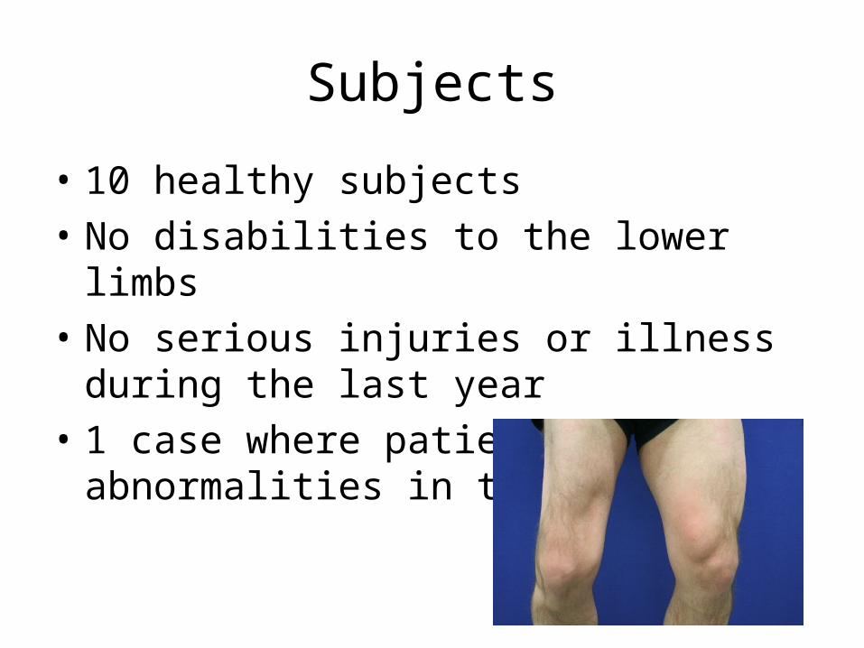
• 10 healthy subjects• No disabilities to the lower limbs• No serious injuries or illness during the last
year• 1 case where patient has abnormalities in the
lower limb

Experiment• Subject transported to local hospital• In a full surgical theater, 3 sterilized bone
pine (Strycker Howmedica) is drilled in to femur, tibia, patella under local anesthesia (Lidocaine 1%, 4 hours)
• Two stereophotogrammetry x-rays are acquired of the knee joint

Experiment• Subject is transported to the gait lab– Skin mounted surface markers is attached– 8 surface electromyography electrodes are attached
with double adhesive tape– Subject is asked to carry out natural movements
• Gait, Crouch, Slow running,– Subject is video recorded for post-evaluation
• Subject is transported back to the hospital– Bone pins are removed– Insertion area is cleaned and bandaged
(Curapor sterile, Lohmann & Rauscher)

Time• Transportation to hospital 90 min (45*2)• Attachment of bone markers
and x-ray, 1.5 hours• Measurement in lab 3-4 hours• Bone markers removal, 0.25 hours• Total: 7.25 hours

Compensation
• 500 euro/day• Lunch and dinner• Full covered health insurance for everything
that can be linked to the experiment
Risk
• Risk for small damage of tissue around the pin• Mild pain during experiment• No side effect of MRI scans• Stereophotogrammetry x-ray classified as
Category 1 risk• 19 known subject has had bone pins attached,
no know side effects have been reported
Subject follow up
• After 1 week and 1 Month– Subject is contacted over phone– Pain?– Discomfort?

Publicity
• Once the dataset is collected and the initial data processing has been carried out, the data will be distributed among the primary partners
• A half years later, the dataset will be published on the Internet, where researchers around the world can download and use data for further research and development of knee models

Approved/Rejected
• Your group is the local ethic board, and its up to you to grant or reject the proposal
• 5 min: talk over the application with your group members
• Questions to scientists?• 2 min: Make your final decision• Approved (Changes ?) / Rejected (Why)


Subjects
• 10 healthy subjects• No disabilities to the lower limbs• No serious injuries or illness during the last
year• 1 case where patient has abnormalities in the
lower limb

Experiment• Subject transported to local hospital• In a full surgical theater, 3 sterilized bone
pine (Strycker Howmedica) is drilled in to femur, tibia, patella under local anesthesia (Lidocaine 1%, 4 hours)
• Two stereophotogrammetry x-rays are acquired of the knee joint

Time
• Transportation to hospital 90 min (45*2)• Attachment of bone markers
and x-ray, 1.5 hours• Measurement in lab 3-4 hours• Bone markers removal, 0.25 hours• Total: 7.25 hours

Experiment
• Subject is transported to the gait lab– Skin mounted surface markers is attached– 8 surface electromyography electrodes are
attached with double adhesive tape– Subject is asked to carry out natural movements• Gait• Crouch• Slow running• Cutting movement (jumping side ways)
– Subject is video recorded for post-evaluation

Compensation
• 500 euro/day (100 DKR/hour (10 euro), max 300 DKR/Day)
• Lunch and dinner• Full covered health insurance for everything
that can be linked to the experiment

Risk
• Infection-risk• CT-risk• Risk for small damage of tissue around the pin• Mild pain during experiment• No side effect of MRI scans• Stereophotogrammetry x-ray classified as
Category 1 risk• 19 known subject has had bone pins attached, no
know side effects have been reported