PRE-LAB EXERCISES - Visible Body - Virtual Anatomy to See ... Manuals/2018 New/lab_man… ·...
47
1
Transcript of PRE-LAB EXERCISES - Visible Body - Virtual Anatomy to See ... Manuals/2018 New/lab_man… ·...

1

2
PRE-LAB EXERCISES
Before coming to lab, get familiar with a few muscle groups we’ll be exploring during lab. Using Visible Body’s Human Anatomy Atlas, go to the Views section. Under Systems, scroll down to the Muscular System views. Select View 11. Shoulder, and find the following muscles. When you select a muscle, note the book icon in the content box. Selecting this icon allows you to read the muscle’s definition.
1. Pectoralis major
2. Latissimus dorsi
Define the following terms:
1. Extension
2. Flexion
3. Abduction
4. Adduction
5. Rotation

3
IN-LAB EXERCISES
Use the following modules to guide your exploration of the shoulder and arm regions of the muscular system. As you explore the modules, locate the muscles on any charts, models, or specimen available. These muscles are located in and act on the shoulder and arm regions. Because the glenoid cavity of the scapula is shallow and does not snugly fit the head of the humerus, the tendons of multiple muscles are involved in securing and stabilizing the humerus at the shoulder to prevent dislocation. Other muscles will cross the shoulder (glenohumeral) joint and insert on the arm, causing the arm to move when they contract.
Movement of the brachium, or upper arm, depends on the fixators of the shoulder to keep the scapula in place so the arm can move freely. Once we move down into the antebrachium (forearm) and hand, the muscles begin to get smaller and more numerous, which grants us our fine motor skills when we write or play the piano. Pay attention to whether the muscle is on the anterior or posterior side of the arm – muscles on the anterior side will flex, while muscles on the posterior side will extend. The long names of some of these muscles can be daunting, but they are often very descriptive. You can find origins, insertions, actions, and/or locations of these muscles simply in the names.
When reviewing the action of a muscle, it will be helpful to think about where the muscle is located and where the insertion is. Muscle physiology requires that a muscle will “pull” instead of “push” during contraction, and the insertion is the part that will move. Imagine that the muscle is “pulling” on the bone or tissue it is attached to at the insertion.
Access 3D views and animated muscle actions in Visible Body’s Human Anatomy Atlas, which will be especially helpful to visualize muscle actions. When you select a structure in the Atlas app, you’ll see options to read the definition and hear the pronunciation in the content box. When you select a muscle, be sure to select the blue pin icon in the content box. This will give you the option to view origins and insertions as visible pins on the muscle (select Attachments), view the blood supply, and/or the nerve supply.
In each module below, identify the following:
• Muscle location
• Origin(s) and insertion(s)
• Muscle action
• Nerve supply

4
A. Muscles of the Shoulder
Muscles of the Shoulder
View the following Muscle Actions:
Shoulder flexion
Shoulder extension
Shoulder horizontal abduction
Shoulder horizontal adduction
Shoulder abduction
Shoulder adduction
Shoulder medial rotation
Try performing these actions yourself and feel which muscles contract.
These muscles primarily act to stabilize the scapula and move the arm. Since the scapula is a moveable bone, it must be stabilized in order for the arm to be able to move.
Some of these muscles are prime movers of the arm. They all cross the shoulder joint to insert on the humerus. Remember that muscles pull, and imagine how the muscle will pull on the humerus as it contracts.
Shoulder lateral rotation
Scapula elevation
Scapula depression
Scapula abduction
Scapula adduction
Scapula upward rotation
Scapula downward rotation

5
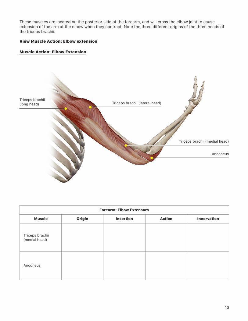
View 11. Shoulder
Deltoid
Pectoralis major
Latissimus dorsi

6
View 11. Shoulder
Deltoid
Teres minor
Coracobrachialis
Latissimus dorsi
Supraspinatus
Infraspinatus
Teres major

7
Insertion
Shoulder
OriginMuscle
Pectoralis major
Pectoralis minor
Deltoid
Action Innervation
Latissimus dorsi
Subscapularis
Infraspinatus
Coracobrachialis
Supraspinatus

8
Insertion
Shoulder (continued)
OriginMuscle
Teres major
Teres minor
Action Innervation

9
B. Muscles of the Torso that Act on the Scapulae
Under the Views section, go to Systems: Muscular System Views and select 20. Muscular System View.
• Rotate the model so you see the posterior side.• Select the left side of the trapezius and hide it. • Observe the following deep muscles that act on the scapulae.
View 20. Muscular System View
Levator scapulae
Rhomboideus major
Rhomboideus minor

10
Insertion
Torso Muscles that Act on the Scapulae
OriginMuscle
Rhomboideus major
Rhomboideus minor
Levator scapulae
Action Innervation

11
C. Muscles of the Elbow
Under the Views section, go to Systems: Muscular System Views and select 12. Elbow.These muscles are all located on the anterior side of the humerus and cross the elbow to insert on the radius or ulna. When these muscles contract, the arm will flex at the elbow. Biceps brachii is named for its “two heads;” note the two different origins of this muscle.
View 12. Elbow
Biceps brachii (long head)
Brachioradialis
Brachialis
Biceps brachii (medial head)

12
Insertion
Forearm: Elbow Flexors
OriginMuscle
Biceps brachii (long and short heads)
Brachialis
Brachioradialis
Action Innervation
13
Muscle Action: Elbow Extension
Triceps brachii(long head)
Anconeus
Triceps brachii (medial head)
Triceps brachii (lateral head)
These muscles are located on the posterior side of the forearm, and will cross the elbow joint to cause extension of the arm at the elbow when they contract. Note the three different origins of the three heads of the triceps brachii.
View Muscle Action: Elbow extension
Insertion
Forearm: Elbow Extensors
OriginMuscle
Triceps brachii (medial head)
Anconeus
Action Innervation

14
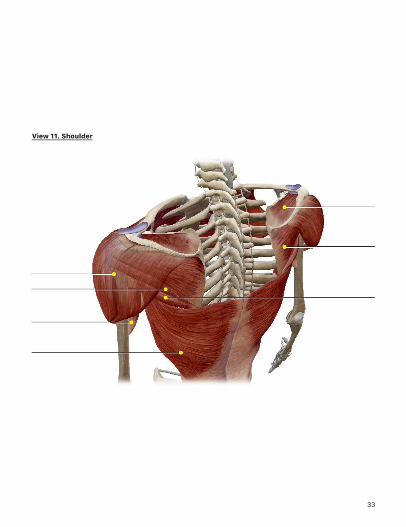
These muscles either pronate the forearm (turn the palm down), or supinate it (turn the palm up).
View Muscle Actions: Forearm pronation and Forearm supination
Muscle Action: Forearm Pronation
Pronator teres
Pronator quadratus

15
Muscle Action: Supination
Supinator
Insertion
Forearm: Pronation and Supination
OriginMuscle
Pronator teres
Pronator quadratus
Supinator
Action Innervation

16
These muscles make up the anterior compartment of the forearm, and cross the wrist to insert on the hand. They all function to flex the wrist and/or the fingers when they contract. These muscles have long names, but the names are very descriptive of where the muscle is located and its action.
View Muscle Actions: Wrist flexionWrist abductionWrist adduction
Muscle Action: Wrist Flexion
Flexor carpi radialis
Flexor digitorum superficialis
Flexor carpi ulnaris
Palmaris longus

17
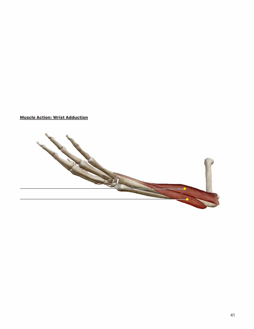
Muscle Action: Wrist Adduction
Flexor carpi ulnaris
Extensor carpi ulnaris
Muscle Action: Wrist Abduction
Flexor carpi radialis
Extensor pollicis longus
Abductor pollicis longus
Extensor carpi radialis longus
Extensor carpi radialis brevis
Extensor pollicis brevis

18
Insertion
Hand: Deep Flexors
OriginMuscle
Flexor carpi ulnaris
Flexor pollicis longus
Flexor digitorum superficialis
Flexor digitorum profundus
Action Innervation
These muscles also flex the hand, and are located deep to the hand flexors above.
Insertion
Hand: Superficial Flexors
OriginMuscle
Flexor carpi radialis
Palmaris longus
Action Innervation

19
These muscles are located on the posterior side of the forearm and cross the wrist to insert on the hand. When these muscles contract, the wrist and/or fingers will extend.
View Muscle Actions: Wrist extension Wrist abductionWrist adduction
Muscle Action: Wrist Extension
Extensor carpi ulnaris
Extensor carpi radialis brevis
Extensor digitorum
Extensor carpi radialis longus

20
Insertion
Hand: Superficial Extensors
OriginMuscle
Extensor carpi radialis longus
Extensor carpi radialis brevis
Extensor digiti minimi
Extensor digitorum
Extensor carpi ulnaris
Action Innervation

21
D. Muscles of the Wrist and Hand
Under the Views section, go to Systems: Muscular System Views and select 13. Wrist and Hand.
View Muscle Actions: Thumb extension
These muscles are also located on the posterior compartment of the forearm, but are located deep to the muscles in the previous section. They will also cross the wrist to insert on the hand, functioning to move the first or second digit when contracted. It will be helpful to remember that “pollicis” is referring to the thumb and “indicis” to the index finger.
Muscle Action: Thumb Extension
Extensor pollicis longus
Extensor pollicis brevis
Abductor pollicis longus

22
Insertion
Hand: Deep Extensors
OriginMuscle
Abductor pollicis longus
Extensor pollicis longus
Extensor indicis
Extensor pollicis brevis
Action Innervation

23
Muscle Action: Thumb Flexion
Flexor pollicis longus
Palmaris longus
Adductor pollicis
Flexor pollicis brevis
These muscles move the first digit – the thumb. Earlier sections have included muscles that move the thumb, but are primarily located in the forearm. Thenar muscles are entirely located within the hand and form the thenar eminence – the fleshy protrusion in the hand at the base of the thumb.
View Muscle Actions: Thumb flexion and Hand digits opposition

24
Muscle Action: Hand Digits Opposition
Opponens digiti minimi
Abductor pollicis brevis
Opponens pollicis
Flexor pollicis brevis

25
Insertion
Finger: Thenar
OriginMuscle
Abductor pollicis brevis
Opponens pollicis
Adductor pollicis
Flexor pollicis brevis
Action Innervation
These muscles all function to move digit 5, the little finger. These muscles are also entirely located within the hand.
Insertion
Finger: Hypothenar
OriginMuscle
Abductor digiti minimi
Flexor digiti minimi brevis
Opponens digiti minimi
Action Innervation

26
These muscles are located within the hand and are responsible for the fine movements of the fingers. The muscles listed in the chart below are actually groups of muscles. The number of muscles normally found in each group is in parentheses after the name.
View Muscle Actions: Hand digits 2-5 flexion and Hand digits 2-5 extension
Muscle Action: Hand Digits 2-5 Flexion
Lumbricals
Palmar interossei
Dorsal interossei

27
Muscle Action: Hand Digits 2-5 Extension
Dorsal interossei
Extensor digitorum
Palmar interossei

28
Insertion
Fingers: Midpalmar
OriginMuscle
Lumbricals (4)
Palmar interossei (3)
Dorsal interossei (4)
Action Innervation

29
PUTTING IT ALL TOGETHER
1. Based on what you’ve learned about the muscles in this exercise, what do you think the following terms mean?
a. Major
b. Minor
c. Extensor
d. Flexor
e. Longus
f. Brevis
g. Spinatus
h. Pollicis
i. Carpi

30
2. Which muscles are part of the rotator cuff that serves to stabilize the shoulder joint?
3. Which muscles are used when performing the following actions?
a. Raising your hand high over your head during class
b. Rowing a boat
c. Reaching behind you, arm extended and pronated
d. Reaching in front of you, arm extended and supinated
e. Bringing your hand to your heart
f. Holding a pencil
4. Carpal tunnel syndrome can result from repetitive motions in the fingers causing inflammation in the carpal tunnel – a space covered by the flexor retinaculum where tendons and nerves pass through the wrist. In this syndrome, the median nerve is compressed, which can lead to tingling, numbness, and muscle weakness. Which muscles are most likely to be affected by carpal tunnel syndrome?

31

32
View 11. Shoulder
33
View 11. Shoulder

34
View 20. Muscular System View

35
View 12. Elbow

36
Muscle Action: Elbow Extension
37
Muscle Action: Forearm Pronation

38
Muscle Action: Supination

39
Muscle Action: Wrist Flexion

40
Muscle Action: Wrist Abduction
41
Muscle Action: Wrist Adduction

42
Muscle Action: Wrist Extension

43
Muscle Action: Thumb Extension

44
Muscle Action: Thumb Flexion

45
Muscle Action: Hand Digits Opposition

46
Muscle Action: Hand Digits 2-5 Flexion

47
Muscle Action: Hand Digits 2-5 Extension