PRACTICAL TECHNIQUES FROM THE NAVC INSTITUTE … · July/August 2014 Today’s Veterinary Practice...
7
July/August 2014 Today’s Veterinary Practice 43 PRACTICAL TECHNIQUES PEER REVIEWED tvpjournal.com PRACTICAL TECHNIQUES FROM THE NAVC INSTITUTE FELINE PERINEAL URETHROSTOMY VENTRAL APPROACH Clara S.S. Goh, BVSc, MS, Diplomate ACVS, and Howard B. Seim III, DVM, Diplomate ACVS Colorado State University O ver the past decade, advances in the medical management of feline lower urinary tract disease (FLUTD) have decreased the requirement for surgical intervention of the blocked male cat. Perineal urethrostomy (PU) is still the surgical treatment of choice for pa- tients with: • Repeated urethral obstruction despite medical management • Obstruction that cannot be relieved by urethral catheterization • Catheterization that has resulted in significant urethral trauma and/or stenosis. 1 INITIAL MEDICAL MANAGEMENT Prior to anesthesia for an elective PU, manage the patient medically with IV fluid therapy and bladder decompression (via retropulsion of urethral crys- tal/mucus plugs or calculi, catheterization, repeat cystocentesis, or a tempo- rary cystostomy tube) until all renal, metabolic, and electrolyte parameters are within normal limits. FELINE PERINEAL URETHROSTOMY (DORSAL RECUMBENCY) Our preferred technique is a slight variation on the traditional PU described by Wilson and Harrison in 1971. 2 In this variation, the cat is placed in dorsal recumbency instead of the standard perineal approach. This positioning is more ergonomic for the surgeon, and allows access to the ventral abdomen for concurrent cystotomy if indicated. The Essentials for Perineal Urethrostomy • Standard general surgery pack, including needle holders, thumb forceps (preferably Debakey forceps), mosquito forceps, scalpel handle, Metzenbaum and Mayo scissors • Stevens tenotomy scissors (4” straight) • 2 Gelpi retractors (3.5” size) • Suture (4-0 to 5-0 monofilament synthetic absorbable, on a taper or taper-cut needle) • Suction device and small Frasier suction tip W elcome to the first article in our new Practical Techniques from the NAVC Institute column. Each year, the NAVC Institute takes place in Orlando, Florida, and specialists in select areas of veterinary medicine provide hands-on, one-on-one continuing education to the Institute attendees. The NAVC and Today’s Veterinary Practice have partnered together to present information from the NAVC Institute 2014 courses. For those unable to attend, this column provides the opportunity to experience the excellent education provided at the Institute. Visit navc.com/ institute for further information. Feline Friendly Article
Transcript of PRACTICAL TECHNIQUES FROM THE NAVC INSTITUTE … · July/August 2014 Today’s Veterinary Practice...
July/August 2014 Today’s Veterinary Practice 43
PRACTICAL TECHNIQUES Peer reviewed
tvpjournal.com
PRACTICAL TECHNIQUES FROM THE NAVC INSTITUTE
Feline Perineal Urethrostomy ventral aPProach Clara S.S. Goh, BVSc, MS, Diplomate ACVS, and Howard B. Seim III, DVM, Diplomate ACVSColorado State University
Over the past decade, advances in the medical management of feline lower urinary tract disease (FLUTD) have decreased the requirement for surgical intervention of the blocked male cat.
Perineal urethrostomy (PU) is still the surgical treatment of choice for pa-tients with:•Repeated urethral obstruction despite medical management•Obstruction that cannot be relieved by urethral catheterization•Catheterization that has resulted in significant urethral trauma and/or
stenosis.1
INITIAL MEDICAL MANAGEMENTPrior to anesthesia for an elective PU, manage the patient medically with IV fluid therapy and bladder decompression (via retropulsion of urethral crys-tal/mucus plugs or calculi, catheterization, repeat cystocentesis, or a tempo-rary cystostomy tube) until all renal, metabolic, and electrolyte parameters are within normal limits.
FELINE PERINEAL URETHROSTOMY (DORSAL RECUMBENCY)Our preferred technique is a slight variation on the traditional PU described by Wilson and Harrison in 1971.2 In this variation, the cat is placed in dorsal recumbency instead of the standard perineal approach. This positioning is more ergonomic for the surgeon, and allows access to the ventral abdomen for concurrent cystotomy if indicated.
The Essentials for Perineal Urethrostomy•Standard general surgery
pack, including needle holders, thumb forceps (preferably Debakey forceps), mosquito forceps, scalpel handle, Metzenbaum and Mayo scissors
•Stevens tenotomy scissors (4” straight)
•2 Gelpi retractors (3.5” size)
•Suture (4-0 to 5-0 monofilament synthetic absorbable, on a taper or taper-cut needle)
•Suction device and small Frasier suction tip
Welcome to the first article in our new Practical Techniques from the
NAVC Institute column. Each year, the NAVC Institute takes place in Orlando, Florida, and specialists in select areas of veterinary medicine provide hands-on, one-on-one continuing education to the Institute attendees.
The NAVC and Today’s Veterinary Practice have partnered together to present information from the NAVC Institute 2014 courses. For those unable to attend, this column provides the opportunity to experience the excellent education provided at the Institute. Visit navc.com/institute for further information.
Feline FriendlyArticle
Today’s Veterinary Practice July/August 201444
| PRACTICAL TECHNIQUES
tvpjournal.com
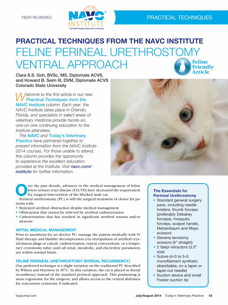
1 Position the cat in dorsal recumbency (see Surgical Insight: Penile Orien-tation) with the hindlimbs tied cra-
nially until the pelvis is slightly elevated off the surgery table. Place a folded towel under the pelvis to support the patient (A and B) and place a purse string suture in the anus. Clip and aseptically prepare the perineal region ± ventral abdomen (if a cystotomy is indicated).
1A
1B
SURGICAL INSIGHT: PENILE ORIENTATION » Pay close attention to penile orien-tation in this technique (ie, dorsal versus ventral side). » When the penis is pulled caudal-ly, toward the tail, the surgeon is working on its ventral aspect. » When the penis is reflected cra-nially, toward the cat’s head, the surgeon is working on the dorsal aspect. » Minimal delicate peripenile dissec-tion is carried out on the dorsal as-pect of the penis, as this is where the primary neurovascular supply is located.3
» When dissecting the dorsal as-pect of the penis, the surgeon is also in close proximity to the rec-tum, which is undesirable because it increases the risk of rectal per-foration
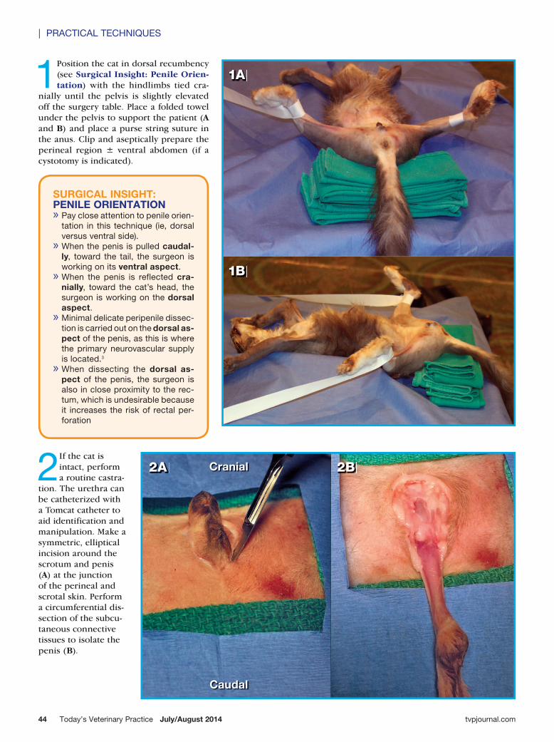
2 If the cat is intact, perform a routine castra-
tion. The urethra can be catheterized with a Tomcat catheter to aid identification and manipulation. Make a symmetric, elliptical incision around the scrotum and penis (A) at the junction of the perineal and scrotal skin. Perform a circumferential dis-section of the subcu-taneous connective tissues to isolate the penis (B).
2A 2BCranial
Caudal
July/August 2014 Today’s Veterinary Practice 45
PRACTICAL TECHNIQUES |
FELI
NE
PE
RIN
EA
L U
RE
THR
oS
ToM
y: V
EN
TRA
L A
PP
Ro
AC
H
tvpjournal.com
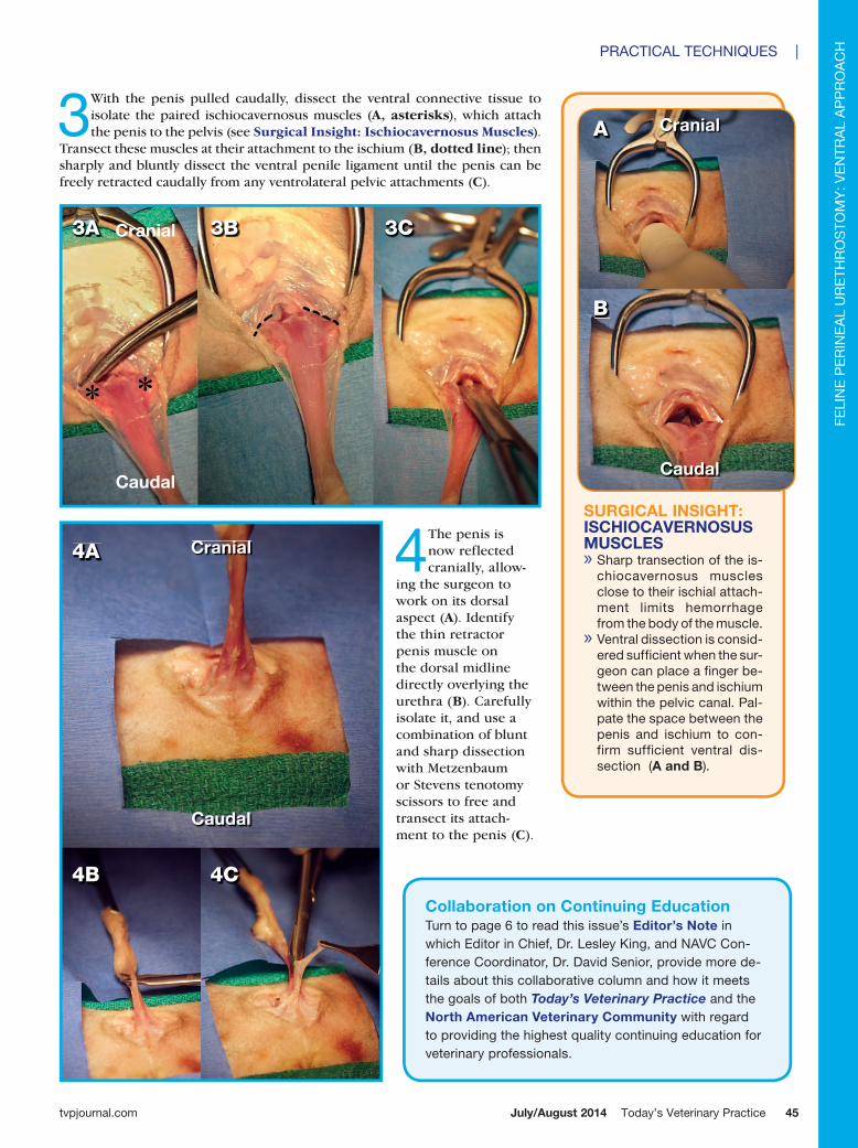
4 The penis is now reflected cranially, allow-
ing the surgeon to work on its dorsal aspect (A). Identify the thin retractor penis muscle on the dorsal midline directly overlying the urethra (B). Carefully isolate it, and use a combination of blunt and sharp dissection with Metzenbaum or Stevens tenotomy scissors to free and transect its attach-ment to the penis (C).
3 With the penis pulled caudally, dissect the ventral connective tissue to isolate the paired ischiocavernosus muscles (A, asterisks), which attach the penis to the pelvis (see Surgical Insight: Ischiocavernosus Muscles).
Transect these muscles at their attachment to the ischium (B, dotted line); then sharply and bluntly dissect the ventral penile ligament until the penis can be freely retracted caudally from any ventrolateral pelvic attachments (C).
SURGICAL INSIGHT: ISCHIOCAVERNOSUS MUSCLES » Sharp transection of the is-chiocavernosus muscles close to their ischial attach-ment limits hemorrhage from the body of the muscle. » Ventral dissection is consid-ered sufficient when the sur-geon can place a finger be-tween the penis and ischium within the pelvic canal. Pal-pate the space between the penis and ischium to con-firm sufficient ventral dis-section (A and B).
A
B
3A 3B 3C
4A
4B 4C
Cranial
Caudal
Cranial
Caudal
Cranial
Caudal
Collaboration on Continuing EducationTurn to page 6 to read this issue’s Editor’s Note in which Editor in Chief, Dr. Lesley King, and NAVC Con-ference Coordinator, Dr. David Senior, provide more de-tails about this collaborative column and how it meets the goals of both Today’s Veterinary Practice and the North American Veterinary Community with regard to providing the highest quality continuing education for veterinary professionals.
Today’s Veterinary Practice July/August 201446
| PRACTICAL TECHNIQUES
tvpjournal.com
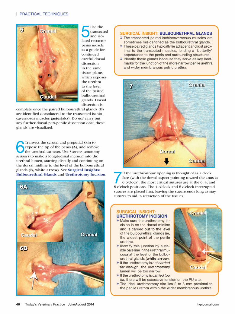
5 Use the transected and iso-
lated retractor penis muscle as a guide for continued careful dorsal dissection in the same tissue plane, which exposes the urethra to the level of the paired bulbourethral glands. Dorsal dissection is
complete once the paired bulbourethral glands (B) are identified dorsolateral to the transected ischio-cavernosus muscles (asterisks). Do not carry out any further dorsal peri-penile dissection once these glands are visualized.
SURGICAL INSIGHT: URETHROTOMY INCISION » Make sure the urethrotomy in-cision is on the dorsal midline and is carried out to the level of the bulbourethral glands (ie, the widest point of the penile urethra). » Identify this junction by a vis-ible pale line in the urethral mu-cosa at the level of the bulbo-urethral glands (white arrow). » If the urethrotomy is not carried far enough, the urethrostomy lumen will be too narrow. » If the urethrotomy is carried too far, there will be excessive tension on the PU site. » The ideal urethrostomy site lies 2 to 3 mm proximal to the penile urethra within the wider membranous urethra.
SURGICAL INSIGHT: BULBOURETHRAL GLANDS » The transected paired ischiocavernosus muscles are sometimes misidentified as the bulbourethral glands. » These paired glands typically lie adjacent and just prox-imal to the transected muscles, lending a “butterfly” appearance to the penis and surrounding structures. » Identify these glands because they serve as key land-marks for the junction of the more narrow penile urethra and wider membranous pelvic urethra.
6 Transect the scrotal and preputial skin to expose the tip of the penis (A), and remove the urethral catheter. Use Stevens tenotomy
scissors to make a longitudinal incision into the urethral lumen, starting distally and continuing on the dorsal midline to the level of the bulbourethral glands (B, white arrow). See Surgical Insights: Bulbourethral Glands and Urethrotomy Incision.
6B
6A
CranialCaudal
Cranial
Caudal
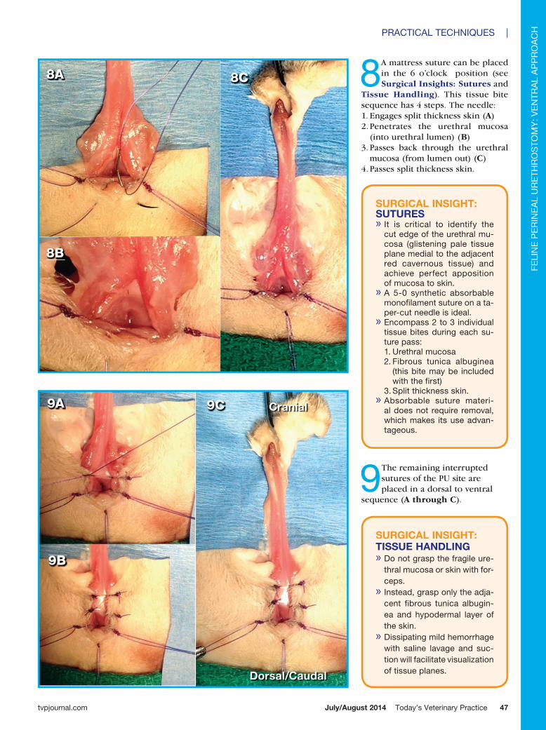
7If the urethrostomy opening is thought of as a clock face (with the dorsal aspect pointing toward the anus at 6 o’clock), the most critical sutures are at the 6, 4, and
8 o’clock positions. The 4 o’clock and 8 o’clock interrupted sutures are placed first, leaving the suture ends long as stay sutures to aid in retraction of the tissues.
7 Cranial
Caudal
Dorsal
5 Cranial
Caudal
July/August 2014 Today’s Veterinary Practice 47
PRACTICAL TECHNIQUES |
FELI
NE
PE
RIN
EA
L U
RE
THR
oS
ToM
y: V
EN
TRA
L A
PP
Ro
AC
H
tvpjournal.com
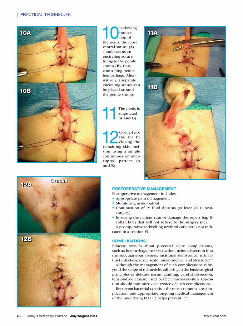
8A mattress suture can be placed in the 6 o’clock position (see Surgical Insights: Sutures and
Tissue Handling). This tissue bite sequence has 4 steps. The needle:1. Engages split thickness skin (A)2. Penetrates the urethral mucosa
(into urethral lumen) (B)3. Passes back through the urethral
mucosa (from lumen out) (C)4. Passes split thickness skin.
SURGICAL INSIGHT: SUTURES » It is critical to identify the cut edge of the urethral mu-cosa (glistening pale tissue plane medial to the adjacent red cavernous tissue) and achieve perfect apposition of mucosa to skin. » A 5-0 synthetic absorbable monofilament suture on a ta-per-cut needle is ideal. » Encompass 2 to 3 individual tissue bites during each su-ture pass: 1. Urethral mucosa2. Fibrous tunica albuginea
(this bite may be included with the first)
3. Split thickness skin. » Absorbable suture materi-al does not require removal, which makes its use advan-tageous.
9 The remaining interrupted sutures of the PU site are placed in a dorsal to ventral
sequence (A through C).
8B
8A 8C
9B
9A 9C Cranial
Dorsal/Caudal
SURGICAL INSIGHT: TISSUE HANDLING » Do not grasp the fragile ure-thral mucosa or skin with for-ceps. » Instead, grasp only the adja-cent fibrous tunica albugin-ea and hypodermal layer of the skin. » Dissipating mild hemorrhage with saline lavage and suc-tion will facilitate visualization of tissue planes.
Today’s Veterinary Practice July/August 201448
| PRACTICAL TECHNIQUES
tvpjournal.com
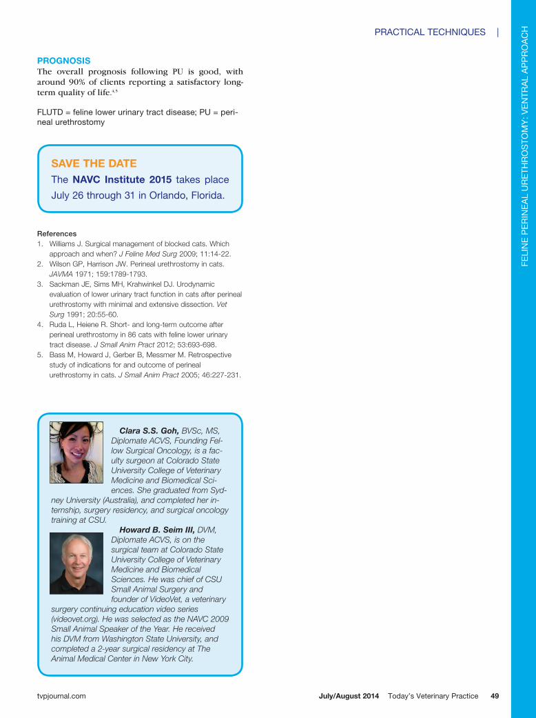
10 Following transec-tion of
the penis, the most ventral suture (A) should act as an encircling suture to ligate the penile stump (B); thus, controlling penile hemorrhage. Alter-natively, a separate encircling suture can be placed around the penile stump.
11 The penis is amputated (A and B).
12 C o mp l e t e the PU by closing the
remaining skin inci-sion using a simple continuous or inter-rupted pattern (A and B).
10B
10A
11B
11A
POSTOPERATIVE MANAGEMENTPostoperative management includes: •Appropriate pain management•Monitoring urine output•Continuation of IV fluid diuresis (at least 24 H post-
surgery)•Ensuring the patient cannot damage the repair (eg, E-
collar, litter that will not adhere to the surgery site).A postoperative indwelling urethral catheter is not indi-
cated in a routine PU.
COMPLICATIONSEducate owners about potential acute complications, such as hemorrhage, re-obstruction, urine dissection into the subcutaneous tissues, incisional dehiscence, urinary tract infection, urine scald, incontinence, and stricture.1,4,5
Although the management of such complications is be-yond the scope of this article, adhering to the basic surgical principles of delicate tissue handling, careful dissection, tension-free closure, and perfect mucosa-to-skin apposi-tion should minimize occurrence of such complications.
Recurrent bacterial cystitis is the most common late com-plication, and appropriate ongoing medical management of the underlying FLUTD helps prevent it.4,5
12B
12ACranial
July/August 2014 Today’s Veterinary Practice 49
PRACTICAL TECHNIQUES |
FELI
NE
PE
RIN
EA
L U
RE
THR
oS
ToM
y: V
EN
TRA
L A
PP
Ro
AC
H
tvpjournal.com
PROGNOSISThe overall prognosis following PU is good, with around 90% of clients reporting a satisfactory long-term quality of life.4,5
FLUTD = feline lower urinary tract disease; PU = peri-neal urethrostomy
References1. williams J. surgical management of blocked cats. which
approach and when? J Feline Med Surg 2009; 11:14-22.2. wilson GP, harrison Jw. Perineal urethrostomy in cats.
JAVMA 1971; 159:1789-1793.3. sackman Je, sims mh, Krahwinkel dJ. Urodynamic
evaluation of lower urinary tract function in cats after perineal urethrostomy with minimal and extensive dissection. Vet Surg 1991; 20:55-60.
4. ruda l, heiene r. short- and long-term outcome after perineal urethrostomy in 86 cats with feline lower urinary tract disease. J Small Anim Pract 2012; 53:693-698.
5. Bass m, howard J, Gerber B, messmer m. retrospective study of indications for and outcome of perineal urethrostomy in cats. J Small Anim Pract 2005; 46:227-231.
Clara S.S. Goh, BVSc, MS, Diplomate ACVS, Founding Fel-low Surgical Oncology, is a fac-ulty surgeon at Colorado State University College of Veterinary Medicine and Biomedical Sci-ences. She graduated from Syd-
ney University (Australia), and completed her in-ternship, surgery residency, and surgical oncology training at CSU.
Howard B. Seim III, DVM, Diplomate ACVS, is on the surgical team at Colorado State University College of Veterinary Medicine and Biomedical Sciences. He was chief of CSU Small Animal Surgery and founder of VideoVet, a veterinary
surgery continuing education video series (videovet.org). He was selected as the NAVC 2009 Small Animal Speaker of the Year. He received his DVM from Washington State University, and completed a 2-year surgical residency at The Animal Medical Center in New York City.
SAVE THE DATEThe NAVC Institute 2015 takes place
July 26 through 31 in orlando, Florida.