
Practical anatomy of the git
72
Practical anatomy of GIT 1 │ BY : Abdelrahman Hilmy N.B : all the theoretical lectures related to practical sessions Should be studied first. ⌔ Content : ▪ 1 st section : Bones (norma lateralis & Mandible ) ▪ 2ed section : Oral cavity &salivary glands, pharynx ▪ 3 rd section : Infratemporal fossa & Muscles of mastication ▪ 4 th section : Ant. & post. Abd. Walls ▪ 5 th section : Stomach & liver ▪ 6 th Section : Small & large intestine and Pancreas Bones (Norma lateralis & Mandible) ⌔ Spot (1) Identify pointed area ? Temporal fossa Give structure attached to it ? Origin of temporalis muscle . Give its action & nerve supply ? anterior fibers : elevate mandible posterior fibers : retract mandible . nerve supply : Deep temporal nerves from the anterior division of mandibular nerve. Give its insertion ? Coronoid process of mandible .
-
Upload
abd-elrahman-hilmy -
Category
Science
-
view
163 -
download
3
Transcript of Practical anatomy of the git

Practical anatomy of GIT
1 │ BY : Abdelrahman Hilmy
N.B : all the theoretical lectures related to practical sessions Should be studied first.
⌔ Content :
▪ 1st section : Bones (norma lateralis & Mandible )
▪ 2ed section : Oral cavity &salivary glands, pharynx
▪ 3rd
section : Infratemporal fossa & Muscles of mastication
▪ 4th
section : Ant. & post. Abd. Walls
▪ 5th
section : Stomach & liver
▪ 6th
Section : Small & large intestine and Pancreas
Bones (Norma lateralis & Mandible)
⌔ Spot (1)
Identify pointed area ?
Temporal fossa
Give structure attached to it ?
Origin of temporalis muscle .
Give its action & nerve supply ?
anterior fibers : elevate mandible
posterior fibers : retract mandible .
nerve supply : Deep temporal nerves
from the anterior division of
mandibular nerve.
Give its insertion ?
Coronoid process of mandible .
Practical anatomy of GIT
2 │ BY : Abdelrahman Hilmy
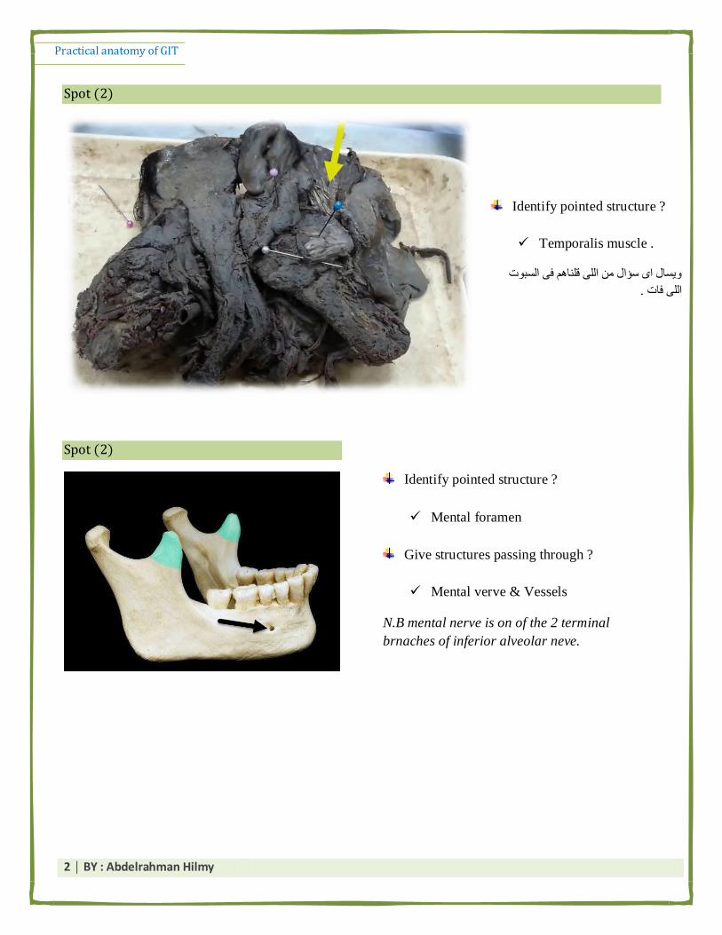
Spot (2)
Spot (2)
Identify pointed structure ?
Mental foramen
Give structures passing through ?
Mental verve & Vessels
N.B mental nerve is on of the 2 terminal
brnaches of inferior alveolar neve.
Identify pointed structure ?
Temporalis muscle .
يظبل ا طؤال ي انه لهبى ف انظجد
.انه فبد
Practical anatomy of GIT
3 │ BY : Abdelrahman Hilmy
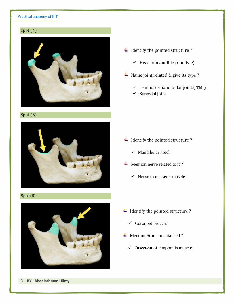
Spot (4)
Spot (5)
Identify the pointed structure ?
Head of mandible (Condyle)
Name joint related & give its type ?
Temporo-mandibular joint.( TMJ)
Synovial joint
Identify the pointed structure ?
Mandibular notch
Mention nerve related to it ?
Nerve to masseter muscle
Spot (6)
Identify the pointed structure ?
Coronoid process
Mention Structure attached ?
Insertion of temporalis muscle .

Practical anatomy of GIT
4 │ BY : Abdelrahman Hilmy
Spot (7)
Spot (8)
Identify the pointed structure ?
Mylohyoid line
Mention str. Attached ?
Origin of mylohoid muscle
Pterygomandibular lig.
Superior Constrictor muscle .
Identify the pointed structures 1 & 2 ?
1- Lingula
2- Mandibular foramen
Mention str. Attached to No. (1) ?
Sphenomandibular lig.
Mention str. Passing through No. (2) ?
Inferior alveolar nerve & vessels .

Practical anatomy of GIT
5 │ BY : Abdelrahman Hilmy
Spot (9)
Spot (10)
Spot (11)
Identify the pointed structures?
Neck of the mandible
Mention str. Attached ?
Tempromandibular ligament
Mention Nerves & Vessels related ?
Maxillary artery : medial to neck
Auriculotemporal nerve . : medial to
neck
Identify the pointed structures No. 1 & 2 ?
1- Sublingual fossa
2- submandibular fosaa
Mention str. Related ?
1- sunlingual salivary gland
2- submandibular salivary gland .
Identify the pointed Str. ?
Pterygoid fovea
Mention str. Attached ?
Insertion of lateral pterygoid muscle

Practical anatomy of GIT
6 │ BY : Abdelrahman Hilmy
Spot (12)
Spot (13)
Spot (14)
Identify the pointed structure ?
Zygomatic arch
Mention str. Attached ?
Temporalis fascia : attachéd to upper
border
Origin of masseter : from lower border
& inner surface .
Tempromandibular ligament .
Identify area pointed by yellow arrow ?
Pterion
Identify the pointed structure ?
Lateral surface of ramus of mandible
Mention str. Attached ?
Insertion of masseter muscle
Identify the pointed structure ?
Angle of mandible
Mention str. Attached ?
Stylomandibular ligament .
Insertion of Medial pterygoid muscle
(in inner aspect of the angle )

Practical anatomy of GIT
7 │ BY : Abdelrahman Hilmy
Spot (15)
Spot (15)
⌔ Geniohyoid muscle :
* Nerve supply : C1 component of hypoglossal N.
* Action : Elevate hyoid bone & depress mandible .
Identify the pointed structures (red arrow)?
Superior temporal line
Mention str. Attached ?
Temporalis fascia
Identify Circled area ?
Asterion
Identify the pointed structures 1 & 2 ?
1- Superior genial tubercle
2- inferior genial tubercle
Mention Str. Attached ?
1- Origin of Genioglossus muscle
2- Origin of geniohyoid muscles

Practical anatomy of GIT
8 │ BY : Abdelrahman Hilmy
Spot (1)
Identify the pointed structures?
Parotid gland
Mention its surfaces ?
Lateral (superfacial) surface
Anteromedial surface
Posteromedial surface
Mention Strs. Within it (in order from
superfascial to deep) ?
Facial nerve
Retromandibular vein
External carotid artery
Mention its blood supply ?
External carotid artery and its terminal -----> superficial temporal + maxillary arteries
Mention its venous drainage ?
Retromandibular vein
Mention its capsule ?
1- inner capsule (true capsule): connective-tissue capsule adherent to the gland and sends
2- Outer capsule (false capsule): fromed by the investing layer of deep cervical fascia.
Give its Motor nerve supply ?
1- Parasympathetic :
Inferior salivary nucleus ⟶ glossopharyngeal nerve ⟶ ⟶⟶ postganglionic fibers are carried by ⟶
auriculo-temporal nerve to supply the gland
2- Sympathetic supply : from plexus surrounding the external carotid artery

Practical anatomy of GIT
9 │ BY : Abdelrahman Hilmy
Mention its lymphatic drainage ?
parotid lymph nodes + deep cervical lymph nodes.
Mention its sensory nerve supply ?
Capsule ⟶ Great auricular nerve .
Parynchema ⟶ auriculo-temporal nerve .
Mention the structures that leave the gland at its upper end (pole) ?
1- Auriculo-temporal nerve
2- Superfascial temporal artery
3- temporal branches of facial nerve .
Mention the structure /s that Enter the gland through its upper end (pole) ?
Superfascial temporal vein
Mention the structures that enter the gland through its posteromedial surface ?
1- Facial nerve
2- External carotid artery
Mention the structures that enter the gland through its anteromedial surface ?
1- Auriculo-temporal nerve
2- Maxillary vein
Mention the Strs. That leave the gland at its lower pole (End) ?
1- Cervical branch of facial nerve
2- Anterior & posterior divisions of retromandibular vein .
Give its anterior & posterior boundaries ?
Anteriorly : masseter muscle (overlies its posterior part )
Posteriorly : sternomastoid muscle (overlies its upper part )

Practical anatomy of GIT
10 │ BY : Abdelrahman Hilmy
Give its superior & inferior boundaries ?
Upwards : Zygomatic arch
Downwards : angle of mandible .
Enumerate the Strs. That leave the gland at its anterior border ?
Zygomatic N.
Buccal nerve
Marginal mandibular N.
⌔ Review the relations of the gland page 26
Spot (2)
Identify pointed str. ?
Parotid Duct
Enumerate Strs. Pierced by it ? (4 Bucc-)
1-Buccal pad of fat
2- Buccopharungeal fascia
3- Buccinator muscle
4-buccal mucosa

Practical anatomy of GIT
11 │ BY : Abdelrahman Hilmy
Mention the site of its opening ? (V-IMP Q)
vestibule of the mouth opposite the upper second molar tooth
Mention the surface marking of it ?
It corresponds to the middle third of a line extending between two points:
① A point midway between the red margin of the upper lip and ala of the nose.
② A point at the lower end of the tragus of the ear.
Spot (3)
Identify pointed Str. ?
Masseter muscle
Give its action & nerve supply ?
Action : Elevation and protraction of the mandible
Nerve supply : anterior division of the mandibular nerve
N.B origin & insertion & action & nerve supply of muscles of mastication is important .

Practical anatomy of GIT
12 │ BY : Abdelrahman Hilmy
Spot (4)
ؼف :ح infratemporal fossaيكظرح ػهؼب اػف انـ mandibleب انـ ⌔
- artery : maxillary artery
يزؼبر ػهي ف انصرح انه فق
- 2 muscles : medial & lateral pterygoid
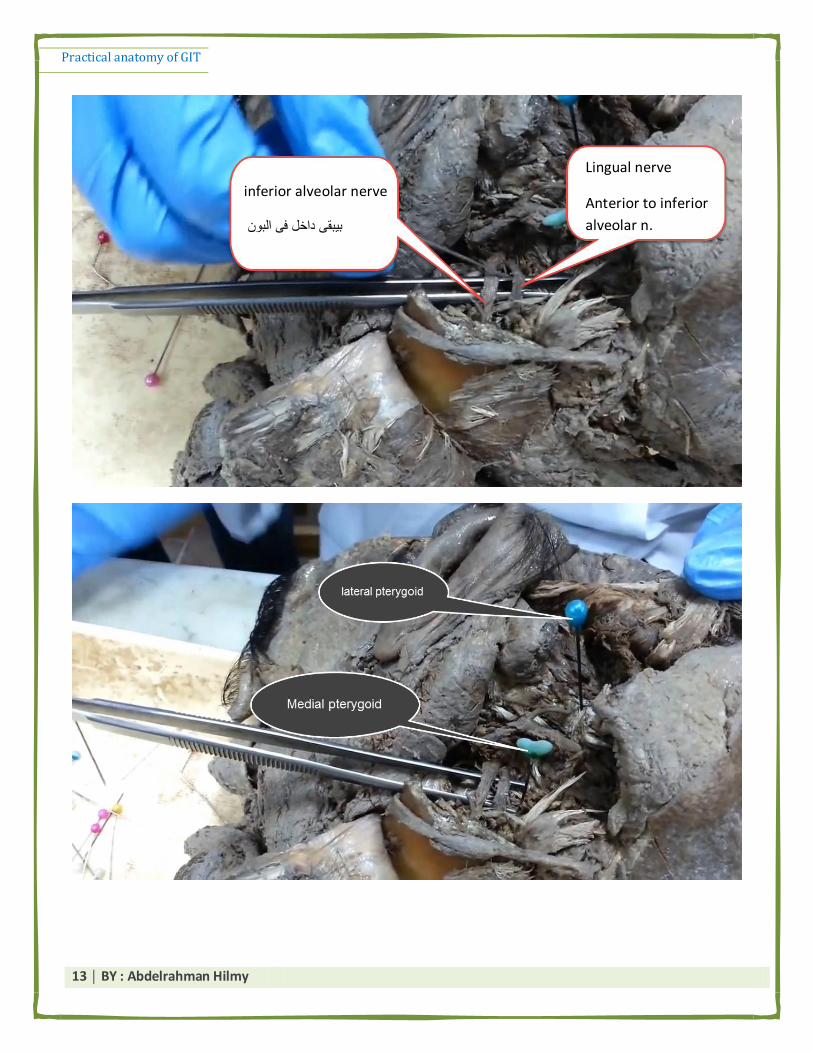
- 2 nerves : inferior alveolar nerve & lingual nerve
انه ثزجم يكظرح طبػبد ثيجم mandibular foramen of mandibleثيجم داخم جا انـ inferior alveolar nerve انـ *
. tongueرايح نهـ ,ني anterior ثيجم lingual nerveيمطع ,, ايب انـ
ثزجم رحذ انـ lateral pterygoidايب انـ medial pterygoid* ن حظ دثص ف انؼضهخ انه رحذ انزفي دل حزجم انـ
maxillary a.
زخيم . ؼف انصر د كذا ػهؼب *
Practical anatomy of GIT
13 │ BY : Abdelrahman Hilmy
inferior alveolar nerve
بيبقى داخل فى البون
Lingual nerve
Anterior to inferior
alveolar n.

Practical anatomy of GIT
14 │ BY : Abdelrahman Hilmy
Spot (5)
Identify pointed Str. ?
Maxillary a.
Mention its origin ? (how & where ?)
It arises from External carotid artery within the substance of the parotid gland at the level of
neck of mandible .
Give its parts ? (3 parts )
1- First (Mandibular) part : Medial to the neck of the mandible
2- Second (pterygoid ) part : On the lateral surface of the lateral pterygoid muscle
3- third part : Passes between the two heads of the lateral pterygoid to reach the pterygopalatine fossa
*انظؤال دا صؼت يج ال يحزبج كزبثخ كزيزح سي انظجد يغ حيكف ثض احب يضطزي ؼزف ال يحذع ػبرف انظزف اي .
.ثؼذ كذا حفظ ا ثزؼي ي كم جشء احزيبط ثزض *
Third part Second part First part
1-posterior superior alveolar
2-greater palatine
3-pharyngeal
4- sphenopalatine arteries,
5- artery of the pterygoid canal.
6- infraorbital artery
Muscular Branches
1- deep temporal as.
2- masseteric a.
3- buccal a.
4- pterygoid branches for medial
and lateral pterygoid muscles
MIADA
1- Middle meningeal a.
2- Inferior alveolar a.
3- Accessory meningeal a.
4- Deep auricular a.
5- Anterior tympanic

Practical anatomy of GIT
15 │ BY : Abdelrahman Hilmy
Spot (6)
Identify pointed Str. 1 & 2 ?
1- inferior alveolar nerve
2- lingual nerve
Mention origin of Str. NO. 1 & 2 ?
Both arise from the posterior Division of mandibular nerve .
Name the Structure that join Str. No 2 ? Mention its type of fibers ?
Chorda tympani nerve
It carries parasympathetic & sensory fibers .
Mention the area supplied by Str. NO 1 & 2 ?
The inferior alveolar nerves Takes the sensation from the lower teeth
It gives the mylohyoid nerve wich supply anterior belly of diagastric muscle & mylohyoid
muscle .
Lingual nerve : takes general sensation from anterior 2 /3 of the tongue .
& Chin & lower lip

Practical anatomy of GIT
16 │ BY : Abdelrahman Hilmy
Mention the terminal branches of str. No 1 ?
inferior alveolar nerves terminates as ---> incisive & mental branches adjacent to first premolar
tooth .
Mention the dangerous area of str. No. 2 ?
Dangerous area of ligual nerve ---> medial surface of mandible adjacent to last molar tooth .
حيزؼت يؼبب ثم د كهب اطئهخ ظز انه يذاكز ظز يزاجغ ػهيب ثظزػخ انه يغ يذاكز *
.ن جبة ا ػضهخ ي االري انه لهب ػهيى حيظبل ػه اكؼ ا زف صجهي *
Spot (7)
Identify pointed Str. ?
Submandibular Salivary gland .
Mention its parts & how they are devided ?
1- large superficial part.
2- Small deep part.
by posterior border of mylohyoid muscle.
Practical anatomy of GIT
17 │ BY : Abdelrahman Hilmy
Mention the site of its superfacial part ?
digastric triangle (Submandibular triangle )
Name the Str. That separate it from parotid gland ?
stylomandibular ligament
Mention the artery related to it ?
Facial artery (grooves it)
Mention its capsule ?
True capsule : connective-tissue capsule adherent to the gland
Flase capsule : derived from the investing layer of deep cervical fascia.
Mention the site of opening of its duct ?
Opens in the floor of mouth on the summit of the sublingual papilla situated at the side of the
frenulum of the tongue.
Mention the nerve related to its duct ?
Lingual nerve (it has a triple relation ship with the duct)
Mention its arterial supply ?
branches from Facial & lingual arteries.
Mention its venous drainage ?
Facial & lingual veins.
Mention its Lymphatic drainage ?
Submandibular and deep cervical lymph nodes
Practical anatomy of GIT
18 │ BY : Abdelrahman Hilmy
Mention its nerve supply ?
Sensory: lingual nerve.
Motor :
⌔ Sympathetic : plexus around facial & lingual arteries arteries.
⌔ Parasympathetic: postganglionic fibres from the Submandibular ganglion
N.B Review relations of each part of the gland .
related to its medial* اب يغ ػبيش اكزجب ب ال كذا انفبيم حيفزح ي حيجم يم ازى رؼزفا انحبجبد انيشح يك يظبل يمل اي انززفبد انه
surface كذا
* Remember :
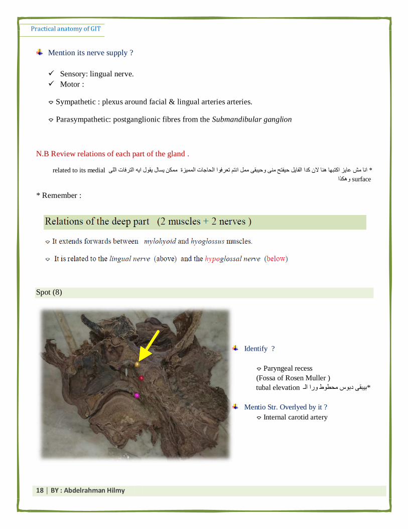
Spot (8)
Identify ?
⌔ Paryngeal recess
(Fossa of Rosen Muller )
tubal elevationص يحطط را انـ ثيجم دث *
Mentio Str. Overlyed by it ?
⌔ Internal carotid artery
Practical anatomy of GIT
19 │ BY : Abdelrahman Hilmy
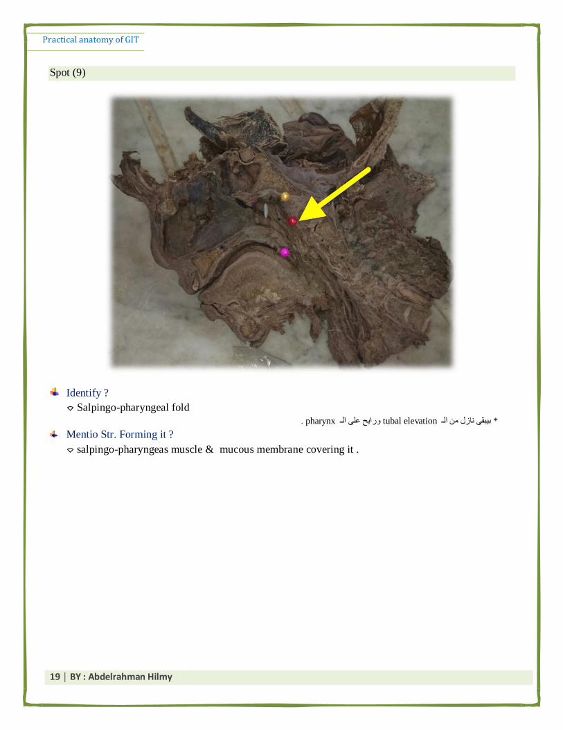
Spot (9)
Identify ?
⌔ Salpingo-pharyngeal fold
. pharynxرايح ػه انـ tubal elevationثيجم بسل ي انـ *
Mentio Str. Forming it ?
⌔ salpingo-pharyngeas muscle & mucous membrane covering it .

Practical anatomy of GIT
20 │ BY : Abdelrahman Hilmy
Spot (10)
Identify ?
⌔ Pharyngeal opening of the auditory tube
Give the site of the opening of other end of this opening ? mention its function ?
⌔ Tympanic cavity of the middle ear
⌔ equalize the pressure on both sides of the ear drum
Name the pointed str. No. 1 ?
⌔ Tubal elevation
Mention the Strs. Forming it ?
1- cartilaginous end of the auditory tube & Mucosa covering it .
2- Tubal tonsil (lymphoid tissue).

Practical anatomy of GIT
21 │ BY : Abdelrahman Hilmy
Spot (11)
1- Bed of palatine tonsil
2- palatoglossal arch
3- palatopharyngeal arch
Mention the Str. Forming No. 2 & 3 ?
1- Palatoglossus arch formed by : palatoglossus muscle & mucosa covering it
2- palatopharyngeus arch formed by : palatopharyngeus & mucosa covering it
Any theoretical Q may be asked about palatine tonsil :
A - arterial supply :
1- Tonsillar artery (from facial artery).
2- Ascending palatine artery (from facial artery).
3- Lingual artery (from external carotid artery).
4- Ascending pharyngeal artery (from external carotid artery).

Practical anatomy of GIT
22 │ BY : Abdelrahman Hilmy
2- nerve supply :
1.Glossopharyngeal nerve. 2. Lesser palatine nerve.
3- Venous drainage :
⌔ Paratonsillar vein
4- Lymphatic Drainage :
⌔Deep cervical lymph nodes (mainly the jugulo-digastric nodes).
⌔ See its relations in page 43.
Spot (12)
Identify ?
⌔ Pyriform fossa
Mention its nerve supply ?
⌔internal laryngeal nerve.
Give its medial boundary ?
⌔ Aryepiglottic fold of the larynx
Give its lateral boundary ?
⌔ Thyrohyoid membrane above and the
lamina of the thyroid cartilage below.

Practical anatomy of GIT
23 │ BY : Abdelrahman Hilmy
Spot (13)
Identify ?
⌔ Genioglossus muscle
Mention its nerve supply ?
⌔ Hypoglossa lverve
Give its action ?
⌔ it Protrudes the tongue . & prevent drop of the tongue
Gie its origin ?
⌔ Superior genial tubercle of the mandible.
Practical anatomy of GIT
24 │ BY : Abdelrahman Hilmy
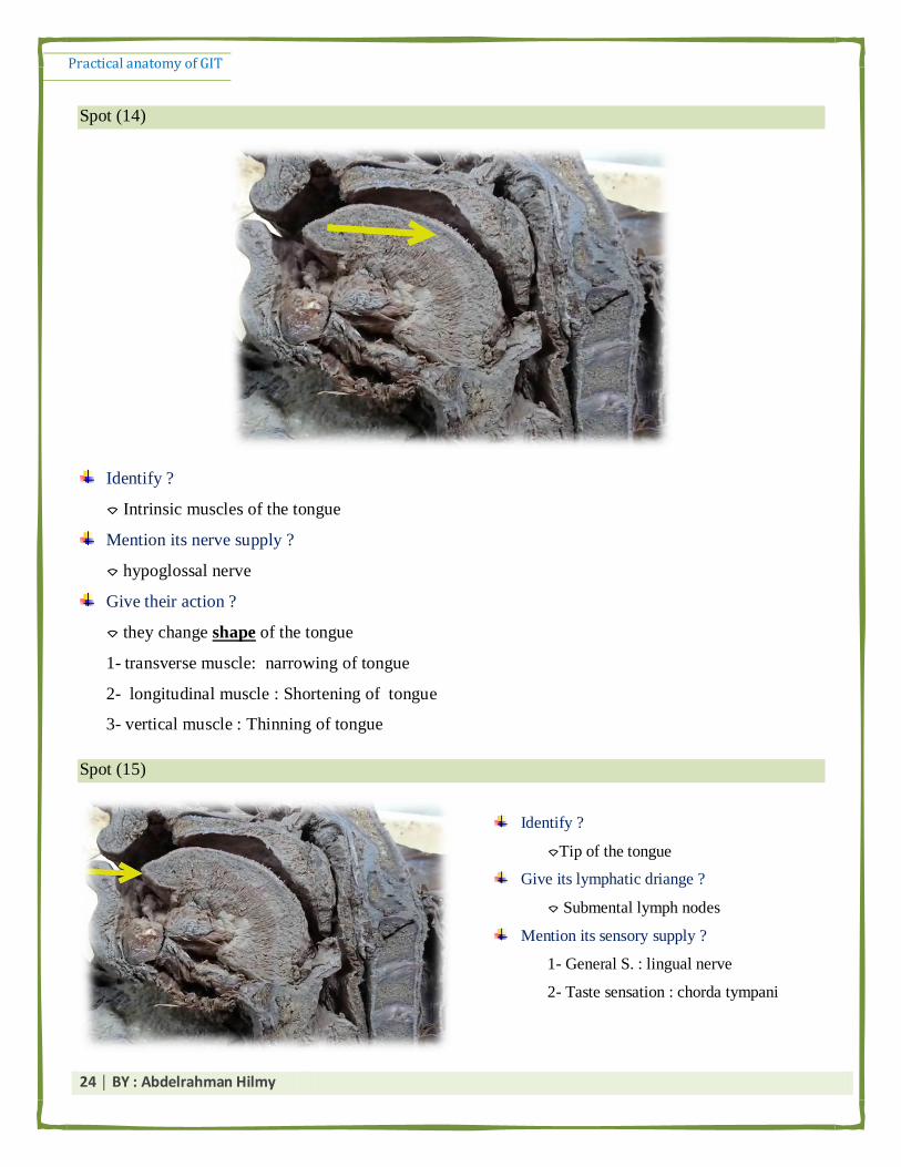
Spot (14)
Identify ?
⌔ Intrinsic muscles of the tongue
Mention its nerve supply ?
⌔ hypoglossal nerve
Give their action ?
⌔ they change shape of the tongue
1- transverse muscle: narrowing of tongue
2- longitudinal muscle : Shortening of tongue
3- vertical muscle : Thinning of tongue
Spot (15)
Identify ?
⌔Tip of the tongue
Give its lymphatic driange ?
⌔ Submental lymph nodes
Mention its sensory supply ?
1- General S. : lingual nerve
2- Taste sensation : chorda tympani
Practical anatomy of GIT
25 │ BY : Abdelrahman Hilmy
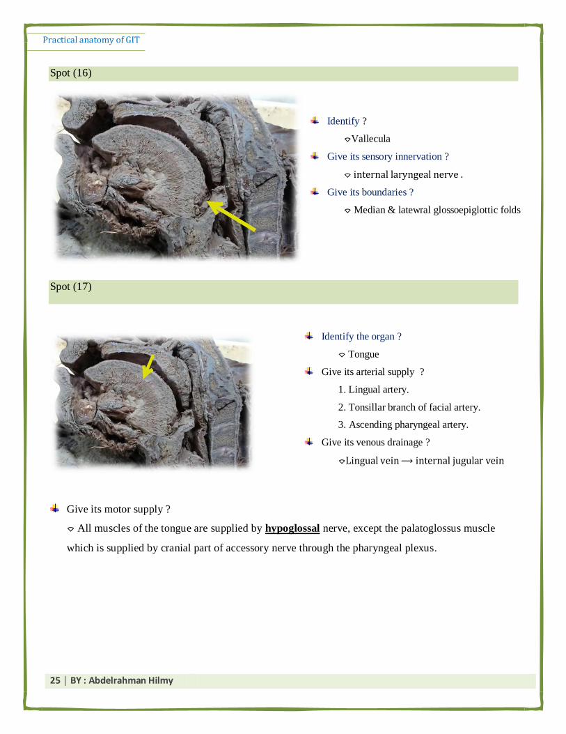
Spot (16)
Spot (17)
Give its motor supply ?
⌔ All muscles of the tongue are supplied by hypoglossal nerve, except the palatoglossus muscle
which is supplied by cranial part of accessory nerve through the pharyngeal plexus.
Identify ?
⌔Vallecula
Give its sensory innervation ?
⌔ internal laryngeal nerve .
Give its boundaries ?
⌔ Median & latewral glossoepiglottic folds
Identify the organ ?
⌔ Tongue
Give its arterial supply ?
1. Lingual artery.
2. Tonsillar branch of facial artery.
3. Ascending pharyngeal artery.
Give its venous drainage ?
⌔Lingual vein ⟶ internal jugular vein

Practical anatomy of GIT
26 │ BY : Abdelrahman Hilmy
Spot (18)
Identify ?
⌔ Soft palate
Mention its content ?
⌔ Palatine aponeurosis.
⌔ Muscles.
⌔ Nerves.
⌔ Vessels.
⌔ Lymphoid tissue.
Enumerate muscles forming it ? which one is intrinsic ?
- Tensor palati
- Tensor tympani
- palatoglossus
- palatopharyngeus
- musculus uvulae (intrinsic one )
Name its motor innervation ?
⌔ All the muscles are supplied by the cranial part of accessory nerve except the tensor palati
muscle which is supplied by the mandibular nerve.
Practical anatomy of GIT
27 │ BY : Abdelrahman Hilmy
Give its sensory innervation ?
1- General sensation :
⌔ Lesser palatine nerve.
⌔ Glossopharyngeal nerve
2- Taste sensation ⟶ lesser palatine nerves.
Give its parasympathetic innervation ? or its Secretomotor innervation ?
The facial nerve ⟶ greater petrosal nerve ⟶ sphenopalatine ganglion. ⟶ lesser palatine nerves to
palatine glands
Mention its arterial supply ?
Mention 2 arteries supplying it ? Give their origin ?
1. Greater palatine artery ⟶ branch from the maxillary artery (3rd
part ).
2. Ascending palatine artery⟶ branch from the facial artery.
Mention its venous drianage ?
⌔ pterygoid + pharyngeal plexuses of veins.
Mention its Lymphatic Drainage ?
⌔ upper deep cervical & retropharyngeal lymph nodes
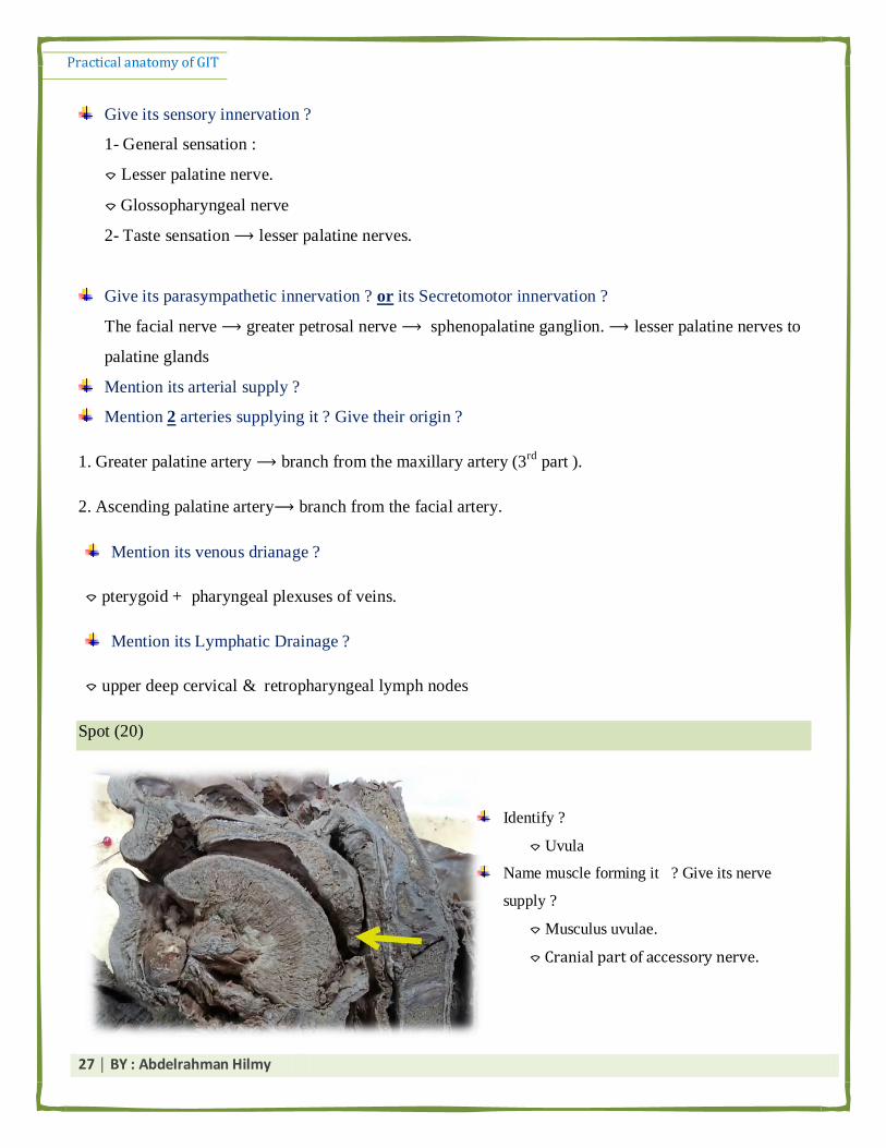
Spot (20)
Identify ?
⌔ Uvula
Name muscle forming it ? Give its nerve
supply ?
⌔ Musculus uvulae.
⌔ Cranial part of accessory nerve.
Practical anatomy of GIT
28 │ BY : Abdelrahman Hilmy
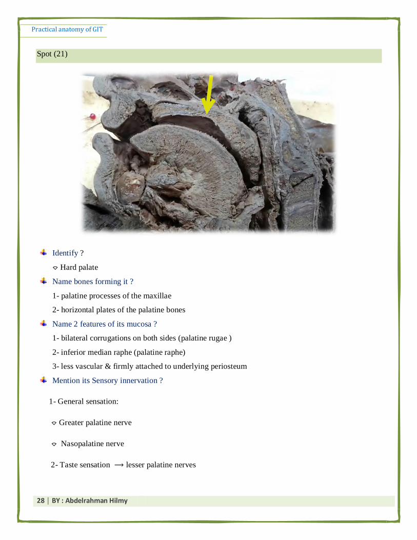
Spot (21)
Identify ?
⌔ Hard palate
Name bones forming it ?
1- palatine processes of the maxillae
2- horizontal plates of the palatine bones
Name 2 features of its mucosa ?
1- bilateral corrugations on both sides (palatine rugae )
2- inferior median raphe (palatine raphe)
3- less vascular & firmly attached to underlying periosteum
Mention its Sensory innervation ?
1- General sensation:
⌔ Greater palatine nerve
⌔ Nasopalatine nerve
2- Taste sensation ⟶ lesser palatine nerves

Practical anatomy of GIT
29 │ BY : Abdelrahman Hilmy
● Don’t forget :
▪ Hard = Greater palatine N. + Nasopalatine N.
▪ Soft = Lesser palatine N. + Glossopharyngeal .
Give its lymphatic drainage ?
⌔ submandibular lymph nodes.
Arterial supply & venous Drianage ⟶ the same as soft palate .
Spot (22)
Spot (23)
Identify ?
⌔ Posterior 1/3 of tongue
Or ( pharyngeal part )
Mention its nerve supply ?
⌔ General sensation & Taste sensation :
By Glossopharyngeal nerve
Name a characteristic feature of
its dorsal surface ?
⌔ Lingual tonsils
Identify ?
⌔ Anterior 2/3 of tongue
Or ( Oral part of the tongue )
Mention its nerve supply ?
⌔ General sensation : By Lingual nerve
⌔ Taste sensation : by Chorda tympani
By Glossopharyngeal nerve
Name a characteristic feature of its dorsal surface ?
⌔ Lingual papillae (Fungiform & filiform & Vallate)

Practical anatomy of GIT
30 │ BY : Abdelrahman Hilmy
Name the Features of its under surface ?
⌔ Attached to floor of mouth by frenulum linguae
⌔ on each side (from medil to lateral ) : lingual artery & lingual nerve & deep lingual vein
Stomach & liver
Spot (1)
Identify the pointed str. ?
Liver , anterior surface
Mention the str. Attached to pointed area ? Give its content ?
Falciform ligament
Ligamentum teres (round ligament of the liver )
Mention its arterial supply ?
1- hepatic artery : from celiac trunk
2- portal vein : formed by union of splenic vein & superior mesenteric vein .
Both of them devides into rt & lt branches

Practical anatomy of GIT
31 │ BY : Abdelrahman Hilmy
Mention its lymphatic drainage ?
portal lymph nodes then into the coeliac lymph nodes
Except bare area of the liver : subphrenic lymph nodes, or Posterior mediastinal lymph nodes.
Mention its venous drainage ?
Right & middle & left hepatic veins ------> IVC
Enumerate its peritoneal ligaments (connections )?
1- Falciform ligament.
2- Upper layer of coronary ligament.
3- Lower layer of coronary ligament.
4- Right triangular ligament.
5- Left triangular ligament.
6- Lesser omentum.
Mention its Embryonic ligaments ? Give the origin of each one ?
1- Ligamentum teres ⟶ obliterated left umblical vein
2- Ligamentum venosum⟶ obliterated ductus venosus
Enumerate its bare areas ?
1- Bare area
2- Groove for IVC
3- Porta hepatic 4- Fossa of gall bladder
5- Fissures for ligamentum teres and for ligamentum venosum
يك يطهت اري ثض يغ كه . ligaments or bare areasطاء ف طؤال انـ *

Practical anatomy of GIT
32 │ BY : Abdelrahman Hilmy
Spot (2)
Identify ?
Bare area of the liver
Mention its boundaries ?
⌔ base : groove for IVC
⌔ apex : right triangular ligament
⌔ sides : the two layers of coronary ligament.
Mention its realtion ?
1-Diaphragm
2- Supra renal gland
Mention its lymphatic drainage ?
subphrenic lymph nodes or Posterior mediastinal lymph nodes
Practical anatomy of GIT
33 │ BY : Abdelrahman Hilmy
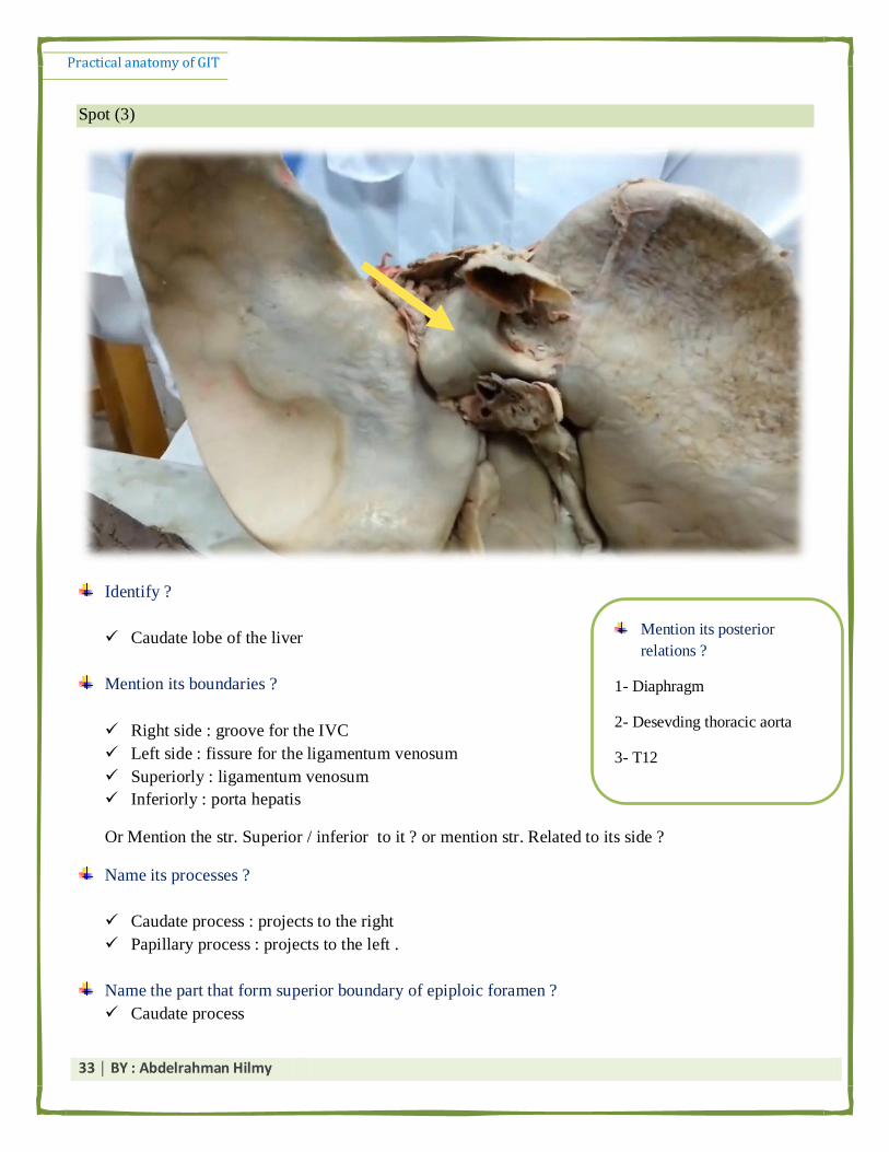
Spot (3)
Identify ?
Caudate lobe of the liver
Mention its boundaries ?
Right side : groove for the IVC
Left side : fissure for the ligamentum venosum
Superiorly : ligamentum venosum
Inferiorly : porta hepatis
Or Mention the str. Superior / inferior to it ? or mention str. Related to its side ?
Name its processes ?
Caudate process : projects to the right
Papillary process : projects to the left .
Name the part that form superior boundary of epiploic foramen ?
Caudate process
Mention its posterior
relations ?
1- Diaphragm
2- Desevding thoracic aorta
3- T12

Practical anatomy of GIT
34 │ BY : Abdelrahman Hilmy
Spot (4)
Identify ?
Porta hepatis
Name the Str. That passing through it ?
a. Hepatic ducts: anterior in position.
b. Hepatic artery: intermediate in position.
c. Portal vein: posterior in position.
d. Lymphatics & sympathetic nerves
Mention str. Attached to it ?
Lesser omentum.
Mention its boundaries ?
Anteriorly : quadrate lobe
Posteriorly : caudate lobe and process

Practical anatomy of GIT
35 │ BY : Abdelrahman Hilmy
Spot (5)
Identify ?
Fissure for ligamentum venosum
Name Str. Related ? give its embryological origin ?
ligamentum venosum >> obliterated ductus venosus .
Spot (6)

Practical anatomy of GIT
36 │ BY : Abdelrahman Hilmy
Identify ?
Quadrate lobe of the liver
Mention its boundaries ?
Anteriorly : inferior border of liver
Posteriorly : porta hepatis
Right side : gall bladder fossa
Left side : fissure for ligamentum teres
Give its relations ? (TPL)
Transverse colon (anteriorly)
Pylorus& 1st part of duodenum (middle )
Lesser omentum (posteriorly).
The order is imp. as the Q may be : give the relations from upward to downward or the reverse .
Spot (7)
Identify Str. No 1 & 2 & 3 ?
1- ligamentum teres
2- gall bladder
3- gastric impression

Practical anatomy of GIT
37 │ BY : Abdelrahman Hilmy
Give the embryological origin of Str. No 1 ?
Obliterated left umblical vein
Mention the parts of Str. No 2 ?
Fundus + Body + Neck
Mention the Blood supply of Str. No. 2 ?
Arterial supply : cystic artery from right branch of hepatic artery
Venous drainage : cystic vein which drains to the right branch of portal vein.
Mention the surface anatomy of Str. No. 2 ? (V-IMp)
Fundus of gall bladder: tip of Right 9th costal cartilage
يؼب ا طؤال ػبو . identifyيك يحظ انذثص ف ا حزخ يظبل impressions* د صرح انـ

Practical anatomy of GIT
38 │ BY : Abdelrahman Hilmy
Spot (8)
Identify ?
Portal vein
Mention its origin ? (how & where ?)
It is formed by union of the superior mesenteric and splenic veins behind the neck of pancreas
and in front of IVC
Name 2 of its tributaries ?
1- Superior mesenteric vein.
2- Splenic vein
3- Right gastric vein.
4- Left gastric vein.
5- Paraumbilical vein (in the left branch).
6- Cystic vein (in the right branch).

Practical anatomy of GIT
39 │ BY : Abdelrahman Hilmy
Spot (9)
Spot (10)
Identify the pointed structure?
Lesser curvature of stomach
Mention str. Attached to it ?
Lesser omentum
Mention str. Related to it ?
Right & left gastric vessels .
Identify the pointed structure?
Greater curvature of stomach
Mention str. Attached to it ?
Greater omentum
Gastrophrenic ligament
Gastrosplenic ligament
Mention str. Related to it ?
Right & left gastroepiploic vessels .
Practical anatomy of GIT
40 │ BY : Abdelrahman Hilmy
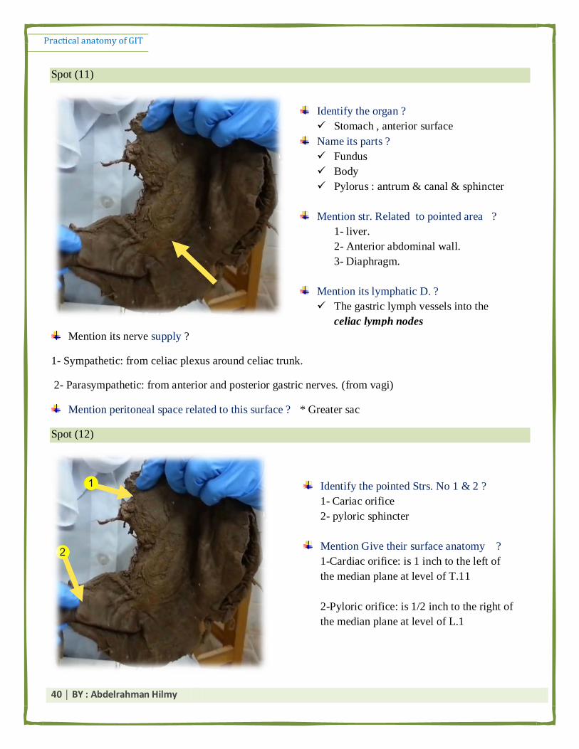
Spot (11)
Mention its nerve supply ?
1- Sympathetic: from celiac plexus around celiac trunk.
2- Parasympathetic: from anterior and posterior gastric nerves. (from vagi)
Mention peritoneal space related to this surface ? * Greater sac
Spot (12)
Identify the organ ?
Stomach , anterior surface
Name its parts ?
Fundus
Body
Pylorus : antrum & canal & sphincter
Mention str. Related to pointed area ?
1- liver.
2- Anterior abdominal wall.
3- Diaphragm.
Mention its lymphatic D. ?
The gastric lymph vessels into the
celiac lymph nodes
Identify the pointed Strs. No 1 & 2 ?
1- Cariac orifice
2- pyloric sphincter
Mention Give their surface anatomy ?
1-Cardiac orifice: is 1 inch to the left of
the median plane at level of T.11
2-Pyloric orifice: is 1/2 inch to the right of
the median plane at level of L.1

Practical anatomy of GIT
41 │ BY : Abdelrahman Hilmy
V- imp : Structures forming stomach bed : (relations of posterior surface of stomach)
4 horizontal structures :
- Body of pancreas
- Splenic artery
- Transverse colon
-Transvesre mesocolon
يظبل ػ posterior surface of stomachا يحظ انذثص ف انـ mention 4 str forming stomach bed* يبيب يج انظؤال يجبػز
. posterior surfaceيبنـ anteriorـ انزيالػش اى حبجخ ظجظ انؼيخ ػهيب كيض ػهؼب ؼزف ان
:انه pylorusيك يظبل ػه اجشاء انـ identify pointed partيحظ دثص ف ا جزخ يظبل *
antrum & canal & Sphincter
4 vertical Str. :
- left kidney
-left suprarenal gland
- left crus od diaphragm
- spleen

Practical anatomy of GIT
42 │ BY : Abdelrahman Hilmy
Spot (13)
Spot (14)
Identify the pointed part ?
⌔ Posterior posterior surface of stomach
Mention 4 Str. Forming its bed ?
( See above )
Name its peritoneal covering ?
⌔ its covered by peritoneum of lesser sac
N.B if you asked about peritoneal covering of
anterior surface the answer will be : peritoneal of
greater sac .
Mention peritoneal space related to this
surface ? Lesser sac (Omental bursa)
Identify the pointed part ?
⌔ Fundus of the stomach
Mention its peritoneal connections ?
1- Gastro-phrenic ligament.
2- Gastro-splenic ligament .
Name its arterial supply ? Origin ?
⌔ Short Gastric arteries
⌔ From : splenic artery .
Practical anatomy of GIT
43 │ BY : Abdelrahman Hilmy
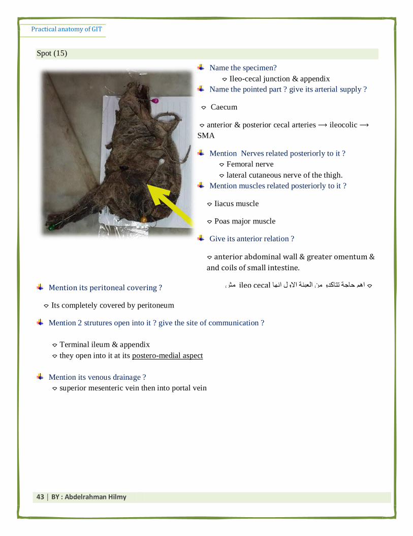
Spot (15)
Mention its peritoneal covering ?
⌔ Its completely covered by peritoneum
Mention 2 strutures open into it ? give the site of communication ?
⌔ Terminal ileum & appendix
⌔ they open into it at its postero-medial aspect
Mention its venous drainage ?
⌔ superior mesenteric vein then into portal vein
Name the specimen?
⌔ Ileo-cecal junction & appendix
Name the pointed part ? give its arterial supply ?
⌔ Caecum
⌔ anterior & posterior cecal arteries ⟶ ileocolic ⟶
SMA
Mention Nerves related posteriorly to it ?
⌔ Femoral nerve
⌔ lateral cutaneous nerve of the thigh.
Mention muscles related posteriorly to it ?
⌔ Iiacus muscle
⌔ Poas major muscle
Give its anterior relation ?
⌔ anterior abdominal wall & greater omentum &
and coils of small intestine.
يغ ileo cecal اى حبجخ رزبكذ ي انؼيخ االل اب ⌔
stomach .

Practical anatomy of GIT
44 │ BY : Abdelrahman Hilmy
Spot (16)
Enumerate its variant positions ? what is the most frequent one ?
1. Retrocecal (65%) ⟶ Most frequent one .
2. Pelvic (30%)
3. Subcecal (3%)
4. Pre or post- ileal (2%)
Mark the surface anatomy of its base ? ( McBurney’s point )
⌔ is represented by a point at the junction of lateral 1/3rd and medial 2/3rd of a line connecting
anterior superior iliac spine (ASIS) and the umbilicus. (Spino-umblical line)
Mention its sensory innervation ? Give the site of its refered pain ?
⌔ sympathetic fibers from 10 th thoracic spinal cord segment .
⌔ umbilical region
Identify (yellow arrow) ?
⌔ Vermiform appendix
Name its Mesentry ? Give its content ?
⌔ Mesoappendix
⌔ appendicular artery in its free border
Name its arterial supply ? Origin ?
⌔ appendicular artery from the posterior caecal
artery from ileocolic artery ⟶ SMA
Mention its venous drainage ?
⌔ superior mesenteric Vein
Mention its Frequent site ?
⌔ Retrocecal in postion

Practical anatomy of GIT
45 │ BY : Abdelrahman Hilmy
Spot (17)
Mention 3 of its chareteristic features that distinguish it from small intestine ? (Leading Q.)
1- taenia coli
2- sacculations
3- Appendices epiploicae
Mention muscles posterior to it ?
1- transverses abdominis
2- quadratus lumborum
3- iliacus
Mention Nerves posterior to it ?
1- iliohypogastric N.
2- ilioinguinal N.
3- lateral cutaneous of the thigh .
The viscera posterior to it >>> right kidney . & ant. Relation : Coils of small intestine + greater omentum
Identify ?
⌔ Ascneding colon
Mention its peritoneal Covering ?
⌔ it is covered by peritoneum from
anterior & its sides only .
Name its arterial supply ? Origin ?
1- Ileocolic artery
2- Right Colic artery
* From superior mesenteric artery.
Where it terminates and how ?
* it ends just below the liver, as the right colic
(hepatic) flexure.

Practical anatomy of GIT
46 │ BY : Abdelrahman Hilmy
Spot (18)
Spot (19)
Identify pointed Str. ?
⌔ Ileum
Name the attachment of root of its mesentery ? & its length ?
⌔ from duodeno-jejunal flexure to iliocecal junction.
⌔ 6inches
Mention its arterial supply ?
⌔ ileal branches of SMA.
Identify ?
⌔ Terminal ileum
Mention its arterial supply ?
⌔ Ileal branches oF SMA
Practical anatomy of GIT
47 │ BY : Abdelrahman Hilmy
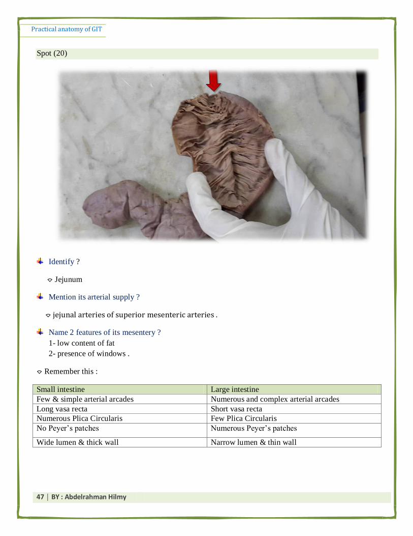
Spot (20)
Identify ?
⌔ Jejunum
Mention its arterial supply ?
⌔ jejunal arteries of superior mesenteric arteries .
Name 2 features of its mesentery ?
1- low content of fat
2- presence of windows .
⌔ Remember this :
Small intestine Large intestine
Few & simple arterial arcades Numerous and complex arterial arcades
Long vasa recta Short vasa recta
Numerous Plica Circularis Few Plica Circularis
No Peyer’s patches Numerous Peyer’s patches
Wide lumen & thick wall Narrow lumen & thin wall
Practical anatomy of GIT
48 │ BY : Abdelrahman Hilmy
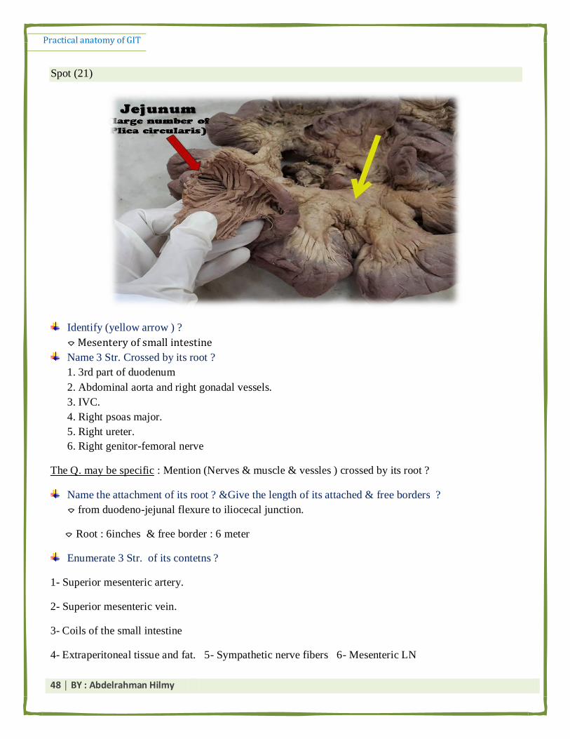
Spot (21)
Identify (yellow arrow ) ?
⌔ Mesentery of small intestine
Name 3 Str. Crossed by its root ?
1. 3rd part of duodenum
2. Abdominal aorta and right gonadal vessels.
3. IVC.
4. Right psoas major.
5. Right ureter.
6. Right genitor-femoral nerve
The Q. may be specific : Mention (Nerves & muscle & vessles ) crossed by its root ?
Name the attachment of its root ? &Give the length of its attached & free borders ?
⌔ from duodeno-jejunal flexure to iliocecal junction.
⌔ Root : 6inches & free border : 6 meter
Enumerate 3 Str. of its contetns ?
1- Superior mesenteric artery.
2- Superior mesenteric vein.
3- Coils of the small intestine
4- Extraperitoneal tissue and fat. 5- Sympathetic nerve fibers 6- Mesenteric LN

Practical anatomy of GIT
49 │ BY : Abdelrahman Hilmy
Spot (22)
Identify ?
⌔ Uncinate process of pancreas
Name the Strs. Anterior to it ?
⌔ Superior mesenteric artery & Vein
Name the Str. Posterior to it ?
⌔ abdominal aorta .
Give its arterial supply ?
⌔ Superior, inferior pancreatico-duodenal arteries
Mention its lymphatic Drainage ?
⌔ superior mesenteric lymph nodes

Practical anatomy of GIT
50 │ BY : Abdelrahman Hilmy
Spot (23)
Identify ?
⌔ Head of pancreas
Mention its arterial supply ? Give the orgin ?
⌔ Superior, inferior pancreatico-duodenal arteries
1- Superior pancreatico-duodenal artery : from gastroduodenal >> hepatic a.
2- inferior pancreatico-duodenal arterys: from SMA
Mention its lymphatic Draingae ?
⌔ upper part : coeliac lymph nodes
⌔ lower part : superior mesenteric lymph nodes
N.B : you must review the relations of each part of pancreas . the Q may be :
Mention vessels related meial to it ?
⌔ Superior, inferior pancreatico-duodenal arteries
Mention its posterior relation ?
⌔IVC, renal veins and common bile duct.
● the anterior relation is >> transverse colon

Practical anatomy of GIT
51 │ BY : Abdelrahman Hilmy
Spot (24)
Identify ?
⌔ Neck of the pancreas
Mention its posterior relation ?
⌔ formation of portal vein from splenic and superior mesenteric veins.
ن طبل احزيبط ثزض اي انـ * duodenal junction-gastroحزجم anterior relation ن جبة انؼيخ د حيجم جبيجب يخصؽ ػهؼب انظؤال دا
Spot (25)
اللى بيطلع من تحتها وكمان لو SMAمن الـ neckبعلم الـ .
duodenumاكيد حيبقى حاطط الدبوس حنب الـ bodyقاصد الـ
حيحط الدبوس فى النص وطبيعة bodyع طوول ولو قاصد الـ
ان شاء هللا . يعنى السؤال اللى بعده بتبين برضو مفهاش لغبطة

Practical anatomy of GIT
52 │ BY : Abdelrahman Hilmy
Identify ?
⌔ Body of the pancreas
Mention its surfaces ? & Borders ?
⌔ Surfaces : anterior, posterior and inferior
⌔ Borders : anterior, superior and inferior
Mention the Strs. Attached to its anterior border ?
⌔ transverse mesocolon
⌔ greater omentum.
Mentios Str. Related to its upper border ? Give a characteristic feature of it ?
⌔ splenic artery & it has a very tourtous course .
Mention 2 branches of the artery running on its upper border ?
1- left gastro-epiploic artery .
2- short gastric artreis
3- arteria pancreatica magna .
Mention the vessels related to its posterior surface ?
1- Aorta and origin of sup. mesenteric artery
2- Splenic and left renal vein
Mention the muscles related to its posterior surface ?
1- Left psoas major
2- Left crus of diaphragm
Mention the Viscera related to its posterior surface ?
1- Left kidney
2- Left supra renal gland
Mention the nervous str . related to its posterior surface ?
⌔ Left sympathetic chain
● Relations of inferior surface (Rare but may come): duodeno-jejunal flexure & loops of jejunum
and end of transverse colon (from right to left).
● Relations of Anterior surface : stomach, separated from it by the lesser sac.
Practical anatomy of GIT
53 │ BY : Abdelrahman Hilmy
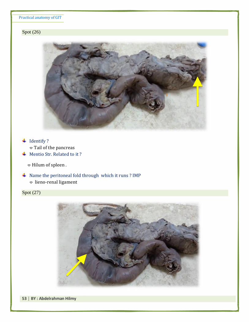
Spot (26)
Identify ?
⌔ Tail of the pancreas
Mentio Str. Related to it ?
⌔ Hilum of spleen .
Name the peritoneal fold through which it runs ? IMP
⌔ lieno-renal ligament
Spot (27)
Practical anatomy of GIT
54 │ BY : Abdelrahman Hilmy
Identify the organ ?
⌔ Duodenum
Mention its arterial supply ? Origin ?
1. Supra-duodenal artery: from the hepatic artery proper .
2. Superior pancreatico-duodenal artery: from gastro-duodenal .
3. Inferior pancreatico-duodenal artery: from superior mesenteric artery.
Mention its beginning ?
⌔ It begins at the pylorus, 1/2 an inch to the right of the median plane at the level of L1
(transpyloric plane).
Mention its termination ?
⌔ at the dudeno-jejunal flexure , one inch to the left of median plane .
What is the most mobile part of this organ ? Why ?
⌔ the first part , because it is covered by peritoneum anteriorly and posteriorly.
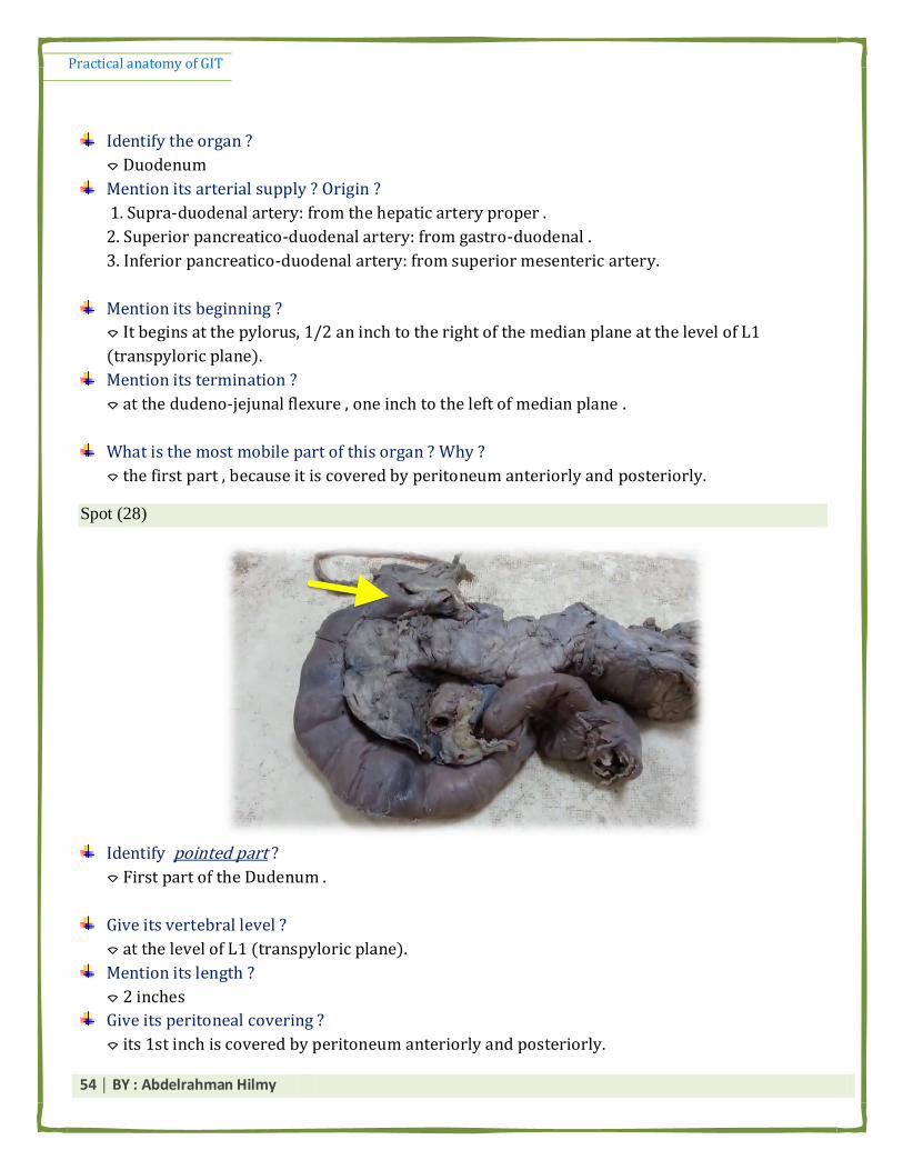
Spot (28)
Identify pointed part ?
⌔ First part of the Dudenum .
Give its vertebral level ?
⌔ at the level of L1 (transpyloric plane).
Mention its length ?
⌔ 2 inches
Give its peritoneal covering ?
⌔ its 1st inch is covered by peritoneum anteriorly and posteriorly.

Practical anatomy of GIT
55 │ BY : Abdelrahman Hilmy
N.B review the relations of each part of the duodnum first .
Mention the Str. Superior to it ?
⌔ Epiploic foramen .
Mention its anterior relations ?
1- quadrate lobe of the liver
2- gallbladder.
Mention its posterior relations ?
1- neck of the pancreas
2- portal vein
3- bile duct
4- gastro-duodenal artery.
Name the artery posterior to it ? Give its origin ? Branches?
⌔ gastro-duodenal artery
⌔ it arises from Hapatic artery .
⌔ Branches : 1- Right gastroepiploic a.
2- Superior pancreatico-duodenal a.
Name the Duct posterior to it ? how its formed ? give its termination ?
⌔ Common bile duct
⌔ it is formed by the union of custic duct of gall bladder & common hepatic duct
⌔ it terminates by the union with common pancreatic duct to form Ampulla of vater wich
opens into major duodenal papilla .
طجؼب يغ حيظبل كم االطئهخ د انفكزح ثض اب جغ انظز كيض زثظ كم االجشاء ثجؼضب ػ ف انظجخد د احب رثطب انـ ⌔
duodenum ثحبضزح انـarterial supply ثحبضزح انـbiliary system .

Practical anatomy of GIT
56 │ BY : Abdelrahman Hilmy
Spot (29)
Identify pointed part ?
⌔ Seconf part of the duodenum
Give its vertebral level ?
⌔ descends vertically from the level of L1 to the level of L3
Mention its length ?
⌔ 3 inches
Mention its peritoneal covering ?
⌔ only covered by peritoneum anteriorly .
Mention its anterior relations ?
⌔ right lobe of the liver & gall bladder & transverse colon & coils of jejunum.
Mentios its posterior relation ?
⌔ hilum of the right kidney
Mention its medial relation ?
⌔ head of pancreas & bile duct & pancreatico –duodenal arteries
Mention vessels related medial to it ?
⌔ Superior & inferior pancreatico –duodenal arteries
Practical anatomy of GIT
57 │ BY : Abdelrahman Hilmy
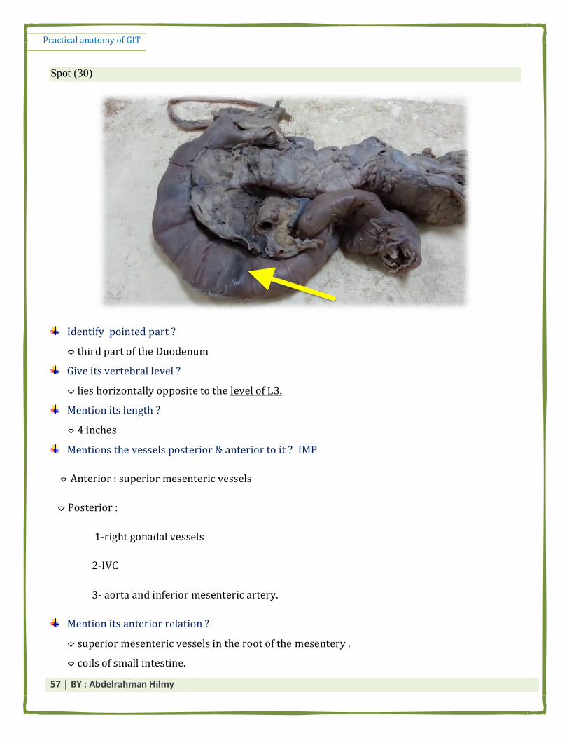
Spot (30)
Identify pointed part ?
⌔ third part of the Duodenum
Give its vertebral level ?
⌔ lies horizontally opposite to the level of L3.
Mention its length ?
⌔ 4 inches
Mentions the vessels posterior & anterior to it ? IMP
⌔ Anterior : superior mesenteric vessels
⌔ Posterior :
1-right gonadal vessels
2-IVC
3- aorta and inferior mesenteric artery.
Mention its anterior relation ?
⌔ superior mesenteric vessels in the root of the mesentery .
⌔ coils of small intestine.

Practical anatomy of GIT
58 │ BY : Abdelrahman Hilmy
Abdominal wall muscles
Spot (1)
Identify ?
⌔ rectus abdominis muscle
Give its action ?
⌔ Flexes the trunk
⌔raises the intra-abdominal pressure.
Give its nerve supply ?
⌔ lower five intercostal verves & subcostal nerve (intercostal nerves 7-11 and subcostal nerve)
Give its origin & insertion ?
Mention the contents of its sheath ?
⌔ Superior & inferior epigastric vessels
⌔ Lower 5 intercostal nerves & Subcostal nerve
⌓ Important : You have to know the Structures forming the anterior & posterior walls of each part of
the sheath (see the Book page 53 & 54 )
For Example : Name the Str. That form the anterior wall of the middle part of its sheath ?
يج فبخذ ثبنب يؼهغ حزؼت ػيخ . * ا طؤال ظز ارد ا

Practical anatomy of GIT
59 │ BY : Abdelrahman Hilmy
ثؼزفى ي ارجب انفيجزس ثزبػزى , يظبنب ا طؤال ظز ػهيى ثض ركشا Muscles of anterior abdominal wallثميذ انـ *
اكزز ػه انزف صجه االكؼ .
⌔ Nerve supply of Transversus abdominis & external oblique & internal oblique :
▪ Intercostal nerves 7-11, subcostal N. ▪ iliohypogastric N.
▪ ilioinguinal nerve
⌔ Action : ▪ Flexes and laterally bends the trunk .
2- External abdominal oblique : fibers runs downward & forwards & medially. ( احذ حطظ ايذ ف جيج)

Practical anatomy of GIT
60 │ BY : Abdelrahman Hilmy
2- internal abdominal oblique : fibers directed upwards, forwards and medially
3 - Transversus abdominis muscle : fibers runs transversely .

Practical anatomy of GIT
61 │ BY : Abdelrahman Hilmy
Spot (2)
Identify ?
⌔ Poas major muscle
Mentio its nerve supply ?
⌔ Lumbar plexus via anterior rami of L1-2-3 nerves.
Give its action ?
⌔ Flexion of the thigh.
⌔Lateral flexion of the trunk.
Give its insertion ?
⌔ By a strong tendon to lesser trochanter of femur
Mention Nerves that appears from its lateral border ?
1- iliohypogastric N.
2- ilioinguinal N.
3- lateral cutaneuos nerve of the thigh
4- Femoral nerve

Practical anatomy of GIT
62 │ BY : Abdelrahman Hilmy
Mention Nerve that emerges from its Medial border?
1- Obturator nerve
2- Lumbosacral trunk
Mention Nerves that emerges from its anterior surface ? give its root value ? its terminal
branches ? area supplied by them ? ( IMP Q )
⌔ genitofemoral nerve
⌔ Root value : L1 & L2 (Ventral rami)
⌔ Terminal brnches :
1- genital branch (enters inguinal canal) : supply cremasteric muscle
2- femoral branch : supply the skin over the femoral triangle.
يك يظبل ػهي ا طؤال ي انه فق anterior surface of poas majorثيجم طبنغ ي انـ genitofemoral nerve* د صرح انـ
دل .

Practical anatomy of GIT
63 │ BY : Abdelrahman Hilmy
حهح يك يظبل ػهيب اطئهخ كزيزح materialػهؼب poas major( اب ركشد ػه انـ 26ؼزف انزف صجهي االكؼ ثزبع كم ػضهخ ي دل )ؽ *
ثض ييؼغ ا انزبيي يك يجا ثصا ػهيى احزيبط. ثبالضبفخ اال اخ يك يزثطب ثبنزفبد س يب ػهب فق كذا .
Vessels & Nerves of Abdomen
Femoral nerve : thick & lateral to poas major (IMP)

Practical anatomy of GIT
64 │ BY : Abdelrahman Hilmy
Identify ?
⌔ Femoral nerve
Give its Root value ? IMP
⌔ posterior division of the L2,3,4 anterior primary rami
How it enters the thigh ?
⌔ behind the inguinal ligament.
Muscle supplied by it ?
⌔ Iliacus
Spot (2)
Identify ?
⌔ Inferior vena cava .
Mention its begining ? (or Mention its formation) ?
⌔ It is formed by union of two common iliac veins at level of 5th lumber vertebra
Mention its termination ?
⌔ it terminates into right atrium
Give the areas drained by it ?
⌔ It Drains the blood from the whole body below the diaphragm

Practical anatomy of GIT
65 │ BY : Abdelrahman Hilmy
Mention its single Tributaries ? (IMP Q )
⌔ Right gonadal vein
⌔ Right suprarenal vein
Mention 2 of its paired Tributaries ?
1- Two common iliac veins
2- Two pairs of lumbar veins: - 3rd, 4th
3- Two renal veins (Rt. & Lt.).
4- Two inferior phrenic veins
5- Two hepatic veins.
Spot (3)
Identify ?
⌔ Abdominal aorta
How it enters the thorax ?
⌔ through aortic hiatus of the Diaphragm at the level of T12.
Mention its termination ?
⌔ It ends by dividing into 2 common iliac arteries opposite the 4th lumbar vertebra.
Practical anatomy of GIT
66 │ BY : Abdelrahman Hilmy
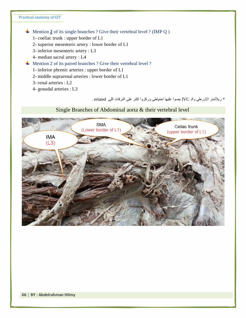
Mention 2 of its single branches ? Give their vertebral level ? (IMP Q )
1- coeliac trunk : upper border of L1
2- superior mesenteric artery : lower border of L1
3- inferior mesenteric artery : L3
4- median sacral artery : L4
Mention 2 of its paired branches ? Give their vertebral level ?
1- inferior phrenic arteries : upper border of L1
2- middle suprarenal arteries : lower border of L1
3- renal arteries : L2
4- gonadal arteries : L3
ب احيبط ركشا اكزز ػه انزفبد انه IVCريالػش االرط انـ * . relatedثصا ػهي
Single Branches of Abdominal aorta & their vertebral level

Practical anatomy of GIT
67 │ BY : Abdelrahman Hilmy
Spot (4)
Identify ?
⌔ Celiac trunk
Give the vertebral level of its origin ?
⌔ upper border of L1
Mention the Str. On each side of it ?
1- Right & left coeliac ganglia
2- Right & left Crura of diaphragm
Give 2 of its Branches ?
1- Left gastric artery
2- Splenic artery
3- Hepatic artery
Mention the parts of the gut supplied by it ? (IMP)
⌔ it supplies Foregut :
1- Esophagus
2- Stomach
3- 1st & ½ of second part od duodenum
4- liver & pancreas & gall bladder
Practical anatomy of GIT
68 │ BY : Abdelrahman Hilmy
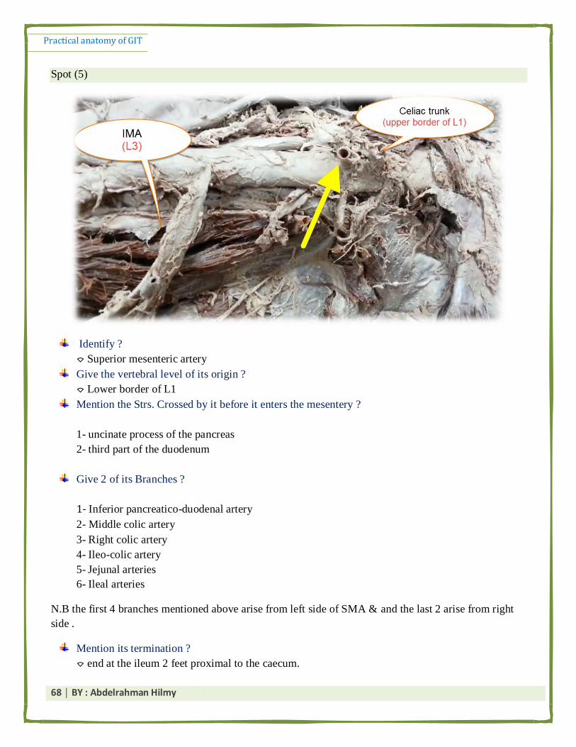
Spot (5)
Identify ?
⌔ Superior mesenteric artery
Give the vertebral level of its origin ?
⌔ Lower border of L1
Mention the Strs. Crossed by it before it enters the mesentery ?
1- uncinate process of the pancreas
2- third part of the duodenum
Give 2 of its Branches ?
1- Inferior pancreatico-duodenal artery
2- Middle colic artery
3- Right colic artery
4- Ileo-colic artery
5- Jejunal arteries
6- Ileal arteries
N.B the first 4 branches mentioned above arise from left side of SMA & and the last 2 arise from right
side .
Mention its termination ?
⌔ end at the ileum 2 feet proximal to the caecum.

Practical anatomy of GIT
69 │ BY : Abdelrahman Hilmy
How ?
⌔ its terminal trunk anastomose with ileal branches of ileo-colic artery .
Mention parts of the gut supplied by it ? (IMP)
⌔ it supplies Midgut :
From the lower ½ of second part of the duodenum till the junction between right 2/3 and left 1/3 of
transverse colon .
Spot (6)
Identify ?
⌔ inferior mesenteric artery .
Mention its origin ?
⌔ It arises from the front of the aorta opposite the third lumber vertebra.
Mention its termination ?
⌔ it terminates as the superior rectal artery at the pelvic brim .
Mention 2 of its branches ?
1- Left colic artery
2- Sigmoid arteries
Mention parts of the gut supplied by it ?
⌔ it supplies the Hindgut :
1- left 1/3 of transverse colon
2- descending colon
3- sigmoid colon & rectum
4- upper ½ of anal canal.

Practical anatomy of GIT
70 │ BY : Abdelrahman Hilmy
Rapid Spots :
Identify ?
1- Esophageal notch
2- Renal imoression
Plus , Any General Q. about the liver we have mentioned above
Spot (2)
Identify ?
1-Epiglottis
2- piriform fossa

Practical anatomy of GIT
71 │ BY : Abdelrahman Hilmy
⌔ Rmember :
▪ Sensory innervation of the pharytnx :
⌔ Mucous membrane of the nasopharynx is supplied by ⟶ Maxillary nerve.
⌔ Mucous membrane of the oropharynx is supplied by ⟶ Glossopharyngeal nerve.
⌔ Mucous membrane of the laryngopharynx is supplied by ⟶ internal laryngeal branch of the
vagus nerve
▪ Arterial supply of the pharynx :
1- Ascending pharyngeal artery
2- Ascending palatine artery
3- Facial and lingual arteries
▪ Motor nerve supply of pharyngeal muscles :
⌔ Through the pharyngeal plexus (by cranial part of accessory nerve) except the stylopharyngeas
muscle which is supplied by the glossopharyngeal nerve
Q : Mention the STr. Forming its wall ?
① Mucosa.
② Inner fibrous coat called ⟶ The pharyngobasilar fascia.
③ Muscle layer ⟶ consisting of the following muscles : (3 constrictors +3 pharyngeus )
④ Outer fibrous layer called ⟶ the buccopharyngeal fascia
Practical anatomy of GIT
72 │ BY : Abdelrahman Hilmy
Q2 : Enumerate Non-Constrictor muscle of the pharynx ?
⌔ Stylopharyngeus
⌔ Palatopharyngeus
⌔ Salpingopharyngeus
.ربد ججشء يظبل ػ انحبجذ د ربو اب يمهزبع فق يغ انظ يك يحظ انذثص ف ا *
⌔ Conjoint tendon is formed by the lower arched fibers of both internal oblique and
transversus abdominis muscles
▪ it narrows the inguinal canal thus prevents passage of the intestine through the inguinal canal.
ع يبريذ ثض يظجغ حبجخ ف انظز ا حزخ لزايخ طزيؼخ كذا GIT* كذا انحذ هلل خهصب ثزاكزيكبل ابري انـ
,, انظزف اب حبد انى انحبجبد انخ ف انظز ػه لذ يمذر ػهؼب يضغ
ػبء هللا ^_^ ثبنزفيك ا


![Cardiovascular System Anatomy Practical [PHL 212].](https://static.fdocuments.net/doc/165x107/5697c01d1a28abf838cd05f5/cardiovascular-system-anatomy-practical-phl-212.jpg)


