PowerPoint Presentation… · PPT file · Web view · 2014-12-25The orbital region is the area of...
56
Kaan Yücel M.D., Ph.D 26.December.2014 Friday Nervous System Anatomy of the
Transcript of PowerPoint Presentation… · PPT file · Web view · 2014-12-25The orbital region is the area of...
Kaan Yücel M.D., Ph.D 26.December.2014 Friday
Nervous System
Anatomy of the
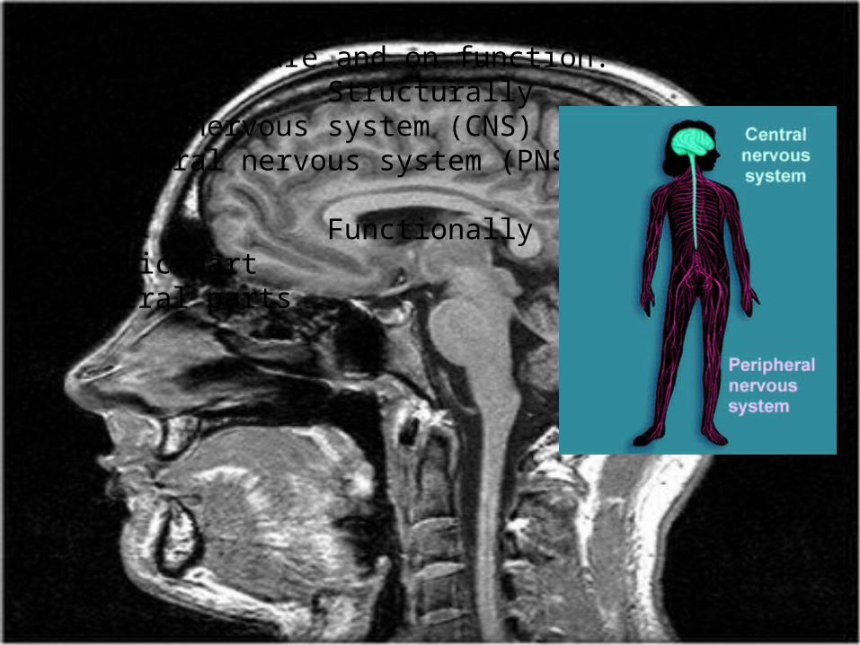
The nervous system can be separated into parts based on structure and on function:
Structurally1) Central nervous system (CNS) 2) Peripheral nervous system (PNS
Functionally3) Somatic part 4) Visceral parts
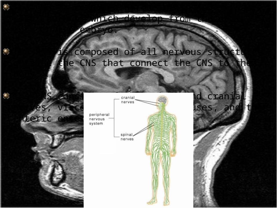
The CNS is composed of the brain and spinal cord, both of which develop from the neural tube in the embryo.
The PNS is composed of all nervous structures outside the CNS that connect the CNS to the body.
The PNS consists of the spinal and cranial nerves, visceral nerves and plexuses, and the enteric enteric system.
CENTRALNERVOUS SYSTEM

Functional subdivisions of the CNSFunctionally, the nervous system can be divided into somatic and visceral parts. The somatic part (soma, from the Greek for "body") innervates structures (skin and most skeletal muscle) derived from somites in the embryo.external environment

The visceral part (viscera, from the Greek for "guts") innervates organ systems in the body and other visceral elements, such as smooth muscle and glands, in peripheral regions of the body. internal environment.
Somatic part of the nervous system consists of:
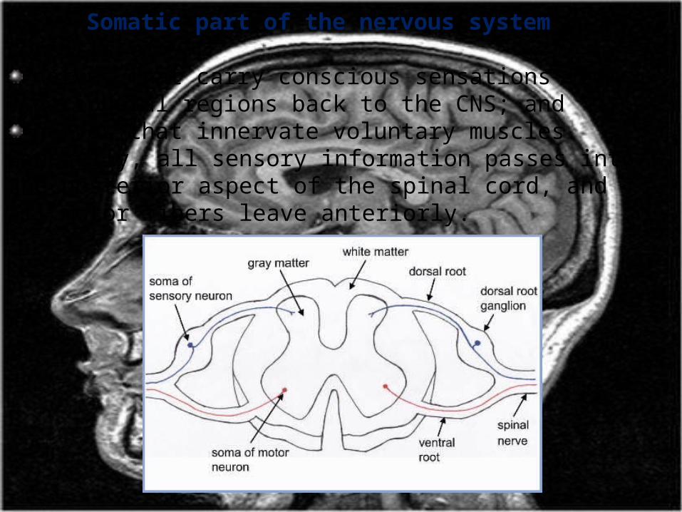
nerves that carry conscious sensations from peripheral regions back to the CNS; andnerves that innervate voluntary muscles.
Generally, all sensory information passes into the posterior aspect of the spinal cord, and all motor fibers leave anteriorly.
Somatic sensory neurons carry information from the periphery into the CNS and are also called somatic sensory afferents or general somatic afferents (GSAs).
The modalities carried by these nerves include temperature, pain, touch, and proprioception.
Proprioception is the sense of determining the position and movement of the musculoskeletal system detected by special receptors in muscles and tendons.
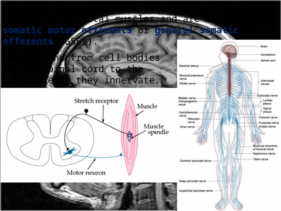
Somatic motor fibers carry information away from the CNS to skeletal muscles and are also called somatic motor efferents or general somatic efferents (GSEs).
They extend from cell bodies in the spinal cord to the muscle cells they innervate.
Dermatomes
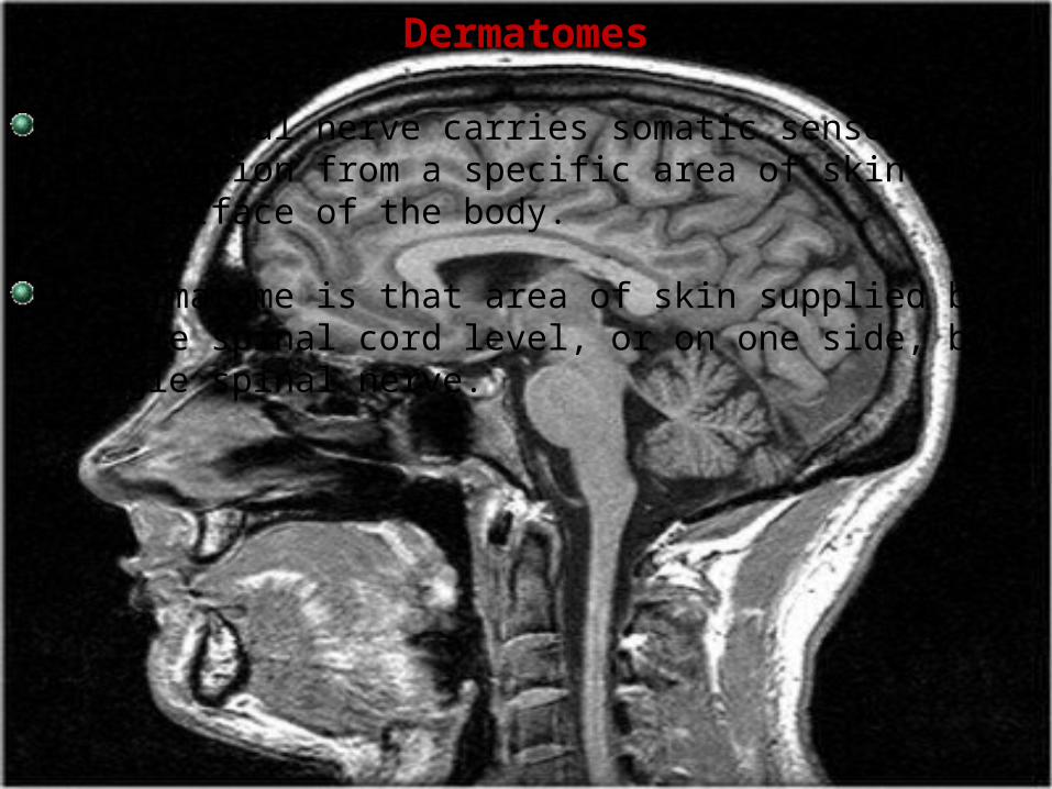
Each spinal nerve carries somatic sensory information from a specific area of skin on the surface of the body.
A dermatome is that area of skin supplied by a single spinal cord level, or on one side, by a single spinal nerve.
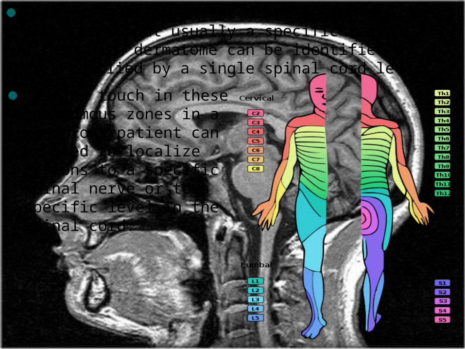
There is overlap in the distribution of dermatomes, but usually a specific region within each dermatome can be identified as an area supplied by a single spinal cord level. Testing touch in these autonomous zones in a conscious patient can be used to localize lesions to a specific spinal nerve or to a specific level in the spinal cord.

MyotomesA myotome is that portion of a skeletal muscle innervated by a single spinal cord level or, on one side, by a single spinal nerve.

Visceral part of the nervous systemConsists of motor and sensory components: sensory nerves monitor changes in the viscera; motor nerves mainly innervate smooth muscle,
cardiac muscle, and glands. The visceral motor component is commonly referred to as the autonomic division of the PNS and is subdivided into sympathetic and parasympathetic parts.
Visceral sensory neurons and their processes are referred to as general visceral afferent fibers (GVAs), and are associated primarily with chemoreception, mechanoreception, and stretch reception.
Visceral motor neurons contain general visceral efferent fibers (GVEs) and provide motor innervation to smooth muscle, cardiac muscle, and glands as part of the autonomic nervous system..
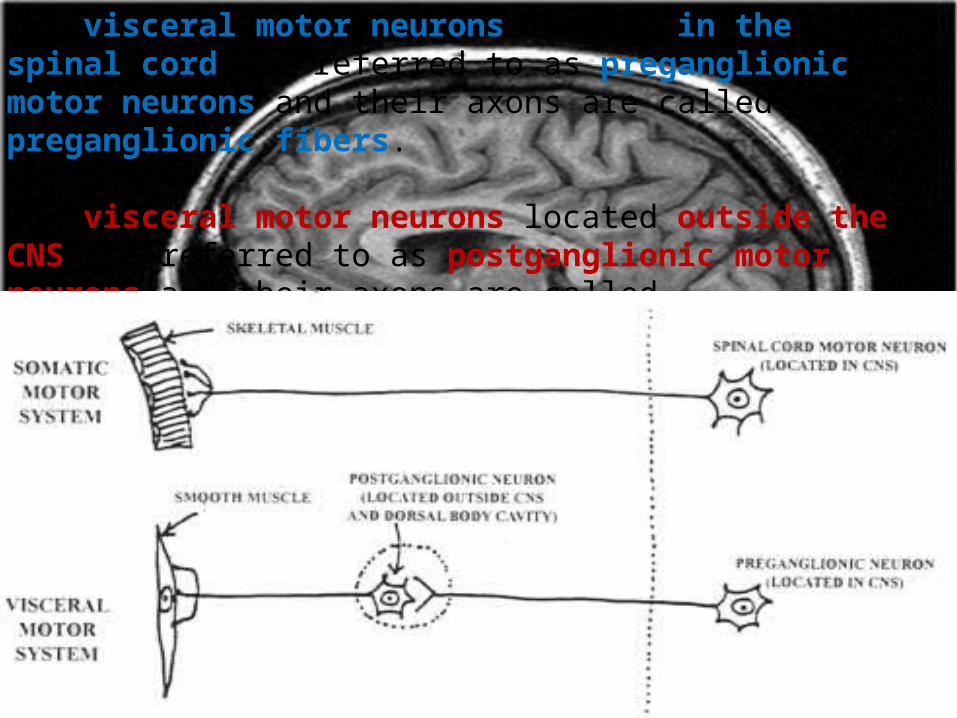
The visceral motor neurons located in the spinal cord are referred to as preganglionic motor neurons and their axons are called preganglionic fibers.
The visceral motor neurons located outside the CNS are referred to as postganglionic motor neurons and their axons are called postganglionic fibers.
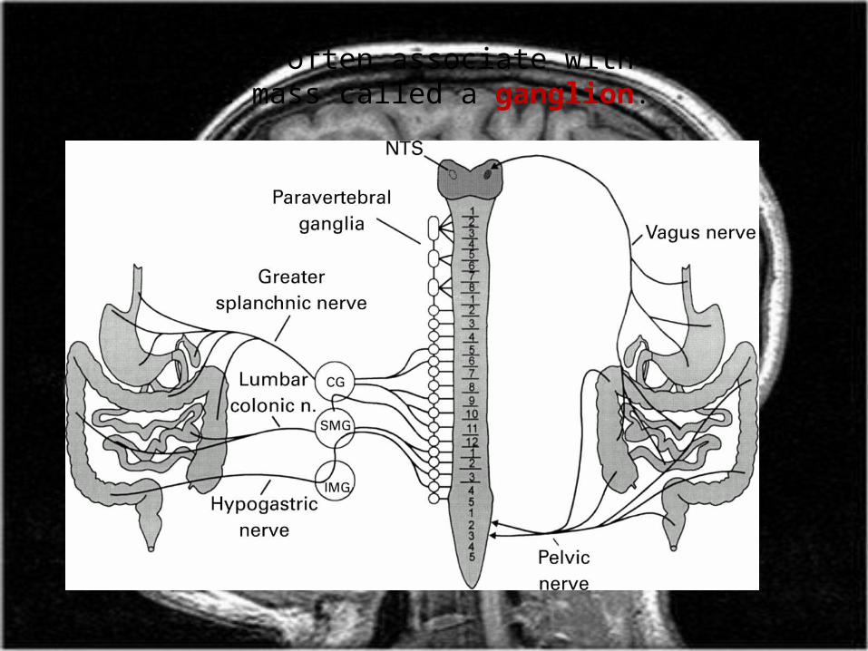
The cell bodies of the visceral motor neurons outside the CNS often associate with each other in a discrete mass called a ganglion.

Visceral sensory and motor fibers enter and leave the CNS with their somatic equivalents.
Visceral sensory fibers enter the spinal cord together with somatic sensory fibers through posterior roots of spinal nerves.
somatic sensory (SS)visceral sensory (VS)visceral motor (VM)somatic motor (SM)
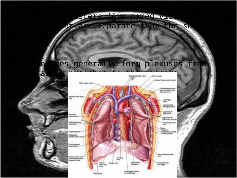
Visceral motor and sensory fibers that travel to and from viscera form named visceral branches that are separate from the somatic branches.
These nerves generally form plexuses from which arise branches to the viscera.
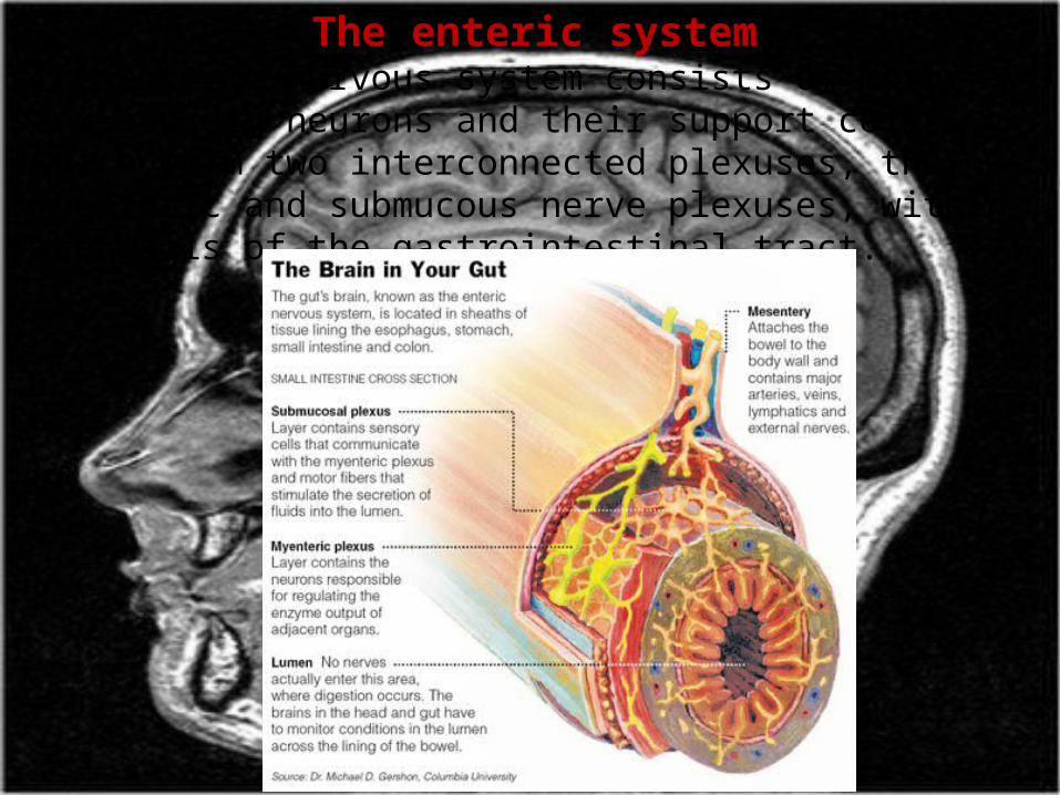
The enteric systemThe enteric nervous system consists of motor and sensory neurons and their support cells, which form two interconnected plexuses, the myenteric and submucous nerve plexuses, within the walls of the gastrointestinal tract.
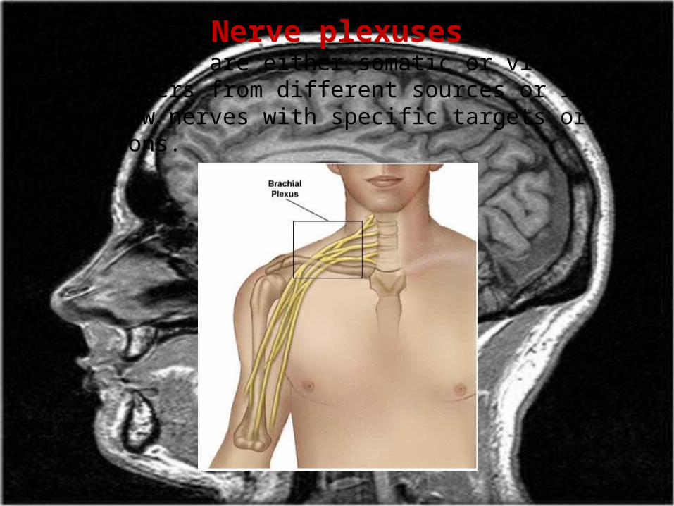
Nerve plexusesNerve plexuses are either somatic or visceral and combine fibers from different sources or levels to form new nerves with specific targets or destinations.
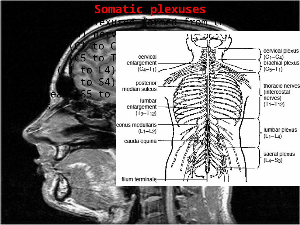
Somatic plexusesMajor somatic plexuses formed from the anterior rami of spinal nerves are: Cervical (C1 to C4) Brachial (C5 to T1) Lumbar (L1 to L4) Sacral (L4 to S4) Coccygeal (S5 to Co) plexuses
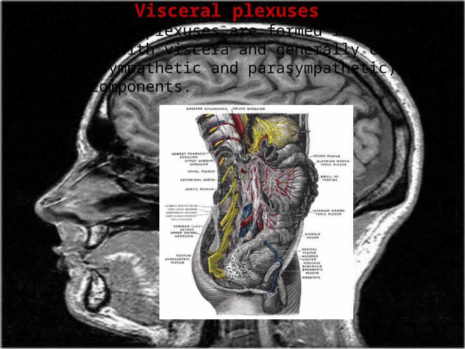
Visceral plexusesVisceral nerve plexuses are formed in association with viscera and generally contain efferent (sympathetic and parasympathetic) and afferent components.
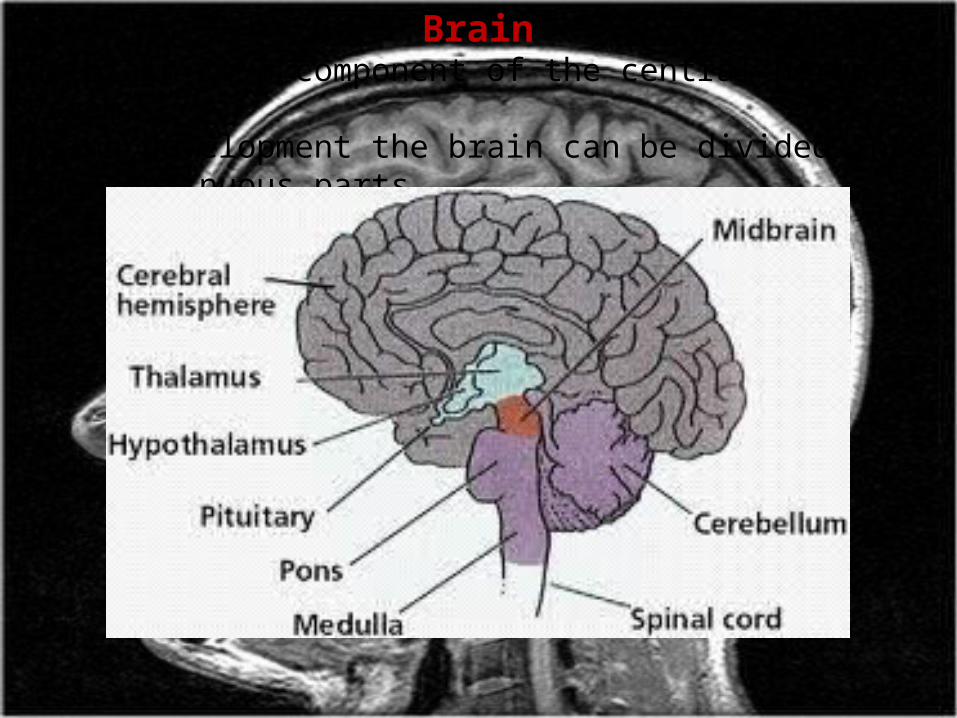
BrainThe brain is a component of the central nervous system. During development the brain can be divided into five continuous parts.
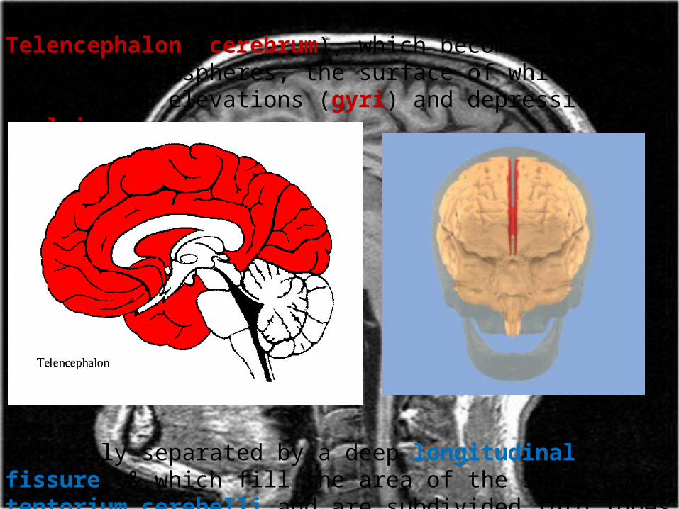
From rostral (or cranial) to caudal they are: Telencephalon (cerebrum), which becomes the large cerebral hemispheres, the surface of which consists of elevations (gyri) and depressions (sulci).
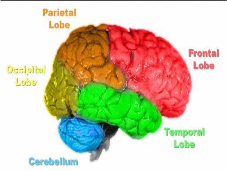
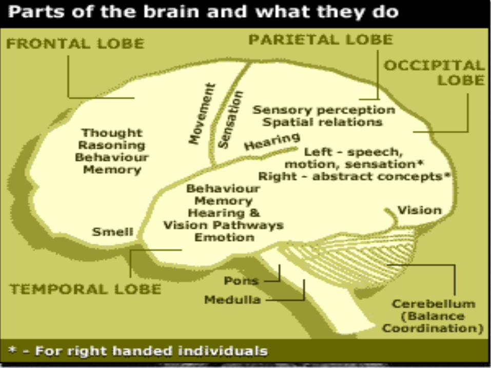
Partially separated by a deep longitudinal fissure, & which fill the area of the skull above tentorium cerebelli and are subdivided into lobes based on their position.
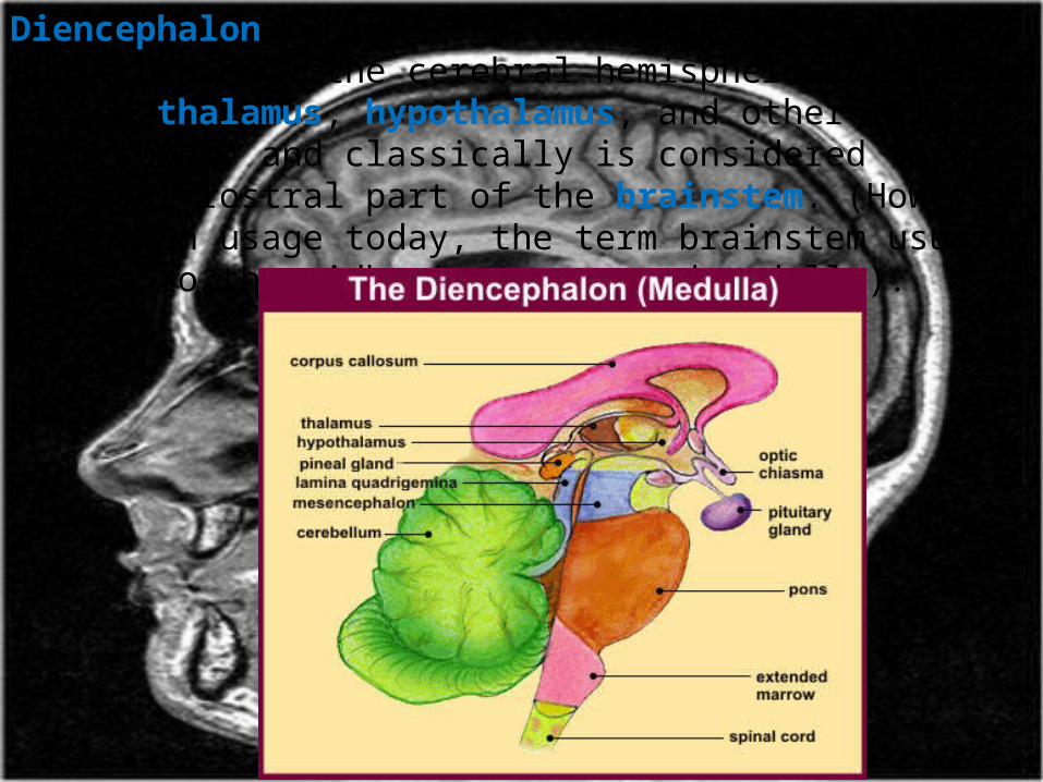
Diencephalon, which is hidden from view in the adult brain by the cerebral hemispheres, consists of the thalamus, hypothalamus, and other related structures, and classically is considered to be the most rostral part of the brainstem. (However, in common usage today, the term brainstem usually refers to the midbrain, pons, and medulla).
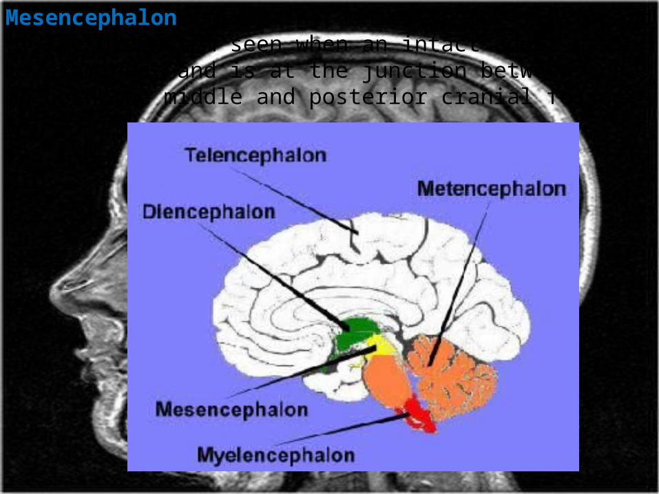
Mesencephalon (midbrain), which is the first part of the brainstem seen when an intact adult brain is examined, and is at the junction between and in both the middle and posterior cranial fossae.
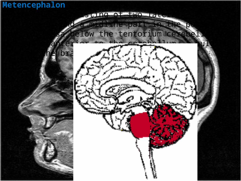
Metencephalon, which gives rise to the cerebellum (consisting of two lateral hemispheres and a midline part in the posterior cranial fossa below the tentorium cerebelli) and the pons (anterior to the cerebellum, a bulging part of the brainstem).

Myelencephalon (medulla oblongata) Caudalmost part of the brainstem, which ends at the foramen magnum.
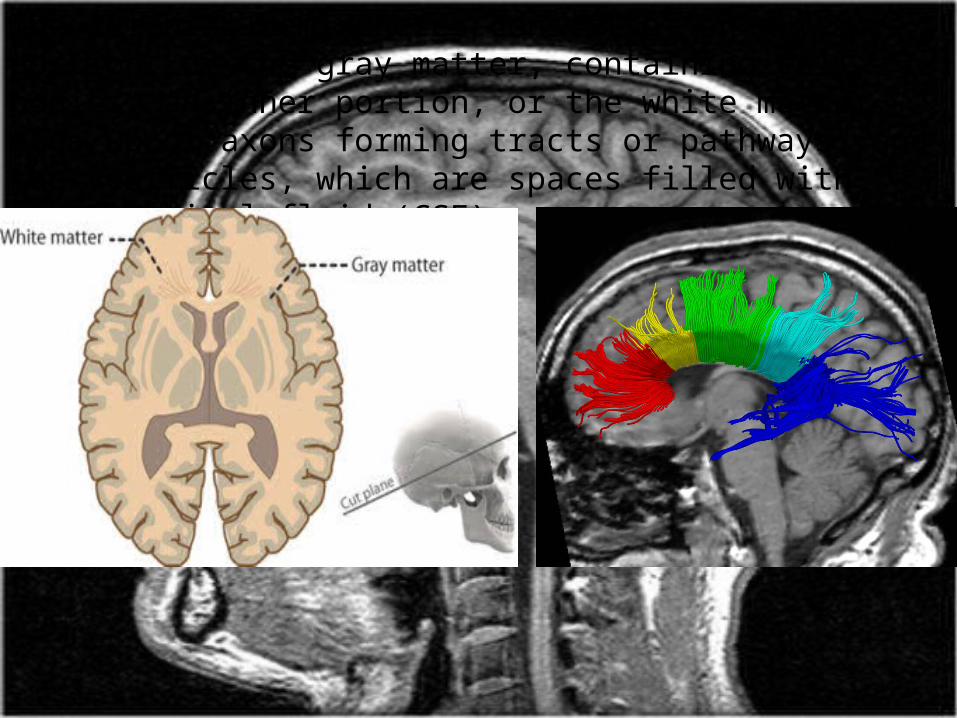
The cerebral hemispheres consist of an outer portion, or the gray matter, containing cell bodies, an inner portion, or the white matter, made up of axons forming tracts or pathways, and the ventricles, which are spaces filled with cerebrospinal fluid (CSF).
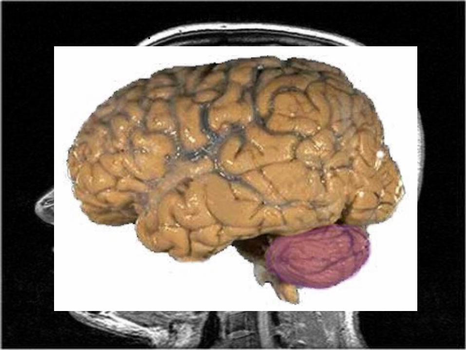
The cerebellum has two lateral lobes and a midline portion.
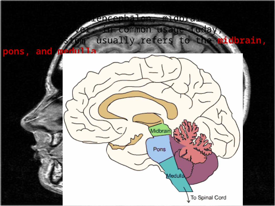
The components of the brainstem are classically defined as the diencephalon, midbrain, pons, and medulla. However, in common usage today, the term "brainstem" usually refers to the midbrain, pons, and medulla.
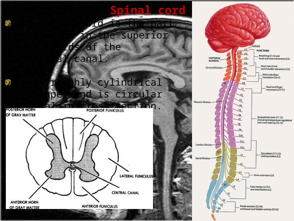
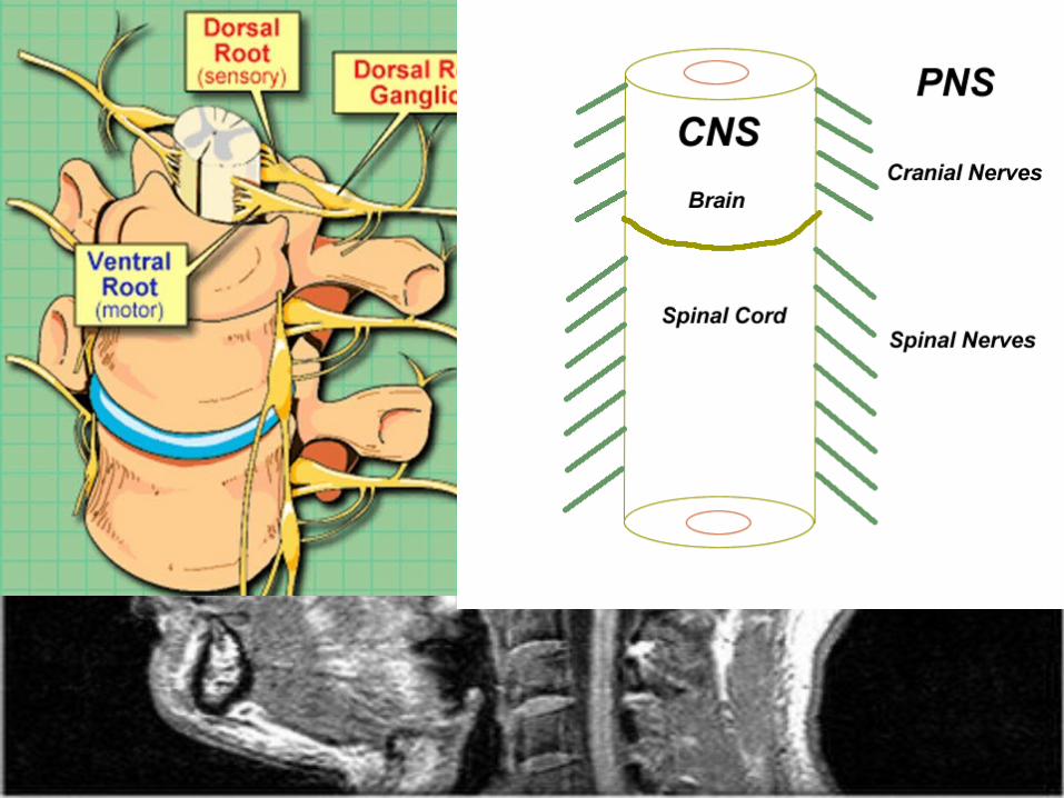
Spinal cordThe spinal cord is the part of the CNS in the superior two- thirds of the vertebral canal.
It is roughly cylindrical in shape, and is circular to oval in cross-section.
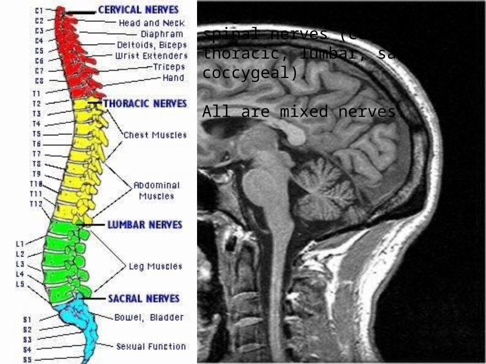
It gives rise to 31 pairs of spinal nerves (cervical, thoracic, lumbar, sacral and coccygeal).
All are mixed nerves.

Spinal nerves arise as rootlets then combine to form dorsal and ventral roots.
Dorsal and ventral roots merge laterally and form the spinal nerve.
Dorsal root is related to the sensory information, whereas the anterior root is related to the motor information.
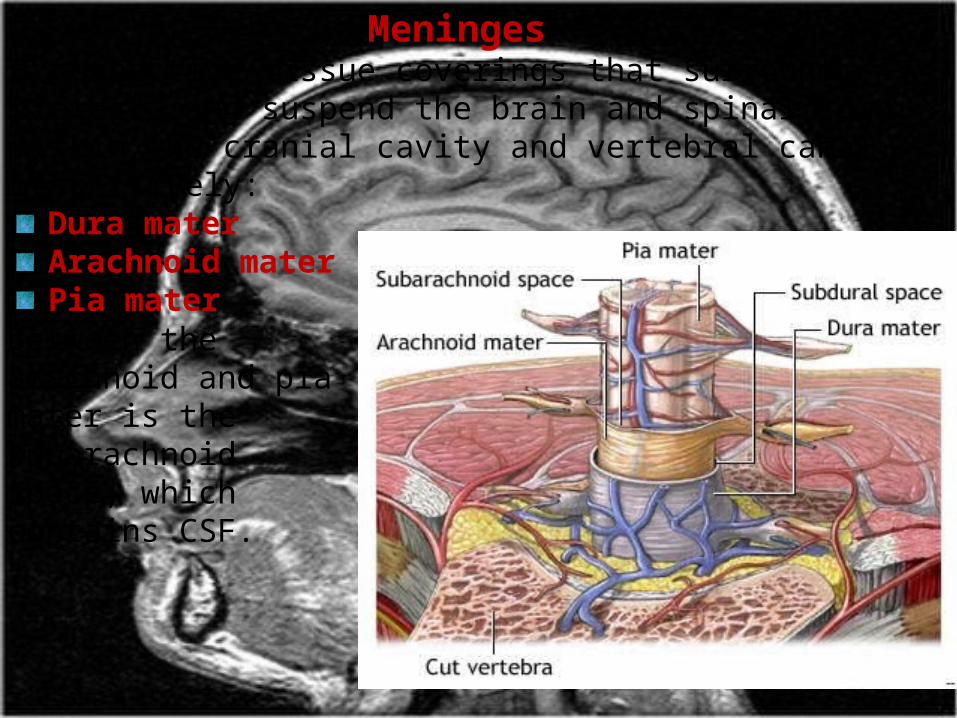
Meninges3 connective tissue coverings that surround, protect, and suspend the brain and spinal cord within the cranial cavity and vertebral canal, respectively:
Dura materArachnoid materPia mater
Between the arachnoid and pia mater is the subarachnoid space, which contains CSF.
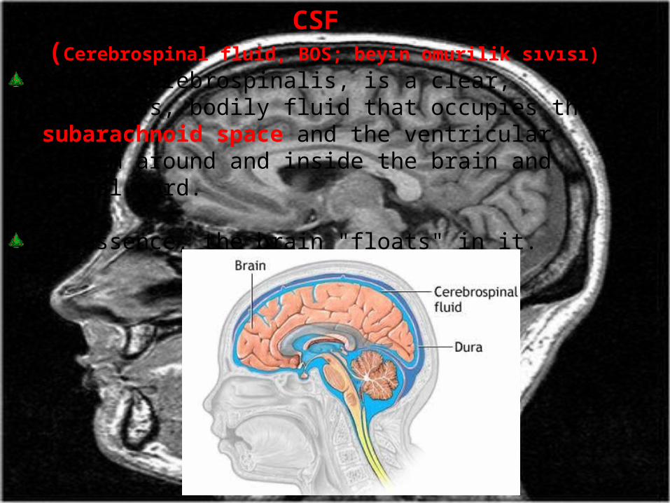
CSF (Cerebrospinal fluid, BOS; beyin omurilik sıvısı)
Liquor cerebrospinalis, is a clear, colorless, bodily fluid that occupies the subarachnoid space and the ventricular system around and inside the brain and spinal cord.
In essence, the brain "floats" in it.

It acts as a "cushion" or buffer for the cortex, providing a basic mechanical and immunological protection to the brain inside the skull.
CSF is produced in the brain by choroid plexus (approx. 50-70%), and the remainder is formed around blood vessels and along ventricular walls.
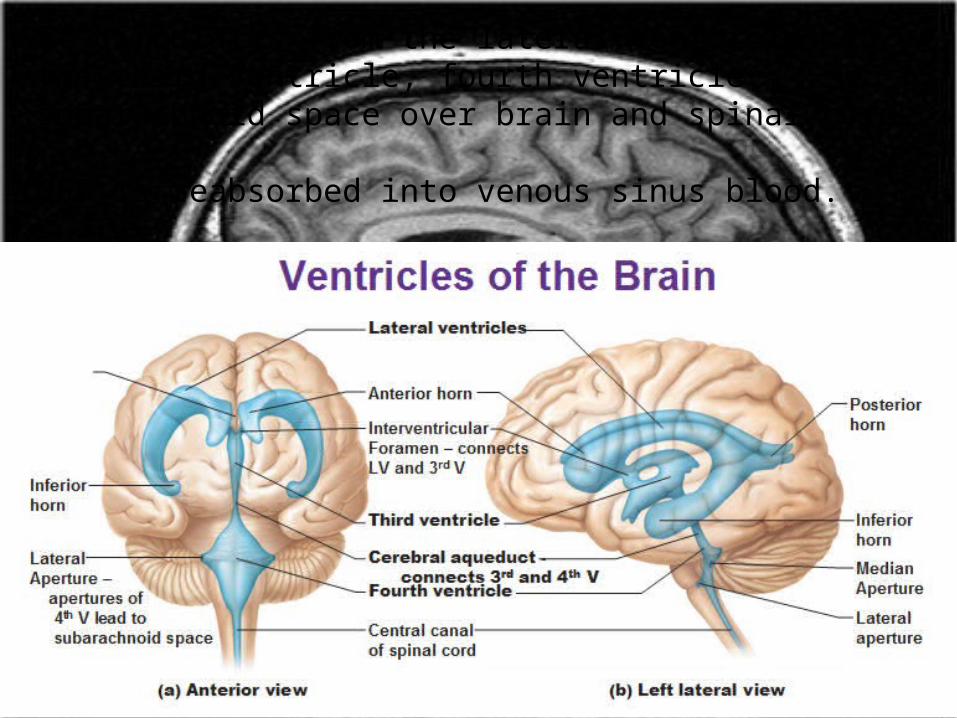
It circulates from the lateral ventricles to the third ventricle, fourth ventricle,; subarachnoid space over brain and spinal cord.
CSF is reabsorbed into venous sinus blood.
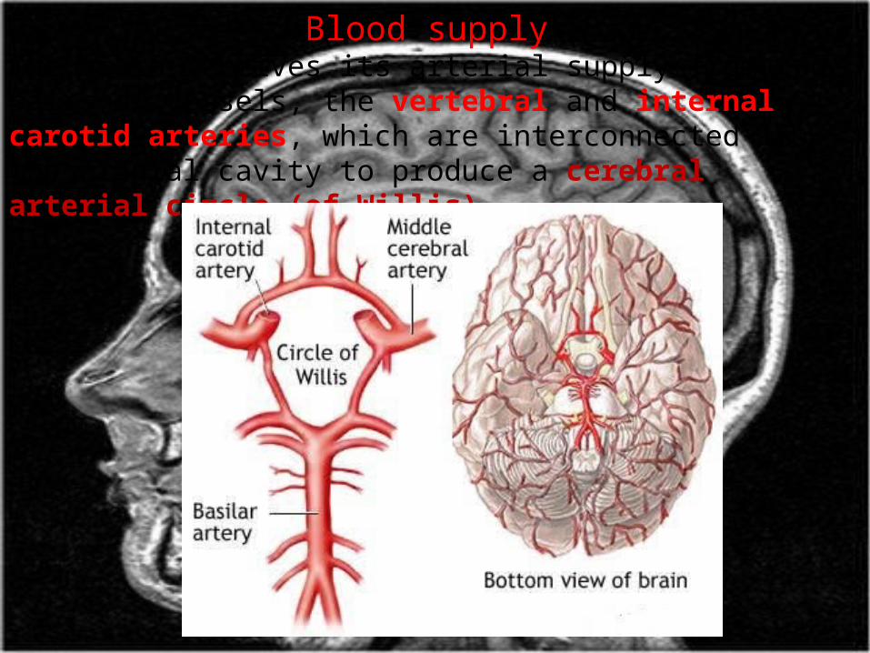
Blood supplyThe brain receives its arterial supply from two pairs of vessels, the vertebral and internal carotid arteries, which are interconnected in the cranial cavity to produce a cerebral arterial circle (of Willis).
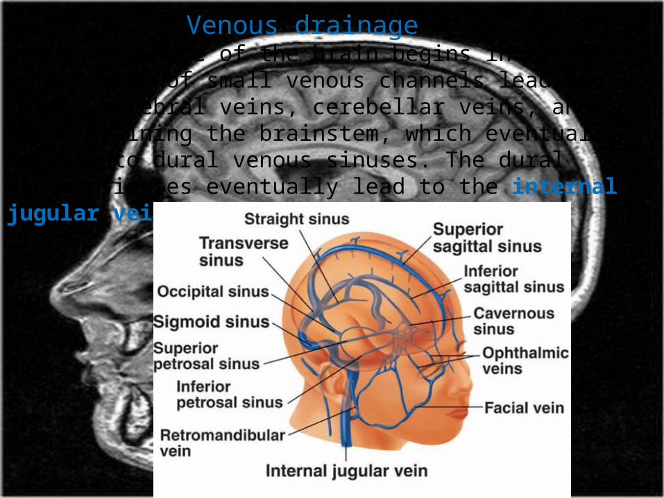
Venous drainage Venous drainage of the brain begins internally as networks of small venous channels lead to larger cerebral veins, cerebellar veins, and veins draining the brainstem, which eventually empty into dural venous sinuses. The dural venous sinuses eventually lead to the internal jugular veins.
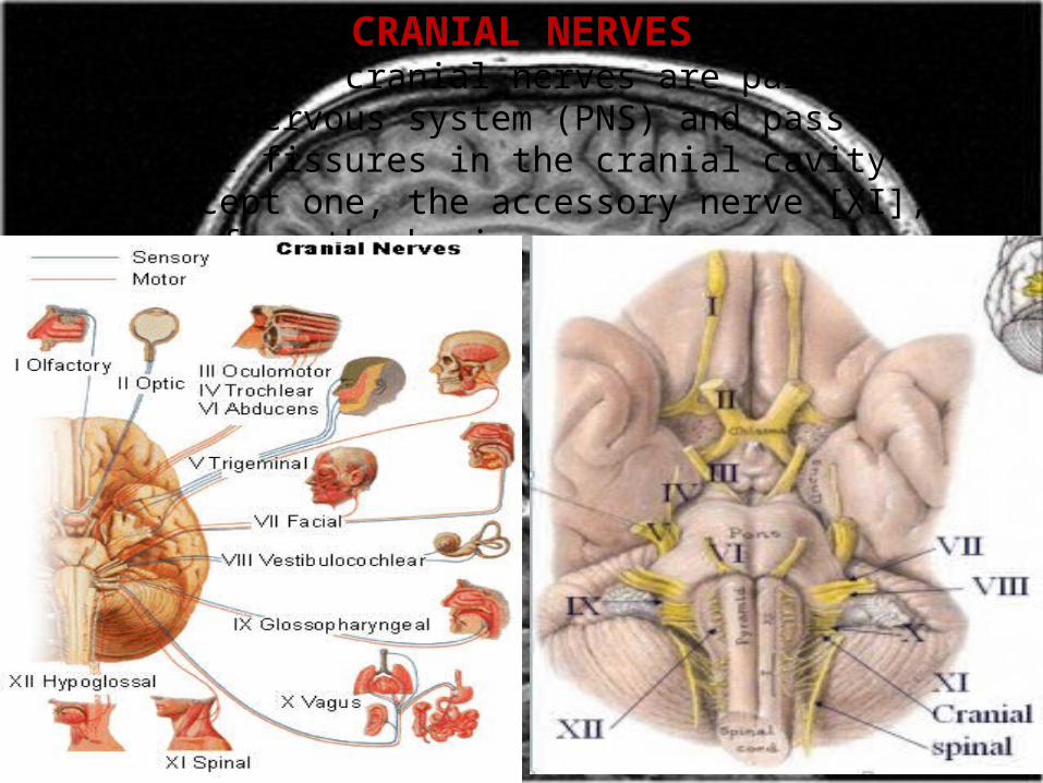
CRANIAL NERVESThe 12 pairs of cranial nerves are part of the peripheral nervous system (PNS) and pass through foramina or fissures in the cranial cavity. All nerves except one, the accessory nerve [XI], originate from the brain.
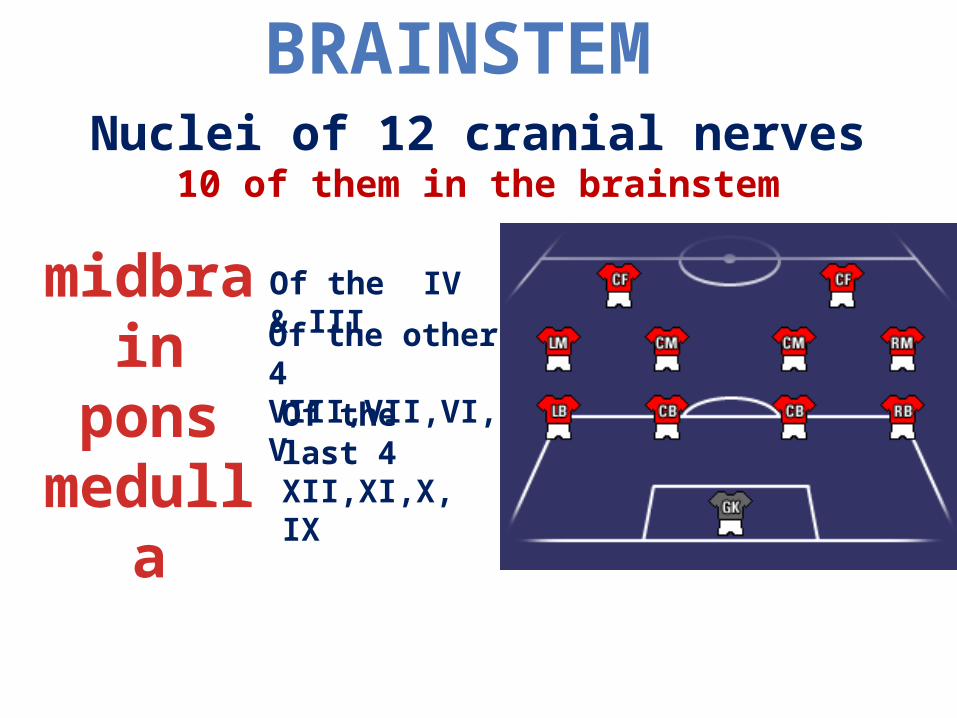
BRAINSTEM
midbrainpons
medulla
Nuclei of 12 cranial nerves10 of them in the brainstem
Of the last 4XII,XI,X, IX
Of the other 4VIII,VII,VI,V
Of the IV & III

I Olfactory Purely sensory Telencephalon
Smelling
II Optic Sensory Retinal ganglion cellsSeeing
III Oculomotor Mainly motor MidbrainEye movements & pupillary reflex
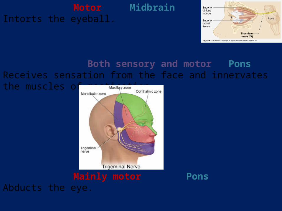
IV Trochlear Motor MidbrainIntorts the eyeball.
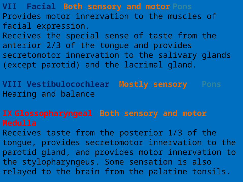
V Trigeminal Both sensory and motor PonsReceives sensation from the face and innervates the muscles of mastication.
VI Abducens Mainly motor PonsAbducts the eye.
VII Facial Both sensory and motor Pons Provides motor innervation to the muscles of facial expression. Receives the special sense of taste from the anterior 2/3 of the tongue and provides secretomotor innervation to the salivary glands (except parotid) and the lacrimal gland.
VIII Vestibulocochlear Mostly sensory PonsHearing and balance
IX Glossopharyngeal Both sensory and motor MedullaReceives taste from the posterior 1/3 of the tongue, provides secretomotor innervation to the parotid gland, and provides motor innervation to the stylopharyngeus. Some sensation is also relayed to the brain from the palatine tonsils.
X Vagus Both sensory and motor Medulla
Innervation to most laryngeal and pharyngeal muscles (except the stylopharyngeus, which is innervated by the glossopharyngeal).
Parasympathetic fibers to nearly all thoracic and abdominal viscera till the proximal two-thirds of the transverse colon.
Receives the special sense of taste from the epiglottis.
A major function: controls muscles for voice and resonance and the soft palate.
XI Accessory (often separated into the cranial accessory and spinal accessory nerves) MedullaMainly motor Cranial and Spinal RootsControls the sternocleidomastoid and trapezius muscles, and overlaps with functions of the vagus nerve (CN X). Symptoms of damage: inability to shrug, weak head movement.
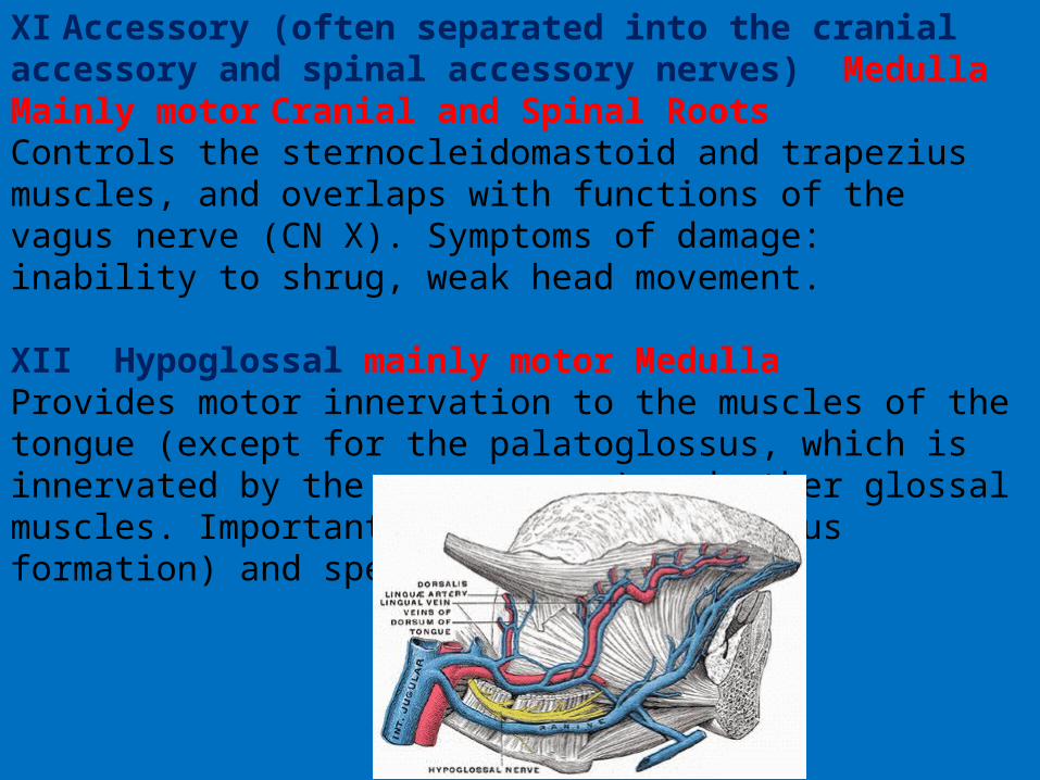
XII Hypoglossal mainly motor MedullaProvides motor innervation to the muscles of the tongue (except for the palatoglossus, which is innervated by the vagus nerve) and other glossal muscles. Important for swallowing (bolus formation) and speech articulation.
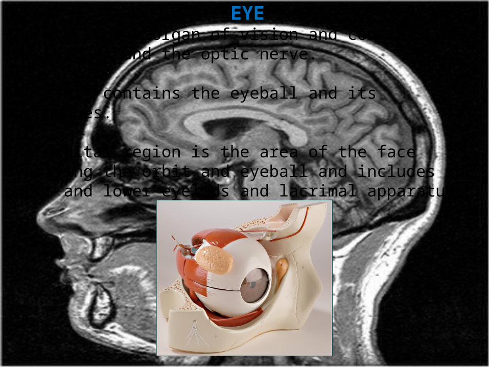
EYEThe eye is the organ of vision and consists of the eyeball and the optic nerve.
The orbit contains the eyeball and its appendages.
The orbital region is the area of the face overlying the orbit and eyeball and includes the upper and lower eyelids and lacrimal apparatus.

The orbits are bilateral bony cavities in the facial skeleton that resemble hollow quadrangular pyramids.
The eyelids and lacrimal fluid, secreted by the lacrimal glands, protect the cornea and eyeballs from injury and irritation (e.g., by dust and small particles).
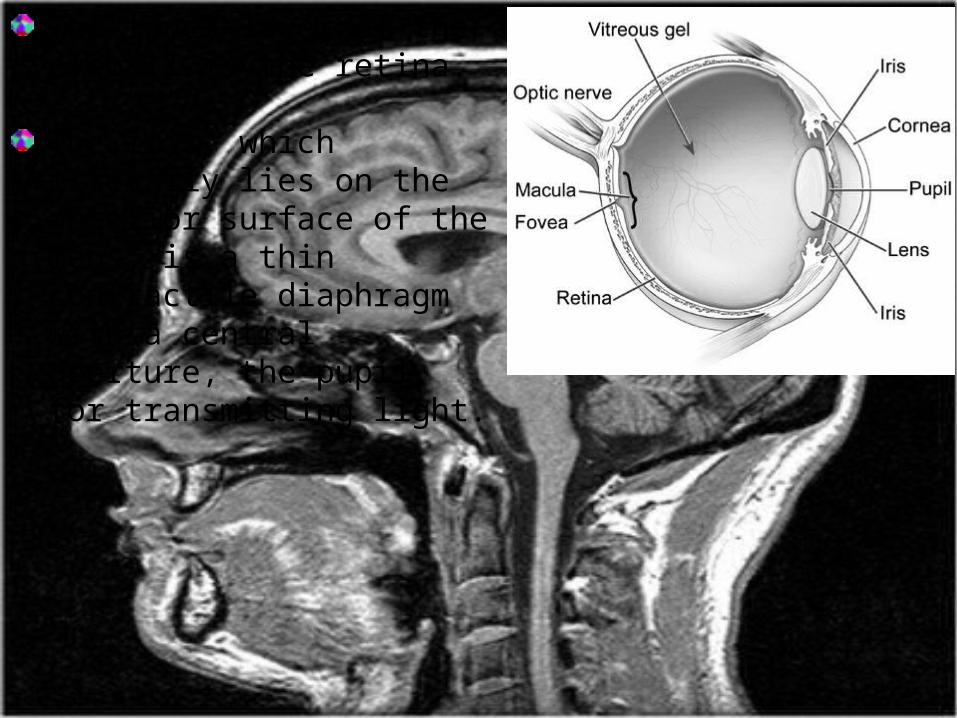
The inner layer of the eyeball is the retina.
The iris, which literally lies on the anterior surface of the lens, is a thin contractile diaphragm with a central aperture, the pupil, for transmitting light.
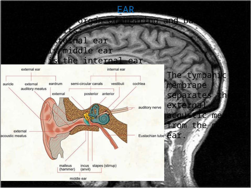
EARThe ear is the organ of hearing and balance. It has three parts: 1st part external ear 2nd part is middle ear3rd part is the internal ear
The tympanic membrane separates the external acoustic meatus from the middle ear.
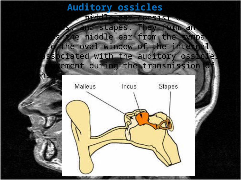
Auditory ossicles The bones of the middle ear consist of the malleus, incus, and stapes. They form an osseous chain across the middle ear from the tympanic membrane to the oval window of the internal ear. Muscles associated with the auditory ossicles modulate movement during the transmission of vibrations.
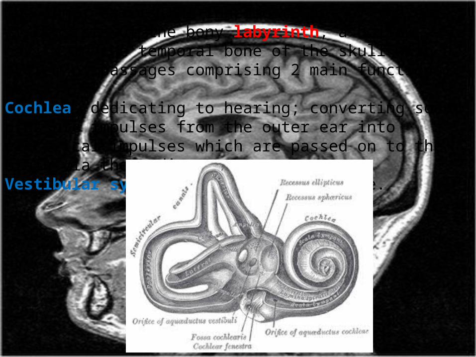
The inner ear is the innermost part of the ear. It consists of the bony labyrinth, a hollow cavity in the temporal bone of the skull with a system of passages comprising 2 main functional parts:Cochlea: dedicating to hearing; converting sound pressure impulses from the outer ear into electrical impulses which are passed on to the brain via the auditory nerve.Vestibular system: dedicated to balance.