
Potential Target for Brain Delivery Search and ...
36
Page 1/36 Search and Characterization of ITM2A as a New Potential Target for Brain Delivery Céline Cegarra ( [email protected] ) Sano-Aventis Recherche Developpement Chilly-Mazarin Longjumeau https://orcid.org/0000-0002- 1710-9796 Catarina Chaves Sano-Aventis Recherche Développement Chilly-Mazarin: Sano-Aventis Recherche Developpement Chilly-Mazarin Longjumeau Catherine Déon Sano-Aventis Recherche Développement Chilly-Mazarin: Sano-Aventis Recherche Developpement Chilly-Mazarin Longjumeau Tuan Minh Do Sano-Aventis Recherche Développement Chilly-Mazarin: Sano-Aventis Recherche Developpement Chilly-Mazarin Longjumeau Bruno Dumas Sano-Aventis Recherche et Développement Vitry-sur-Seine: Sano-Aventis Recherche et Developpement Vitry-sur-Seine Alfortville André Frenzel Universität Braunschweig: Technische Universitat Braunschweig Philip Kuhn Universität Braunschweig: Technische Universitat Braunschweig Valérie Roudieres Sano-Aventis Recherche Développement Chilly-Mazarin: Sano-Aventis Recherche Developpement Chilly-Mazarin Longjumeau Jean-Claude Guillemot Sano-Aventis Recherche Développement Chilly-Mazarin: Sano-Aventis Recherche Developpement Chilly-Mazarin Longjumeau Dominique Lesuisse Sano-Aventis Recherche Développement Chilly-Mazarin: Sano-Aventis Recherche Developpement Chilly-Mazarin Longjumeau Research Keywords: Blood Brain Barrier, ITM2A, antibodies, transcytosis
Transcript of Potential Target for Brain Delivery Search and ...

Page 1/36
Search and Characterization of ITM2A as a NewPotential Target for Brain DeliveryCéline Cegarra ( celine.cegarra@sano�.com )
Sano�-Aventis Recherche Developpement Chilly-Mazarin Longjumeau https://orcid.org/0000-0002-1710-9796Catarina Chaves
Sano�-Aventis Recherche Développement Chilly-Mazarin: Sano�-Aventis Recherche DeveloppementChilly-Mazarin LongjumeauCatherine Déon
Sano�-Aventis Recherche Développement Chilly-Mazarin: Sano�-Aventis Recherche DeveloppementChilly-Mazarin LongjumeauTuan Minh Do
Sano�-Aventis Recherche Développement Chilly-Mazarin: Sano�-Aventis Recherche DeveloppementChilly-Mazarin LongjumeauBruno Dumas
Sano�-Aventis Recherche et Développement Vitry-sur-Seine: Sano�-Aventis Recherche etDeveloppement Vitry-sur-Seine AlfortvilleAndré Frenzel
Universität Braunschweig: Technische Universitat BraunschweigPhilip Kuhn
Universität Braunschweig: Technische Universitat BraunschweigValérie Roudieres
Sano�-Aventis Recherche Développement Chilly-Mazarin: Sano�-Aventis Recherche DeveloppementChilly-Mazarin LongjumeauJean-Claude Guillemot
Sano�-Aventis Recherche Développement Chilly-Mazarin: Sano�-Aventis Recherche DeveloppementChilly-Mazarin LongjumeauDominique Lesuisse
Sano�-Aventis Recherche Développement Chilly-Mazarin: Sano�-Aventis Recherche DeveloppementChilly-Mazarin Longjumeau
Research
Keywords: Blood Brain Barrier, ITM2A, antibodies, transcytosis

Page 2/36
Posted Date: December 3rd, 2021
DOI: https://doi.org/10.21203/rs.3.rs-1125315/v1
License: This work is licensed under a Creative Commons Attribution 4.0 International License. Read Full License

Page 3/36
Abstract
BackgroundIntegral membrane protein 2A (ITM2A) is a transmembrane protein whose function is not well described.This target was identi�ed as highly enriched in human brain vs peripheral endothelial cells bytranscriptomic and proteomic studies during the European Collaboration on the Optimization ofMacromolecular Pharmaceutical Innovative Medicines Initiative (COMPACT IMI) consortium. The objectof the present paper is to report the work we have undertaken to characterize this protein as a potentialbrain delivery target.
MethodsA series of ITM2A constructs, cell lines and speci�c anti-human and mouse ITM2A antibodies weregenerated. Binding and internalization in Human Embryonic Kidney 293 (HEK293) cells overexpressingITM2A and in brain microvascular endothelial cells from mouse and non-human primate (NHP) wereperformed with these tools. The best antibody was evaluated in an in vitro human blood brain barrier(BBB) model and in a mouse in vivo pharmacokinetic study to validate blood brain barrier crossing.
ResultsAntibodies speci�cally recognizing extracellular parts of ITM2A or tags inserted in its extracellulardomain showed selective binding and uptake in ITM2A-expressing cells. However, despite high RNAexpression in mouse and human microvessels, this protein, is rapidly downregulated upon endothelialcells culture, probably explaining why transcytosis could not be evidenced in vitro. An attempt to directlydemonstrate in vivo transcytosis in mice turned out unconvincing, using a cross reactive anti ITM2Aantibody on one hand and in vivo phage panning of an anti ITM2A phage library on the other hand.
ConclusionsThe present article describes the work we have undertaken to explore the potential of ITM2A as target tomediate transcytosis through BBB. This work highlights the multiple challenges linked to theidenti�cation of new brain delivery targets.
BackgroundBrain is a highly protected tissue. The endothelial cells lining the blood vessels that are at the interface ofblood and brain are different than the peripheral endothelial cells, and as opposed to them are extremelytight, non-fenestrated and equipped with many e�ux systems. This blood brain barrier is only permeableto very small lipophilic compounds but is actively preventing most molecules to enter and in particular

Page 4/36
large or polar molecules such as biotherapeutics and antibodies(1, 2). This explains why some enormousmedical needs remain to be addressed in particular for targets for which biologics are the main modalityin therapeutic area such as neurosciences, oncology (e.g. central nervous system (CNS) lymphoma orglioblastoma) or rare diseases. For instance, the use of therapeutic antibodies for CNS disorders such asAlzheimer’s, Parkinson’s, Huntington’s diseases or brain cancers has been very limited so far(3) owing tothe presence of the BBB. This is also explaining why so few biologics are in development in CNS. Thebiologics that are on the market in neurology are most certainly acting peripherally (or else givenintrathecally)(4). Therefore, strategies to increase brain exposure of biotherapeutics are key to thesuccess of biotherapeutics in this area.
So far, the most successful strategy to carry biotherapeutics to the brain using systemic route has beenmaking use of a ligand or antibody against a receptor-mediated transcytosis (sometimes referred to asthe ‘Trojan horse’ approach). Several receptors such as insulin(5), transferrin(6), lipoprotein-relatedproteins(7, 8), low density lipoprotein(9) or Insulin-like Growth Factor 1 (10) receptors have been used.Several challenges however remain in the �eld. One of them is linked to the fact that all these receptorsare ubiquitously expressed. So far, no brain-speci�c receptor capable of mediating such transcytosis hasbeen discovered.
During the COMPACT IMI consortium (https://www.imi.europa.eu/projects-results/project-factsheets/compact ) an approach integrating different omics levels of proteomic, MicroArray and RNAsequencing approaches was engaged from human primary endothelial cells from brain, liver and lungaimed at identifying candidate membrane proteins highly enriched in brain vs liver or lung(11). Theresulting over 60000 RNA’s were then processed through several �lters, downsizing to mRNA’s notdetected at all in liver and/or lung or with high differential level in brain, mRNA’s with mostlytransmembrane expression, association to the BBB vasculature, expression and selectivity comparablebetween rodents and humans and high degree of conservation between orthologs. This led to a fewmRNA’s such as basigin (12) and Low-density lipoprotein receptor-related protein 8 (LRP8)(13) alreadyreported as brain transport mechanisms validating therefore the approach. ITM2A was identi�ed amongthese mRNA’s.
ITM2A (alias E25A or BRICD2A) is a 263-amino acid protein with a single transmembrane domain(14). Itsubiquitous expression is higher in thymus where it was shown to be an activation marker in thymocytedevelopment(15). ITM2A is mainly believed to be associated with cell differentiation duringmyogenesis(16, 17), chondrogenesis(18–22) and odontogenesis(23). The overall homology betweenmouse and human is more than 95% in the extracellular domain(24). The protein has a motopsin-bindingBrichos domain within the COOH-terminal extracellular domain(25) whose signi�cance is poorlyunderstood(25). Brichos domains appear to bear a chaperone function in different biological situationsand particularly bind amyloid �brils (26). Recently ITM2A expression was shown to negatively regulateautophagic �ux by inhibiting lysosomal function through physical interaction with vacuolar Adenosyl TriPhosphatase(27).

Page 5/36
Expression in mouse and human brain has been reported to be homogenous in all brain (Protein AtlasITM2A) and quite speci�c of endothelial cells vs neurons, microglia, oligodendrocytes or astrocytes asshown in brain RNA seq database (Fig. 1). In fact, it is referred to as an endothelial cell-speci�c gene(28).An analysis of �ve human and murine cell type-speci�c transcriptome-wide RNA expression data setsthat were generated within the past several years also identi�ed ITM2A as one of the top expressed genesin endothelial cells(29). A recent single cell RNA seq analysis of 20 organs in mice also established theITM2A gene speci�c for brain endothelial cells(30).
Two precedents point to speci�c association of ITM2A with the blood brain barrier. ITM2A was identi�edas microvasculature speci�c through the screening of subtractive cDNA libraries from rat brain capillariesversus kidney/liver on one hand(31, 32) and from porcine brain and aortic endothelial cells on the otherhand(33). In addition, the expression of ITM2A found in freshly isolated porcine Brain MicrovascularEndothelial Cells (BMEC) was shown to be lost upon culture similarly to some known BBB markers (33).
ITM2A as an endothelial brain speci�c transmembrane protein has not been associated in the literaturewith brain transcytosis or transport. The present report describes the work we have done to characterizethis target as a potential brain delivery candidate.
Materials And MethodsAnimals
Male and Female Cynomolgus monkeys (Macaca fascicularis) aged from 4.8 to 5.9 years werepurchased from Le Tamarinier and Noveprim Ltd. (Mahebourg, Mauritius). Six animals were group-housed in aviaries or interconnected mobile cages and two animals were individually housed ininterconnected mobile cages. Animals were housed under controlled conditions (20–2 4°C, 40–70%humidity, 10–15 renewals per hour of �ltered, non-recycled air, 12-h light cycle) with free access to �lteredtap water and daily distribution of expanded diet (sodium dodecyl sulfate SDS, France) and fruits orvegetables. Animals used for the isolation of brain microvessels were at disposal following pre-clinicalstudies. Prior to the isolation of brain cortical microvessels, animals were submitted to a washout periodof a minimum of 1 month.
Male mice C57BL6/J aged from 6 to 8 weeks were purchased from Charles River Laboratories (France)
Pregnant mice C57BL/6JRj were purchased from Janvier Lab’s (France) between E10 and E12.
After arrival, mice were housed individually (for pregnant mice) or grouped (for male, 6 animals per cage)in an enriched environment in a pathogen-free facility at a constant temperature of 22 ± 2°C and humidity(50 ± 10%) on a 12-h light/dark cycle with ad libitum access to food and water.
Male rats Wistar: crl WI were purchased from Charles River Laboratories (Germany) After arrival, rats werehoused individually in an enriched environment in a pathogen-free facility at a constant temperature of22 ± 2°C and humidity (50 ± 10%) on a 12-h light/dark cycle with ad libitum access to food and water.

Page 6/36
Isolation of Brain Microvessels from Cynomolgus Monkey and rodents Cortex
Brains from Cynomolgus monkeys or rodents were collected shortly after the euthanasia of the animalinto ice-cold Hibernate A medium (ThermoFisher). All subsequent steps were performed at 4 °C and undera biological safety cabinet. Brain cortex was isolated and placed in petri dishes containing ice-coldHanks’ Balanced Salt solution HBSS. The meninges and the cortical white matter were removed. Thecollected tissues were transferred into a new sterile container with HBSS, �nely minced with a scalpel,and then pelleted by centrifugation (5 min at 600 g, 4°C). The pellet was resuspended in acollagenase/dispase solution (Roche, Meylan, France, Collagenase 0.1 U/mL; Dispase 0.8 U/mL preparedin Ca2+/Mg2+ free HBSS) containing type I DNAse at 20 U/mL and Tosyl-L-lysyl-chloromethanehydrochloride (TLCK) at 0.147 g/mL, and incubated at 37°C for 60 min, under vigorous agitation. Thedigested tissue was carefully homogenized, and centrifuged for 5 min at 600 g, 4°C. The resultant pelletwas resuspended in HBSS containing 20% Bovine serum albumin (BSA) and centrifuged at for 30 min at2000 g, 4°C. The myelin ring-containing supernatants were discarded, and the vessel-containing pelletwas resuspended and re-incubated in the collagenase/dispase solution in presence of DNAse and TLCKfor 30 min at 37°C (except microvessels from mice). This suspension was re-pelleted by centrifugation 5min at 600 g, 4°C, and the �nal pellet (named passage 0 Day in vitro 0 (P0D0) fraction from this pointonwards) is resuspended in endothelial cell medium endothelial basal medium (EBM-2) supplementedwith Kit endothelial cell growth medium micro vascular (EGM-2 MV) Single Quots, Lonza, Basel,Switzerland) containing 3 g/mL puromycin, before set up in pre-coated (collagen IV 100 g/mL, �bronectin10 g/mL, Sigma, Saint Quentin Fallavier, France) cell culture �asks, and incubated at 37°C, 5% CO2 for 7days. Every 2 days the cell medium was changed, the supplemented puromycin concentration lowered to2 g/mL, and subsequently removed. Following 7 days of expansion at P0D7, Brain Endothelial Cell(BEC)s from cortex, were further singularized and re-plated de novo for further 7-day cell expansion(P1D7).
Stable cell lines generation
Plasmid constructs
Various human in�uenza hemagglutinin (HA) human ITM2A cDNA with coding sequence: NM_004867and its variants were synthetically made by GeneART and introduced in a PiggyBac® transposonmammalian expression vector pBH 6450 with a Cytomegalo virus (CMV) promoter. HA tag is inserted atdifferent position in ITM2A sequence: at NH2 or COOH side or in the Brichos domain: at NH2 or COOHside of the Brichos domain or in the middle in order to disrupt the eventual function of this domain.Plasmids were named: pBH-hITM2A HA NH2, pBH-hITM2A HA COOH, pBH-hITM2A wild type (wt), pBH-hITM2A Brichos HA COOH, pBH-hITM2A Brichos HA NH2, pBH-hITM2A4 Brichos HA mid.
Various Green Fluorescent Protein (GFP) ITM2A constructs with coding sequence: NM_004867 for humanand NM_008409 for mouse were introduced in pcDNA6.2™C-Emerald (Em)GFP or pcDNA6.2™N-EmGFPexpression vectors with CMV promoter and blasticidin resistance. GFP tag is inserted at differentpositions in mouse or human ITM2A sequences: at NH2 or COOH sides. Plasmids were named:
Page 7/36
pcDNA6.2™N-EmGFP -hITM2A, pcDNA6.2™N-EmGFP -mITM2A, pcDNA6.2™C-EmGFP -hITM2A,pcDNA6.2™C-EmGFP -mITM2A.
Cells and cloning
HEK293/CVCL_0045 cells were obtained from Deutsche Sammlung von Mikroorganismen undZellkulturen (DSMZ). For thawing: cells are thawed rapidly in water bath at 37°C, centrifuge at 900 g for4mn, pellet is re-suspended in culture medium. Cells are cultured in the following medium: Dulbecco’sModi�ed Eagle Medium Gibco 21969 (glutamine-free: selection pressure for HA tag or Blasticidin15µg/mL: selection pressure for GFP tag); 10% Foetal Calf Serum Eurobio CVFSVF06 heat inactivatedAustralian; Penicillin / Streptomycin Gibco 15140 100 U / mL �nal. At con�uence, the cells are rinsed withPhosphate-buffered saline (PBS) (-Ca2+, -Mg2+), detached by enzymatic treatment (Accutase SigmaA6964 or trypsin) at 37°C for 3mn, centrifuge at 900g for 4mn. The pellet is re-suspended in culturemedium and diluted to 1/10 in fresh medium for seeding in new �asks.
Human Cerebral Microvascular Endothelial Cell (hCMEC)/D3 cells were obtained from Cedarlane. Cellsare cultured in the medium cell biologics add with supplements for endothelial cells H1168 as describedabove.
HEK293/CVCL_0045 cells were transfected with pcDNA6.2™-EmGFP or cotransfected with PiggyBac®transposon expression vectors bearing wt ITM2A and variants cDNAs and transposase plasmids 6209(10:1) by using Lipofectamine 2000®, following manufacturer instructions. Transfections wereperformed at 50% con�uence in 24 wells plate with 500 ng of plasmids. For stable transfections, clonesare obtained by limit dilution in 96 well plates with blasticidin selection for pcDNA 6.2 or in glutamine freemedia corresponding to the glutamine synthase selection marker of pBH 6450. Each individual cell isampli�ed in a 6 wells plate, then ITM2A expression is checked �rst with �uorescent microscope for GFPtag, second by Western Blot for all ITM2A constructions.
Antibody generation
Antigen preparation
The human and murine ITM2A extra-cellular domain (ECD) gene sequence was synthesized and fused toa human Fc in a mammalian expression vector. After preparation of transfection-grade DNA, a transienttransfection of HEK293 cells was performed. After 7 days of cultivation, the ITM2A-Fc containing culturesupernatant was isolated and puri�ed by Protein A a�nity chromatography according to standardprotocols. After buffer exchange to PBS, the puri�ed antigen was analyzed by UV/VIS spectrometry andSDS-PAGE.
Antibody-phage selection
The target protein (ITM2A-human Fc) and negative antigen (Protein N Standard, Siemens, QQIM13) wereimmobilized onto the wells of an Enzyme-Linked Immunosorbent Assay (ELISA) plate (Corning, 9018) for

Page 8/36
1 h at room temperature (RT) (1 µg each). After removal of non-bound antigen, ELISA wells were blockedwith a 2% BSA solution for 16 h at 4 °C. After washing of the plates with PBS-T (PBS containing 0.05%Tween 20), the antibody-phage library was added to the immobilized negative antigen and incubated for1 h at RT to remove Fc speci�c or polyreactive antibody-phage. Additionally, 5 µg Protein N Standard wasadded as soluble competitor. Non-bound antibody-phage were recovered and incubated on theimmobilized target antigen for 2 h at RT. Non-bound or weakly bound antibody-phage were removed bywashing with PBS-T (10x) before antigen-speci�c phage were recovered by Trypsin (10 µg/ml) elution for30 min at 37 °C. The antibody-phage were rescued by infection of TG1 cells (OD600=0.5) for 30 min at 37°C. After propagation of the cells for additional 30 min at 37 °C and 500 rpm, ampicillin (100 µg/ml) andglucose (100 mM) were added to the 2YT culture medium. Bacterial propagation was continued for 1 h at37 °C and 500 rpm. Then, bacteria were co-infected with M13K07 helper phage, incubated at 30 min at 37°C followed by another incubation for 30 min, 37 °C and 500 rpm. Double-infected bacteria werecentrifuged (4000 g, 10 min) and the cell pellet was resuspended in fresh 2YT, containing Ampicillin (100µg/ml) and Kanamycin (50 µg/ml). For the ampli�cation of antibody-phage particles, the incubation wascontinued for 16 h at 30 °C. Then, the culture was centrifuged (4000 g, 10 min) and antibody-phagecontaining supernatant was recovered and used for the next panning cycle. Three panning cycles wereperformed in total. In each cycle, the number of washing steps was increased (cycle 2: 20x, Cycle 3: 30x)to increase the stringency of the selection.
Antibody screening and sequence analysis
After the third panning cycle, the eluted phage were used to infect XL1 (OD600=0.5) for 30 min at 37 °Cand streaked out on 2YT agar plates, containing ampicillin (100 µg/ml), glucose (100 mM) andtetracycline (20 µg/ml). Incubation was continued at 37 °C until single colonies were observed. Singleclones were isolated and transferred into 96-well plates, containing 2YT, ampicillin (100 µg/ml), glucose(100 mM) and tetracycline (20 µg/ml). The bacteria were cultivated for 16 h at 37 °C and 300 rpm. Then,15 µl of the overnight cultures were used to inoculate new 96-well plates, containing 2YT medium,ampicillin (100 µg/ml), tetracycline (20 µg/ml) and IPTG (50 µM). The bacteria were cultivated for 16 h at30 °C and 300 rpm. The cultures were centrifuged (4000 g, 10 min) and Single-Chain Variable Fragment(scFv) containing supernatants recovered. The scFv containing supernatants were used for antibodyscreening. In brief, supernatants were diluted with a 2% BSA solution (in PBS, containing 0.05%Tween20), added to the immobilized antigens in a 384 well ELISA plate (20 ng/well) and incubated for 1h at RT. After washing, binding of the scFv antibodies to human ITM2A-human Fc, murine ITM2A-humanFc, Protein N Standard or BSA was detected via a Myc-tag using a secondary horseradish Peroxidase(HRP) conjugated antibody. The binding was quanti�ed by TMB reaction and absorbance reading at 450nm. Target speci�c antibody clones were isolated, and the DNA sequence of the respective scFv antibodyanalyzed by sanger sequencing.
Immunoglobulin G (IgG) expression

Page 9/36
The VH and VL sequence of selected antibody clones was ampli�ed by PCR and cloned into mammalianIgG expression vectors. After preparation of transfection-grade DNA, a transient transfection of HEK293cells was performed. After 7 days of cultivation, the IgG containing culture supernatant was isolated andpuri�ed by Protein A a�nity chromatography according to standard protocols. After buffer exchange toPBS, puri�ed antibodies were analyzed by UV/VIS spectrometry, SDS-PAGE and �ow cytometry analysisfor cell binding.
Transcriptomic
Transcriptome Sequencing (RNAseq) and mRNA Expression Analysis COMPACT IMI consortium
As described previously by Li et al (11).
RNA Samples for Library Preparation
As described previously by Chaves et al (34) Frozen cell pellets from the cellular P0D0, P0D7 and P1D7fractions were lysed using QIAzol Lysis Reagent (Qiagen, #79306, Courtaboeuf, France). Total RNA wasthen isolated from the lysates on a QIAcube instrument (Qiagen) using the Rneasy Mini QIAcube kit(Qiagen, #74116) and following the manufacturer’s instructions. The RNA concentration was determinedusing the Qubit RNA HS Assay Kit (Invitrogen, #Q32852, Illkirch-Graenstaden, France) and the quality andintegrity was assessed on a Bioanalyzer 2100 (Agilent Technology, Les Ulis, France) with an Agilent RNA6000 nano kit (Agilent Technology, #5067-1511).
RNA Sequencing (RNAseq)
As described previously by Chaves et al (34), the RNAseq libraries were prepared with 30 ng of input totalRNA using the NEBNext Ultra II Directional RNA Library Prep Kit for Illumina (New England Biolabs, #E-7765S, Évry-Courcouronnes, France) with the NEBNext rRNA Depletion Kit (New England Biolabs,#E6310L) and following the manufacturer’s instructions. The libraries were then paired end sequenced(75 cyclesx2) on the NovaSeq 6000 instrument (Illumina, Paris, France) using the NovaSeq 6000 SPReagent Kit (300 cycles; #20027465, Illumina).
Reverse transcription quantitative Polymerase Chain Reaction (RTqPCR)
Cells or tissus were lysed with RLT buffer from Qiagen added with 1% β-mercapto-ethanol according tomanufacturer instructions. mRNA was extracted with Qiagen MiniKit followed by DNase step using theQiacube robot. Reverse Transcription is achieved from 250 ng of mRNA with High Capacity cDNA ReverseTranscription Kit from AppliedBiosystem Ref 4368813. qPCR is performed from cDNA diluted 1/20 usingQuantStudio™ 7 in 384 wells with the TaqMan system standard mode and following manufacturerinstructions. Primers and Probes Taqman were bought at thermo�cher scienti�c inventoried assay ref4331182, ITM2A: Mm00515208_m1; Actin: Mm01205647_g1; glyceraldehyde-3-phosphatedeshydrogenase (GAPDH) Mm99999915_g1; Platelet endothelial cell adhesion molecule (PECAM) 1Mm01242576_m1. Raw data were analyzed with QuantStudio™ Real-Time PCR software.
Page 10/36
Proteomics
Immunocytochemistry
Cultured cells were �xed in 4% paraformaldehyde for 15 min, at RT, and subsequently permeabilized andblocked in Blocking Buffer Odyssey LiCor containing 0.2% Triton X-100. Primary antibodies wereincubated overnight at 4°C (anti-ITM2A polyclonal AF4876 and 14407-1-AP, monoclonal non-commercialprovider Yumab: YU93-G04, YU147-A01 YU147-E02 YU147-H07, anti-EmGFP A31852 or A11122, anti-HA901509), and appropriate secondary antibodies conjugated with Alexa �uorophores (Invitrogen) andHoechst 33432 (Invitrogen) for nuclei staining were subsequently used for 2h at RT. Images wereacquired on a Perkin Elmer Operetta CLS system.
Colocalization confocal imaging
Confocal microscope images were acquired with SP8 LEICA microscope, 40X objectives. For theacquisition ITM2A was directly visualized by EmGFP 488. Organelles were stained with following primaryantibodies anti Lysosomal-associated membrane protein 1 (LAMP1) ab24170 for lysosome detection,anti-giantin ab37266 for Golgi. Antibody anti-organelle were detected by staining with appropriatesecondary antibody Alexa 633 goat anti mouse A21050 1/1000 or goat anti rabbit A21070 1/1000
Western Blot
NHP-derived BEC were lysed using ice-cold radio-immunoprecipitation assay (RIPA) or cell lysis buffercontaining protease inhibitor cocktail (ThermoFisher) centrifuged at 15,000 g for 15 min, and supernatantfractions were collected. Samples were added with SDS and loading buffer then denaturized by heat 95°Cfor 5 min. These were loaded into 4–12% Tris-Glycine SDS-page gels (Invitrogen), and let to migrate for1h at 180V. Samples were then transferred onto polyvinylidene �uoride (PVDF) or nitrocellulosemembranes using an iBlot 2 Dry Blotting System (Invitrogen) on the P0 program (20 V for 1 min, 23 V for4 min, 25 V for 2 min). PVDF membranes were then rinsed with Tris-buffered saline with 0.1% Tween 20(TBST) and blocked for 1 h in 5% non-fat dry milk in TBST (blocking buffer). Membranes were �rstprobed overnight at 4°C with primary antibodies in blocking buffer (anti-ITM2A polyclonal AF4876 and18306-1-AP, EmGFP A11122, anti-HA 901509, anti α-tubulin T9026) and then probed with secondaryantibodies diluted in TBST for 1 h at RT (1:10,000 diluted HRP-coupled goat anti-mouse IgG or goat anti-rabbit IgG, GEHealthcare). Following secondary antibody incubation, membranes were rinsed thoroughlywith TBST, imaged using a LICOR Odyssey Imager and bands quanti�ed using Multi-Gauge v3.0.
Mass spectrometry
Post-mortem human brain samples (occipital cortex, devoid of pathological �ndings) from three 69 to 79-year-old, male, non-demented, control donors were obtained from external biological resource centers infull accordance with legislation and ethical standards. Microvessels were isolated as described above formonkeys. Tissus or cells were lysed in Preomics 2X buffer then crushed gently with gentle MACSDissociator Miltenyi Biotec, program Protein for 1min. Samples were centrifugated 10min at 4000g, 4°C,

Page 11/36
supernatants were collected and dilute to 1X Preomics buffer. Benzonase 1/100 was added andincubated 10 min à 95°C, 1000 rpm. Samples were centrifugated 20mn at 13000g and supernatants werecollected. Proteins are quanti�ed by spectrophotometry using bicinchoninic acid method and spectraMaxi3x. 25 µg of protein were digested on Preomics �lter in 50µL following manufacturer instructions during3h at 37°C, after evaporation, samples were diluted at 0,5µg/µL in 50µL LC-Load buffer, vortexed andsonicated. Heavy peptides were added to the sample solution to inject 5 fmol of heavy peptides and 2µgof proteins. 7 heavy stable isotope labelling (SIL) peptides spiked in �nal digest prior to Parallel ReactionMonitoring-Mass Spectrometry analysis on Q-Exactive HF /NanoRSLC 3500. From ITM2A_MOUSEIntegral membrane protein 2A Q61500, spiked SIL peptides for detection were: IAFNTPTAVQK, NLVELFGK,EDLVAVEEIR, DLLLGFNK. Spiked SIL peptides used for hTFRC detection were: DSAQNSVIIVDK,LTVSNVLK, SGVGTALLLK, AAAEVAGQFVIK, LTTDFGNAEK.
Transport assays
Internalization assay
HEK293 ITM2A cells are cultured on 96 wells plate coated with poly-L-lysine at 50000 cells/well. After24h cells were incubated 1h at 4°C in cell medium with 5µg/mL of studied antibody (Anti ITM2ARandDsystem AF4876, Yumab antibodies anti ITM2A YU147-A01, E02 and H07, YU93-G04, GFPantibodies A31852 or A11122, anti-HA 901509). Cells were washed with ice cold PBS then incubated 1hat 37°C or at 4°C for control. Cells were washed 2 times with ice cold PBS, then 2mn with acid buffer(glacial acid acetic 1/167 in PBS, pH = 2,5) and �nally 2 times with ice cold PBS. Cells were �xed andstained as described above. Images were acquired with Operetta High Content Screening (PerkinElmer)objective 40X water confocal mode.
In Vitro Transcytosis and Permeability Measurements Brainplotting models
All human samples were provided by Brainplotting (35)(iPEPS, Institut du Cerveau et de la Moelle épinière,Hôpital Universitaire de la Pitié-Salpêtrière, Paris, France) in partnership with Sainte-Anne Hospital, Paris(neurosurgeon Dr. Johan Pallud) and harvested during tumor scheduled resection surgery with writteninformed consent from the patients (authorization number CODECOH DC-2014-2229). Human brainmicrovessels were obtained from surgical resections of one patient: a 35-years-old female suffering fromof a diffuse oligoastrocytic grade III glioma. Microvessels were isolated from peritumoral or healthy braintissue using an enzymatic procedure(34) adapting methods previously published for rats(36, 37). Brie�y,tissue samples were carefully cleaned from meninges and excess of blood; then, an enzymatic mix wasused to dissociate the tissues and microvessels were isolated by retention on a 10 mM mesh. Cells werecultured in EBM-2 medium (Lonza, Basel, Switzerland) supplemented with 20% serum and growth factors(Sigma)(36, 37). After seeding brain capillaries in petri dishes, brain primary microvascular endothelialcells were shortly ampli�ed and seeded (P1D0) on Transwell (Corning) with microporous membranes(pore size: 0.4 mm) in monoculture.
Page 12/36
Test (1 µg/mL of internal anti-human/cynomolgus Transferrin Receptor C (TFRC) antibody or anti-ITM2AYU93-G04) and control antibodies (1µg/mL mouse IgG, clone MG1-45, BioLegend) were added onto theupper chamber on day adapted to the transport assay de�ned by Brainplotting. Fresh endothelial cellmedium with none of these compounds was added onto the bottom chamber. Final aliquots from bothchambers were taken 240 min following incubation at 37°C, 5% CO2. Compound levels in mothersolutions (T = 0 min), upper and lower compartments (T = 240 min) were determined by ELISA assay(MESOQuickPlex SQ120, MesoScale Discovery, Rockville, MD, USA). Apparent permeability (Papp)coe�cients were calculated using the following formula:
Papp (cm/min) = (V /(A x Cluminal)) x (Cabluminal/ t)
where V = volume of cell medium in the bottom chamber (mL), A = surface area of the insert (cm2),Cluminal = compound concentration loaded in the upper chamber (µM), Cabluminal = compoundconcentration measured in the bottom chamber (µM); t = time of the assay (min).
Immunoassay Method ELISA (in vitro and in vivo studies)
Standard 96-well sector plates (Meso Scale Discovery) were coated with 0.5 μg/mL of Fab’2 anti-Human(709-006-098 Jacksonimmuno) or anti-mouse (M0284 Sigma-aldrich) IgG in PBS and then incubated for1 h under agitation at RT. After incubation, plates were washed three times with PBS-Tween 0.05%(Calbiochem, 524653) and blocked for 1 h at RT with 0.1% BSA solution (A7030, Sigma). After blockingthe plates, collected samples in transport assay and standards were incubated on plates for 2 h at RT.After incubation, plates were washed three times with PBS-Tween 0.05%, and bound antibody wasdetected with SULFO-TAG conjugated goat anti-mouse antibody (R32AC-1, Meso Scale Discovery) orgoat anti-human antibody (R32AJ-1, Meso Scale Discovery) using Tripropylamine containing read buffer(R92TC-2, Meso Scale Discovery). Concentrations were determined from the standard curve using a four-parameter non-linear regression program (Discovery Workbench version 4.0 software).
In vivo experimentations
Mouse brain collection
Each mouse was anaesthetized in iso�urane gas chamber then transcardiacally perfused with Li-heparinate solution at �nal concentration 20 U/mL in sterile PBS. Perfusion was realized with 48mLdelivered at the speed of 8 mL/mn. Brain samples were collected. Each mouse was decapitatedimmediately after perfusion. Perfused brain was removed, cerebellum and brainstem were separated andeliminated. Brain cortex were washed in ice-cold PBS, collected in preweighted Precellys tube and storedimmediately at -80°C freezer (or dry ice) until use. Then, the preweighted hemispheres were thawed andhomogenized in 5 vol. (v/w) of brain lysis buffer (1% NP-40 in PBS containing complete mini ethylenediamine tetra-acetic acid - free protease inhibitor cocktail tablets, Pierce) using bead homogenizer.Homogenized brain samples were then rotated at 4°C for 1 hour before centrifuge at 20000 g for 20 min.
Page 13/36
Pharmacokinetic study in vivo in mouse
5-10 mg/kg (35-70 nmol/kg) in a single dose of anti-ITM2A (clone YU93-G04) or control(anti- trinitrophenyl (TNP), batch VA2-17-419-1, internal production) antibodies were administrated bycaudal intravenous injection into mice, C57Bl/6, male, 20-25g (n = 3/condition). 5h post-injection, plasmaand saline-perfused brains were collected, then concentration was determined by ELISA immunoassay asdescribed above
In vivo panning Yumab in mice
Yumab provided ampli�ed phage library to Sano�. First, a naïve human antibody phage library wasenriched for antigen speci�c antibodies against ITM2A proteins. The antibody phage output wasampli�ed and puri�ed by poly-ethylene glycol (PEG)/ NaCl puri�cation. Puri�ed antibody phages weredirectly used for the in vivo panning: 1011 antibody phage particles (~10 µl) were mixed from eachpanning output. The antibody phage mix was injected into the mouse. Brains were isolated after 1 h and24 h (2x each). Brains were collected as described above.
Antibody phages in the homogenate were used for infection of E. coli. Bacteria were selected and usedfor production of monoclonal scFv antibodies. About 800 clones were picked and screened by scFv inELISA assay.
Statistical Analysis
The statistical signi�cance of differences between groups was analyzed using GraphPad Prism v9.0.0software (GraphPad Software, San Diego, CA, USA) Ordinary one-way Analysis of Variance (ANOVA) (non-parametric or mixed) with Dunnett’s multiple comparisons test, Ordinary two-way ANOVA (mixed) withSidak’s, Kruskal Wallis or Mann-Whitney multiple comparison test or unpaired t test (non-parametric). Theapplication of these statistical methods to speci�c experiments is noted in the Fig. legends.
ResultsOur strategy is outlined in Fig. 2. The �rst objective was to produce tools to study the mechanisms: cellsexpressing ITM2A and the extracellular domain of the protein to produce antibodies preferably mouse-human cross reactive. With this in hand, uptake, internalization, and tra�cking in either the aboveoverexpressing cells or in BEC’s would provide the �rst �lter. Transcytosis and in vivo brainpharmacokinetic would lead to �nal validation (Fig. 2).
Building up cell lines expressing ITM2A
In the absence of available monoclonal antibodies, we devised a strategy that could bring clues on thepotential of this protein to carry biologics into the brain using speci�c well characterized cargos. To thisend, we engineered several constructs of human or murine ITM2A. One plasmid, named wt, was designedwith no tag, the others bearing either an HA-tag (HA-tag are amino acids 98-106 of human in�uenza

Page 14/36
hemagglutinin glycoprotein) or a GFP-tag at different positions, namely on the C-Terminal (C-Ter)extracellular domain or on the N-Terminal (N-Ter) intracellular domain of ITM2A. In addition, the HA tagswere positioned at three distinct locations of the Brichos domain within the extracellular portion to bringadditional insight on the potential position involved in internalization. This information could be exploitedlater for the generation of antibodies speci�cally recognizing a part of the antigen to optimize theirtranscytosis. Designed plasmids are summarized in Fig. 3.
We selected adherent HEK293 cells well suited for immuno�uorescence analyses and produced 10 clonalcells lines by recombination with the various tagged or untagged mouse or human plasmids displayed inFig. 3 (Table 1). Clones were obtained after limit dilution and selection by either �uorescence and WesternBlot for GFP tag or by Western Blot for HA tag and wt ITM2A (Additional �le 1).
Table 1
Engineered HEK293 cells expressing various tagged or untagged human or mouseITM2A
Mouse itm2a Human ITM2A
GFP tag HA tag GFP tag No tag
N-Ter
C-Ter
N-Ter
C-Ter
Brichos 1 C-Ter
Brichos 2 N-Ter
Brichos 3 Mid
N-Ter
C-Ter
wt
Several constructs of human or murine ITM2A with or without tags as GFP or HA
Fluorescence ITM2A visualization
After cell line validation for the ITM2A construct protein expression by Western Blot (WB), cellularlocalization of both human and mouse ITM2A was visualized by �uorescence microscopy in cell linesusing GFP �uorescence (Fig. 4). GFP �uorescence was detected in all N-Ter and C-Ter constructs inHEK293 expressing both human and mouse ITM2A.
Cellular localization of ITM2A
Aside from membrane localization as could be showed by the above binding experiments, ITM2A couldbe localized in Golgi of the overexpressing HEK293 cells after evidencing co-localization with Giantin(Fig. 5A). No ITM2A could be seen in the lysosomes as shown by LAMP1 co-labeling (Fig. 5B).
Cell uptake of antibodies
Page 15/36
With these cell lines in hand, we went on to evaluate their capacity to uptake antibodies after binding withthe protein. We could perform these experiments using several handles.
First, we performed immuno�uorescence studies on HEK293 cells expressing human ITM2A with twocommercially available polyclonal antibodies against extracellular epitopes of human ITM2A: AF4876and 14407-1-AP (Fig. 6A and B). The �rst one displayed membrane labeling at 4°C while the second onecould not be detected at 4°C maybe linked to a lower a�nity or different behavior after acid washing(performed before analysis). Both antibodies readily internalized at 37°C leading to punctate labellingand demonstrating active uptake. The weaker signal observed with AF4876 on the C-Ter GFP cell linecould result from steric hindrance of the GFP tag for antibody binding.
The same results were obtained using anti-GFP antibodies either directly �uorescent (A31852, Fig. 6C), orafter detection of an unlabeled anti-EmGFP antibody (A11122) with a secondary labeled Alexa 647 anti-Fc rabbit antibody (Fig. 6D). In both cases, very clear membrane labeling could be observed at 4°Cfollowed by internalization at 37°C with the HEK293 cells expressing human and murine C-Ter GFPITM2A, but not N-Ter GFP ITM2A con�rming the need for the GFP to be extracellular for recognition.
We generated analogous results with anti HA antibodies on HEK293 cells (Fig. 6E) where clearinternalization could be observed when the label was in C-Ter leading to punctate labelling con�rmingthat ITM2A was functional and able to internalize cargos such as antibodies into cells.
Similar results were obtained with anti GFP antibodies and mouse ITM2A GFP constructs in HEK293 cellsexpressing mouse ITM2A with C-Ter and N-Ter GFP demonstrating functional internalization of mouseITM2A too (Fig. 7):
Production of monoclonal ITM2A antibodies
A campaign was launched in collaboration with Yumab and the COMPACT IMI consortium to generatemouse/human cross-reactive monoclonal antibodies. The genes of the murine and human ITM2A ECDwere synthesized and fused to the human IgG1 Fc part on a mammalian antigen expression vector. Aftertransient transfection of HEK293 cells, the ITM2A-Fc fusion proteins were expressed, secreted by the cellsinto the culture medium and puri�ed. A total of four different antibody selections were performed usingtwo antibody libraries consisting of human kappa or lambda antibodies, comprising together a diversityof more than 1010 different antibody sequences.
From 2304 clones tested for binding activity on the murine and human ITM2A, 220 cross-reactive and 15mouse speci�c hits were identi�ed. 19 cross reactive antibodies with unique sequences were selected,produced and puri�ed as fully human IgG in HEK293 cells. Testing these antibodies on human ITM2Aoverexpressing HEK293 cells revealed low correlation between binding to the recombinant protein andcell binding. 4 IgG antibodies did exhibit potent cell binding. These antibodies could demonstrate bindingand internalization analogously as shown previously. For instance, when HEK293 cells expressing wt, C-Ter or N-Ter GFP-ITM2A were exposed to G04, internalization occurred readily at 37°C (Fig. 8A).

Page 16/36
Similarly Yumab A01, E02 et H07 and the control AF4876 displayed binding at 4°C with the cells. Uponwashing and warming to 37°C they were internalized as evidenced by a punctiform labeling inside cells(Fig. 8B).
ITM2A expression in BEC’s
With antibodies demonstrating binding and uptake in hand, our next step was to evaluate their capacityto perform transcytosis. Before we went on to evaluate our ITM2A antibodies in models of transcytosiswe checked whether we could �nd ITM2A expression in our BEC’s.
First, we checked ITM2A mRNA level in brain endothelial primary cells and cell lines. RNA levels wererapidly lost upon culture of non-human primate brain endothelial cells (Fig. 9A, cortex shown). Similardownregulation after culture was also observed in mouse primary BEC’s (Fig. 9B). In comparison, clusterof differentiation 31’s (CD31) expression, a speci�c marker of endothelial cells retained high expressionthroughout the culture. This downregulation most certainly also explains why no expression of itm2acould be found in bEnd.3 cell lines (Fig. 9B).
Likewise, we could not detect endogenous ITM2A protein in primary endothelial cells from brain cortex ofmouse or rats by immunolabelling compared to mouse ITM2A overexpressing HEK293 cells whichdisplayed nice immunolabeling with anti-mouse ITM2A antibodies (AF4876, 18306-1-AP and YumabG04). (Fig. 10)
Similarly, we could not detect endogenous ITM2A protein in hCMEC/D3 by immunolabelling compared toTFRC probably due to the downregulation of ITM2A in culture (Fig. 11)
As immunolabelling showed no signal for ITM2A, we checked the level of ITM2A by WB usingcommercially available polyclonal antibody AF4876 and as a control we evaluated the mouse ITM2A-GFP fusion in HEK293 cells using an anti GFP antibody. The protein was well detected at the expectedmolecular weight (MW) (30 kDa for ITM2A+ 27 kDa for GFP) in the HEK293 overexpressing cells butcould not be detected in mouse, rat or monkey primary endothelial cells or astrocytes nor could it bedetected in other cell lines (bEnd.3, Madin-Darby Canine Kidney (MDCK1), hCMEC/D3) (Fig. 12).
We checked mRNA expression at several mice ages but could not see any marked difference. Decentexpression (itm2a Cycle Threshold (Ct) = 25 vs gapdh Ct = 21) could be counted throughout post birth(P1) to six weeks of age in the cortex of mice (Fig. 13).
However, when these brain fractions were analyzed by WB, no band at 30 kDa could be identi�ed (Fig.14).
Proteomic studies to quantify ITM2A
To clarify whether ITM2A protein was or not present in the mouse, proteomic studies were engaged inmouse cortex and muscle of newborn (P1 and P2 stage) and adult mice, along with freshly prepared

Page 17/36
astrocytes and brain microvessels in comparison with HEK293 cells overexpressing ITM2A. By LiquidChromatography/ Mass Spectrometry (LC/MS), six endogenous peptides belonging to C-Ter or N-TerITM2A were used for detection and quanti�cation. The results are shown in Fig. 15. While ITM2A waswell detected in newborn muscle samples and in brain microvessels, less ITM2A was detected in bothnewborn and adult cortex samples and in adult muscle sample and no ITM2A was detected in astrocytes.
Protein intensities were calculated by averaging peptides intensity values (Fig. 15). After quanti�cation ofITM2A in each sample, it could be calculated that the newborn cortex and muscle, brain microvessels andHEK293 cells overexpressing mITM2A averaged 0,1; 1,7; 0,6 and 3,6 fmol of ITM2A per µg of total proteincontent respectively.
From these experiments, it was concluded that ITM2A could be quanti�ed in mouse brain and musclealbeit to a much lower extent in adult muscle and in cortex from newborn and adult. The protein washigher in brain microvessels freshly isolated from adult wt mice than in cortex homogenate pointing toendothelial cell enrichment. Levels of TFRC in mouse brain microvessels were quanti�ed and foundaround the same range (0.8 fmol/µg).
These results suggest that WB conditions might not have been sensitive enough to detect the levels ofITM2A present. This was veri�ed by diluting HEK293 cell lysate overexpressing mouse ITM2A. From theabove-determined levels of 3.6 fmol ITM2A/µg of protein we determined that our limit of detection usingWestern blotting (additional �le 4) was 7.2 fmol of ITM2A, over 10-fold the levels found in brainmicrovessels.
Finally, ITM2A was quanti�ed in human microvessels and found to be four times lower than TFRC(ITM2A 0.11 fmol/µg of total protein vs TFRC 0.4 fmol/µg of total protein which could suggest lowertranscytosis e�cacy (Fig. 16).
In vitro transcytosis in human primary BBB model
As the ITM2A protein could be quanti�ed, we decided to study one of our anti-ITM2A antibodies in atranscytosis model. Because of the strong ITM2A downregulation upon culture, cell line derived modelswere excluded. Even primary models which require a minimal culture to access enough endothelial cellswere ruled out as we showed that in our NHP primary model ITM2A was already strongly downregulated.Preparation of human primary endothelial cells suitable for transcytosis was di�cult owing to the hurdleof obtaining very fresh post-mortem human brains. We decided to turn to a human primary BEC modelprepared from freshly resected brain tissue from glioblastoma or epilepsy surgeries provided byBrainplotting.
Anti ITM2A YU93-G04 antibody was evaluated in the model. As seen on Fig. 17A, the antibody did notperform better than a control antibody. In this model, an anti-TFRC receptor antibody performedsystematically better than the control antibody (Fig. 17B).
In vivo mouse brain exposure of ITM2A antibodies

Page 18/36
To �nally conclude on the potential of this protein to enhance brain exposure of antibodies, two in vivomouse experiments were conducted.
A single administration study in mice of the anti ITM2A monoclonal antibody (mAb) G04 vs a controlanti-TNP mAb was performed and levels of the mAbs in brain and plasma after 5h were determined. Themice were perfused with PBS before brain collection. The level of the antibodies was quanti�ed using anELISA assay. There was a weak (>= 2-fold) increase in brain exposure of the anti-ITM2A mAb comparedto the anti-TNP (control) antibody (Fig. 18).
In a second experiment, a naïve human antibody-phage library was enriched for antigen speci�cantibodies against ITM2A. The antibody-phage output was ampli�ed and puri�ed by PEG/N antibodypuri�cation. The puri�ed phage display library of >6 million phages (panning campaign PC084, StrategyS1-1-10, panning rounds 3, diversity 6.3 10E6, titer ~1.10E13 cfu/ml) anti ITM2A ScFv’s was used for invivo panning and injected in mice. Brain were isolated after 1h and 24h and the brain homogenates wereused to perform infection of E.coli. Picking of ~800 clones, screening of scFv supernatant in ELISA andon cells did not allow to identify any ITM2A phages in the brain.
DiscussionEndothelial cells form biological barriers that regulate exchanges and maintain a low and selectivepermeability to �uid and solutes under normal physiological conditions. Understanding their speci�c orenriched membrane proteins has been critical to facilitate drug delivery to speci�c organs. Within theseorgans, brain is indeed the most highly protected tissue. The blood brain barrier with its network of tightjunctions, e�ux pumps and speci�c metabolic systems represents a huge challenge for xenobiotics,drugs and especially large and polar biomolecules(1, 2). Several strategies are actively pursued toenhance brain exposure of biotherapeutics, one the most successful is certainly making use of anendogenous transcytotic receptor located on the BBB such as transferrin or insulin receptor(4). However,the mechanisms so far identi�ed for brain enhancement have been mostly ubiquitously expressedleading to exposure in other tissues than brain which could potentially lead to pleiotropic and adverseeffects. Identi�cation of brain-speci�c mechanisms remains the ultimate unreached goal and is the activefocus of current research in this area.
Two main work�ows have been reported for the search of new mechanisms of brain delivery: On onehand transcriptomic and proteomic approaches from either brain microvessels or endothelials cells ofhuman(38), cynomolgus monkey(39), bovine(40), rat(31, 41) or mouse(12, 42–45), including human(32,46–48) diseased brains. On the other hand, phenotypic in vitro or in vivo screening of antibody’s andpeptide libraries displayed in various formats including phage and yeast(49–51). Only a few of themhave delivered new brain delivery targets. Proteomics of rodent BEC’s have led to CD98 heavy chain (asolute carrier) and Basigin (a matrix metalloprotease) along with known Lrp1 and InsR. Phenotypicpanning of naïve lama single-domain antibody phage display for binding and internalizing in primaryhuman BEC versus primary human lung endothelial cells led to FC5 and FC44(50). It was later shown that
Page 19/36
FC5 binds to Cdc50A (energy-dependent clathrin endocytosis)(52). Our approach is combining bothstrategies.
Using proteomic, MicroArray and RNA sequencing approaches, the COMPACT IMI consortium identi�edcandidate genes with high enrichment for brain, liver or lung of human primary endothelial cells(53).These identi�ed proteins could have potential in understanding biological differences among thesebarriers and developing drugs to target speci�c organ. Analysis of the Next Generation Sequencing (NGS)data from human brain, liver and lung endothelial cells, selection of the genes with the highest expressionin brain, differential expression versus peripheral tissues, annotation of human tissue, cell type andmembrane localization using several public databases, led to few genes, some of them previouslyreported as brain delivery receptors such as LRP8(54) or Basigin(12) (54). Among them, ITM2A stood outwith the highest differential expression. This protein has been reported as brain endothelial speci�c andidenti�ed from other omics efforts on rat(31) (32) and pig(33) BEC’s. However, the function of this proteinremains largely unknown, and it was never reported as a brain transporter.
To validate a putative transport mechanism for ITM2A along with potential for delivering drugs to thebrain, we �rst developed cells overexpressing the protein with the aim to validate the protein membranelocalization and look at binding and uptake of anti-ITM2A antibodies. At the start of this effort, we did nothave access to monoclonal anti-ITM2A antibodies. Several anti-ITM2A antibodies were reported(15, 27)or commercially available but mostly polyclonal. We reasoned that engineering the protein with GFP andHA tags C-Ter (extracellular domain) position of the protein, could serve the double purpose of visualizingits cellular location using GFP �uorescence and allow the study of binding and uptake with antibodiesagainst these tags. This strategy of tagging a protein to circumvent the absence of monoclonalantibodies has actually been reported for ITM2A for deciphering its role in hedgehog signalling pathway(55). Tags at the N-Ter were also engineered as controls along with several positions within theextracellular Brichos domain which could later bring information on the precise site for endocytosis.Exposure of the cells to anti-GFP antibodies �rst allowed to con�rm ITM2A membrane localization.Cellular localization of ITM2A has been looked at in a few cell systems and shown on the plasmicmembrane along with large cytoplasmic vesicles, possibly endosomes and the Golgi apparatus (15). Inparticular, cytosolic localization has been reported in HEK293 cells overexpressing ITM2A and that theprotein could be found colocalized with LAMP1 in lysosomes (27). We have con�rmed the presence ofITM2A in Golgi but not in lysosomes by colocalization experiments using GFP tags. In addition tobinding, HEK293 cells demonstrated nice uptake of anti GFP or HA antibodies respectively. These bindingand uptake were speci�c of cells overexpressing ITM2A bearing extracellular tags. The cellsoverexpressing ITM2A bearing intracellular tags did not lead to binding or uptake with these antibodiescon�rming that this uptake was speci�cally linked to extracellular binding to ITM2A. Internalization ofreceptors genetically engineered with extracellular tags such as HA, cMyc, EGFP, have been documentedin the literature with some G-coupled receptors (56), Transforming Growth Factor β(57) orerythropoietin(58) receptors. However, this uptake is far from systematic and many antibodies do notinternalize upon binding their antigen receptors as was shown for instance by Jacobsen et al with ananti-Myc antibody and myc-engineered GPR6 and β2-adrenergic receptors(59). The speci�c uptake of

Page 20/36
these antibodies upon binding to the extracellular part of the protein was interpreted as a positive signaland gave us the second go for our validation �owchart. Monoclonal anti ITM2A antibodies were laterdesigned and generated using the extracellular domain of ITM2A as antigen and the resulting antibodiescon�rmed the uptake seen previously.
Our next objective was to demonstrate that this uptake could lead to transcytosis in endothelial polarizedcells. For this we needed a transcytosis model expressing the protein. It could be rodent or human as ouranti ITM2A antibodies were cross reactive. From the COMPACT IMI consortium studies, it had beenshown that the protein could no longer be found after an additional cell passage. We showed that ITM2Aexpression is strongly downregulated upon culture of either non-human primate(34) or mouse primaryBEC’s (Fig. 9) as is the case for several BBB genes after cell line establishment or culture (60, 61) and hasalready been shown speci�cally with ITM2A(33).
Most expression studies reported in the literature about ITM2A are transcript-based. In human, a goodamount of the ITM2A transcript can be found in the brain as detailed in databases such as Gtex,Stanford, GenCard or open target platform (Fig. 1). In addition, according to Zhang et al, in humans, theamount of ITM2A mRNA in endothelial cells was evaluated at 150 Fragments Per Kilobase Million(FPKM) versus 23 FPKM for TFRC a well-known transcytotic receptor while in total brain the two proteinsaccounted for 2 FPKM. In mice, the amount of ITM2A mRNA was evaluated at 2000 FPKM in endothelialcells against 800 FPKM for TFRC and respectively 80 and 60 FPKM in the total brain for the two proteins(28).
Our own RNA seq study in non-human primate brain microvessels had shown intermediate expression ofITM2A in cortex, hippocampus and septum (FPKM 9-16) while very low expression in liver(34). Bycomparison in the same study, TFRC had shown a high expression (FPKM 75-125) in brain structures(34). Mitsui(25) reported that ITM2A protein was strongly detected in the lysates of mouse cerebral cortexbetween P0 and P10, and gradually decreased towards adulthood. Our own experiments on mice from P0to 6 weeks of age showed that their ITM2A mRNA content remains constant throughout age. However,ITM2A protein could not be detected by WB in any of the samples, nor could it be detected in mouse, rator monkey primary endothelial cells or astrocytes or in other cell lines (bEnd.3, MDCK1, hCMEC/D3) asopposed to the HEK293 cells overexpressing ITM2A where a strong band could be seen (Fig. 12). Thetheoretical MW of ITM2A is of ~30 kDa (Gencards or Proteintech) and it was reported that posttranslational modi�cations lead to an apparent MW of 43 and 45 kDa probably resulting from N-glycosylation at amino acid position 166 (15). Nevertheless, no protein could be detected around this MWeither. Fluorescence gave the same results with strong signal for the HEK293 cells and no signal forendothelial rat or mouse cells or hCMEC/D3 cells con�rming results from Masuda et al(62).
Using more sensitive proteomics, we were able to quantitate levels of ITM2A in mouse brainmicrovessels. These levels have been con�rmed to be under the limit of detection of our WB conditions.Nevertheless, as these levels were in the same range as the ones found for TFRC in the same study, weconsidered that it was worth engaging into a transcytosis experiment.

Page 21/36
We decided to evaluate one of our monoclonal anti ITM2A antibody in a model based on primary culturesof human BMEC from Brainplotting(35). These cells are prepared from fresh brains derived from surgicalresections after very short time culture which gives them a better chance to keep their phenotype. In thismodel, a TFRC antibody reproducibly shows enhanced apparent permeability vs a control antibody.However, when the ITM2A antibody was evaluated in the same conditions, no difference in permeabilityvs the control could be evidenced (Papp ITM2A 0,97.10-6 vs control 1,02.10-6 cm.min-1). As we had noinformation regarding the status of ITM2A levels in this model, we could not clearly conclude on thisexperiment.
The area of predictivity of in vitro blood brain barrier models is still the matter of intense research anddebate. Even if some antibodies brain exposures have been linked to their apparent permeabilities in invitro transcytosis models(63, 64), this was shown in cases where in vitro and in vivo experiments wereperformed in the same species as described by Stanimirovic et al in rat(63). In vivo brain exposure,distribution and pharmacokinetics are dependent on a series of dynamic processes, linked also to targetengagement, localization and cellular tra�cking. All these would be di�cult to recapitulate in an in vitromodel even more so for species-to-species predictions where additional parameters such as anatomy,capillary bed density, molecular composition, as well as the density of speci�c BBB transporters(63) needto be taken into account.
To conclude about the potential of ITM2A to transport antibodies to the brain, we engaged one of ourspeci�c anti ITM2A antibodies directly in vivo in mice to quantify its brain exposure. When injected at 5mg/Kg YU93-G04 could be quantitated in brain parenchyma 5h after injection with a 2-fold higher levelthan a control antibody. The brain/plasma ratio were not very different though for ITM2A and controlantibodies (around 0.3%). This ratio was in the range of what is described in the literature regarding brain/plasma ratio for antibodies with no modi�cation to enter the brain : 0.1% in the rat(65, 66) and 0.01% inprimates(65–67) .To further evaluate if this modest brain exposure increase observed was mechanism-related and monitor an early time after injection (1h), we performed in vivo panning of a library of antiITM2A antibodies. A naïve human antibody-phage library was enriched for ITM2A speci�c antibodiesagainst recombinant protein ITM2A and the antibody-phage output was ampli�ed and puri�ed byPEG/NaCl puri�cations before injection to a single mouse. The brain was harvested at 1 and 24h afterinjection and the homogenates were used for infection of E. coli. From this no hit was identi�edsuggesting that none of them was able to speci�cally reach the brain parenchyma. Phages are hugeentities, and their barrier crossing might be more di�cult than isolated antibodies. In addition, our antiITM2A antibody could be trapped in the vessels or recycling(68). Alternatively, the epitope recognized bythe antibody we selected for in vivo study might not be the one leading to transcytosis. To �nallyconclude about the fate of ITM2A antibodies after in vivo injection and their potential to enhance brainexposure, several antibodies recognizing distinct epitopes should be compared, and the antibodiesquantitated in both parenchyma and vessels. At this stage, we considered that the enhancement obtainedwas not at the level that could be of interest for potential application to a therapeutic project.

Page 22/36
ConclusionsOur work combines both transcriptomic pro�ling leading to selection of ITM2A as a potential brainspeci�c target, and in vivo phage panning of an anti ITM2A phage library. Our approach illustrates thecomplexity of such endeavor. Not even mentioning the technical challenge of getting access to purehuman primary endothelial cells, highly expressed targets are often down regulated upon culture makingit di�cult to study them in functional cellular models, cross-reactive monoclonal antibodies are necessaryfor validation in rodent models. In addition, targets might have different function in rodent and humanalthough we have no indication that this could be the case for ITM2A. ITM2A might remain a valid targetfor human brain enhancement but its validation might prove quite complex.
AbbreviationsANOVA: Analysis of Variance
BBB: Blood Brain Barrier
BEC: Brain Endothelial Cell
BMEC: Brain Microvascular Endothelial Cells
BSA: Bovine Serum Albumin
CD: Cluster of Differentiation
CMV: CytoMegaloVirus
CNS: Central Nervous System
COMPACT: Collaboration on the Optimization of Macromolecular Pharmaceutical
Ct: Cycle Treshold
C-Ter: C-Terminal
EBM: Endothelial Basal Medium
ECD: ExtraCellular Domain
ELISA: Enzyme-Linked Immunosorbent Assay
Em: Emerald
FPKM: Fragments Per Kilobase Million
GAPDH: GlycerAldehyde-3-Phosphate DesHydrogenase

Page 23/36
GFP: Green Fluorescent Protein
HA: HemAgglutinin
HBSS: Hanks' Balanced Salt Solution
hCMEC/D3: Human Cerebral Microvascular Endothelial Cell
HEK293: Human Embryonic Kidney
HRP: horseradish Peroxidase
ICD: IntraCellular Domain
IgG: Immunoglobulin G
IMI: Innovative Medicines Initiative
ITM2A: InTegral Membrane protein 2A
LAMP1: Lysosomal-Associated Membrane Protein 1
LC/MS: Liquid Chromatography/ Mass Spectrometry
LRP8: Low-density lipoprotein Receptor-related Protein 8
mAb: monoclonal Antibody
MDCK: Madin-Darby Canine Kidney
MW: molecular weight
NGS: Next Generation Sequencing
NHP: Non-Human Primate
N-Ter: N-Terminal
P0D0: Passage 0 Day in vitro 0
P1: Post-birth 1
Papp: Apparent permeability
PBS: Phosphate-Buffered Saline
PEG: Poly-Ethylene Glycol

Page 24/36
PVDF: PolyVinyliDene Fluoride
RIPA: Radio-ImmunoPrecipitation Assay
RT : Room Temperature
RTqPCR : Reverse Transcription Quantitative Polymerase Chain Reaction
scFv: Single-Chain Variable Fragment
SDS: Sodium Dodecyl Sulfate
SIL: Stable Isotope Labelling
TBST: Tween Tris-Buffered Saline
TFRC: Transferrin Receptor C
TLCK: Tosyl-L-lysyl-Chloromethane Hydrochloride
TM: TransMembrane domain
TNP: TriNitroPhenyl
WB: Western Blot
Wt: Wild Type
DeclarationsEthics declarations
Experiments were performed at Sano� in our Association for Assessment and Accreditation of LaboratoryAnimal Care (AAALAC)-accredited facility in full compliance with standards for the care and use oflaboratory animals, according to the French and European Community (Directive 2010/63/EU) legislation.All procedures were approved by the local animal ethics committee (Ethical Committee on AnimalExperimentation [CEEA] #24 and #21), of Sano�, Vitry-Alfortville and Chilly Mazarin Research Centers,France, and the French Ministry for Research.
Consent for publication
Not applicable.
Availability of data and materials
Page 25/36
All data generated or analysed during this study are included in this published article and itssupplementary information �les.
Competing interests
The authors declare that they have no competing interests.
Funding
Funding for the present work was supported by Sano� and COMPACT IMI consortium.
Author’s Contributions
CC, CCH, TMD, VR and DL were involved in the design, analysis, and interpretation of data andmanuscript writing. CD and JCG performed the mass spectrometry analysis. BD generated taggedplasmids. AF and PK performed ITM2A monoclonal antibodies production and phage display study. Allauthors participated in the critical editing of the manuscript. All authors read and approved the �nalmanuscript.
Acknowledgements
We are grateful to Axel Meyer for his strong involvement in the COMPACT IMI consortium data analysesand his active reading of the manuscript. We are thankful to Martin Lenter, Klaus Fuchs and SimonPlythe for bringing Yumab to the project and active reading of the manuscript. We warmly thank Anne-Céline Le-Fevre for colocalization images, Corinne Célinain for mass spectrometry experiments andPatricia Senneville for the copyright requests.
Author informations
A�liations
1Sano�, Rare and Neurologic Diseases Research Therapeutic Area, Sano�, Chilly Mazarin, France; 2Sano�,Proteomics, Translational Science, Sano�, Chilly Mazarin, France; 3Sano� Biological Research, Sano�,Vitry-Sur-Seine, France; 4 Yumab GmBH, Braunschweig, Germany
Corresponding authors
Correspondence to Céline Cegarra.
References1. Banks WA. From blood-brain barrier to blood-brain interface: new opportunities for CNS drug delivery.
Nat Rev Drug Discov. 2016;15(4):275-92.

Page 26/36
2. Obermeier B, Daneman R, Ransohoff RM. Development, maintenance and disruption of the blood-brain barrier. Nat Med. 2013;19(12):1584-96.
3. Kumar NN, Pizzo ME, Nehra G, Wilken-Resman B, Boroumand S, Thorne RG. PassiveImmunotherapies for Central Nervous System Disorders: Current Delivery Challenges and NewApproaches. Bioconjug Chem. 2018;29(12):3937-66.
4. Lesuisse D. Increasing brain exposure of antibodies BBB Book. 2020.
5. Boado RJ, Hui EK, Lu JZ, Pardridge WM. Glycemic control and chronic dosing of rhesus monkeyswith a fusion protein of iduronidase and a monoclonal antibody against the human insulin receptor.Drug Metab Dispos. 2012;40(10):2021-5.
�. Johnsen KB, Burkhart A, Thomsen LB, Andresen TL, Moos T. Targeting the transferrin receptor forbrain drug delivery. Prog Neurobiol. 2019;181:101665.
7. Demeule M, Regina A, Che C, Poirier J, Nguyen T, Gabathuler R, et al. Identi�cation and design ofpeptides as a new drug delivery system for the brain. J Pharmacol Exp Ther. 2008;324(3):1064-72.
�. Gabathuler R. Development of new peptide vectors for the transport of therapeutic across the blood-brain barrier. Ther Deliv. 2010;1(4):571-86.
9. Molino Y, David M, Varini K, Jabes F, Gaudin N, Fortoul A, et al. Use of LDL receptor-targeting peptidevectors for in vitro and in vivo cargo transport across the blood-brain barrier. FASEB J.2017;31(5):1807-27.
10. Stanimirovic DB, Sandhu JK, Costain WJ. Emerging Technologies for Delivery of Biotherapeutics andGene Therapy Across the Blood-Brain Barrier. BioDrugs. 2018;32(6):547-59.
11. J.Li AM, I.Mäger, H.J. Johansson, M.Lenter, T. Hildebrandt, G.Leparc, C.Untucht, J.Sim, M.Gumbleton,S.Plyte, W.Zhang, T.Letoha, S.El Andaloussi, R.Bell, D.Lesuisse, M.J. A. Wood. Proteomic andtranscriptomic study to identify membrane proteins highly enriched in brain, lung and livermicrovascular endothelial cells using human primary cultures. Scienti�c reports, manuscript inpreparation.
12. Zuchero YJ, Chen X, Bien-Ly N, Bumbaca D, Tong RK, Gao X, et al. Discovery of Novel Blood-BrainBarrier Targets to Enhance Brain Uptake of Therapeutic Antibodies. Neuron. 2016;89(1):70-82.
13. R. Watts YJ, M. Dennis, P.-O. Freskgard. WO2011/091304A1. 2011.
14. Deleersnijder W, Hong G, Cortvrindt R, Poirier C, Tylzanowski P, Pittois K, et al. Isolation of markers forchondro-osteogenic differentiation using cDNA library subtraction. Molecular cloning andcharacterization of a gene belonging to a novel multigene family of integral membrane proteins. JBiol Chem. 1996;271(32):19475-82.
15. Kirchner J, Bevan MJ. ITM2A is induced during thymocyte selection and T cell activation and causesdownregulation of CD8 when overexpressed in CD4(+)CD8(+) double positive thymocytes. J ExpMed. 1999;190(2):217-28.
1�. Lagha M, Mayeuf-Louchart A, Chang T, Montarras D, Rocancourt D, Zalc A, et al. Itm2a is a Pax3target gene, expressed at sites of skeletal muscle formation in vivo. PLoS One. 2013;8(5):e63143.

Page 27/36
17. Van den Plas D, Merregaert J. Constitutive overexpression of the integral membrane protein Itm2Aenhances myogenic differentiation of C2C12 cells. Cell Biol Int. 2004;28(3):199-207.
1�. Boeuf S, Borger M, Hennig T, Winter A, Kasten P, Richter W. Enhanced ITM2A expression inhibitschondrogenic differentiation of mesenchymal stem cells. Differentiation. 2009;78(2-3):108-15.
19. Pittois K, Wauters J, Bossuyt P, Deleersnijder W, Merregaert J. Genomic organization andchromosomal localization of the Itm2a gene. Mamm Genome. 1999;10(1):54-6.
20. Richter GH, Fasan A, Hauer K, Grunewald TG, Berns C, Rossler S, et al. G-Protein coupled receptor 64promotes invasiveness and metastasis in Ewing sarcomas through PGF and MMP1. J Pathol.2013;230(1):70-81.
21. Tuckermann JP, Pittois K, Partridge NC, Merregaert J, Angel P. Collagenase-3 (MMP-13) and integralmembrane protein 2a (Itm2a) are marker genes of chondrogenic/osteoblastic cells in boneformation: sequential temporal, and spatial expression of Itm2a, alkaline phosphatase, MMP-13, andosteocalcin in the mouse. J Bone Miner Res. 2000;15(7):1257-65.
22. Van den Plas D, Merregaert J. In vitro studies on Itm2a reveal its involvement in early stages of thechondrogenic differentiation pathway. Biol Cell. 2004;96(6):463-70.
23. Kihara M, Kiyoshima T, Nagata K, Wada H, Fujiwara H, Hasegawa K, et al. Itm2a expression in thedeveloping mouse �rst lower molar, and the subcellular localization of Itm2a in mouse dentalepithelial cells. PLoS One. 2014;9(7):e103928.
24. Liu GY, Ge CR, Zhang X, Gao SZ. Isolation, sequence identi�cation and tissue expression distributionof three novel porcine genes--RAB14, S35A3 and ITM2A. Mol Biol Rep. 2008;35(2):201-6.
25. Mitsui S, Osako Y, Yuri K. Mental retardation-related protease, motopsin (prss12), binds to theBRICHOS domain of the integral membrane protein 2a. Cell Biol Int. 2014;38(1):117-23.
2�. Biverstal H, Kumar R, Schellhaus AK, Sarr M, Dantuma NP, Abelein A, et al. Functionalization ofamyloid �brils via the Bri2 BRICHOS domain. Sci Rep. 2020;10(1):21765.
27. Namkoong S, Lee KI, Lee JI, Park R, Lee EJ, Jang IS, et al. The integral membrane protein ITM2A, atranscriptional target of PKA-CREB, regulates autophagic �ux via interaction with the vacuolarATPase. Autophagy. 2015;11(5):756-68.
2�. Zhang Y, Sloan SA, Clarke LE, Caneda C, Plaza CA, Blumenthal PD, et al. Puri�cation andCharacterization of Progenitor and Mature Human Astrocytes Reveals Transcriptional and FunctionalDifferences with Mouse. Neuron. 2016;89(1):37-53.
29. McKenzie AT, Wang M, Hauberg ME, Fullard JF, Kozlenkov A, Keenan A, et al. Brain Cell Type Speci�cGene Expression and Co-expression Network Architectures. Sci Rep. 2018;8(1):8868.
30. Feng W, Chen L, Nguyen PK, Wu SM, Li G. Single Cell Analysis of Endothelial Cells Identi�ed Organ-Speci�c Molecular Signatures and Heart-Speci�c Cell Populations and Molecular Features. FrontCardiovasc Med. 2019;6:165.
31. Li JY, Boado RJ, Pardridge WM. Rat blood-brain barrier genomics. II. J Cereb Blood Flow Metab.2002;22(11):1319-26.
Page 28/36
32. Pardridge WM. Blood-brain barrier genomics. Stroke. 2007;38(2 Suppl):686-90.
33. Bangsow T, Baumann E, Bangsow C, Jaeger MH, Pelzer B, Gruhn P, et al. The epithelial membraneprotein 1 is a novel tight junction protein of the blood-brain barrier. J Cereb Blood Flow Metab.2008;28(6):1249-60.
34. Chaves C, Do TM, Cegarra C, Roudières V, Tolou S, Thill G, et al. Non-Human Primate Blood-BrainBarrier and In Vitro Brain Endothelium: From Transcriptome to the Establishment of a New Model.Pharmaceutics. 2020;12(10).
35. Chaves C, Campanelli F, Chapy H, Gomez-Zepeda D, Glacial F, Smirnova M, et al. An InterspeciesMolecular and Functional Study of Organic Cation Transporters at the Blood-Brain Barrier: FromRodents to Humans. Pharmaceutics. 2020;12(4).
3�. Perrière N, Demeuse P, Garcia E, Regina A, Debray M, Andreux JP, et al. Puromycin-based puri�cationof rat brain capillary endothelial cell cultures. Effect on the expression of blood-brain barrier-speci�cproperties. J Neurochem. 2005;93(2):279-89.
37. Perrière N, Yousif S, Cazaubon S, Chaverot N, Bourasset F, Cisternino S, et al. A functional in vitromodel of rat blood-brain barrier for molecular analysis of e�ux transporters. Brain Res. 2007;1150:1-13.
3�. Uchida Y, Ohtsuki S, Katsukura Y, Ikeda C, Suzuki T, Kamiie J, et al. Quantitative targeted absoluteproteomics of human blood-brain barrier transporters and receptors. J Neurochem. 2011;117(2):333-45.
39. Ito K, Uchida Y, Ohtsuki S, Aizawa S, Kawakami H, Katsukura Y, et al. Quantitative membrane proteinexpression at the blood-brain barrier of adult and younger cynomolgus monkeys. J Pharm Sci.2011;100(9):3939-50.
40. Li JY, Boado RJ, Pardridge WM. Blood-brain barrier genomics. J Cereb Blood Flow Metab.2001;21(1):61-8.
41. Enerson BE, Drewes LR. The rat blood-brain barrier transcriptome. J Cereb Blood Flow Metab.2006;26(7):959-73.
42. Daneman R, Zhou L, Agalliu D, Cahoy JD, Kaushal A, Barres BA. The mouse blood-brain barriertranscriptome: a new resource for understanding the development and function of brain endothelialcells. PLoS One. 2010;5(10):e13741.
43. Badhwar A, Stanimirovic DB, Hamel E, Haqqani AS. The proteome of mouse cerebral arteries. J CerebBlood Flow Metab. 2014;34(6):1033-46.
44. Chun HB, Scott M, Niessen S, Hoover H, Baird A, Yates J, 3rd, et al. The proteome of mouse brainmicrovessel membranes and basal lamina. J Cereb Blood Flow Metab. 2011;31(12):2267-81.
45. Tam SJ, Richmond DL, Kaminker JS, Modrusan Z, Martin-McNulty B, Cao TC, et al. Death receptorsDR6 and TROY regulate brain vascular development. Dev Cell. 2012;22(2):403-17.
4�. Boado RJ, Li JY, Wise P, Pardridge WM. Human LAT1 single nucleotide polymorphism N230K doesnot alter phenylalanine transport. Mol Genet Metab. 2004;83(4):306-11.

Page 29/36
47. Shusta EV, Boado RJ, Mathern GW, Pardridge WM. Vascular genomics of the human brain. J CerebBlood Flow Metab. 2002;22(3):245-52.
4�. Shawahna R, Uchida Y, Decleves X, Ohtsuki S, Yousif S, Dauchy S, et al. Transcriptomic andquantitative proteomic analysis of transporters and drug metabolizing enzymes in freshly isolatedhuman brain microvessels. Mol Pharm. 2011;8(4):1332-41.
49. Jones AR, Stutz CC, Zhou Y, Marks JD, Shusta EV. Identifying blood-brain-barrier selective single-chain antibody fragments. Biotechnol J. 2014;9(5):664-74.
50. Muruganandam A, Tanha J, Narang S, Stanimirovic D. Selection of phage-displayed llama single-domain antibodies that transmigrate across human blood-brain barrier endothelium. FASEB J.2002;16(2):240-2.
51. Stutz CC, Zhang X, Shusta EV. Combinatorial approaches for the identi�cation of brain drug deliverytargets. Curr Pharm Des. 2014;20(10):1564-76.
52. Farrington GK, Caram-Salas N, Haqqani AS, Brunette E, Eldredge J, Pepinsky B, et al. A novel platformfor engineering blood-brain barrier-crossing bispeci�c biologics. The FASEB Journal.2014;28(11):4764-78.
53. Li J, Meyer, A., Mäger, I.,Johansson, H.J., Lenter, M., Hildebrandt, T., Leparc, G., Untucht, C., Sim, J.,Gumbleton, M., Plyte, S., Zhang, W., Letoha, T., El Andaloussi, S., Bell, R., Lesuisse, D., Wood, M.J.A.Manuscript in preparation. 2020.
54. Watts RJ, Dennis, M., Freskgard, P.O, Tam, S. WO. WO2011 091304 A1.
55. Morales-Alcala CC, Georgiou IC, Timmis AJ, Riobo-Del Galdo NA. Integral Membrane Protein 2A Is aNegative Regulator of Canonical and Non-Canonical Hedgehog Signalling. Cells. 2021;10(8):2003.
5�. McKeown L, Robinson P, Greenwood SM, Hu W, Jones OT. PIN-G--a novel reporter for imaging andde�ning the effects of tra�cking signals in membrane proteins. BMC Biotechnol. 2006;6:15.
57. Ehrlich M, Shmuely A, Henis YI. A single internalization signal from the di-leucine family is critical forconstitutive endocytosis of the type II TGF-beta receptor. J Cell Sci. 2001;114(Pt 9):1777-86.
5�. Bulut GB, Sulahian R, Ma Y, Chi NW, Huang LJ. Ubiquitination regulates the internalization,endolysosomal sorting, and signaling of the erythropoietin receptor. J Biol Chem. 2011;286(8):6449-57.
59. Jacobsen SE, Ammendrup-Johnsen I, Jansen AM, Gether U, Madsen KL, Bräuner-Osborne H. TheGPRC6A receptor displays constitutive internalization and sorting to the slow recycling pathway. JBiol Chem. 2017;292(17):6910-26.
�0. Helms HC, Abbott NJ, Burek M, Cecchelli R, Couraud PO, Deli MA, et al. In vitro models of the blood-brain barrier: An overview of commonly used brain endothelial cell culture models and guidelines fortheir use. J Cereb Blood Flow Metab. 2016;36(5):862-90.
�1. Urich E, Lazic SE, Molnos J, Wells I, Freskgard PO. Transcriptional pro�ling of human brainendothelial cells reveals key properties crucial for predictive in vitro blood-brain barrier models. PLoSOne. 2012;7(5):e38149.

Page 30/36
�2. Masuda T, Hoshiyama T, Uemura T, Hirayama-Kurogi M, Ogata S, Furukawa A, et al. Large-ScaleQuantitative Comparison of Plasma Transmembrane Proteins between Two Human Blood-BrainBarrier Model Cell Lines, hCMEC/D3 and HBMEC/ciβ. Mol Pharm. 2019;16(5):2162-71.
�3. Stanimirovic DB, Bani-Yaghoub M, Perkins M, Haqqani AS. Blood-brain barrier models: in vitro to invivo translation in preclinical development of CNS-targeting biotherapeutics. Expert Opin DrugDiscov. 2015;10(2):141-55.
�4. Haqqani AS, Delaney CE, Brunette E, Baumann E, Farrington GK, Sisk W, et al. Endosomal tra�ckingregulates receptor-mediated transcytosis of antibodies across the blood brain barrier. J Cereb BloodFlow Metab. 2018;38(4):727-40.
�5. Chang HY, Morrow K, Bonacquisti E, Zhang W, Shah DK. Antibody pharmacokinetics in rat braindetermined using microdialysis. MAbs. 2018;10(6):843-53.
��. Pepinsky RB, Shao Z, Ji B, Wang Q, Meng G, Walus L, et al. Exposure levels of anti-LINGO-1 Li81antibody in the central nervous system and dose-e�cacy relationships in rat spinal cordremyelination models after systemic administration. J Pharmacol Exp Ther. 2011;339(2):519-29.
�7. Pardridge WM. Blood-Brain Barrier and Delivery of Protein and Gene Therapeutics to Brain. FrontAging Neurosci. 2019;11:373.
��. Goldenring JR. Recycling endosomes. Curr Opin Cell Biol. 2015;35:117-22.
Figures
Figure 1
ITM2A mRNA quanti�cation by RNAseq from http://www.brainrnaseq.org/ ITM2A mRNA is highlyrepresented in mouse (upper graph) and human (lower graph) brain endothelial cells(28).Copyright fromSteven Sloan.
Figure 2
Flowchart for full validation of ITM2A as a potential brain delivery target. Go steps are highlighted withblue arrow, no-go steps are highlighted with red arrow.
Figure 3
Human or mouse tagged ITM2A plasmid design. Each construct has intracellular domain (ICD) in blue,transmembrane domain (TM) in yellow and ECD in green with brichos domain. 3A: HA tag localisation inred, 3B: GFP tag in �uo green and wt construct.
Page 31/36
Figure 4
Human or mouse GFP tagged ITM2A expression in HEK293 cells. HEK293 cells are stably recombinedwith human or mouse ITM2A with GFP tag at different positions. GFP tag ITM2A is visualized in green,and nuclei are stained in blue by Hoechst.
Figure 5
Highlighting the ITM2A colocalization with HEK293 human ITM2A N-Ter GFP cell compartments. A:confocal images of N-Ter GFP ITM2A in green and antibody anti-Giantin (ab37266) in red B: confocalimages of N-Ter GFP ITM2A in green and antibody anti-LAMP1 (ab24170) in red. Yellow color indicatescolocalization in the same focal plan, red and green colors indicate no colocalization in the same focalplan.
Figure 6
HEK293 human ITM2A uptake and internalization of several antibodies (anti-ITM2A, anti-GFP or anti-HA).A : HEK293 ITM2A wt, ITM2A GFP N-Ter and ITM2A GFP C-Ter : internalization of anti ITM2A AF4876revealed with anti-Rabbit Alexa 647 in red; B : HEK293 ITM2A wt, ITM2A GFP N-Ter and ITM2A GFP C-Ter :internalization of anti ITM2A 14407-1-AP revealed with anti-Rabbit Alexa 647 in red, C : HEK293 ITM2Awt, ITM2A GFP N-Ter and ITM2A GFP C-Ter : internalization of anti GFP A31852 Alexa 647 in red; D :HEK293 ITM2A GFP N-Ter and ITM2A GFP C-Ter : internalization of anti GFP A31852 revealed with anti-Rabbit Alexa 647 in red E : HEK293 ITM2A HA N-Ter and ITM2A HA C-Ter : internalization of anti HA901509 Alexa 488 in green.
Figure 7
HEK293 mouse ITM2A uptake of anti-GFP Antibodies. Internalization of anti GFP A31852 revealed withanti-Rabbit Alexa 647 in red
Figure 8
Anti-ITM2A antibodies internalization. A: Anti ITM2A Yumab G04 internalization with 3 differentconstructions in HEK293 cells expressing human ITM2A B: Anti ITM2A Yumab 93- G04, 147-A01, 147-E02& 147-H07 cell internalization in HEK293 cells expressing human N-Ter GFP ITM2A
Page 32/36
Figure 9
ITM2A RNA detection in mouse and monkey endothelial cells. P0D0 = fresh microvessels, P0D7 =endothelial cells after 1 week in culture, P1D7 = endothelial cells after 1 thawing then 1 week in culture. A:ITM2A mRNA levels in the monkey brain endothelium from cortex analyzed with RNA seq at differenttime of culture. B: Differences in the mRNA levels of itm2a and CD31 in mouse endothelial cells. CD31 inorange and itm2a in blue. Microvessels named P0D0, primary cells selected by puromycin named P0D7,primary cells selected by CD31+ named P0D0 CD31+ selected and P0D7 CD31+selected. Housekeepinggene used was actin.
Figure 10
Immuno�uorescence staining of mouse ITM2A protein in rodent endothelial cells with three ITM2Aantibodies. Labelling is visualized with secondary antibodies anti-human or rabbit with Alexa 647. Nucleiare labelled with Hoechst in blue. Immunolabelling has shown no or non-speci�c signal in primary cellscontrary to HEK293 overexpressing ITM2A. Similarly, we could not detect endogenous ITM2A protein inhCMEC/D3 by immunolabelling compared to TFRC probably due to the downregulation of ITM2A inculture (Fig. 11)
Figure 11
Immuno�uorescence staining of hCMEC/D3 with TFRC and ITM2A antibodies. Labelling is visualizedwith secondary antibodies anti human Alexa 647 in red. Nuclei are labelled with Hoechst in blue.

Page 33/36
Figure 12
Relative quanti�cation of ITM2A protein expression in different cells by Western Blot. In green proteinquanti�cation at 60 kDa (ITM2A + GFP) in pink protein quanti�cation at 30 kDa (ITM2A). Western Blotmembranes are in additional �le 2.

Page 34/36
Figure 13
itm2a mRNA relative quantity (RQ) in new-born and adult mice brain homogenate. Housekeeping genewas gapdh. Results are expressed as mean ± S.D. (n = 1 experiment, performed in triplicates). P-valueswere obtained by Ordinary One-way ANOVA with Dunnett’s multiple comparisons test vs P1 ns: nostatistical signi�cance = p > 0.05.
Figure 14
Relative quanti�cation of ITM2A protein expression in newborn mice by Western Blot. HEK293 mouseITM2A GFP is used as control. In green protein quanti�cation at 60 kDa (ITM2A + GFP) in pink proteinquanti�cation at 30 kDa (ITM2A) using antibody anti-ITM2A AF4876. Western Blot membranes inadditional �le 3
Figure 15

Page 35/36
Quanti�cation of ITM2A in samples of cells and tissues from mice by LC/MS. LC/MS Cortex results are inpurple, Muscle results in blue and cells results in green. Results are expressed as mean ±S.D. (3 quanti�edpeptides). P-values were obtained by Ordinary One-way ANOVA with Kruskal-Wallis multiple comparisonstest vs Cortex P1 or Muscle P1 or by Mann-Whitney test for comparison between Cortex Adult and Brainmicrovessels ns: no statistical signi�cance = p > 0.05.
Figure 16
Quanti�cation of ITM2A in samples of brain microvessels from human by LC/MS. Protein quanti�cationresults for ITM2A are in green and for TFRC are in red. Results are expressed as mean ±S.D. (4 quanti�edpeptides for ITM2A and 5 quanti�ed peptides for hTFRC).
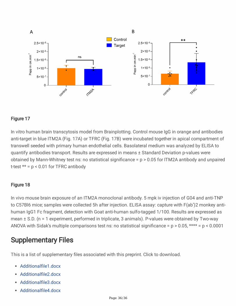
Page 36/36
Figure 17
In vitro human brain transcytosis model from Brainplotting. Control mouse IgG in orange and antibodiesanti-target in blue ITM2A (Fig. 17A) or TFRC (Fig. 17B) were incubated together in apical compartment oftranswell seeded with primary human endothelial cells. Basolateral medium was analyzed by ELISA toquantify antibodies transport. Results are expressed in means ± Standard Deviation p-values wereobtained by Mann-Whitney test ns: no statistical signi�cance = p > 0.05 for ITM2A antibody and unpairedt-test ** = p < 0.01 for TFRC antibody
Figure 18
In vivo mouse brain exposure of an ITM2A monoclonal antibody. 5 mpk iv injection of G04 and anti-TNPto C57Bl6 mice; samples were collected 5h after injection. ELISA assay: capture with F(ab’)2 monkey anti-human IgG1 Fc fragment, detection with Goat anti-human sulfo-tagged 1/100. Results are expressed asmean ± S.D. (n = 1 experiment, performed in triplicate, 3 animals). P-values were obtained by Two-wayANOVA with Sidak’s multiple comparisons test ns: no statistical signi�cance = p > 0.05, **** = p < 0.0001
Supplementary Files
This is a list of supplementary �les associated with this preprint. Click to download.
Additional�le1.docx
Additional�le2.docx
Additional�le3.docx
Additional�le4.docx


![Academic Sciences International Journal of Pharmacy and ... · Brain drug delivery strategies 1. Conventional strategies [2] ... that can be exploited as a means to target drugs to](https://static.fdocuments.net/doc/165x107/6029ff5c5dc3fc227174e0c0/academic-sciences-international-journal-of-pharmacy-and-brain-drug-delivery.jpg)


