Post-Transfer Outcomes in Cultured Bovine Embryos ... · Growth Factor, Fibroblast Growth Factor 2,...
86
Post-Transfer Outcomes in Cultured Bovine Embryos Supplemented with Epidermal Growth Factor, Fibroblast Growth Factor 2, and Insulin-Like Growth Factor 1 McCauley T. Vailes Thesis submitted to the faculty of the Virginia Polytechnic Institute and State University in partial fulfillment of the requirements for the degree of Master of Science In Animal and Poultry Sciences Alan D. Ealy Vitor R. G. Mercadante David A. Roper May 5, 2017 Blacksburg, Virginia Keywords: Embryogenesis, Pregnancy, Placenta
-
Upload
truongdien -
Category
Documents
-
view
218 -
download
0
Transcript of Post-Transfer Outcomes in Cultured Bovine Embryos ... · Growth Factor, Fibroblast Growth Factor 2,...

Post-Transfer Outcomes in Cultured Bovine Embryos Supplemented with Epidermal Growth Factor, Fibroblast Growth Factor 2, and Insulin-Like Growth Factor 1
McCauley T. Vailes
Thesis submitted to the faculty of the Virginia Polytechnic Institute and State University
in partial fulfillment of the requirements for the degree of
Master of Science In
Animal and Poultry Sciences
Alan D. Ealy Vitor R. G. Mercadante
David A. Roper
May 5, 2017 Blacksburg, Virginia
Keywords: Embryogenesis, Pregnancy, Placenta

Post-Transfer Outcomes in Cultured Bovine Embryos Supplemented with Epidermal Growth Factor, Fibroblast Growth Factor 2, and Insulin-Like Growth Factor 1
McCauley T. Vailes
Abstract
The high incidence of pregnancy loss is a major issue facing the cattle industry.
Use of in vitro fertilized (IVF) bovine embryos has become increasingly popular to help
alleviate several of these reproductive issues and provide a means to enhance genetic
gain for production traits. An uterine paracrine factor cocktail containing epidermal
growth factor (EGF), fibroblast growth factor 2 (FGF2), and insulin-like growth factor 1
(IGF1) (collectively termed EFI) was recently identified as a means for improving in vitro
derived bovine embryo development and trophectoderm cell numbers. The objectives of
this work were to determine if EFI treatment during in vitro bovine embryo culture
improves transferable embryo quality and post-transfer placental and fetal development.
For each replicate (3 total), slaughterhouse-derived bovine oocytes were matured and
fertilized in vitro. At day 4 post-fertilization, ≥8 cell embryos were harvested, pooled, and
exposed to either the EFI treatment (10ng/ml EGF, 10ng/ml FGF2, 50ng/ml IGF1) or
carrier only (1% Bovine Serum Albumin). At day 7, individual embryos were transferred
to estrous synchronized beef cattle. Artificial insemination (AI) was completed on a
subset of cows. The EFI treatment increased (P<0.05) the percentage of transferable
embryos. Pregnancy rate at day 28 post-estrus was similar among treatments.
Circulating concentrations of pregnancy-associated glycoproteins (PAGs) were
determined from plasma harvested at day 28, 42 and 56. Transrectal ultrasonography
was used to measure fetal crown-rump length (CRL) at day 42 and 56 and to determine
fetal sex at day 60. There were no main effect differences observed across days for
PAG concentration. Fetus sex by ET/AI group interactions were absent at day 28 but
existed at days 42 and 56 (P<0.05). At both days, this interaction reflected fetus sex-
dependent changes within the ET control group, where PAG concentrations were
greater (P<0.05) in male fetuses than female fetuses. No CRL differences or
interactions existed among fetal sex and pregnancy group. In summary, addition of the
EFI cocktail during bovine embryo culture improved the quality of transferable embryos,
but did not affect placental function or embryonic/fetal development. Increasing the
numbers of transferable embryos is of value given the cost of in vitro embryo
production, but no apparent increases in embryo or placental competency were
detected. The EFI treatment increased (P<0.05) the percentage of transferable
embryos.
Post-Transfer Outcomes in Cultured Bovine Embryos Supplemented with Epidermal Growth Factor, Fibroblast Growth Factor 2, and Insulin-Like Growth Factor 1
McCauley T. Vailes
General Audience Abstract
The high incidence of pregnancy loss is a major issue facing the cattle industry.
Use of in vitro fertilized (IVF) bovine embryos has become increasingly popular to help
alleviate several of these reproductive issues and provide a means to enhance genetic
gain for production traits. An uterine paracrine factor cocktail containing epidermal
growth factor (EGF), fibroblast growth factor 2 (FGF2), and insulin-like growth factor 1
(IGF1) (collectively termed EFI) was recently identified as a means for improving in vitro
derived bovine embryo development and trophectoderm cell numbers. The objectives of
this work were to determine if EFI treatment during in vitro bovine embryo culture
improves transferable embryo quality and post-transfer placental and fetal development.
For each replicate, slaughterhouse-derived bovine oocytes were matured and fertilized
in vitro and at day 4 post-fertilization, embryos were exposed to either the EFI treatment
(10ng/ml EGF, 10ng/ml FGF2, 50ng/ml IGF1) or carrier only (1% Bovine Serum
Albumin). Artificial insemination (AI) was completed on a subset of cows and the
remaining cattle receive embryos at day 7. The EFI treatment increased (P<0.05) the
percentage of transferable embryos. Pregnancy rate at day 28 post-estrus was similar
among treatments. Circulating concentrations of pregnancy-associated glycoproteins
(PAGs) were determined from plasma harvested at day 28, 42 and 56. Transrectal
ultrasonography was used to measure fetal crown-rump length (CRL) at day 42 and 56
and to determine fetal sex at day 60. There were no main effect differences observed
across days for PAG concentration. Fetus sex by ET/AI group interactions were absent
at day 28 but existed at days 42 and 56 (P<0.05). At both days, this interaction reflected
fetus sex-dependent changes within the ET control group, where PAG concentrations
were greater (P<0.05) in male fetuses than female fetuses. No CRL differences or
interactions existed among fetal sex and pregnancy group. In summary, addition of the
EFI cocktail during bovine embryo culture improved the quality of transferable embryos,
but did not affect placental function or embryonic/fetal development. Increasing the
numbers of transferable embryos is of value given the cost of in vitro embryo
production, but no apparent increases in embryo or placental competency were
detected. The EFI treatment increased (P<0.05) the percentage of transferable
embryos.

iv
Acknowledgements
I would first like to thank my parents and my sister for the endless support and
love and always letting me know how proud they are of my accomplishments. I would
also like to thank Jake Bailey for being there for me and being a shoulder to lean on. To
Erin Fitzgerald, thank you for knowing what to say and encouraging me throughout the
years. Also, to my lab mates and office mates, thank you for bringing laughter to my
days. Thank you to my awesome committee members, Dr. Vitor Mercadante and Dr.
David Roper, for your guidance and talent. Finally, to Dr. Alan Ealy, thank you for your
inspiration and teaching me how to think and write like a scientist.

v
Table of Contents
Acknowledgements ...................................................................................................... iv
List of Figures ............................................................................................................. vii
List of Abbreviations .................................................................................................. viii
Chapter 1 - Literature Review ....................................................................................... 1
Introduction ................................................................................................................ 1
Stages of Pregnancy Losses in Cattle ..................................................................... 2
Economic Impacts of Pregnancy Loss .................................................................... 4
Events Associated with Pregnancy Success in Cattle ........................................... 5
Estrous Synchronization and Detection ................................................................... 5
Oocyte Maturation .................................................................................................... 6
Fertilization ............................................................................................................... 7
Early Embryonic Development ................................................................................. 8
Maternal Recognition of Pregnancy ....................................................................... 10
Implantation ........................................................................................................... 12
Placental Development .......................................................................................... 12
Major Contributors to Pregnancy Loss in Cattle .................................................. 13
Oocyte Competency ............................................................................................... 13
Uterine Development and Function ........................................................................ 15
The Progesterone-Uterus Link ............................................................................... 16
Maternal Recognition of Pregnancy ....................................................................... 17
Implantation and Placental Failures ....................................................................... 18
Physiological and Environmental Contributors to Pregnancy Losses in Cattle 19
Metabolic Status ..................................................................................................... 20
Environmental Factors ........................................................................................... 21
Clinical/Subclinical Diseases .................................................................................. 23
Using Bovine In Vitro Fertilization to Study Early and Late Embryonic Losses 25
Epidermal Growth Factor ....................................................................................... 26
Fibroblast Growth Factor-2 .................................................................................... 27
Insulin-like Growth Factor 1 ................................................................................... 27
Multiple Uterine Factor Supplementation Schemes ............................................. 28
vi
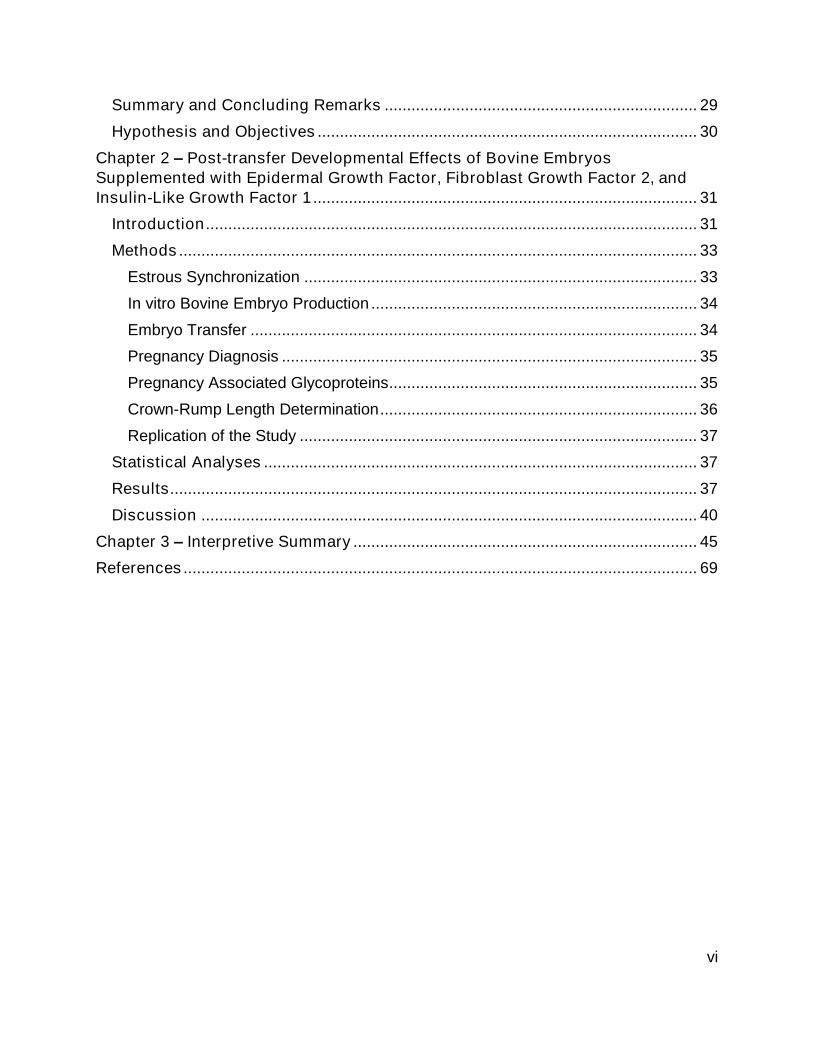
Summary and Concluding Remarks ...................................................................... 29
Hypothesis and Objectives ..................................................................................... 30
Chapter 2 – Post-transfer Developmental Effects of Bovine Embryos
Supplemented with Epidermal Growth Factor, Fibroblast Growth Factor 2, and
Insulin-Like Growth Factor 1 ...................................................................................... 31
Introduction .............................................................................................................. 31
Methods .................................................................................................................... 33
Estrous Synchronization ........................................................................................ 33
In vitro Bovine Embryo Production ......................................................................... 34
Embryo Transfer .................................................................................................... 34
Pregnancy Diagnosis ............................................................................................. 35
Pregnancy Associated Glycoproteins ..................................................................... 35
Crown-Rump Length Determination ....................................................................... 36
Replication of the Study ......................................................................................... 37
Statistical Analyses ................................................................................................. 37
Results ...................................................................................................................... 37
Discussion ............................................................................................................... 40
Chapter 3 – Interpretive Summary ............................................................................. 45
References ................................................................................................................... 69

vii
List of Figures
Figure 2-1 Experimental Design. .................................................................................. 47 Figure 2-2 Information on number of cows synchronized, detected in estrus, and
selected for AI or ET after completing a CIDR-Ovsynch protocol. .......................... 49
Figure 2-3 Cow AI/ET information. ............................................................................... 51 Figure 2-4 The influence of EFI treatment on the transferable quality of in vitro
produced bovine embryos. ..................................................................................... 53
Figure 2-5 Percentage of cows pregnant at day 28 and 56 of gestation....................... 55 Figure 2-6 Circulating concentrations of pregnancy specific protein B (PSPB) at days
28, 42, and 56 of gestation. .................................................................................... 57 Figure 2-7 Effect of fetal sex on circulating pregnancy specific protein B (PSPB)
concentrations in pregnant cows at days 28, 42, and 56. ...................................... 59
Figure 2-8 The interaction between fetal sex and treatment on circulating PSPB
concentrations. ....................................................................................................... 61
Figure 2-9 The effect of treatment on crown-rump lengths (CRL) in cattle at days 42
and 56 of gestation. ................................................................................................ 63 Figure 2-10 The effect of fetal sex on CRL at day 42 and 56. ...................................... 65
Figure 2-11 Influence of treatments and fetal sex on CRL at day 42 and 56. ............... 67

viii
List of Abbreviations
AI Artificial insemination ANOVA Analysis of variance bFGF Basic fibroblast growth factor CIDR Controlled internal drug release CNL Crown-nose length COC Cumulus-oocyte complex CRL Crown-rump length EGA Embryonic genome activation EGF Epidermal growth factor ET Embryo transfer FGF Fibroblast growth factor FGF2 Fibroblast growth factor 2 FGF2 Fibroblast growth factor 2 FGF7 Fibroblast growth factor 7 FSH Follicle stimulating hormone GLM General linear model GM-CSF Granulocyte-macrophage colony-stimulating factor GnRH Gonadotropin releasing hormone ICM Inner cell mass IFN Interferon IFNT Interferon-tau IGF1 Insulin-like growth factor 1 IGF2 Insulin-like growth factor 2 ISG Interferon stimulating genes IVF In vitro fertilization IVP In vitro produced LH Luteinizing hormone LIF Leukemia inhibitory factor MET Maternal to embryonic transition MRP Maternal recognition of pregnancy OPU Ovum pickup P4 Progesterone PAG Pregnancy associated glycoproteins PBL Peripheral blood leukocytes PDIFF Probability of difference PGF2α Prostaglandin F2 alpha SAS Statistical analysis system SOF Synthetic oviduct fluid TE Trophectoderm TGF-β Transforming growth factor-beta UGKO Uterine gland knockout ZP Zona pellucida

1
Chapter 1 - Literature Review
Introduction
Pregnancy loss is one of the most predominant issues facing the dairy and beef
cattle industries, causing reduced production efficiency and economic loss. Failure of
lactating dairy cows to establish and carry a pregnancy to term results in an average
loss of $555 per pregnancy [1]. Various environmental and physiological factors
contribute to pregnancy loss, with uterine function being one of the major contributors to
infertility. The use of various assisted reproductive techniques has been implemented to
evade the damaging effects suboptimal uterine environments and other environmental
and physiological factors have on embryonic development. In vitro production (IVP) of
bovine embryos and embryo transfer (ET) is gaining popularity globally as a means for
improving genetic potential in beef and dairy cattle [2].
There are three primary stages of pregnancy loss established by the Committee
on Bovine Reproductive Nomenclature (1972). The first stage is early embryonic death,
which encompasses the period of undetected losses that occur within the normal range
of an estrous cycle (16-24 days) in cattle [3]. The second stage of pregnancy loss is
termed late embryonic death which occurs between 25 and 42 days of gestation [3].
During this period, corpus luteum (CL) function is maintained beyond a normal estrous
cycle length. The third period of pregnancy losses, which occurs after day 42 when a
fetus is distinguishable, are termed fetal losses [3]. Of the three stages, the majority of
pregnancy losses occur during the early embryonic stage [4]. Improvements in early
embryonic development to reduce these early pregnancy losses must, therefore, be
explored. The following literature review will go into further detail of the stages of

2
pregnancy loss, economic impacts of losses, events of pregnancy success, major
contributors to pregnancy loss and potential ways to reduce these losses.
Stages of Pregnancy Losses in Cattle
Pregnancy loss is loosely defined as losses in pregnancies that occur throughout
gestation immediately after sygamy of the male and female pronuclei and creation of a
new life form. Infertility in cattle is typically reflected in embryonic or fetal mortality,
which is seen through poor fertilization rates and improper pregnancy maintenance.
Fertilization rates have not changed much over the past several decades, therefore
problems with fertility usually are caused by increases in embryonic or fetal losses.
Losses are more prevalent in lactating cows due to the metabolic stresses of milk
production. Cows will normally experience a negative energy balance in early lactation
due to the metabolic demands of lactation, and this has detrimental effects on
embryonic development. Pregnancy losses also are influenced by various
environmental factors. For example, fertilization rate in lactating cattle is reduced to
55% during heat stress periods [5].
There are three primary stages of pregnancy loss in the cow. These stages have
been established by the Committee on Bovine Reproductive Nomenclature (1972). The
first stage is early embryonic death. This occurs between days 1 and 24 of pregnancy.
This period encompasses undetected losses that occur within the normal range in
estrous cycle lengths in cattle (16 to 24 days) [3]. These pregnancy losses occur prior to
the time of corpus luteum (CL) maintenance, or maternal recognition of pregnancy,
which will be described later in this review. Periods of early embryonic loss include pre-
embryonic genome activation (EGA), post-EGA, and maternal recognition of pregnancy.

3
The bovine EGA becomes activated 3-4 days post-fertilization, therefore any alterations
in this process may cause pregnancy failure [6]. Additional early embryonic losses occur
between EGA and maternal recognition of pregnancy. Maternal recognition of
pregnancy, which will be discussed in further detail in subsequent sections, occurs at
day 16 of pregnancy.
Lactation is a prominent cause for early embryonic losses. In moderate-
producing cows, early embryonic losses are about 40% based on a 90% fertilization
rate. Also, it is estimated that 70-80% of these early embryonic losses occur between 8
and 16 days after insemination [4]. Embryo viability studies completed in dairy cows
found approximately 50.0, 57.9, and 71.9% of potential pregnancies in parous lactating,
parous nonlactating, and nulliparous heifers, respectively, to be viable by days 5-6 post-
insemination [3]. A similar pattern is noted in beef cows where viability of embryos
collected from parous lactating, parous nonlactating, and nulliparous heifers is 57.5,
79.5, and 77.6%, respectively [3]. These data confirm the impact lactation has on
escalating early embryonic losses and limiting pregnancy maintenance.
The second stage of pregnancy loss is termed late embryonic death. This occurs
between 25 and 42 days of gestation. This is the period where CL function is
maintained beyond a normal estrous cycle length. It also contains the initiation of
implantation, placentome formation and completion of much of embryonic differentiation
[7]. Lactating dairy cows display extensive late embryonic death compared to beef
cattle, with an average loss of 12.8% during this period [3, 8]. Greater late embryonic
losses could be attributed to the increased demand of lactation on the body in the dairy

4
cow. Additionally, placental insufficiencies such as reduced blood flow or inadequate
nutrient supply can contribute to late embryonic losses.
At day 42-45 of gestation, the embryo has fully attached to the uterus and a fetus
has become distinguishable. Thus, pregnancies failures after day 42, the third period of
pregnancy losses, are termed fetal losses [3]. Fetal losses are less frequent in both beef
and dairy cattle. The causes for these losses generally go undetermined if not caused
by known infectious agents [3]. In beef and dairy cows, fetal losses are generally low
averaging 2.5 and 4.2%, respectively.
Economic Impacts of Pregnancy Loss
Substantial economic losses result from pregnancy failures in cattle. This has
been well described in dairy cattle. The value of pregnancy varies depending on stage
of lactation, lactation number, milk yield, and reproductive status. Cattle that have a
longer interval between post-calving and re-breeding are more costly to the producer,
therefore retaining pregnancies is economically crucial [9-11]. The average loss per
pregnancy in lactating dairy cows is $555, while the average added value of a new
pregnancy is $278 [1]. For example, a 100-head dairy operation with a 40% early and
late embryonic loss rate will lose $22,200 in revenue from pregnancy losses. By
contrast, a 100-cow dairy with only a 15% early and late embryonic loss rate reduce
total economic losses to $8,325 and gain $6,950 with added pregnancies. The
economic impact of pregnancy loss outweighs the value of a new pregnancy, so it is
crucial to reduce early pregnancy losses and minimize economic losses.

5
The negative economic impact of early and late embryonic losses also exists in
the beef industry. The timing of calving is coupled with conception rates, thus the ability
to get cows pregnant in a short timespan has an important influence on economic
return. Most of the income cow-calf operations generate is determined by the number of
calves sold and by the weight of these calves when they are sold. Cows that achieve
pregnancy after first insemination and avoid pregnancy loss contribute to improved calf
crop and greater weaning weights. Producers can increase net return by $204 per cow
by simply improving net calf crop by 10% and increasing weaning weight by 200 pounds
[12].
Events Associated with Pregnancy Success in Cattle
Various time-sensitive events must occur to ensure pregnancy success in cattle.
The following paragraphs will go into these specific developmental events in more
detail. Among these events, heat detection has improved timing of fertilization and
pregnancy rates. Also, advancements in estrous synchronization protocols have
eliminated the heat detection component and improved conception rates when
implemented. Oocyte maturation, fertilization, and cleavage events are all necessary for
a normal pregnancy. Early embryonic development and pregnancy maintenance is
regulated by maternal recognition of pregnancy and subsequent implantation and
placentation. Progression through these events is imperative for pregnancy success in
cattle.
Estrous Synchronization and Detection
Based on several sources, heat detection has drastically improved throughout
recent history, and especially over the past 15-20 years. This has reduced days to first

6
conception and increased the number of inseminations within a set breeding season.
Both of these outcomes have increased resulting pregnancy rates. Prior to the use of
drug-based ovulation strategies, detecting estrus was a major contributor to infertility in
operations that artificially inseminated cattle. Only 30-70% of cows that exhibit heat are
detected in heat, which leads to inadequate timing of fertilization and failure to maintain
pregnancy [13]. High producing dairy cows are more likely to have decreased heat
expression due to the high metabolic hormone turnover.
The development of synchronization protocols has alleviated the heat detection
issue. The first timed artificial insemination (AI) protocol developed targeted the
synchronization of ovulation using gonadotropin releasing hormone (GnRH) and
prostaglandin F2 alpha (PGF2α) [14]. Estrus detection is associated with greater fertility
when using a timed AI protocol. Current estrous synchronization protocols focus on
synchronizing CL function and follicular waves. Progesterone inserts, or Controlled
Internal Drug Release (CIDR) allow emergence of a synchronized follicular wave
around 4 days after initiation of protocol [15, 16]. Timed AI protocols are used in
conjunction with intravaginal inserts to remove the heat detection component and result
in reasonable conception rates.
Oocyte Maturation
Oocyte competency is defined as the ability of an oocyte to complete maturation,
undergo successful fertilization and cleavage, develop into a blastocyst, and achieve a
viable offspring [17, 18]. Oogenesis, the process of developing oocytes, begins during
pre-natal development and is completed by birth in cattle [18]. Primordial germ cells
proliferate through mitotic divisions before birth, producing primary oocytes. At birth, the

7
nucleus of the primary oocytes become dormant, a phenomenon termed nuclear arrest.
This nuclear arrest continues until fertilization occurs. However, cytoplasmic
development continues. This is especially evident during folliculogenesis, when
maternal mRNA and proteins are loaded and transferred into oocytes [19]. Cytoplasmic
reserves of RNA accumulate until oocyte becomes full size, and this material will
sustain the oocyte and early-developing embryo until the embryonic genome is
activated [20].
Oogenesis progresses in synchrony with folliculogenesis through the
interdependence of cellular communications between the oocyte and follicle [18].
Folliculogenesis begins with primordial follicles and continues development though
primary, secondary, tertiary, and antral follicles. The Graafian, or antral, follicle contains
theca and granulosa cells which have receptors for luteinizing hormone (LH) and follicle
stimulating hormone (FSH), respectively. Follicle growth is stimulated by FSH and the
LH surge causes ovulation. Waves of follicle growth and atresia occur continuously
throughout the reproductive period and contribute to oocyte competency.
Fertilization
Fertilization rates in cattle are generally high. Lactating beef cattle have an
average fertilization rate of 75%, likely due to the prevalence of post-partum anestrous
periods, whereas non-lactating beef cattle have an average rate of 98.6% [3, 21-23].
Following a similar pattern, fertilization rates in lactating and non-lactating dairy cattle
are around 75% [3]. Lactating dairy cows have much lower conception rates due to the
increased nutritional demand for milk production, which negatively affects embryo

8
quality [5]. Lactating beef cattle can recover body condition and resume cyclicity, with
conception rates ranging from 50-65% [3].
Successful fertilization in the cow requires proper sperm travel and capacitation
processes in the oviduct, concurrent with oocyte ovulation. After sperm capacitation has
occurred in the uterus or oviduct, the sperm encounters the egg and attaches to the
zona pellucida (ZP) initiating the acrosomal reaction. This reaction releases hydrolytic
enzymes packaged within the acrosome, which digest the ZP and allow spermatozoa
penetration of the ZP. The fusion of the female and male pronuclei, or syngamy, results
in formation of a zygote. The first cleavage events occur 23-31 hours post fertilization,
producing a 2-cell stage embryo [24]. Cleavage to the 4- and 8-cell stage occurs 36-50
hours and 56-64 hours post-fertilization, respectively. The final cleavage event, which
yields a 16-cell stage embryo, occurs 80-86 hours post-fertilization. Cells produced by
cleavage events are referred to as blastomeres. A single cell (blastomere) can give rise
to a complete individual and is considered totipotent at these initial stages of
development. Cell number increases post-fertilization, but the cytoplasm mass remains
the same. The embryo travels through the uterine horn during cleavage events to the
uterine body to continue development.
Early Embryonic Development
The oocyte and early developing embryo must rely on stored mRNA and protein
for survival and developmental signals. Thus, the activation of the embryonic genome is
essential for continued survival of embryos. In cattle, this event occurs around the 8- or
16-stage cell embryo, or 3-4 days post-fertilization [6]. At this time, the embryo begins to
experience maternal to embryonic transition (MET). The depletion of maternal

9
transcripts, replacement of maternal transcripts by embryonic transcripts, and
generation of new embryo specific transcripts are involved in MET [25]. By day 5,
maternal stores are usually depleted and the embryo controls its own development. The
embryonic genome transition is imperative to survival, as it allows the embryo to
respond to environmental stresses and any perturbations that may harm its
development [6].
As blastomere cleavages continue, the embryo begins a process referred to as
compaction. Compaction occurs when blastomeres increase contact with neighboring
cells, forming a compact sphere of cells. This change in cellular behavior, in which
number of cells becomes difficult to count, is known as the beginning of the morula
stage. As the morula progresses through development, a cavity is formed by a process
known as cavitation. Cavitation and formation of the internal fluid cavity, termed
blastocoel, marks the progression of the morula to the blastocyst stage of development.
This process typically occurs around day 7 post-fertilization in cattle.
After cell divisions have concluded, two distinct cell populations are formed. The
outer cells are termed trophectoderm (TE) and the inner cells are termed inner cell
mass (ICM) [26]. The ICM will give rise to the embryo proper, or body of the embryo
which eventually forms the fetus. Additionally, extraembryonic membranes including the
yolk sac, amnion, and allantois will be formed from the ICM cells. Trophectoderm cells
give rise to the chorion, ultimately forming the placenta. Extraembryonic membrane
development is essential for embryonic development and subsequent uterine
attachment.

10
The developing embryo reaches the blastocyst stage by day 7, begins hatching
from the ZP, and emerges as a spherical conceptus. Following the blastocyst stage,
bovine TE cells undergo extensive proliferation termed conceptus elongation.
Simultaneously with TE proliferation, the ICM-derived yolk sac continues growth and
reaches peak development by day 20 of pregnancy. After peak development is reached,
the yolk sac regresses as its nutritive function is taken over by the emerging allantois
[27]. This elongation process occurs between day 14 and 21 of gestation in cattle [28].
By day 16 of pregnancy, the bovine conceptus reaches a length of 10-30 cm. By day
21, or time of uterine attachment, the conceptus reaches a length of 50-200 cm.
Elongation is essential to increase surface area which aids conceptus apposition,
attachment, and adhesion to the uterine wall [29].
Maternal Recognition of Pregnancy
Pregnancy recognition and maintenance requires cross talk between the
conceptus and maternal system. Signaling from the embryo between days 16-25
determines if pregnancy is prolonged, or a new estrous cycle begins. Improper signaling
results in embryonic death and pregnancy loss.
During elongation, the conceptus signals its presence to prolong CL life, thus
termed the period maternal recognition of pregnancy (MRP). Interferon tau (IFNT) is a
member of the Type 1 interferon (IFN) family and is the maternal recognition of
pregnancy factor in ruminants [30-32]. Type 1 IFNs are involved in immune responses
and anti-proliferative activities [30, 33]. However, unlike other Type 1 IFNs, IFNT has
evolved to serve a key role in pregnancy recognition in ruminant species.

11
Interferon tau is secreted from the mononuclear cells of the trophectoderm.
Interferon tau mRNA is present during the preimplantation stage of development, with
the earliest detection around day 7, or the morula/early blastocyst period [34-37]. During
elongation, IFNT secretions increase with maximal secretion around day 12-13 of
gestation in sheep and day 16-19 in cattle [30, 38]. The conceptus of ruminants
transforms through spherical, tubular, and filamentous stages with increases in
trophoblast cell numbers, corresponding to higher IFNT secretion and attachment
preparation [31, 39, 40].
The primary action of IFNT is to function as an antiluteolytic agent so CL
regression can be prevented [30-32]. Through paracrine signaling, IFNT acts locally on
the endometrium prevent CL regression [30, 31, 40]. Estrogen and oxytocin receptor
expression is suppressed by IFNT, and this blocks the pulsatile release of PGF2α and
its ability to induce CL regression [30, 31, 41]. Ovine trophoblast protein-1, the ovine
specific IFNT, extends the lifespan of the CL and prevents luteal regression when
injected into the uterine lumen of non-pregnant ewes [42]. Similarly, bovine trophoblast
protein-1 significantly increases the inter-estrous interval when infused through the
cervix of cyclic cows [43].
Additionally, IFNT induces expression of classical interferon-stimulating genes
(ISG) in the uterus, including ISG15 and OAS [44, 45]. Increased expression of these
and other ISGs are correlated with regulation of uterine receptivity, conceptus
elongation, and implantation [31, 45]. Consequently, impaired conceptus development,
insufficient production of IFNT, and MRP inhibition will be translated in reduced or no
ISG expression in maternal circulation. Expression of ISG in maternal blood circulation

12
can be used as an indicator of early embryonic loss, specifically around MRP and
implantation events [46]. Interferon stimulating genes will be discussed in further detail
in subsequent sections.
Implantation
Approximately 10 to 12 days after hatching and 7 to 9 days after elongation
begins, the conceptus begins making contact with the uterine luminal epithelium. This
occurs on or shortly after day 19 of pregnancy in cattle [47]. At this time, the conceptus
typically spans the length of the uterine horn ipsilateral to the CL, and extends into the
contralateral horn. The steps that precede implantation include apposition and
adhesion of conceptus trophectoderm to uterine and luminal epithelium [29, 48].
Placental Development
As implantation is underway, post-implantation placental development
commences. One major event that occurs in cattle and other ruminants is the formation
and migration of bincucleate cells from the chorion into the maternal endometrial
epithelium. As apposition and adhesion occur, the trophectoderm begins producing a
specialized, invasive cell termed the binucleate cell. These cells will migrate to fuse with
the endometrial luminal epithelium [48, 49].
This invasive cell type is one way that a placental unit is established. The other
way this is established is by the great surface area contact developed in these
placentae. Cattle have a cotyledonary placenta, and thus the uterus contains numerous
aglandular nodules termed cotyledons. These serve as placental attachments with
cotyledons, structures that contain the ability to produce highly interdigitated, villous

13
networks with caruncles. These attachments are collectively called placentomes.
Around day 25, the chorion initiates attachment to the caruncles of the uterus. By day
40, placentomes are well established [26]. Placentome growth occurs throughout
gestation and cotyledons increase many-fold, measuring up to 6 inches in diameter in
late pregnancy. This growth provides greater surface area to support placental transfer
of nutrients from the maternal side and metabolic wastes from the fetus. Fetal
development through the remainder of gestation is completely reliant on nutrients
delivered through the placenta.
Major Contributors to Pregnancy Loss in Cattle
There are many contributors to pregnancy failure in cattle. Among these are
oocyte competency, uterine development and function, MRP, and implantation/placental
development. As has already been discussed, the collective functioning of these factors
is essential to establish and maintain pregnancy. Therefore, any perturbation along the
way can result in pregnancy loss. The following section will describe the major
contributors to pregnancy loss in greater detail.
Oocyte Competency
Follicle size is one factor that has a direct effect on oocyte competence. Higher
blastocyst rates are observed in oocytes retrieved from large follicles (2-6mm) and
oocytes surrounded by more cumulus cell layers collected from large follicles [50, 51].
Additionally, higher cleavage rates (early embryonic cell division) and blastocyst number
are noted when retrieving oocytes from >4-8mm (large) and >2-4mm (medium) follicles
[52]. Conversely, small follicles had no day 7 blastocysts and low cleavage rates [52].
Oocytes collected from small follicles have reduced quality, validating the relationship
14
between follicle size and oocyte quality [51, 53, 54]. There is a likely factor inherent in
oocytes from small follicles that limits development, but the mechanism responsible for
these differences are unclear. However, ultrastructural changes in the oocyte and
estradiol limit achievement of developmental competence, thus maturation of the oocyte
and exposure to estrogens seems to facilitate competency [51-53].
The specific phase of follicle development is another factor affecting oocyte
competence. There is improved development and increased quality of transferable
blastocysts from oocytes in the growth phase (day 2 and 10) compared to the dominant
phase (day 7 and 15) of folliculogenesis [17, 54]. In addition, large follicles collected
during the growth phase (day 3) have higher blastocyst development rates compared to
oocytes from the dominant phase (day 7), which demonstrate lesser developmental
potential [17].
Atretic follicles can impact oocyte competency positively or negatively, depending
on the stage of atresia. Oocytes showing beginning signs of degeneration (Class 3), are
more likely to develop than late atretic follicles [53]. Class 3 oocytes appear to be more
differentiated and therefore are still able to obtain developmental competence. A small
degree of cumulus expansion still allows a competent oocyte to form [55]. There are
higher blastocysts rates in early atretic follicles with minor cumulus expansion, but not
late atretic follicles [56]. The level of atresia and cumulus expansion is highly correlated
with oocyte developmental potential, as demonstrated by the findings of these studies.
One major determinant of oocyte competency is the level of P4 that exists during
follicle selection and dominance. Oocyte quality, rate of embryo development, and

15
uterine activity is altered by low P4 levels [57, 58]. One time that low P4 exists is during
anestrous periods. Postpartum cows producing large amounts of milk may not be able
to support the early embryo as a result of elevated steroid metabolism and lower P4
concentrations, though a large volume of luteal tissue is present [59, 60]. Thus, lower
blastocyst rates are noted. Oocytes transferred to heifers have a higher blastocyst rate
of 33.9±3.6% compared to 18.3±7.9% blastocyst rate in postpartum cows, but no
difference in blastocyst quality was reported [60].
Uterine Development and Function
The positive interaction between the conceptus and uterus is necessary for
maintenance of pregnancy in the cow. Although most female reproductive organs are
fully developed at birth, the uterus is differentiated and developed postnatally in cattle
[61, 62]. Ruminants have a bicornuate uterus with a common corpus and single cervix
[63]. The endometrium in cattle and sheep consists of aglandular caruncular areas and
glandular intercaruncular areas, which provide nutrients to the developing embryo
through fusion of cotyledons, forming a placentome [64]. Adenogenesis, or the process
whereby uterine glands develop, involves the differentiation of lumen epithelium to
glandular epithelium and coiling and branching of germinal epithelium through stroma
[62]. Uterine glands synthesize and secrete or transport a variety of enzymes, growth
factors, cytokines, hormones, and proteins that provide histotrophic nutrition to the
conceptus [65].
Proper functioning uterine glands are essential for uterine receptivity, blastocyst
implantation, and embryonic growth in sheep and mice [62, 66-68]. In uterine gland
knock out (UGKO) ewes, blastocysts are present and development is normal, however

16
conceptuses are not detected in day 14 uteri flush [62]. Fertilization and transport
defects were eliminated by transferring day 7 sheep embryos from normal, non-UGKO
donor sheep thus confirming the importance of uterine glands for establishment of
pregnancy.
The Progesterone-Uterus Link
Progesterone during the peri-implantation stage is critical for establishment and
maintenance of pregnancy in ruminants. Early conceptus growth and development, and
specifically elongation, is regulated by P4 acting on the uterus [57, 69-71]. Progesterone
regulates endometrial receptivity and secretions by changing gene expression, which
alters the composition of histotroph essential for conceptus survival and growth [72].
Histotroph is comprised of several hormones, cytokines, growth factors, lipids, amino
acids and nutrients which are vital for embryonic development [31]. Larger day 14
conceptuses were found in P4-treated cows than in non-treated cows [57, 73]. Also,
providing P4 early after ovulation can hasten conceptus development. Vaginal insertion
of a P4-releasing device at day 3 of gestation enhanced P4 concentrations four- to five-
fold and increased conceptus length in vivo. This treatment induced changes in the
uterine environment that advanced elongation and increased interferon-tau
concentrations [72, 74].
It may be possible to use this knowledge about P4’s role in early pregnancy to
improve pregnancy outcomes. One study also found that early P4 exposure increased
pregnancy rates, but others found no such effect of this treatment [75, 76]. This type of
treatment likely does not work effectively because although an advancement in
conceptus development occurs, uterine events also are advanced, and cattle return to
17
estrus earlier than normal. Thus, any advantages seen in conceptus development likely
will be offset by the shorted window for maternal recognition of pregnancy. However, it
may be possible to enhance pregnancy retention by providing supplemental P4 later in
early pregnancy. In one recent study, P4 supplementation beginning at day 4, and
again at day 7 post-AI improved pregnancy per AI rates when cows were inseminated
following estrus detection [77]. When a timed-AI protocol was implemented, P4
supplementation beginning at day 4, or during metestrus, improved pregnancy per AI
rates. Supplemental P4 is beneficial to improving pregnancy rates, but the magnitude of
benefit depends on stage of the cycle and days post-AI when supplementation occurs.
Maternal Recognition of Pregnancy
Improper IFNT signaling during MRP leads to pregnancy loss, with losses during
this period averaging around 30% [78]. There are several potential reasons as to why
MRP might fail. First, the developing embryo must fend off the maternal immune
response that attempts to terminate pregnancy because of the paternally inherited
proteins [78]. Irregular elongation, insufficient histotroph, and disruption in
trophectoderm formation can also restrict IFNT signaling by the embryo. Finally,
suboptimal circulations of P4 during the early luteal phase, which suppress embryonic
growth, reduce pregnancy success during the MRP period [72].
One measurable means for determining how well IFNT is acting in early
pregnancy is to examine how it influences the expression of ISGs in peripheral
leukocytes [46, 79]. Transcripts for these ISGs are measured in the maternal peripheral
blood leukocytes (PBL) and are used to identify non-pregnant cattle [46, 80]. Increases
in ISG mRNA abundance can be detected as early as day 17-18 of pregnancy, but the

18
greatest amount of ISG are seen on days 20-22 of pregnancy, likely due to the
conceptus secreting larger quantities of IFNT at this time [80]. Mx1, Mx2, and ISG-15
are a few of the most common ISG detected in PBL [46, 81, 82].
Implantation and Placental Failures
Poor placental development during gestation will ultimately cause pregnancy
failures. This impaired development can result from improper implantation, poor
fetal/maternal interaction, and reduced nutrient exchange. The best way to examine
placental fitness and fetal well-being is to look at PAG concentrations.
Pregnancy associated glycoproteins are aspartic proteases that are secreted into
the bovine maternal bloodstream throughout pregnancy from binucleate cells of the
placenta [83-86]. Measured through presence of pregnancy specific proteins in plasma,
PAG are used as pregnancy predictors by day 24 of pregnancy. Plasma PAG
concentrations increase around day 24, peak at approximately day 30 of gestation, and
decline through the second month of gestation before increasing again in late
pregnancy [87]. Pregnant cows that will maintain their pregnancy to term have greater
plasma PAG concentrations at day 30 than cows that will experience late embryonic
mortality [87, 88]. However, cows with late fetal losses have relatively normal PAG
concentrations at day 32 [87, 88].
High circulating PAG concentrations may also be indicative of impending
pregnancy loss compared to average PAG concentrations [89]. These increased PAG
concentrations could be due to the mother overcoming placental insufficiency, ultimately
destructing the fetal-placental unit and releasing the placental products into circulation

19
[88]. Concentrations of PAG are associated with parity, AI service number, milk
production, and metabolic diseases linked with pregnancy failure [88, 89]. Breed is also
associated with PAG concentration. This concentration difference is noted in Brangus
cattle, which have higher concentrations than Angus cattle throughout pregnancy [86].
Another way to quantify the normal or abnormal progress of pregnancy is to
examine fetometric growth parameters, such as crown-rump length (CRL) or crown-
nose length (CNL) [90]. Crown-rump length is the distance between the highest point of
the head, or crown, and the most caudal extent of the buttocks at the tail head [91]. The
entire bovine fetus can be scanned in month 2 of gestation, with accessibility of the
thorax, abdomen, and pelvis declining to about 50% in months four and five [92].
Crown-rump length is limited approximately to day 70 of gestation as fetuses larger than
10 centimeters are difficult to project onto the screen [92]. The length of the head has
linear correlation with CRL and therefore can be substituted for CRL if hard to obtain
[90]. In Kahn’s [92] study, the head was accessible 80% of the time and correlated with
fetal age. Crown-rump length showed the least variation and highest correlation to age
of pregnancy [92]. Thus, the size of the bovine fetus is directly proportional to fetal age
and demonstrates linear growth throughout gestation.
Physiological and Environmental Contributors to Pregnancy Losses in Cattle
These common causes of pregnancy loss can manifest in several ways. The
animal’s metabolic status, especially the occurrence of a negative energy balance, can
impact various aspects of the reproductive process. For example, the developing
embryo is very sensitive to stress during early pregnancy. In addition to metabolic
stress, heat stress and other environmental factors can negatively affect embryo

20
competence. Clinical and subclinical periparturient diseases are also major issues in
high producing dairy cows. The incidence of multiple diseases further reduces
conception and pregnancy rates. This section will describe these and other major
contributors to pregnancy loss in cattle.
Metabolic Status
Metabolic status of the cow, such as high nutrition, negative energy balance, and
body condition score, greatly affects embryonic and fetal survival. Cattle grazing in a
high-energy pasture prior to AI have more than a 30-percentage point reduction in
embryo survival compared to cattle grazing a lower energy pasture [93]. By contrast,
during periods of extreme negative energy balance, intake is insufficient for general
body maintenance, therefore reproductive parameters and oocyte quality are greatly
compromised [94]. Body condition loss is another aspect that significantly affects
fertility. Underweight cows are subjected to longer periods of anestrous, lengthening the
breeding interval [95]. Additionally, a one unit drop in body condition score from calving
to 30 days post-partum increases the odds ratio for pregnancy loss by 2.41-fold [96].
Another important facet of metabolism is how it may control circulating steroid
hormone concentrations. A high plane of nutrition (i.e. consumption of high quantities of
feedstuff, and especially grains) will reduce circulating estradiol and P4 concentrations
in cattle [97]. This occurs, at least in part, because the high metabolic activity of the
liver inadvertently increases steroid hormone metabolism, and this reduces circulating
concentrations of these hormones [93, 97].

21
This scenario of lowering steroid hormones is especially dramatic in lactating
dairy cows. The increased feed intake needed to supply the energy requirements for
lactation and resulting liver steroid metabolism are likely major contributors to infertility.
Reductions in estradiol adversely affect ovarian dynamics, and specifically follicle
development, creating a smaller follicle containing a less competent oocyte [97].
Reductions in P4 are thought to reduce uterine activity, and reductions in uterine
histotroph production likely reduce conceptus development and hinder maternal
recognition of pregnancy success [72, 74, 78].
Environmental Factors
One environmental factor that can have dramatic adverse effects on fertility is
heat stress. To manage the effects of heat stress, cattle exert energy to maintain body
homeostasis and dissipate heat, which negatively impacts reproductive parameters.
The effects of heat stress can be attributed to decreased thermoregulatory ability,
hyperthermia, or elevated uterine temperature [98, 99]. The temperature at which stress
events begin, or the upper critical temperature, is 25-26°C in high-yielding dairy cows
[100].
Several facets of reproduction are adversely affected by heat stress.
Folliculogenesis, specifically follicular development and dominance, is altered during
high heat exposure, and this decreases oocyte and subsequent embryo quality [101,
102]. Oocytes are extremely temperature sensitive, and any perturbation can inhibit
their development [103]. Heat stressed oocytes have reduced competence to develop
into blastocysts as the uterus is unable to support embryo metabolism [98, 104].

22
Additionally, uterine blood flow is decreased, diminishing the delivery of nutrients to
support oocyte development while increasing uterine temperature [105].
Heat stress also contributes to reduced reproductive efficiency by limiting
reproductive estrus expression/detection and decreasing conception rate [5]. There are
decreases in estradiol-17β concentrations in heat stressed cows, and these reductions
in estradiol limit estrus behavior [101, 102, 106, 107]. Mounting activities are especially
reduced during the summer not only by reduced hormonal expression but physical
lethargy [108].
There also are major problems with early embryo development during heat
stress. Final oocyte maturation and ovulation are disrupted by elevated temperatures,
inhibiting subsequent embryo development. Embryos retrieved from cattle heat stressed
on day 1 post-estrus have decreased viability and development [109]. Additionally, there
is a reduced percentage of embryos at the blastocyst stage of development when heat
stress occurs on day 1 post-estrus. There is no effect of heat stress on embryonic
development when stress occurs on day 3, 5, or 7 [109]. This is indicative of the
embryo’s developmental resistance and thermotolerance to maternal heat stress.
One way to overcome many of the detrimental effects of heat stress on the
oocyte and early embryo is through ET. Transferring embryos from non-heat-stressed
cows into heat stressed recipients increases pregnancy rates [110]. Prior to EGA, heat
stress alters steroid synthesis, disrupts oocyte maturation, and blocks development at
the 1-4 cell stage embryo. Once EGA occurs, the embryo becomes heat resistant and
can defend itself from the detrimental effects heat stress imposes on subsequent

23
embryonic growth. Defense against heat stress improves as pregnancy progresses,
therefore ET bypasses the most heat sensitive time and allows proper embryonic
development [108, 109].
Fescue toxicosis is another environmental factor that causes infertility in cattle.
Endophyte-infected fescue contains Acremonium coenophialum, a fungus that resides
in fescue grass which produces toxins termed ergo alkaloids [15]. Cattle grazing
infected fescue have reduced pregnancy rates [15, 111]. The mechanisms by which
fescue toxicosis reduces reproductive performance are not fully understood, but at least
two major causes for infertility exist. The first is a potential direct effect of alkaloid toxins
on oocyte and embryo development [112]. Early stage embryos are extremely sensitive
to ergot alkaloid and pregnancies are typically lost before day 30 of gestation [112]. A
contributing, secondary factor to this infertility occurs because fescue toxicosis
heightens thermal sensitivity. Cattle feeding on infected fescue spring and summer
months are undoubtedly less fertile because of the damaging effects increased body
temperature has on embryonic survival [15].
Clinical/Subclinical Diseases
Clinical and sub-clinical periparturient diseases are associated with reduced
reproductive performance in cattle [113]. The prevalence of periparturient diseases in
dairy cattle range from ~8-50%, depending on the disease. Inflammation, discomfort,
and fever are characteristic for disease-ridden cows. Elevated body temperature and a
suboptimal uterine environment increase the risk of pregnancy loss, as the cow is
unable to achieve and maintain a normal pregnancy with these issues.

24
Mastitis is one of the most prominent diseases affecting lactating dairy cows
[114]. Clinical mastitis is identified by alterations in milk composition and appearance,
decreased milk production, swelling or heat in the infected quarters, or elevated body
temperature [115]. There is a decreased conception rate both at first postpartum AI and
in overall pregnancy rate in cows experiencing clinical mastitis [116]. Another study
determined that cows with clinical mastitis between the time of AI to pregnancy
confirmation were 2.8 times more likely to lose the pregnancy than cows without
mastitis [117]. Also, conception rates were reduced in cows experiencing clinical or
subclinical mastitis at insemination, and cows that did conceive experienced greater
embryonic losses than non-infected cows [116, 117]. Subclinical mastitis, as defined as
the presence of the pathogen but no visual abnormalities in the milk, has detrimental
effects on reproductive performance. Days open, days to first service, and services per
conception are increased in cows diagnosed with subclinical mastitis [115].
The mechanism by which clinical or subclinical mastitis affects reproductive
performance is not known. Perhaps adverse consequences resulting from mammary
gland inflammation changes endocrine profiles. Also, fever associated with some
clinical mastitis events could compromise oocyte and embryo developmental potential.
Retained placenta, metritis, and ovarian cysts are other factors that contribute to
lower conception rates in cows [113]. Additionally, milk fever is associated with reduced
conception rate and endometritis results in delayed days to first service and greater
services per conception [117, 118].

25
Pregnancy outcomes in dairy cattle are more profoundly reduced in cows
experiencing two or more periparturient disorders [88, 119, 120]. Healthy dairy cows
have a 51% pregnancy rate, whereas cows one case of a disease have a reduced
pregnancy rate of 43% [119]. The incidence of more than one disease further reduces
the pregnancy rate to 34% [119]. Also, there is a clear link between peri- and
postpartum cow health on subsequent fertility, as seen with increased risk of pregnancy
loss. These losses typically occur within the first 60 days in cattle with disorders [119].
There is a 40% prevalence rate across cows affected by multiple clinical and subclinical
diseases [119, 120]. Consequently, a further reduction in reproductive efficiency is
noted in cows affected by more than one disease.
The mechanisms that cause more profound infertility and pregnancy losses in
these cattle are unclear. These diseases adversely affect milk production, body
condition and overall animal health, therefore it is likely that the overall severity and
duration of each disease state has indirect effects on reproductive processes. Long-
term health effects can remain in cattle with disorders, which ultimately carry over and
inhibit proper reproductive function. Resulting limitations in pregnancy status may occur
because cattle are not at a physiological state required for optimal reproduction.
Using Bovine In Vitro Fertilization to Study Early and Late Embryonic Losses
The high incidence of embryonic losses has prompted the utilization of in vitro
bovine embryo production to examine potential reasons for these losses. This
laboratory uses an in vitro bovine embryo production system to study bovine embryo
development. In vitro production (IVP) of embryos (fertilization and culture of embryos in
a laboratory setting) is gaining popularity globally as a means for improving genetic

26
potential in beef and dairy cattle. Ovum Pickup (OPU) and IVF methods have been
implemented in South America, more specifically Brazil and Argentina, where
production and transfer of IVF embryos jumped from 2,858 in 2010 to over 400,000 in
2013. [2]. We are using IVF-ET as a model for investigating ways to improve embryo
competency.
We have focused efforts on examining whether three uterine-derived growth
factors can be used to encourage early trophectoderm development in bovine embryos.
These factors are epidermal growth factor (EGF), fibroblast growth factor 2 (FGF2), and
insulin-like growth factor 1 (IGF1).
Epidermal Growth Factor
Epidermal growth factor (EGF) is a uterine produced factor that stimulates
cellular proliferation, differentiation, and migration in various cell types [121, 122]. EGF
is expressed in the peri-implantation mouse uterus and enhances blastocyst formation,
expansion, and zona hatching [123]. Receptors for EGF are present in the early mouse
embryo between the 4- and 8-cell stage and the day 13-14 porcine conceptus
trophectoderm [124, 125]. In vitro maturation of mouse, rat, human, porcine, and bovine
embryos is encouraged with the addition of EGF to medium [126]. Specifically, addition
of EGF in vitro increases protein synthesis and enhances differentiation in the mouse
embryo [127]. In the bovine trophoblast cell line, EGF improves cell numbers and
enhances cell proliferation, motility, and migration [128-130]. Furthermore, EGF
stimulates proliferation and motility of bovine trophoblast cells during the post-
implantation stage [131]. Also, as will be discussed later, early bovine embryonic and
conceptus development is mediated by EGF [131].

27
Fibroblast Growth Factor-2
Fibroblast growth factor 2 (FGF2), part of the FGF superfamily, is a locally
produced hormone expressed by the uterine epithelium [131, 132]. The FGF family
function as key regulators of cell proliferation, differentiation, and migration [133]. They
also positively influence embryonic and trophectoderm development [133, 134]. Basic
fibroblast growth factor (bFGF), FGF2, and FGF7 are detected in the uterine luminal
fluid of mice, sheep/cattle, and pigs, respectively [132, 133, 135-137]. FGF2 and its
receptor are present on bovine conceptuses during early development [132, 134]. The
rise in FGF2 concentrations coincides with conceptus elongation and increased
interferon-tau concentrations [136]. In ruminants, FGF2 stimulates IFNT mRNA
expression and protein levels in trophectoderm [130, 132, 134, 136].
Insulin-like Growth Factor 1
Insulin-like growth factor I (IGF1) stimulates tissue growth, mitogenesis,
proliferation, differentiation, and survival in various cell types [138, 139]. Both IGF1 and
insulin like growth factor-II (IGF2) are produced by placental cells during pregnancy and
levels of expression are maximal during the peri-implantation stage of embryo
development [138, 140]. Reduction in IGF expression has been implicated in
intrauterine growth restriction [138]. Moreover, IGFs regulate peri-conceptus and
placental development in humans, rodents, and domestic animals [139, 141]. The
addition of IGF1 to porcine trophectoderm cells (pTr) increases cell numbers and
stimulates cell migration [139]. Furthermore, migration of ovine trophoblast cells (oTr) to
the maternal epithelium is stimulated with IGF2 treatment [141]. Lastly, bovine

28
blastocyst and hatched blastocyst development is increased with the addition of IGF-I
[126].
It also is noteworthy to mention that IGF1 improves post-transfer embryo survival
in cattle [142]. Addition of IGF-1 advances development of bovine embryos to the
blastocyst stage and improves blastocyst number. These advancements are especially
noted in heat stress situations. The treatment of bovine preimplantation embryos
improves resistance to heat shock by reducing effects of raised temperature on
blastomere apoptosis and development to the blastocyst stage [142]. In bovine
embryos, IGF1 regulates cell survival through the Akt-dependent pathway that prevents
apoptosis [143]. The mechanism by which IGF1 works is not well understood, but the
improvement in cell numbers by the addition of IGF1 may be able to block the increase
of PGF2α through increased interferon-tau production. The improvements in cell
number, pregnancy rates, and calving rates of IGF1 treated embryos are only significant
during heat stress, demonstrating an interaction between season and culture with IGF1
[142].
Multiple Uterine Factor Supplementation Schemes
Although individual growth factor treatments have been successful in improving
embryo competency, even greater improvements in developmental potential of bovine
embryos may be possible by combining these uterine factors together during embryo
culture. Xie et al. [131] reported that combining EGF, FGF2 and IGF1 treatments
enhances the proliferation trophoblast CT1 cell line. This combination treatment also
increases bovine IVP blastocyst rates, and increases trophoblast cell numbers in these
blastocysts [131]. The percent of hatched blastocysts and blastocyst diameter are

29
increasingly improved with the growth factor treatments, suggesting an additive effect of
EGF, FGF2, and IGF1 [131]. Other examples of this phenomenon also exist. For
example, combining IGF1, IGF2, FGF2, transforming growth factor-β (TGF-β),
granulocyte-macrophage colony-stimulating factor (GM-CSF), and leukemia inhibitory
factor (LIF) promote IVP bovine embryo development to the blastocyst stage [126].
Higher rates of blastocyst and hatched blastocyst development on day 7 and 8 and
increases in overall cell numbers were also observed following this treatment [126].
To date, no information about post-transfer survival of embryos supplemented
with multiple growth factors in vitro is available. Success of IVP bovine embryo
development with the addition of various growth factors has been established, however
these embryos have not been transferred to examine the effects on fetal/maternal
fitness. Additionally, early indicators of pregnancy success such as PAGs and fetal CRL
prove useful to predict success outcomes of IVP embryos without needing the hundreds
of cows required to examine calving outcomes.
Summary and Concluding Remarks
Pregnancy loss has grave economic repercussions to producers, therefore it is
imperative for cows to achieve and maintain pregnancy. The majority of losses occur
during the early embryonic stage when the embryo fails to implant and signal its
presence to the maternal system. Any perturbations throughout normal pregnancy
progression can contribute to pregnancy loss. Metabolic status, environmental factors,
and periparturient diseases are major factors impeding early embryonic development.
Limiting various maternal stress effects is key to ensure successful embryonic
development. Addition of a growth factor cocktail (EGF, FGF2, and IGF1) to IVP bovine

30
embryos increases trophoblast cell number and blastocyst development. Placental
development is mediated through trophoblast cells and is imperative for fetal growth and
nourishment. Embryo competency is improved in vitro, however the effects of growth
factor treatment on subsequent pregnancy establishment and maintenance and
embryonic/fetal health remains to be explored.
Hypothesis and Objectives
The work completed in this thesis will expand upon previous observations made
by this laboratory on combining uterine factors during IVP embryo production as a
means for improving embryo competency. This research will test the hypothesis that
promoting trophectoderm and thus placental development during early IVP embryo
development will encourage positive interactions between the placenta and maternal
system, and this will ultimately improve pregnancy retention in cattle. Animal number
limitations prevent completion of an ET study where pregnancy rates throughout
gestation can be calculated. Rather, alternative indicators of pregnancy success/failure
will be examined in this work. The specific objectives of this study were to:
1. Determine if supplementing EGF, FGF2, and IGF1 to IVP bovine embryos
improves the proportion of transfer-quality bovine embryos and the
trophectoderm size of these embryos at day 7 of development, and
2. Determine if supplementing EGF, FGF2, and IGF1 to IVP bovine embryos
influences fetal size and circulating PAG concentrations during early pregnancy.

31
Chapter 2 – Post-transfer Developmental Effects of Bovine Embryos Supplemented with Epidermal Growth Factor, Fibroblast Growth
Factor 2, and Insulin-Like Growth Factor 1
Introduction
Pregnancy loss is one of the most predominant issues facing the dairy and beef
cattle industries, causing reduced production efficiency and economic loss. Failure of
lactating dairy cows to establish and carry a pregnancy to term results in an average
loss of $555 per pregnancy [1]. Executing management strategies to minimize
pregnancy losses is essential to reduce economic losses. Various environmental and
physiological factors contribute to pregnancy loss, with uterine function being one of the
major contributors to infertility. The use of various assisted reproductive techniques,
namely ET, have been implemented to evade the damaging effects suboptimal uterine
environments and other environmental and physiological factors have on embryonic
development. In vitro production of bovine embryos and ET is gaining popularity globally
as a means for improving genetic potential in beef and dairy cattle [2]. We are using
IVF-ET as a model to further investigate ways to improve embryo competency and
subsequent pregnancy maintenance.
There are three primary stages of pregnancy loss established by the Committee
on Bovine Reproductive Nomenclature (1972). Losses are typically reflected in
embryonic and fetal mortality. The first stage is early embryonic death, which
encompasses the period of undetected losses that occur within the normal range of an
estrous cycle (16-24 days) in cattle [3]. Losses during this period result from failures in
embryonic genome activation, elongation, and maternal recognition of pregnancy. The
second stage of pregnancy loss is termed late embryonic death which occurs between
32
25 and 42 days of gestation [3]. During this period, corpus luteum (CL) function is
maintained beyond a normal estrous cycle length. Failures in initiation of implantation,
placentome formation, or completion of embryonic differentiation can result in
pregnancy loss during this period [7]. The third period of pregnancy losses, which
occurs after day 42 when a fetus is distinguishable, are termed fetal losses [3]. The
causes for losses during this period generally go undetermined if not caused by known
infectious agents. Of the three stages, the majority of pregnancy losses occur during the
early embryonic stage [4]. Improvements in early embryonic development to reduce
these early pregnancy losses must, therefore, be explored.
This work focused on examining whether three uterine-derived growth factors
can be used to encourage early trophectoderm development in bovine embryos and
limit early embryonic loss. Epidermal growth factor (EGF), fibroblast growth factor 2
(FGF2), and insulin-like growth factor 1 (IGF1) are uterine produced growth factors
involved in cell proliferation, differentiation, and migration [121, 132, 138]. These
specific growth factors play a role in early bovine embryonic and conceptus
development. Increasing concentrations of EGF and FGF2 coincide with trophectoderm
development and conceptus elongation [130, 136]. Cell survival, peri-implantation
embryonic growth, and placental development is regulated by IGF1 [138].
Although individual growth factor treatments have been successful in improving
embryo competency, even greater improvements in developmental potential of bovine
embryos may be possible by combining these uterine factors together during embryo
culture. Combining EGF, FGF2, and IGF1 treatments enhances the proliferation of the

33
trophoblast CT1 cell line [131]. This combination treatment also increases bovine IVP
blastocyst rates and increases trophoblast cell numbers in blastocysts [131].
This work will further pursue the addition of uterine-derived factors, which are
known to improve embryonic development, to embryos prior to transfer. We hope to
promote trophectoderm/placental development, encourage positive fetal and maternal
interactions, and ultimately improve pregnancy rates and retention. Due to limited
animal numbers, cows were aborted for reuse in future replicates. Markers were utilized
to determine pregnancy success and fitness. These markers were circulating pregnancy
associated glycoprotein (PAG) concentrations and crown-rump lengths (CRL),
indicators of early placental and fetal fitness, respectively.
Methods
A timeline of events used for this study is provided in Figure 2-1.
Estrous Synchronization
A CIDR-Ovsynch protocol was used to synchronize beef cows [144]. The
protocol was initiated by administering GnRH (100 μg; Cystorelin, Merial, Duluth, GA)
and inserting an intravaginal progesterone device (Eazi-Breed CIDR containing 1.38 g
P4, Zoetis Animal Health, New York). After 7 days, the insert was removed and PGF2α
(25 mg; Lutalyse, Zoetis Animal Health) was administered intramuscularly.
Approximately 55-60 hours after the Lutalyse injection, GnRH was administered. Cows
were observed for estrus expression (day -1). Approximately one-third of the cows were
artificially inseminated (AI) the morning following GnRH injection (day 0). The remaining
cows were slated for ET. A multi-sire pool of Holstein semen was used for each AI. This
same semen pool was used for in vitro fertilization (described below).

34
In vitro Bovine Embryo Production
The IVP of bovine embryos was completed as described previously [131, 145].
Bovine oocytes were purchased from DeSoto Biosciences (Seymour, TN). These
oocytes were derived from Brown Packing (Gaffney, SC), and cumulus-oocyte
complexes (COCs) were isolated and shipped overnight in a transport incubator at 38.5
C. Individual tubes containing COCs were gassed with 5% CO2 in air before sealing.
Cumulus-oocyte complexes were transferred to fertilization medium, and exposed to
BoviPure™ gradient-purified (Nidacon, Spectrum Technologies, Healdsburg, CA)
bovine spermatozoa derived from the pool of frozen semen from 3 Holstein bulls
described previously. After 18 h at 38.5°C in 5% CO2 in humidified air, cumulus was
removed by vortexing, and groups of 20-30 presumptive zygotes were placed in 50 μl
drops of SOF containing 20 μg/ml essential amino acids (Sigma-Aldrich), 10 μg/ml
nonessential amino acids (Life Technologies), 4 mg/ml fatty acid free bovine serum
albumin (Sigma-Aldrich), and 25 μg/ml gentamicin sulfate (Life Technologies), and
cultured at 38.5°C in 5% O2, 5% CO2, 90% N2 [145]. At day 4 post-fertilization, ≥8-cell
embryos were harvested, pooled and exposed either to the EFI treatment (10 ng/ml
EGF, 10 ng/ml FGF2, 50 ng/ml IGF1) or carrier only (1% [w/v] bovine serum albumin).
At day 7, embryo stage and quality were assessed. Only embryos that were categorized
as compact morulae, blastocysts or expanded blastocysts and contained quality scores
of 1 or 2 were used for ET [146].
Embryo Transfer
At day 7, individual transferable embryos exposed to either the EFI treatment or
carrier only were placed into ET straws, maintained in a shipper incubator during

35
transport to the VA Tech Beef Center, and transferred to beef cattle at day 7 post-
estrus. Transrectal ultrasonography was completed to determine the presence and
location (left or right ovary) of a functional corpus luteum (CL). Those cows with no CL
were excluded from the study. Information about the specific stage and number of
embryos transferred within each treatment group is shown in Figure 2-2.
For ET, an epidural was administered using Lidocaine (5mL; VEDCO, St.
Joseph, Missouri) injected into the spinal junction to aid manipulation of the cervix and
uterine horns. The ET straw was loaded into an ET rod and prepared for transfer. The
vulva was cleaned and spread for insertion of the rod by the technician. Embryos were
placed in the upper one-third of the uterine horn ipsilateral to the CL.
Pregnancy Diagnosis
Pregnancy was diagnosed on day 28 post-ovulation using an Ibex portable
transrectal ultrasound, equipped with a linear 8–5 MHz multifrequency transducer (E.I.
Medical Imaging, Loveland, Colorado). A functional CL, presence of the amniotic sac
(determined as fluid accumulation within the uterus) and presence of an embryo were
features used to determine pregnancy.
Pregnancy Associated Glycoproteins
Blood samples from pregnant cows were collected via coccygeal vein on days
28, 42, and 56 of gestation. Presence of pregnancy associated glycoproteins (PAGs),
and specifically pregnancy specific protein B (PSPB), was determined from blood
plasma. The BioPRYN Quantitative PSPB ELISA (Enzyme-Linked Immunosorbent
Assay) (BioTracking, Inc, Moscow, ID) was used for PSPB quantification. Standards of
known PSPB concentrations were run with each assay to determine the concentration

36
of PSPB in each sample and all solutions needed for the antigen-capture ELISA were
included in the kit. Study samples and kit standards were run in duplicate. Briefly,
sample buffer, bovine plasma, and standards were added to antibody coated wells. The
plates were incubated overnight (>5 hours) at room temperature (25ºC). After the
sample incubation, the plate was washed using a blot dry method. Next, buffer solution
containing an antibody (Detector Solution) was added to the wells and incubated for one
hour at room temperature. Following incubation, plates were washed and enhancer
solution containing horseradish peroxidase enzyme (HRP) was added to wells and
incubated for one hour. After the hour incubation, wells were washed and indicator
solution containing TMB Substrate was added for 15-minutes. Then, stop solution was
added and plates were read at a wavelength of 640nm using a Tecan Infinite Pro
(Tecan Trading Ag, Switzerland) plate reader. The optical density plate reading was
exported into the excel sheet provided by BioTracking for analysis.
Crown-Rump Length Determination
Transrectal ultrasonography was completed using an Ibex portable ultrasound,
equipped with a linear 8–5 MHz multifrequency transducer (E.I. Medical Imaging,
Loveland, Colorado) on all cows at gestational days 42 and 56. Fetal crown-rump length
(CRL) was measured on day 42 and 56 and crown-nose length (CNL) was measured on
day 56 [86]. Fetal CRL is classified as the distance from the top of the head (crown) to
the base of the tail (rump). Fetal CNL is the distance from the crown to the tip of the
nose. Additionally, fetal sex was determined on day 56-61 [8].

37
Replication of the Study
Three replicates of the study were completed. In the first replicate, 8 cows were
AI and 14 cows received embryos (6 control and 8 EFI). For the second replicate, 9
cows were AI and 19 cows received embryos (10 control and 9 EFI). Finally, for the
third replicate, 5 cows were AI and 19 cows received embryos (9 control and 10 EFI).
Pregnancies were aborted by administering Lutalyse post-fetal sexing to permit the
reuse of cows for subsequent study replicates. Termination of pregnancy was verified
by ultrasound and cows were given a minimum of 4 weeks to allow sufficient uterine
recovery. To avoid effects of heat stress, replicates were completed outside of the
summer months.
Statistical Analyses
All data were analyzed using Analysis of Variance (ANOVA) with the General
Linear Model (GLM) of the Statistical Analysis System (SAS). Main effects in the model
included treatment group (AI vs. ET groups), replicate, and day (when appropriate).
Pairwise comparisons were completed using the Probability of Difference (PDIFF)
option in SAS. Data are presented as arithmetic means and standard errors of the mean
(SEM).
Results
Stage of embryo development (morula, blastocyst, expanded blastocyst) was
determined in control and EFI (10 ng/ml EGF, 10 ng/ml FGF2, 50 ng/ml IGF1) embryos
on day 7 prior to ET (Fig. 2-2A). No differences were noted between ET groups at the
morula or blastocyst stages of development. There was, however, a greater percentage
of advanced, expanded blastocysts in the EFI group compared to the control group

38
(P<0.05). Embryo quality was also assessed on day 7 before transfer (Fig. 2-2B). The
EFI group had a greater percentage of transferable embryos than the control group
(P<0.05).
Pregnancy diagnosis was performed at day 28 (Fig. 2-3A). There were no
differences in pregnancy rate at day 28 post-estrus among the AI and ET groups (EFI
and control). Pregnancy rate at day 56 was not different, however 3 control ET and 2
EFI ET pregnancies were lost by day 56 (Fig. 2-3B).
The main effects of treatment and fetal sex on circulating concentrations of PSPB
were examined on day 28, 42, and 56 (Fig. 2-4 and 2-5). There were no differences
detected in concentrations at day 28 (Fig. 2-4A), day 42 (Fig. 2-4B), and 56 (Fig. 2-4C)
among the AI and ET groups. There also were no main effects of fetal sex on circulating
PSPB concentrations on day 28 (Fig. 2-5A), day 42 (Fig. 2-5B), and day 56 (Fig. 2-5C).
The interaction between fetal sex and treatment group on circulating PSPB
concentrations at day 28, day 42, and day 56 was also examined (Fig. 2-6). No
differences between treatment groups and fetal sex were evident at day 28, but there
was a tendency for a sex by treatment interaction effect in cows with control embryos at
day 28 (Fig. 2-6A). Heifer pregnancies tended to have lower circulating PSPB
concentrations than bulls in the control embryos (P= 0.08). A treatment by fetal sex
interaction was present at day 42 (Fig. 2-6B). This effect was observed in the control ET
group, where cows with bull fetuses contained greater (P <0.05) PSPB concentrations
than cows with heifer fetuses. Also, cows carrying heifers from control embryos
contained less (P < 0.05) PSPB than cows with bull fetuses in the EFI ET group.
Differences in circulating PSPB concentrations also existed at day 56 (Fig. 2-6C).

39
Again, this effect occurred because cows with bull fetuses from control embryos had
greater (P < 0.05) circulating PSPB concentrations than pregnancies with heifer fetuses
in the control group. No other differences among the groups were detected.
Crown-rump lengths (CRL) were measured via transrectal ultrasonography at
day 42 and day 56 (Fig. 2-7). At day 42, there were no differences in CRL between
treatment groups (Fig. 2-7A). There were differences in CRL at day 56 (Fig. 2-7B). The
AI group had larger (P <0.05) CRL than both ET groups. There was no effect of fetal
sex on CRL was noted at day 42 (Fig. 2-8A) or day 56 (Fig. 2-8B).
The interaction of treatment and fetal crown-rump length (CRL) on day 42 and
day 56 also was assessed (Fig. 2-9). There were no effects of treatment group and fetal
sex on CRL at day 42 (Fig. 2-9A). At day 56, there were CRL differences among
treatment groups (Fig. 2-9B). Heifer fetuses from AI cows were larger (P < 0.05) than
bull fetuses from cows receiving either a control or EFI embryo.

40
Discussion
This work established the beneficial effects of combinatorial growth factor
supplementation in bovine embryo culture. The basis for this work stemmed from the
increasing global use of IVF and OPU to improve genetic potential of IVP bovine
embryos [2]. In vitro supplementation of a combination of growth factors (EGF, FGF2,
and IGF1) has been previously studied in this lab [131]. However, embryonic
development of IVP bovine embryos post-transfer has not been established. The focus
of this research was to examine the embryonic and placental developmental of IVP-
growth factor supplemented bovine embryos post-transfer.
The specific growth factors used in this study are involved in cell proliferation,
differentiation and migration during early embryonic development. Supplementation of
embryo culture media with EGF, FGF2, and IGF1 advanced embryo development and
increased the number of transferable embryos. The in vitro embryonic development
observed with growth factor supplementation was comparable to previously completed
studies. Addition of IGF1, IGF2, FGF2, TGF-β, GM-CSF, and LIF promoted IVP bovine
embryo development to the blastocyst stage and increased overall cell numbers [126].
These results suggest cooperative actions of multiple growth factors to improve embryo
development in vitro and enhance the transferable quality.
There have been mixed findings on post-transfer consequences (maternal and
fetal) of IVP bovine embryos. Pregnancy loss during the late embryonic and fetal period
and abnormal placental development has been reported in cattle with IVP embryos
[147, 148]. Heavier fetal weights, birth weights, and higher incidence of dystocia have
also resulted from IVP embryos [149-151]. However, these developmental outcomes

41
have not been observed in IVP-derived offspring in more recent publications [152, 153].
Due to limitations in animal numbers, pregnancy associated glycoproteins (PAG) and
crown-rump length (CRL) were used as alternative markers to examine placental fitness
and fetal well-being.
Circulating concentrations of PAG of pregnant cows, specifically pregnancy
specific protein B (PSPB), were analyzed at gestation days 28, 42, and 56.
Concentrations of PAG typically increase around day 30 of gestation and then gradually
decrease until late gestation [87]. In this study, concentrations of PSPB tended to follow
a similar pattern, with the greatest average concentration across groups at day 28, and
lowest concentrations at day 56. However, PSPB concentration levels were higher
across days (~4-6 ng/ml) than previously reported concentrations (~2-3 ng/ml) analyzed
by the same ELISA [154]. This difference could be attributed to the variation in cattle
population. Non-lactating beef cows were used in this study, whereas lactating beef
cattle were used in the previous study [154]. The reduced PAG concentrations in
lactating cattle may reflect the elevated degradation of PAG in the liver or clearance by
the kidney [155].
Lower PAG concentrations are typically associated with pregnancy loss [88].
Cattle experiencing embryonic losses (before day 42) tended to have lower circulating
concentrations of PSPB compared to pregnant cows in the present study (data not
shown). However, cattle losing pregnancy by day 56 had normal PSPB concentrations.
This association could suggest PAG production is important for early placental
development, but the circulating concentration is not indicative of fetal losses likely due
to placental insufficiencies [3]. Additionally, PSPB concentrations did not differ between

42
the AI group and ET groups. These findings indicate sufficient placental support and
development in cows receiving an IVP embryo, contrary to previous reports [148].
The effect of fetal sex by treatment on circulating PAG concentrations was also
examined in this study. Interestingly, bull fetuses had higher circulating concentrations,
but only within the control ET group. This could potentially be a carryover effect of in
vitro embryo culture. Male embryos typically develop quicker in vitro and the advanced
development could result in more active binucleate cells secreting PAG. Additionally, at
day 42 the growth factor treated (EFI) heifer embryos had higher PAG concentrations
than control heifer embryos. Again, this could be a result of culture conditions and
improved development in the EFI embryos. Interestingly, higher PAG concentrations
were not observed between male embryos of the ET groups. Taken together, these
results suggest a potential improvement in placental fitness of EFI heifer embryos
compared to control embryos. Because male embryos are more advanced in vitro, this
effect may not be as noticeable.
Crown-rump length measurements are used to evaluate fetal age and
developmental status [90]. Fetal size is not different during early development, however
subspecies differences in fetal development are noted at day 54/55 of gestation [86].
Angus cows contain larger fetuses than Brangus cows at day 54/55 [86]. This deviation
could be related the shorter gestation length in Angus cattle compared to Brangus cattle
that have longer gestations and more time to develop. Due to these findings, we
hypothesized any variations in fetal development between the AI and ET groups would
be noted by day 56. Fetal CRL at day 42 was not different among groups, but at day 56
the AI fetuses were larger than both the EFI fetuses and control fetuses. Growth of ET

43
fetuses was not retarded or advanced which suggests addition of the growth factors
improves early embryonic development, but these effects are nonexistent later in
development.
There was no interaction of fetal sex and CRL at day 42, but CRL appeared to be
influenced by fetal sex among treatment group at day 56. The AI cows carrying heifer
fetuses had longer CRL than bull fetuses from both ET groups. In bovine embryo
culture, male embryos are programmed during early development to be increasingly
stress resistant and develop quicker than female embryos [156]. Although male
embryos develop earlier in vitro, the culture conditions are suboptimal. After transfer,
the embryo must adjust to the optimal in vivo environment and use more energy for
survival rather than growth, which is likely why shorter CRL are seen in ET embryos.
Compensatory growth could be attributed to the weight gained by the ET calves to
reach normal birth weight. Bull calves tend to have longer gestation lengths and greater
birth weights than heifer calves [157]. Additionally, calves produced by ET are similar in
size at birth to conventional AI calves, but are more likely to experience alterations in
birth weight [153]. Early embryo culture conditions and sensitivity could be to blame for
the early lag in growth seen in the ET fetuses.
To conclude, growth factor supplementation improved embryo quality and
advanced development in culture. Circulating PAG concentrations were not altered
within the ET groups, however fetal sex effects were noted within the control group.
Additionally, CRL did not differ between treatment groups at day 42, but at day 56 AI
fetuses were larger than both ET groups. Our goal was to improve embryonic
development without inducing large offspring syndrome and dystocia. Although

44
pregnancies were not carried to term, we can infer from the CRL measurements that
EFI embryos were not advanced in growth. Future studies should investigate late
gestation outcomes of growth factor treated embryos. We successfully improved the
quality and development of embryos with growth factor supplementation, without
affecting fetal and maternal interactions.

45
Chapter 3 – Interpretive Summary
Pregnancy loss is one of the major issues facing the beef and cattle industries,
causing reduced production efficiency and economic loss. Various physiological and
environmental factors contribute to losses throughout gestation. Any perturbations in the
normal progression of pregnancy can result in pregnancy loss. The three stages of
pregnancy loss include early embryonic loss, late embryonic loss, and fetal loss. Most
losses occur during the early embryonic stage and are attributed to failures between the
embryo and uterine environment. Use of bovine embryo transfer (ET) has been
explored to evade the damaging effects of stress and suboptimal uterine environments
on embryonic development. We implemented a bovine in vitro production (IVP)-ET
scheme to further investigate ways to improve embryo competency and pregnancy
retention. The goals of this work were to improve embryo quality with the addition of
growth factors known to advance embryonic development (Epidermal growth factor,
Fibroblast growth factor 1, and Insulin-like growth factor 1) and to encourage positive
fetal/maternal interactions, ultimately improving pregnancy retention.
Supplementation with growth factors during in vitro culture prior to transfer
improved embryo quality and development compared to control embryos. To adjust for
limited cow numbers, pregnancies were aborted and cows were recycled for additional
study replications. Alternative markers were used to determine fetal development and
placental fitness, as pregnancies were not carried to term. Circulating concentrations of
pregnancy associated glycoproteins (PAG) did not differ among treatment groups.
There was a fetal sex by treatment effect within the control embryo group, where bull
fetuses had higher PAG concentrations than heifer fetuses. This sex effect could be

46
attributed to the embryo culture conditions and advanced development of male
embryos. Crown-rump length (CRL) measurements were used to determine fetal well-
being. At day 56, fetuses (regardless of sex) in artificially inseminated cattle (AI) were
larger than both ET groups.
Overall, with growth factor supplementation, we successfully produced higher
quality embryos and improved the percentage of transferable embryos. This will prove
helpful to improve efficiency of the bovine IVP industry. Alterations in PAG
concentrations were not observed in the ET groups. Additionally, fetal CRL in the ET
groups were shorter than AI fetuses. The objective was to produce a more developed
embryo without creating damaging effects post-transfer. From these results, we can
conclude addition of the growth factors did not harm embryonic/fetal growth or placental
development. Altogether, this work provides new insight into the post-transfer effects of
growth factor supplemented embryos on fetal and placental development. Further
studies should examine the effects of growth factor supplementation on peri- and post-
natal calf development and production.

47
Figure 2-1 Experimental Design. Cattle were synchronized using a CIDR-Ovsynch
protocol initiated on day -10. Estrus was observed on day -1 and ~1/3 of the cows were
artificially inseminated the following morning, day 0. The remaining cows were slated for
embryo transfer, and in vitro produced bovine embryos were transferred on day 7.
Blood samples were taken on day 21 and peripheral blood mononuclear cells (PBMC)
were isolated to examine expression of interferon stimulating genes (ISG). Pregnancy
was diagnosed on day 28 post-ovulation. Presence of pregnancy-associated
glycoproteins (PAG) were determined from blood plasma on day 28, 42, and 56. Crown-
rump lengths (CRL) were measured on day 42 and 56. Fetal sex was determined
between day 56-60 and pregnancies were aborted to provide more animal numbers for
future replicates.

48
Figure 2-1
49
Figure 2-2 Information on number of cows synchronized, detected in estrus, and
selected for AI or ET after completing a CIDR-Ovsynch protocol. Those cows not
expressing estrus received a second administration of GnRH on day 0. Cows that were
not AI and had no functioning CL were excluded from the replicate. Number of cows
that were AI and number of cows that were used for ET (CL at day 7) are also included
in the table.

50
Figure 2-2
Replicate 1 Replicate 2 Replicate 3
CIDR-Ovsynch (#) 25 32 25
Detected in estrus day -1 (#) 9 22 14
Second GnRH day 0 (#) 16 10 11
Not used (#) 3 4 1
AI day 0 (#) 8 9 5
CL at Day 7 for ET (#) 14 19 19

51
Figure 2-3 Cow AI/ET information. For each replicate, the number of cows used for
treatment groups and stage of embryo at transfer, when applicable, is shown.
*ML= morula, BL= blastocyst, EB= expanded blastocyst

52
Figure 2-3
Replicate 1 Replicate 2 Replicate 3
# Cows Used
ML BL EB # Cows Used
ML BL EB # Cows Used
ML BL EB
AI 8 - - - 9 - - - 5 - - -
Control 6 6 - - 10 2 5 3 9 - 4 5
EFI 8 3 1 4 9 - 3 6 10 - 5 5

53
Figure 2-4 The influence of EFI treatment on the transferable quality of in vitro
produced bovine embryos. In vitro-produced bovine embryos were created, and at day
4 post-fertilization, embryos were cultured in medium containing a mix of three growth
factors (EFI; 10 ng/ml Epidermal Growth Factor, 10 ng/ml Fibroblast Growth Factor 2,
50 ng/ml Insulin-like Growth Factor 1) or carrier only (Control; 1% BSA). At day 7,
embryo development (morula, regular blastocyst, expanded blastocyst) and percent
transferable embryos were examined. Panel A: Percentage of embryos that achieved
the compact morula, blastocyst and expanded blastocyst stages by day 7 post-
fertilization. The asterisk indicates differences between treatment groups at the
expanded blastocyst stage (P=0.02). Panel B: Percentage of embryos considered
transferable. Embryos represent those that achieved compact morulae, blastocyst or
expanded blastocyst stages and were given quality scores of 1 or 2. Different
superscripts denote differences (P=0.05) (n=60 embryos/replicate).

54
Figure 2-4

55
Figure 2-5 Percentage of cows pregnant at day 28 and 56 of gestation. Cows either
were artificially inseminated (AI) or received an in vitro-produced embryo treated with
growth factors (EFI; 10 ng/ml Epidermal Growth Factor, 10 ng/ml Fibroblast Growth
Factor 2, 50 ng/ml Insulin-like Growth Factor 1) or carrier only (Control; 1% BSA) from
day 4 to 7 post-fertilization. Panel A: At day 28, transrectal ultrasonography was
performed to diagnose pregnancy. Panel B: Percent pregnant at day 56. No differences
were detected among treatment groups (n=22-28).

56
Figure 2-5

57
Figure 2-6 Circulating concentrations of pregnancy specific protein B (PSPB) at days
28, 42, and 56 of gestation. Cows either were artificially inseminated (AI) or received an
in vitro-produced embryo treated with growth factors (EFI; 10 ng/ml Epidermal Growth
Factor, 10 ng/ml Fibroblast Growth Factor 2, 50 ng/ml Insulin-like Growth Factor 1) or
carrier only (Control; 1% BSA) from day 4 to 7 post-fertilization. Pregnancy was
diagnosed at day 28 by transrectal ultrasonography. Cows that aborted pregnancies
between days 28 and 56 were not included in these analyses. At days 28, 42, and 56,
blood was collected via coccygeal venipuncture. Circulating concentrations of PSPB
were determined from harvested plasma. Panel A: PSPB concentrations at day 28.
Panel B: PSPB concentration at day 42. Panel C: PSPB concentrations at day 56.
Different superscripts within each panel denote differences (P<0.05) (n=8-9).

58
Figure 2-6

59
Figure 2-7 Effect of fetal sex on circulating pregnancy specific protein B (PSPB)
concentrations in pregnant cows at days 28, 42, and 56. Cows either were artificially
inseminated (AI) or received an in vitro-produced embryo treated with growth factors
(EFI10 ng/ml Epidermal Growth Factor, 10 ng/ml Fibroblast Growth Factor 2, 50 ng/ml
Insulin-like Growth Factor 1) or carrier only (Control; 1% BSA) from day 4 to 7 post-
fertilization. Pregnancy was diagnosed at day 28. Cows that aborted pregnancies
between days 28 and 56 were not included in these analyses. Fetal sex was determined
by transrectal ultrasonography at day 56-60. At days 28, 42, and 56, blood was
collected via coccygeal venipuncture. Circulating concentrations of PSPB were
determined from harvested plasma. Panel A: PSPB concentrations for each fetal sex at
day 28. Panel B: PSPB concentrations for each fetal sex at day 42. Panel C: PSPB
concentrations for each fetal sex at day 56. Different superscripts within each panel
denote differences (P<0.05) (n=11-15).
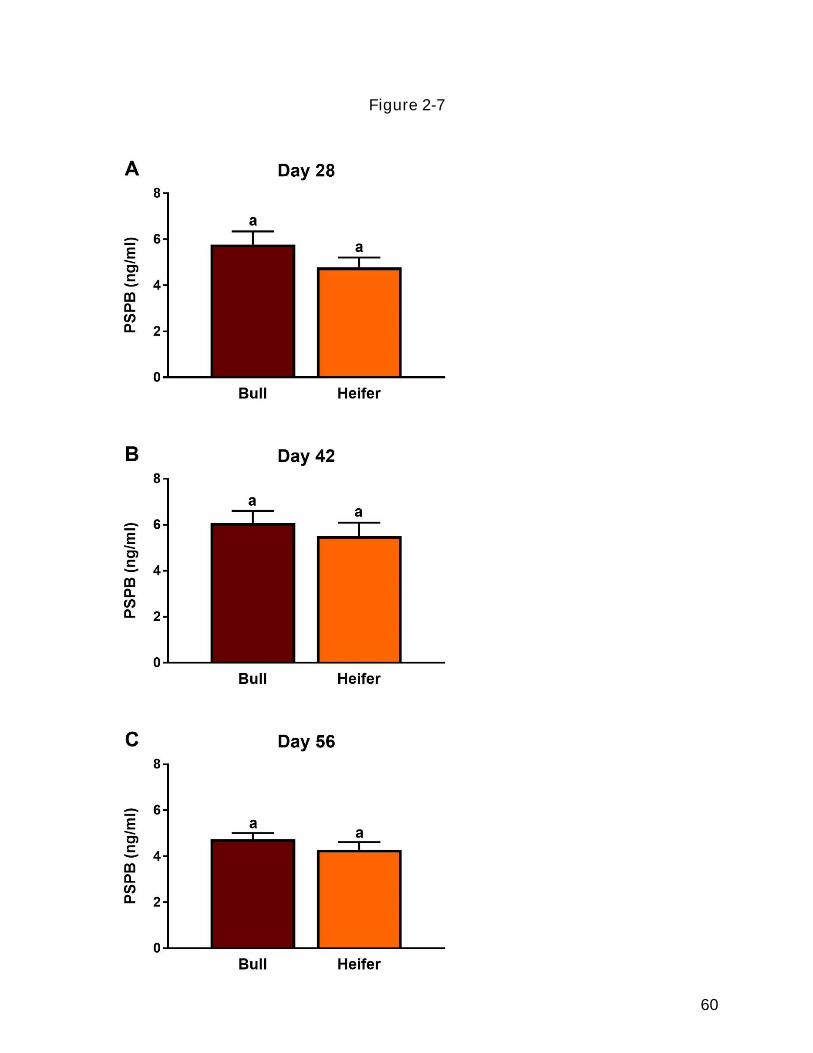
60
Figure 2-7

61
Figure 2-8 The interaction between fetal sex and treatment on circulating PSPB
concentrations. Cows either were artificially inseminated (AI) or received an in vitro-
produced embryo treated with growth factors (EFI; 10 ng/ml Epidermal Growth Factor,
10 ng/ml Fibroblast Growth Factor 2, 50 ng/ml Insulin-like Growth Factor 1) or carrier
only (Control; 1% BSA) from day 4 to 7 post-fertilization. Pregnancy was diagnosed at
day 28. Cows that aborted pregnancies between days 28 and 56 were not included in
these analyses. Fetal sex was determined by transrectal ultrasonography at day 56-60.
Panel A: PSPB concentrations for each fetal sex in each treatment group at day 28.
Panel B: PSPB concentrations for each fetal sex in each treatment group at day 42.
Panel C: PSPB concentrations for each fetal sex in each treatment group at day 56.
Different superscripts denote differences between groups (P<0.05) (n=3-6). The
asterisk represents a sex-based tendency (P=0.08) within the control group.

62
Figure 2-8

63
Figure 2-9 The effect of treatment on crown-rump lengths (CRL) in cattle at days 42
and 56 of gestation. Cows either were artificially inseminated (AI) or received an in vitro-
produced embryo treated with growth factors (EFI; 10 ng/ml Epidermal Growth Factor,
10 ng/ml Fibroblast Growth Factor 2, 50 ng/ml Insulin-like Growth Factor 1) or carrier
only (Control; 1% BSA) from day 4 to 7 post-fertilization. Pregnancy was diagnosed at
day 28. Cows that aborted pregnancies between days 28 and 56 were not included in
these analyses. CRL was measured via transrectal ultrasonography at day 42 and 56
post-AI. Panel A: CRL at day 42. Panel B: CRL at day 56. Different superscripts within
each panel denote differences (P<0.05) (n=7-9).

64
Figure 2-9
65
Figure 2-10 The effect of fetal sex on CRL at day 42 and 56. Cows either were
artificially inseminated (AI) or received an in vitro-produced embryo treated with growth
factors (EFI; 10 ng/ml Epidermal Growth Factor, 10 ng/ml Fibroblast Growth Factor 2,
50 ng/ml Insulin-like Growth Factor 1) or carrier only (Control; 1% BSA) from day 4 to 7
post-fertilization. Pregnancy was diagnosed at day 28. Cows that aborted pregnancies
between days 28 and 56 were not included in these analyses. CRL was measured via
transrectal ultrasonography on day 42 and 56 post-AI. Fetal sex was determined by
transrectal ultrasonography at day 56-60. post-AI. Panel A: CRL measurements for
each feta sex at day 42. Panel B: CRL measurements for each fetal sex at day 56.
Different superscripts within each day denote differences (P<0.05) (n=10-14).

66
Figure 2-10

67
Figure 2-11 Influence of treatments and fetal sex on CRL at day 42 and 56. Cows either
were artificially inseminated (AI) or received an in vitro-produced embryo treated with
growth factors (EFI; 10 ng/ml Epidermal Growth Factor, 10 ng/ml Fibroblast Growth
Factor 2, 50 ng/ml Insulin-like Growth Factor 1) or carrier only (Control; 1% BSA) from
day 4 to 7 post-fertilization. Pregnancy was diagnosed at day 28. Cows that aborted
pregnancies between days 28 and 56 were not included in these analyses. CRL was
measured via transrectal ultrasonography on day 42 and 56 post-AI. Fetal sex was
determined by transrectal ultrasonography at day 56-60. post-AI. Panel A: CRL
measurements for each fetal sex and treatment at day 42. Panel B: CRL measurements
for each fetal sex and treatment at day 56 Different superscripts denote differences
(P<0.05) (n=3-6).

68
Figure 2-11

69
References
1. De Vries, A., Economic value of pregnancy in dairy cattle. J Dairy Sci, 2006. 89(10): p. 3876-85.
2. Perry, G.A., 2013 Statistics of Embryo Collection and Transfer in Domestic Farm Animals. 2013, International Embryo Transfer Society (IETS) Data Retreival Committee.
3. Santos, J.E., et al., The effect of embryonic death rates in cattle on the efficacy of estrus synchronization programs. Anim Reprod Sci, 2004. 82-83: p. 513-35.
4. Sreenan, J.M., M.G. Diskin, and Commission of the European Communities. Coordination of Agricultural Research., Embryonic mortality in farm animals : a seminar in the CEC programme of coordination of research on livestock productivity and management. Current topics in veterinary medicine and animal science. 1986, Dordrecht ; Boston, Hingham, MA, USA: M. Nijhoff for the Commission of the European Communities ; Distributors for the U.S. and Canada, Kluwer Academic. vii, 280 p.
5. Sartori, R., et al., Fertilization and early embryonic development in heifers and lactating cows in summer and lactating and dry cows in winter. J Dairy Sci, 2002. 85(11): p. 2803-12.
6. Graf, A., et al., Genome activation in bovine embryos: review of the literature and new insights from RNA sequencing experiments. Anim Reprod Sci, 2014. 149(1-2): p. 46-58.
7. Humblot, P., Use of pregnancy specific proteins and progesterone assays to monitor pregnancy and determine the timing, frequencies and sources of embryonic mortality in ruminants. Theriogenology, 2001. 56(9): p. 1417-33.
8. Beal, W.E., R.C. Perry, and L.R. Corah, The use of ultrasound in monitoring reproductive physiology of beef cattle. J Anim Sci, 1992. 70(3): p. 924-9.
9. Holmann, F.J., et al., Economic Value of Days Open for Holstein Cows of Alternative Milk Yields with Varying Calving Intervals. Journal of Dairy Science, 1984. 67(3): p. 636-643.
10. Groenendaal, H., D.T. Galligan, and H.A. Mulder, An economic spreadsheet model to determine optimal breeding and replacement decisions for dairy cattle. J Dairy Sci, 2004. 87(7): p. 2146-57.
11. Meadows, C., P.J. Rajala-Schultz, and G.S. Frazer, A spreadsheet-based model demonstrating the nonuniform economic effects of varying reproductive performance in Ohio dairy herds. J Dairy Sci, 2005. 88(3): p. 1244-54.
12. Dyer, T.G., Reproductive Management of Commercial Beef Cows UGA Cooperative Extension, 2012( Bulletin 864).
13. Diskin, M.G. and J.M. Sreenan, Expression and detection of oestrus in cattle. Reprod Nutr Dev, 2000. 40(5): p. 481-91.
14. Pursley, J.R., M.R. Kosorok, and M.C. Wiltbank, Reproductive management of lactating dairy cows using synchronization of ovulation. Journal of Dairy Science, 1997. 80(2): p. 301-306.
15. Burke, C.R., et al., Effects of maturity of the potential ovulatory follicle on induction of oestrus and ovulation in cattle with oestradiol benzoate. Anim Reprod Sci, 2001. 66(3-4): p. 161-74.
70
16. Souza, A.H., et al., Effects of equine chorionic gonadotropin and type of ovulatory stimulus in a timed-AI protocol on reproductive responses in dairy cows. Theriogenology, 2009. 72(1): p. 10-21.
17. Machatkova, M., et al., Developmental competence of bovine oocytes: effects of follicle size and the phase of follicular wave on in vitro embryo production. Theriogenology, 2004. 61(2-3): p. 329-35.
18. Gilbert, I., A. Macaulay, and C. Robert, Oocyte developmental competence and embryo quality: distinction and new perspectives. Animal Reproduction, 2015. 12(3): p. 397-407.
19. Sternlicht, A.L. and R.M. Schultz, Biochemical studies of mammalian oogenesis: kinetics of accumulation of total and poly(A)-containing RNA during growth of the mouse oocyte. J Exp Zool, 1981. 215(2): p. 191-200.
20. Macaulay, A.D., et al., The gametic synapse: RNA transfer to the bovine oocyte. Biol Reprod, 2014. 91(4): p. 90.
21. Breuel, K.F., et al., Factors affecting fertility in the postpartum cow: role of the oocyte and follicle in conception rate. Biol Reprod, 1993. 48(3): p. 655-61.
22. Maurer, R.R. and J.R. Chenault, Fertilization failure and embryonic mortality in parous and nonparous beef cattle. J Anim Sci, 1983. 56(5): p. 1186-9.
23. Dunne, L.D., M.G. Diskin, and J.M. Sreenan, Embryo and foetal loss in beef heifers between day 14 of gestation and full term. Anim Reprod Sci, 2000. 58(1-2): p. 39-44.
24. Barnes, F.L. and W.H. Eyestone, Early Cleavage and the Maternal Zygotic Transition in Bovine Embryos. Theriogenology, 1990. 33(1): p. 141-152.
25. Sirard, M.A., Activation of the embryonic genome. Reproduction in Domestic Ruminants Vii, 2010. 67: p. 145-158.
26. Senger, P.L., Pathways to pregnancy & parturition. 3rd ed. 2012, Redmond, OR: Current Conceptions. ix, 381 p.
27. Greenstein, J.S., R.W. Murray, and R.C. Foley, Observations on the morphogenesis and histochemistry of the bovine preattachment placenta between 16 and 33 days of gestation. Anat Rec, 1958. 132(3): p. 321-41.
28. Bazer, F.W., et al., Uterine receptivity to implantation of blastocysts in mammals. Front Biosci (Schol Ed), 2011. 3: p. 745-67.
29. Spencer, T.E., Early pregnancy: Concepts, challenges, and potential solutions. Animal Frontiers 2013. 3(4).
30. Bazer, F.W., T.E. Spencer, and T.L. Ott, Placental interferons. Am J Reprod Immunol, 1996. 35(4): p. 297-308.
31. Spencer, T.E., et al., Conceptus signals for establishment and maintenance of pregnancy. Anim Reprod Sci, 2004. 82-83: p. 537-50.
32. Roberts, R.M., Interferon-tau, a Type 1 interferon involved in maternal recognition of pregnancy. Cytokine Growth Factor Rev, 2007. 18(5-6): p. 403-8.
33. Roberts, R.M., J.C. Cross, and D.W. Leaman, Interferons as hormones of pregnancy. Endocr Rev, 1992. 13(3): p. 432-52.
34. Hernandez-Ledezma, J.J., et al., Expression of bovine trophoblast interferon in conceptuses derived by in vitro techniques. Biol Reprod, 1992. 47(3): p. 374-80.

71
35. Kubisch, H.M., M.A. Larson, and R.M. Roberts, Relationship between age of blastocyst formation and interferon-tau secretion by in vitro-derived bovine embryos. Mol Reprod Dev, 1998. 49(3): p. 254-60.
36. Roberts, R.M., et al., Trophoblast interferons. Placenta, 1999. 20(4): p. 259-264. 37. Ealy, A.D., et al., Polymorphic Forms of Expressed Bovine Interferon-tau Genes:
Relative Transcript Abundance during Early Placental Development, Promoter Sequences of Genes and Biological Activity of Protein Products. Endocrinology, 2001. 142(7): p. 2906-2915.
38. Farin, C.E., et al., Expression of trophoblastic interferon genes in sheep and cattle. Biol Reprod, 1990. 43(2): p. 210-8.
39. Guillomot, M., J.E. Flechon, and S. Wintenberger-Torres, Conceptus attachment in the ewe: an ultrastructural study. Placenta, 1981. 2(2): p. 169-82.
40. Bazer, F.W., T.L. Ott, and T.E. Spencer, Maternal recognition of pregnancy: Comparative aspects. Placenta, 1998. 19: p. 375-386.
41. Ott, T.L., et al., Effects of ovine conceptus secretory proteins and progesterone on oxytocin-stimulated endometrial production of prostaglandin and turnover of inositol phosphate in ovariectomized ewes. J Reprod Fertil, 1992. 95(1): p. 19-29.
42. Vallet, J.L., et al., Effect of ovine conceptus secretory proteins and purified ovine trophoblast protein-1 on interoestrous interval and plasma concentrations of prostaglandins F-2 alpha and E and of 13,14-dihydro-15-keto prostaglandin F-2 alpha in cyclic ewes. J Reprod Fertil, 1988. 84(2): p. 493-504.
43. Thatcher, W.W., et al., Antiluteolytic effects of bovine trophoblast protein-1. J Reprod Fertil Suppl, 1989. 37: p. 91-9.
44. Hansen, T.R., et al., Mechanism of action of interferon-tau in the uterus during early pregnancy. J Reprod Fertil Suppl, 1999. 54: p. 329-39.
45. Dorniak, P., F.W. Bazer, and T.E. Spencer, Physiology and Endocrinology Symposium: biological role of interferon tau in endometrial function and conceptus elongation. J Anim Sci, 2013. 91(4): p. 1627-38.
46. Gifford, C.A., et al., Regulation of interferon-stimulated genes in peripheral blood leukocytes in pregnant and bred, nonpregnant dairy cows. J Dairy Sci, 2007. 90(1): p. 274-80.
47. Spencer, T.E., N. Forde, and P. Lonergan, Insights into conceptus elongation and establishment of pregnancy in ruminants. Reproduction, Fertility and Development, 2016. 29(1): p. 84-100.
48. Bazer, F.W., et al., Novel pathways for implantation and establishment and maintenance of pregnancy in mammals. Mol Hum Reprod, 2010. 16(3): p. 135-52.
49. Spencer, T.E., O. Sandra, and E. Wolf, Genes involved in conceptus-endometrial interactions in ruminants: insights from reductionism and thoughts on holistic approaches. Reproduction, 2008. 135(2): p. 165-79.
50. Tan, S.J. and K.H. Lu, Effects of different oestrous stages of ovaries and sizes of follicles on generation of bovine embryos in vitro. Theriogenology, 1990. 33(1): p. 335.

72
51. Lonergan, P., et al., Effect of follicle size on bovine oocyte quality and developmental competence following maturation, fertilization, and culture in vitro. Mol Reprod Dev, 1994. 37(1): p. 48-53.
52. Pavlok, A., A. Lucas-Hahn, and H. Niemann, Fertilization and developmental competence of bovine oocytes derived from different categories of antral follicles. Mol Reprod Dev, 1992. 31(1): p. 63-7.
53. Blondin, P. and M.A. Sirard, Oocyte and follicular morphology as determining characteristics for developmental competence in bovine oocytes. Mol Reprod Dev, 1995. 41(1): p. 54-62.
54. Hagemann, L.J., et al., Development during single IVP of bovine oocytes from dissected follicles: interactive effects of estrous cycle stage, follicle size and atresia. Mol Reprod Dev, 1999. 53(4): p. 451-8.
55. de Loos, F., et al., Morphology of immature bovine oocytes. Gamete Res, 1989. 24(2): p. 197-204.
56. de Wit, A.A., Y.A. Wurth, and T.A. Kruip, Effect of ovarian phase and follicle quality on morphology and developmental capacity of the bovine cumulus-oocyte complex. J Anim Sci, 2000. 78(5): p. 1277-83.
57. Garrett, J.E., et al., Evidence for maternal regulation of early conceptus growth and development in beef cattle. J Reprod Fertil, 1988. 84(2): p. 437-46.
58. Nephew, K.P., et al., Relationship between variation in conceptus development and differences in estrous cycle duration in ewes. Biol Reprod, 1991. 44(3): p. 536-9.
59. Lopez, H., et al., Relationship between level of milk production and multiple ovulations in lactating dairy cows. J Dairy Sci, 2005. 88(8): p. 2783-93.
60. Rizos, D., et al., Contribution of the female reproductive tract to low fertility in postpartum lactating dairy cows. J Dairy Sci, 2010. 93(3): p. 1022-9.
61. Bartol, F.F., et al., Uterine differentiation as a foundation for subsequent fertility. J Reprod Fertil Suppl, 1999. 54: p. 287-302.
62. Gray, C.A., et al., Developmental biology of uterine glands. Biol Reprod, 2001. 65(5): p. 1311-23.
63. Spencer, T.E., K.A. Dunlap, and J. Filant, Comparative developmental biology of the uterus: insights into mechanisms and developmental disruption. Mol Cell Endocrinol, 2012. 354(1-2): p. 34-53.
64. Atkinson, B.A., G.J. King, and E.C. Amoroso, Development of the caruncular and intercaruncular regions in the bovine endometrium. Biol Reprod, 1984. 30(3): p. 763-74.
65. Bazer, F.W., Uterine protein secretions: Relationship to development of the conceptus. J Anim Sci, 1975. 41(5): p. 1376-82.
66. Stewart, C.L., et al., Blastocyst implantation depends on maternal expression of leukaemia inhibitory factor. Nature, 1992. 359(6390): p. 76-9.
67. Cooke, P.S., et al., Uterine glands: development, function and experimental model systems. Mol Hum Reprod, 2013. 19(9): p. 547-58.
68. Filant, J. and T.E. Spencer, Uterine glands: biological roles in conceptus implantation, uterine receptivity and decidualization. Int J Dev Biol, 2014. 58(2-4): p. 107-16.

73
69. Satterfield, M.C., F.W. Bazer, and T.E. Spencer, Progesterone regulation of preimplantation conceptus growth and galectin 15 (LGALS15) in the ovine uterus. Biol Reprod, 2006. 75(2): p. 289-96.
70. Carter, F., et al., Effect of increasing progesterone concentration from Day 3 of pregnancy on subsequent embryo survival and development in beef heifers. Reprod Fertil Dev, 2008. 20(3): p. 368-75.
71. Lonergan, P., N. Forde, and T.E. Spencer, Progesterone and conceptus-derived factors important for conceptus survival and growth. 2016, 2016. 13(3): p. 10.
72. Forde, N., et al., Progesterone-regulated changes in endometrial gene expression contribute to advanced conceptus development in cattle. Biol Reprod, 2009. 81(4): p. 784-94.
73. Clemente, M., et al., Progesterone and conceptus elongation in cattle: a direct effect on the embryo or an indirect effect via the endometrium? Reproduction, 2009. 138(3): p. 507-17.
74. Lonergan, P., Influence of progesterone on oocyte quality and embryo development in cows. Theriogenology, 2011. 76(9): p. 1594-601.
75. Larson, S.F., W.R. Butler, and W.B. Currie, Pregnancy rates in lactating dairy cattle following supplementation of progesterone after artificial insemination. Anim Reprod Sci, 2007. 102(1-2): p. 172-9.
76. Arndt, W.J., et al., Effect of post-insemination progesterone supplementation on pregnancy rate in dairy cows. Can J Vet Res, 2009. 73(4): p. 271-4.
77. Monteiro, P.L., Jr., et al., Effects of supplemental progesterone after artificial insemination on expression of interferon-stimulated genes and fertility in dairy cows. J Dairy Sci, 2014. 97(8): p. 4907-21.
78. Wiltbank, M.C., et al., Pivotal periods for pregnancy loss during the first trimester of gestation in lactating dairy cows. Theriogenology, 2016. 86(1): p. 239-53.
79. Austin, K.J., et al., Localization of ISG15 and conjugated proteins in bovine endometrium using immunohistochemistry and electron microscopy. Endocrinology, 2004. 145(2): p. 967-75.
80. Green, J.C., et al., Measurement of interferon-tau (IFN-tau) stimulated gene expression in blood leukocytes for pregnancy diagnosis within 18-20d after insemination in dairy cattle. Anim Reprod Sci, 2010. 121(1-2): p. 24-33.
81. Johnson, G.A., et al., Pregnancy and interferon-tau induce conjugation of bovine ubiquitin cross-reactive protein to cytosolic uterine proteins. Biol Reprod, 1998. 58(4): p. 898-904.
82. Han, H., et al., Low blood ISG15 mRNA and progesterone levels are predictive of non-pregnant dairy cows. J Endocrinol, 2006. 191(2): p. 505-12.
83. Sasser, R.G., et al., Detection of pregnancy by radioimmunoassay of a novel pregnancy-specific protein in serum of cows and a profile of serum concentrations during gestation. Biol Reprod, 1986. 35(4): p. 936-42.
84. Zoli, A.P., et al., Purification and characterization of a bovine pregnancy-associated glycoprotein. Biol Reprod, 1991. 45(1): p. 1-10.
85. Green, J.A., S. Xie, and R.M. Roberts, Pepsin-related molecules secreted by trophoblast. Rev Reprod, 1998. 3(1): p. 62-9.

74
86. Mercadante, P.M., et al., Subspecies differences in early fetal development and plasma pregnancy-associated glycoprotein concentrations in cattle. J Anim Sci, 2013. 91(8): p. 3693-701.
87. Thompson, I.M., et al., Effects of resynchronization programs on pregnancy per artificial insemination, progesterone, and pregnancy-associated glycoproteins in plasma of lactating dairy cows. J Dairy Sci, 2010. 93(9): p. 4006-18.
88. Mercadante, P.M., et al., Associations between pregnancy-associated glycoproteins and pregnancy outcomes, milk yield, parity, and clinical diseases in high-producing dairy cows. J Dairy Sci, 2016. 99(4): p. 3031-40.
89. Lopez-Gatius, F., et al., Plasma concentrations of pregnancy-associated glycoprotein-1 (PAG-1) in high producing dairy cows suffering early fetal loss during the warm season. Theriogenology, 2007. 67(8): p. 1324-30.
90. Riding, G.A., et al., Conceptus-related measurements during the first trimester of bovine pregnancy. Vet J, 2008. 175(2): p. 266-72.
91. Evans, H.E. and W.O. Sack, Prenatal development of domestic and laboratory mammals: growth curves, external features and selected references. Zentralbl Veterinarmed C, 1973. 2(1): p. 11-45.
92. Kahn, W., Sonographic fetometry in the bovine. Theriogenology, 1989. 31(5): p. 1105-21.
93. Dunne, L.D., et al., The effect of pre- and post-insemination plane of nutrition on embryo survival in beef heifers. Animal Science, 1999. 69: p. 411-417.
94. Van Hoeck, V., et al., Elevated non-esterified fatty acid concentrations during bovine oocyte maturation compromise early embryo physiology. PLoS One, 2011. 6(8): p. e23183.
95. Diskin, M.G., et al., Effects of nutrition and metabolic status on circulating hormones and ovarian follicle development in cattle. Anim Reprod Sci, 2003. 78(3-4): p. 345-70.
96. Lopez-Gatius, F., et al., Factors affecting pregnancy loss from gestation Day 38 to 90 in lactating dairy cows from a single herd. Theriogenology, 2002. 57(4): p. 1251-61.
97. Wiltbank, M., et al., Changes in reproductive physiology of lactating dairy cows due to elevated steroid metabolism. Theriogenology, 2006. 65(1): p. 17-29.
98. Ryan, D.P., et al., Comparing early embryo mortality in dairy cows during hot and cool seasons of the year. Theriogenology, 1993. 39(3): p. 719-37.
99. Hansen, P.J., Effects of heat stress on mammalian reproduction. Philosophical Transactions of the Royal Society B-Biological Sciences, 2009. 364(1534): p. 3341-3350.
100. Berman, A., et al., Upper critical temperatures and forced ventilation effects for high-yielding dairy cows in a subtropical climate. J Dairy Sci, 1985. 68(6): p. 1488-95.
101. Wolfenson, D., et al., Seasonal and acute heat stress effects on steroid production by dominant follicles in cows. Anim Reprod Sci, 1997. 47(1-2): p. 9-19.
102. De Rensis, F. and R.J. Scaramuzzi, Heat stress and seasonal effects on reproduction in the dairy cow--a review. Theriogenology, 2003. 60(6): p. 1139-51.

75
103. Rutledge, J.J., et al., Seasonality of cattle embryo production in a temperate region. Theriogenology, 1999. 51(1): p. 330-330.
104. Al-Katanani, Y.M., et al., Pregnancy rates following timed embryo transfer with fresh or vitrified in vitro produced embryos in lactating dairy cows under heat stress conditions. Theriogenology, 2002. 58(1): p. 171-82.
105. Roman-Ponce, H., et al., Thermal stress effects on uterine blood flow in dairy cows. J Anim Sci, 1978. 46(1): p. 175-80.
106. Gwazdauskas, F.C., et al., Hormonal patterns during heat stress following PGF(2)alpha-tham salt induced luteal regression in heifers. Theriogenology, 1981. 16(3): p. 271-85.
107. Vanselow, J., et al., Exposure of Lactating Dairy Cows to Acute Pre-Ovulatory Heat Stress Affects Granulosa Cell-Specific Gene Expression Profiles in Dominant Follicles. PLoS One, 2016. 11(8): p. e0160600.
108. Hansen, P.J., et al., Adverse impact of heat stress on embryo production: causes and strategies for mitigation. Theriogenology, 2001. 55(1): p. 91-103.
109. Ealy, A.D., M. Drost, and P.J. Hansen, Developmental changes in embryonic resistance to adverse effects of maternal heat stress in cows. J Dairy Sci, 1993. 76(10): p. 2899-905.
110. Drost, M., et al., Conception rates after artificial insemination or embryo transfer in lactating dairy cows during summer in Florida. Theriogenology, 1999. 52(7): p. 1161-7.
111. Porter, J.K. and F.N. Thompson, Jr., Effects of fescue toxicosis on reproduction in livestock. J Anim Sci, 1992. 70(5): p. 1594-603.
112. Schuenemann, G.M., et al., Embryo development and survival in beef cattle administered ergotamine tartrate to simulate fescue toxicosis. Reprod Biol, 2005. 5(2): p. 137-50.
113. Grohn, Y.T. and P.J. Rajala-Schultz, Epidemiology of reproductive performance in dairy cows. Anim Reprod Sci, 2000. 60-61: p. 605-14.
114. DeGraves, F.J. and J. Fetrow, Economics of mastitis and mastitis control. Vet Clin North Am Food Anim Pract, 1993. 9(3): p. 421-34.
115. Schrick, F.N., et al., Influence of subclinical mastitis during early lactation on reproductive parameters. J Dairy Sci, 2001. 84(6): p. 1407-12.
116. Santos, J.E., et al., Effect of timing of first clinical mastitis occurrence on lactational and reproductive performance of Holstein dairy cows. Anim Reprod Sci, 2004. 80(1-2): p. 31-45.
117. Chebel, R.C., et al., Factors affecting conception rate after artificial insemination and pregnancy loss in lactating dairy cows. Anim Reprod Sci, 2004. 84(3-4): p. 239-55.
118. Gilbert, R.O., et al., Prevalence of endometritis and its effects on reproductive performance of dairy cows. Theriogenology, 2005. 64(9): p. 1879-88.
119. Santos, J.E., et al., Applying nutrition and physiology to improve reproduction in dairy cattle. Soc Reprod Fertil Suppl, 2010. 67: p. 387-403.
120. Ribeiro, E.S., et al., Prevalence of periparturient diseases and effects on fertility of seasonally calving grazing dairy cows supplemented with concentrates. J Dairy Sci, 2013. 96(9): p. 5682-97.
76
121. Simmen, F.A. and R.C. Simmen, Peptide growth factors and proto-oncogenes in mammalian conceptus development. Biol Reprod, 1991. 44(1): p. 1-5.
122. Wei, Z., et al., Effect of epidermal growth factor on preimplantation development and its receptor expression in porcine embryos. Mol Reprod Dev, 2001. 60(4): p. 457-62.
123. Adamson, E.D., Activities of growth factors in preimplantation embryos. Journal of Cellular Biochemistry, 1993. 53(4): p. 280-287.
124. Wiley, L.M., et al., Epidermal growth factor receptor mRNA and protein increase after the four-cell preimplantation stage in murine development. Dev Biol, 1992. 149(2): p. 247-60.
125. Jeong, W., et al., Epidermal growth factor stimulates proliferation and migration of porcine trophectoderm cells through protooncogenic protein kinase 1 and extracellular-signal-regulated kinases 1/2 mitogen-activated protein kinase signal transduction cascades during early pregnancy. Mol Cell Endocrinol, 2013. 381(1-2): p. 302-11.
126. Neira, J.A., et al., Effect of the association of IGF-I, IGF-II, bFGF, TGF-beta1, GM-CSF, and LIF on the development of bovine embryos produced in vitro. Theriogenology, 2010. 73(5): p. 595-604.
127. Wood, S.A. and P.L. Kaye, Effects of epidermal growth factor on preimplantation mouse embryos. J Reprod Fertil, 1989. 85(2): p. 575-82.
128. Munson, L., S.K. Chandler, and D.H. Schlafer, Long-term culture of bovine trophoblastic cells. Journal of tissue culture methods, 1988. 11(3): p. 123-128.
129. Dilly, M., et al., Epidermal growth factor (EGF) induces motility and upregulates MMP-9 and TIMP-1 in bovine trophoblast cells. Mol Reprod Dev, 2010. 77(7): p. 622-9.
130. Yang, Q.E., M.I. Giassetti, and A.D. Ealy, Fibroblast growth factors activate mitogen-activated protein kinase pathways to promote migration in ovine trophoblast cells. Reproduction, 2011. 141(5): p. 707-14.
131. Xie, M., et al., Combinatorial effects of epidermal growth factor, fibroblast growth factor 2 and insulin-like growth factor 1 on trophoblast cell proliferation and embryogenesis in cattle. Reprod Fertil Dev, 2015.
132. Cooke, F.N., et al., Several fibroblast growth factors are expressed during pre-attachment bovine conceptus development and regulate interferon-tau expression from trophectoderm. Reproduction, 2009. 137(2): p. 259-69.
133. Carlone, D.L. and V. Rider, Embryonic modulation of basic fibroblast growth factor in the rat uterus. Biol Reprod, 1993. 49(4): p. 653-65.
134. Michael, D.D., et al., Fibroblast growth factor-2 is expressed by the bovine uterus and stimulates interferon-tau production in bovine trophectoderm. Endocrinology, 2006. 147(7): p. 3571-9.
135. Ka, H., et al., Regulation of expression of fibroblast growth factor 7 in the pig uterus by progesterone and estradiol. Biol Reprod, 2007. 77(1): p. 172-80.
136. Ocon-Grove, O.M., et al., Ovine endometrial expression of fibroblast growth factor (FGF) 2 and conceptus expression of FGF receptors during early pregnancy. Domest Anim Endocrinol, 2008. 34(2): p. 135-45.
137. Ozawa, M., Q.E. Yang, and A.D. Ealy, The expression of fibroblast growth factor receptors during early bovine conceptus development and pharmacological

77
analysis of their actions on trophoblast growth in vitro. Reproduction, 2013. 145(2): p. 191-201.
138. Forbes, K., et al., Insulin-like growth factor I and II regulate the life cycle of trophoblast in the developing human placenta. Am J Physiol Cell Physiol, 2008. 294(6): p. C1313-22.
139. Jeong, W., et al., Insulin-like growth factor I induces proliferation and migration of porcine trophectoderm cells through multiple cell signaling pathways, including protooncogenic protein kinase 1 and mitogen-activated protein kinase. Mol Cell Endocrinol, 2014. 384(1-2): p. 175-84.
140. Ko, Y., et al., Insulin-like growth factors in sheep uterine fluids: concentrations and relationship to ovine trophoblast protein-1 production during early pregnancy. Biol Reprod, 1991. 45(1): p. 135-42.
141. Kim, J., et al., Insulin-like growth factor II activates phosphatidylinositol 3-kinase-protooncogenic protein kinase 1 and mitogen-activated protein kinase cell Signaling pathways, and stimulates migration of ovine trophectoderm cells. Endocrinology, 2008. 149(6): p. 3085-94.
142. Block, J. and P.J. Hansen, Interaction between season and culture with insulin-like growth factor-1 on survival of in vitro produced embryos following transfer to lactating dairy cows. Theriogenology, 2007. 67(9): p. 1518-29.
143. Vincent, A.M. and E.L. Feldman, Control of cell survival by IGF signaling pathways. Growth Horm IGF Res, 2002. 12(4): p. 193-7.
144. El-Zarkouny, S.Z., et al., Pregnancy in dairy cows after synchronized ovulation regimens with or without presynchronization and progesterone. J Dairy Sci, 2004. 87(4): p. 1024-37.
145. Fields, S.D., P.J. Hansen, and A.D. Ealy, Fibroblast growth factor requirements for in vitro development of bovine embryos. Theriogenology, 2011. 75(8): p. 1466-75.
146. Bo, G.A. and R.J. Mapletoft, Evaluation and classification of bovine embryos. Animal Reproduction, 2013. 10(3): p. 344-348.
147. Agca, Y., et al., Normal calves from transfer of biopsied, sexed and vitrified IVP bovine embryos. Theriogenology, 1998. 50(1): p. 129-45.
148. Miles, J.R., et al., Effects of embryo culture on angiogenesis and morphometry of bovine placentas during early gestation. Biol Reprod, 2005. 73(4): p. 663-71.
149. Miles, J.R., et al., Angiogenesis and morphometry of bovine placentas in late gestation from embryos produced in vivo or in vitro. Biol Reprod, 2004. 71(6): p. 1919-26.
150. Breukelman, S.P., et al., Plasma concentrations of bovine pregnancy-associated glycoprotein (bPAG) do not differ during the first 119 days between ongoing pregnancies derived by transfer of in vivo and in vitro produced embryos. Theriogenology, 2005. 63(5): p. 1378-89.
151. van Wagtendonk-de Leeuw, A.M., B.J. Aerts, and J.H. den Daas, Abnormal offspring following in vitro production of bovine preimplantation embryos: a field study. Theriogenology, 1998. 49(5): p. 883-94.
152. Rasmussen, S., et al., Pregnancy rates of lactating cows after transfer of in vitro produced embryos using X-sorted sperm. Theriogenology, 2013. 79(3): p. 453-61.

78
153. Bonilla, L., et al., Consequences of transfer of an in vitro-produced embryo for the dam and resultant calf. J Dairy Sci, 2014. 97(1): p. 229-39.
154. Mercadante, V.R., et al., Effects of recombinant bovine somatotropin administration at breeding on cow, conceptus, and subsequent offspring performance of beef cattle. J Anim Sci, 2016. 94(5): p. 2128-38.
155. Sangsritavong, S., et al., High feed intake increases liver blood flow and metabolism of progesterone and estradiol-17beta in dairy cattle. J Dairy Sci, 2002. 85(11): p. 2831-42.
156. Xu, K.P., et al., Sex-related differences in developmental rates of bovine embryos produced and cultured in vitro. Mol Reprod Dev, 1992. 31(4): p. 249-52.
157. Holland, M.D. and K.G. Odde, Factors Affecting Calf Birth-Weight - a Review. Theriogenology, 1992. 38(5): p. 769-798.