
Post-operative outcomes and complications of suspensory ...
12
REVIEW ARTICLE Post-operative outcomes and complications of suspensory loop fixation device versus hook plate in acute unstable acromioclavicular joint dislocation: a systematic review and meta-analysis Alisara Arirachakaran 1 • Manusak Boonard 2 • Peerapong Piyapittayanun 1 • Wichan Kanchanatawan 3 • Kornkit Chaijenkij 4 • Akom Prommahachai 5 • Jatupon Kongtharvonskul 6 Received: 21 March 2016 / Accepted: 9 February 2017 / Published online: 25 February 2017 Ó The Author(s) 2017. This article is published with open access at Springerlink.com Abstract Background Treatment of acute (B3 weeks) acromio- clavicular joint dislocation type III–VI is still controversial. Currently, the two modern techniques that are widely used are hook plate (HP) fixation and coracoclavicular ligament fixation using a suspensory loop device that consists of either a tightrope (single or double), endo-button (single or double), or synthetic ligament and absorbable polydiox- ansulfate sling. Materials and methods This systematic review was con- ducted according to the PRISMA guidelines. Relevant studies that reported Constant-Murley score (CMS), Pain Visual Analog score (VAS) and postoperative complica- tions of either technique were identified from Medline and Scopus from inception to 5 October 2015. Results Sixteen studies were included for the analysis of HP fixation, and 25 studies were included for analysis of loop suspensory fixation (LSF). Pooling of mean CMS and VAS scores gave 90.35 (95% CI 87.16, 93.54), 1.51 (95% CI 0.73, 2) in the HP group, and 92.48 (95% CI 90.91, 94.05), 0.32 (95% CI 0, 0.64) in the suspensory loop devices group, respectively. The pooled unstandardized mean differences (UMD) scores of CMS and VAS in LSF were 2.13 (95% CI -1.43, 5.69) and -1.19 (95% CI -2.03, -0.35) when compared to hook plating. The pooled prevalence of LSF and hook plating were 0.08 (95% CI 0.06, 0.10) and 0.05 (95% CI 0.02, 0.08) scores. The chance of having complications in the LSF group was 1.69 (95% CI 1.07, 2.60), which was statistically significantly higher than in the HP group. Conclusion LSF have higher shoulder function scores (CMS) and lower postoperative pain when compared to HP fixation; however, there are higher complication rates with LSF when compared to hook plating. Level of evidence IV. Keywords Hook plate Loop suspensory fixation AC injury Systematic review Tightrope Endobutton & Jatupon Kongtharvonskul [email protected] Alisara Arirachakaran [email protected] Manusak Boonard [email protected] Peerapong Piyapittayanun [email protected] Wichan Kanchanatawan [email protected] Kornkit Chaijenkij [email protected] Akom Prommahachai [email protected] 1 Orthopedics Department, Police General Hospital, Bangkok, Thailand 2 Orthopedics Department, Srinakarin Hospital, Khonkan, Thailand 3 Orthopedic Department, Lerdsin General Hospital, Bangkok, Thailand 4 Orthopedics Department, College of Sports Science and Technology, Mahidol University, Bangkok, Thailand 5 Orthopedic Department, Aek Udon International Hospital, Udontani, Thailand 6 Sport and orthopedic center, Samitivej Hospital, Bangkok, Thailand 123 J Orthop Traumatol (2017) 18:293–304 DOI 10.1007/s10195-017-0451-1
Transcript of Post-operative outcomes and complications of suspensory ...
REVIEW ARTICLE
Post-operative outcomes and complications of suspensory loopfixation device versus hook plate in acute unstableacromioclavicular joint dislocation: a systematic reviewand meta-analysis
Alisara Arirachakaran1 • Manusak Boonard2 • Peerapong Piyapittayanun1 •
Wichan Kanchanatawan3 • Kornkit Chaijenkij4 • Akom Prommahachai5 •
Jatupon Kongtharvonskul6
Received: 21 March 2016 / Accepted: 9 February 2017 / Published online: 25 February 2017
� The Author(s) 2017. This article is published with open access at Springerlink.com
Abstract
Background Treatment of acute (B3 weeks) acromio-
clavicular joint dislocation type III–VI is still controversial.
Currently, the two modern techniques that are widely used
are hook plate (HP) fixation and coracoclavicular ligament
fixation using a suspensory loop device that consists of
either a tightrope (single or double), endo-button (single or
double), or synthetic ligament and absorbable polydiox-
ansulfate sling.
Materials and methods This systematic review was con-
ducted according to the PRISMA guidelines. Relevant
studies that reported Constant-Murley score (CMS), Pain
Visual Analog score (VAS) and postoperative complica-
tions of either technique were identified from Medline and
Scopus from inception to 5 October 2015.
Results Sixteen studies were included for the analysis of
HP fixation, and 25 studies were included for analysis of
loop suspensory fixation (LSF). Pooling of mean CMS and
VAS scores gave 90.35 (95% CI 87.16, 93.54), 1.51 (95%
CI 0.73, 2) in the HP group, and 92.48 (95% CI 90.91,
94.05), 0.32 (95% CI 0, 0.64) in the suspensory loop
devices group, respectively. The pooled unstandardized
mean differences (UMD) scores of CMS and VAS in LSF
were 2.13 (95% CI -1.43, 5.69) and -1.19 (95% CI
-2.03, -0.35) when compared to hook plating. The pooled
prevalence of LSF and hook plating were 0.08 (95% CI
0.06, 0.10) and 0.05 (95% CI 0.02, 0.08) scores. The
chance of having complications in the LSF group was 1.69
(95% CI 1.07, 2.60), which was statistically significantly
higher than in the HP group.
Conclusion LSF have higher shoulder function scores
(CMS) and lower postoperative pain when compared to HP
fixation; however, there are higher complication rates with
LSF when compared to hook plating.
Level of evidence IV.
Keywords Hook plate � Loop suspensory fixation � AC
injury � Systematic review � Tightrope � Endobutton
& Jatupon Kongtharvonskul
Alisara Arirachakaran
Manusak Boonard
Peerapong Piyapittayanun
Wichan Kanchanatawan
Kornkit Chaijenkij
Akom Prommahachai
1 Orthopedics Department, Police General Hospital, Bangkok,
Thailand
2 Orthopedics Department, Srinakarin Hospital, Khonkan,
Thailand
3 Orthopedic Department, Lerdsin General Hospital, Bangkok,
Thailand
4 Orthopedics Department, College of Sports Science and
Technology, Mahidol University, Bangkok, Thailand
5 Orthopedic Department, Aek Udon International Hospital,
Udontani, Thailand
6 Sport and orthopedic center, Samitivej Hospital, Bangkok,
Thailand
123
J Orthop Traumatol (2017) 18:293–304
DOI 10.1007/s10195-017-0451-1
Introduction
Acromioclavicular (AC) joint (ACJ) dislocation is a com-
mon injury in active young adults [5]. The prevalence was
approximately 9% of shoulder girdle injuries [3, 28]. AC
dislocation is associated with AC and coracoclavicular
(CC) ligaments injuries [37]; such injuries are classified
into type I–VI on the basis of the radiographic findings
using the Rockwood criteria [26, 37]. Non-operative
treatment has generally been accepted as the gold standard
of treatment in Rockwood I and II lesions [52], whereas the
optimal method of treatment for grade III–VI lesions
remains a matter of controversy [16, 30, 59]. Although type
IV–VI injuries are treated operatively because of their
severe instability [45], treatment for type III injury is still
controversial [22, 52]. The aim of any surgical approach
addressing the instability of the ACJ should be an anatomic
reduction and restoration of normal arthrokinetics [18].
Surgical methods employed for the treatment of AC dis-
locations include extraarticular fixation by CC restoration
with metallic cables, autologous ligaments or LARS arti-
ficial ligaments, transarticular fixation by hook plate (HP)
and Kirschner wires. Kirschner wires and threaded pins are
frequently used for temporary fixation of the ACJ. How-
ever, serious concerns still exist regarding pin migration or
breakage, pin-site infection, fixation failure, and recurrent
instability after pin removal [14, 21, 31]. Currently, two
modern techniques that are widely used include HP fixation
[2, 7, 10, 29, 35, 59] and CC ligament fixation using a
suspensory loop device [tightrope (single or double), endo-
button (single or double), synthetic ligament and absorb-
able polydioxansulfate sling (PDS)] [7, 8, 10, 12, 19, 31,
32, 35, 36, 39, 51]. Many case series have reported safe and
effective results with the treatment of acute unstable ACJ
dislocations by loop suspensory fixation (LSF) [1, 8, 9, 12,
20, 24, 40, 42, 43, 50, 55] and hook plating [7, 17, 25, 38,
39, 49, 56, 57]. More recently, several retrospective cohort
studies have aimed to investigate the results of treatment
for unstable acute AC dislocations (type III–VI) with HP
and LSF. However, there has been no consensus as to
which is better for treatment of acute unstable ACJ dislo-
cation. Some studies show benefits of the AC or CC aug-
mentations in pain and Constant-Murley score (CMS) [29,
59], whereas other studies do not [2, 10, 18]. However,
LSF consists of variable types of fixation, which include
tightrope (single or double), endo-button (single or double),
synthetic ligaments and absorbable PDS. Moreover, no
high quality methodological quality study [prospective
cohort or randomized controlled trials (RCT)] has recently
been published. We hypothesized that the impact of each
type of fixation with LSF would be comparable to that the
HP fixation in acute unstable AC joint dislocation. We
therefore conducted a systematic review and meta-analysis
that analyzes the available literature, with the aim of
comparing the outcomes and safety of LSF, including all
different types of implants (tightrope, endo-button, syn-
thetic ligament and PDS) with HP fixation for treatment of
acute unstable AC joint dislocation. These clinical out-
comes consist of the CMS, Pain Visual Analog score
(VAS) and postoperative complications.
Materials and methods
Search strategy
The Medline and Scopus databases were used for identi-
fying relevant studies published in English since the date of
inception to 5 October 2015. The PubMed and Scopus
search engines were used to locate studies with the fol-
lowing search terms: {[(acromioclavicular joint) OR AC
joint] AND [(separation) OR dislocation OR trauma OR
injury] AND [(hook plate) OR locking plate OR fixation
OR tightrope OR dogbone] AND [(Constant score) OR
Constant Murley scale OR CMS OR pain OR UCLA]}.
Search strategies for Medline and Scopus are described in
detail in the ‘‘Appendix 1’’. References from the reference
lists of included trials and previous systematic reviews
were also explored.
Inclusion criteria
Observational studies (e.g., cross-sectional or cohort) that
reported clinical outcomes of hook plate or fixation of the
CC ligament using an LSF device for treatment of acute
unstable ACJ injury were eligible if they met the following
criteria:
• Reported at least one of the following outcomes: CMS,
VAS, and postoperative complications.
• Had sufficient data to extract and pool, i.e., the reported
mean, standard deviation (SD), number of subjects
according to treatments for continuous outcomes, and
number of patients according to treatment for dichoto-
mous outcomes.
The use of a combination of LSF or HP with other
methods of fixation and non-English studies were exclu-
ded. The reference lists of the retrieved articles were also
reviewed to identify publications on the same topic. Where
there were multiple publications from the same study group
on the same population, the most complete and recent
results were used.
294 J Orthop Traumatol (2017) 18:293–304
123
Data extraction
Two reviewers (J.K. and A.A.) independently performed
data extraction using standardized data extraction forms.
General characteristics of the study [i.e., mean age, gender,
body mass index (BMI), mean follow up time, mean
duration after injury, pain VAS and CMS score at baseline]
were extracted. The number of subjects, mean, and SD of
continuous outcomes (i.e., VAS and CMS) between groups
were extracted. Cross-tabulated frequencies between
treatment and all dichotomous outcomes (post-operative
complications) were also extracted. Any disagreements
were resolved by discussion and consensus with a third
party (M.B.).
Outcomes of interest
The outcomes of interests included CMS, VAS, and post-
operative complications. These outcomes were measured
as reported in the original studies, which were VAS pain
scale from 0 to 10 cm (lower values of these scores refer to
better outcomes), CMS (0–100, higher values are equiva-
lent to better outcomes). Postoperative complications
(wound problems, loss of reduction, implant migration and
osteolysis) were considered.
Statistical analysis
For continuous outcomes (CMS and VAS), unstandardized
mean differences (UMD) was pooled and calculated using
the method as follows [53]: UMD ðdIÞ ¼ �x1i��x2i; varðdIÞ ¼ sd2
1i
n1iþ sd2
2i
n2i; wI ¼ 1
varðdIÞ ; where wI is the
weighting factor, dI is the standardized/unstandardized
difference of means, DI is the pooled difference of means,
n1i and n2i are the number of subjects in group 1 and 2, nI is
n1i ? n2i, sdI is the pooled SD, var(dI) is variance of dif-
ference, and the subscript I is the study I. Heterogeneity
was checked using Q statistic as follows: Q ¼Pk
i wiðdi�
DÞ2; D ¼Pk
i¼1widi
Pk
i¼1wi
; wi ¼ 1varðdiÞ : The Q statistic follows a
Chi square distribution with k - 1 degrees of freedom (df).
For dichotomous outcomes (complications), the preva-
lence was pooled and calculated using the inverse variance
method as follows [53] �p ¼P
wipiPwi
where p was the pooled
prevalence, pi was the prevalence of complications of each
study, wi was 1/var(pi), which was the weight of each
study. Heterogeneity of prevalence across studies p was
checked as follows:P
wiðpi � �pÞ2. The Q statistic follows
a v2 distribution with number of studies (k) - 1 degree of
freedom (df). The degree of heterogeneity was also quan-
tified using the I2 statistic [15]. This value can range from 0
to 100%, the closer to 100%, the higher the heterogeneity.
If heterogeneity was present, between studies variation was
then estimated as follows: s2 ¼ Q�ðk�1ÞP
wi�P
w21P
w1
if Q k 1 or 0
otherwise. This was used to calculate a weight term that
accounted for variations between studies w�i ¼ 1
varðp1Þ¼s2
and then the pooled prevalence was estimated using the
random effects model as follows: 95% CI ¼ �p� � 1:96ffiffiffiffiffiffiffiffiffiffiPw�i
p :
Meta-regression analysis was then applied to explore
causes of heterogeneity [15, 54]. Coverable parameters,
i.e., type of implants (single and double loops), mean age,
percentage male, and type of injuries (III, IV, V and VI)
were considered in the meta-regression model. Power of
the test for meta-regression was also assessed [44]. The
UMD and odds ratio (OR) were estimated by indirect meta-
analysis using a random effects model, otherwise a fixed
effects model was applied. All analyses were performed
using STATA version 14.0 [48].
Results
In all, 231 and 387 studies were identified from Medline
and Scopus respectively, as described in Fig. 1, of which
49 studies were duplicates, leaving 569 studies for review
of titles and abstracts. Of these, 36 articles were relevant
and the full papers were retrieved. Characteristics of these
studies are described in Table 1: 28 studies were case
series reports, 7 were cohort studies and 1 study was a
cross-sectional study. Twenty studies reported results of
LSF, 11 studies reported results of HP fixation, and 5
studies compared LSF to HP fixation. All 36 studies
reported postoperative complications, 25 studies for LSF
(22 studies were reported CMS and 12 studies were VAS
for pain), and 16 studies for HP fixation (13 studies were
reported CMS and 10 studies were VAS for pain). Mean
age, percentages of male gender, duration from injury and
mean follow up of LSF participants varied from 26 years to
45.6 years, 72.7% to 94.4%, 4.2 days to 13 days and
3 months to 70 months, while HP varied from 29 years to
42.3 years, 84.4% to 100%, 3.5 days to 9.2 days and
3 months to 50.4 months. In all 36 studies, fixation was
performed in ACJ injury types III–VI. Twenty-two studies
were type III and V, 6 studies were type III, 6 studies were
type V, 1 study was type III–IV, 1 study was type IV–V
and 1 study was type IV–VI. In the LSF group, 13/24
studies used arthroscopically assisted techniques, as did
4/16 studies in the hook plate group; 12/25 studies used
double loop and 13/25 studies used single loop fixation. In
the HP group, 14/16 studies reported the time of plate
removal, with 8 studies removing the plates within
J Orthop Traumatol (2017) 18:293–304 295
123

3 months of initial operation, 4 studies removing the plates
at 4 months, and 2 studies removing the plates after
4 months.
Pooled mean CMS in LSF and HP
Thirty-eight studies using LSF and HP fixation in high-
grade acute ACJ injury were included for pooling of means
and 95% confidence intervals (CI) (Table 2). Among 23
LSF studies, 15 were single bundle fixation, 10 were
double bundle fixation, 12 were arthroscopically assisted
and 13 were open technique. In terms of CMS, with the
LSF group containing 663 patients and HP fixation having
394 patients, the pooled mean CMS of LSF varied greatly
across studies (I2 = 75.2) and was 92.48 (95% CI 90.91,
94.05) (Table 2). The pooled mean of CMS of 16 HP
studies varied across studies (I2 = 85.47) at 90.35 (95% CI
87.16, 93.54). From the result of the indirect meta-analysis,
the pooled UMD were 2.13 (95% CI -1.43, 5.69), which
translates to the mean CMS of LSF scoring 2.13 higher
than HP fixation but the difference was not statistically
significant.
Pooled mean VAS in LSF and HP
Ten LSF studies and seven hook plate studies were pooled
for VAS pain scores. For the LSF group of 288 patients and
HP group of 234 patients, the pooled mean VAS of LSF
and HP were homogenous across studies (I2 = 0 and
15.09), scoring 0.32 (95% CI 0, 0.64) and 1.51 (95% CI
0.73, 2) (Table 2). From indirect meta-analysis, the pooled
UMD were -1.19 (95% CI -2.03, -0.35), translating to
the mean VAS of LSF being about 1.2 scores statistically
significantly lower when compared to hook plating.
231 studies retrieved from Medline
387 studies retrieved from
Scopus
569 left after removed duplicates
533 studies deleted:
499 studies: other diseases
13 studies: other interventions
21 studies: other languages
(16: Chinese, 2 Germany, 1: France,
1:
Hook plate: 11 studies
Constant-Murley Score : 8 studies Visual Analog Score pain: 7 studiesComplications : 11 studies
36 studies were eligible
Loop suspensory fixation: 20 studies
Constant-Murley Score : 17 studies Visual Analog Score pain: 9 studiesComplications : 20 studies
Loop suspensory fixation vs Hook plate: 5 studies
Constant-Murley Score : 5 studies Visual Analog Score pain: 3 studiesComplications : 5 studies
●
●
●
●
●
●
)
Fig. 1 Flow of study selection
296 J Orthop Traumatol (2017) 18:293–304
123

Table
1C
har
acte
rist
ics
of
incl
ud
edst
ud
ies
Au
tho
rY
ear
Ty
pe
of
stu
dy
Fo
llo
wu
p
(mo
nth
s)
Art
hro
sco
pic
assi
sted
Su
spen
sory
loo
p(s
ing
le
or
do
ub
le)
or
HP
Imp
lan
t
typ
es
Imp
lan
t
rem
ov
al
tim
e(m
on
th)
Ag
eT
yp
eo
f
AC
inju
ry
Mal
e(%
)T
ime
inte
rval
fro
min
jury
to
surg
ery
(day
s)
Ou
tco
me
Sta
mL
1991
Cas
ese
ries
46.8
NS
ingle
Dac
ron
–40
III–
V73.9
4.4
Com
pli
cati
ons
Dim
akopoulo
sP
2006
Cas
ese
ries
33.2
ND
ouble
Eth
ibond
–33.5
III/
V91.2
10
CM
S,
com
pli
cati
ons
Ryhan
enJ
2006
Cas
ese
ries
12
NS
ingle
C-h
ook
337
III
––
Com
pli
cati
ons
Choi
S2008
Cas
ese
ries
41.2
NS
ingle
Eth
ibond
–33.5
III–
V80
–V
AS
,co
mpli
cati
ons
Mure
na
L2009
Cas
ese
ries
31
ND
ouble
Endobutt
on
–33.3
III–
V93.8
4.3
CM
S,
com
pli
cati
ons
Gre
iner
S2009
Cas
ese
ries
70
NS
ingle
PD
S–
35.3
III–
V86
–C
MS
,co
mpli
cati
ons
Sal
zman
nG
M2010
Cas
ese
ries
24
YD
ouble
Tig
htr
ope
–37.5
III–
V91.3
11.3
CM
S,
VA
S,
com
pli
cati
ons
Sch
eibel
M2011
Cas
ese
ries
26.5
YD
ouble
Tig
htr
ope
–38.6
III/
V92.9
7.3
CM
S,
com
pli
cati
ons
Wai
HF
2011
Cas
ese
ries
12
ND
ouble
Endobutt
on
–42.8
III–
V86.7
6.5
CM
S,
VA
S,
com
pli
cati
ons
El
Sal
lakh
SA
2012
Cas
ese
ries
24
YS
ingle
Tig
htr
ope
–30
V90
–C
MS
,co
mpli
cati
ons
San
dm
ann
GH
2012
Cas
ese
ries
32
ND
ouble
Tig
htr
ope
–39
III–
V91
5C
MS
,V
AS
,co
mpli
cati
ons
Ber
isA
2013
Cas
ese
ries
18.2
5N
Sin
gle
Tig
htr
ope
–27.5
III–
IV75
5C
MS
,V
AS
,co
mpli
cati
ons
Kra
us
N2013
Cohort
24
ND
ouble
(V)
Tig
htr
ope
–37.7
V93.3
–C
MS
,co
mpli
cati
ons
Kra
us
N2013
Cohort
24
ND
ouble
Tig
htr
ope
–40.9
V92.3
–C
MS
,co
mpli
cati
ons
Ven
jakob
AJ
2013
Cas
ese
ries
58
YD
ouble
Tig
htr
ope
––
III–
V91.3
–C
MS
,V
AS
,co
mpli
cati
ons
Spoli
til
M2014
Cas
ese
ries
5Y
Sin
gle
Tig
htr
ope
–33
III–
V84.2
–C
MS
,V
AS
,co
mpli
cati
ons
Aca
rM
A2015
Cas
ese
ries
13.6
YS
ingle
Zip
loop
–43.4
III
92.3
7.9
2C
MS
,V
AS
,co
mpli
cati
ons
Kat
senis
DL
2015
Cas
ese
ries
42
NS
ingle
Fli
ppta
ck–
35.5
IV–V
76
4.2
CM
S,
com
pli
cati
ons
Pan
Z2015
Cas
ese
ries
24
YS
ingle
Endobutt
on
–26
III/
V72.7
6.1
CM
S,
com
pli
cati
ons
Shin
SJ
2015
Cas
ese
ries
25.6
YS
ingle
Tig
htr
ope
–45.4
III–
V94.4
6.1
CM
S,
com
pli
cati
ons
Str
uhl
S2015
Cas
ese
ries
62.4
YS
ingle
Endobutt
on
–45.7
III–
V88.9
13
CM
S,
com
pli
cati
ons
Koukak
isA
2008
Cas
ese
ries
10.6
NH
PH
P2–3
–II
I–V
–7.3
CM
S,
VA
S,
com
pli
cati
ons
Sal
emK
H2009
Cas
ese
ries
30
NH
PH
P2.5
41
III–
V92
7C
MS
,co
mpli
cati
ons
Kie
nas
tB
2011
Cas
ese
ries
36
NH
PH
P3
38.4
III–
V83.7
6C
om
pli
cati
ons
Fra
nce
sco
AD
2012
Cas
ese
ries
12
NH
PH
P3
27.5
III/
V66.7
3.5
CM
S,
com
pli
cati
ons
Gil
leJ
2013
Cas
ese
ries
7Y
HP
HP
––
III/
V–
–C
MS
,co
mpli
cati
ons
Sar
rafa
nN
2012
Cas
ese
ries
12
NH
PH
P8
38
III
90
–V
AS
,co
mpli
cati
ons
Guiz
ziP
2012
Cas
ese
ries
21
NH
PH
P–
–II
I–
–C
MS
,co
mpli
cati
ons
Hei
dek
enJV
2013
Cas
eco
ntr
ol
32
NH
PH
P4
40
V77.3
0.3
CM
S,
VA
S,
com
pli
cati
ons
Jafa
ryD
2014
Cas
ese
ries
19
NH
PH
P5
40.3
III/
V91.7
–C
MS
,co
mpli
cati
ons
Ste
inbac
her
G2014
Cas
ese
ries
50.4
NH
PH
P4
29
III
73.7
–V
AS
,co
mpli
cati
ons
Kum
arN
2015
Cas
ese
ries
23.5
NH
PH
P4
34.2
4II
I100
9.0
6C
MS
,co
mpli
cati
ons
Esc
her
A2011
Cohort
31.2
NS
ingle
vs
HP
PD
S4
42.3
V88.5
8.5
VA
S,
CM
S,
com
pli
cati
ons
Andre
ani
L2014
Cohort
3N
Sin
gle
vs
HP
Tig
htr
ope
3–
IV–V
I100
7.2
CM
S,
com
pli
cati
ons
Jense
nG
2014
Cohort
48
YD
ouble
vs
HP
Tig
htr
ope
339
III/
V91.1
–V
AS
,C
MS
,co
mpli
cati
ons
Met
zlaf
fS
2016
Cohort
32
YS
ingle
vs
HP
Fli
ppT
ack
337.6
III/
V79.5
14
CM
S,
com
pli
cati
ons
Yoon
JP2015
Cohort
–Y
Double
vs
HP
LIG
AS
TIC
240
V78.6
9.2
VA
S,
CM
S,
com
pli
cati
ons
HP
Ho
ok
pla
te,AC
acro
mio
clav
icu
lar,PDS
po
lyd
iox
ansu
lfat
esl
ing
,CMS
Co
nst
ant-
Mu
rley
sco
re,VAS
Vis
ual
An
alo
gsc
ore
J Orthop Traumatol (2017) 18:293–304 297
123
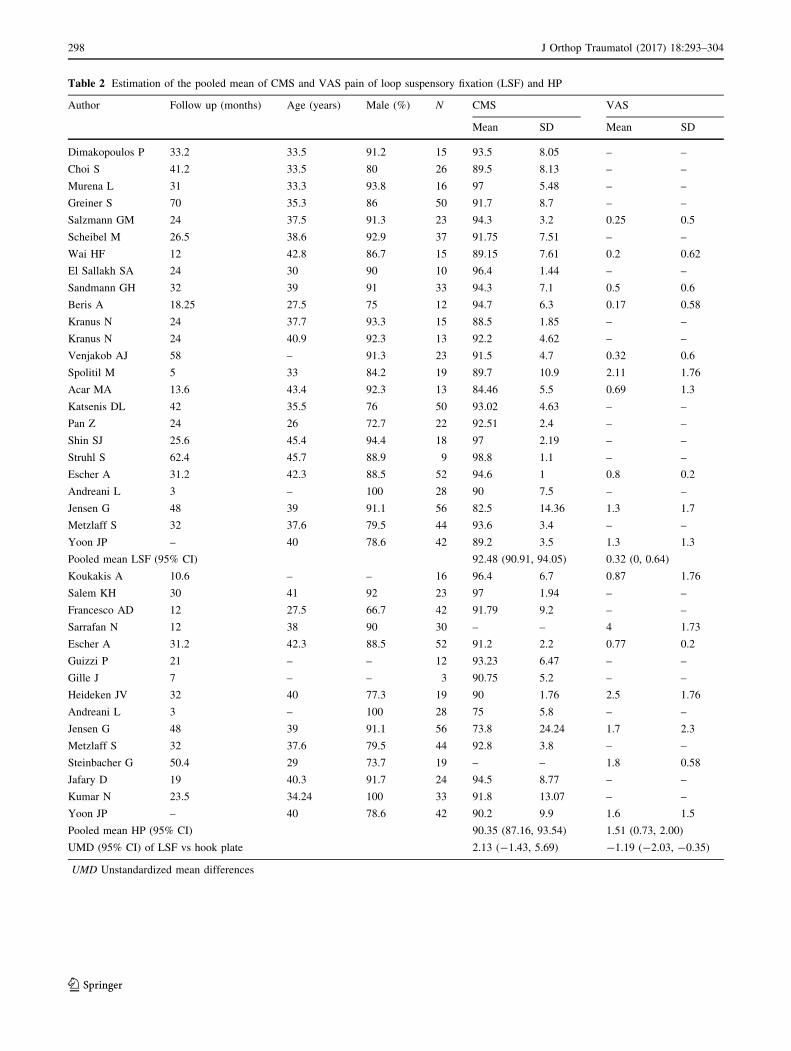
Table 2 Estimation of the pooled mean of CMS and VAS pain of loop suspensory fixation (LSF) and HP
Author Follow up (months) Age (years) Male (%) N CMS VAS
Mean SD Mean SD
Dimakopoulos P 33.2 33.5 91.2 15 93.5 8.05 – –
Choi S 41.2 33.5 80 26 89.5 8.13 – –
Murena L 31 33.3 93.8 16 97 5.48 – –
Greiner S 70 35.3 86 50 91.7 8.7 – –
Salzmann GM 24 37.5 91.3 23 94.3 3.2 0.25 0.5
Scheibel M 26.5 38.6 92.9 37 91.75 7.51 – –
Wai HF 12 42.8 86.7 15 89.15 7.61 0.2 0.62
El Sallakh SA 24 30 90 10 96.4 1.44 – –
Sandmann GH 32 39 91 33 94.3 7.1 0.5 0.6
Beris A 18.25 27.5 75 12 94.7 6.3 0.17 0.58
Kranus N 24 37.7 93.3 15 88.5 1.85 – –
Kranus N 24 40.9 92.3 13 92.2 4.62 – –
Venjakob AJ 58 – 91.3 23 91.5 4.7 0.32 0.6
Spolitil M 5 33 84.2 19 89.7 10.9 2.11 1.76
Acar MA 13.6 43.4 92.3 13 84.46 5.5 0.69 1.3
Katsenis DL 42 35.5 76 50 93.02 4.63 – –
Pan Z 24 26 72.7 22 92.51 2.4 – –
Shin SJ 25.6 45.4 94.4 18 97 2.19 – –
Struhl S 62.4 45.7 88.9 9 98.8 1.1 – –
Escher A 31.2 42.3 88.5 52 94.6 1 0.8 0.2
Andreani L 3 – 100 28 90 7.5 – –
Jensen G 48 39 91.1 56 82.5 14.36 1.3 1.7
Metzlaff S 32 37.6 79.5 44 93.6 3.4 – –
Yoon JP – 40 78.6 42 89.2 3.5 1.3 1.3
Pooled mean LSF (95% CI) 92.48 (90.91, 94.05) 0.32 (0, 0.64)
Koukakis A 10.6 – – 16 96.4 6.7 0.87 1.76
Salem KH 30 41 92 23 97 1.94 – –
Francesco AD 12 27.5 66.7 42 91.79 9.2 – –
Sarrafan N 12 38 90 30 – – 4 1.73
Escher A 31.2 42.3 88.5 52 91.2 2.2 0.77 0.2
Guizzi P 21 – – 12 93.23 6.47 – –
Gille J 7 – – 3 90.75 5.2 – –
Heideken JV 32 40 77.3 19 90 1.76 2.5 1.76
Andreani L 3 – 100 28 75 5.8 – –
Jensen G 48 39 91.1 56 73.8 24.24 1.7 2.3
Metzlaff S 32 37.6 79.5 44 92.8 3.8 – –
Steinbacher G 50.4 29 73.7 19 – – 1.8 0.58
Jafary D 19 40.3 91.7 24 94.5 8.77 – –
Kumar N 23.5 34.24 100 33 91.8 13.07 – –
Yoon JP – 40 78.6 42 90.2 9.9 1.6 1.5
Pooled mean HP (95% CI) 90.35 (87.16, 93.54) 1.51 (0.73, 2.00)
UMD (95% CI) of LSF vs hook plate 2.13 (-1.43, 5.69) -1.19 (-2.03, -0.35)
UMD Unstandardized mean differences
298 J Orthop Traumatol (2017) 18:293–304
123
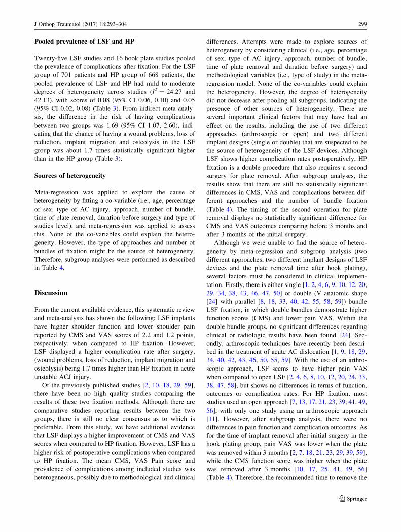
Pooled prevalence of LSF and HP
Twenty-five LSF studies and 16 hook plate studies pooled
the prevalence of complications after fixation. For the LSF
group of 701 patients and HP group of 668 patients, the
pooled prevalence of LSF and HP had mild to moderate
degrees of heterogeneity across studies (I2 = 24.27 and
42.13), with scores of 0.08 (95% CI 0.06, 0.10) and 0.05
(95% CI 0.02, 0.08) (Table 3). From indirect meta-analy-
sis, the difference in the risk of having complications
between two groups was 1.69 (95% CI 1.07, 2.60), indi-
cating that the chance of having a wound problems, loss of
reduction, implant migration and osteolysis in the LSF
group was about 1.7 times statistically significant higher
than in the HP group (Table 3).
Sources of heterogeneity
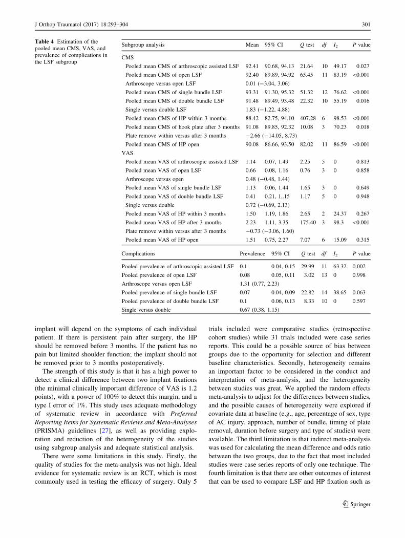
Meta-regression was applied to explore the cause of
heterogeneity by fitting a co-variable (i.e., age, percentage
of sex, type of AC injury, approach, number of bundle,
time of plate removal, duration before surgery and type of
studies level), and meta-regression was applied to assess
this. None of the co-variables could explain the hetero-
geneity. However, the type of approaches and number of
bundles of fixation might be the source of heterogeneity.
Therefore, subgroup analyses were performed as described
in Table 4.
Discussion
From the current available evidence, this systematic review
and meta-analysis has shown the following: LSF implants
have higher shoulder function and lower shoulder pain
reported by CMS and VAS scores of 2.2 and 1.2 points,
respectively, when compared to HP fixation. However,
LSF displayed a higher complication rate after surgery,
(wound problems, loss of reduction, implant migration and
osteolysis) being 1.7 times higher than HP fixation in acute
unstable ACJ injury.
Of the previously published studies [2, 10, 18, 29, 59],
there have been no high quality studies comparing the
results of these two fixation methods. Although there are
comparative studies reporting results between the two
groups, there is still no clear consensus as to which is
preferable. From this study, we have additional evidence
that LSF displays a higher improvement of CMS and VAS
scores when compared to HP fixation. However, LSF has a
higher risk of postoperative complications when compared
to HP fixation. The mean CMS, VAS Pain score and
prevalence of complications among included studies was
heterogeneous, possibly due to methodological and clinical
differences. Attempts were made to explore sources of
heterogeneity by considering clinical (i.e., age, percentage
of sex, type of AC injury, approach, number of bundle,
time of plate removal and duration before surgery) and
methodological variables (i.e., type of study) in the meta-
regression model. None of the co-variables could explain
the heterogeneity. However, the degree of heterogeneity
did not decrease after pooling all subgroups, indicating the
presence of other sources of heterogeneity. There are
several important clinical factors that may have had an
effect on the results, including the use of two different
approaches (arthroscopic or open) and two different
implant designs (single or double) that are suspected to be
the source of heterogeneity of the LSF devices. Although
LSF shows higher complication rates postoperatively, HP
fixation is a double procedure that also requires a second
surgery for plate removal. After subgroup analyses, the
results show that there are still no statistically significant
differences in CMS, VAS and complications between dif-
ferent approaches and the number of bundle fixation
(Table 4). The timing of the second operation for plate
removal displays no statistically significant difference for
CMS and VAS outcomes comparing before 3 months and
after 3 months of the initial surgery.
Although we were unable to find the source of hetero-
geneity by meta-regression and subgroup analysis (two
different approaches, two different implant designs of LSF
devices and the plate removal time after hook plating),
several factors must be considered in clinical implemen-
tation. Firstly, there is either single [1, 2, 4, 6, 9, 10, 12, 20,
29, 34, 38, 43, 46, 47, 50] or double (V anatomic shape
[24] with parallel [8, 18, 33, 40, 42, 55, 58, 59]) bundle
LSF fixation, in which double bundles demonstrate higher
function scores (CMS) and lower pain VAS. Within the
double bundle groups, no significant differences regarding
clinical or radiologic results have been found [24]. Sec-
ondly, arthroscopic techniques have recently been descri-
bed in the treatment of acute AC dislocation [1, 9, 18, 29,
34, 40, 42, 43, 46, 50, 55, 59]. With the use of an arthro-
scopic approach, LSF seems to have higher pain VAS
when compared to open LSF [2, 4, 6, 8, 10, 12, 20, 24, 33,
38, 47, 58], but shows no differences in terms of function,
outcomes or complication rates. For HP fixation, most
studies used an open approach [7, 13, 17, 21, 23, 39, 41, 49,
56], with only one study using an arthroscopic approach
[11]. However, after subgroup analysis, there were no
differences in pain function and complication outcomes. As
for the time of implant removal after initial surgery in the
hook plating group, pain VAS was lower when the plate
was removed within 3 months [2, 7, 18, 21, 23, 29, 39, 59],
while the CMS function score was higher when the plate
was removed after 3 months [10, 17, 25, 41, 49, 56]
(Table 4). Therefore, the recommended time to remove the
J Orthop Traumatol (2017) 18:293–304 299
123
Table 3 Estimation of the
pooled prevalence of post-
operative complication of LSF
and HP
Author Follow up (months) Age (years) Male (%) N Complication
Yes No
Stam L 46.8 40 73.9 23 0 23
Dimakopoulos P 33.2 33.5 91.2 15 2 32
Ryhanen J 12 37 – 15 1 14
Choi S 41.2 33.5 80 26 1 19
Murena L 31 33.3 93.8 16 2 14
Greiner S 70 35.3 86 50 4 46
Salzmann GM 24 37.5 91.3 23 1 22
Scheibel M 26.5 38.6 92.9 37 0 40
Wai HF 12 42.8 86.7 15 2 13
El Sallakh SA 24 30 90 10 0 10
Sandmann GH 32 39 91 33 3 30
Beris A 18.25 27.5 75 12 0 12
Kranus N 24 37.7 93.3 15 0 13
Kranus N 24 40.9 92.3 13 1 14
Venjakob AJ 58 – 91.3 23 2 21
Spolitil M 5 33 84.2 19 3 16
Acar MA 13.6 43.4 92.3 13 1 12
Katsenis DL 42 35.5 76 50 6 50
Pan Z 24 26 72.7 22 2 20
Shin SJ 25.6 45.4 94.4 18 8 10
Struhl S 62.4 45.7 88.9 9 4 5
Escher A 31.2 42.3 88.5 52 0 25
Andreani L 3 – 100 28 2 17
Jensen G 48 39 91.1 56 12 14
Metzlaff S 32 37.6 79.5 44 1 23
Yoon JP – 40 78.6 42 6 12
Pooled prevalence LSF (95% CI) 0.08 (0.06, 0.10)
Koukakis A 10.6 – – 16 0 16
Salem KH 30 41 92 23 7 16
Kienast B 36 38.4 – 225 24 201
Francesco AD 12 27.5 66.7 42 5 37
Sarrafan N 12 38 90 30 1 29
Escher A 31.2 42.3 88.5 52 5 22
Guizzi P 21 – – 12 1 1
Gille J 7 – – 3 0 3
Heideken JV 32 40 77.3 19 0 19
Andreani L 3 – 100 28 0 7
Jensen G 48 39 91.1 56 19 11
Metzlaff S 32 37.6 79.5 44 0 20
Steinbacher G 50.4 29 73.7 19 0 19
Jafary D 19 40.3 91.7 24 2 22
Kumar N 23.5 34.24 100 33 0 33
Yoon JP – 40 78.6 42 9 15
Pooled prevalence HP (95% CI) 0.05 (0.02, 0.08)
RR of LSF vs HP 1.69 (1.07, 2.60)
300 J Orthop Traumatol (2017) 18:293–304
123
implant will depend on the symptoms of each individual
patient. If there is persistent pain after surgery, the HP
should be removed before 3 months. If the patient has no
pain but limited shoulder function; the implant should not
be removed prior to 3 months postoperatively.
The strength of this study is that it has a high power to
detect a clinical difference between two implant fixations
(the minimal clinically important difference of VAS is 1.2
points), with a power of 100% to detect this margin, and a
type I error of 1%. This study uses adequate methodology
of systematic review in accordance with Preferred
Reporting Items for Systematic Reviews and Meta-Analyses
(PRISMA) guidelines [27], as well as providing explo-
ration and reduction of the heterogeneity of the studies
using subgroup analysis and adequate statistical analysis.
There were some limitations in this study. Firstly, the
quality of studies for the meta-analysis was not high. Ideal
evidence for systematic review is an RCT, which is most
commonly used in testing the efficacy of surgery. Only 5
trials included were comparative studies (retrospective
cohort studies) while 31 trials included were case series
reports. This could be a possible source of bias between
groups due to the opportunity for selection and different
baseline characteristics. Secondly, heterogeneity remains
an important factor to be considered in the conduct and
interpretation of meta-analysis, and the heterogeneity
between studies was great. We applied the random effects
meta-analysis to adjust for the differences between studies,
and the possible causes of heterogeneity were explored if
covariate data at baseline (e.g., age, percentage of sex, type
of AC injury, approach, number of bundle, timing of plate
removal, duration before surgery and type of studies) were
available. The third limitation is that indirect meta-analysis
was used for calculating the mean difference and odds ratio
between the two groups, due to the fact that most included
studies were case series reports of only one technique. The
fourth limitation is that there are other outcomes of interest
that can be used to compare LSF and HP fixation such as
Table 4 Estimation of the
pooled mean CMS, VAS, and
prevalence of complications in
the LSF subgroup
Subgroup analysis Mean 95% CI Q test df I2 P value
CMS
Pooled mean CMS of arthroscopic assisted LSF 92.41 90.68, 94.13 21.64 10 49.17 0.027
Pooled mean CMS of open LSF 92.40 89.89, 94.92 65.45 11 83.19 \0.001
Arthroscope versus open LSF 0.01 (-3.04, 3.06)
Pooled mean CMS of single bundle LSF 93.31 91.30, 95.32 51.32 12 76.62 \0.001
Pooled mean CMS of double bundle LSF 91.48 89.49, 93.48 22.32 10 55.19 0.016
Single versus double LSF 1.83 (-1.22, 4.88)
Pooled mean CMS of HP within 3 months 88.42 82.75, 94.10 407.28 6 98.53 \0.001
Pooled mean CMS of hook plate after 3 months 91.08 89.85, 92.32 10.08 3 70.23 0.018
Plate remove within versus after 3 months -2.66 (-14.05, 8.73)
Pooled mean CMS of HP open 90.08 86.66, 93.50 82.02 11 86.59 \0.001
VAS
Pooled mean VAS of arthroscopic assisted LSF 1.14 0.07, 1.49 2.25 5 0 0.813
Pooled mean VAS of open LSF 0.66 0.08, 1.16 0.76 3 0 0.858
Arthroscope versus open 0.48 (-0.48, 1.44)
Pooled mean VAS of single bundle LSF 1.13 0.06, 1.44 1.65 3 0 0.649
Pooled mean VAS of double bundle LSF 0.41 0.21, 1,.15 1.17 5 0 0.948
Single versus double 0.72 (-0.69, 2.13)
Pooled mean VAS of HP within 3 months 1.50 1.19, 1.86 2.65 2 24.37 0.267
Pooled mean VAS of HP after 3 months 2.23 1.11, 3.35 175.40 3 98.3 \0.001
Plate remove within versus after 3 months -0.73 (-3.06, 1.60)
Pooled mean VAS of HP open 1.51 0.75, 2.27 7.07 6 15.09 0.315
Complications Prevalence 95% CI Q test df I2 P value
Pooled prevalence of arthroscopic assisted LSF 0.1 0.04, 0.15 29.99 11 63.32 0.002
Pooled prevalence of open LSF 0.08 0.05, 0.11 3.02 13 0 0.998
Arthroscope versus open LSF 1.31 (0.77, 2.23)
Pooled prevalence of single bundle LSF 0.07 0.04, 0.09 22.82 14 38.65 0.063
Pooled prevalence of double bundle LSF 0.1 0.06, 0.13 8.33 10 0 0.597
Single versus double 0.67 (0.38, 1.15)
J Orthop Traumatol (2017) 18:293–304 301
123
operation cost or post-operative satisfaction and quality of
life. However, these factors could not be analyzed because
of insufficient data. The last limitation is that most studies
had a mean follow up time of approximately 1–2 years;
therefore mid-term to long-term effects of the different
types of fixation are still unknown.
In summary, for acute high-grade ACJ injuries, both HP
and LSF had acceptable post-operative outcomes. LSF pro-
vided better postoperative shoulder function (CMS) when
compared to HP fixation, but the difference was not statis-
tically significant. LSF provided clinically and statistically
significant lower pain VAS when compared to HP fixation.
However, LSF had higher complication rates when com-
pared to the HP fixation group. This study shows that both
options have advantages and disadvantages and should be
chosen based on patient status. In the future, prospective
randomized controlled studies are needed to confirm these
findings as the current literature is still insufficient.
Compliance with ethical standards
Conflict of interest All authors declare that they have no conflicts of
interests.
Patient consent For this type of study informed consent is not
required.
Ethical approval Not applicable as no new patients were involved in
this research.
Funding None.
Open Access This article is distributed under the terms of the
Creative Commons Attribution 4.0 International License (http://crea
tivecommons.org/licenses/by/4.0/), which permits unrestricted use,
distribution, and reproduction in any medium, provided you give
appropriate credit to the original author(s) and the source, provide a
link to the Creative Commons license, and indicate if changes were
made.
Appendix
Search term and strategy
#1 acromioclavicular joint
#2 AC joint
#3 #1 OR #2
#4 separation
#5 dislocation
#6 trauma
#7 injury
#8 #4 OR #5 OR #6 OR #7
#9 hook plate
#10 locking plate
#11 fixation
#12 tightrope
#13 dogbone
#14 #9 OR #10 OR #11 OR #12 OR #13
#15 Constant score
#16 Constant Murley scale
#17 CMS
#18 pain
#19 UCLA
#20 #15 OR #16 OR #17 OR #18 OR #19
#21 #3 AND #8 AND #14 AND #20
References
1. Acar MA, Gulec A, Erkocak OF, Yilmaz G, Durgut F, Elmadag
M (2015) Percutaneous double-button fixation method for treat-
ment of acute type III acromioclavicular joint dislocation. Acta
Orthop Traumatol Turc 49(3):241–248. doi:10.3944/AOTT.2015.
14.0230
2. Andreani L, Bonicoli E, Parchi P, Piolanti N, Michele L (2014)
Acromio-clavicular repair using two different techniques. Eur J
Orthop Surg Traumatol 24(2):237–242. doi:10.1007/s00590-013-
1186-1
3. Babhulkar A, Pawaskar A (2014) Acromioclavicular joint dislo-
cations. Curr Rev Musculoskelet Med 7(1):33–39. doi:10.1007/
s12178-013-9199-2
4. Beris A, Lykissas M, Kostas-Agnantis I, Vekris M, Mitsionis G,
Korompilias A (2013) Management of acute acromioclavicular
joint dislocation with a double-button fixation system. Injury
44(3):288–292. doi:10.1016/j.injury.2013.01.002
5. Bishop JY, Kaeding C (2006) Treatment of the acute traumatic
acromioclavicular separation. Sports Med Arthrosc Rev
14(4):237–245. doi:10.1097/01.jsa.0000212330.32969.6e
6. Choi SW, Lee TJ, Moon KH, Cho KJ, Lee SY (2008) Minimally
invasive coracoclavicular stabilization with suture anchors for
acute acromioclavicular dislocation. Am J Sports Med
36(5):961–965. doi:10.1177/0363546507312643
7. Di Francesco A, Zoccali C, Colafarina O, Pizzoferrato R, Flamini
S (2012) The use of hook plate in type III and V acromio-clav-
icular Rockwood dislocations: clinical and radiological midterm
results and MRI evaluation in 42 patients. Injury 43(2):147–152.
doi:10.1016/j.injury.2011.04.002
8. Dimakopoulos P, Panagopoulos A, Syggelos SA, Pana-
giotopoulos E, Lambiris E (2006) Double-loop suture repair for
acute acromioclavicular joint disruption. Am J Sports Med
34(7):1112–1119. doi:10.1177/0363546505284187
9. El Sallakh SA (2012) Evaluation of arthroscopic stabilization of
acute acromioclavicular joint dislocation using the TightRope
system. Orthopedics 35(1):e18–e22. doi:10.3928/01477447-
20111122-13
10. Eschler A, Gradl G, Gierer P, Mittlmeier T, Beck M (2012) Hook
plate fixation for acromioclavicular joint separations restores
coracoclavicular distance more accurately than PDS augmenta-
tion, however presents with a high rate of acromial osteolysis.
Arch Orthop Trauma Surg 132(1):33–39. doi:10.1007/s00402-
011-1399-x
11. Gille J, Heinrichs G, Unger A, Riepenhof H, Herzog J, Kienast B,
Oheim R (2013) Arthroscopic-assisted hook plate fixation for
acromioclavicular joint dislocation. Int Orthop 37(1):77–82.
doi:10.1007/s00264-012-1691-6
12. Greiner S, Braunsdorf J, Perka C, Herrmann S, Scheffler S (2009)
Mid to long-term results of open acromioclavicular-joint
302 J Orthop Traumatol (2017) 18:293–304
123
reconstruction using polydioxansulfate cerclage augmentation.
Arch Orthop Trauma Surg 129(6):735–740. doi:10.1007/s00402-
008-0688-5
13. Guizzi P, Quadri V, Claudio M, Moretta M (2012) Rockwood
type iii acromioclavicular dislocation: clinical and x ray review
after treatment with clavicle Hook Plate. Minerva Ortop Trau-
matol 63(4):271–278
14. Guttmann D, Paksima NE, Zuckerman JD (2000) Complications
of treatment of complete acromioclavicular joint dislocations.
Instr Course Lect 49:407–413
15. Higgins JP, Thompson SG (2002) Quantifying heterogeneity in a
meta-analysis. Stat Med 21(11):1539–1558. doi:10.1002/sim.
1186
16. Horst K, Dienstknecht T, Pishnamaz M, Sellei RM, Kobbe P,
Pape HC (2013) Operative treatment of acute acromioclavicular
joint injuries graded Rockwood III and IV: risks and benefits in
tight rope technique vs. k-wire fixation. Patient Saf Surg 7:18.
doi:10.1186/1754-9493-7-18
17. Jafary D, Keihan Shokouh H, Najd Mazhar F, Shariat Zadeh H,
Mochtary T (2014) Clinical and radiological results of fixation of
acromioclavicular joint dislocation by hook plates retained for
more than five months. Trauma Mon 19(2):e13728. doi:10.5812/
traumamon.13728
18. Jensen G, Katthagen JC, Alvarado LE, Lill H, Voigt C (2014)
Has the arthroscopically assisted reduction of acute AC joint
separations with the double tight-rope technique advantages over
the clavicular hook plate fixation? Knee Surg Sports Traumatol
Arthrosc 22(2):422–430. doi:10.1007/s00167-012-2270-5
19. Jobe FW, Bradley JP (1989) The diagnosis and nonoperative
treatment of shoulder injuries in athletes. Clin Sports Med
8(3):419–438
20. Katsenis DL, Stamoulis D, Begkas D, Tsamados S (2015) Min-
imally invasive reconstruction of acute type IV and Type V
acromioclavicular separations. Orthopedics 38(4):e324–e330.
doi:10.3928/01477447-20150402-62
21. Kienast B, Thietje R, Queitsch C, Gille J, Schulz AP, Meiners J
(2011) Mid-term results after operative treatment of rockwood
grade III–V acromioclavicular joint dislocations with an AC-
hook-plate. Eur J Med Res 16(2):52–56
22. Kim S, Blank A (2014) Management of type 3 acromioclavicular
joint dislocations—current controversies. Bull Hosp Jt Dis (2013)
72(1):53–60
23. Koukakis A, Manouras A, Apostolou CD, Lagoudianakis E,
Papadima A, Triantafillou C, Korres D, Allen PW, Amini A
(2008) Results using the AO hook plate for dislocations of the
acromioclavicular joint. Expert Rev Med Devices 5(5):567–572.
doi:10.1586/17434440.5.5.567
24. Kraus N, Haas NP, Scheibel M, Gerhardt C (2013) Arthroscop-
ically assisted stabilization of acute high-grade acromioclavicular
joint separations in a coracoclavicular Double-TightRope tech-
nique: V-shaped versus parallel drill hole orientation. Arch
Orthop Trauma Surg 133(10):1431–1440. doi:10.1007/s00402-
013-1804-8
25. Kumar N, Sharma V (2015) Hook plate fixation for acute
acromioclavicular dislocations without coracoclavicular ligament
reconstruction: a functional outcome study in military personnel.
Strateg Trauma Limb Reconstr (Online) 10(2):79–85. doi:10.
1007/s11751-015-0228-0
26. Li Q, Hsueh PL, Chen YF (2014) Coracoclavicular ligament
reconstruction: a systematic review and a biomechanical study of
a triple endobutton technique. Medicine 93(28):e193. doi:10.
1097/md.0000000000000193
27. Liberati A, Altman DG, Tetzlaff J, Mulrow C, Gotzsche PC,
Ioannidis JP, Clarke M, Devereaux PJ, Kleijnen J, Moher D
(2009) The PRISMA statement for reporting systematic reviews
and meta-analyses of studies that evaluate health care interven-
tions: explanation and elaboration. PLoS Med 6(7):e1000100
28. Macdonald PB, Lapointe P (2008) Acromioclavicular and stern-
oclavicular joint injuries. Orthop Clin N Am 39(4):535–545.
doi:10.1016/j.ocl.2008.05.003
29. Metzlaff S, Rosslenbroich S, Forkel PH, Schliemann B, Arshad
H, Raschke M, Petersen W (2016) Surgical treatment of acute
acromioclavicular joint dislocations: hook plate versus minimally
invasive reconstruction. Knee Surg Sports Traumatol Arthrosc
24(6):1972–1978. doi:10.1007/s00167-014-3294-9
30. Modi CS, Beazley J, Zywiel MG, Lawrence TM, Veillette CJ
(2013) Controversies relating to the management of acromio-
clavicular joint dislocations. Bone Joint J 95-B(12):1595–1602.
doi:10.1302/0301-620x.95b12.31802
31. Morrison DS, Lemos MJ (1995) Acromioclavicular separation.
Reconstruction using synthetic loop augmentation. Am J Sports
Med 23(1):105–110
32. Motamedi AR, Blevins FT, Willis MC, McNally TP, Shahinpoor
M (2000) Biomechanics of the coracoclavicular ligament com-
plex and augmentations used in its repair and reconstruction. Am
J Sports Med 28(3):380–384
33. Murena L, Vulcano E, Ratti C, Cecconello L, Rolla PR, Surace
MF (2009) Arthroscopic treatment of acute acromioclavicular
joint dislocation with double flip button. Knee Surg Sports
Traumatol Arthrosc 17(12):1511–1515. doi:10.1007/s00167-009-
0838-5
34. Pan Z, Zhang H, Sun C, Qu L, Cui Y (2015) Arthroscopy-assisted
reconstruction of coracoclavicular ligament by Endobutton fixation
for treatment of acromioclavicular joint dislocation. Arch Orthop
Trauma Surg 135(1):9–16. doi:10.1007/s00402-014-2117-2
35. Patzer T, Clauss C, Kuhne CA, Ziring E, Efe T, Ruchholtz S,
Mann D (2013) Arthroscopically assisted reduction of acute
acromioclavicular joint separations: comparison of clinical and
radiological results of single versus double TightRope technique.
Der Unfallchirurg 116(5):442–450. doi:10.1007/s00113-011-
2135-2
36. Petersen W, Wellmann M, Rosslenbroich S, Zantop T (2010)
Minimally invasive acromioclavicular joint reconstruction
(MINAR). Oper Orthop Traumatol 22(1):52–61. doi:10.1007/
s00064-010-3004-4
37. Rieser GR, Edwards K, Gould GC, Markert RJ, Goswami T,
Rubino LJ (2013) Distal-third clavicle fracture fixation: a
biomechanical evaluation of fixation. J Shoulder Elb Surg
22(6):848–855. doi:10.1016/j.jse.2012.08.022
38. Ryhanen J, Leminen A, Jamsa T, Tuukkanen J, Pramila A,
Raatikainen T (2006) A novel treatment of grade III acromio-
clavicular joint dislocations with a C-hook implant. Arch Orthop
Trauma Surg 126(1):22–27. doi:10.1007/s00402-005-0074-5
39. Salem KH, Schmelz A (2009) Treatment of Tossy III acromio-
clavicular joint injuries using hook plates and ligament suture.
J Orthop Trauma 23(8):565–569. doi:10.1097/BOT.
0b013e3181971b38
40. Salzmann GM, Walz L, Buchmann S, Glabgly P, Venjakob A,
Imhoff AB (2010) Arthroscopically assisted 2-bundle anatomical
reduction of acute acromioclavicular joint separations. Am J
Sports Med 38(6):1179–1187. doi:10.1177/0363546509355645
41. Sarrafan N, Nasab SAM, Gholaminejad S (2012) Comparison of
the efficacy of hook plate versus pinning in treatment of acute
acromioclavicular joint dislocation. Pak J Med Sci 28(5):879–882
42. Scheibel M, Droschel S, Gerhardt C, Kraus N (2011) Arthro-
scopically assisted stabilization of acute high-grade acromio-
clavicular joint separations. Am J Sports Med 39(7):1507–1516.
doi:10.1177/0363546511399379
43. Shin SJ, Kim NK (2015) Complications after arthroscopic cora-
coclavicular reconstruction using a single adjustable-loop-length
J Orthop Traumatol (2017) 18:293–304 303
123
suspensory fixation device in acute acromioclavicular joint dis-
location. Arthrosc J Arthrosc Relat Surg 31(5):816–824. doi:10.
1016/j.arthro.2014.11.013
44. Simmonds MC, Higgins JP (2007) Covariate heterogeneity in
meta-analysis: criteria for deciding between meta-regression and
individual patient data. Stat Med 26(15):2982–2999. doi:10.1002/
sim.2768
45. Smith TO, Chester R, Pearse EO, Hing CB (2011) Operative
versus non-operative management following Rockwood grade III
acromioclavicular separation: a meta-analysis of the current
evidence base. J Orthop Traumatol 12(1):19–27. doi:10.1007/
s10195-011-0127-1
46. Spoliti M, De Cupis M, Via AG, Oliva F (2014) All arthroscopic
stabilization of acute acromioclavicular joint dislocation with
fiberwire and endobutton system. Muscles Ligaments Tendons J
4(4):398–403
47. Stam L, Dawson I (1991) Complete acromioclavicular disloca-
tions: treatment with a Dacron ligament. Injury 22(3):173–176
48. StataCorp. (2015) Stata statistical software: release 14. StataCorp
LP, College Station
49. Steinbacher G, Sallent A, Seijas R, Boffa JM, Espinosa W, Cugat
R (2014) Clavicular hook plate for grade-III acromioclavicular
dislocation. J Orthop Surg (Hong Kong) 22(3):329–332
50. Struhl S, Wolfson TS (2015) Continuous loop double endobutton
reconstruction for acromioclavicular joint dislocation. Am J
Sports Med 43(10):2437–2444. doi:10.1177/0363546515596409
51. Taft TN, Wilson FC, Oglesby JW (1987) Dislocation of the
acromioclavicular joint. An end-result study. J Bone Joint Surg
Am 69(7):1045–1051
52. Tauber M (2013) Management of acute acromioclavicular joint
dislocations: current concepts. Arch Orthop Trauma Surg
133(7):985–995. doi:10.1007/s00402-013-1748-z
53. Thakkinstian A, McEvoy M, Minelli C, Gibson P, Hancox B,
Duffy D, Thompson J, Hall I, Kaufman J, Leung TF, Helms PJ,
Hakonarson H, Halpi E, Navon R, Attia J (2005) Systematic
review and meta-analysis of the association between {beta}2-
adrenoceptor polymorphisms and asthma: a HuGE review. Am J
Epidemiol 162(3):201–211. doi:10.1093/aje/kwi184
54. Thompson SG, Higgins JP (2002) How should meta-regression
analyses be undertaken and interpreted? Stat Med
21(11):1559–1573. doi:10.1002/sim.1187
55. Venjakob AJ, Salzmann GM, Gabel F, Buchmann S, Walz L,
Spang JT, Vogt S, Imhoff AB (2013) Arthroscopically assisted
2-bundle anatomic reduction of acute acromioclavicular joint
separations: 58-month findings. Am J Sports Med 41(3):615–621.
doi:10.1177/0363546512473438
56. von Heideken J, Bostrom Windhamre H, Une-Larsson V, Eke-
lund A (2013) Acute surgical treatment of acromioclavicular
dislocation type V with a hook plate: superiority to late recon-
struction. J Shoulder Elbow Surg 22(1):9–17. doi:10.1016/j.jse.
2012.03.003
57. Wang Y, Zhang J (2014) Acromioclavicular joint reconstruction
by coracoid process transfer augmented with hook plate. Injury
45(6):949–954. doi:10.1016/j.injury.2013.12.013
58. Wei HF, Chen YF, Zeng BF, Zhang CQ, Chai YM, Wang HM,
Lu Y (2011) Triple endobuttton technique for the treatment of
acute complete acromioclavicular joint dislocations: preliminary
results. Int Orthop 35(4):555–559. doi:10.1007/s00264-010-
1057-x
59. Yoon JP, Lee BJ, Nam SJ, Chung SW, Jeong WJ, Min WK, Oh
JH (2015) Comparison of results between hook plate fixation and
ligament reconstruction for acute unstable acromioclavicular
joint dislocation. Clin Orthop Surg 7(1):97–103. doi:10.4055/
cios.2015.7.1.97
304 J Orthop Traumatol (2017) 18:293–304
123


