Complications During A Laparoscopic Cholecystectomy Bill Cavatassi.
Upload
zeeshanrahman86Category
view
127download
3
Post- cholecystectomy complications
Dr. Zeeshan
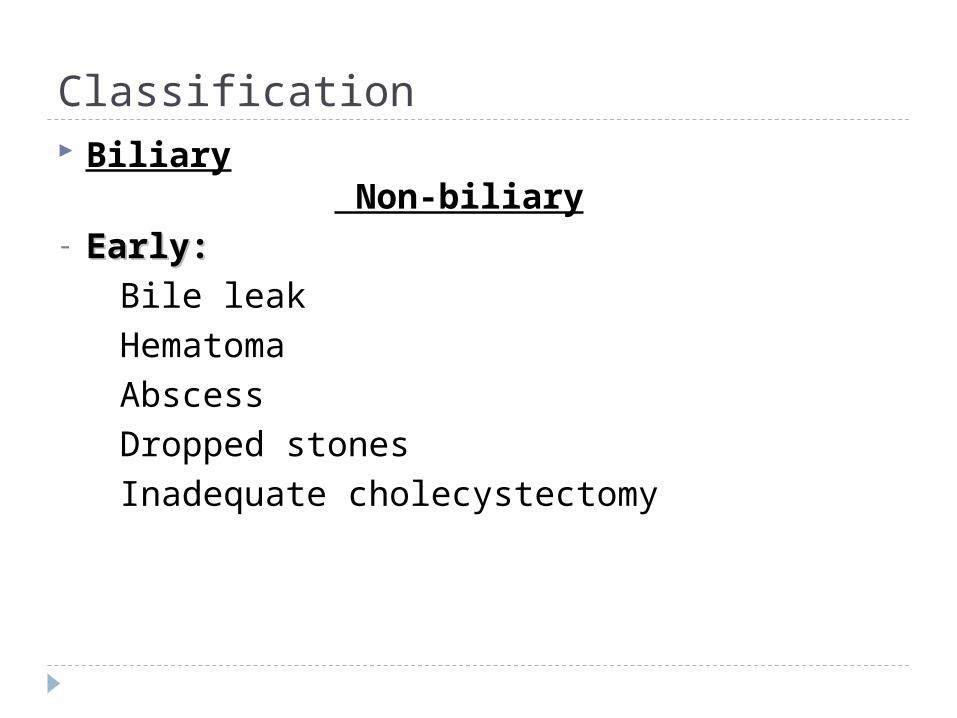
Classification Biliary Non-
biliary- Early:Early: Bile leak Hematoma Abscess Dropped stones Inadequate cholecystectomy
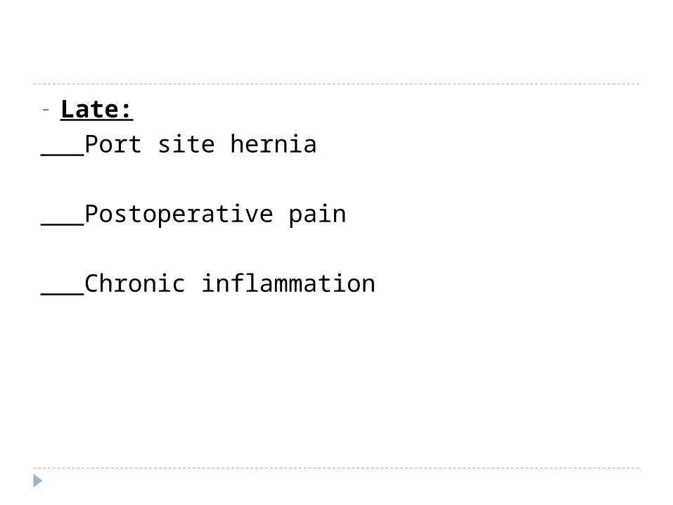
- Late: Port site hernia
Postoperative pain
Chronic inflammation
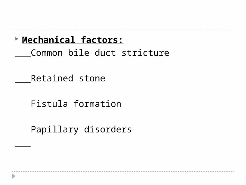
Mechanical factors: Common bile duct stricture
Retained stone
Fistula formation
Papillary disorders
Bile duct injury Incidence : - Laparoscopic cholecystectomy: 0.2 – 0.7 %- Open : 0.1 – 0.04 %
Most common factors associated:- Acute cholecystitis- Inexperience of the surgeon (1st 15
operations)
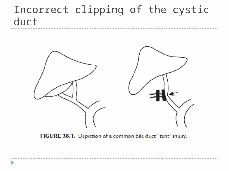
Incorrect clipping of the cystic duct
Important factors to prevent bile duct injury:- Surgical experience- Preoperative imaging- Precise operative procedure- Conversion to open in difficult cases- Visualisation of “critical view”- Abandoning infundibular dissection
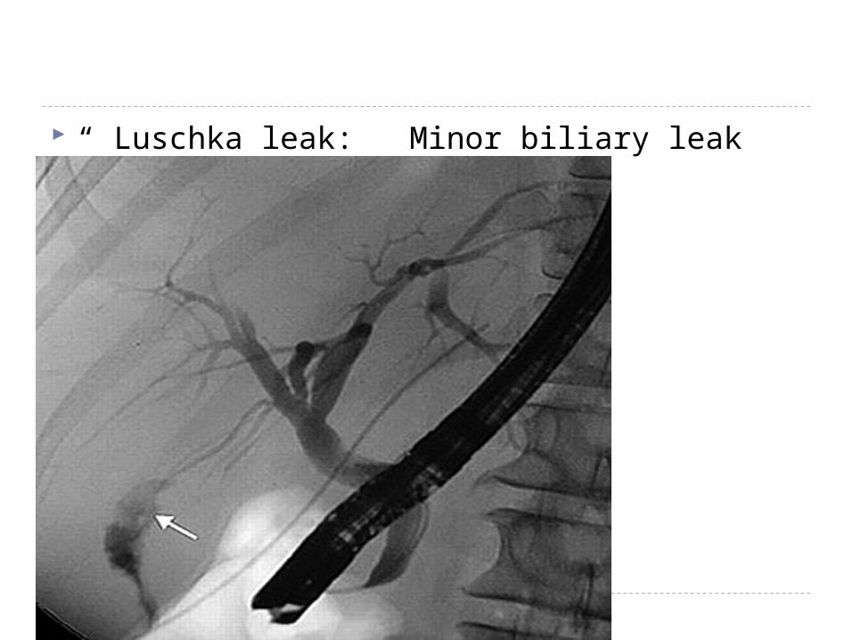
“ Luschka leak: Minor biliary leak
Luschka leak Accessory bile duct in the liver bed
If drainage > 500 ml over 24 hours --- ERCP + sphincterotomy + stenting of the CBD
Hemorrhage Incidence:- Intraop : 2.3%
Most common treatment : Open procedure with hemostasis – suturing of the GB peritoneum
Most common source: GB bed
Usually seen in - Acute cholecystitis- Cirrhosis
Other sites of bleeding:- Incursion into deeper planes Distal tributaries of middle hepatic veins
encountered(10%)- Port site
- Cystic artery (1%)
Dropped stone Incidence : 6% - 40% dropped stones
following GB perforation Stones lost : 3% - 32%
Complications:- Fever- Abdominal pain – chronic type- Intestinal obstruction- Liver abscess / bacteremia- Empyema / broncholithiasis
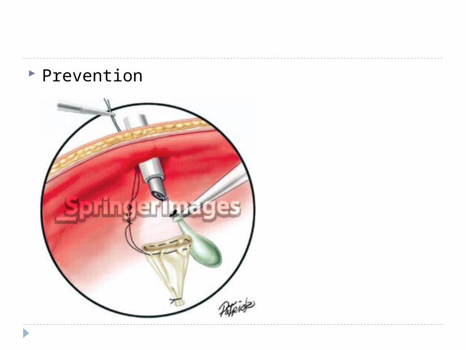
Prevention
Insufficient cholecystectomy Indication :- Acute cholecystitis - Inability to obtain critical view – SUBTOTAL CHOLE
Study in UK: 26 cases of subtotal cholecystectomy
5 cases – Postop ERCP for biliary leak / retained stones
1 case – Subphrenic abscess – re-laparotomy 1 case – Gall stone retained in pouch – completion
LC 2 cases – Port site hernia
In Lap. Subtotal cholecystectomy:
- Experienced surgeon required
- Always place a drain at level of transection
- If port site retained - coagulate
Infection CDC study done over 7 years ( n = 54,504
patients) SSI in lap vs open : 0.62 % - 1.82% Pathogens are similar : Enterococcus, E.coli,
Staph. In open approach : Infection limited to
subcutaneous plane In lap approach : Infection predominantly intra-
abdominal SSI higher in : - Emergency surgery - ASA >= 3
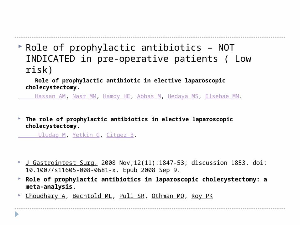
Role of prophylactic antibiotics – NOT INDICATED in pre-operative patients ( Low risk)
Role of prophylactic antibiotic in elective laparoscopic cholecystectomy.
Hassan AM, Nasr MM, Hamdy HE, Abbas M, Hedaya MS, Elsebae MM.
The role of prophylactic antibiotics in elective laparoscopic cholecystectomy.
Uludag M, Yetkin G, Citgez B.
J Gastrointest Surg. 2008 Nov;12(11):1847-53; discussion 1853. doi: 10.1007/s11605-008-0681-x. Epub 2008 Sep 9.
Role of prophylactic antibiotics in laparoscopic cholecystectomy: a meta-analysis.
Choudhary A, Bechtold ML, Puli SR, Othman MO, Roy PK
Late complications Abdominal pain - Pathophysiology : Bile duct neuromas
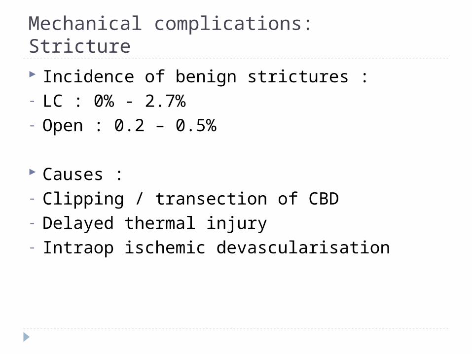
Mechanical complications:Stricture Incidence of benign strictures :- LC : 0% - 2.7%- Open : 0.2 – 0.5%
Causes :- Clipping / transection of CBD- Delayed thermal injury- Intraop ischemic devascularisation
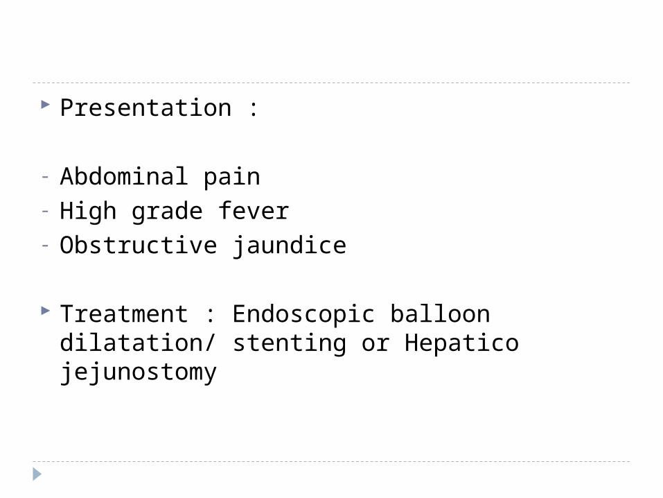
Presentation :
- Abdominal pain- High grade fever- Obstructive jaundice
Treatment : Endoscopic balloon dilatation/ stenting or Hepatico jejunostomy
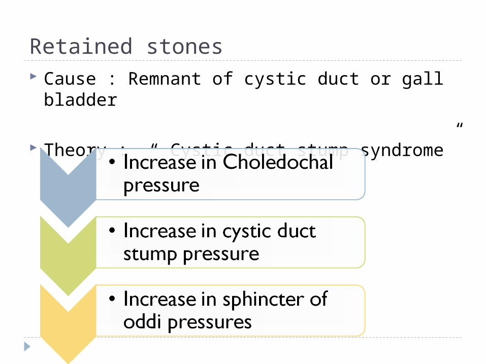
Retained stones Cause : Remnant of cystic duct or gall bladder
Theory : “ Cystic duct stump syndrome”
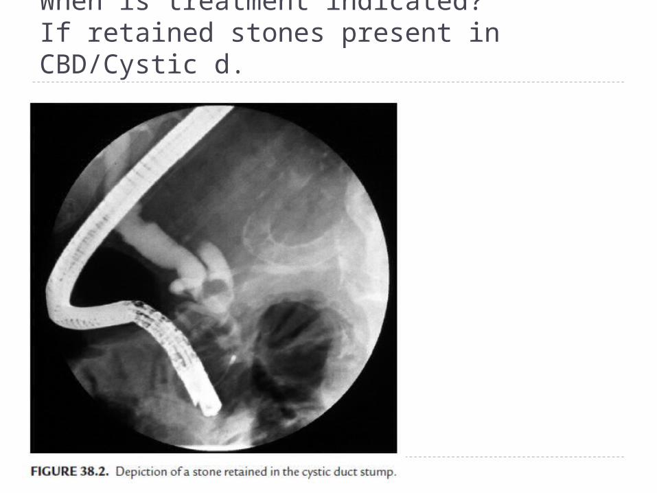
When is treatment indicated?If retained stones present in CBD/Cystic d.
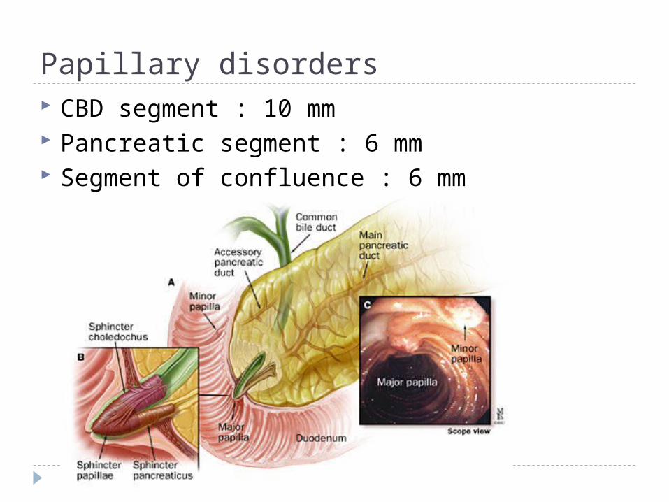
Papillary disorders CBD segment : 10 mm Pancreatic segment : 6 mm Segment of confluence : 6 mm
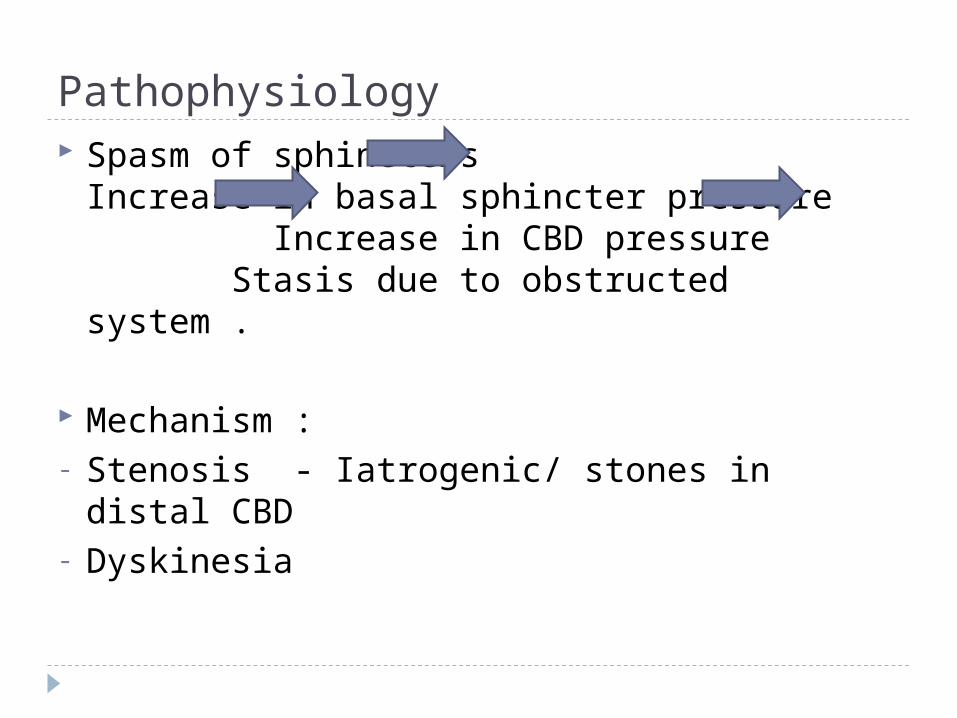
Pathophysiology Spasm of sphincters Increase in basal
sphincter pressure Increase in CBD pressure Stasis due to obstructed system .
Mechanism : - Stenosis - Iatrogenic/ stones in distal CBD- Dyskinesia
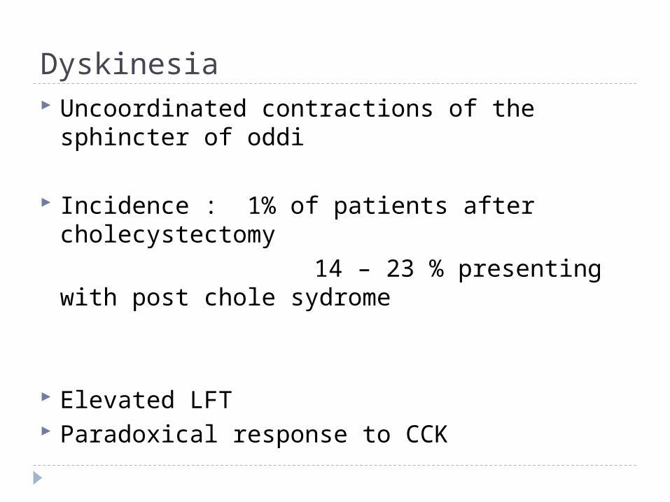
Dyskinesia Uncoordinated contractions of the sphincter of
oddi
Incidence : 1% of patients after cholecystectomy
14 – 23 % presenting with post chole sydrome
Elevated LFT Paradoxical response to CCK
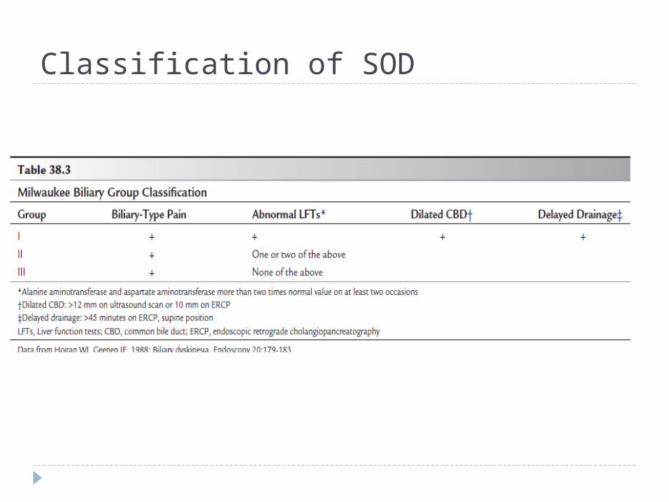
Classification of SOD
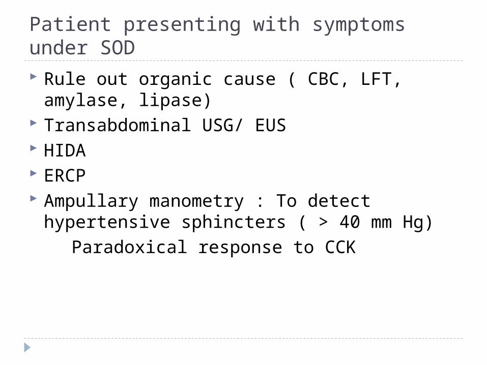
Patient presenting with symptoms under SOD Rule out organic cause ( CBC, LFT, amylase,
lipase) Transabdominal USG/ EUS HIDA ERCP Ampullary manometry : To detect
hypertensive sphincters ( > 40 mm Hg) Paradoxical response to CCK
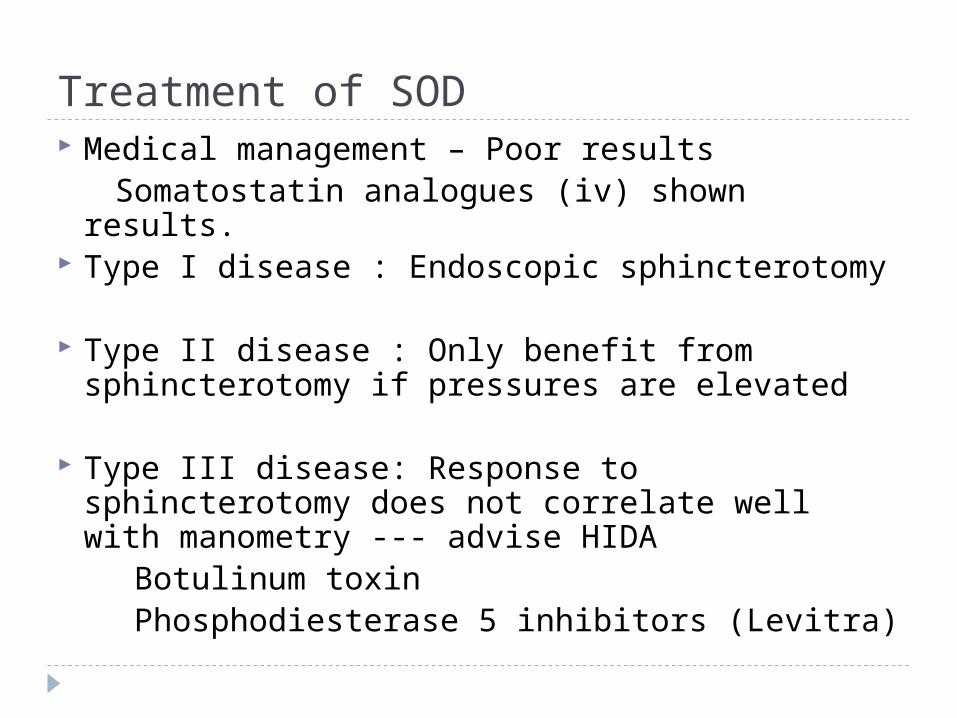
Treatment of SOD Medical management – Poor results Somatostatin analogues (iv) shown results. Type I disease : Endoscopic sphincterotomy
Type II disease : Only benefit from sphincterotomy if pressures are elevated
Type III disease: Response to sphincterotomy does not correlate well with manometry --- advise HIDA
Botulinum toxin Phosphodiesterase 5 inhibitors (Levitra)
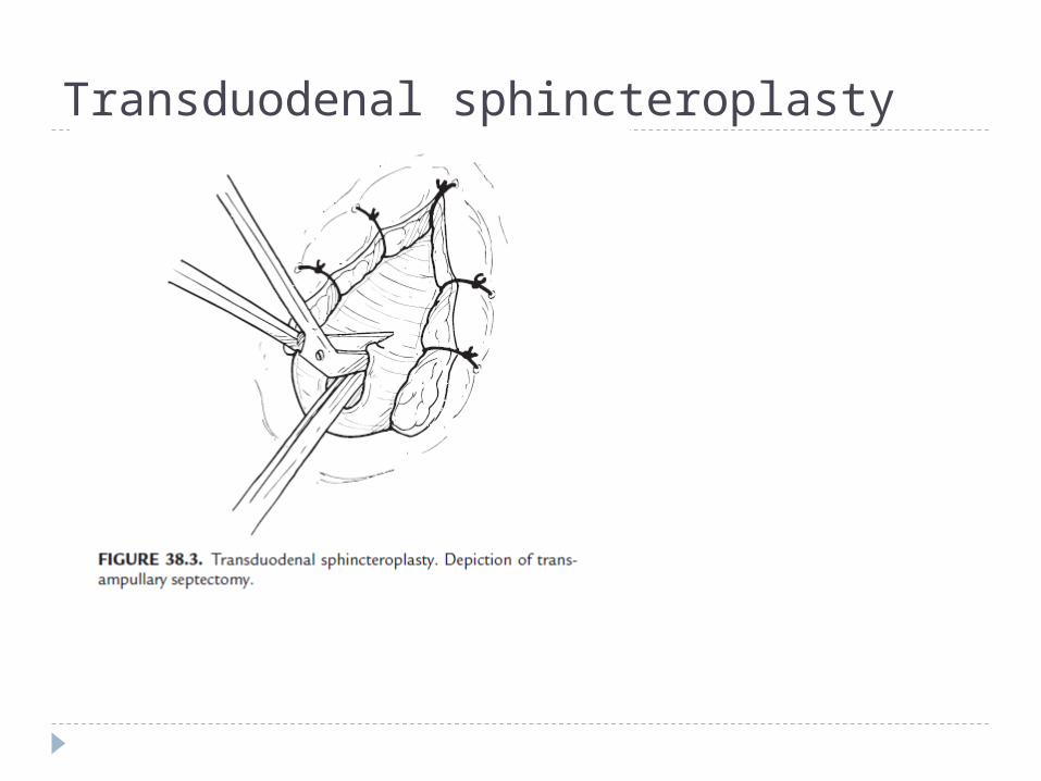
Transduodenal sphincteroplasty

Advantages:- Accurate apposition of duodenal and ductal
mucosa- Access to transampullary septum