Possible Role of Escherichia coli Penicillin-Binding Protein 6...
7
JOURNAL OF BACTERIOLOGY, Dec. 1992, p. 7572-7578 0021-9193/92/237572-07$02.00/0 Copyright © 1992, American Society for Microbiology Vol. 174, No. 23 Possible Role of Escherichia coli Penicillin-Binding Protein 6 in Stabilization of Stationary-Phase Peptidoglycan MARK P. G. VAN DER LINDEN, LOLKE DE HAAN, MAARTEN A. HOYER, AND WOLFGANG KECK* BIOSON Research Institute, Department of Biochemistry, University of Groningen, Nijenborgh 4, 9747AG Groningen, The Netherlands Received 29 July 1992/Accepted 23 September 1992 Plasmids for high-level expression of penicillin-binding protein 6 (PBP6) were constructed, giving rise to overproduction of PBP6 under the control of the lambdapR promoter in either the periplasmic or the cytoplasmic space. In contrast to penicillin-binding protein 5 (PBP5), the presence of high amounts of PBP6 in the periplasm as well as in the cytoplasm did not result in growth as spherical ceUls or in lysis. Deletion of the C-terminal membrane anchor of PBP6 resulted in a soluble form of the protein (PBP6'3-). Electron micrographs of thin sections of cells overexpressing both native membrane-bound and soluble PBP6 in the periplasm revealed a polar retraction of the cytoplasmic membrane. Cytoplasmic overexpression of native PBP6 gave rise to the formation of membrane vesicles, whereas the soluble PBP6 formed inclusion bodies in the cytoplasm. Both the membrane- bound and the soluble forms of PBP6 were purifie to homogeneity by using the immobilized dye Procion rubine MX-B. Purified preparations of PBP6 and PBP6S3se formed a 14[Cjpenicillin-protein complex at a 1:1 stoichiometry. The half-lives of the complexes were 8.5 and 6 min, respectively. In contrast to PBP5, no DD-carboxypeptidase activity could be detected for PBP6 by using bisacetyl-L-Lys-D-Ala-D-Ala and several other substrates. These findings led us to conclude that PBP6 has a biological function clearly distinct from that of PBP5 and to suggest a role for PBP6 in the stabilization of the peptidogiycan during stationary phase. Three enzymes having D-alanine carboxypeptidase activ- ity were isolated from Escherichia coli by Tamura et al. (27). These enzymes appeared to be identical to penicillin-binding proteins 4 (PBP4), 5 (PBP5), and 6 (PBP6). PBP5 and PBP6 are the major penicillin-binding components in the cell, accounting for 85% of the penicillin-binding capacity (23, 25). PBP5 and PBP6 are encoded by two distinct genes, dacA and dacC. Their primary structures show 62% se- quence identity (5). PBP5 has been thoroughly studied with respect to membrane association, cellular location, penicillin binding, and DD-carboxypeptidase activity (8, 14, 29, 30). PBP5 (molecular mass, 41,337 Da) catalyzes the cleavage of the D-alanine-D-alanyl peptide bond in the pentapeptide side chain of the peptidoglycan polymer, resulting in a tetrapep- tide (18, 25). The proposed biological function for this enzyme is the regulation of peptide cross-linkage in the peptidoglycan (17). PBP6 (molecular mass, 40,804 Da) is translated as a preprotein and translocated across the cytoplasmic mem- brane by an N-terminal signal sequence 27 amino acids long. Cleavage of the signal sequence yields a mature pro- tein of 373 residues (21). PBP6 is bound to the cytoplasmic membrane by a C-terminal membrane anchor, the bulk of the protein sticking out into the periplasm. The C-terminal 22 residues can form an amphiphilic helix associating the protein with the membrane (14). PBP6 was purified to homogeneity, and its penicillin-binding characteristics and enzymatic activity were determined by Amanuma and Strominger (1). The protein was shown to have DD-carboxy- peptidase activity by using various natural and synthetic substrates. The specific activity was determined to be three- to fourfold lower than that of PBP5. However, there is some evidence that the two enzymes do not fulfill the same * Corresponding author. function. Buchanan and Sowell described a considerable rise in expression level of PBP6 in stationary-phase cells com- pared with exponentially growing cells (7). Therefore, we undertook a more thorough study of PBP6. PBP6 is present in the cell in relatively small amounts, 570 molecules per cell (23). To investigate the physiological effects of elevated levels of PBP6, the dacC gene was cloned into the earlier-described pROFIT expression vector, giving rise to high-level expression of PBP6 in either the periplas- mic or the cytoplasmic space (30). In this paper, we describe the effects on the cell of high-level expression of both membrane-bound and soluble forms of PBP6. We further report on the purification and enzymatic characterization of these proteins. A possible physiological role of PBP6, clearly different from that of PBP5, is discussed. MATERIALS AND METHODS Bacterial strains and plasmids. E. coli MC1061 was used as a host strain for subcloning and expression of proteins (19). Site-directed mutagenesis according to the method of Kunkel et al. (15) was performed with E. coli CJ236 (Bio-Rad Laboratories, Richmond, Calif.) and JM101 and phage M13mpl8 (20). The plasmid pBS102, harboring the dacC gene, was a gift from B. Spratt, University of Sussex, Brighton, United Kingdom. Growth of bacteria was at 30°C in Luria-Bertani medium supplemented with kanamycin (50 p,g ml-') for antibiotic-resistant strains. To induce the lambdapR promoter, the growth temperature was shifted to 42°C at an A6w of 0.6. Enzymes and radiochemicals. Restriction enzymes, T4 DNA ligase, T4 polynucleotide kinase, and Klenow poly- merase were used as recommended by the supplier (Boehr- inger). at-35S-dATP used for DNA sequencing and [1 CJben- zylpenicillin (specific activity, 1.97 GBq mmol-1) used for 7572 on May 12, 2018 by guest http://jb.asm.org/ Downloaded from
Transcript of Possible Role of Escherichia coli Penicillin-Binding Protein 6...
JOURNAL OF BACTERIOLOGY, Dec. 1992, p. 7572-75780021-9193/92/237572-07$02.00/0Copyright © 1992, American Society for Microbiology
Vol. 174, No. 23
Possible Role of Escherichia coli Penicillin-Binding Protein 6in Stabilization of Stationary-Phase Peptidoglycan
MARK P. G. VAN DER LINDEN, LOLKE DE HAAN, MAARTEN A. HOYER,AND WOLFGANG KECK*
BIOSON Research Institute, Department ofBiochemistry, University of Groningen,Nijenborgh 4, 9747AG Groningen, The Netherlands
Received 29 July 1992/Accepted 23 September 1992
Plasmids for high-level expression of penicillin-binding protein 6 (PBP6) were constructed, giving rise tooverproduction ofPBP6 under the control ofthe lambdapR promoter in either the periplasmic or the cytoplasmicspace. In contrast to penicillin-binding protein 5 (PBP5), the presence of high amounts ofPBP6 in the periplasmas well as in the cytoplasm did not result in growth as spherical ceUls or in lysis. Deletion of the C-terminalmembrane anchor of PBP6 resulted in a soluble form of the protein (PBP6'3-). Electron micrographs of thinsections of cells overexpressing both native membrane-bound and soluble PBP6 in the periplasm revealed a polarretraction of the cytoplasmic membrane. Cytoplasmic overexpression of native PBP6 gave rise to the formationof membrane vesicles, whereas the soluble PBP6 formed inclusion bodies in the cytoplasm. Both the membrane-bound and the soluble forms of PBP6 were purifie to homogeneity by using the immobilized dye Procion rubineMX-B. Purified preparations of PBP6 and PBP6S3se formed a 14[Cjpenicillin-protein complex at a 1:1stoichiometry. The half-lives of the complexes were 8.5 and 6 min, respectively. In contrast to PBP5, noDD-carboxypeptidase activity could be detected for PBP6 by using bisacetyl-L-Lys-D-Ala-D-Ala and several othersubstrates. These findings led us to conclude that PBP6 has a biological function clearly distinct from that ofPBP5and to suggest a role for PBP6 in the stabilization of the peptidogiycan during stationary phase.
Three enzymes having D-alanine carboxypeptidase activ-ity were isolated from Escherichia coli by Tamura et al. (27).These enzymes appeared to be identical to penicillin-bindingproteins 4 (PBP4), 5 (PBP5), and 6 (PBP6). PBP5 and PBP6are the major penicillin-binding components in the cell,accounting for 85% of the penicillin-binding capacity (23,25). PBP5 and PBP6 are encoded by two distinct genes,dacA and dacC. Their primary structures show 62% se-quence identity (5). PBP5 has been thoroughly studied withrespect to membrane association, cellular location, penicillinbinding, and DD-carboxypeptidase activity (8, 14, 29, 30).PBP5 (molecular mass, 41,337 Da) catalyzes the cleavage ofthe D-alanine-D-alanyl peptide bond in the pentapeptide sidechain of the peptidoglycan polymer, resulting in a tetrapep-tide (18, 25). The proposed biological function for thisenzyme is the regulation of peptide cross-linkage in thepeptidoglycan (17).PBP6 (molecular mass, 40,804 Da) is translated as a
preprotein and translocated across the cytoplasmic mem-brane by an N-terminal signal sequence 27 amino acidslong. Cleavage of the signal sequence yields a mature pro-tein of 373 residues (21). PBP6 is bound to the cytoplasmicmembrane by a C-terminal membrane anchor, the bulk ofthe protein sticking out into the periplasm. The C-terminal22 residues can form an amphiphilic helix associating theprotein with the membrane (14). PBP6 was purified tohomogeneity, and its penicillin-binding characteristics andenzymatic activity were determined by Amanuma andStrominger (1). The protein was shown to have DD-carboxy-peptidase activity by using various natural and syntheticsubstrates. The specific activity was determined to be three-to fourfold lower than that of PBP5. However, there is someevidence that the two enzymes do not fulfill the same
* Corresponding author.
function. Buchanan and Sowell described a considerable risein expression level of PBP6 in stationary-phase cells com-pared with exponentially growing cells (7). Therefore, weundertook a more thorough study of PBP6.PBP6 is present in the cell in relatively small amounts, 570
molecules per cell (23). To investigate the physiologicaleffects of elevated levels of PBP6, the dacC gene was clonedinto the earlier-described pROFIT expression vector, givingrise to high-level expression of PBP6 in either the periplas-mic or the cytoplasmic space (30).
In this paper, we describe the effects on the cell ofhigh-level expression of both membrane-bound and solubleforms of PBP6. We further report on the purification andenzymatic characterization of these proteins. A possiblephysiological role of PBP6, clearly different from that ofPBP5, is discussed.
MATERIALS AND METHODS
Bacterial strains and plasmids. E. coli MC1061 was used asa host strain for subcloning and expression of proteins (19).Site-directed mutagenesis according to the method ofKunkel et al. (15) was performed with E. coli CJ236 (Bio-RadLaboratories, Richmond, Calif.) and JM101 and phageM13mpl8 (20). The plasmid pBS102, harboring the dacCgene, was a gift from B. Spratt, University of Sussex,Brighton, United Kingdom. Growth of bacteria was at 30°Cin Luria-Bertani medium supplemented with kanamycin (50p,g ml-') for antibiotic-resistant strains. To induce thelambdapR promoter, the growth temperature was shifted to42°C at an A6w of 0.6.Enzymes and radiochemicals. Restriction enzymes, T4
DNA ligase, T4 polynucleotide kinase, and Klenow poly-merase were used as recommended by the supplier (Boehr-inger). at-35S-dATP used for DNA sequencing and [1 CJben-zylpenicillin (specific activity, 1.97 GBq mmol-1) used for
7572
on May 12, 2018 by guest
http://jb.asm.org/
Dow
nloaded from

CONSTRUCTION, OVEREXPRESSION, AND PURIFICATION OF PBP6 7573
penicillin-binding assays were obtained from Amersham,Amersham, United Kingdom.
Site-directed mutagenesis and DNA sequencing. The SmaIand SalI restriction sites of M13mpl8 were used to clone the1.5-kb AhaIII-SalI fragment from pBS102, containing thedacC gene, into the phage.The method described by Kunkel et al. (15) was used for
oligonucleotide-directed mutagenesis. Primers were synthe-sized by Eurosequence, Groningen, The Netherlands. Se-quences of the primers are given throughout the text. Thedesired mutations and the complete sequence of the dacCgene were verified by DNA sequencing according to Sangeret al. (22).
Protein purification. Cultures overexpressing PBP6 (cul-ture volume, 2 liters; 4.5 g [wet mass] of cells) wereharvested by centrifugation and resuspended in 10 ml of 10mM Tris-HCl, pH 7.0, containing 1 mM 1,4-dithiothreitol.Cells were disrupted by passage through an Aminco Frenchpress at 68.9 MPa. The soluble and particulate fractions wereseparated by ultracentrifugation (45 min, 100,000 x g, 4°C).Membrane-bound forms of PBP6 were purified from theparticulate fraction in the presence of 0.2% Triton X-100. Inthe purification of the soluble forms of PBP6, the detergentwas omitted. The crude protein fractions were directlyapplied to a column (200 by 24 mm) of Procion rubine MX-Bcoupled to Fractogel TSK HW-65F, and the purificationprocedure was further carried out as described earlier forPBP5 (30).SDS-PAGE and Western blotting (immunoblotting). So-
dium dodecyl sulfate-polyacrylamide gel electrophoresis(SDS-PAGE) was performed according to the method ofLugtenberg et al. (16). A 10% (mass/vol) running gel and a4.5% (mass/vol) stacking gel were used, the acrylamide/bisacrylamide ratio being 30:0.5. Proteins were electro-phoretically transferred onto nitrocellulose as described byBittner et al. (3). Rabbit antisera against PBP6 were obtainedin this laboratory from rabbits immunized with purifiedprotein. PBP6 was immunochemically detected by usingantiserum, goat anti-rabbit immunoglobulin G-conjugatedhorseradish peroxidase (Tago, Inc., Burlingame, Calif.) andchloronaphthol-H202 (26).
Penicillin-binding assays. The penicillin-binding activity ofcytoplasmically and periplasmically located soluble andmembrane-bound forms of PBP6 was assayed as describedearlier (30). The binding stoichiometry of the different formsof PBP6 was determined by incubating protein samples of 25pmol each with 200 pmol of [14C]benzylpenicillin at 37°C for10 min. The molar ratio of bound [14Cqbenzylpenicillin andthe time course of the release of the [ 4C]benzylpenicilloylmoiety from PBP6 were determined as described by van derLinden et al. (30).
Carboxypeptidase activity assays. DD-Carboxypeptidaseactivity was determined qualitatively by thin-layer chroma-tography (TLC) and high-pressure liquid chromatography(HPLC) detection of UDP-muramyl-tetrapeptide and D-ala-nine release after incubation of PBP6 with UDP-muramyl-pentapeptide. TLC was performed on Silicagel 60-kieselguhrF254-precoated aluminum sheets (Merck) with n-butanol-acetic acid-H20 (4:1:1) as a solvent. Samples of PBP6 (1 to2 ,ug each) were incubated with 6.5 nmol of UDP-muramyl-pentapeptide for 10 min at 37°C in 100mM Tris-HCl, pH 7.0.The reaction mixture was spotted on a TLC sheet, dried witha hot gun, and eluted. The peptides and released D-alaninewere detected with ninhydrin. HPLC analysis was per-formed as follows. PBP6 (1 to 2 jig) was incubated with 6.5nmol of UDP-muramyl-pentapeptide at 37°C for 10 min in
100 mM Tris-HCl, pH 7.0. The sample volume was adjustedto 250 ,ul with water, and the sample was boiled for 5 min.The pH was adjusted to 2.0 by using H3PO4, and the samplewas applied to a Lichrosorb RP18 column (particle size, 7,um; 250 by 4 mm), mounted in a Merck-Hitachi HPLCsystem. UDP-muramyl-pentapeptide and UDP-muramyl-te-trapeptide were eluted by using a 0 to 30% methanol gradientin 100 mM Na2HPO4 (pH 2.0). Peaks were detected at 205nm with a Hitachi L-4200 UV-VIS detector.
Kinetic measurements. DD-Carboxypeptidase activity wasquantitatively assayed by incubating 10 ,ug of purified PBP6in 100 mM Tris-HCl, pH 8.0, at 37°C with various con-centrations of substrates. Bisacetyl-L-Lys-D-Ala-D-Ala, bis-acetyl-L-Lys-D-Ala-D-Lac, L-Ala-D-y-Glu-L-Lys-D-Ala-D-Ala,and UDP-muramyl-pentapeptide were used as substrates.The amount of released D-alanine was determined by usingD-aminoacid oxidase and O-dianisidine chloride as describedby Frere et al. (9).
Secondary structure prediction by hydrophobic clusteranalysis. Gaboriaud et al. (10) described a method for theprediction of secondary structure elements based on a two-dimensional representation of the distribution of hydropho-bic residues along the protein sequence. Clustered hydro-phobic residues supposed to indicate secondary structureelements are used to align weakly related primary structures.
Electron microscopy. For morphological studies, cellswere fixed in 3% (vol/vol) glutaraldehyde in 0.1 M sodiumcacodylate buffer (pH 7.2) for 1 h at 0°C. Postfixation wasperformed in 1% OS04 and 2.5% K2Cr2O7 in the same bufferfor 1 h at room temperature. Subsequently, the material wasstained en bloc overnight with 1% (wt/vol) uranyl acetate indistilled water, dehydrated in a graded series of ethanolsolutions, and embedded in Epon 812. Ultrathin sectionswere cut with a diamond knife on an LKB ultramicrotome.Sections were examined in a Philips CM10 microscope.Other techniques. Protein concentrations were determined
by using the method of Bradford (4). This assay had beencalibrated by a PBP5 standard of known protein concentra-tion as was described earlier (30).
RESULTS
Construction of plasmids for high-level expression of PBP6.The 1.5-kbAhaIII-SalI fragment from pBS102 containing thedacC gene coding for PBP6 was cloned into M13mpl8 byusing the SmaI and Sall restriction sites (M13mpl8dacC).The dacC gene was cloned from M13mpl8 into the expres-sion vector pROFIT by using the EcoRI and PstI restrictionsites (30). In the resulting vector pROFIT6An, the dacC geneis placed under the control of the temperature-induciblelambda PR promoter, giving rise to overexpression of thenative membrane-bound PBP6, with the bulk of the proteinsticking out into the periplasmic space (Fig. 1).For cytoplasmic overexpression of PBP6, a BglII site was
created by using site-directed mutagenesis at the signalpeptide cleavage point in the dacC gene. The primer used forthis mutation was CGCTlCAACGGTTAGATC/TGCCGCGAATGCCG (mutated bases are underlined, and the BglIIsite is indicated by a slash). Verification of the mutated geneby DNA sequencing showed no further mutations. By usingthe BglII and PstI restriction sites, the mutated gene wascloned into the BamHI and PstI sites of pROFIT, resulting inthe vector pROFIT6Cn (Fig. 1). pROFIT6Cn gave rise tooverexpression of the native membrane-bound PBP6, in-serted with its C-terminal membrane anchor into the mem-
VOL. 174, 1992
on May 12, 2018 by guest
http://jb.asm.org/
Dow
nloaded from

7574 VAN DER LINDEN ET AL.
s.p.pROFIT-6A~nL d CC It2.-
EcoRI
pROFIT-6CnPsti
dacC sT0-y ->BgIll Pstl
5966,BamHI
Pstl,657
FIG. 1. Construction of plasmids for the overexpression ofPBP6A. The vector pROFIT-6AW for the accumulation of theoverexpressed PBP6 in the periplasm was obtained by cloning the1.5-kb EcoRl-PstI fragment containing the dacC gene fromM13mpl8dacC into the vector pROFIT. A BgIII site introduced bysite-directed mutagenesis was used to clone the BglII-PstI fragmentfrom M13mpl8dacC into pROFIT, resulting in the vector pROFIT-6Cn giving rise to overexpression of PBP6 in the cytoplasm. Thefingerprints conserved in the family of active-site seine penicillin-interacting enzymes are indicated by a square (SxxK), a triangle(SxN), and a circle (KTG). s.p., signal peptide.
brane from the cytoplasmic side, with the bulk of the proteinsticking out into the cytoplasmic space.
Construction of soluble forms of PBP6. The C-terminalresidues 350 to 367 of PBP6, which are believed to form an
amphiphilic helix serving as a membrane anchor, weredeleted by the introduction of stop codons at positionsVal-347 and Gly-351, respectively. The two positions werechosen on the basis of primary structure comparisons ofPBP5 and PBP6 in combination with hydrophobic clusteranalysis to possibly avoid destruction of secondary structureelements (Fig. 2 and reference 10). The regions P353EGN inPBP5 and E348EGG in PBP6 are located between twopredicted structural elements at the C-terminal ends of the
stop354
PBP5 ----EQR P VVLQP E G Np P FGK I I D Y I K L M F H H
PBPS E Q RP C E G GJ[ F G R V W D F V M M K F H QWF S W F S
stop347 stop351
FIG. 2. C-terminal amino acid sequence alignment of PBP5 andPBP6. The C-terminal residues of PBP5 and PBP6 were aligned.Identical residues are shaded. The boxed parts indicate secondarystructure elements predicted by hydrophobic cluster analysis. Inboth proteins, the extreme C-terminal structural element is theamphiphilic helix serving as a membrane anchor. Stop codonsintroduced to create soluble forms of PBP5 and PBP6 are indicatedby arrows.
proteins. The primers used to create the stop codons wereGCCTCTlCCT-AATT'ITCCATCAC and CCGACCAAAGAATIL/CICACTCITJ7CCAC, respectively (mutated basesare underlined, and the EcoRI cleavage site is indicated by aslash). Mutated genes were verified by DNA sequenceanalysis. The mutated genes were recloned into pROFIT6A'and pROFIT6C', resulting in the vectors pROFIT6AS346,pROFIT6AS35O, pROFIT6CS346, and pROFIT6CS350. TheVal-347 mutation was recloned by using the BamHI and PstIsites. Recloning of the Gly-351 mutation by using the newlycreated EcoRI site resulted in a complete deletion of thesequence coding for the membrane anchor.
Overexpression of membrane-bound and soluble forms ofPBP6. Each of the pROFIT6A and pROFIT6C plasmids,coding for both membrane-bound and soluble forms ofPBP6, was introduced into E. coli MC1061. Cells weregrown at 28°C to anAwo of 0.6. The growth temperature wasthen raised to 420C to induce the lambdaPR promoter. ThepROFIT6A constructs gave rise to overproduction of PBP6and accumulation in the periplasm whereas for thepROFIT6C constructs, the overexpressed protein remainedin the cytoplasm. Neither the periplasmically nor the cyto-plasmically located forms of PBP6 resulted in lysis of thecells upon overexpression, and the cells could be grown at42°C for several hours. The level of overproduction wasdifferent from one construct to another. PBP6An was ex-pressed to about 200 times the wild-type level as wasdetermined by a Western blot analysis of a dilution series.Overexpression of PBP6C' was 100-fold whereasPBP6AS346, PBP6AS350, PBP6Cs346, and PBP6Cs35O wereexpressed to 50 times the wild-type level.
Cells were disrupted by passage through an AmincoFrench press at 68.9 MPa followed by centrifugation at100,000 x g for 45 min. The soluble and the particulatefractions were separated and analyzed for the presence ofPBP6. PBP6A7 and PBP6Cn were localized exclusively inthe particulate fraction. PBP6AS346, PBP6AS350, PBP6CS346,and PBP6CS350 were found in the soluble fraction whenanalyzed on Coomassie blue-stained SDS-PAGE, by West-ern blot, and by penicillin-binding assay. PBP6AS346 andPBP6CI'6 both appeared to be unstable, resulting in theappearance of a 30-kDa degradation product during overex-pression of the proteins (Fig. 3).
Purification of soluble and membrane-bound PBP6. Purifi-cation of PBP5 by using the immobilized dye Procion rubineMX-B was described earlier (30). PBP6 appeared to haveaffinity for the same dye. Both the membrane-bound and thesoluble forms of PBP6 were purified to homogeneity on aProcion rubine MX-B column in a one-step procedure (Fig.4). The conditions for this purification procedure were thesame as those described for PBP5, and both proteins elutedat the same NaCl concentration (30). Purified protein wasobtained for PBP6An, PBP6Cn, PBP6AS350, and PBP6CS350.PBP6AS346 and PBP6CS346 showed no affinity for the dye andtherefore could not be purified by using this method. Startingfrom 4.5 g (wet mass) of cells, the yield was 15 mg of purePBP6A7. For the soluble form PBP6C350, the yield was 10times lower, mainly because of the lower expression level.
Polyclonal antibodies against purified PBP6 were raised inrabbits in this laboratory. These antibodies showed affinityfor all the various forms of PBP6 on Western blots (Fig. 3B).
Characterization of purified forms of PBP6. After purifica-tion, all the forms of PBP6 described above retained theirability to bind [14C]benzylpenicillin in a 1:1 stoichiometry.Like PBP5, PBP6 shows a rather fast release of the boundpenicillin. A 50% degradation of the protein-[14C]benzylpen-
J. BACTERIOL.
on May 12, 2018 by guest
http://jb.asm.org/
Dow
nloaded from

CONSTRUCTION, OVEREXPRESSION, AND PURIFICATION OF PBP6 7575
94 kDa
67
Iw.t$- 43
30
B
C_
FIG. 3. Overexpression of membrane-bound and soluble formsof PBP6. Coomassie blue-stained SDS-PAGE (A), Western blotafter immunostaining (B), and fluorogram after incubation with["4C]benzylpenicillin (C) of cells overexpressing membrane-boundand soluble forms of PBP6. Cells were grown at 30°C to an A6w of0.6, after which the temperature was shifted to 42°C. Total protein ofthe cells after 2 h of growth was applied to the gel. Lanes 1 to 6,PBP6As346-, PBP6A!350_, PBP6An-, PBP6Cs346-, PBP6C350-, andPBP6Cn-overproducing cells, respectively; lane 7, molecular massmarkers. Arrows indicate the positions of PBP6AW and PBP6Cn (A)or the position of the 30-kDa degradation product of PBP6A3 andPBP6C'346 (B).
icillin complex was observed after 8.5 min for the mem-brane-bound forms and after 6 min for the soluble forms ofPBP6 (Table 1). Purified PBP6A7 was assayed for DD-carboxypeptidase activity by using the artificial substratesbisacetyl-L-Lys-D-Ala-D-Ala, bisacetyl-L-Lys-D-Ala-D-lac-tate, and Ala-D--y-Glu-L-Lys-D-Ala-D-Ala. No release ofD-alanine was observed with any of these substrates. When,however, PBP5 was incubated with these substrates underexactly the same conditions, a considerable release of D-al-anine could be detected (Table 1). Incubation of PBP6A7 orPBP6A350 with isolated UDP-muramyl-pentapeptide andsubsequent analysis by HPLC or TLC, again in contrast toPBP5, did not show any appearance of UDP-muramyl-tetrapeptide or released D-alanine. DD-Carboxypeptidaseactivity measurements with membrane fractions from thePBP6An overproducer, in contrast to PBP5Cn membranefractions, did not show any increase in release of D-alaninefrom bisacetyl-L-Lys-D-Ala-D-Ala (data not shown). Thesefindings are in clear contradiction to the earlier-describedDD-carboxypeptidase activity of purified PBP6 with the verysame substrates (1, 2).
Morphological observations. Electron micrographs of thinsections of cells overexpressing membrane-bound or soluble
1 2 3 4 5 6 7 8
94kDa
67
43
30
20
FIG. 4. Dye affinity purification of PBP6AW and PBP6CS350. Themembrane-bound and soluble forms of PBP6 were purified by usingthe immobilized dye Procion rubine MX-B. PBP6AS350 and PBP6Cncould be purified by the same methods. PBP6A346 and PBP6Cs346showed no affinity for the dye. Lanes 1 and 8, molecular massmarkers; lane 2, Triton X-100-solublized membrane proteins fromthe PBP6AW-overproducing strain; lane 3, flowthrough of the Pro-cion rubine MX-B dye affinity column; lane 4, PBP6An eluted fromthe Procion rubine MX-B column by an NaCl gradient; lane 5,soluble fraction of the PBP6C635O-overproducing strain; lane 6,flowthrough of the dye affinity column; lane 7, PBP6C350 elutedfrom the dye column by an NaCl gradient.
forms of PBP6 in either the periplasm or the cytoplasm areshown in Fig. 5. Overexpression of membrane-bound PBP6in the cytoplasm (PBP6Cn) gave rise to the formation ofintracellular membrane vesicles throughout the cytoplasm ofmany cells (Fig. 5B). These vesicles seemed to consist of amembrane similar in general appearance and thickness to thecytoplasmic membrane. Cells overexpressing the nativemembrane-bound PBP6 in the periplasm (PBP6AW) do notshow formation of membrane vesicles. However, a retrac-tion of the cytoplasmic membrane at the polar tips of thecells was observed (Fig. 5A). The same phenomenon wasfound in cells overexpressing the periplasmically locatedsoluble form of PBP6, PBP6A350 (Fig. 5C). Overexpressionof the c toplasmically located soluble form of PBP6,PBP6CS3 , gave rise to intracellular structures visible ashomogeneously stained areas with a distinct border with thecytoplasm. These areas most probably are cross sections ofinclusion bodies formed in the cells.
DISCUSSION
Despite the accumulation of information on PBP5 andPBP6 in recent years, no evident physiological role for thesetwo major PBPs in E. coli could be established. Bothproteins are usually considered the major DD-carboxypepti-dases and because of their similarities in primary structureand biochemical characteristics are believed to fulfill com-parable functions in determining the degree of cross-linkagein the peptidoglycan polymer.The previously described minor but nevertheless essential
differences between the two proteins, like the differences inexpression during exponential or stationary phase and thefinding that peptidoglycan isolated from log-phase cells of adacC deletion strain did not show the increase in pentapep-tide side chains that was observed for peptidoglycan isolatedfrom a dacA deletion strain, did not gain much attention (11).The expression of a DD-carboxypeptidase in stationary-phase cells, the peptidoglycan of which does not contain the
A
1 2 3 4 5 6 7
VOL. 174, 1992
I 4It, .,.,
'-"?r ..i
'P7 '; Mll
on May 12, 2018 by guest
http://jb.asm.org/
Dow
nloaded from

7576 VAN DER LINDEN ET AL.
TABLE 1. Kinetic constants and purification results of membrane-bound and soluble forms of PBP6
Hydrolysis of [Cl4CbenzylpenicillieaProtein form Dye DD-Carboxypeptidase ['4CJbenzylpenicilhinpurification activity" bindingd t,2 k3
(s) (10-1 S-1)
PBP6AW + + 517 ± 30 1.34 ± 0.08PBP6A" - ND + ND NDPBP6A350 + + 363 ± 50 1.91 ± 0.26PBP6Cn + + 510 ± 50 1.36 ± 0.13PBP6Cs34 - ND + ND NDPBP6Cs350 + + 377 + 16 1.84 ± 0.08PBP5Cn + + + 260±10 2.7±0.1
a t12, half-life; k3, rate constant.b Proteins were purified (+) or could not be purified (-) with the immobilized dye Procion rubine MX-B.c DD-Carboxypeptidase activity was assayed as described in the text. + and -, release of alanine was or was not detected. ND, not detected.d Penicillin binding capacity was assayed as described in the text. +, binding ability was retained.
supposed substrate-pentapeptide side chains any more,raises again the question of its specific role in vivo (7, 12). Allthese observations were taken as indications for a physio-logical function of PBP6 distinct from that of PBP5.To assess the differences in function, overproducers of
PBP6 in either the native or the soluble form were estab-lished. Transport of the various forms of PBP6 into theperiplasm does not cause the detrimental effect on theproducing cells that had been observed during PBP5 over-expression (17, 30). As in the case of PBP5, high-levelexpression of PBP6 in the cytoplasm also did not affectcellular growth, which indicates that the amount of pen-
tapeptide containing precursors is not decreased to an extentthat could abolish peptidoglycan polymerization.
Deletion of the predicted membrane anchor of PBP6yielded two soluble forms. In PBP6S35O, the C-terminala-helix predicted by hydrophobic cluster analysis was de-leted, resulting in a stable soluble form. The other solubleform, PBP6SN6, appeared to be very unstable, probablybecause of cleavage of a secondary structure element pre-dicted by hydrophobic cluster analysis. A stop codon intro-duced in the homologous region of PBP5 (Val-348) resultedin a totally inactive and insoluble protein (29). Both solubleforms of PBP6 still bound penicillin in a 1:1 ratio, which
B
V
p<J D W W_W
FIG. 5. Electron micrographs of ultrathin sections of PBP6-overproducing cells. Cells overproducing PBP6AW (A), PBP6C' (B), PBP6AS350(C), and PBP6CI350 (D) are shown. Bars, 0.5 p,m unless otherwise stated.
J. BACrERIOL.
IMF
4
on May 12, 2018 by guest
http://jb.asm.org/
Dow
nloaded from
CONSTRUCTION, OVEREXPRESSION, AND PURIFICATION OF PBP6 7577
indicates that PBP6S346 is structurally not completely dis-turbed as was the case for the analogous form of PBP5.However, the structural disturbance was such that no affin-ity for the dye resin was observed. The 30-kDa degradationproduct of PBP6S" still carried the radiolabelled penicillin,indicating that it still contains the active-site serine, whichmeans that a 10-kDa portion of the protein has been removedfrom the C terminus.The unexpected tolerance of the cells for high-level
expression of PBP6 led us to investigate whether we weredealing with a functional form of PBP6.The [P4C]benzylpenicillin-protein complexes of the soluble
forms PBP6V35 and PBP6C,3" have considerably shorterhalf-lives compared with the respective membrane-boundforms (PBP6An and PBP6C"). We do not have an explana-tion for this difference, but the same effect was observed forPBP5 (30).
In contrast to results published by Amanuma andStrominger (1), we were not able to detect DD-carboxypep-tidase activity for either the native membrane-bound or thesoluble forms of PBP6 by using various artificial substratesas well as UDP-muramyl-pentapeptide. Samples of PBP6purified in our laboratory from a nonoverproducing, dacAdeletion strain (SP 5003) by conventional covalent affinitychromatography also showed no DD-carboxypeptidase activ-ity. This is again in strong contrast to the findings ofAmanuma and Strominger (2). The discrepancy betweentheir and our results so far remains unexplained. Lack ofactivity in assays with membrane fractions from PBP6An-overproducing cells proved that the proteins had not beeninactivated during purification.Resequencing of the entire dacC gene revealed one differ-
ence from the published sequence. At the codon correspond-ing to residue 204, our sequence reads GTJ instead of GAA.This results in a valine rather than a glutamate at position204. It is not likely that this difference could cause acomplete inactivation of PBP6 as DD-carboxypeptidase sincea valine is also found at the corresponding position in PBP5.The most likely explanation is that there has been a readingerror in the original sequence (24).These results contradict the current opinion that PBP6 is
fulfilling the very same role as PBP5. Nevertheless, PBP6might still be a DD-carboxypeptidase, being active only on aspecific substrate under specific conditions. Stationary-phase cells show elevated amounts of PBP6 (7). Apparently,there is a need for PBP6 at this particular stage, since onresumption of exponential growth the amount of PBP6 fallsto the original level. Nongrowing cells were found to stabi-lize their peptidoglycan by increasing the level of Dap-Dapcross-linkages (12), and the same effect was found for aminoacid-starved cells (13). We suggest a role for PBP6 instabilization of the peptidoglycan during stationary phase inanalogy to the sporulation-specific PBP5a (PBP5*) fromBacillus subtilis for which, with a partially purified batch,only a very low specific activity as DD-carboxypeptidase hasbeen detected (6, 28). The morphological alterations ob-served in the ultrathin sections of cells overexpressingvarious forms of PBP6 are obviously a direct consequence ofthe presence of high amounts of protein. Morphologicaleffects of this kind were not found in PBP5-overproducingstrains. However, extensive membrane formation was pre-viously reported for cells producing large amounts of themembrane proteins fumarate reductase and F1-Fo proton-translocating ATPase (31, 32). Apparently, cells producenew membrane structures to accommodate an excess ofmembrane-bound protein. The cytoplasmically overex-
pressed soluble form of PBP6, however, is accumulating ininclusion bodies. Large amounts of either the membrane-bound or the soluble form of PBP6 in the periplasm do notresult in extra membrane structures. The cause of theobserved polar retraction of the cytoplasmic membraneremains unclear.
ACKNOWLEDGMENT
We thank Klaas Sjollema for the electron microscopy work.
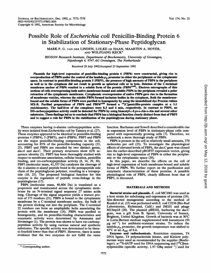
REFERENCES1. Amanuma, H., and J. L. Strominger. 1980. Purification and
properties of penicillin-binding proteins 5 and 6 from Esche-richia coli membranes. J. Biol. Chem. 255:11173-11180.
2. Amanuma, H., and J. L. Strominger. 1984. Purification andproperties of penicillin-binding proteins 5 and 6 from the dacAdeletion strain of Escherchia coli (JE11191). J. Biol. Chem.259:1294-1298.
3. Bittner, M., P. Kupferer, and C. F. Morris. 1980. Electropho-retic transfer of proteins and nucleic acids from slab gels todiazobenzyloxymethyl cellulose or nitrocellulose sheets. Anal.Biochem. 102:459-471.
4. Bradford, M. M. 1976. A rapid and sensitive method for thequantitation of microgram quantities of protein utilizing theprinciple of protein-dye binding. Anal. Biochem. 72:248-254.
5. Broome-Smith, J. K., I. Ioannidis, A. Edelman, and B. G. Spratt.1988. Nucleotide sequences of the penicillin-binding protein 5and 6 genes of Escherichia coli. Nucleic Acids Res. 16:1617.
6. Buchanan, C. E., and M.-L. Ling. 1992. Isolation and sequenceanalysis of dacB, which encodes a sporulation-specific penicil-lin-binding protein in Bacillus subtilis. J. Bacteriol. 174:1717-1725.
7. Buchanan, C. E., and M. 0. Soweli. 1982. Synthesis of penicil-lin-binding protein 6 by stationary-phase Escherichia coli. J.Bacteriol. 151:491-494.
8. Ferreira, L. C. S., U. Schwarz, W. Keck, P. Charlier, 0.Dideberg, and J.-M. Ghuysen. 1988. Properties and crystalliza-tion of a genetically engineered, water-soluble derivative ofpenicillin-binding protein 5 of Escherichia coli K12. Eur. J.Biochem. 171:11-16.
9. Frere, J.-M., M. Leyh-Bouille, J.-M. Ghuysen, M. Nieto, andH. R. Perkins. 1976. Exocellular DD-carboxypeptidases-transpeptidases from Streptomyces. Methods Enzymol. 45:610-636.
10. Gaboriaud, C., V. Bissery, T. Bencheritt, and J. P. Mornon.1987. Hydrophobic cluster analysis: an efficient new way tocompare and analyse amino acid sequences. FEBS Lett. 224:149-155.
11. Glauner, B. 1986. Ph.D. thesis. University of Tubingen, Tubin-gen, Germany.
12. Glauner, B., and J.-V. Holtje. 1990. Growth pattern of themurein sacculus of Eschenichia coli. J. Biol. Chem. 265:18988-18996.
13. Goodell, W., and A. Tomasz. 1980. Alteration of Escherichiacoli murein during amino acid starvation. J. Bacteriol. 144:1009-1016.
14. Jackson, M. E., and J. M. Pratt. 1987. An 18 amino acidamphiphilic helix forms the membrane-anchoring domain of theEschenichia coli penicillin-binding protein 5. Mol. Microbiol.1:23-28.
15. Kunkel, T. A., J. D. Roberts, and R. A. Zakour. 1987. Rapid andefficient site-specific mutagenesis without phenotypic selection.Methods Enzymol. 154:367-382.
16. Lugtenberg, B., J. Mejers, R. Peters, P. van der Hoek, and L.van Alphen. 1975. Electrophoretic resolution of the 'major outermembrane protein' of Eschenichia coli K12 into four bands.FEBS Lett. 58:254-258.
17. Marliewicz, Z., J. K Broome-Smith, U. Schwarz, and B. G.Spratt. 1982. Spherical E. coli due to elevated levels of D-ala-nine carboxypeptidase. Nature (London) 297:702-704.
VOL. 174, 1992
on May 12, 2018 by guest
http://jb.asm.org/
Dow
nloaded from

7578 VAN DER LINDEN ET AL.
18. Matsuhashi, M., S. Tamaki, S. J. Curtis, and J. L. Strominger.1979. Mutational evidence for identity of penicillin-bindingprotein 5 in Escherichia coli with the major D-alanine carboxy-peptidase IA activity. J. Bacteriol. 137:644-647.
19. Meissner, P. S., W. P. Sisk, and M. L. Berman. 1987. Bacterio-phage lambda cloning system for the construction of directionalcDNA libraries. Proc. Natl. Acad. Sci. USA 84:4171-4175.
20. Norrander, J., T. Kempe, and J. Messing. 1983. Construction ofimproved M13 vectors using oligodeoxynucleotide-directed mu-tagenesis. Gene 26:101-106.
21. Pratt, J. M., B. Holland, and B. G. Spratt. 1981. Precursorforms of penicillin-binding proteins 5 and 6 of E. coli cytoplas-mic membrane. Nature (London) 293:307-309.
22. Sanger, F., S. Nicklen, and A. R. Coulson. 1977. DNA sequenc-ing with chain-terminating inhibitors. Proc. Natl. Acad. Sci.USA 74:5463-5467.
23. Spratt, B. G. 1977. Properties of the penicillin-binding proteinsof Escherichia coli K12. Eur. J. Biochem. 72:341-352.
24. Spratt, B. G. (University of Sussex, Brighton, United Kingdom).1992. Personal communication.
25. Spratt, B. G., and J. L. Strominger. 1976. Identification of themajor penicillin-binding proteins of Eschenchia coli as D-ala-nine carboxypeptidase IA. J. Bacteriol. 127:660-663.
26. Talbot, P. V., R. L. Knobler, and M. Buchmeier. 1984. Westernand dot immunoblotting analysis of viral antigens and antibod-
ies: application to murine hepatitis virus. J. Immunol. Methods73:177-188.
27. Tamura, T., I. Yasuo, and J. L. Strominger. 1976. Purification tohomogeneity and properties of two D-alanine carboxypeptidasesfrom Escherichia coli. J. Biol. Chem. 251:414-423.
28. Todd, J. A., E. J. Bone, and D. J. Ellar. 1985. The sporulation-specific penicillin-binding protein 5a from Bacillus subtilis is aDD-carboxypeptidase in vitro. Biochem. J. 230:825-828.
29. van der Linden, M. P. G., L. de Haan, and W. Keck. Domainorganization of penicillin-binding protein 5 from Escherichiacoli analysed by C-terminal truncation. Submitted for publica-tion.
30. van der linden, M. P. G., H. Mottl, and W. KecL 1992.Cytoplasmic high-level expression of a soluble enzymaticallyactive form of the Escherichia coli penicillin-binding protein 5and purification by dye-chromatography. Eur. J. Biochem.204:197-202.
31. von Meyenburg, K., B. B. J0rgensen, and B. van Deurs. 1984.Physiological and morphological effects of overproduction ofmembrane-bound ATP synthase in Escherichia coli K-12.EMBO J. 3:1791-1797.
32. Weiner, J. H., B. D. Lemire, M. L. Elmes, R. D. Bradley, andD. G. Scraba. 1984. Overproduction of fumarate reductase inEscherichia coli induces a novel intracellular lipid-protein or-ganelle. J. Bacteriol. 158:590-596.
J. BACTERIOL.
on May 12, 2018 by guest
http://jb.asm.org/
Dow
nloaded from