Positive Feedback Loops for Factor V and Factor VII Activation Supply Sensitivity to Local Surface...
9
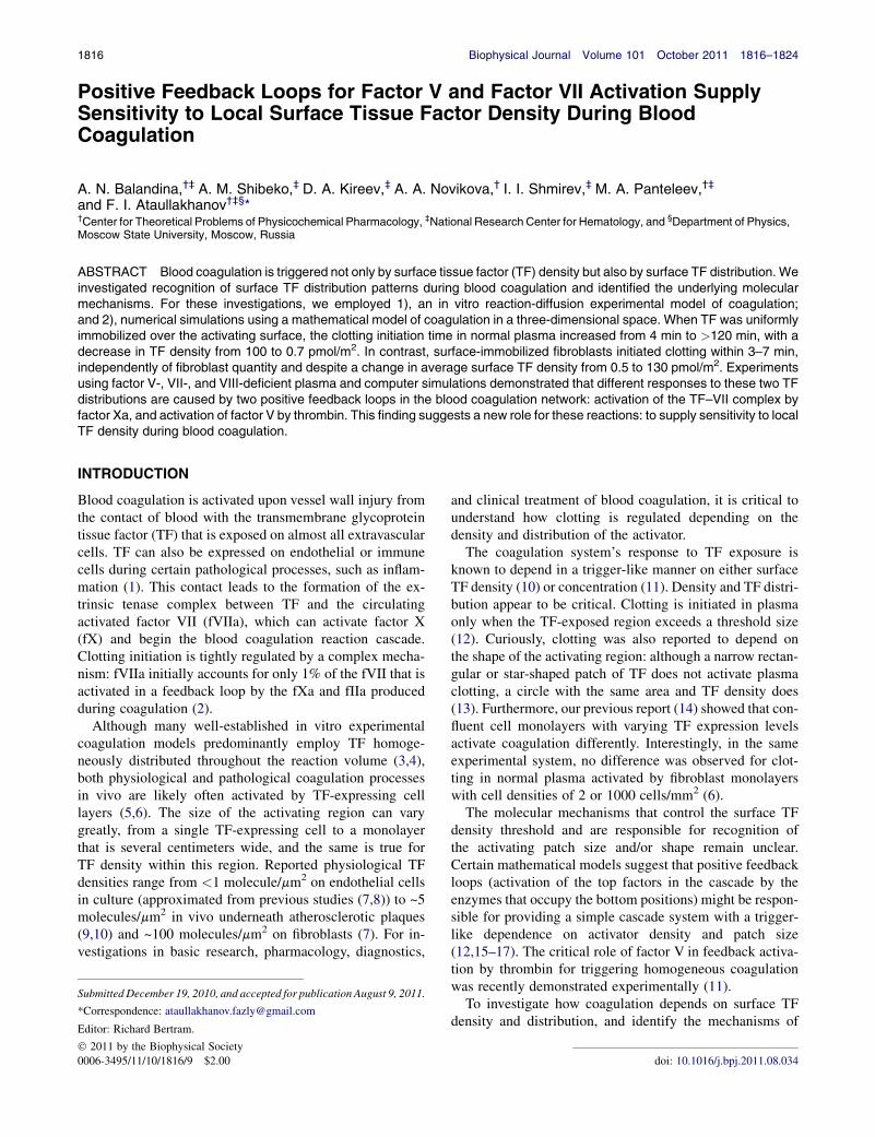
Positive Feedback Loops for Factor V and Factor VII Activation Supply Sensitivity to Local Surface Tissue Factor Density During Blood Coagulation A. N. Balandina, †‡ A. M. Shibeko, ‡ D. A. Kireev, ‡ A. A. Novikova, † I. I. Shmirev, ‡ M. A. Panteleev, †‡ and F. I. Ataullakhanov †‡§ * † Center for Theoretical Problems of Physicochemical Pharmacology, ‡ National Research Center for Hematology, and § Department of Physics, Moscow State University, Moscow, Russia ABSTRACT Blood coagulation is triggered not only by surface tissue factor (TF) density but also by surface TF distribution. We investigated recognition of surface TF distribution patterns during blood coagulation and identified the underlying molecular mechanisms. For these investigations, we employed 1), an in vitro reaction-diffusion experimental model of coagulation; and 2), numerical simulations using a mathematical model of coagulation in a three-dimensional space. When TF was uniformly immobilized over the activating surface, the clotting initiation time in normal plasma increased from 4 min to >120 min, with a decrease in TF density from 100 to 0.7 pmol/m 2 . In contrast, surface-immobilized fibroblasts initiated clotting within 3–7 min, independently of fibroblast quantity and despite a change in average surface TF density from 0.5 to 130 pmol/m 2 . Experiments using factor V-, VII-, and VIII-deficient plasma and computer simulations demonstrated that different responses to these two TF distributions are caused by two positive feedback loops in the blood coagulation network: activation of the TF–VII complex by factor Xa, and activation of factor V by thrombin. This finding suggests a new role for these reactions: to supply sensitivity to local TF density during blood coagulation. INTRODUCTION Blood coagulation is activated upon vessel wall injury from the contact of blood with the transmembrane glycoprotein tissue factor (TF) that is exposed on almost all extravascular cells. TF can also be expressed on endothelial or immune cells during certain pathological processes, such as inflam- mation (1). This contact leads to the formation of the ex- trinsic tenase complex between TF and the circulating activated factor VII (fVIIa), which can activate factor X (fX) and begin the blood coagulation reaction cascade. Clotting initiation is tightly regulated by a complex mecha- nism: fVIIa initially accounts for only 1% of the fVII that is activated in a feedback loop by the fXa and fIIa produced during coagulation (2). Although many well-established in vitro experimental coagulation models predominantly employ TF homoge- neously distributed throughout the reaction volume (3,4), both physiological and pathological coagulation processes in vivo are likely often activated by TF-expressing cell layers (5,6). The size of the activating region can vary greatly, from a single TF-expressing cell to a monolayer that is several centimeters wide, and the same is true for TF density within this region. Reported physiological TF densities range from <1 molecule/mm 2 on endothelial cells in culture (approximated from previous studies (7,8)) to ~5 molecules/mm 2 in vivo underneath atherosclerotic plaques (9,10) and ~100 molecules/mm 2 on fibroblasts (7). For in- vestigations in basic research, pharmacology, diagnostics, and clinical treatment of blood coagulation, it is critical to understand how clotting is regulated depending on the density and distribution of the activator. The coagulation system’s response to TF exposure is known to depend in a trigger-like manner on either surface TF density (10) or concentration (11). Density and TF distri- bution appear to be critical. Clotting is initiated in plasma only when the TF-exposed region exceeds a threshold size (12). Curiously, clotting was also reported to depend on the shape of the activating region: although a narrow rectan- gular or star-shaped patch of TF does not activate plasma clotting, a circle with the same area and TF density does (13). Furthermore, our previous report (14) showed that con- fluent cell monolayers with varying TF expression levels activate coagulation differently. Interestingly, in the same experimental system, no difference was observed for clot- ting in normal plasma activated by fibroblast monolayers with cell densities of 2 or 1000 cells/mm 2 (6). The molecular mechanisms that control the surface TF density threshold and are responsible for recognition of the activating patch size and/or shape remain unclear. Certain mathematical models suggest that positive feedback loops (activation of the top factors in the cascade by the enzymes that occupy the bottom positions) might be respon- sible for providing a simple cascade system with a trigger- like dependence on activator density and patch size (12,15–17). The critical role of factor V in feedback activa- tion by thrombin for triggering homogeneous coagulation was recently demonstrated experimentally (11). To investigate how coagulation depends on surface TF density and distribution, and identify the mechanisms of Submitted December 19, 2010, and accepted for publication August 9, 2011. *Correspondence: [email protected] Editor: Richard Bertram. Ó 2011 by the Biophysical Society 0006-3495/11/10/1816/9 $2.00 doi: 10.1016/j.bpj.2011.08.034 1816 Biophysical Journal Volume 101 October 2011 1816–1824
Transcript of Positive Feedback Loops for Factor V and Factor VII Activation Supply Sensitivity to Local Surface...
1816 Biophysical Journal Volume 101 October 2011 1816–1824
Positive Feedback Loops for Factor V and Factor VII Activation SupplySensitivity to Local Surface Tissue Factor Density During BloodCoagulation
A. N. Balandina,†‡ A. M. Shibeko,‡ D. A. Kireev,‡ A. A. Novikova,† I. I. Shmirev,‡ M. A. Panteleev,†‡
and F. I. Ataullakhanov†‡§*†Center for Theoretical Problems of Physicochemical Pharmacology, ‡National Research Center for Hematology, and §Department of Physics,Moscow State University, Moscow, Russia
ABSTRACT Blood coagulation is triggered not only by surface tissue factor (TF) density but also by surface TF distribution. Weinvestigated recognition of surface TF distribution patterns during blood coagulation and identified the underlying molecularmechanisms. For these investigations, we employed 1), an in vitro reaction-diffusion experimental model of coagulation;and 2), numerical simulations using a mathematical model of coagulation in a three-dimensional space. When TF was uniformlyimmobilized over the activating surface, the clotting initiation time in normal plasma increased from 4 min to >120 min, with adecrease in TF density from 100 to 0.7 pmol/m2. In contrast, surface-immobilized fibroblasts initiated clotting within 3–7 min,independently of fibroblast quantity and despite a change in average surface TF density from 0.5 to 130 pmol/m2. Experimentsusing factor V-, VII-, and VIII-deficient plasma and computer simulations demonstrated that different responses to these two TFdistributions are caused by two positive feedback loops in the blood coagulation network: activation of the TF–VII complex byfactor Xa, and activation of factor V by thrombin. This finding suggests a new role for these reactions: to supply sensitivity to localTF density during blood coagulation.
INTRODUCTION
Blood coagulation is activated upon vessel wall injury fromthe contact of blood with the transmembrane glycoproteintissue factor (TF) that is exposed on almost all extravascularcells. TF can also be expressed on endothelial or immunecells during certain pathological processes, such as inflam-mation (1). This contact leads to the formation of the ex-trinsic tenase complex between TF and the circulatingactivated factor VII (fVIIa), which can activate factor X(fX) and begin the blood coagulation reaction cascade.Clotting initiation is tightly regulated by a complex mecha-nism: fVIIa initially accounts for only 1% of the fVII that isactivated in a feedback loop by the fXa and fIIa producedduring coagulation (2).
Although many well-established in vitro experimentalcoagulation models predominantly employ TF homoge-neously distributed throughout the reaction volume (3,4),both physiological and pathological coagulation processesin vivo are likely often activated by TF-expressing celllayers (5,6). The size of the activating region can varygreatly, from a single TF-expressing cell to a monolayerthat is several centimeters wide, and the same is true forTF density within this region. Reported physiological TFdensities range from <1 molecule/mm2 on endothelial cellsin culture (approximated from previous studies (7,8)) to ~5molecules/mm2 in vivo underneath atherosclerotic plaques(9,10) and ~100 molecules/mm2 on fibroblasts (7). For in-vestigations in basic research, pharmacology, diagnostics,
Submitted December 19, 2010, and accepted for publication August 9, 2011.
*Correspondence: [email protected]
Editor: Richard Bertram.
� 2011 by the Biophysical Society
0006-3495/11/10/1816/9 $2.00
and clinical treatment of blood coagulation, it is critical tounderstand how clotting is regulated depending on thedensity and distribution of the activator.
The coagulation system’s response to TF exposure isknown to depend in a trigger-like manner on either surfaceTF density (10) or concentration (11). Density and TF distri-bution appear to be critical. Clotting is initiated in plasmaonly when the TF-exposed region exceeds a threshold size(12). Curiously, clotting was also reported to depend onthe shape of the activating region: although a narrow rectan-gular or star-shaped patch of TF does not activate plasmaclotting, a circle with the same area and TF density does(13). Furthermore, our previous report (14) showed that con-fluent cell monolayers with varying TF expression levelsactivate coagulation differently. Interestingly, in the sameexperimental system, no difference was observed for clot-ting in normal plasma activated by fibroblast monolayerswith cell densities of 2 or 1000 cells/mm2 (6).The molecular mechanisms that control the surface TF
density threshold and are responsible for recognition ofthe activating patch size and/or shape remain unclear.Certain mathematical models suggest that positive feedbackloops (activation of the top factors in the cascade by theenzymes that occupy the bottom positions) might be respon-sible for providing a simple cascade system with a trigger-like dependence on activator density and patch size(12,15–17). The critical role of factor V in feedback activa-tion by thrombin for triggering homogeneous coagulationwas recently demonstrated experimentally (11).
To investigate how coagulation depends on surface TFdensity and distribution, and identify the mechanisms of
doi: 10.1016/j.bpj.2011.08.034

Feedback Loops Supply Sensitivity to Local Tissue Factor Density 1817
this regulation, we used two different patterns of clottingactivation: TF-expressing cells and uniformly distributedsurface-immobilized TF (Fig. 1). For both experimentaldesigns, the average surface TF density varied by two ordersof magnitude. Experiments in normal plasma and fV-, fVII-,and fVIII-deficient plasma, using a reaction-diffusionin vitro experimental model, were performed with computersimulations that employed a detailed three-dimensional(3D) mathematical model of blood coagulation. Clot forma-tion was controlled by local, small-scale TF density; forexample, highly procoagulant fibroblasts rapidly activatedclotting independently of the average surface TF density.This sensitivity to TF density was controlled by two positivefeedback loops: activation of the TF-VII complex by fXa,and activation of fV by thrombin.
MATERIALS AND METHODS
Proteins
The following materials were purchased from the sources shown in
parentheses: human fVa, fV, fIXa, thrombin, and RVV-Vactivator (Haema-
tologic Technologies, Essex Junction, VT); recombinant factor VIIa and
NovoSeven (Novo Nordisk, Copenhagen, Denmark); human fVIII (Hemo-
phil M, Baxter Russia, Moscow, Russia); EDTA (Helicon, Moscow,
Russia); PPACK (Calbiochem, Darmstadt, Germany); fXa-specific chro-
mogenic substrate S2765 (Chromogenix, Milano, Italy); and human
prothrombin and fXa (Enzyme Research Laboratories, South Bend, IN).
The thrombin-specific chromogenic substrate, Gly-Pro-Arg-NA, was syn-
thesized in the Russian Cardiology Research and Production Complex
of Ministry of Health Care and Social Development (Moscow, Russia).
The specific fXIIa inhibitor, corn trypsin inhibitor (CTI), was prepared
from corn seeds as described previously (18). Phospholipid (PL) vesicles
were prepared by extrusion from phosphatidylcholine and phos-
phatidylserine (Avanti, Alabaster, AL) at a molar ratio of 7:3, as described
previously (19).
FIGURE 1 Blood clotting activation on TF-expressing surfaces in
a vessel (in vivo) and in a reaction-diffusion experimental model (in vitro).
Under normal conditions, TF is expressed by subendothelial cells only. In
many pathological conditions, such as inflammation and cancer, TF can
also appear on the surface of blood cells and endothelial cells, either iso-
lated or in monolayers. TF density varies over a wide range. By analogy,
coagulation in the in vitro model system used herein was activated by films
with either uniformly distributed TF or TF-bearing cells. In both cases, the
average surface TF density varied over two orders of magnitude. Spatial
clot growth was recorded by means of light scattering. Initiation times,
clot growth rates, and clot sizes after 40 min were determined as shown.
Plasma preparation
Normal plasma was prepared from the whole blood of healthy volunteers
and collected with the approval of the ethics committees of the Center
for Theoretical Problems of Physicochemical Pharmacology and the
National Research Center for Hematology. Blood was drawn into 3.8%
sodium citrate (pH 5.5) at a 9:1 blood/anticoagulant volume ratio and
supplemented with CTI at a 0.2 mg/ml final concentration to inhibit contact
activation. A routine protocol was used to obtain plasma with stable
and reproducible procoagulant lipid content. Blood was centrifuged at
1600 � g for 15 min, and the supernatant was collected and centrifuged
again at 10,000 � g for 5 min to obtain platelet-free plasma, which was
lactate-treated to stabilize the pH at 7.2–7.4 (6). fV-, fVII-, and fVIII-
deficient plasma (George King Bio-Medical, Overland Park, KS) was
thawed in a 37�C water bath, supplemented with CTI, incubated at room
temperature for 4 h to inhibit the enzymes formed from cold-promoted
activation (14), and then processed as normal plasma.
Preparation of the fibroblast films
A human fetal lung fibroblast line was obtained from the Ivanovskii
Research Institute of Virology (Moscow, Russia). The films were prepared
as described previously (6). A detailed description is available in the
Supporting Material.
TF immobilization on the films
TF was biotinylated and immobilized on films with covalently bound
streptavidin (SAM2 Biotin Capture Membrane, Promega, Madison, WI).
A detailed description is available in the Supporting Material.
Assembly of intrinsic tenase on filmswith immobilized TF or fibroblasts
The ability of the TF-bearing films to provide binding sites for the intrinsic
tenase was evaluated based on their acceleration of fX activation. A mixture
with TF-bearing films, fIXa, fVIIIa (prepared immediately), and fX was
used. We evaluated fXa activity using the rate of S2765 hydrolysis as
measured by absorbance at 405 nm using a Thermomax microplate reader
(Molecular Devices, Sunnyvale, CA). A detailed description is available in
the Supporting Material.
Assembly of prothrombinase on filmswith immobilized TF or fibroblasts
To evaluate prothrombinase-supporting activity, we prepared a mixture of
TF-bearing films, fXa, RVV-Vactivated fV, and prothrombin. We evaluated
thrombin activity using the rate of Gly-Pro-Arg-NA hydrolysis as measured
with a Thermomax reader. A detailed description is available in the
Supporting Material.
Spatial clot growth in the reaction-diffusionsystem and image processing
We studied spatial fibrin clot formation using a light-scattering videomicro-
scopy system, and determined the parameters of spatial clot formation from
the experimental image series as described previously (6). The initiation
time for clot formation was defined as the time interval required for the
mean light-scattering intensity near the activator to reach the half-
maximum value for a clot activated by the film with the maximum TF
density. For each frame, the clot size was determined as the distance
between the activation film and the edge of the clot (the clot edge was
Biophysical Journal 101(8) 1816–1824

1818 Balandina et al.
defined as the point where the light-scattering intensity is equal to the half-
maximum value for a clot activated by the film with the maximum TF
density). The rate of clot growth was derived from the clot size versus
time curve as a mean rate within the range of 10–40 min after the onset
of clotting (Fig. 1). The clot size after 40 min from the beginning of clot
activation was also registered. A detailed description is available in the
Supporting Material.
Computer simulations
We performed computer simulations of blood clotting using a detailed
mechanism-driven mathematical clotting model in a 3D reaction-diffusion
system. The model was based on another model that was developed earlier
by our group (11,20,21). The reaction kinetics was described by a set of
partial differential equations. All model constants were taken from the ex-
perimental reports without adjustment. The 3D equation system was solved
using finite element method software (COMSOL Multiphysics, Comsol,
Burlington, MA). The model was biphasic, i.e., reactions took place both
at the surface with TF and in the plasma volume. The activator was located
on the bottom surface as uniformly distributed TF or TF spots (see Fig. S1,
A and B, in the Supporting Material). Clotting parameters were calculated
from fibrin concentrations based on the assumption that the fibrin concen-
tration is proportional to light scattering (14), as an average fibrin concen-
tration across the surface corresponds better to the spatial experiments. The
average fibrin concentration was calculated as the mean concentration at
some distance from the surface and treated as a light-scattering profile. A
detailed description of the model is provided in Fig. S1.
RESULTS
Clot formation as a function of surface TF density
To investigate clotting regulation by TF density, we acti-vated coagulation in plasma by TFs that were uniformly im-mobilized on films at densities from 0.7 to 107 pmol/m2.Fig. 2 A shows pictures that are typical of clots formedupon activation with 96 and 6 pmol/m2 TF density at 5and 50 min from the beginning of the experiment. Acomparison of light-scattering profiles showed that clotstructure depended on TF density. The clots growing fromthe films with high TF densities (30–107 pmol/m2) had steepfronts (Fig. 2 B, top, and Fig. S6) and propagated intoplasma without changing shape. Light-scattering intensity
Imm
obili
zed
TF6
pmol
/m2
96 p
mol
/m2
5 min 50 min Profi les CBA
1 mm
densities, clot formation was delayed and clot size was smaller. (B) Correspond
corresponds to a different time point during clot growth; the left and lowest c
are separated by 5-min intervals. As the clot grows and increases in size, the cu
(the clot becomes larger). The first 50 min are shown. (C) Clot size as a function o
Biophysical Journal 101(8) 1816–1824
was proportional to fibrin concentration (14), and thus onecan conclude that fibrin density remained constant withinthese clots. With a decrease in TF density, light-scatteringprofiles became diffusive; there was neither a steep frontnor a plateau (Fig. 2 B, bottom).
The parameters for clot growth were determined for theentire range of TF densities. We found that the initiationtime dramatically depended on TF density and steadilydecreased with an increase in TF density (Fig. 3 A). For TFdensities below 1.4 pmol/m2, clots did not form for at least2 h. Differences in initiation time values were up to30-fold within the experimental range of TF densities. Theclot growth rate decreased with a decrease in TF density(Fig. 3 B). Clot growth rate decreased ~2-fold for a TFdensity decrease from 107 to 5 pmol/m2. An initiation timeincrease with a clot growth rate decrease led to a dramaticclot-size dependence on activator density (Fig. 3 C).
Thus, throughout the experiment, large fibrin clots wereformed on the films with high TF densities, but no clotformation was detected for those with the smallest TF densi-ties. With lower TF densities, delayed clot formation andsmaller clot sizes were observed. Thus, clotting is highlysensitive to changes in the density of TF that is uniformlydistributed on a surface. We also sought to determinewhether the behavior of the clotting system changes whenthe same quantity of TF is redistributed nonuniformly ona surface as localized spots.
A single fibroblast is sufficient to activate clotting
We investigated how the density of TF-expressing cellsinfluences blood-clotting activation. Films with cell densi-ties ranging from 10 to 1000 cells/mm2 were used. Thisrange of cell densities included both dense fibroblast mono-layers and sparsely distributed cells (Fig. S8). Using filmswith extremely low cell densities, we can study how singlecells activate clotting. The maximal distance between adja-cent cells was 300 mm, and cell size was 71 � 11 mm. Thus,clots were formed by individual cells.
Clot size FIGURE 2 Effect of uniformly immobilized TF on
fibrin clot growth in normal plasma depends on the
TF density. Typical experimental results for platelet-
free plasma coagulation activated by TF at 96 and
6 pmol/m2 are shown. (A) Images of fibrin clots
for different TF densities as registered by light scat-
tering are shown. Clotting was performed in thin
layers of plasma from healthy donors. The activating
surface was located on the vertical wall of the experi-
mental chamber and is shown as a vertical stripe at
the left end of each image. During the experiment
(2 h), large fibrin clots were formed on the films
with high TF densities, but no clot formation was
detected with the lowest TF density. For lower TF
ing profiles of clot growth (note the different scales). Each curve (profile)
urve corresponds to 5 min after recalcification, and the subsequent curves
rves shift upward (implying that the clot becomes denser) and to the right
f time. The plots were obtained using the series of profiles shown in panel B.

FIGURE 3 Clotting activation by fibroblasts was
independent of their density, whereas clot formation
by uniformly immobilized activators was sensitive
to TF density. Initiation time (A), clot growth rate
(B), and clot size after 40 min (C) are plotted as func-
tions of average TF density for uniformly distributed
TF (solid gray circles) and TF-expressing cells (solid
dark gray circles). Normal plasma from 23 donors
was used. For data approximation, the mathematical
functions used were a second-order exponential (A),
hyperbolic (B), and hyperbolic plus linear (C). Appro-
ximation curves are supplemented with the corre-
sponding mean 5 SE.
Feedback Loops Supply Sensitivity to Local Tissue Factor Density 1819
Average TF densities for fibroblast-containing filmsranged by two orders ofmagnitude, from 0.5 to 135 pmol/m2,and overlapped with the corresponding range of TF densi-ties for uniformly distributed TF.
In contrast to experiments with uniformly distributed TF,clot formation activated with TF-expressing cells mostly didnot depend on the average TF density throughout the entireTF density range. Initiation times and clot growth rates hadonly a weak dependence on average TF density. Initiationtime increased only twofold when the average TF densitywas decreased by two orders of magnitude (Fig. 3 A). Aslight decrease in clot growth rate was observed, althoughit was smaller than in experiments with TFs that wereuniformly immobilized on a surface (Fig. 3 B). Clots prop-agated from films with cells, and after 40 min, the clot sizewas >700 mm in all experiments (Fig. 3 C).
We controlled the activity of the TF-VIIa complex onfilms with both immobilized thromboplastin and fibroblastsusing a functional assay. This result suggests that the exper-imentally observed differences in clot formation parameterswere not caused by differences in the TF source but by theTF distribution. Thus, the process of clot activation usingthe same TF quantities dramatically depends on TF presen-tation on the surface. Clotting is initiated quickly, and theclot grows rapidly upon activation with fibroblasts through-out the entire range of TF densities.
Model design
Thus far, we have discussed two effects: 1), the trigger-likedependence of blood plasma clotting initiation time onthe density of the TF uniformly distributed over a surface;and 2), the independence of initiation time on average TFdensity over the same density range for fibroblasts. To testwhether these results can be explained by differences inTF distribution only, and propose hypotheses that explainsuch results, we used a mathematical model. We performedcomputer simulations of blood clotting using a mathe-matical clotting model in a reaction-diffusion system (seeSupporting Material for details). The principal equationscheme is shown in Fig. 4 A. The reaction kinetics wasdescribed by a set of differential equations.
A simple cascade of enzymatic reactions withoutfeedback loops does not sense surface activatordistribution
We wanted to ascertain whether TF distribution on the sur-face is necessary and sufficient for experiments with bothuniformly distributed TFs and cells. Thus, we calculated theinitiation times using a model and the same range of averageTF densities that was used in the experiments. Two con-ditions were simulated: uniformly distributed surface-boundTFs and uniformly distributed rectangular spots of boundTFs with a size and density similar to those of fibroblasts.In both cases, average TF density was varied over a widerange. TF density at the spots was not changed, but distancebetween the spots was altered. Local TF sensitivity was notobserved in a simple cascade system without feedbackloops because, in that case, fibrin quantity was proportionalto the average density of the activator (Fig. 4 B, open circles).
The system becomes sensitive to local TF densitywhen positive feedback loops are introduced
In contrast, there was a difference between two types ofactivation in the model with positive feedback loops(Fig. 4 B, solid circles). Initiation time plots over averageTF densities qualitatively corresponded with the experi-mental data in both cases (Fig. 4 C). Initiation time wasmostly independent of average TF density for the modeledspot-activated clotting with high TF density. In contrast,TF uniformly immobilized on the surface dramaticallyenhanced the TF-density dependence of the clotting process.Initiation times at low average TF densities were substan-tially different between activation by uniformly distributedTFs and TF-bearing spots. Thus, the same TF quantitiesyielded the formation of different fibrin quantities depend-ing on TF distribution at the surface. To study the roles ofindividual feedback loops, the rates for fV, fVII, fVIII,and fXI activation were each set to zero (Fig. 4 D). Onlytwo positive feedback loops, for fV activation by thrombinand fVII activation by fXa, led to a difference in the twotypes of TF localization (Fig. S3). This result suggeststhat the absence of these reactions led to a loss of sensitivity
Biophysical Journal 101(8) 1816–1824

A
DC
BB
FIGURE 4 fV activation by thrombin and fVII acti-
vation by fXa are essential for clotting system recog-
nition of the local TF concentration. A detailed
mechanism-driven mathematical model was used.
(A) General equations (F, factor; A, activator; I, inhib-
itor; t, time; x, spatial coordinate). (B) The ratio of
total fibrin generated upon clotting activation by
spotted TF to that generated upon stimulation with
the same quantity of uniformly distributed TF in the
models with and without feedback loops. (C) Depen-
dence of the initiation time on the average surface
TF density. Clotting was activated by uniformly
distributed TF (gray lines) or by rectangular spots
(dark gray lines) with a high TF density (60 � 15 mm
size and 500 pmol/m2 TF density) in a 3D region.
To evaluate the role of feedback loops in clotting
sensitivity to TF distribution, we individually
switched off positive feedback loops for fV, fVII,
fVIII, and fXI activation by setting the rates of these
reactions to zero in our computer simulations (D).
The average TF density for this series of simulations
was 12.5 pmol/m2.
1820 Balandina et al.
to the surface distribution of the activator. For the activationof fVIII and fXI by thrombin, the influence was minimal;these feedback loops only slightly increased the differencebetween types of TF localization. A severalfold differencein the binding ability of uniformly immobilized TF andTF spots did not affect the phenomena (Fig. S2).
Roles of factors V and VII in feedback activation:acceleration of clotting initiation
We calculated the contributions from fVand fVII activationusing the model system to clarify why fVand fVII activationis essential for the clotting system’s sensitivity to surface TFdistribution. We simulated two cases: homogenous clottingactivation and activation at a surface with uniformly distrib-
Homogeneous TFdestribution
TF uniformly immobilized on surface
Surface TF dtendency to hom
tion of clotting disappeared, suggesting that fVII activation by fXa is importa
additional fVIIa activated fXa and compensated for its diffusion.
Biophysical Journal 101(8) 1816–1824
uted TFs (Fig. 5, left andmiddle). The fVII activation by fXawas important for clotting initiation only when the activatorwas localized to the surface. What changes in the systemduring translocation of TF from the entire volume to thesurface? We suggest that, in the latter case, diffusion of allthe clotting system components is essential. When weanalyzed the contribution from diffusion of individualcomponents, we found that fXa diffusion was the onlyessential activity (data not shown). When the fXa diffusioncoefficient was increased, the initiation time curves for thesystem, with and without fVII activation, were the sameas for homogeneous TF activation (Fig. 5 right).
To summarize, the mathematical model showed that thepositive feedback loops for fV and fVII activation werenecessary and likely sufficient to supply clotting activation
istribution at ogeneous model
FIGURE 5 Spatial TF distribution defines the role
of positive feedback for fVII activation in clotting.
To compare the roles of fV and fVII feedback loops,
initiation time dependencies on TF concentration and
density were calculated for normal plasma and
plasma without either fVII or fV activation. A
detailed, mechanism-driven mathematical model
was used. (Left) When TF was distributed homoge-
neously, only the fV activation feedback loop was
essential for clotting. (Middle) When immobilized
TF was uniformly distributed over the surface in
3D computer simulations, the feedback loops for
both fV and fVII activation influenced the initiation
time. (Right) TF was uniformly immobilized at the
surface, and the fXa diffusion coefficient was
increased by 104 to mimic homogeneous TF distribu-
tion. The role of positive feedback in the fVII activa-
nt for the clotting process only when fXa is removed by diffusion; thus,

Feedback Loops Supply Sensitivity to Local Tissue Factor Density 1821
sensitivity to high TF density at individual spots and thesame quantity of TF uniformly distributed on the surface.
Feedback loops for activation of factors V and VIIsupply clotting system sensitivity to TFdistribution at the surface
To test the prediction that fV and fVII activation feedbackloops render blood coagulation sensitive to the spatial distri-bution of TF, we performed experiments using fV-, fVII-,and fVIII-deficient plasma (Fig. S9). To shorten initiationtimes, fVII- and fV-deficient plasma was supplementedwith small quantities of fVIIa and fVa, respectively. As acontrol, a mixture of fV- and fVII-deficient plasma at a1:1 volume ratio was used. In agreement with the computersimulations, the difference in initiation times for activationby immobilized TF and cells almost disappeared in thefV- and fVII-deficient plasma. For fVIII-deficient plasma,the difference remained the same as in normal plasma.This result confirms the model prediction that positive feed-back loops for fV and fVII activation are essential forclotting system sensitivity to surface TF distribution andits local density (Fig. 6).
In contrast to normal plasma, the clot growth rates in fV-and fVII-deficient plasma were similar for clotting activa-tion with either uniformly distributed or spotted TF, andthe clot sizes 40 min after the beginning of the experimentwere also similar (Fig. S10). Without fVIII, however, differ-ences in clot growth rates and clot sizes for these two cases
BA
FIGURE 6 Positive feedback loops for fVand fVII activation render clot-
ting sensitive to surface TF distribution. To validate the mathematical
model prediction that the clotting system can recognize local TF concentra-
tion only after fV and fVII activation, we performed experiments with fV-,
fVII-, and fVIII-deficient plasma. The mathematical model (A) and exper-
imental data (B) are shown. The average TF density was 6.25 pmol/m2 for
the mathematical model and 8 pmol/m2 for the experiments. Plots show the
initiation times for the control plasma and the fV-, fVII-, and fVIII-deficient
plasmas. Immobilized TF and TF-bearing cells were used to activate clot-
ting. For the control plasma, a mixture of fV- and fVII-deficient plasma
at a 1:1 volume ratio was used. To enhance clot activation, fV-deficient
plasma (<1%) was supplemented with fVa (0.1% of the normal experi-
mental fV concentration and 1% for the model), and fVII-deficient plasma
was supplemented with <1% VIIa (1% of the normal fVII concentration).
Control and fVIII-deficient plasmas were sensitive to TF distribution in the
clotting process, whereas fV- and fVII-deficient plasma showed only small
differences between activation by uniform immobilized TFs and fibroblasts.
Experimental data are shown with the corresponding SD (n ¼ 3O5).
of surface TF distribution increased, even compared withnormal plasma. This result indicates that the feedback loopsfor fV activation by thrombin and TF-fVII complex activa-tion by fXa are essential for fast clot growth with a concen-trated activator and slow clot growth with uniformlydistributed surface TFs. Clots growing in fV- and fVIII-deficient plasma had more diffuse profiles than normaland fVII-deficient plasma, and did not have a distinctboundary between the fibrin clot and liquid plasma, in con-trast to the control and fVII-deficient plasma (Fig. S11).Thus, the feedback loops for fV and fVIII activation bythrombin abruptly switched between fibrin clots and liquidplasma.
Effect of plasma and cell surface lipidson the ability of the coagulation systemto sense local TF density
To ensure that the difference between fibroblasts and immo-bilized TF was not caused by factors other than the TFdistribution (for example, a difference in tenase and pro-thrombinase activity), we performed control experimentsto evaluate their procoagulant activity. The ability of thefilms to support prothrombinase was both low and identical(Fig. 7 A). Immobilized TF had higher procoagulant activitycompared with intrinsic tenase in fibroblasts (Fig. 7 B).However, the effects were also small (films with the maxi-mum density, at 4 mm2/60 ml, were similar to PL at200 nM). Thus, a higher procoagulant activity in immobi-lized TF cannot support the enhanced fibroblast clottingobserved in this study.
To test whether the phenomena observed in this studydepend on plasma lipid concentration, and to exclude thepossibility of artifacts from insufficient surface binding forfactors such as Xa, Va, and VIIa, we performed controlexperiments in plasma supplemented with PL at 10 mM.These data, presented in Fig. 7 C, show that PLs shortenedthe clotting initiation times by ~2-fold in all cases but had noeffect on the phenomenon itself; fibroblast clotting was stillactivated more rapidly than uniformly immobilized TF ofthe same surface density by an order of magnitude. As anadditional control, we added both fVIIa (10 nM) and PL(10 mM) to normal plasma activated with either fibroblastsor immobilized TF, and the difference between the activa-tors was greatly decreased (Fig. 7 C). These data confirmthat it is the positive feedback loops, not lipid (un)avail-ability, that determine coagulation sensitivity to local TFdensity independently of other conditions used in this study.
DISCUSSION
The purpose of the study was to investigate the dependenceof spatial clot formation on surface TF density and distribu-tion. The principal results were as follows: 1), the clottingsystem is sensitive to the density of uniformly distributed
Biophysical Journal 101(8) 1816–1824

A B C FIGURE 7 Effect of plasma and cell surface
lipids on the sensitivity to local TF density during
coagulation. (A) Thrombin production by pro-
thrombinase assembled on PL, films with fibro-
blasts, or films with immobilized TFs. Shown are
the mean 5 SD, n ¼ 8O16. (B) fXa production
by intrinsic tenase assembled on either PL, films
with fibroblasts, or films with immobilized TFs.
Shown are the means 5 SD, n ¼ 4O6. (C) Effect
of PL (0 and 10 mM) and fVIIa (0 and 10 nM) on
the clotting initiation time. Clot formation was initiated by films with fibroblasts or immobilized TFs, both with an average surface density of 8 pmol/m2.
Control films are films without TFs. Shown are the means5 SD, n¼ 3O7. Asterisks show a significant difference, and double asterisks show a lack thereof,
as calculated using an unpaired, two-sample t-test at p ¼ 0.05.
1822 Balandina et al.
TF, as clotting initiation time increased with a decrease inTF density; 2), in contrast, immobilized fibroblast cellsalways activated clotting rapidly, independently of theirmonolayer density; and 3), the distinction in the clottingresponse to surface TF distribution was determined by twopositive feedback loops, TF-fVII complex activation byfXa and fV activation by thrombin.
These results confirm the concept that the blood clottingsystem can, in a way, recognize patterns (13). For our exper-iments, it recognized regions with high local TF density andrapidly generated a fibrin clot proximal to these spots. Whenthe same TF quantity was uniformly distributed acrossa larger surface, the clotting system was activated slowlyand clots did not form. When the activator was a high-density TF spot, the clots always grew rapidly and did notdepend on the average nonlocal TF density.
Previous theoretical studies (16,22–24) predicted that inenzyme cascades with positive feedback loops, activationthresholds and sensitivities to local concentration and sur-face density can arise. A previous experimental study (13)demonstrated that clotting can depend not only on the areaof a TF-containing spot but also on its shape. The principalnew results from our study are identification of specificcoagulation factors and feedback loops that are importantfor recognizing regions with high local TF density. Thisability is controlled by two positive feedback loops: fVacti-vation by thrombin and TF-fVII complex activation by fXa.Our simulations suggest that all of the thresholds for coag-ulation (i.e., thresholds for TF density, TF-bearing spot size,and blood flow rate (21)) in this reaction-diffusion systemare determined by these two feedback loops. Two addi-tional positive feedback loops, fVIII and fXI activation bythrombin, do not affect clotting system sensitivity to TFdistribution.
Our conclusions from this study are based on a compar-ison of two types of activators: TF-expressing cells anduniformly distributed TF. Although the use of native cellsrenders the experimental conditions more physiological,and we quantitatively determined TF density in all cases,one cannot rule out differences between cells and noncellTF in addition to surface distribution. We performed thefollowing additional control experiments to test this possi-bility: First, we determined the ability of the cells and non-
Biophysical Journal 101(8) 1816–1824
cell TF to support intrinsic tenase and prothrombinaseactivity. Second, we performed 3D computer simulationsof clotting activation by uniform and spotted TF. Finally,we tested the effects of Kd differences between cells andnoncell TFs. These results support the conclusion that theprincipal cause of the difference between cells and noncellTF activation is a difference in surface distribution. Contri-butions from additional factors were detectable and likelydo not affect our conclusions.
Feedback from fV activation aids the clotting system informing a clear boundary between fibrin clots and liquidplasma. Clots with a wide range of unstable states, betweensolid fibrin polymer and liquid plasma, were formed in fV-deficient plasma. In a previous study (11), we showed thatfor homogeneous clotting activation, this reaction functionssimilarly: fV activation feedback minimizes the temporaland parametric (TF concentration) intervals of clot insta-bility. Patients with an fV deficiency were previously re-ported to experience thromboembolisms (25). That reportmay be consistent with our results, which show a role forfV-activation feedback. One can speculate that blood flowdetaches unstable clot portions and causes the embolism.
The role of fVII activation by fXa was previously unclear.Its participation in activation threshold formation has beensuggested (22), but it has not been observed in a mecha-nism-driven clotting model for a homogeneous system(11). FVII activation has also been suggested as a triggerfor kinetic changes in coagulation during blood flow alongthe activation region (21), as fXa is removed from the gener-ation region by flow. Here, we propose a novel (to ourknowledge) role for this reaction in the regulation of spatialclotting with low surface TF density (Fig. 8). FXa removalthrough diffusion is essential in the clotting system, becauseit is slowly generated. The special role of the fVII activationfeedback loop is to enhance the rate of clot growth at spotswith high local TF density. Using a mathematical model, wedemonstrated that this reaction is only essential in the reac-tion-diffusion system. The component that is sensitive tospatial heterogeneity is fXa. The influence of the feedbackincreases as the contribution from diffusion increases.Thus, fXa accumulation near a TF-bearing surface enhancesthe influence of fVII feedback activation on the clottingsystem compared with uniform surface TF distribution.

FIGURE 8 Positive feedback loops supply sensitivity to surface TF
distribution during coagulation. (Left) When clotting is activated by cells
with a high surface TF density, fX activation is more important than the
removal of fXa due to diffusion and inhibition. This result is reinforced
by the feedback loop for fVII activation, and the clot rapidly forms near
the cells and propagates from them. (Right) In contrast, if the same quantity
of TF is distributed uniformly across the surface, then fXa removal and
inactivation predominate, and clot formation is prevented.
Feedback Loops Supply Sensitivity to Local Tissue Factor Density 1823
When the same quantity of fXa was uniformly distributedthroughout the volume, fV and fVII activation played thesame role in clotting as it did in homogeneous distribution.The role of fV activation was retained, whereas the role offVII activation disappeared. Thus, our study shows thatfVII feedback activation is not important at a high TFdensity, but is critical at low TF density. Previous studiesshowed that flow can enhance the influence of this feedbackloop even at high TF density (21), whereas this feedbackloop had no effect on homogeneous clotting activation(11). These three findings parallel one another, i.e., they indi-cate that fVII feedback during coagulation forms additionalthresholds under unfavorable, spatially nonuniform activa-tion conditions (when TF is low or rapidly removed by flow).
These results supplement earlier work on the function ofthe clot system. We can assume that the complicated reac-tion system for clotting can be decomposed into functionalmodules with unique roles (11). Currently identified rolesinclude fibrin polymerization, activation triggered by thepositive feedback loops for fV and fVII activation, spatialpropagation by positive feedback loops for fVIII and fXIactivation, and termination of spatial propagation by a nega-tive feedback loop in the protein C pathway (20).
The practical significance of the results obtained in thisstudy is the potential for selectively regulating various pro-cesses and properties in the coagulation response. Whenclotting is activated in vivo, the activation signal (i.e., TFdensity and distribution, and activating region size) canvary greatly. TF is expressed by endothelial cells and mono-cytes upon the action of toxins, cytokines, lipids, TFN-6,immune complexes, and other signals (26–30). Thus, at alow density of uniformly distributed activators, clottingbegins after tens and even hundreds of minutes. This amountof time is sufficient for the removal of activated enzymes byblood flow and the blood clearance system. Thus, clots arenot formed at all when the cells begin expressing TF atlow densities, even on a large region. In contrast, a singlecell with high TF expression can effectively cause clotformation. Information regarding each reaction’s role during
clotting formation may aid in specifically influencing thesystem during pathological conditions. For example, inhibi-tion or stimulation of fVand fVII activation canmodulate theclotting system’s sensitivity to activator distribution.
SUPPORTING MATERIAL
Detailed descriptions and 11 figures are available at http://www.biophysj.
org/biophysj/supplemental/S0006-3495(11)01007-1.
We thank Dr. Ljudmila I. Ulyanova (Institute of Immunology, Moscow,
Russia) for her help with fibroblast cell culture, Prof. Vitaly Volpert
(University of Lyon 1, Lyon, France) for providing access to computational
facilities and software, and Alexey Tokarev (National Research Center for
Hematology) for his advice on numerical simulations in COMSOL.
This work was supported by the Russian Academy of Sciences Presidium
basic research programs Molecular and Cellular Biology, and Basic
Sciences for Medicine; the Russian Foundation for Basic Research (grants
09-04-00232, 09-04-00357, 09-02-00018, 10-01-91055, 11-04-00303);
and a Russian Federation President Grant for Young Scientists (MK-
155.2010.4). The authors declare no financial conflicts of interest.
REFERENCES
1. Monroe, D. M., and N. S. Key. 2007. The tissue factor-factorVIIa complex: procoagulant activity, regulation, and multitasking.J. Thromb. Haemost. 5:1097–1105.
2. Butenas, S., and K. G. Mann. 1996. Kinetics of human factor VII acti-vation. Biochemistry. 35:1904–1910.
3. Hemker, H. C., R. Al Dieri,., S. Beguin. 2006. Thrombin generation,a function test of the haemostatic-thrombotic system. Thromb.Haemost. 96:553–561.
4. Rand, M. D., J. B. Lock,., K. G. Mann. 1996. Blood clotting in mini-mally altered whole blood. Blood. 88:3432–3445.
5. Monroe, D. M., M. Hoffman, and H. R. Roberts. 1996. Transmission ofa procoagulant signal from tissue factor-bearing cell to platelets. BloodCoagul. Fibrinolysis. 7:459–464.
6. Ovanesov, M. V., J. V. Krasotkina, ., F. I. Ataullakhanov. 2002.Hemophilia A and B are associated with abnormal spatial dynamicsof clot growth. Biochim. Biophys. Acta. 1572:45–57.
7. Bach, R., and D. B. Rifkin. 1990. Expression of tissue factor procoagu-lant activity: regulation by cytosolic calcium. Proc. Natl. Acad. Sci.USA. 87:6995–6999.
8. Zwaginga, J. J., H. C. de Boer, ., P. G. de Groot. 1990. Thromboge-nicity of vascular cells. Comparison between endothelial cells isolatedfrom different sources and smooth muscle cells and fibroblasts.Arteriosclerosis. 10:437–448.
9. Hatakeyama, K., Y. Asada, ., A. Sumiyoshi. 1997. Localizationand activity of tissue factor in human aortic atherosclerotic lesions.Atherosclerosis. 133:213–219.
10. Okorie, U. M., W. S. Denney,., S. L. Diamond. 2008. Determinationof surface tissue factor thresholds that trigger coagulation at venousand arterial shear rates: amplification of 100 fM circulating tissuefactor requires flow. Blood. 111:3507–3513.
11. Panteleev, M. A., A. N. Balandina,., F. I. Ataullakhanov. 2010. Task-oriented modular decomposition of biological networks: trigger mech-anism in blood coagulation. Biophys. J. 98:1751–1761.
12. Kastrup, C. J., F. Shen, ., R. F. Ismagilov. 2007. Characterization ofthe threshold response of initiation of blood clotting to stimulus patchsize. Biophys. J. 93:2969–2977.
13. Kastrup, C. J., F. Shen, and R. F. Ismagilov. 2007. Response to shapeemerges in a complex biochemical network and its simple chemicalanalogue. Angew. Chem. Int. Ed. Engl. 46:3660–3662.
Biophysical Journal 101(8) 1816–1824

1824 Balandina et al.
14. Ovanesov, M. V., N. M. Ananyeva, ., E. L. Saenko. 2005. Initiationand propagation of coagulation from tissue factor-bearing cell mono-layers to plasma: initiator cells do not regulate spatial growth rate.J. Thromb. Haemost. 3:321–331.
15. Anand, M., K. Rajagopal, and K. R. Rajagopal. 2004. A model incor-porating some of the mechanical and biochemical factors underlyingclot formation and dissolution in flowing blood. J. Theor. Med.6:183–218.
16. Beltrami, E., and J. Jesty. 2001. The role of membrane patch size andflow in regulating a proteolytic feedback threshold on a membrane:possible application in blood coagulation. Math. Biosci. 172:1–13.
17. Kuharsky, A. L., and A. L. Fogelson. 2001. Surface-mediated controlof blood coagulation: the role of binding site densities and plateletdeposition. Biophys. J. 80:1050–1074.
18. Hojima, Y., J. V. Pierce, and J. J. Pisano. 1980. Hageman factor frag-ment inhibitor in corn seeds: purification and characterization. Thromb.Res. 20:149–162.
19. Panteleev, M. A., N. M. Ananyeva,., E. L. Saenko. 2006. Factor VIIIaregulates substrate delivery to the intrinsic factor X-activatingcomplex. FEBS J. 273:374–387.
20. Panteleev, M. A., M. V. Ovanesov, ., F. I. Ataullakhanov. 2006.Spatial propagation and localization of blood coagulation are regulatedby intrinsic and protein C pathways, respectively. Biophys. J. 90:1489–1500.
21. Shibeko, A. M., E. S. Lobanova, ., F. I. Ataullakhanov. 2010. Bloodflow controls coagulation onset via the positive feedback of factor VIIactivation by factor Xa. BMC Syst. Biol. 4:5.
Biophysical Journal 101(8) 1816–1824
22. Beltrami, E., and J. Jesty. 1995. Mathematical analysis of activationthresholds in enzyme-catalyzed positive feedbacks: application tothe feedbacks of blood coagulation. Proc. Natl. Acad. Sci. USA.92:8744–8748.
23. Jesty, J., and E. Beltrami. 2005. Positive feedbacks of coagulation: theirrole in threshold regulation. Arterioscler. Thromb. Vasc. Biol. 25:2463–2469.
24. Khanin, M. A., and V. V. Semenov. 1989. A mathematical model of thekinetics of blood coagulation. J. Theor. Biol. 136:127–134.
25. Reich, N. E., G. C. Hoffman,., H. S. Van Ordstrand. 1976. Recurrentthrombophlebitis and pulmonary emboli in congenital factor 5 defi-ciency. Chest. 69:113–114.
26. Ahamed, J., F. Niessen, ., W. Ruf. 2007. Regulation of macrophageprocoagulant responses by the tissue factor cytoplasmic domain inendotoxemia. Blood. 109:5251–5259.
27. Bryant, A. E., S. M. Hayes-Schroer, and D. L. Stevens. 2003. M type 1and 3 group A streptococci stimulate tissue factor-mediated procoagu-lant activity in human monocytes and endothelial cells. Infect. Immun.71:1903–1910.
28. Drake, T. A., K. Hannani,., J. A. Berliner. 1991. Minimally oxidizedlow-density lipoprotein induces tissue factor expression in culturedhuman endothelial cells. Am. J. Pathol. 138:601–607.
29. Haubitz, M., M. Gerlach, ., R. Brunkhorst. 2001. Endothelial tissuefactor stimulation by proteinase 3 and elastase. Clin. Exp. Immunol.126:584–588.
30. Riewald, M., and W. Ruf. 2003. Science review: role of coagulationprotease cascades in sepsis. Crit. Care. 7:123–129.