
Mammography Linda Haun Bryan Medical Center Mammography Coordinator.

Positioning in MammographyUnit 4
Bonnie A. Barnes, BA, R.T., (R)(M)(CT)(f)Edited by Xuan Ho, Ph.D., R.T. (R)
Positioning
• Clinical data of patients• Screening mammography• Diagnostic mammography

Clinical Data of Patients
• It is very important to obtain the patient’s history.
• Not only does this help you to better understand your patient, her needs and anxieties, but it also helps the interpreting radiologist.
Clinical Data of Patients
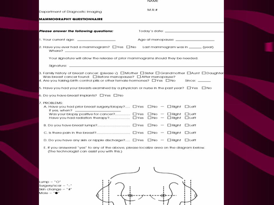
History includes • Gender• Age• Age of onset of menses• Parity (pregnancy history):
– Nulliparity (no pregnancies)– Multiparity (have they had multiple live births?)– Age of primiparity (first pregnancy)

Clinical Data of PatientsHistory:• Menstrual status
– Last menstrual cycle – age at menopause– has she had a hysterectomy and oophorectomy
• Medications– estrogen– progesterone – prolactin – thyroid or diabetes medications – steroids, or – estrogen inhibitors
Clinical Data of Patients
History • Previous breast biopsies
– Surgical biopsies and pathologic results– Core biopsies and pathologic results– Cyst aspirations
• Previous breast surgery– Augmentation– Reduction– Other (breast lift, tram flap etc.)
• Family history of breast cancer– Maternal and paternal
Clinical Data of Patients
History • Miscellaneous:
– Previous chest surgery (open heart, etc.)– Port-O-Caths– Moles– Accessory nipple– Unusual landmarks
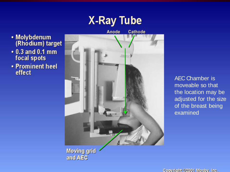
AEC Chamber is moveable so thatthe location may beadjusted for the sizeof the breast being examined
Distinction• Screening mammography
– Used to detect breast changes in women who have no sign, or symptom, or observable breast anomalies
– The goal is to detect cancer before any clinical signs are noticeable.
– At least two mammograms from different angles of each breast• Diagnostic mammography
– To investigate suspicious breast changes, such as• a breast lump, breast pain, an unusual skin appearance, nipple thickening or
nipple discharge.
– Also used to evaluate abnormal findings on a screening mammogram.
– Additional images can be made from other angles or focus on areas of concern at higher magnification.
Screening Mammography
• In positioning patients for a routine screening mammogram, the following views are considered standard for the exam:
– Craniocaudal (CC)– Mediolateral oblique (MLO)
Screening Mammography
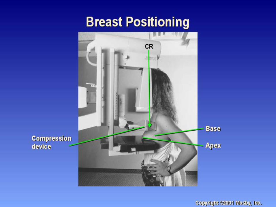
• Proper breast positioning is based on an understanding of the normal breast anatomy and the normal mobility of the breast.
• The mobile aspects of the breast are the lateral and inferior margins;
• the medial and superior margins are fixed.
Screening Mammography
• While it is desirable to have the nipple in profile on the routine views, the primary goal in breast positioning is to show as much tissue as possible.
• Therefore, breast tissue should not be sacrificed to show the nipple in profile. The nipple should be shown in profile, in at least one view. When the nipple is not shown in profile on any view, an extra view for nipple profile can be done.
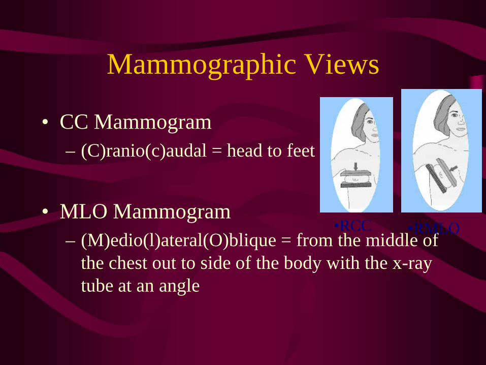
Mammographic Views
• CC Mammogram– (C)ranio(c)audal = head to feet
• MLO Mammogram– (M)edio(l)ateral(O)blique = from the middle of
the chest out to side of the body with the x-ray tube at an angle
•RCC •RMLO
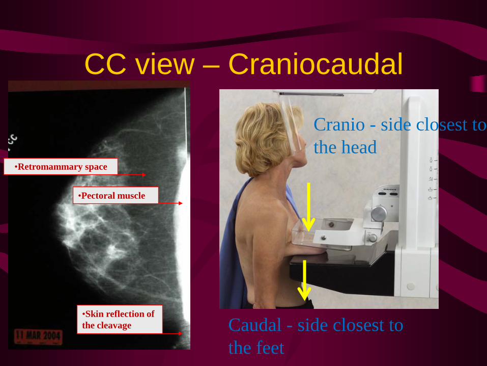
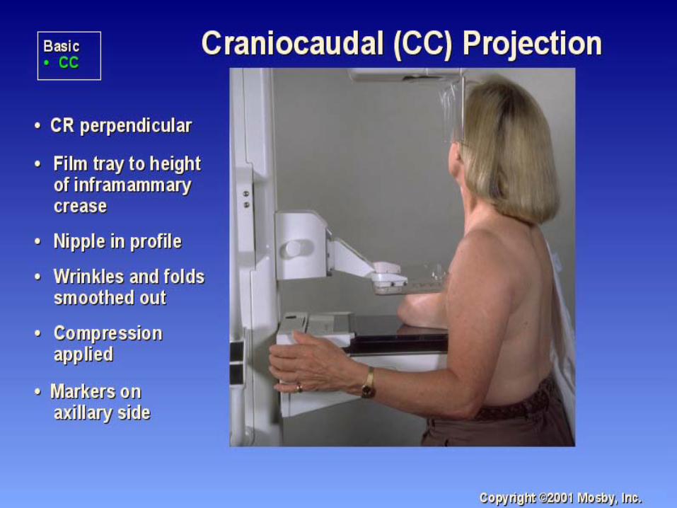
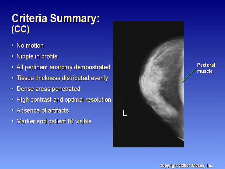
CC view – Craniocaudal
Cranio - side closest to the head
Caudal - side closest to the feet
•Retromammary space
•Pectoral muscle
•Skin reflection of the cleavage
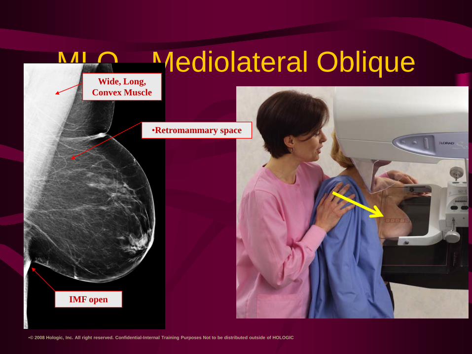
MLO – Mediolateral Oblique
•© 2008 Hologic, Inc. All right reserved. Confidential-Internal Training Purposes Not to be distributed outside of HOLOGIC
Wide, Long, Convex Muscle
•Retromammary space
IMF open
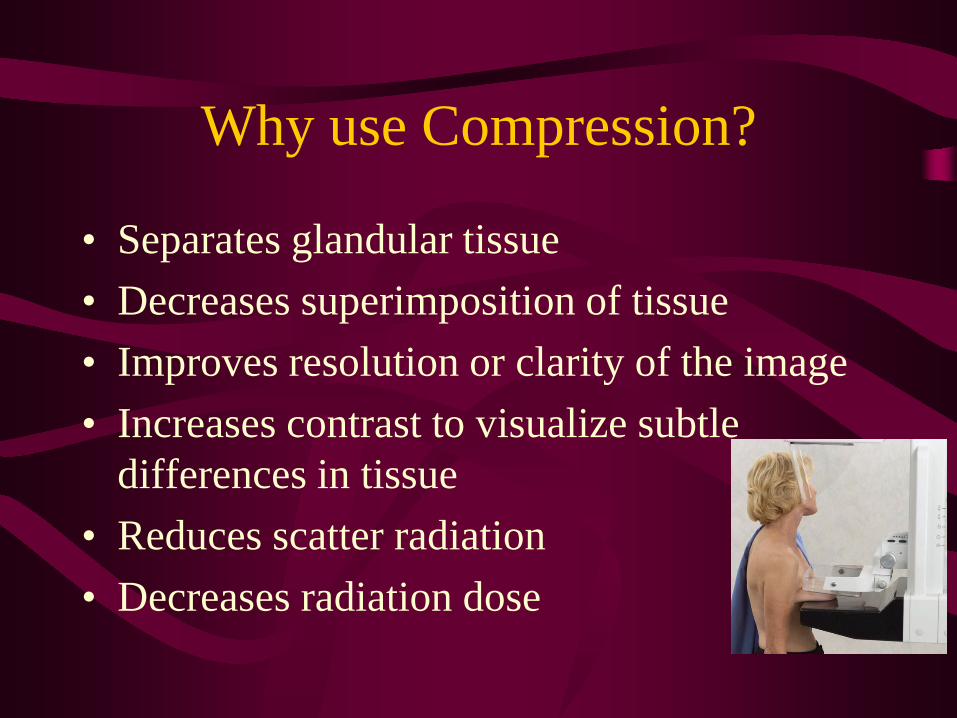
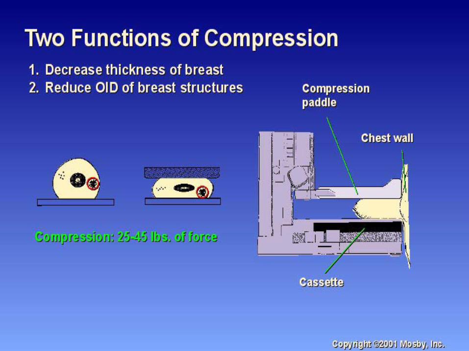
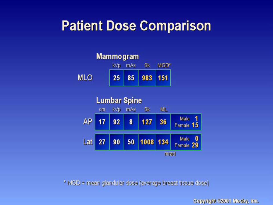
Why use Compression?
• Separates glandular tissue• Decreases superimposition of tissue• Improves resolution or clarity of the image• Increases contrast to visualize subtle
differences in tissue• Reduces scatter radiation• Decreases radiation dose
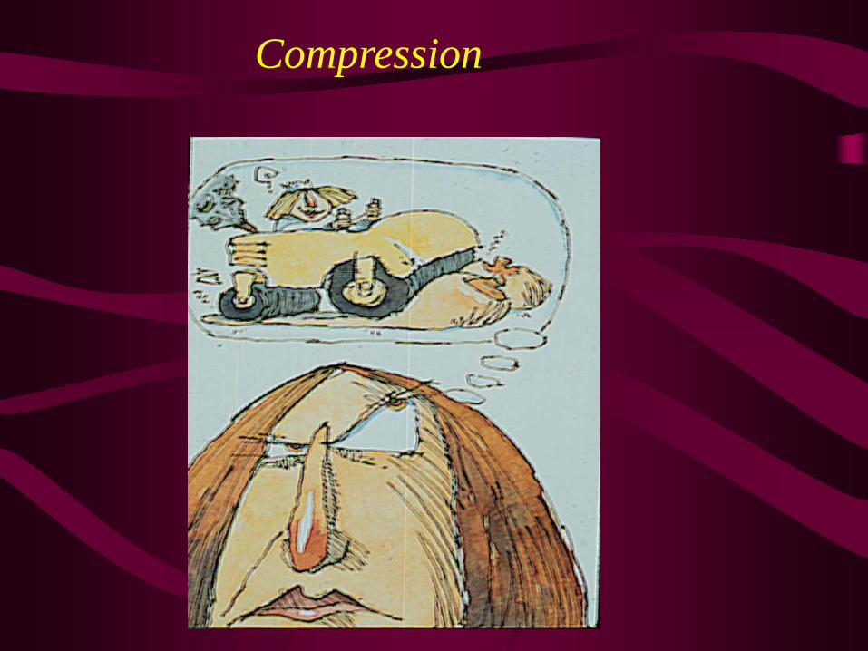
Compression
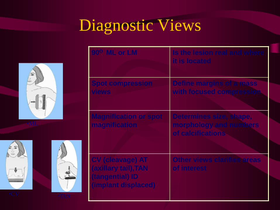
Diagnostic Views90O ML or LM Is the lesion real and where
it is located
Spot compression views
Define margins of a mass with focused compression
Magnification or spot magnification
Determines size, shape, morphology and numbers of calcifications
CV (cleavage) AT (axillary tail),TAN (tangential) ID (implant displaced)
Other views clarifies areas of interest
•ML
•TAN•CV
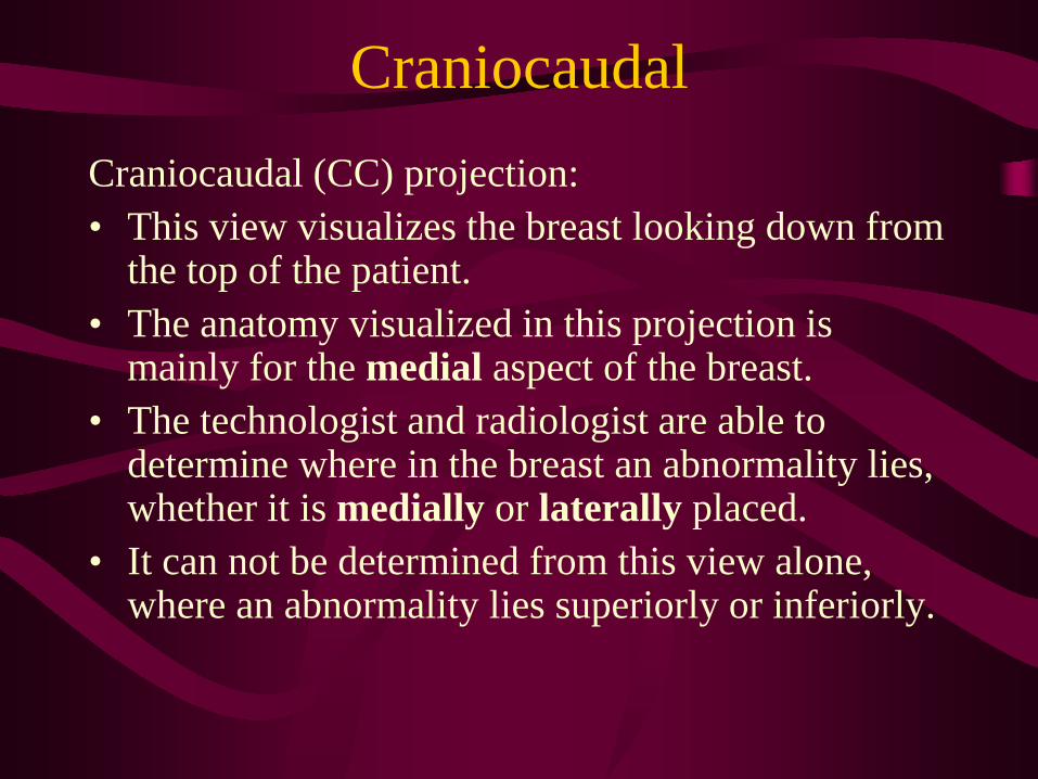
CraniocaudalCraniocaudal (CC) projection:• This view visualizes the breast looking down from
the top of the patient.• The anatomy visualized in this projection is
mainly for the medial aspect of the breast.• The technologist and radiologist are able to
determine where in the breast an abnormality lies, whether it is medially or laterally placed.
• It can not be determined from this view alone, where an abnormality lies superiorly or inferiorly.
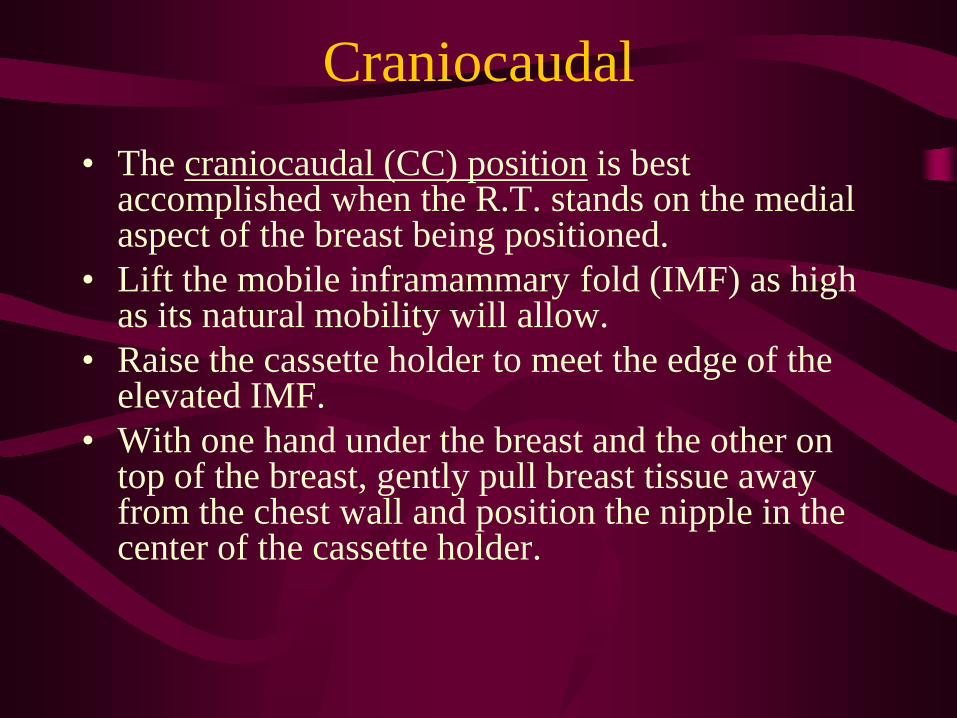
Craniocaudal• The craniocaudal (CC) position is best
accomplished when the R.T. stands on the medial aspect of the breast being positioned.
• Lift the mobile inframammary fold (IMF) as high as its natural mobility will allow.
• Raise the cassette holder to meet the edge of the elevated IMF.
• With one hand under the breast and the other on top of the breast, gently pull breast tissue away from the chest wall and position the nipple in the center of the cassette holder.
Craniocaudal
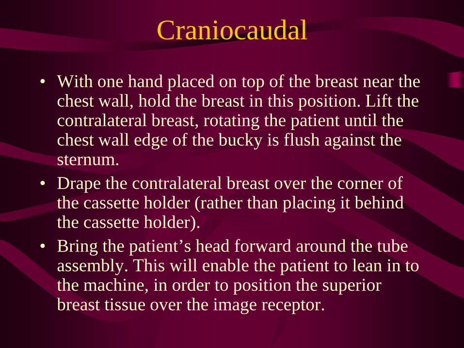
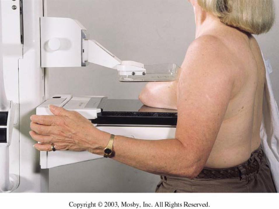
• With one hand placed on top of the breast near the chest wall, hold the breast in this position. Lift the contralateral breast, rotating the patient until the chest wall edge of the bucky is flush against the sternum.
• Drape the contralateral breast over the corner of the cassette holder (rather than placing it behind the cassette holder).
• Bring the patient’s head forward around the tube assembly. This will enable the patient to lean in to the machine, in order to position the superior breast tissue over the image receptor.

Craniocaudal• Place the other hand, not holding the breast,
behind the patient’s back, to keep her from pulling away from the mammography unit.
• As compression is applied, the hand on top of the breast gently pulls the tissue forward to prevent wrinkles in the skin from occurring.

Craniocaudal
• It is important to have the patient relax the arm of the breast being imaged. This will allow more lateral breast tissue to be imaged. If wrinkles appear, on the lateral side of the breast, use one finger to laterally smooth them out.
• Do not push the wrinkles back towards the chest wall. This would move breast tissue out of the image.
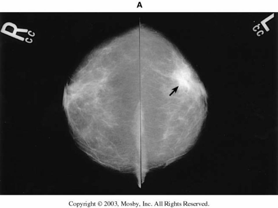
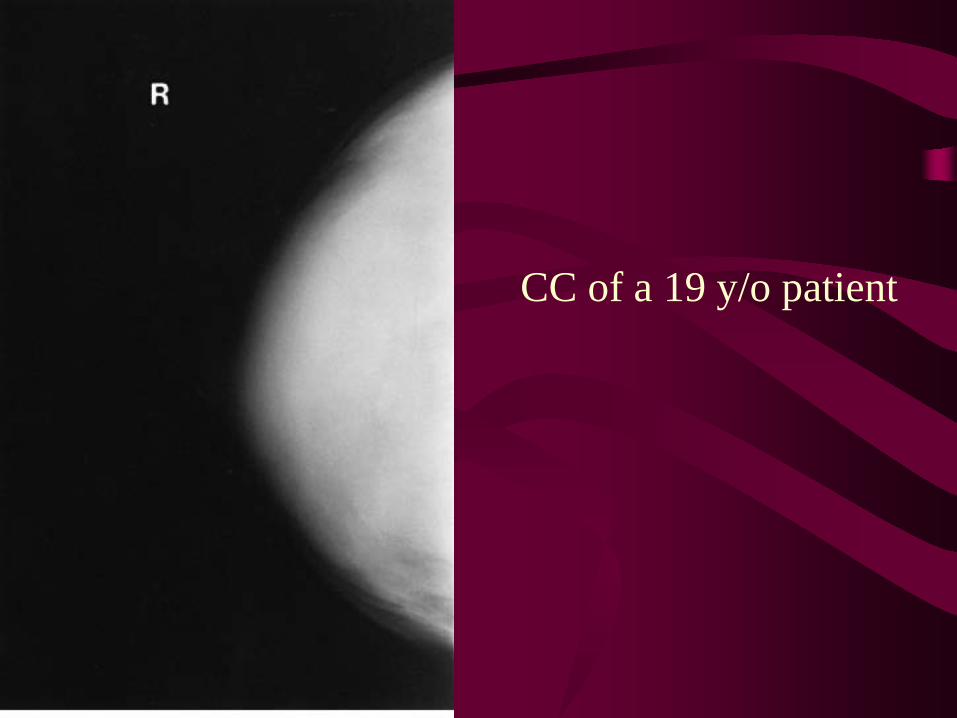
CC of a 19 y/o patient
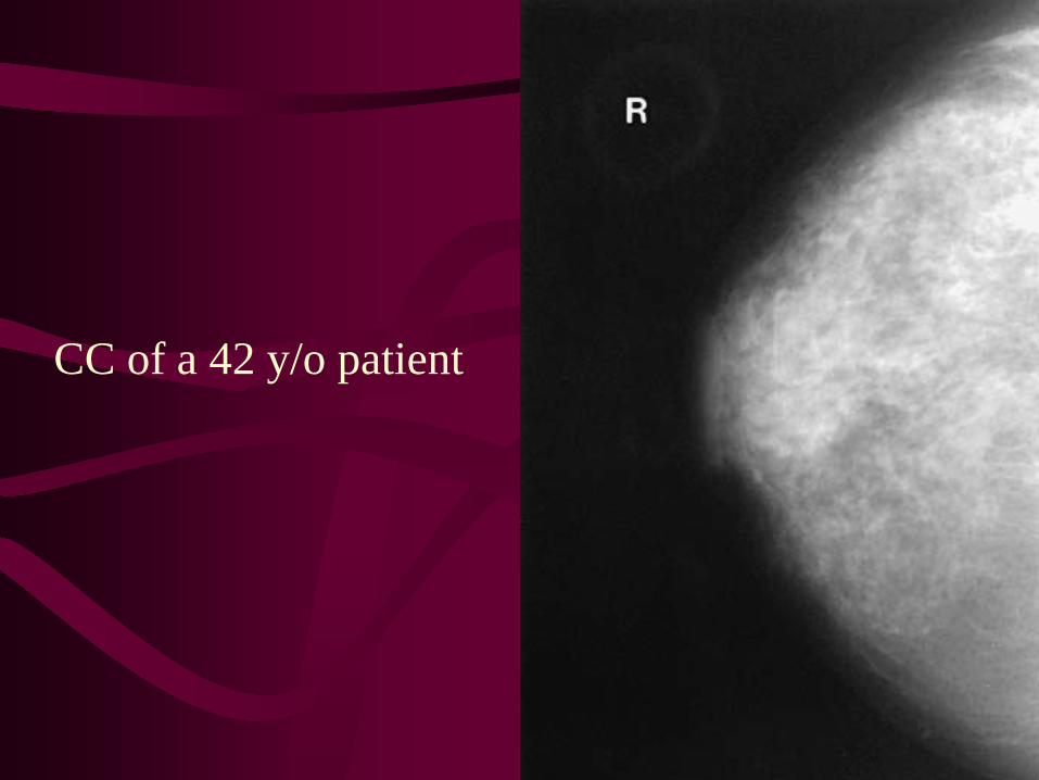
CC of a 42 y/o patient
Craniocaudal
• The CC projection may need to be modified with more challenging patients.
• Male patients have very little breast tissue to manipulate; it is best to try gently pinching the tissue around the areola, to maximize the amount of tissue being imaged.
• In patients with pacemakers or port-o-caths, may be challenging to position, it is best to try maximizing the amount of tissue being imaged, without compromising the object in the chest.
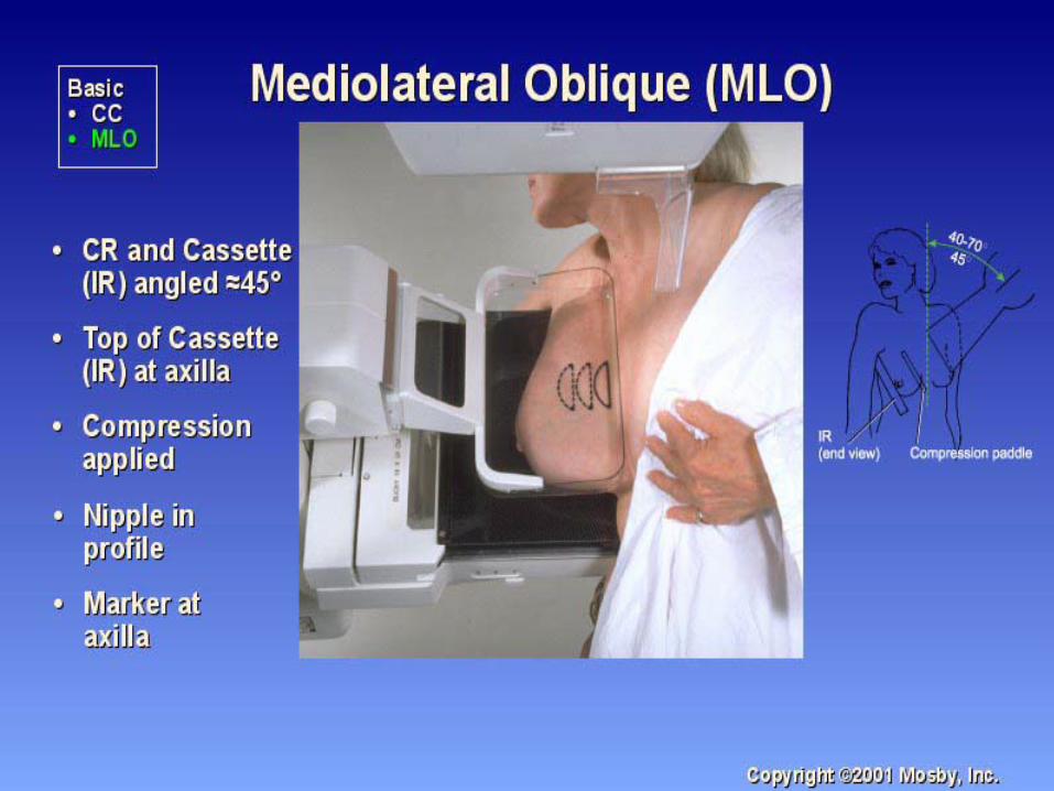
Mediolateral Oblique
• The mediolateral oblique (MLO) offers the best opportunity to visualize the maximum amount of breast tissue in a single view.
• For the MLO, the plane of the cassette holder can be angled anywhere from 30 to 60 degrees from the horizontal, so that the cassette is parallel to the pectoral muscle.
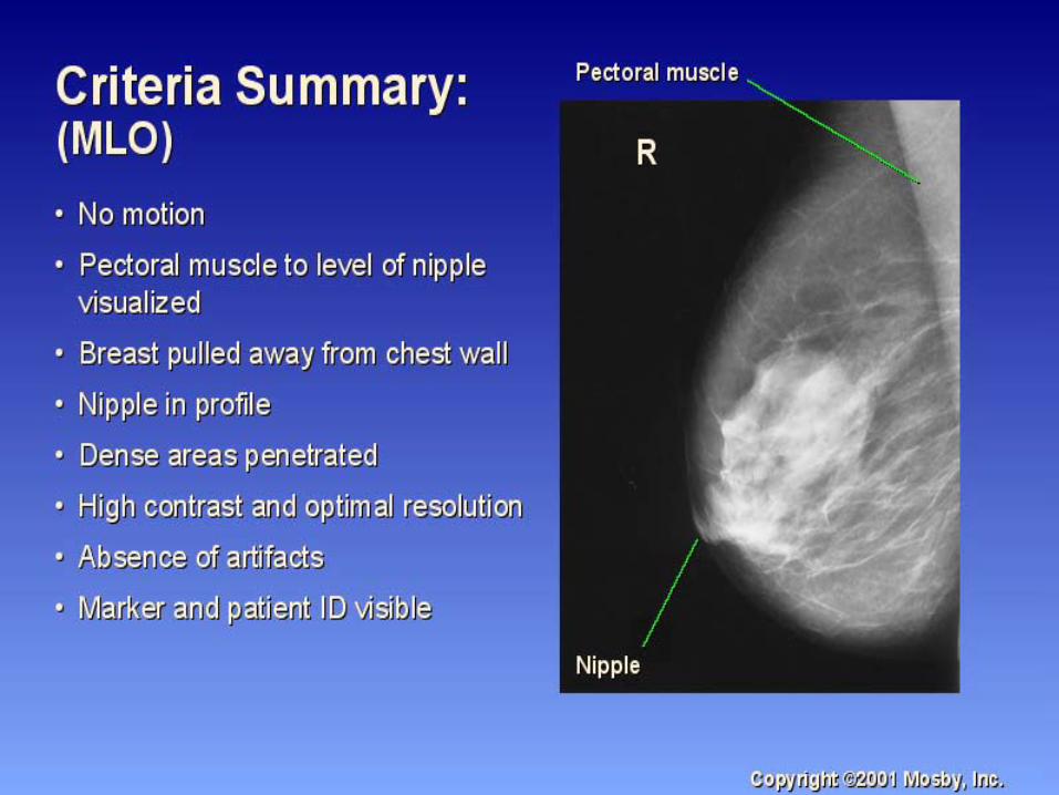
Mediolateral Oblique• The criteria on the mammogram indicating that
positioning for the MLO is optimal include: – pectoral muscle is wide superiorly with a convex
anterior border, extending to or below the posterior nipple line;
– fat is visualized posterior to all of the fibroglandular tissues;
– deep and superficial breast tissues are well separated; – close inspection shows no evidence of motion blur; and– the inframammary fold is open.
Mediolateral Oblique• In the MLO projection, the X-ray beam is directed
from the superomedial to the inferolateral aspect of the breast. In order to image the maximum amount of tissue, it is imperative that the angle of the image receptor is parallel to the angle of the pectoral muscle of the individual patient.
• To determine the angle of the pectoral muscle, the technologist places her fingers in the patient’s axilla behind the muscle. The patient’s shoulder is should be relaxed in neutral rotation.
• The technologist gently moves the pectoral muscle forward to accentuate the movable lateral border.
Mediolateral Oblique
• In the MLO projection, tall and thin patients typically use a steeper angle (50 to 60 degrees).
• Short and heavy patients typically use lesser of an angle (30 to 40 degrees).
• If the pectoral muscle is not parallel to the image receptor, less tissue will be imaged.
Mediolateral Oblique
• Applying the principle of moving the mobile tissue toward the fixed tissue, lift the breast, then pull both breast tissue and the pectoral muscle anteriorly and medially.
• The patient’s hand on the side being imaged should be resting on the handlebar. Move the patient’s shoulder as close to the center of the bucky as possible.
• This will place the corner of the cassette holder posterior to the axilla, behind the pectoral muscle, but in front of the latissimus dorsi.
Mediolateral Oblique• The patient’s arm is draped behind the cassette
holder to relax the pectoral muscle. • Rotate the patient toward the cassette holder so
that the edge of the cassette holder replaces your hand in maintaining the breast and muscle in its mobilized position.
• Hold the breast up and out, away from the chest wall to prevent overlapping of tissue.
• Begin to apply compression. • While compressing, have the patient move their
hips and feet in toward the mammography unit.
Mediolateral Oblique• The upper corner of the compression paddle
should be just below the clavicle. While moving your hand out of the field, continue to support the anterior aspect of the breast with your hand until there is enough compression to maintain the breast in this position.
• We call the combined hand movements the “out and up” maneuver. It is very important to use this technique, otherwise breast tissue will overlap as a result of improper technique.
Mediolateral Oblique
• The final step in positioning the MLO projection of the breast involves pulling abdominal tissue down in order to open the inframammary fold.
• The entire breast, from inframammary fold to axilla, should be centered on the cassette holder.
• The photo cell should be placed at the level of the retroareolar tissue. This is the most dense area of the breast.
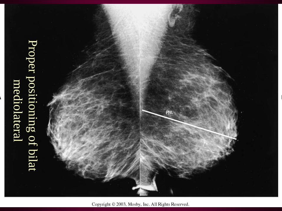
Proper positioning of bilat m
ediolateral
Mediolateral Oblique• In special circumstances, you may be working
with an obese patient. In these cases, try to push as much of the abdominal tissue back behind the image receptor, after compressing the breast.
• You may also be working with patients with pectus excavatum. These patients may be difficult to position, so a reverse MLO may be necessary. This is called an LMO. This view will improve visualization of medial breast tissue.
Craniocaudal
Medio Lateral
Oblique

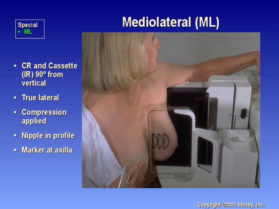
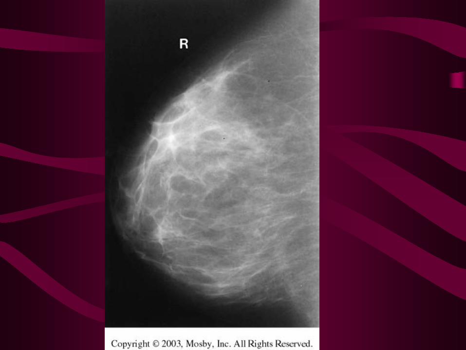
Lateral• Ninety degrees or true lateral ( or straight lateral)
projection: is the most commonly used additional view.
• This view is used to triangulate the exact location of lesions in the breast.
• The 90 degree lateral view is also used to demonstrate gravity-dependent calcifications. (Milk of calcium).
• When an abnormality shows on one standard view but not the other (CC or MLO), a lateral view is taken to determine if the abnormality is real, superimposed tissue, artifact on the radiograph, or in the skin.
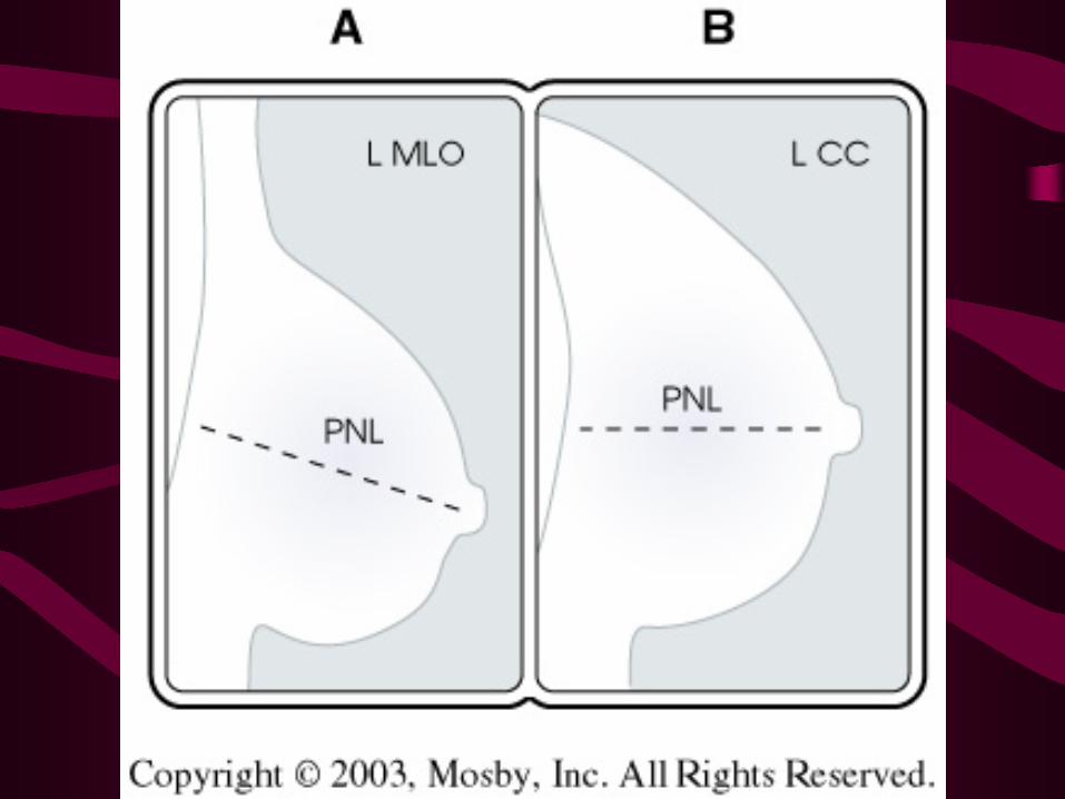
Lateral• A change in location of a lesion relative to
its distance from the nipple of the 90 degree lateral view can be used to determine whether the lesion is in the lateral, central, or medial aspect of the breast.
• When an abnormality has been identified, the most appropriate lateral view, medial to lateral versus lateral to medial, is the one that provides the shortest object-to-image receptor distance, to reduce geometric unsharpness.
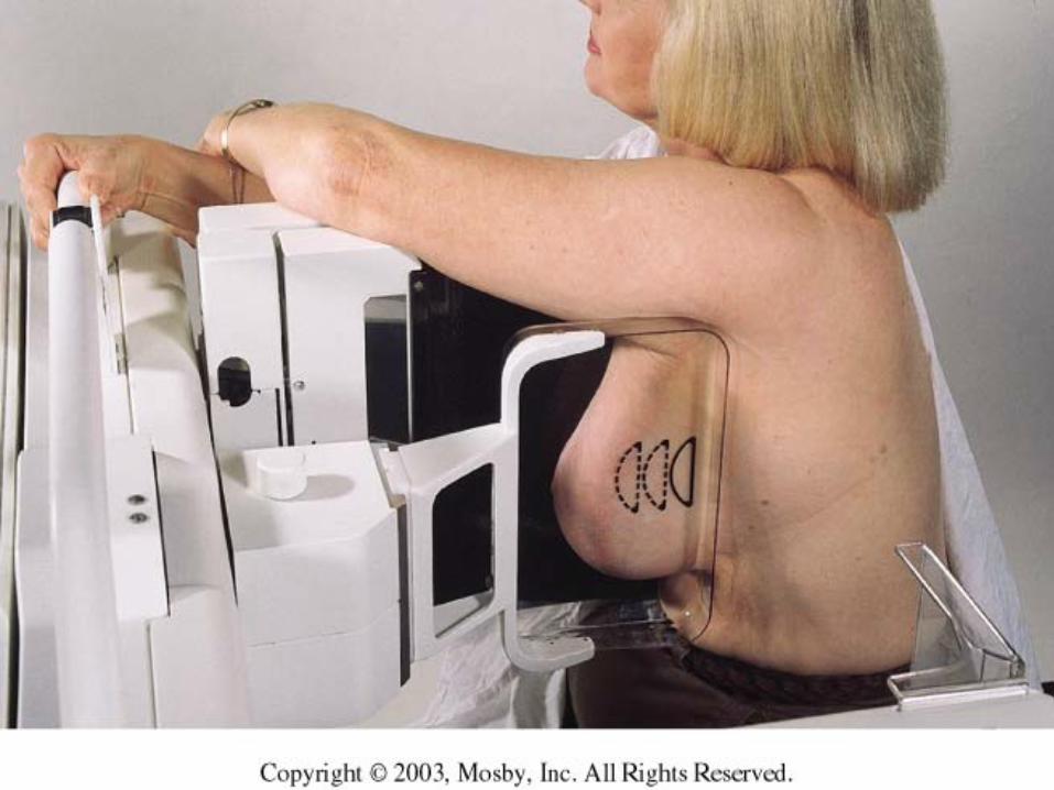
Lateral• Medial-to-lateral 90 degree lateral projection
• The tube arm is rotated 90 degrees. The patient’s arm on the side being examined is abducted 90 degrees resting across the top of the cassette holder.
Lateral• Pull the breast tissue and pectoral muscle
anteriorly and medially. Lift the breast out and up while gently pulling the breast away from the chest wall.
• Rotate the patient toward the cassette holder and begin compression. Continue to compress, while holding the breast tissue up and out away from the chest wall.
• When you are finished compressing, pull down on the abdominal tissue, to open up the inframammary fold.
Lateral• For the lateromedial view, the tube arm is
90 degrees with the top of the cassette holder at the level of the suprasternal notch.
• The patient is positioned with her sternum against the edge of the cassette holder, her neck extended with her chin resting on the top of the cassette holder.
Lateral• Pull the mobile lateral and inferior tissue up and
toward the cassette holder. Bring the compression paddle down past the latissimus dorsi, lift the patient’s arm on the side being imaged over the cassette holder.
• Continue rotating the patient until the breast is in a true lateral position centered on the cassette holder. Open the inframammary fold by gently pulling abdominal tissue down.

Diagnostic and Additional Projections
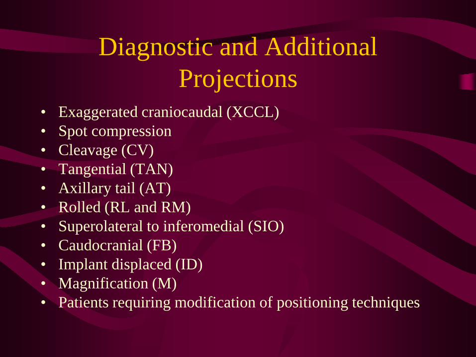
• Exaggerated craniocaudal (XCCL)• Spot compression • Cleavage (CV)• Tangential (TAN) • Axillary tail (AT)• Rolled (RL and RM)• Superolateral to inferomedial (SIO)• Caudocranial (FB)• Implant displaced (ID)• Magnification (M)• Patients requiring modification of positioning techniques
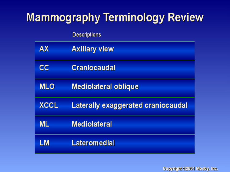
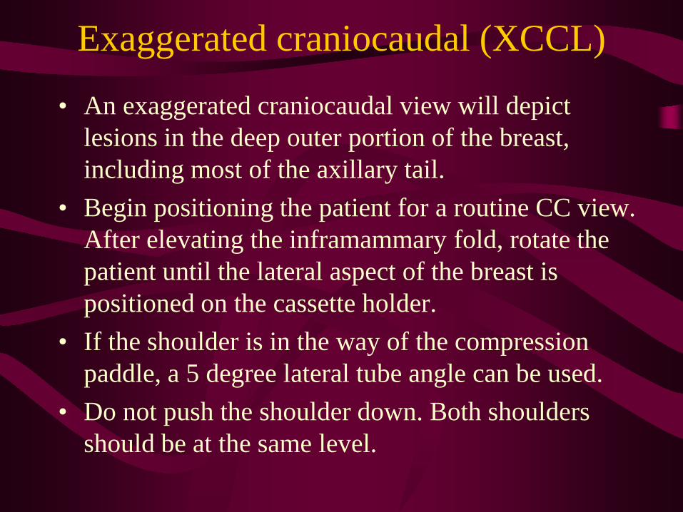
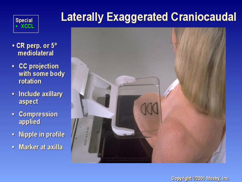
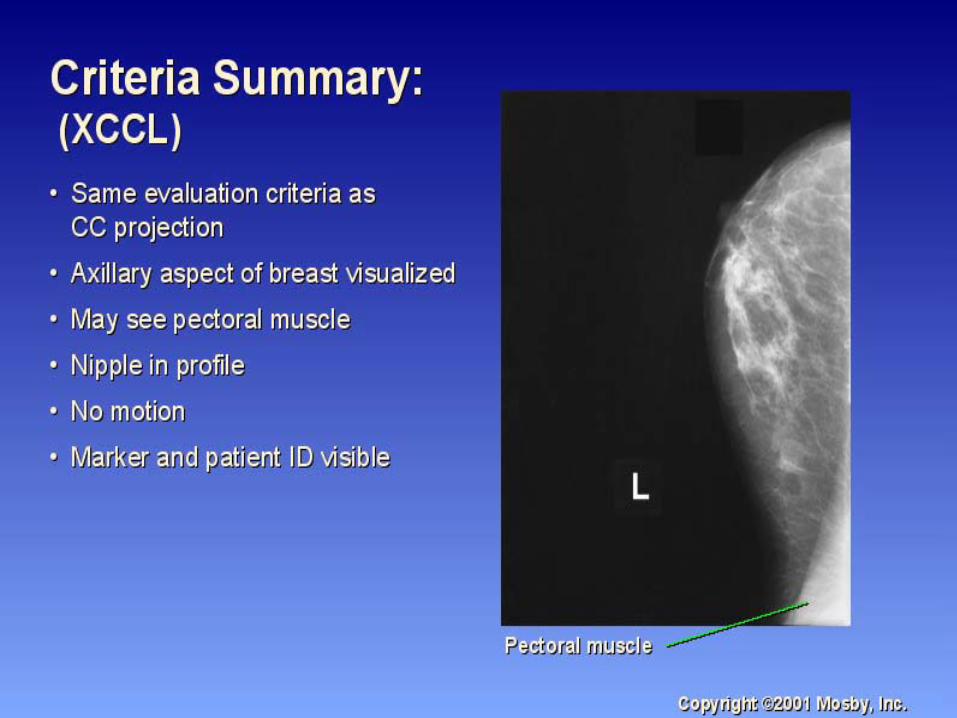
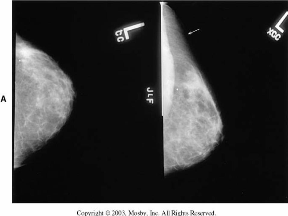
Exaggerated craniocaudal (XCCL)
• An exaggerated craniocaudal view will depict lesions in the deep outer portion of the breast, including most of the axillary tail.
• Begin positioning the patient for a routine CC view. After elevating the inframammary fold, rotate the patient until the lateral aspect of the breast is positioned on the cassette holder.
• If the shoulder is in the way of the compression paddle, a 5 degree lateral tube angle can be used.
• Do not push the shoulder down. Both shoulders should be at the same level.
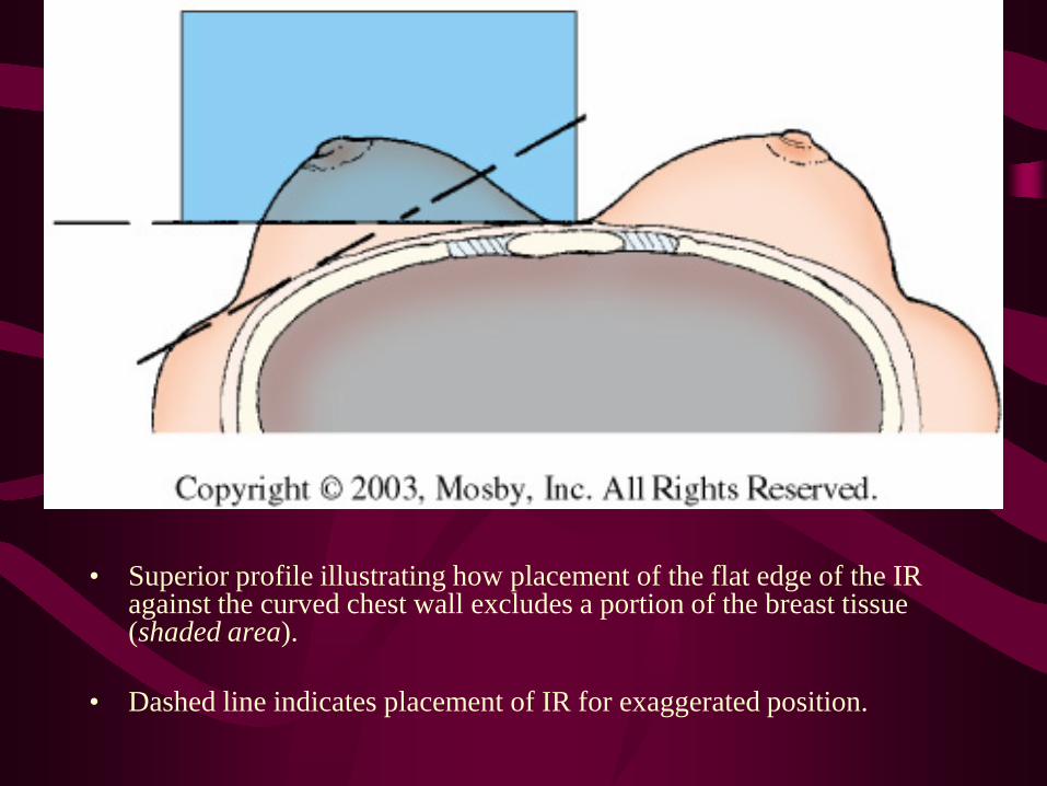
• Superior profile illustrating how placement of the flat edge of the IR against the curved chest wall excludes a portion of the breast tissue (shaded area).
• Dashed line indicates placement of IR for exaggerated position.
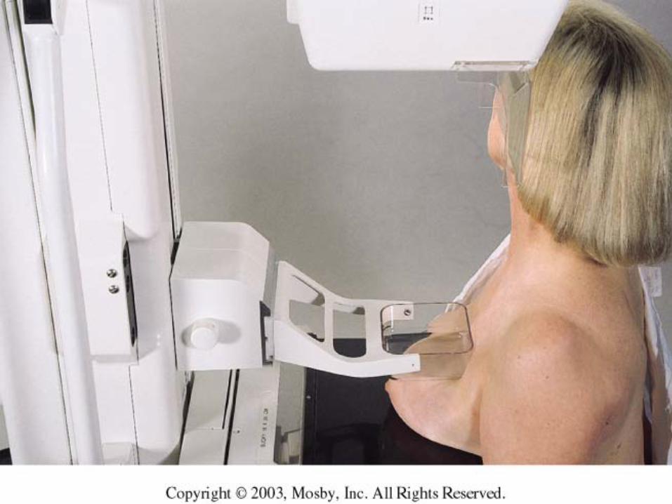
Spot Compression
• Spot or coned compression is a simple technique. It is especially helpful with obscure or equivocal findings in areas of dense tissue.
• Spot compression allows for more localized compression of an area of the breast. It allows for higher contrast, and more precise evaluation of findings.
Spot Compression
• Using the original mammogram, the technologist determines the placement of the small compression device by determining the location of the lesion.
• To determine the location of the lesion, measure the depth relative to a line drawn directly posterior from the nipple, the distance from that line to the lesion in the superior-to inferior or medial-to-lateral direction, and the distance from the lesion to the skin surface.
Spot Compression
• Reposition the patient, using your hand to simulate compression.
• Transfer the three measurements to the breast and use a marker to identify the location of the lesion.
• Reposition to center the spot compression device over the lesion.
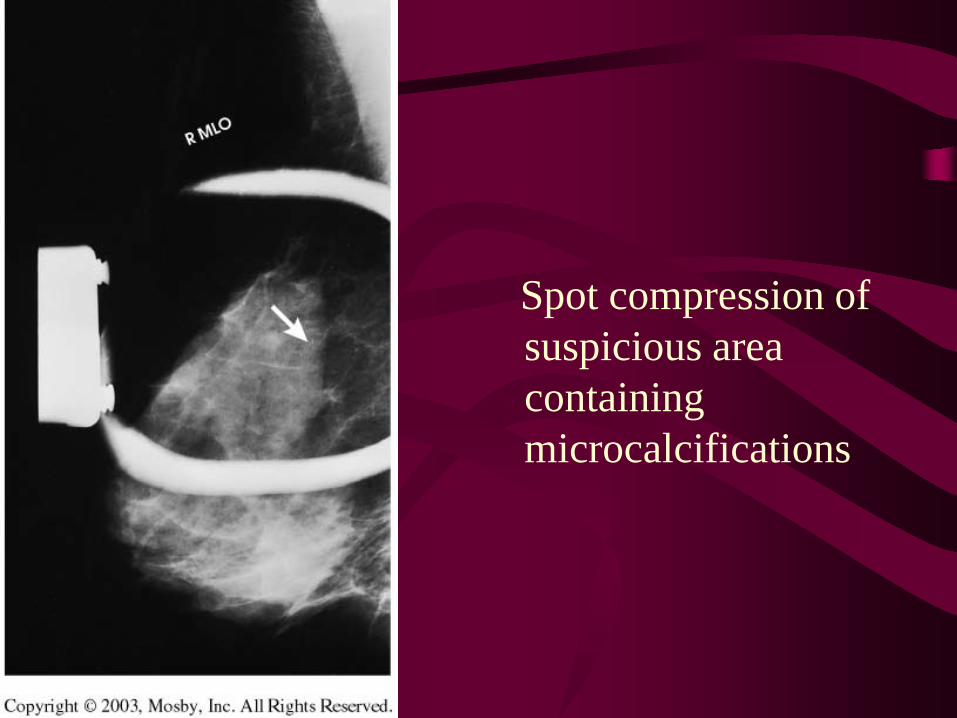
Spot compression of suspicious area containing microcalcifications
Cleavage (CV)
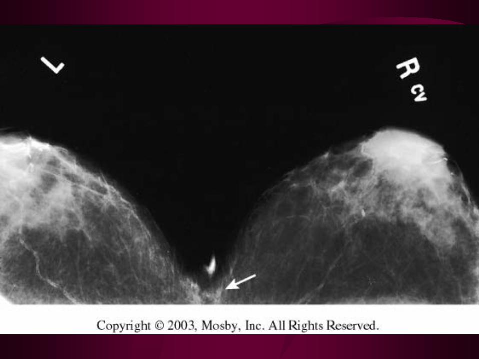
• The cleavage view (valley view, double breast compression view) is performed to visualize deep lesions in the posteromedial aspect of the breast.
• The patients head is turned away from the side of interest.
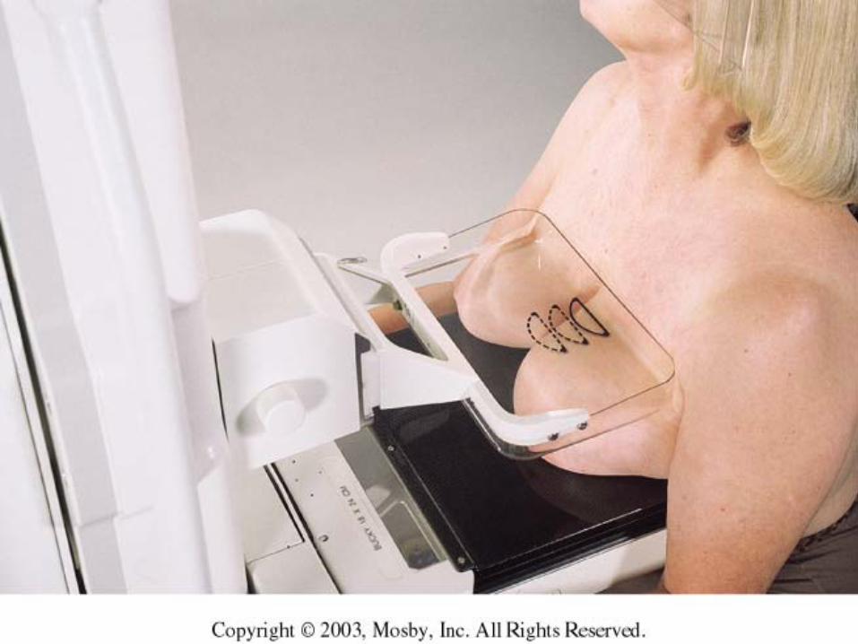
Cleavage (CV)• Positioning is done with the technologist standing
behind the patient, and wrapping her arms around the patient to reach her breasts. Make sure to pull all of the medial tissue of both breasts anteriorly in order to image the cleavage.
• Automatic exposure can be obtained, by placing the breast of interest over the photocell with the cleavage slightly off center.
• Manual technique must be used if the photocell is under an open cleavage.
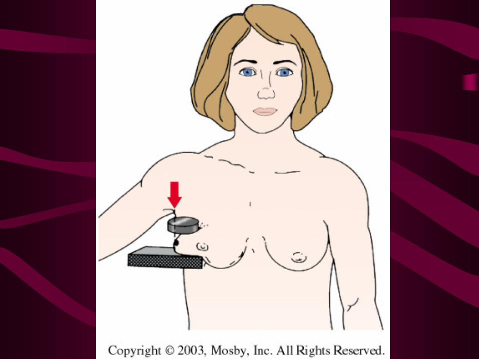
Tangential (TAN)
• This view is used for palpable lesions that are obscured by surrounding dense glandular tissue on the mammogram. The C-arm is rotated and the patient is turned so that the X-ray beam is tangential to the palpable lump.
• These views can be obtained by placing a lead marker (BB) directly over the lump and directing the X-ray beam tangential to the lead marker.
• These views can also be used to verify that calcifications seen on a mammogram are located within the skin.
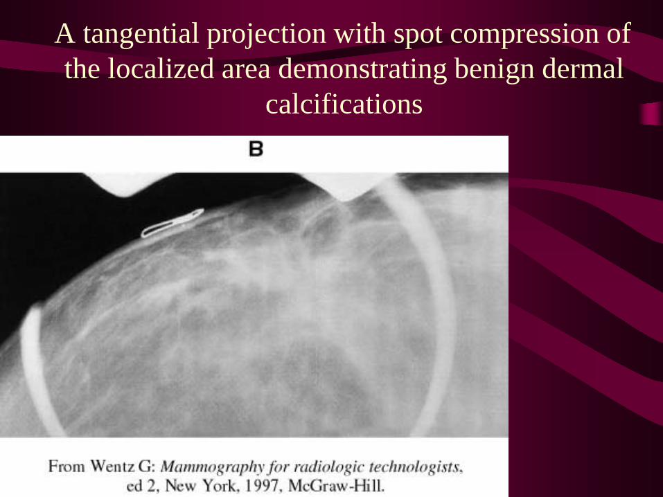
A tangential projection with spot compression of the localized area demonstrating benign dermal
calcifications
Axillary Tail (AT)• This view is used to demonstrate the entire axillary
tail as well as most of the lateral aspect of the breast. The tube is rotated to an angle that will place the cassette holder parallel to the axillary tail.
• The patient is turned to bring the axillary tail in contact with the cassette holder. The patient’s arm on the side being imaged is draped behind the top of the cassette holder.
• Gently pull the axillary aspect of the breast out and away from the chest wall and place it on the cassette holder. Hold the axillary tail in place while slowly applying compression.

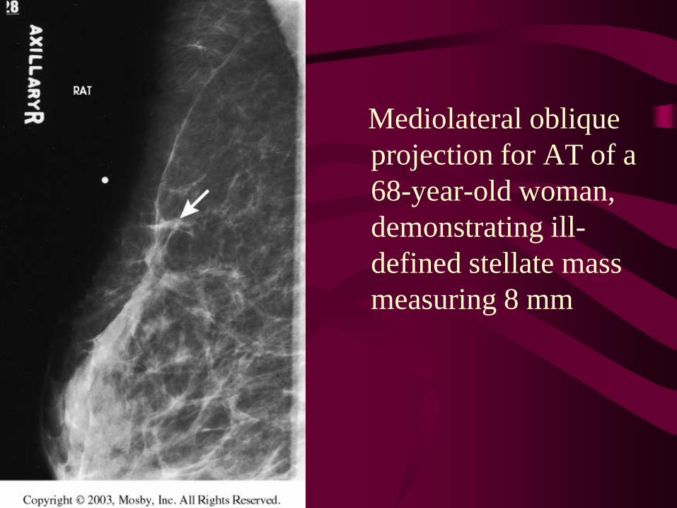
Mediolateral oblique projection for AT of a 68-year-old woman, demonstrating ill-defined stellate mass measuring 8 mm

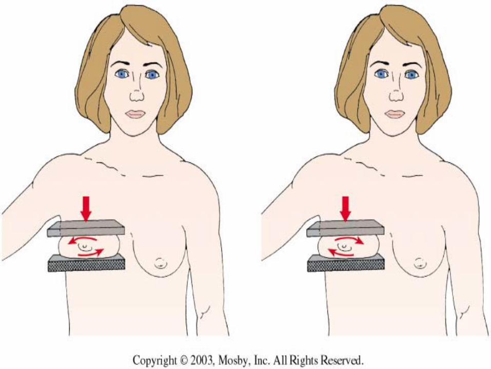
Rolled (RL and RM)• The roll view is used to separate superimposed
breast tissues. The purpose is to confirm the presence of an abnormality, to better define a lesion, or to determine the location of a finding seen on only one of the standard views.
• The patient is repositioned using the same projection that demonstrated the abnormality. Placing your hands on either side of the breast, “roll” the tissue in opposite directions.
• Compression will maintain the breast in the “rolled” position. A radiopaque marker indicating the direction of the roll should be placed on the image receptor.
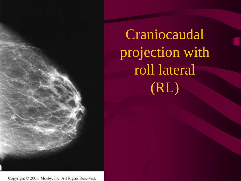
Craniocaudal projection with
roll lateral (RL)
Superolateral to inferomedial oblique (SIO)
• This is an oblique view that can be performed with the central ray directed upper-outer to lower-inner.
• This view has been incorrectly termed as a reverse oblique. As a whole-breast projection it has limited usefulness.
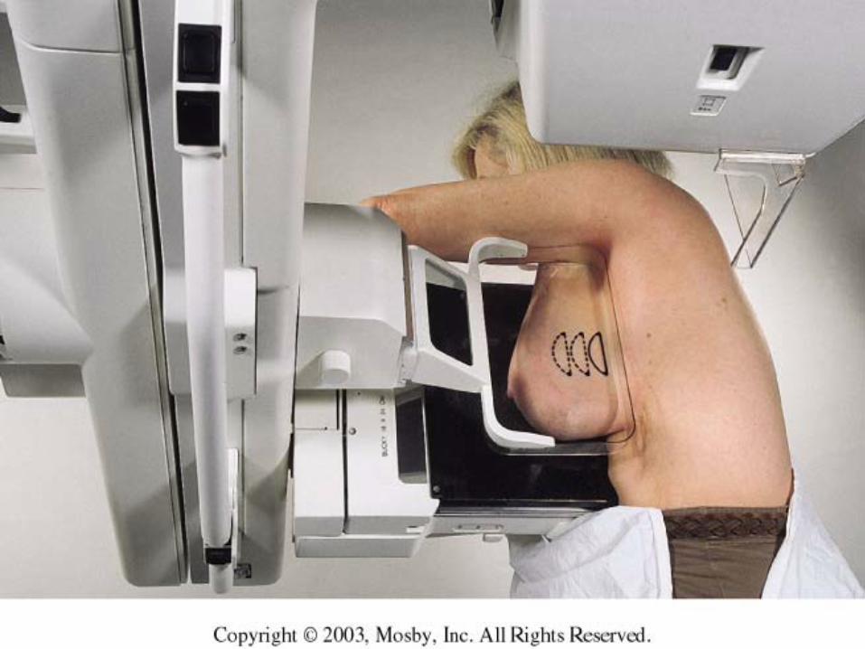
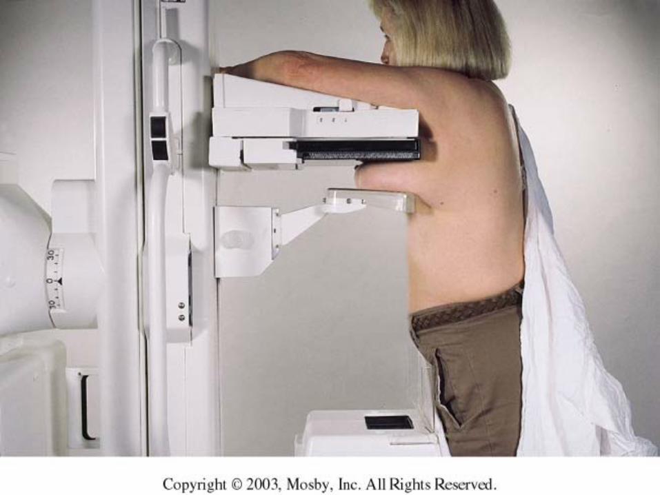
Caudocranial view (reverse CC):
• This view will improve visualization of lesions in the uppermost aspect of the breast due to the reduced object-to-image receptor distance.
• Since the compression device comes from below, this view will not exclude the fixed posterior tissue in the superior aspect of the breast.
• This view is also commonly used in patients who are kyphotic, or in male patients.
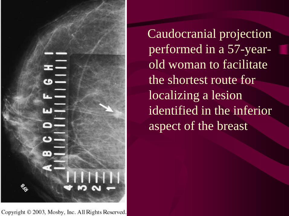
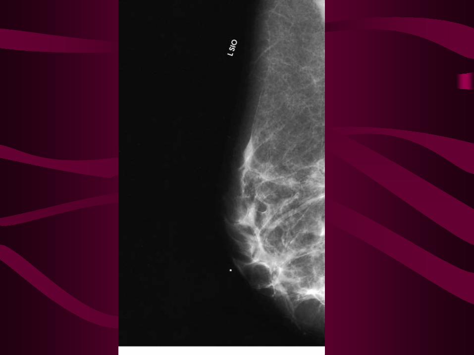
Caudocranial projection performed in a 57-year-old woman to facilitate the shortest route for localizing a lesion identified in the inferior aspect of the breast
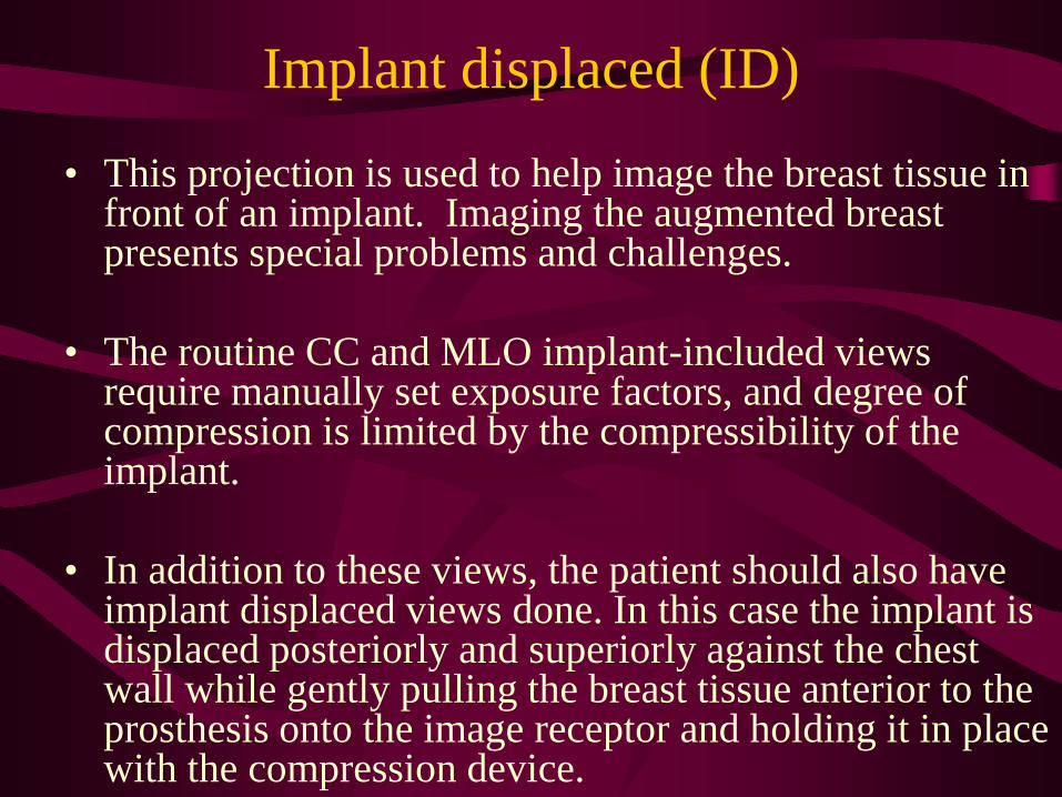
Implant displaced (ID)
• This projection is used to help image the breast tissue in front of an implant. Imaging the augmented breast presents special problems and challenges.
• The routine CC and MLO implant-included views require manually set exposure factors, and degree of compression is limited by the compressibility of the implant.
• In addition to these views, the patient should also have implant displaced views done. In this case the implant is displaced posteriorly and superiorly against the chest wall while gently pulling the breast tissue anterior to the prosthesis onto the image receptor and holding it in place with the compression device.
Implant displaced (ID)
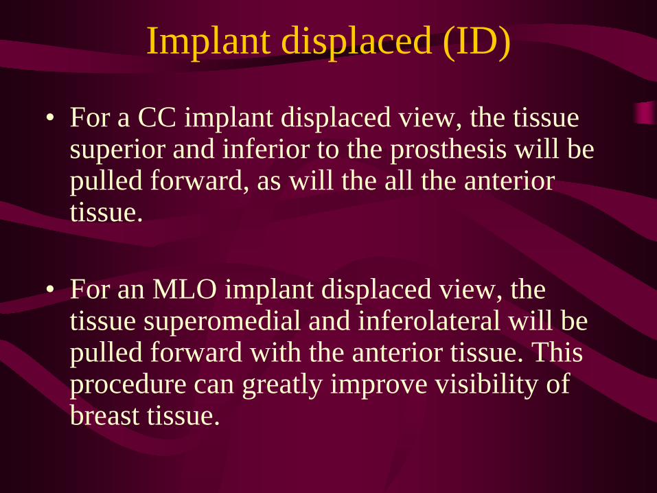
• For a CC implant displaced view, the tissue superior and inferior to the prosthesis will be pulled forward, as will the all the anterior tissue.
• For an MLO implant displaced view, the tissue superomedial and inferolateral will be pulled forward with the anterior tissue. This procedure can greatly improve visibility of breast tissue.
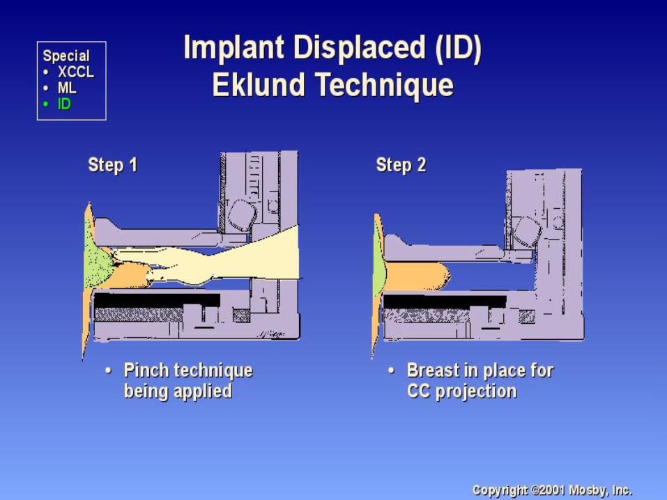
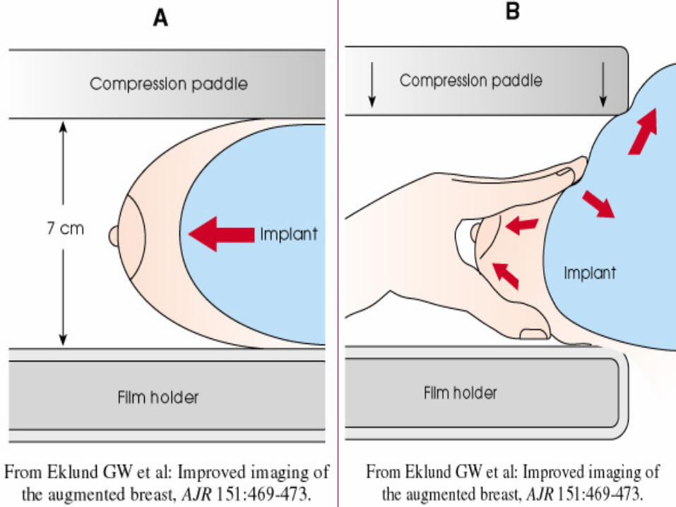
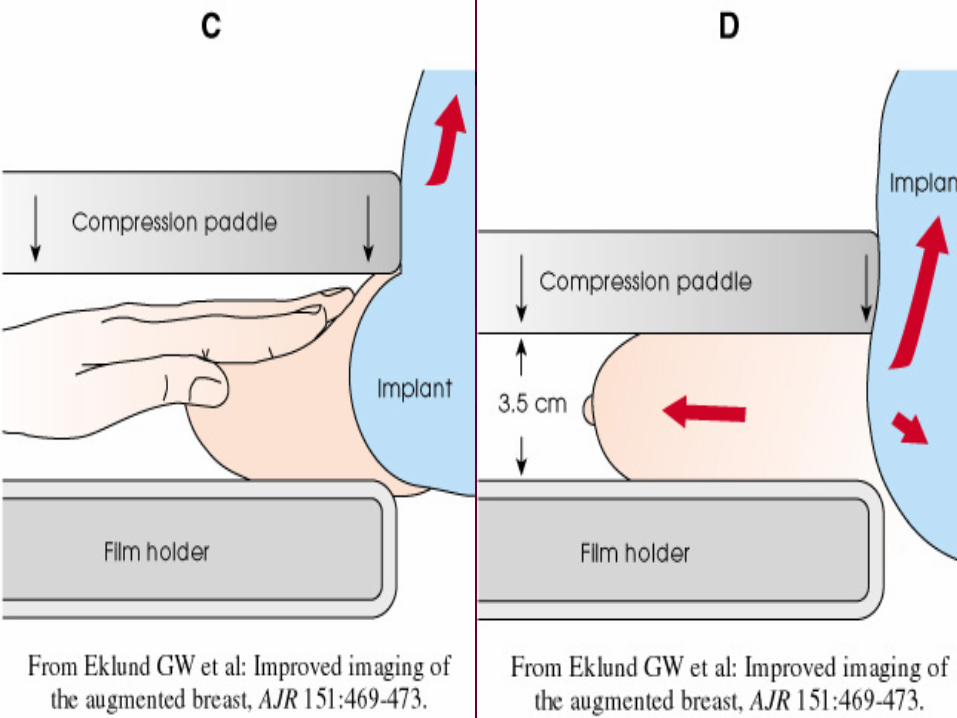
Implant displaced (ID)
Steps to positioning the CC and MLO implant displaced views:
• Have the patient bend forward from the waist, use your fingers to pull the breast tissue forward while displacing the implant posteriorly, and then have the patient stand again.
• Gently pull the breast tissue onto the cassette holder and place the edge of your fingers, holding the inferior tissue, against the edge of the bucky.
• Ask the patient to push her body against the image receptor.
• Apply compression.

Magnification (M)
• Magnification views with or without spot compression can be helpful in differentiating benign from malignant lesions by permitting a more precise evaluation of margins and other architectural characteristics of a focal density or mass.
• These views also permit better delineation of the number, distribution and morphology of calcifications.
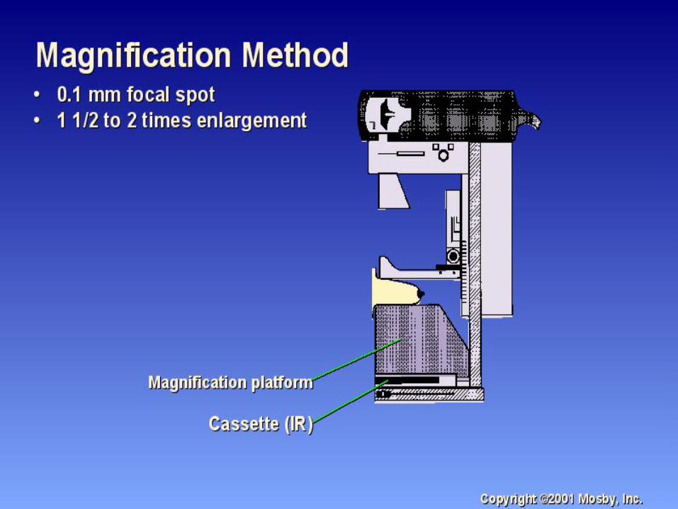
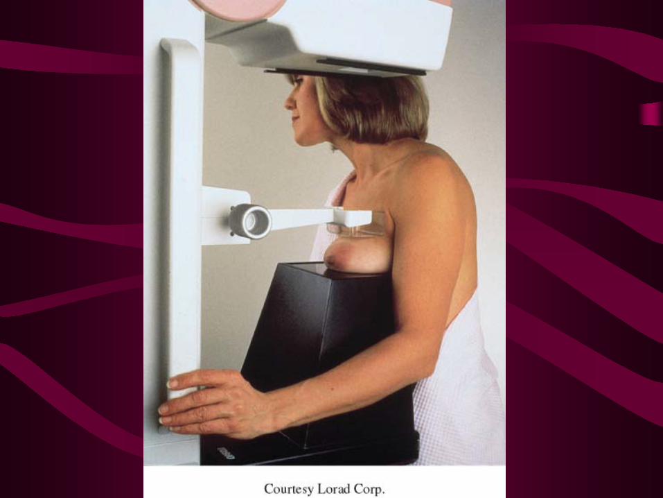
Magnification (M)• To perform magnification views, there has to be an
X-ray tube with a micro focal spot to offset the geometric unsharpness. It also requires a magnification platform to separate the compressed breast from the cassette for a 1.5 to 2.0 times magnification. In making the exposure, the patient will need to hold still longer than for a normal mammogram.
• The air gap resulting from separation of the breast from the image receptor prevents a significant amount of scattered radiation from reaching the image receptor, and a grid is not used.
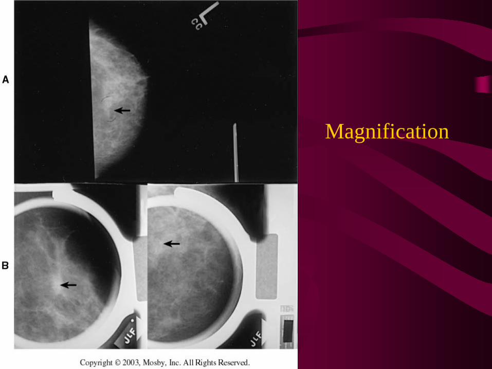
Magnification
Diagnostic and Additional Projections
Patients requiring modification of positioning techniques:• Males• Kyphotic patients• Large breasts• Small breasts• Encapsulated implants• Pectus excavatum/carinatum• Protruding abdomens• Pacemaker• Stretcher• Wheelchair• Port-a-cath• Physically/mentally challenged