
Portraying emotions at their unfolding: A multilayered approach for probing dynamics of neural...
14
Portraying emotions at their unfolding: A multilayered approach for probing dynamics of neural networks Gal Raz a, d, e , Yonatan Winetraub a, b , Yael Jacob a, d , Sivan Kinreich a, c , Adi Maron-Katz a, d , Galit Shaham f , Ilana Podlipsky a , Gadi Gilam a, c , Eyal Soreq a, c, g , Talma Hendler a, c, d, ⁎ a Functional Brain Center, Tel Aviv Sourasky Medical Center, 6 Weizmann Street, Tel Aviv 64239, Israel b Adi Lautman Interdisciplinary Program for Outstanding Students, Tel Aviv University, Tel Aviv 69978, Israel c School of Psychological Sciences, Tel Aviv University, Tel Aviv 69978, Israel d Physiology and Pharmacology Department, Sackler Faculty of Medicine, Tel Aviv University, Tel Aviv 69978, Israel e Film and Television Department, Tel Aviv University, Tel Aviv 69978, Israel f Psychology Department, The Hebrew University of Jerusalem, Jerusalem, Israel g Screen Based Art Department, Bezalel Academy of Arts and Design, Jerusalem, Israel abstract article info Article history: Received 27 July 2011 Revised 23 December 2011 Accepted 24 December 2011 Available online 8 January 2012 Keywords: Emotion regulation Emotional cinematic experience fMRI Functional connectivity Emotional dynamics Network cohesion Dynamic functional integration of distinct neural systems plays a pivotal role in emotional experience. We introduce a novel approach for studying emotion-related changes in the interactions within and between networks using fMRI. It is based on continuous computation of a network cohesion index (NCI), which is sen- sitive to both strength and variability of signal correlations between pre-defined regions. The regions encom- pass three clusters (namely limbic, medial prefrontal cortex (mPFC) and cognitive), each previously was shown to be involved in emotional processing. Two sadness-inducing film excerpts were viewed passively, and comparisons between viewer's rated sadness, parasympathetic, and inter-NCI and intra-NCI were obtained. Limbic intra-NCI was associated with reported sadness in both movies. However, the correlation between the parasympathetic-index, the rated sadness and the limbic-NCI occurred in only one movie, pos- sibly related to a “deactivated” pattern of sadness. In this film, rated sadness intensity also correlated with the mPFC intra-NCI, possibly reflecting temporal correspondence between sadness and sympathy. Further, only for this movie, we found an association between sadness rating and the mPFC–limbic inter-NCI time courses. To the contrary, in the other film in which sadness was reported to commingle with horror and anger, dra- matic events coincided with disintegration of these networks. Together, this may point to a difference be- tween the cinematic experiences with regard to inter-network dynamics related to emotional regulation. These findings demonstrate the advantage of a multi-layered dynamic analysis for elucidating the uniqueness of emotional experiences with regard to an unguided processing of continuous and complex stimulation. © 2012 Elsevier Inc. All rights reserved. Introduction Dynamism has been a key issue in the field of empirical research of emotion since its early days. The notion that exploration of the temporal characteristics of emotions is crucial to their psychological conceptualization is a red thread running through classical and con- temporary theories (e.g. those of James (1884), Schachter and Singer (2000), Lazarus and Folkman (1984), Frijda (1986), Scherer (2001), Russell (2003)). These theories have paid much attention to temporal aspects of emotional processes, such as order, duration, la- tency, sequentiality and simultaneity. Biologically oriented re- searchers of emotion (e.g. Cannon, 1929; Damasio et al., 1996; LeDoux, 1996; MacLean, 1955; Papez, 1937) have reformulated this issue into a neuroscientific problem, proposing various models of temporal relations between different emotional processes, which are instantiated in sub-cortical and cortical regions. In contemporary theory (e.g. Barrett, 2006; Frijda, 1986; Mesquita et al., 2010; Scherer, 2009) the interactive and contextualized nature of emotional dynamism is increasing in prominence. Emotions are regarded less as a reflex-like deterministic execution of automatic or innate “affect programs”, and more as processes which are dynam- ically emerging and interactively shaping vis-à-vis changing internal and external factors. Neuroscientifically, this focus on interacting pro- cesses is related to a system level perspective, which highlights the unfolding integration and disintegration of neural networks. Re- searchers such as Scherer (2009), Lewis (2005), and Barrett (2006) maintain that emotions wax and wane in correspondence with neural synchronization and de-synchronization respectively, assuming that synchronization allows a coherent response from the interacting net- works. Specifically, Barrett (2006, 2009) highlights two interacting NeuroImage 60 (2012) 1448–1461 ⁎ Corresponding author at: Functional Brain Imaging Unit, Tel Aviv Sourasky Medical Center, 6 Weizmann Street, Tel Aviv 64239, Israel. Fax: + 972 3 6973080. E-mail address: [email protected] (T. Hendler). 1053-8119/$ – see front matter © 2012 Elsevier Inc. All rights reserved. doi:10.1016/j.neuroimage.2011.12.084 Contents lists available at SciVerse ScienceDirect NeuroImage journal homepage: www.elsevier.com/locate/ynimg
Transcript of Portraying emotions at their unfolding: A multilayered approach for probing dynamics of neural...

NeuroImage 60 (2012) 1448–1461
Contents lists available at SciVerse ScienceDirect
NeuroImage
j ourna l homepage: www.e lsev ie r .com/ locate /yn img
Portraying emotions at their unfolding: A multilayered approach for probingdynamics of neural networks
Gal Raz a,d,e, Yonatan Winetraub a,b, Yael Jacob a,d, Sivan Kinreich a,c, Adi Maron-Katz a,d, Galit Shaham f,Ilana Podlipsky a, Gadi Gilam a,c, Eyal Soreq a,c,g, Talma Hendler a,c,d,⁎a Functional Brain Center, Tel Aviv Sourasky Medical Center, 6 Weizmann Street, Tel Aviv 64239, Israelb Adi Lautman Interdisciplinary Program for Outstanding Students, Tel Aviv University, Tel Aviv 69978, Israelc School of Psychological Sciences, Tel Aviv University, Tel Aviv 69978, Israeld Physiology and Pharmacology Department, Sackler Faculty of Medicine, Tel Aviv University, Tel Aviv 69978, Israele Film and Television Department, Tel Aviv University, Tel Aviv 69978, Israelf Psychology Department, The Hebrew University of Jerusalem, Jerusalem, Israelg Screen Based Art Department, Bezalel Academy of Arts and Design, Jerusalem, Israel
⁎ Corresponding author at: Functional Brain Imaging UCenter, 6 Weizmann Street, Tel Aviv 64239, Israel. Fax:
E-mail address: [email protected] (T. Hendl
1053-8119/$ – see front matter © 2012 Elsevier Inc. Alldoi:10.1016/j.neuroimage.2011.12.084
a b s t r a c t
a r t i c l e i n f oArticle history:Received 27 July 2011Revised 23 December 2011Accepted 24 December 2011Available online 8 January 2012
Keywords:Emotion regulationEmotional cinematic experiencefMRIFunctional connectivityEmotional dynamicsNetwork cohesion
Dynamic functional integration of distinct neural systems plays a pivotal role in emotional experience. Weintroduce a novel approach for studying emotion-related changes in the interactions within and betweennetworks using fMRI. It is based on continuous computation of a network cohesion index (NCI), which is sen-sitive to both strength and variability of signal correlations between pre-defined regions. The regions encom-pass three clusters (namely limbic, medial prefrontal cortex (mPFC) and cognitive), each previously wasshown to be involved in emotional processing. Two sadness-inducing film excerpts were viewed passively,and comparisons between viewer's rated sadness, parasympathetic, and inter-NCI and intra-NCI wereobtained. Limbic intra-NCI was associated with reported sadness in both movies. However, the correlationbetween the parasympathetic-index, the rated sadness and the limbic-NCI occurred in only one movie, pos-sibly related to a “deactivated” pattern of sadness. In this film, rated sadness intensity also correlated with themPFC intra-NCI, possibly reflecting temporal correspondence between sadness and sympathy. Further, onlyfor this movie, we found an association between sadness rating and the mPFC–limbic inter-NCI time courses.To the contrary, in the other film in which sadness was reported to commingle with horror and anger, dra-matic events coincided with disintegration of these networks. Together, this may point to a difference be-tween the cinematic experiences with regard to inter-network dynamics related to emotional regulation.These findings demonstrate the advantage of a multi-layered dynamic analysis for elucidating the uniquenessof emotional experiences with regard to an unguided processing of continuous and complex stimulation.
© 2012 Elsevier Inc. All rights reserved.
Introduction
Dynamism has been a key issue in the field of empirical researchof emotion since its early days. The notion that exploration of thetemporal characteristics of emotions is crucial to their psychologicalconceptualization is a red thread running through classical and con-temporary theories (e.g. those of James (1884), Schachter andSinger (2000), Lazarus and Folkman (1984), Frijda (1986), Scherer(2001), Russell (2003)). These theories have paid much attention totemporal aspects of emotional processes, such as order, duration, la-tency, sequentiality and simultaneity. Biologically oriented re-searchers of emotion (e.g. Cannon, 1929; Damasio et al., 1996;LeDoux, 1996; MacLean, 1955; Papez, 1937) have reformulated this
nit, Tel Aviv Sourasky Medical+972 3 6973080.er).
rights reserved.
issue into a neuroscientific problem, proposing various models oftemporal relations between different emotional processes, whichare instantiated in sub-cortical and cortical regions.
In contemporary theory (e.g. Barrett, 2006; Frijda, 1986; Mesquitaet al., 2010; Scherer, 2009) the interactive and contextualized natureof emotional dynamism is increasing in prominence. Emotions areregarded less as a reflex-like deterministic execution of automaticor innate “affect programs”, and more as processes which are dynam-ically emerging and interactively shaping vis-à-vis changing internaland external factors. Neuroscientifically, this focus on interacting pro-cesses is related to a system level perspective, which highlights theunfolding integration and disintegration of neural networks. Re-searchers such as Scherer (2009), Lewis (2005), and Barrett (2006)maintain that emotions wax and wane in correspondence with neuralsynchronization and de-synchronization respectively, assuming thatsynchronization allows a coherent response from the interacting net-works. Specifically, Barrett (2006, 2009) highlights two interacting
1449G. Raz et al. / NeuroImage 60 (2012) 1448–1461
processes, which are hypothesized to be underpinned by distinctbrain systems: (a) core affect—a continuous stream of neurophysio-logical data, experienced as a feeling of pleasantness/unpleasantnesswith a varying degree of arousal; (b) conceptualization—attributionof the core affect to specific mental contents, which facilitates the cat-egorization and conscious elaboration of the affective state. Barrettfurther maintains that with a growing extent of re-entrance betweenthe systems that render these processes during emotional experience,neural activities are constrained and tunneled into a coherent “solu-tion” in terms of interpretation and action plans.
While the theoretical interest in dynamism of emotions and therelated neural network dynamics is growing, the empirical inquiryinto these issues appears disproportionately limited, especially inhuman neuroscience (cf. Frijda, 2009). Neuroimaging studies havetended to focus on peaks of emotional intensity, rather than on itsunfolding, thus experientially “chunking” the changing feelings intosupposedly static emotion states (Scherer, 2009). A considerable lim-itation of the empirical scope on dynamism particularly appears instudies, which introduce instantaneous emotional cues, such as im-ages of facial expressions and abrupt aversive stimuli.
As theoretical emphasis has been placed on the dynamics of coor-dination within and between neural networks rather than on thechange of local activation, an analysis tool for probing inter-regionalcrosstalk in the brain is required. Functional connectivity, coherenceand synchronization are such measures of neural coupling. This hasbeen established by numerous studies of various brain functionsand dysfunctions, using mainly functional Magnetic Resonance Imag-ing (fMRI), ElectroEncephaloGraphy (EEG) and MagnetoEncephaloG-raphy (MEG). In these cases, the neural correlate of a specific functionis not assumed to be a result of activation in a group of brain regions,but rather of one or more connection paths between regions (Sporns,2010).
Experience of emotions has also been correlated with certain pat-terns of synchronization and coherence in EEG (Garcia-Garcia et al.,2010; Keil et al., 2007), and functional connectivity measures infMRI, mostly between the amygdala and cortical regions (e.g. Lerneret al., 2009; Morris et al., 1999; Williams, 2006). Further, the strengthof such relations has been reported to covary with factors such as per-sonality traits (Cremers et al., 2010; Matsumoto et al., 2006), and psy-chopathological states (Admon et al., 2009; Bleich-Cohen et al., 2009;Chen et al., 2008). However, while these studies examined couplingof brain activities during emotional experience, the dynamics ofsuch coupling remains largely unexplored. Similar to other researchin the field, most of the studies that examine the impact of emotionon fMRI correlation and EEG synchronization of brain signals used in-stantaneous stimuli (mainly brief presentation of affective images),thus limiting the scope of research to transient affects. While somestudies (Aftanas et al., 1998; Eryilmaz et al., 2010) have introducedprolonged experimental stimuli (e.g., video clips, personalized recall),their authors report on functional connectivity or synchronization in-dices computed over the entire recording period rather than tracingthe dynamic unfolding of these measures. Moreover, while the ana-tomic delineation of the networks of interest is a central issue for the-ories of emotion, the spatial resolution of EEG does not allow suchspecification. On the other hand, fMRI studies have often focused onthe correlativity of one or a few regions of interest, but have notaccounted for temporal aspects of networking.
Alternatively, here we used a dynamic measure of correlativitywithin and between anatomically defined groups of regions, whichcan be functional referred to as networks. This study, in line with con-temporary theories of emotion, aims to characterize the dynamic in-teractions within and between brain networks, which instantiateprocessing of distinct aspects of the emotional experience. The ana-tomical definition of the networks was based on the findings of acomprehensive meta-analysis of 162 emotion studies (Kober et al.,2008), which clustered six distributed groups of regions according
to the extent to which they were significantly co-activated acrossstudies. Importantly, while the data-driven clustering algorithmused in this meta-analysis was blind to the assigned functions of theregions, the resulting clusters appear to correspond with distinct as-pects of emotional processing beyond stimulus type. Rather thanaccounting for the co-activity of these functional networks acrossstudies, in this study we were interested in their unfolding co-activity during an experience of the same emotional stimulus.
Since the “conceptual act model” (Barrett, 2006) provides specificpredictions of the dynamic coordination between basic affective andhigher cognitive processes, we selected three of the region groups,which have been shown to play a role in these functions in the con-text of emotional processing (for coordinates see Table S2): (a) Corelimbic group (from here on referred to as the limbic network). In ourstudy, this network is comprised of the periaqueductal gray, ventralstriatum nuclei, amygdala, hypothalamus, and central medial thala-mus. These regions have been related to quick, automatic, and oftenpreattentive, “low-level” appraisals of the affective value of emotionalstimuli, as well as to modulation of autonomic emotional reactions(cf. Kober et al., 2008; LeDoux, 1996). (b) Cognitive/motor group (cog-nitive network)—includes lateral prefrontal cortical areas and the pre-supplementary motor area. These regions have been associated withcognitive appraisal of emotional information (e.g. conceptualization)and with cognitive control over emotional reactions, including alloca-tion of attention, inhibition, and the selection of appropriate action(e.g. Kober et al., 2008; Pessoa, 2008). (c) The medial prefrontalgroup (mPFC network)—encompassing the pre-genual and rostro-dorsal anterior cingulated cortex (ACC) and the dorsomedial prefron-tal cortex (dmPFC). The mPFC has been implicated in the monitoringof one's own or other's affective states in a social context (e.g.Northoff et al., 2006; Ochsner and Gross, 2008), as well as empathy(Shamay-Tsoory, 2008). Anatomical evidence (e.g. Öngür and Price,2000) has suggested that the regions in this network mediate corticalvisceromotor output to the hypothalamus and brainstem, and aplethora of functional evidence (cf. Nakao et al., 2009) associatesmPFC structures with regulation of emotions. While both lateral andmedial prefrontal regions have been shown to play a key role in emo-tion regulation, mPFC seems to be more dominant in forms of regula-tion, which do not involve cognitive reappraisal of the emotionalstimuli (Ochsner and Gross, 2008).
In order to empirically tackle the issue of dynamism of these net-works, we applied the following steps: (a) dynamically manipulatingemotions by utilizing prolonged and complex stimuli by movie clips;(b) developing a method for probing changes in the coordinated ac-tivities within and between functional networks; (c) comparing theresultant temporal patterns of connectivity with corresponding be-havioral and physiological indices of the emotional reaction.
In keeping with the conceptual act model, we were interested inemotions, which apparently engage both “low” and “high” levels of pro-cessing, and are also readily elicited by motion pictures. Sadness, whichinvolves well-known autonomic reactions, as well as cognitive proces-sing of social information (cf. Averill, 1968), seems to meet these cri-teria, and therefore it was selected as a case study. To note, a previousmeta-analysis reported that ACC and dorsomedial areas, which are in-cluded in themPFC networkdescribed above, are specifically implicatedin sadness (Murphy et al., 2003). Following this rationale, two excerptsfrom the films Stepmom (Columbus, 1998) and Sophie's Choice (Pakula,1982, here termed Sophie) were used in the study. Both excerpts, whichwere previously shown to effectively induce sadness (Goldin et al.,2005; Oatley, 1996), present a fatal separation of a mother from herchildren. In Sophie, the act of separation is forced by a Nazi officer; inthe farewell scene from Stepmom, the separation of a terminally-illmother from her children is discussed as a future event.
To test the unfolding of co-activity of the three brain networks ofinterest, we propose a new index, probing the dynamics of coordina-tion both within a defined network (intra-network cohesion index;

sliding windows
Inte
nsity
Extracting continuous rating
Time
Inte
nsity
1. Defining networks2. Computing correlations in sliding windows
rr
Inter-network correlations
Intra-network correlations
a
Minor
Very DeepDeepModerate to DeepModerateMinor to Moderate
NeutralRating of emotion intensityfMRI during passive film viewing
t test on network correlations
rH0
test group
Calculating network cohesion index (NCI)
Time
t
Retrospective emotion rating (ERI)
Inte
nsity
b
Rs
Brain Behavior
Spearman correlation (NCI vs ERI)
Z-test
Z
Computing intensity level in
1450 G. Raz et al. / NeuroImage 60 (2012) 1448–1461

1451G. Raz et al. / NeuroImage 60 (2012) 1448–1461
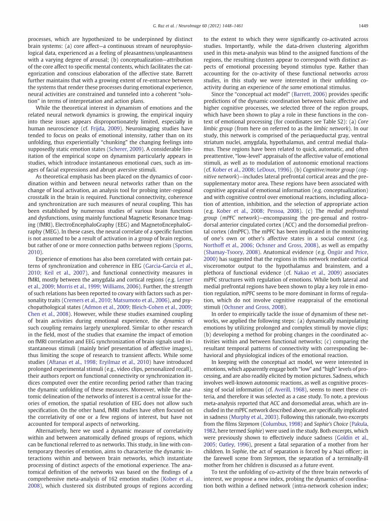
intra-NCI) and between networks (inter-network cohesion index;inter-NCI). Cohesion is measured here in a way that reflects boththe strength of the average correlations between signals in a groupof regions and the variation about this average, with higher valuesfor correlations that are narrowly distributed about a high average.The temporal patterns of the NCIs are then compared with corre-sponding time courses of behavioral and physiological indices ofemotionality (see Fig. 1 for an outline of the procedure).
It was expected that these indices of emotionality will covarywhen testing brain networks, which are associated with emotionalprocessing in general, and sadness in specific. Assuming this couplingto be consistent across the two instances of cinematic sadness induc-tion (i.e. Sophie and Stepmom), we specifically tested the followinghypotheses: (a) the intra-NCI of the limbic and mPFC networks willcovary with behavioral and physiological indices of emotional reac-tion to the films as an indication of their key involvement in thebasic processing of affective information; and (b) the inter-NCI of lim-bic–cognitive networks will correlate with the continuous behavioralindices, indicating enhanced conceptual processing of affect during anintense emotional experience and reduced processing as it wanes.
Methods
Material and procedure
Induction of emotional experienceTwo video excerpts, taken from the commercial films Sophie's
Choice (Pakula, 1982) and Stepmom (Columbus, 1998), were used inthis study. In the scene from Sophie, a mother is forced by a Nazi offi-cer to choose which of her two children lives and which dies. The clipfrom Stepmom included two farewell scenes in which a mother talkswith her children about her future death from a terminal disease.The durations of the clips were 10:00 and 8:27 min, respectively,and their display was preceded and followed by a 3-minute epochduring which the participants passively gazed at an all-black slide.
Retrospective self-reporting of emotional experience
Emotion label rating. An inventory containing 76 emotion labels wascreated on the basis of a comprehensive list of emotion words sug-gested by Shaver et al. (2001). The labels were translated to Hebrewand presented along with their corresponding annotations, adaptedfrom the Rav-Milim Hebrew dictionary (Choueka et al., 1997). Afterscanning, the participants rated the intensity to which they experi-enced each labeled emotion on a seven point Likert-like scale, con-sulting the annotations in case of unclarity.
Continuous emotion rating. The participants continuously reported onshifts in intensity of sadness they have experienced while watchingthe clip during scanning, i.e. retrospectively. The data were acquiredvia designated homemade software. By using the computer-mouse,the subjects indicated changes in their felt intensity of sadness in re-spect with a vertical scale continuously presented on the screen. Thescale included 7 levels of sadness—from neutral to very deep—eachcontaining 3° of change (21° in total). The feedback was sampled atthe rate of 10 Hz.
Electrocardiography recordingElectrocardiography (ECG) was recorded continuously during
scanning via an MRI-compatible system. The sampling rate was
Fig. 1. Analysis approach. (a) Schematic description of the computation of the network coheThe locations of the ROIs encompassing the limbic (red), mPFC (green), and cognitive (bluethalamus; 6 and 7—L and R ventral striatum; 8—central medial thalamus; 9—dorsomedial PFfrontal gyrus; 15 and 16—L and R dorsolateral prefrontal cortex (coordinates in Table S2).Brain Atlas (Talos et al., 2008) registered to the Talairach coordinate system and visualized u
5000 Hz. The measurements were obtained using a BrainAmp ExGMR psychophysiological monitoring system (BrainProducts,Munich,Germany). For each participant, Ag/AgCl electrodes were attached tothe right and left side of the chest.
fMRI acquisitionStructural and functional scans were performed using a GE 3 T
Signa Excite echo speed scanner with an 8-channel head coil, and aresonant gradient echoplanar imaging system. The scanner is locatedat the Wohl Institute for Advanced Imaging at the Tel-Aviv SouraskyMedical Center. A T1-weighted 3D axial spoiled gradient echo(SPGR) pulse sequence (TR/TE=7.92/2.98 ms, flip angle=15º, pixelsize=1 mm, FOV=256×256 mm, slice thickness=1 mm) was ap-plied to provide high-resolution structural images. Functionalwhole-brain scans were performed in an interleaved top-to-bottomorder, using a T2*-weighted gradient echo planar imaging pulse se-quence (TR/TE=3000/35 ms, flip angle=90º, pixel size=1.56 mm,FOV=200×200 mm, slice thickness=3 mm, 39 slices per volume).
General procedure
All the participants in this study were healthy volunteers withoutknown history of neurological or psychiatric disorder with at least12 years of education living in Israel most of their life with Hebrewas their spoken language. All participants signed an informed consentform approved by the ethical committees of the Tel Aviv SouraskyMedical Center. The participants in Experiment 1 additionally signedan informed consent form approved by the department of psychologyat the Tel Aviv University. For a summary of details on the differentexperiments included in this study, see Table S1.
Experiment 1: validation of the behavioral measureTo examine the reliability and validity of our tool for retrospective
moment-to-moment emotion rating we used a test–retest designwith the same rating software. 20 healthy volunteers (13 females;23.18±2.56 years) were randomly and equally assigned to partici-pate in either the reliability or validity protocols and viewed one ofthe clips mentioned above. In both test subgroups, participantswere randomly and equally assigned to view either Sophie's Choiceor Stepmom.
The reliability of the behavioral measure was tested on a groupof 10 participants. These participants went through three sessionsduring which the same clip was presented to them on a computerscreen. Following the first and second sessions, the participantswere asked to fill two personality questionnaires: the Sensitivity toPunishment and Sensitivity to Reward Questionnaire (Torrubia etal., 2001) and the NEO Five-Factor Inventory (Costa and McCrae,1992), also serving as a buffer in this procedure. In the second andthird sessions, but not in the first, the participants were asked to ret-rospectively report on their emotional experience during the firstsession, as described in the main text. Test–retest reliability wasdetermined by calculating the moment-by-moment correlation be-tween the ratings performed during the second and third sessionsover both film clips. Together, these series showed high moment-by-moment correlation (average r=0.93), indicating a considerablereliability.
To examine the construct validity of the tool, we tested whetherthe retrospective emotion rating indeed fits with the participants' re-port of emotional effect during the initial viewing. 10 additional par-ticipants were instructed to rate their felt sadness intensity
sion index, the behavioral index, and the comparison between them (see Methods). (b)) network: 1 and 2—L and R amygdala; 3—periaqueductal gray; 4 and 5 L and R hypo-C; 10—pregenual ACC; 11—rostrodorsal ACC; 12—pre-SMA; 13 and 14—R and L inferiorThe regions are projected on a 3 D model of brain anatomy adopted from the SPL-PNLsing 3D slicer (Pieper et al., 2004) and Maya (Autodesk Inc., San Rafael, CA).

1452 G. Raz et al. / NeuroImage 60 (2012) 1448–1461
throughout two sessions during which the same clip was presented.During the first session, the participants were requested to rate theintensity of sadness they were currently feeling, while in the secondsession they were asked to retrospectively report on sadness experi-enced during the first session. Other instructions were identical tothose given to the other group, as described in the previous para-graph. Between the sessions the participants filled the SPSRQ ques-tionnaire. Moment-by-moment correlation between the on-line andthe retrospective ratings was calculated over both film clips.
Analyzed together, the average moment-by-moment correlationbetween the data series was 0.89, indicating that retrospective ratingconsiderably reflects of reported experience of the initial viewing.These findings are in line with the reports of Hutcherson et al.(2005), who reported an average r of 0.86 between online and retro-spective ratings, which were obtained using a similar method.
Experiment 2: fMRI and retrospective emotional ratingThe fMRI data were acquired from 31 right-handed healthy volun-
teers (16 females; 26.47±4.79 years) during passive viewing of thetwo film excerpts described above, displayed in a counterbalancedorder across participants and intersected by 10 min of anatomicalscans. ECGwas recorded during the scan for 13 of these subjects (7 fe-males; 26.6±5.59 years) as described above (Material and proceduresection). To avoid distraction during film viewing, continuous emo-tional rating was obtained only retrospectively immediately follow-ing scanning in a quiet room using a computer screen and a mousein a similar manner as described above (see Methods).
Experiment 3: physiological characterization of the emotional experienceTo further characterize the physiological reaction to the clips with
regard to retrospective emotional rating, ECG was recorded outsidethe scanner from additional group of 26 healthy volunteers (15 fe-males; 25.23±2.7 years). In addition, emotional rating and labelingwere performed retrospectively in a similar manner to Experiment 2.
Data preprocessing and analysis
Behavioral indices
Emotion label rating. Overall, 51 self-report questionnaires (25 fe-males; 25.75±3.68 years and 23 females; 25.87±3.81 years forSophie and Stepmom, respectively) were included in the analysisafter 5 of the forms were excluded for each film due to incorrect com-pletion. The median values and the frequency of their rating higherthan minimal were computed for each label.
Rated sadness intensity. The rating was collected from 64 (35 females;25.55±3.68 years) and 59 (31 females; 25.87±3.81 years) subjectsacross the three experiments described above. Technical problemsprevented the inclusion of 3 and 8 ratings for Sophie and Stepmom,respectively.
Parasympathetic indexThe high-frequency (0.15 to 0.4 Hz) component of the power
spectrum of heart rate variability (HF–HR) is considered to representan autonomic parasympathetic vagal influence on the sino-atrialnode of the heart (Camm et al., 1996). Significant changes in HF–HRpower were demonstrated during the experience of various emo-tions, including sadness (Kreibig, 2010). We therefore consideredthe fluctuations in this frequency band as indicating an emotional re-sponse. Preprocessing of ECG was done offline using Matlab software(MathWorks Inc.) to yield a continuous heart rate (HR) index, basedon RR interval analysis. The ECG signal was analyzed offline usingMatlab software (MathWorks Inc.) to yield a continuous heart rate(HR) index, based on R–R interval analysis. Artifacts related to theMR gradients were removed from all the ECG datasets using the
FASTR algorithm (Niazy et al., 2005) implemented in FMRIB plug-infor EEGLAB (Delorme and Makeig, 2004) and provided by the Univer-sity of Oxford Centre for Functional MRI of the Brain (FMRIB). Theresulting clean data was downsampled to 250 Hz.
R peak detection using the FMRIB plug-in for EEGLAB. A trained ratermonitored the performance of the algorithm, hand-correcting mis-marking of R peaks. The average correction rate over subjects was0.45% (sd: 1.01%, maximum: 5.78%). The inter-beat intervals or RR in-tervals were obtained as differences between successive R-wave oc-currence times. A sliding window approach was used to correct thetime series for irregular RR intervals. Irregular ectopic RR intervalswere defined as intervals shorter than 2 standard deviations withina window of 21 s. These intervals were considered abnormal beatsand their RR value was set to be the average RR interval within thatwindow. A cubic spline interpolation was used to obtain an equidis-tantly sampled time series of RR intervals.
HF–HR calculation included Hilbert transform (Le Van Quyenet al., 2001; Peng et al., 2004) to explore the dynamics in the high fre-quency component. A time series of the instantaneous power is theparasympathetic index we use in our investigation of HF componentof the RR time series: first the equidistantly sampled RR time serieswas band pass filtered to the HF frequency band (0.15–0.4 Hz), thenthe Hilbert transform was applied to it and the result was squaredto obtain instantaneous power estimate. The obtained HF estimateswere Z-transformed to allow inter-subject comparisons.
Relationships between behavioral and parasympathetic indicesWhen using Spearman's rank test to compare continuous time se-
ries, such as the behavioral and physiological indices, dependenciesbetween sequential samples should be minimized to avoid violationof the assumption of independence among samples. Temporal auto-correlations resulting from the cyclic nature of the signal is a sourceof such dependencies. Since the HF–HR signal was high pass filteredat 0.15 Hz, the longest duration of such a cycle in this signal is1
0:15=6.67 s. In other words, any information with dependency timeconstant higher than 6.67 s was removed from the signal followingthe filtering. However, the information in windows shorter than6.67 s may still be dependent due to the cyclic nature of the signal.In that case, resampling the signal in time windows longer than6.67 guarantees independence. Therefore, the HF–HR signal was aver-aged in non-overlapping windows of 7 s. To allow the comparison ofthe time series of sadness rating with this physiological index, themedian values of the former were also computed in non-overlapping windows of 7 s. The correlations between physiologicaland behavioral data were calculated for each subject, yielding foreach pair of series one value. A two sided Z-test was used to testwhether the correlation between the pair is 0, as Spearman correla-tion values are approximately normally distributed.
fMRI network indices
Preprocessing. Seventeen (9 females in both cases; 26.12±4.66 and26.56±4.66 years in Sophie and Stepmom, respectively) participantswere included for final analysis of the brain data, and 7 participantswere included in the analysis of the simultaneous ECG (4 females;24.14±3.06 and 3 females; 25.93±3.94 years for Sophie and Step-mom, respectively). Since the viewing of the onset of the clip may en-gage emotion-related neural processing associated with appraisal ofthe novelty, and not with its content, the rest epochs and additionalseven first TRs recorded during the film viewing (approximately thetime span of hemodynamic response to the onset of the film) wereexcluded from the statistical analysis.
Preprocessing and statistical analysis were performed using Brain-Voyager (BV) QX version 2.1.2.1545. Head motions were corrected byrigid body transformations, using 3 translation and 3 rotation

1453G. Raz et al. / NeuroImage 60 (2012) 1448–1461
parameters. The middle volume served as a reference in this proce-dure. Trilinear interpolation was applied to detect head motions,which were corrected using sinc interpolation. Slice scan time correc-tion was performed using sinc interpolation. The temporal smoothingprocess included linear trend removal and usage of high pass filter of0.008 Hz. 6 mm FWHM Gaussian spatial smoothing was used to re-duce noise. The voxel size of the SPGR images was standardized to1×1×1 mm using trilinear interpolation. Structural maps were thentransformed into Talairach space and manually coregistered withthe corresponding functional maps.
Brain data of 13 of the participants were excluded from analysisdue to various technical problems during scanning. Head motion ex-clusion criteria were >1.5 mm translational or >1.5° rotation in anyof the axes. For each of the cinematic conditions, one data set was dis-carded due to these criteria. 6 of the ECG data sets for each film werediscarded due to technical problems and poor quality of ECG signal.
Selection of regions of interest (ROIs) was anatomically based onthe work of Kober et al. (2008) with few modifications (see Supple-ments). The final location of regions included in our study and theircoordinates are presented in Fig. 1b and Table S2. The transformationof the MNI coordinates to Talairach space was performed using theBrett transform (Brett et al., 2002). The ROIs were delineated on thebasis of these coordinates, using 6 mm Gaussian smoothing kernel.
Computation of intra-network cohesion index (intra-NCI)To analyze the dynamics of the functional connectivity within
each of the networks, we first extracted the signal of each ROI(signr) from the raw data, using a Gaussian mask with a radius of3 mm around the seed coordinates of the ROI. Next, a set of all pair-wise Pearson correlation values were calculated for each subject, net-work, and time window. Cohesion indices were computed over timewindows of 30 s (10 TRs) in keeping with the findings of an in silicostudy of functional connectivity at multiple time scales (Honey etal., 2007). In this study, the authors simulated dynamic brain activityand estimated the related BOLD signal on the basis of anatomical con-nectivity data. Dynamic time-dependent patterns of functional con-nectivity were observed in the simulated data when sampled insliding windows of 30 s (but not of 240 s). Accordingly, we expectedto find function-related connectivity fluctuations in our data using asimilar time frame.
The correlation values computed over these windows were thentransformed using Fisher's transformation, resulting in values thatare approximately normally distributed, with mean at the indicatedcorrelation. For each time window Δt: [t∗Δ,(t+1)∗Δ) and each sub-ject (sub), sub=1,2,…,N, and each one of the networks k=1,2,3 wedefine the set of Fisher-transformed pair-wise correlations betweenregions i and j belonging to network k
Rsubk Δtð Þ ¼ Rsub
ij Δtð Þ i; j∈kj gn
ð1Þ
Rsubij Δtð Þ ¼ arctanh corr sigi Δtð Þ; sigj Δtð Þ
� �h i: ð2Þ
For each subject, network region nr1 ::nrn∈Kð Þ and time window(Δt) we calculated a network cohesion index (NCIsub
k ), being the t-statistic on the set of R values (Eq. (3)). A right-tailed t-statistic withthe null hypothesis of μR=0 was computed. This measurement takesinto consideration the standard deviation of the correlation values.This was required in order to make sure that a high mean correlationvalue is not the result of a single strong connection.
NCIsubk Δtð Þ ¼ Rsubk Δtð Þ
se Rsubk Δtð Þ� � : ð3Þ
Here we made use of the t-statistic like measure, which is alsoproportional to the inverse of the square root of the coefficient of
variation, as it yields high values when the mean correlation betweenthe regional signals in the network is high and the variability in thecorrelations is low. Note that this measure cannot be used for testingthe hypothesis of 0 correlation, because a BOLD signal across the brainis known to be highly correlated, as are the pairs themselves. It servesmerely as an indicator for both the strength and distribution of thesecorrelations. A different approach was adopted in order to validatethe significance of our downstream results (see below).
Computation of inter-network cohesion index (inter-NCI)Cohesion indices were calculated also for each pair of networks in
the same manner, while considering only correlation values betweenregions that are not in exclusive networks. Once again, the score wascalculated using a right-tailed t-statistic assuming null hypothesis ofmean 0 and unknown variance. For each pair of regions Kk′ ;Kk″
� �we defined:
Rk′ k″ Δtð Þ ¼ Rsubpq Δtð Þ p∈Kk′ ; q∈Kk″ ; k
′≠k″���
o:
nð4Þ
Having defined intra-network and inter-network functional con-nectivity indices, we next examined the relationships between thetemporal pattern of these indices and the corresponding behavioraland physiological indices.
Examining the relationship between NCI and behavioral indexTo fit the sampling method applied on the brain data we
resampled the behavioral data. Median values of the rated sadnesswere computed in time windows of 7 time repetitions (TRs) withan overlap of 6 TRs (21 and 18 s respectively). Spearman ranked cor-relation coefficient (Rs) between the resampled behavioral and thecohesion time series (NCIsub, Kk) were computed for each subject sep-arately. Spearman coefficient was used here due to the fact that therelationship between the values of the emotion rating and BOLD sig-nals need not be linear.
When it came to testing, in order to reduce the dependence be-tween values in different time windows, only samples of non-overlapping time windows were utilized. Calculating Spearman'scorrelation coefficient yielded a correlation value for each subject(sub), sub=1,2…N and network (k), k=1,2,3. Two-tailed Z-testwas used to test whether the average correlation is different from 0,yielding one p-value for each network.
Examining the relationship between NCI and the parasympathetic indexInstantaneous HF estimate was downsampled to allow its compar-
ison with the network cohesion. The signal was averaged across timewindows of 7 time repetitions (TRs) with an overlap of 6 TRs (21 and18 s respectively), using a similar window averaging approach as ap-plied for the NCI. Since only 7 valid ECG recordings of the scannedsubjects were available, and the statistic power that can be achievedwhen performing individual comparisons in such a small samplegroup is low, the NCIs and the parasympathetic index were comparedat the group level (rather than individually as in the case of the be-havioral index). To moderate the effect of outlier data in this smallgroup, the medians rather than the mean values were compared.Spearman's ranked correlation coefficient between the two time se-ries was calculated for each network and between networks. Onlysamples of non-overlapping time windows were compared.
Correction for multiple comparisonsAdjusting for multiplicity, over all the 24 NCI-behavioral and NCI-
parasympathetic comparisons, was done by controlling the false dis-covery rate (FDR) at level 0.05. The procedure used for that purposeis the Benjamini–Hochberg procedure (Benjamini and Hochberg,1995), which is valid even under dependency for normally distribut-ed test statistics as is the case in the current study (Benjamini and

1454 G. Raz et al. / NeuroImage 60 (2012) 1448–1461
Yekutieli, 2001; Reiner-Benaim, 2007). As for the two research ques-tions (on NCI relations with the behavioral index on one side and theparasympathetic index on the other), each comprising of a family of12 hypotheses, the combined evidence per family was derived usingthe Benjamini–Hochberg procedure. Both families were significantso no further selection was needed. In such a case each family canbe tested separately at 0.05 level, and this was done using again theBenjamini–Hochberg procedure. The procedure assures FDR controlon the average over the selected families.
Estimating the spatial specificity of the resultsIn order to examine the extent to which the observed relation-
ships are part of a “whole brain effect” (i.e., their specificity in thebrain space), we compared the resulting correlation values with cor-responding values generated by an identical analysis of a set of ran-dom groups of regions in the gray matter. The original coordinateswere randomly rotated and translocated in a sampling space, whichincluded gray matter voxels. This gray matter mask was generatedby applying a segmentation algorithm (BrainVoyager QX version2.1.2.1545) on the anatomical data collected for the study. The ran-domization of the stereotactic coordinates was carried out in a way,which preserved the Euclidean distances between the nodes of theoriginal network with a precision of 1 mm.
The cohesion indices for the random groups of regions and the be-havioral and physiological indices were compared using a similar pro-tocol as described above. This procedure was repeated for onethousand times. The specificity of the tested result was defined asthe proportion of the random cases with lower Z value (behavioral)or Rs in the sampling space.
Percent signal change computation during limbic–mPFC NCI minimaA complementary post-hoc analysis of the percent of BOLD signal
change was performed to further examine the neural activations co-inciding with dips in limbic–mPFC NCI in the case of Sophie. We ex-amined data recorded during 8 cinematic events, which werereported to elicit at least “minor” intensity of sadness during thefilm viewing. The first time window during which the median ratingof sadness intensity was null, was used as an emotional baseline,thus, the baseline condition included the first ten TRs.
The signals were Z-transformed across all time points, and a meanbaseline signal level was then calculated for each region in the corelimbic and the mPFC networks. The percent signal change was calcu-lated for each network (k), subject (sub) and local minimum (min) asfollows:
PSCk;min;sub ¼1n∑
ni¼1
�Xk;i−�Xk;baseline
� �
�X k;baseline� 100 ð5Þ
where �Xk is the mean level of the BOLD signal within the network k, iis the index of the TR within a certain window, n is the number of TRsin the window (in this case, 7), and �Xk:baseline is the signal in net-work k averaged over the baseline window. A two-tailed t-test wasperformed on the PSC over subjects to assess the significance of theresults. FDR correction was applied to the resulting p values of (16comparisons) comparisons (Benjamini and Hochberg, 1995;Benjamini and Yekutieli, 2001)
Results
Behavioral and physiological characterization of the emotional impact ofthe film excerpts
To identify the most commonly used labels for the emotional ex-perience we used a specially designed inventory administrated to allparticipants (n=51, see Methods). Fig. 2a and Table S3 present the
labels of the emotions that were reported by the participants to bemost intensively experienced during the viewing of each of the ex-cerpts. The highest rate was obtained for “sadness”, “compassion”and “mercy” in both films (5 out of 7 in median, corresponding tothe category “moderate to high intensity”). On the other hand, “sym-pathy” was additionally top-rated only in Stepmom, while “horror”,“hate”, “fear”, and “anger” were reported high (4 out of 7 in median)only in Sophie.
To characterize the dynamic aspects of the subjective emotionalexperience evoked by the films, we recorded retrospective continu-ous reports on sadness intensity during a second viewing of the filmoutside the scanner. Fig. 2b shows the median time course obtainedfor all participants for this rating (across experiments; n=64 andn=59 for Sophie and Stepmom, respectively). The peak intensitylevels indicate an effective induction of sadness, which was signifi-cantly higher for Sophie than for Stepmom (pb0.005, Mann–Whitney,two-tailed test) rating 85.7% (i.e. deep sadness) and 59.5% (i.e. “mod-erate to deep sadness”) of the maximal value, respectively. The emo-tional response to the excerpts differed not only in terms of themaximal values, but also in the patterns of its unfolding in time—amonotonic increase in Sophie vis à vis double-humped shape inStepmom.
As evident in Figs. 2d and e, for each of the films, similar patternswere obtained for participants whose first time viewing had takenplace inside (i.e. Experiment 2; n=16, n=17 in Sophie and Stepmom,respectively) or outside (i.e. Experiments 1 and 3; n=22, n=19 inSophie and Stepmom, respectively) the scanner. The high correlationbetween the median time-courses obtained from these differentstudy groups (Rs=0.95, pb5×10−37, Rs=0.9, pb5×10−23 forSophie and Stepmom, respectively) also indicates a similarity in theiremotional experience.
The behavioral data was compared with the parasympatheticindex for a group who viewed the excerpts outside the scanner (i.e.Experiment 3; n=24, n=19 for Sophie and Stepmom, respectively).The temporal pattern of the parasympathetic index, which wasobtained during the first uninterrupted film viewing is presented inFig. 2c (see Methods for details on calculations), and is overlaidwith the retrospective sadness rating in Fig. 2d. To note, the within-film similarity of parasympathetic patterns, which is evident for thetwo independent groups tested inside or outside the scanner(Rp=0.384, pb0.001 and Rp=0.46, pb0.0002 for Sophie and Step-mom, respectively) indicates the reliability of this measurement.
The individual parasympathetic index and sadness ratings werepositively correlated for Stepmom (Z=3.84, pb0.0002) but not forSophie (Z=−1.90, pb0.06). Importantly, similar relationships(Z=3.46, pb0.0006) were observed for a separate group who viewedStepmom inside the scanner (i.e. Experiment 2).
Correction for multiple comparisons
Adjusting for the multiplicity of the tests of the association betweenthe cohesion index with the other two indices was done by controllingthe false discovery rate at level 0.05 (qBH
FDR≤0.05), using theBenjamini–Hochberg procedure (see Methods, Data preprocessingand analysis section). The comparisons yielded four p-values smallerthan 0.0042, which are statistically significant (for the entire list of p-values, see Table S4). When viewing the research questions as our rele-vant frameof inference, one family of hypotheses about the associationsbetween the cohesion and behavioral indices, and the second familyabout associations between the cohesion and the parasympathetic indi-ces, we find that both are statistically significant after adjusting formul-tiplicity. We can therefore add an analysis within each family, adjustingseparately for multiplicity using the Benjamini–Hochberg procedure.Wefind that afifth hypothesiswith p-value of 0.0163 (on an associationbetween limbic intra-NCI and the behavioral index in the case of Sophie)can also be rejected (see Data preprocessing and analysis section).

a
b
c
d
e
StepmomSophie’s Choice
N=64
N=51 N=51
Rat
ed s
adne
ss in
tens
ity
0
3
69
1215
1821
0 600 N=590 500
Minor
Very Deep
Deep
Moderate to Deep
Moderate
Minor to Moderate
Neutral
n.u
HF-HR inside the magnetRated sadness intensity
0.2
1.8x 10
-5
N=24 N=19
N=24 N=19
N=7 N=7
HF
-HR
pow
er (
ms2
)
Time (seconds)
n.u
0 600 Time (seconds)0 500
HF-HR outside the magnetRated sadness intensity
Z= -1.90 Z= 3.84**
Z= 1.70 Z= 3.46*
Fig. 2. Behavioral and physiological reactions to the film excerpts.(a) Emotion label rating—font size is proportional to the median value of the rating (the maximal size correspondsto the level of 5 out of 7). Only labels rated higher than 1 in median are presented (see Table S3 for more details). (b) Rated sadness intensity—median values (black) and inter-quartile range (blue) of rated sadness intensity. (c) Parasympathetic index (mean±standard error) in a group of subjects whose ECG data were recorded while viewing thefilm excerpts outside the scanner. (d and e) Parasympathetic (mean) and behavioral (median) indices for the groups of subjects whose ECG was recorded outside (d) and inside(e) the scanner. Z values for tests on Rs coefficients between the behavioral and parasympathetic indices are presented in (d). *pb0.006; **pb0.0002.
1455G. Raz et al. / NeuroImage 60 (2012) 1448–1461
Relations between dynamics of NCI and rated sadness intensity and acomplementary percent signal change analysis
A comparison between the temporal patterns of intra-NCI and thebehavioral index revealed both similarity and difference between theemotional states elicited by the two film excerpts.
In line with our first hypotheses, for both films the pattern of therated sadness intensity significantly correlated with the time courseof the limbic intra-NCI (Z=2.40, pb0.02, qBH
FDR≤0.05; Z=2.86,pb0.005, qBH
FDR≤0.05 for Sophie and Stepmom, respectively, Fig. 3ai and ii and Table 1). To test the specificity of this association, a boot-strapping analysis was performed. Corresponding temporal patterns
of network cohesion were computed for 1000 clusters of voxelswhose coordinates were randomly chosen. For each of the randomclusters, the NCI time course was compared with the ratings of sad-ness intensity (see Data preprocessing and analysis section for moredetails). In the case of Sophie, the association between the limbicNCI and the rated sadness intensity was stronger than 99.4% of the as-sociations measured for the randomized clusters, indicating its highspecificity (Fig. S1). A lower specificity of 84.4% for the limbic–behavioral association was found in Stepmom.
Similar analyses were performed for the mPFC and cognitive net-works. The pattern of the rated sadness intensity significantly corre-lated with mPFC intra-NCI in Stepmom but not in Sophie (Z=3.24,

a
b
c
StepmomSophie’s Choice
Rated sadness intensity
Time (seconds) Time (seconds)
Z = 2.40*^ Z = 2.86**^
Z = 3.24**^
Z = 0.79
Z = 0.13
Z = 0.94
n.u
n.u
n.u
n.u
n.u
n.u
Core Limbic network
Limbic intra-NCI
mPFC network mPFC intra-NCI
Cognitive network
Limbic & mPFC networks
Limbic & Cognitive networks
Cognitive intra-NCI
Limbic-mPFC inter-NCI
Limbic-Cognitive inter-NCI
0 600 0 500
Rs = 0.72**^Rs = -0.23
Rs = 0.05Rs = 0.08
Rs =0.50*Rs = 0.52*
HF-HR
d
e
I
I
I
II
III IV
II
II
I II
I II
Fig. 3. Relations between indices. (a–c) Comparison between network cohesion (colored), behavioral (black) and parasympathetic (gray) indices. (d and e) Comparison betweeninter-network cohesion (black) and parasympathetic (gray) indices. The median values of the indices are presented (NCI is smoothed with Loess of 0.08; for presentation of theunsmoothed signal, see Fig. S2). Z and Rs values for tests on individual behavior–NCI and group parasympathetic–NCI comparisons, respectively, are shown. *pb0.05;**pb0.005, ^qBH
FDR≤0.05.
Table 1Z values for tests of individual Spearman's coefficients between rated sadness intensityand network cohesion.
Limbic Cognitive MPF Limbic–cognitive
Limbic–MPF
Cognitive–MPF
Sophie 2.40⁎,^ 0.94 0.13 1.40 1.20 0.42Stepmom 2.86⁎⁎,^ 0.79 3.24^,⁎⁎ 1.94 3.68⁎⁎⁎,^ 1.84
qBHFDR stands for FDR adjusted q-value using the BH procedure (see Methods, fMRI
network indices section)⁎ pb0.05.
⁎⁎ pb0.005.⁎⁎⁎ pb0.0005.
^ qBHFDR≤0.05.
1456 G. Raz et al. / NeuroImage 60 (2012) 1448–1461
pb0.002, qBHFDR≤0.05; and Z=0.13, pb0.89, respectively, Fig. 3b i
and ii). The bootstrapping analysis indicated a 97.2% specificity ofthis association (Fig. S1). For both films there were no significant as-sociations between cognitive intra-NCI and the patterns of reportedsadness (see Fig. 3c and Table 1).
Significant association between the fluctuations of inter-NCI andthe pattern of continuous rated sadness intensity was found to be sig-nificant only in one case: mPFC—limbic inter-NCI in Stepmom(Z=3.68, pb0.0003, qBH
FDR≤0.05; Fig. 4). The specificity of this asso-ciation in the brain space was 97.1% (Fig. S1). In contrast, not onlythat limbic–mPFC inter-NCI were not found to be significantly associ-ated in the case of Sophie, but in fact some dips in the median value of

0 5000 600
mPFC Inter-NCI
Rated sadness intensity
10-1
StepmomSophie’s Choice
Newcomers entering
the Auschwitz concentration
camp
Sophie holds herweeping daughter
and tells a Nazi
she is not Jewish
after an anticipation period, looking at
Sophie's boy
Sophie cries while she tells the Nazis
to take her daughter
The son tells his mother that
he realizes that she is dying
The mother and her son are hugging
The crying mother promises her
daughter that she “will be with her”
after death
The mother and her daughter are
hugging
Fig. 4. Descriptive examination of local extrema in the course of limbic–mPFC inter-NCI. Peaks andminima of interest are numbered at the top panel, and the corresponding brain data andcinematic events are represented below (outer panels). Textual description rather than images are presented due to copyright reasons. More details may be obtained directly from theauthors upon request. The heat maps (at the center) represent matrices of correlations between ROIs (see Fig. 1 for nomenclature) in the corresponding time windows. For each pairof ROIs, the median correlation values are computed over the group of subjects. The inter-NCI computation was based on these matrices. For each pair-wise connection, a t-test wasperformed on the set of Fisher-transformed Rp coefficients, computed across participants with the null hypothesis of μR=0. The lines indicate connections, which were found to be sig-nificantly correlated (pb0.01, FDR corrected) in this test. For video illustration of the changing correlation matrices in relation to cinematic content, see Video S1.
Table 2Spearman r coefficients for correlation between the parasympathetic index and net-work cohesion.
1457G. Raz et al. / NeuroImage 60 (2012) 1448–1461
this neural index were evident especially throughout a period, whichwas reported to induce increased sadness intensity.
To further examine these events of uncoupling, we tested which ofthe networks activates/deactivates during these dips in Sophie. Acomplementary post-hoc analysis of the percent of BOLD signalchange within the limbic and the mPFC networks during local minimaof limbic–mPFC inter-NCI was performed (see Supplements for moredetails on analysis, and Fig. S3 for the selected time points). Of 8 min-ima analyzed for Sophie, 3 significant (qBH
FDRb0.05) elevations ofcore limbic activity were found. One significant activation (qBH-FDRb0.05) of the mPFC network was also observed in the case of an-other limbic–mPFC inter-NCI minimum.
Limbic Cognitive MPF Limbic–cognitive
Limbic–MPF
Cognitive–MPF
Sophie −0.23 0.34 0.36 0.52⁎ 0.08 0.45Stepmom 0.72⁎⁎,^ −0.04 −0.37 0.50⁎ 0.05 0.30
⁎ pb0.05.⁎⁎ pb0.005.^ qBH
FDR≤0.05.
Relations between dynamics of NCI and the parasympathetic index
The limbic intra-NCI, which was significantly associated with thebehavioral index in the case of Stepmom, also significantly correlatedwith the parasympathetic index for that film excerpt, but not for
Sophie (Rs=0.72, pb0.003, qBHFDR≤0.05; Rs=−0.23, pb0.35, re-
spectively, Fig. 3a iii and iv and Table 2). This association in Stepmomwas found to be highly specific in the brain space, as lower NCI-parasympathetic associations were found in 99.8% of the randomizedclusters (Fig. S1).
Lastly, for both films a correlation between cognitive–limbic inter-NCI and the parasympathetic index was observed at pb0.05(Rs=0.50 and Rs=0.52 in Stepmom and Sophie, respectively),

1458 G. Raz et al. / NeuroImage 60 (2012) 1448–1461
showing considerable specificity (95.7% and 97.9% in Stepmom andSophie, respectively). However, both correlations did not survive thecorrection for multiple comparisons.
Discussion
In line with the growing emphasis of theories of emotion on thedynamism of synchronization within and between distinct neuralnetworks, we propose a novel approach for a multi-layered character-ization of emotional experiences as they unfold. The findings pre-sented above point to the potential of this approach to revealcommonalities, as well as key specificities, of emotional experiencesinduced by prolonged stimuli. Correspondences in results across thebehavioral, autonomic and brain levels suggest a rich and complex,but also reconcilable, picture of the processes taking place duringthe emotional experiences elicited by the two film excerpts used inthis study.
Multi-layered dynamics of emotional response related to the limbicnetwork
The theme of separation of a mother from her children, which ispresented in both of the film scenes, was commonly reported byviewers to induce primarily “sadness” in both cases (Fig. 2a). Accord-ingly, for both films the development of the rated sadness intensityover time significantly correlated with the temporal pattern of cohe-sion within the limbic network. To the extent to which signal similar-ity between regions indicates their coordinated functioning, thesefindings support our first hypothesis, based on the conceptual actmodel (Barrett, 2006). In both experimental conditions, dynamics ofcohesion within the limbic network, which is assumed to mediatecore aspects of emotion including the evaluation of the affectivevalue of the stimulus and the triggering of bodily responses to it,was associated with the rated sadness intensity level. Our findingssuggest that the involvement of this limbic network in affective pro-cessing crosses experimental cinematic conditions, which arereported to differ in the mixture of emotions they elicit. It is stillopen to further investigation whether the relation found betweenthe limbic cohesion and the aware emotional rating contradictsprior work showing that the amygdala activity and connectivity ismost prominent during implicit processing (e.g. Williams, 2006).
When parasympathetic index is considered, a difference betweenthe cinematic experiences arises. While in Stepmom, this autonomicindex significantly correlated with the time courses of both therated sadness and the limbic cohesion, no such associations werefound for Sophie. These findings may be interpreted in light of an ob-servation made in a recent meta-analytical study (Kreibig, 2010),according to which the widely-shared lay concept of sadness con-flates at least two distinct major patterns of autonomic modulations:an “activating sadness response”, which implicates increased cardio-vascular sympathetic control, versus a “deactivating sadness re-sponse” (observed in most of the studies that induce sadness usingfilms) involving a decrease in cardiac activity, mostly driven by aparasympathetic control. The limbic–parasympathetic–behavioral as-sociation in Stepmom indicates that the emotional experience elicitedby this film excerpt better fits with the later “parasympathetic mode”of sadness. This interpretation is congruent with the emotion labelingprofiles of these excerpts. In the case of Stepmom, sadness wasreported by the participants to commingle with the related feelingsof sympathy and affection. Notably, the tendency to sympathizewith fictional characters during film viewing was previously relatedto parasympathetic activity (Fabes et al., 1993). Alternatively, emo-tions of horror, fear, and anger, which are associated with an immedi-ate urge to act, were reported to accompany feelings of sadness whileviewing Sophie. Further studies could examine the involvement of thesympathetic system in such a case.
Multi-layered dynamics of emotional responses related to the mPFCnetwork cohesion
Another key difference between the two experimental conditionsregards behavioral and brain aspects of social cognition. “Sympathy”,which was rated high in Stepmom but not Sophie, denotes in the in-ventory we used “fondness; positive attitude; sharing the emotionsof the other”. We therefore suspect that its elicitation involved pro-cesses of social cognition during the film viewing. This may corre-spond with the finding of a significant positive correlation betweenthe mPFC intra-NCI and the rated sadness intensity only in Stepmom.The emergence of this coupling when witnessing the sorrow of othersfits with a body of evidence which links medial prefrontal structureswith empathic processing. A number of neuropsychiatric, neuroimag-ing and lesion studies have indicated that more ventral aspects of themPFC have a key role in appreciation of emotional states of the other(affective theory of mind), while a more dorsal locus is also involvedin mentalizing the state of another person, possibly a more cognitiveaspect of empathic processing (Shamay-Tsoory, 2008). Lastly, thedorsal ACC has been implicated in appraisal and expression of nega-tive affects (Etkin et al., 2010a). Thus, the correspondence betweenthe mPFC–NCI and the rated sadness indicates that the affective re-sponse in Stepmom may be guided by the encoding of the character'semotional states and the evaluation of relevance to the viewer.
Since the empathy-related emotions of “Compassion” and “Mercy”were also rated high in Sophie, it is not clear why mPFC–limbic cou-pling with sadness rating was not evident in this case. While a directexamination of this question is yet to be carried out, the differencebetween the ways in which the loss is presented in the films may pro-vide a clue. In Stepmom, the separation of the mother from her chil-dren is a future imagined event, while in Sophie it is overtlypresented as an event which is taking place at present. In the firstcase, sadness intensification possibly coincides with mentalizing pro-cesses, while in the second it may couple with the process of “senso-rimotor resonance”, which relies on brain circuits related toprocessing of empathy through simulation, including the pars oper-cularis and the anterior insula (Lamm et al., 2011). Specifically, thisapparent neuro-behavioral difference between Stepmom and Sophiemay align with the empirically supported distinction between empa-thy systems driven by cognitive and somatic related processes(Shamay-Tsoory, 2008) with a prominent role of the mPFC only inthe former.
Ebb and flow of the cross talk between prefrontal cortical networks
At odds with our second hypothesis, the behavioral index was notfound to be significantly associated with cognitive–limbic inter-NCI ineither of the cinematic conditions. Thus, the findings do not supportthe notion that the reported intensification of emotion is necessarilyassociated with an increasing correlativity between the limbic andthe cognitive networks. It is possible that the relations between theinter-network cohesion and the awareness of emotional intensityare non-linear or non-monotonic (see the descriptive “reverse engi-neering” analysis presented below), and therefore the regressionmethod adopted in this study fails to capture such an effect. Alterna-tively, cognitive and limbic processing may cohere linearly for shorttime intervals following emotional stimuli, and then quickly de-coordinate. Such a process is largely unobservable in fMRI-basedfunctional connectivity analysis due to the limited temporal resolu-tion of this technique, and call for further investigation via EEG/MEG-based methods.
While the temporal pattern of limbic–cognitive inter-NCI was notfound to be related to the time course of reported sadness intensity,we did find indications for its association with parasympathetic activ-ity. For both movies, this association showed high specificity in thebrain space, though its significance at the 0.05 level did not survive

1459G. Raz et al. / NeuroImage 60 (2012) 1448–1461
correction for multiple comparisons. While further examination ofthis association in a larger group of subjects is needed, our results in-dicate a possible coupling between the parasympathetic reaction andthe dynamics of cognitive–limbic crosstalk. This coupling may reflecta dependency of a cognitive processing of limbic information on para-sympathetic modulation of arousal. Alternatively, parasympatheticreaction to the cinematic content may result from top-down corticalprocesses mediated by lateral prefrontal structures, included in thecognitive network. In line with this interpretation is a study, whichdemonstrated that cognitive re-appraisal of emotional stimuli in-volves the coupling of apparent regulatory effect of the ventrolateralPFC on the amygdala with parasympathetically-mediated constrictionof the pupil (Johnstone et al., 2007).
An examination of the case of the coordination between the medi-al PFC and the limbic networks indicates additional dissimilarity be-tween the cinematic experiences elicited by the two films. Thisassociation between the limbic–mPFC inter-NCI and the behavioralindex was statistically significant in Stepmom, but not in Sophie(Fig. 4). When examining the data from a descriptive perspective,the peaks of the rated sadness intensity temporally coincide withpeaks of limbic–mPFC inter-NCI in Stepmom. The corresponding cine-matic events include notable moments of mother–child physicalbonding (see Fig. 4 and Supplements for details).
While a peak of limbic–mPFC inter-NCI also corresponds to anearly phase of rated sadness intensification in Sophie (as Sophie andher children arrive to the concentration camp), other major reportedemotional events are not accompanied with such a global peak. Infact, prominent limbic–mPFC inter-NCI local minima in Sophie coin-cide with significant dramatic events (see Fig. 4 and supplements).
What may be the functional meaning of such local minima andpeaks of inter-NCI? Anatomically, the medial PFC and structures ofthe limbic network are directly connected: highly ordered innerva-tions of specific hypothalamic and PAG substructures have beenmapped in detail in the rat's brain (Floyd et al., 2000, 2001); other an-imal studies have also shown that the ventromedial prefrontal cortex(VMPFC) and dorsal aspects of the ACC project to the nucleus accum-bens and adjacent areas in the ventral striatum (Ernst and Fudge,2009) and dense projections reciprocally connect the mPFC and theACC with the amygdala (Kim et al., 2011).
In terms of the functionality of such connections, accumulating ev-idence from human neuroimaging studies indicates that the DMPFC,the dorsal extension of the VMPFC, and the ACC are activated whenone appraises an affective state, probably through interaction withcore limbic structures (Barrett et al., 2007; Ochsner and Gross,2008). This interaction is thought to mediate introspection and mon-itoring of one's own stream of feelings (Barrett et al., 2007), and ac-cordingly hypo-activity of the DMPFC and ACC was shown to beassociated with alexithymia, a deficiency in the ability to attend emo-tional states (Aleman, 2005; Heinzel et al., 2010).
Another explanation may be derived from a line of research,which has related this limbic cortical connection to the process ofemotional regulation. This assumption has been based on animalstudies demonstrating an inhibitory effect of medial PFC projectionson output from the central nucleus of the amygdala (Milad andQuirk, 2002; Quirk et al., 2003). Furthermore, a line of neuroimagingstudies in humans has reported that successful regulation of emotioninvolves simultaneous increase in mPFC and ACC activity along withdecreased amygdala (Kim et al., 2011), and ventral striatum activity(Phan et al., 2005; Staudinger et al., 2009).
In contrast to the congruency of the reports on medial PFC andlimbic activity relative to baseline conditions, evidence of their func-tional connectivity during emotional regulation is less clear in termsof the direction of the correlativity. Increased coupling between theamygdala and the DMPFC/pregenual cortex in this condition wasdemonstrated (Banks et al., 2007; Erk et al., 2010). Moreover, thisinter regional connectivity was stronger for subjects who reported
successful reduction of negative affect (Banks et al., 2007), andweaker for patients with a major depressive disorder (Erk et al.,2010). On the other hand, several fMRI studies of emotion regulationreported on negative correlativity between the amygdala and the ros-tral ACC/pregenual cortex—an effect which is not evident among anx-iety disorder patients (Etkin et al., 2006, 2010b).
Considering the contradicting evidence reviewed above, limbic–mPFC inter-NCI minima could reflect either down-regulation of lim-bic activity via mPFC projections or decoupling of these networks ina state of under-regulation. In order to examine these options, a com-plementary post-hoc calculation of the percent of signal change in themPFC and core limbic networks was obtained for Sophie (Fig. S3). Theanalysis revealed significantly increased core limbic signal (but not ofmPFC activation) for three out of five limbic–mPFC NCI minima, ob-served during the interaction between Sophie and the Nazi officer.To note, two of these significant effects were measured at the climaxof the scene, as indicated by the rated sadness intensity (Fig. 4, Fig.S3). One case of mPFC (but not limbic) increased activation wasfound in an earlier time point, corresponding to the presentation ofa line of newcomers to the Auschwitz concentration camp (see Fig.S3).
Howmay these findings contribute to the interpretation of mPFC–limbic NCI dips, coinciding with highly emotional events in Sophie? Arecent review on post-traumatic stress disorder (PTSD)-related pa-thologies of emotion modulation (Lanius et al., 2010) may provide aclue. Based on functional imaging studies, Lanius and colleagues dis-tinguish between two forms of emotion dysregulation in PTSD:hyperarousal of limbic regions due to their under-regulation bymPFC structures, and disengagement with emotional content, whichis entailed with excessive inhibition of limbic activity following acti-vation of the mPFC. Furthermore, another recent review, which focus-es on studies of emotion regulation (Ochsner and Gross, 2008),suggests that medial (rather than lateral) PFC regions are more activewhen subjects utilize methods of detachment and distancing. Thus,the combination of mPFC–limbic discoordination with the mPFC acti-vation during the presentation of the entrance of the Jews to theAuschwitz concentration camp (see the upper image from Sophie inFig. 4) may reflect emotion regulation via disengagement while cop-ing with this cinematic content. On the other hand, the dips coincid-ing with limbic activation, especially during the event of forcedseparation of Sophie from her daughter (see the lower image fromSophie in Fig. 4), may indicate unregulated limbic bursts, while facinga traumatic content.
In summary, the significant correlation between the limbic–mPFCinter-NCI and the emotional rating in Stepmom indicates that in thiscase, sadness involves regulated processes of mentalization and intro-spection. Together with the evidence of the associations betweenbrain, behavior and parasympathetic indices, the findings suggestthat the emotional experience elicited by this film excerpt corre-sponds with a parasympathetic profile of “deactivated sadness”, gov-erned by cognitive processes. Conversely, it is possible that abrupteffects of dysregulated limbic arousal and dissociation drive emotion-al experience in key moments of Sophie, resulting in non-monotonicrelations between the time course of the cohesion and the reportedemotional intensity.
Caveats and future directions
The strength of the reported associations, although significant, ismodest in some cases. This shortcoming may result for various rea-sons, which should be taken into account in a future work. First, aspreviously mentioned, the behavioral, physiological and neural indi-ces may also be related in non-linear and non-monotonic fashions,producing effects which were undetected by our methods. Further,time-shifts between the indices may appear due to causal relation-ships and other factors, such as hemodynamic delay.

1460 G. Raz et al. / NeuroImage 60 (2012) 1448–1461
Accounting for the hemodynamic delay between a certain stimu-lus and the peak of the signal, which reflect a response to it, is a com-mon practice in studies that test the levels of BOLD signal. However,BOLD correlativity may behave differently in time. The signals of twohypothetical ROIs, which are reactive to a certain stimulus, may be-come correlated immediately in response to it and maintainedwhile the hemodynamic reaction is reaching its peak and afterwards.In the absence of an established assumption about the nature of thetemporal alignment of the cohesion and behavioral indices, no lag be-tween the indices was introduced in the current study. However, thisissue is to be examined in future work. As for the temporal alignmentof the physiological and behavioral indices, shifting forward the HF–HR time series seems to be theoretically plausible. The parasympa-thetic response may at least partially reflect pre-attentive processingof an emotional input, whether in a form of a reflexive and sub-cognitive reaction in Jamesian notions or as a prompt result of aprimary non-conscious appraisal. The conscious assessment of theemotional intensification may accordingly lag after this physiologicalreaction. Thus, the temporal relations between the rated emotionalintensity and the HF–HR index have yet to be explored.
Furthermore, while the correlations between the indices were com-puted over the entire experimental epoch, significant relationsmay lastfor shorter time periods. Thus, additional analysis methods (e.g. cross-correlation and spectral methods), which are sensitive for such effects,may offer complementary information on the relationships betweenthese processes. Moreover, the anatomical delineation of the regionsof interest in this work relied on meta-analytical data, which was aver-aged over many brains. This method of functional localization is insen-sitive to individual differences. A semi-supervised approach fordelineating seed regions on the basis of signal similarity at the individ-ual level may improve such sensitivity.
Regardless, the behavioral, physiological, and brain indices shouldnot be expected to fully overlap, since they provide information ondifferent and nonsynonymous aspects of the emotional experience.Therefore, rather than expecting full correspondence between the in-dices, it seems more productive to treat them as related componentsof a complex image of emotional experience. The examination of thevariability in these sets of relations accordingly provides importantclues regarding the uniqueness of certain emotional states.
The duration of the time-frame in which functional connectivity isexamined should also be taken into consideration. While in this work,connectivity fluctuations were monitored at a time scale of seconds,inter-network synchronization events, triggered by processing ofemotion-related information occur within milliseconds. Methods forintracranial measurement of neural activity provide the opportunityto trace such events. Slower fluctuations in network synchronizationmeasured intracranially may be compared with cohesion indices,computed for a similar cinematic stimulus on the basis of fMRI data.
As for the lower specificity of the measured effects in the brainspace in the case of the limbic NCI in Stepmom, it may indicate awide-scale association between inter-regional correlativity and thespecific temporal patterns of the reported sadness. It is possible thatthis finding results from a global pattern of enhanced multi-systemic synchrony in moments of high emotional intensity. Sinceemotional arousal involves the activation of multiple processes, itmay also increase the functional correlativity within other relevantcircuits. In that case, a global effect is expected to overshadow thespecific effect in the tested network.
Finally, it is worth noting the possibility of meaningful emotionalevents, which are neurally represented not as a unidirectional in-crease or decrease in the cohesion index of a specific network, butrather as a “split” of the network into sub-components with opposingpatterns of functional connectivity. An intra-network cohesion index,which relates to the entire network, will probably fail to capture sucheffects (although it should be evident when the correlation matricesof the networks are examined, as in Fig. 4). Indeed, similarly to
other research methods, the approach presented above is limited bythe definition of the elementary unit of analysis—the single networkin this case. Thus, theoretically or empirically justifiable hypothesesregarding patterns of dynamics of functional connectivity betweensub-components of a certain network (e.g. striatal structures vs.amygdala in the core limbic network) may be tested using the sameanalysis protocol with re-defined sets of ROIs.
Conclusions
In keeping with contemporary theoretical concerns in the field ofemotion research, this work presents a new approach for monitoringthe dynamics of connectivity within and between functional net-works. Tested on two sadness inducing films, the subjective feelingwas significantly coupled with the cohesion of a core limbic networkfor both excerpts. However, the limbic cohesion and the rated sadnesscorrelated with the parasympathetic index only in Stepmom, suggest-ing a specific autonomic profile for the emotional state elicited by thismovie. Unlike the case of the limbic cohesion, the relations betweenthe mPFC cohesion and the rated sadness varied across cinematicconditions, providing insights about the uniqueness of the neural dy-namics as related to affective-social processing. Lastly, the cohesionbetween the limbic and mPFC networks demonstrated either integra-tion or disintegration at points of prominent dramatic events duringthe movies. Only in Stepmom did this correlate with the behavior,pointing to different dynamics of emotional regulation during theviewing of these films. Together, the findings suggest that unravelingthe relations between various indices of emotional response may con-tribute to the understanding of the individual ways in which we ex-perience emotions.
Supplementary materials related to this article can be found on-line at doi:10.1016/j.neuroimage.2011.12.084.
Acknowledgments
This work was supported by the University of Chicago's Arete Ini-tiative - Science of Virtues Program (TH), a grant from the U.S. De-partment of Defense award number W81XWH-11-2-0008 (TH), theDan David Scholarship Award (GR), and Human Frontier Science Pro-gram Grant RGP0054/2004-C, by European Commission-fundedCOBOL Contract 043403 (TH,GR). The authors thank Yoav Benjamini(Department of Statistics and Operations Research, Tel Aviv Universi-ty) for helpful statistical consultation, and Gali Ellenblum and Tal Nis-senson for conducting Experiment 3.
References
Admon, R., Lubin, G., Stern, O., Rosenberg, K., Sela, L., Ben-Ami, H., Hendler, T., 2009.Human vulnerability to stress depends on amygdala's predisposition and hippo-campal plasticity. Proc. Natl. Acad. Sci. 106, 14120.
Aftanas, L.I., Lotova, N.V., Koshkarov, V.I., Popov, S.A., 1998. Non-linear dynamical cou-pling between different brain areas during evoked emotions: an EEG investigation.Biol. Psychol. 48, 121–138.
Aleman, A., 2005. Feelings you can't imagine: towards a cognitive neuroscience of alex-ithymia. Trends Cogn. Sci. 9, 553–555.
Averill, J.R., 1968. Grief: its nature and significance. Psychol. Bull. 70, 721–748.Banks, S.J., Eddy, K.T., Angstadt, M., Nathan, P.J., Phan, K.L., 2007. Amygdala frontal con-
nectivity during emotion regulation. Social cognitive and affective neuroscience 2,303–312.
Barrett, L.F., 2006. Are emotions natural kinds? Perspect. Psychol. Sci. 1, 28.Barrett, L.F., 2009. Variety is the spice of life: a psychological construction approach to
understanding variability in emotion. Cognit. Emot. 23, 1284–1306.Barrett, L.F., Mesquita, B., Ochsner, K.N., Gross, J.J., 2007. The experience of emotion.
Annu. Rev. Psychol. 58, 373.Benjamini, Y., Hochberg, Y., 1995. Controlling the false discovery rate: a practical and
powerful approach to multiple testing. J. R. Stat. Soc. B Methodol. 289–300.Benjamini, Y., Yekutieli, D., 2001. The control of the false discovery rate in multiple
testing under dependency. Ann. Stat. 1165–1188.Bleich-Cohen, M., Strous, R.D., Even, R., Rotshtein, P., Yovel, G., Iancu, I., Olmer, A.,
Hendler, T., 2009. Diminished neural sensitivity to irregular facial expression infirst-episode schizophrenia. Hum. Brain Mapp. 30, 2606–2616.

1461G. Raz et al. / NeuroImage 60 (2012) 1448–1461
Brett, M., Johnsrude, I.S., Owen, A.M., 2002. The problem of functional localization inthe human brain. Nat. Rev. Neurosci. 3, 243–249.
Camm, A.J., Malik, M., Bigger, J.T., Breithardt, G., Cerutti, S., Cohen, R.J., Coumel, P., Fallen,E.L., Kennedy, H.L., Kleiger, R.E., et al., 1996. Heart rate variability: standards of mea-surement, physiological interpretation, and clinical use. Circulation 93, 1043–1065.
Cannon, W.B., 1929. Bodily changes in pain, hunger, fear and rage. South. Med. J. 22, 870.Chen, S.S., Tu, P.C., Su, T.P., Hsieh, J.C., Lin, Y.C., Chen, L.F., 2008. Impaired frontal syn-
chronization of spontaneous magnetoencephalographic activity in patients withbipolar disorder. Neurosci. Lett. 445, 174–178.
Choueka, Y., Freidkin, U., Hakohen, H.A., Zachi-Yannay, Y., 1997. Rav Milim: A Compre-hensive Dictionary of Modern Hebrew. Center for Educacional Technology. Israel:Bar-Ilan University, Miskal.
Columbus, C., 1998. Stepmom.Costa Jr., P.T., McCrae, R.R., 1992. Revised neo personality inventory (neo pi-r) and neo
five-factor inventory (neo-ffi).Cremers,H.R., Demenescu, L.R., Aleman, A., Renken, R., van Tol,M.J., van derWee,N.J., Velt-
man, D.J., Roelofs, K., 2010. Neuroticismmodulates amygdala–prefrontal connectivityin response to negative emotional facial expressions. Neuroimage 49, 963–970.
Damasio, A.R., Everitt, B.J., Bishop, D., 1996. The somatic marker hypothesis and thepossible functions of the prefrontal cortex. Philos. Trans. R. Soc. Lond. B Biol. Sci.351, 1413–1420.
Delorme, A., Makeig, S., 2004. EEGLAB: an open source toolbox for analysis of single-trial EEG dynamics including independent component analysis. J. Neurosci.Methods 134, 9–21.
Erk, S., Mikschl, A., Stier, S., Ciaramidaro, A., Gapp, V., Weber, B., Walter, H., 2010. Acuteand sustained effects of cognitive emotion regulation in major depression. J. Neu-rosci. 30, 15726.
Ernst, M., Fudge, J.L., 2009. A developmental neurobiological model of motivated be-havior: anatomy, connectivity and ontogeny of the triadic nodes. Neurosci. Biobe-hav. Rev. 33, 367–382.
Eryilmaz, H., Van De Ville, D., Schwartz, S., Vuilleumier, P., 2010. Impact of transientemotions on functional connectivity during subsequent resting state: a waveletcorrelation approach. NeuroImage 3, 2481–2491.
Etkin, A., Egner, T., Peraza, D.M., Kandel, E.R., Hirsch, J., 2006. Resolving emotional con-flict: a role for the rostral anterior cingulate cortex in modulating activity in theamygdala. Neuron 51, 871–882.
Etkin, A., Prater, K.E., Hoeft, F., Menon, V., Schatzberg, A.F., 2010a. Failure of anteriorcingulate activation and connectivity with the amygdala during implicit regulationof emotional processing in generalized anxiety disorder. American Journal of Psy-chiatry 167, 545–554.
Etkin, A., Prater, K.E., Hoeft, F., Menon, V., Schatzberg, A.F., 2010b. Failure of anteriorcingulate activation and connectivity with the amygdala during implicit regula-tion of emotional processing in generalized anxiety disorder. Am. J. Psychiatry167, 545.
Fabes, R.A., Eisenberg, N., Eisenbud, L., 1993. Behavioral and physiological correlates ofchildren's reactions to others in distress. Dev. Psychol. 29, 655.
Floyd, N.S., Price, J.L., Ferry, A.T., Keay, K.A., Bandler, R., 2000. Orbitomedial prefrontalcortical projections to distinct longitudinal columns of the periaqueductal gray inthe rat. J. Comp. Neurol. 422, 556–578.
Floyd, N.S., Price, J.L., Ferry, A.T., Keay, K.A., Bandler, R., 2001. Orbitomedial prefrontalcortical projections to hypothalamus in the rat. J. Comp. Neurol. 432, 307–328.
Frijda, N.H., 1986. The Emotions. Cambridge Univ Pr, Cambridge.Frijda, N.H., 2009. Emotions, individual differences and time course: reflections. Cognit.
Emot. 23, 1444–1461.Garcia-Garcia, M., Yordanova, J., Kolev, V., Domínguez-Borrās, J., Escera, C., 2010. Tun-
ing the brain for novelty detection under emotional threat: the role of increasinggamma phase-synchronization. Neuroimage 49, 1038–1044.
Goldin, P.R., Hutcherson, C.A., Ochsner, K.N., Glover, G.H., Gabrieli, J.D., Gross, J.J.,2005. The neural bases of amusement and sadness: a comparison of block con-trast and subject-specific emotion intensity regression approaches. Neuroimage27, 26–36.
Heinzel, A., Schäfer, R., Müller, H.W., Schieffer, A., Ingenhag, A., Eickhoff, S.B., Northoff, G.,Franz, M., Hautzel, H., 2010. Increased activation of the supragenual anterior cingulatecortex during visual emotional processing in male subjects with high degrees ofalexithymia: an event-related fMRI study. Psychother. Psychosom. 79, 363–370.
Honey, C.J., Kötter, R., Breakspear, M., Sporns, O., 2007. Network structure of cerebralcortex shapes functional connectivity on multiple time scales. Proc. Natl. Acad.Sci. 104, 10240–10245.
Hutcherson, C.A., Goldin, P.R., Ochsner, K.N., Gabrieli, J.D., Barrett, L.F., Gross, J.J., 2005.Attention and emotion: does rating emotion alter neural responses to amusing andsad films? Neuroimage 27, 656–668.
James, W., 1884. What is an emotion? Mind 9, 188–205.Johnstone, T., van Reekum, C.M., Urry, H.L., Kalin, N.H., Davidson, R.J., 2007. Failure to
regulate: counterproductive recruitment of top-down prefrontal-subcortical cir-cuitry in major depression. J. Neurosci. 27, 8877.
Keil, A., Stolarova, M., Moratti, S., Ray, W.J., 2007. Adaptation in human visual cortexas a mechanism for rapid discrimination of aversive stimuli. Neuroimage 36,472–479.
Kim, M.J., Loucks, R.A., Palmer, A.L., Brown, A.C., Solomon, K.M., Marchante, A.N., Whalen,P.J., 2011. The structural and functional connectivity of the amygdala: from normalemotion to pathological anxiety. Behavioural Brain Research 223, 403–410.
Kober, H., Barrett, L.F., Joseph, J., Bliss-Moreau, E., Lindquist, K., Wager, T.D., 2008. Func-tional grouping and cortical–subcortical interactions in emotion: a meta-analysisof neuroimaging studies. Neuroimage 42, 998–1031.
Kreibig, S.D., 2010. Autonomic nervous system activity in emotion: a review. Biol. Psy-chol. 84, 394–421.
Lamm, C., Decety, J., Singer, T., 2011. Meta-analytic evidence for common and distinctneural networks associated with directly experienced pain and empathy for pain.Neuroimage 54, 2492–2502.
Lanius, R.A., Vermetten, E., Loewenstein, R.J., Brand, B., Schmahl, C., Bremner, J.D.,Spiegel, D., 2010. Emotion modulation in PTSD: clinical and neurobiological evi-dence for a dissociative subtype. Am. J. Psychiatry 167, 640.
Lazarus, R.S., Folkman, S., 1984. Stress, Appraisal, and Coping. Springer PublishingCompany.
Le Van Quyen, M., Foucher, J., Lachaux, J.P., Rodriguez, E., Lutz, A., Martinerie, J., Varela,F.J., 2001. Comparison of Hilbert transform and wavelet methods for the analysis ofneuronal synchrony. J. Neurosci. Methods 111, 83–98.
LeDoux, J., 1996. The Emotional Brain. New York.Lerner, Y., Papo, D., Zhdanov, A., Belozersky, L., Hendler, T., 2009. Eyes wide shut:
amygdala mediates eyes-closed effect on emotional experience with music. PLoSOne 4, e6230.
Lewis, M.D., 2005. Bridging emotion theory and neurobiology through dynamic sys-tems modeling. Behav. Brain Sci. 28, 169–194.
MacLean, P., 1955. The limbic system (‘visceral brain’) and emotional behavior. Arch.Neurol. Psychiatry 73, 130.
Matsumoto, A., Ichikawa, Y., Kanayama, N., Ohira, H., Iidaka, T., 2006. Gamma band ac-tivity and its synchronization reflect the dysfunctional emotional processing inalexithymic persons. Psychophysiology 43, 533–540.
Mesquita, B., Barrett, L.F., Smith, E.R., 2010. Emoting: a contextualized process. Themind in context, pp. 83–104.
Milad, M.R., Quirk, G.J., 2002. Neurons in medial prefrontal cortex signal memory forfear extinction. Nature 420, 70–74.
Morris, J.S., Öhman, A., Dolan, R.J., 1999. A subcortical pathway to the right amygdalamediating “unseen” fear. Proc. Natl. Acad. Sci. 96, 1680–1685.
Murphy, F.C., Nimmo-Smith, I., Lawrence, A.D., 2003. Functional neuroanatomy of emo-tions: a meta-analysis. Cogn. Affect. Behav. Neurosci. 3, 207.
Nakao, T., Takezawa, T., Miyatani, M., Ohira, H., 2009. Psychologia 52, 93–109.Niazy, R.K., Beckmann, C.F., Iannetti, G.D., Brady, J.M., Smith, S.M., 2005. Removal of
FMRI environment artifacts from EEG data using optimal basis sets. Neuroimage28, 720–737.
Northoff, G., Heinzel, A., de Greck, M., Bermpohl, F., Dobrowolny, H., Panksepp, J., 2006.Self-referential processing in our brain—a meta-analysis of imaging studies on theself. Neuroimage 31, 440–457.
Oatley, K.G., 1996. Emotions, rationality and informal reasoning. In: Oakhill, J.,Garnham, A. (Eds.), Mental Models in Cognitive Science: Essays in Honour of PhilJohnson-Laird. Psychology Press, East Sussex, pp. 175–196.
Ochsner, K.N., Gross, J.J., 2008. Cognitive emotion regulation. Curr. Dir. Psychol. Sci. 17,153–158.
Öngür, D., Price, J.L., 2000. The organization of networks within the orbital and medialprefrontal cortex of rats, monkeys and humans. Cereb. Cortex 10, 206–219.
Pakula, A.J., 1982. Sophie's Choice.Papez, J.W., 1937. A proposed mechanism of emotion. Arch. Neurol. Psychiatry 38, 725.Peng, C.K., Henry, I.C., Mietus, J.E., Hausdorff, J.M., Khalsa, G., Benson, H., Goldberger,
A.L., 2004. Heart rate dynamics during three forms of meditation. Int. J. Cardiol.95, 19–27.
Pessoa, L., 2008. On the relationship between emotion and cognition. Nat. Rev. Neu-rosci. 9, 148–158.
Phan, K.L., Fitzgerald, D.A., Nathan, P.J., Moore, G.J., Uhde, T.W., Tancer, M.E., 2005. Neu-ral substrates for voluntary suppression of negative affect: a functional magneticresonance imaging study. Biol. Psychiatry 57, 210–219.
Pieper, S., Halle, M., Kikinis, R., 2004. 3D slicer. Biomedical Imaging: Nano to Macro,2004: IEEE International Symposium On, pp. 632–635.
Quirk, G.J., Likhtik, E., Pelletier, J.G., Paré, D., 2003. Stimulation of medial prefrontal cor-tex decreases the responsiveness of central amygdala output neurons. J. Neurosci.23, 8800.
Reiner-Benaim, A., 2007. FDR control by the BH procedure for two-sided correlatedtests with implications to gene expression data analysis. Biom. J. 49, 107–126.
Russell, J.A., 2003. Core affect and the psychological construction of emotion. Psychol.Rev. 110, 145–172.
Schachter, S., Singer, J.E., 2000. Cognitive, social and physiological determinants ofemotional state. Emotions in social psychology: essential readings, p. 76.
Scherer, K.R., 2001. Appraisal considered as a process of multilevel sequential checking.Appraisal processes in emotion: theory, methods, research, pp. 92–120.
Scherer, K.R., 2009. The dynamic architecture of emotion: evidence for the componentprocess model. Cognit. Emot. 23, 1307–1351.
Shamay-Tsoory, S.G., 2008. Empathic processing: its cognitive and affective dimensionsand neuroanatomical basis. In: Decety, J., Ickes, W. (Eds.), The Social Neuroscienceof Empathy. MIT Press, Cambridge, Mass., pp. 215–232.
Shaver, P., Schwartz, J., Kirson, D., O'Connor, C., 2001. Emotion knowledge: furtherexploration of a prototype approach. Emotions in Social Psychology: Essentialreadings 26–56.
SpOrnS, O., 2010. Brain networks and embodiment. The mind in context, p. 42.Staudinger, M.R., Erk, S., Abler, B., Walter, H., 2009. Cognitive reappraisal modulates
expected value and prediction error encoding in the ventral striatum. Neuroimage47, 713–721.
Talos, I.-F., Jakab, M., Kikinis, R., Shenton, M., 2008. SPL-PNL Brain Atlas [WWW Docu-ment]URL http://www.slicer.org/publications/item/view/12652008.
Torrubia, R., Avila, C., Moltó, J., Caseras, X., 2001. The sensitivity to punishment and sen-sitivity to reward questionnaire (SPSRQ) as a measure of Gray's anxiety and impul-sivity dimensions. Pers. Individ. Differ. 31, 837–862.
Williams, L.M., 2006. Mode of functional connectivity in amygdala pathways dissoci-ates level of awareness for signals of fear. J. Neurosci. 26, 9264–9271.