Porphyromonas gingivalisParticipates in Pathogenesis of ... · Porphyromonas gingivalisParticipates...
17
Porphyromonas gingivalis Participates in Pathogenesis of Human Abdominal Aortic Aneurysm by Neutrophil Activation. Proof of Concept in Rats Sandrine Delbosc 1,2 , Jean-Marc Alsac 1,4 , Clement Journe 1,2 , Liliane Louedec 1,2 , Yves Castier 3 , Martine Bonnaure-Mallet 5 , Raymond Ruimy 6 , Patrick Rossignol 7 , Philippe Bouchard 2,8 , Jean-Baptiste Michel 1,2 , Olivier Meilhac 1,2 * 1 INSERM (Institut National de la Sante ´ et de la Recherche Me ´dicale) U698, Paris, France, 2 Universite ´ Denis Diderot, Paris, France, 3 Service de chirurgie thoracique et vasculaire, Ho ˆ pital Xavier Bichat-Claude Bernard, APHP (Assistance Publique Ho ˆ pitaux de Paris), Paris, France, 4 Service de chirurgie cardiovasculaire, Ho ˆ pital Europe ´en Georges Pompidou, APHP (Assistance Publique Ho ˆ pitaux de Paris), Paris, France, 5 Equipe de Microbiologie, UPRES-EA (Unite ´ Propre de Recherche de l’Enseignement Superieur-Equipe d’Accueil) 1254, Universite ´ Europe ´enne de Bretagne, Universite ´ de Rennes I, Rennes, France, 6 Service de bacte ´riologie et virologie, Ho ˆ pital Xavier Bichat-Claude Bernard, APHP (Assistance Publique Ho ˆ pitaux de Paris), Paris, France, 7 CHU (Centre Hospitalier Universitaire) de Nancy, CIC (Centre d’Investigation Clinique); CIC9501; Universite ´ Nancy, Faculte ´ de Me ´ decine; Inserm, U961, Vandoeuvre le ` s Nancy, France; Service de me ´ decine vasculaire et hypertension, Ho ˆ pital Europe ´en Georges Pompidou, Paris, France, 8 De ´partement de Parodontologie, Service d’odontologie, Ho ˆ pital Garancie `re Rothschild, APHP (Assistance Publique Ho ˆ pitaux de Paris), Paris, France Abstract Background: Abdominal Aortic Aneurysms (AAAs) represent a particular form of atherothrombosis where neutrophil proteolytic activity plays a major role. We postulated that neutrophil recruitment and activation participating in AAA growth may originate in part from repeated episodes of periodontal bacteremia. Methods and Findings: Our results show that neutrophil activation in human AAA was associated with Neutrophil Extracellular Trap (NET) formation in the IntraLuminal Thrombus, leading to the release of cell-free DNA. Human AAA samples were shown to contain bacterial DNA with high frequency (11/16), and in particular that of Porphyromonas gingivalis (Pg), the most prevalent pathogen involved in chronic periodontitis, a common form of periodontal disease. Both DNA reflecting the presence of NETs and antibodies to Pg were found to be increased in plasma of patients with AAA. Using a rat model of AAA, we demonstrated that repeated injection of Pg fostered aneurysm development, associated with pathological characteristics similar to those observed in humans, such as the persistence of a neutrophil-rich luminal thrombus, not observed in saline-injected rats in which a healing process was observed. Conclusions: Thus, the control of periodontal disease may represent a therapeutic target to limit human AAA progression. Citation: Delbosc S, Alsac J-M, Journe C, Louedec L, Castier Y, et al. (2011) Porphyromonas gingivalis Participates in Pathogenesis of Human Abdominal Aortic Aneurysm by Neutrophil Activation. Proof of Concept in Rats. PLoS ONE 6(4): e18679. doi:10.1371/journal.pone.0018679 Editor: Pieter H. Reitsma, Leiden University Medical Center, Netherlands Received December 15, 2010; Accepted March 8, 2011; Published April 13, 2011 Copyright: ß 2011 Delbosc et al. This is an open-access article distributed under the terms of the Creative Commons Attribution License, which permits unrestricted use, distribution, and reproduction in any medium, provided the original author and source are credited. Funding: This work was supported by European Community Fighting Aneurysmal Diseases (FAD) project (FP-7, HEALTH F2-2008-200647) and by the Fondation de la Recherche Medicale (FRM, DCV 2007, AAA diagnostics and therapeutics). The funders had no role in study design, data collection and analysis, decision to publish or preparation of the manuscript. Competing Interests: The authors have declared that no competing interests exist. * E-mail: [email protected] Introduction Abdominal Aortic Aneurysms (AAAs) may be considered as a particular form of atherothrombosis characterized by high levels of proteolytic activity [1,2,3] leading to dilation and eventually to rupture of the aortic wall. AAA progression towards rupture is not linear, but usually presents periods of stability alternating with periods of growth (‘‘staccato’’ growth) [4,5]. A multilayered IntraLuminal Thombus (ILT) usually lines the aneurysm and presents an interface with the circulating blood components [6]. This blood-ILT interface generates biological activity linked to activation of platelets and the coagulation cascade [7], red blood cell retention and hemoglobin release[8], and neutrophil accumulation leading to retention and/or release of proteases and oxidative activities [6,9,10]. Neutrophil activation leads to the release of markers measurable in plasma of patients with AAA, such as MMP- 9, elastase-a1 antitrypsin complexes and myeloperoxidase [11]. Elastase that remains associated with the most luminal layer of the ILT inhibits the colonization of the fibrin network by mesenchymal cells, thus impeding the subsequent healing process [12]. We and others have pointed at potential mediators of neutrophil recruitment such as L and P-selectin [7,13] and chemoattractants such as RANTES, IL-8 and Leukotriene B4 [10]. Nevertheless, these mediators alone do not appear sufficient to explain the abundance of activated neutrophils within the ILT and the staccato progression observed in human AAA. Recent epidemiological data indicate that chronic periodontitis, the most common form of periodontal disease, is associated with PLoS ONE | www.plosone.org 1 April 2011 | Volume 6 | Issue 4 | e18679
Transcript of Porphyromonas gingivalisParticipates in Pathogenesis of ... · Porphyromonas gingivalisParticipates...

Porphyromonas gingivalis Participates in Pathogenesisof Human Abdominal Aortic Aneurysm by NeutrophilActivation. Proof of Concept in RatsSandrine Delbosc1,2, Jean-Marc Alsac1,4, Clement Journe1,2, Liliane Louedec1,2, Yves Castier3, Martine
Bonnaure-Mallet5, Raymond Ruimy6, Patrick Rossignol7, Philippe Bouchard2,8, Jean-Baptiste Michel1,2,
Olivier Meilhac1,2*
1 INSERM (Institut National de la Sante et de la Recherche Medicale) U698, Paris, France, 2 Universite Denis Diderot, Paris, France, 3 Service de chirurgie thoracique et
vasculaire, Hopital Xavier Bichat-Claude Bernard, APHP (Assistance Publique Hopitaux de Paris), Paris, France, 4 Service de chirurgie cardiovasculaire, Hopital Europeen
Georges Pompidou, APHP (Assistance Publique Hopitaux de Paris), Paris, France, 5 Equipe de Microbiologie, UPRES-EA (Unite Propre de Recherche de l’Enseignement
Superieur-Equipe d’Accueil) 1254, Universite Europeenne de Bretagne, Universite de Rennes I, Rennes, France, 6 Service de bacteriologie et virologie, Hopital Xavier
Bichat-Claude Bernard, APHP (Assistance Publique Hopitaux de Paris), Paris, France, 7 CHU (Centre Hospitalier Universitaire) de Nancy, CIC (Centre d’Investigation
Clinique); CIC9501; Universite Nancy, Faculte de Medecine; Inserm, U961, Vandoeuvre les Nancy, France; Service de medecine vasculaire et hypertension, Hopital Europeen
Georges Pompidou, Paris, France, 8 Departement de Parodontologie, Service d’odontologie, Hopital Garanciere Rothschild, APHP (Assistance Publique Hopitaux de Paris),
Paris, France
Abstract
Background: Abdominal Aortic Aneurysms (AAAs) represent a particular form of atherothrombosis where neutrophilproteolytic activity plays a major role. We postulated that neutrophil recruitment and activation participating in AAA growthmay originate in part from repeated episodes of periodontal bacteremia.
Methods and Findings: Our results show that neutrophil activation in human AAA was associated with NeutrophilExtracellular Trap (NET) formation in the IntraLuminal Thrombus, leading to the release of cell-free DNA. Human AAAsamples were shown to contain bacterial DNA with high frequency (11/16), and in particular that of Porphyromonasgingivalis (Pg), the most prevalent pathogen involved in chronic periodontitis, a common form of periodontal disease. BothDNA reflecting the presence of NETs and antibodies to Pg were found to be increased in plasma of patients with AAA. Usinga rat model of AAA, we demonstrated that repeated injection of Pg fostered aneurysm development, associated withpathological characteristics similar to those observed in humans, such as the persistence of a neutrophil-rich luminalthrombus, not observed in saline-injected rats in which a healing process was observed.
Conclusions: Thus, the control of periodontal disease may represent a therapeutic target to limit human AAA progression.
Citation: Delbosc S, Alsac J-M, Journe C, Louedec L, Castier Y, et al. (2011) Porphyromonas gingivalis Participates in Pathogenesis of Human Abdominal AorticAneurysm by Neutrophil Activation. Proof of Concept in Rats. PLoS ONE 6(4): e18679. doi:10.1371/journal.pone.0018679
Editor: Pieter H. Reitsma, Leiden University Medical Center, Netherlands
Received December 15, 2010; Accepted March 8, 2011; Published April 13, 2011
Copyright: � 2011 Delbosc et al. This is an open-access article distributed under the terms of the Creative Commons Attribution License, which permitsunrestricted use, distribution, and reproduction in any medium, provided the original author and source are credited.
Funding: This work was supported by European Community Fighting Aneurysmal Diseases (FAD) project (FP-7, HEALTH F2-2008-200647) and by the Fondationde la Recherche Medicale (FRM, DCV 2007, AAA diagnostics and therapeutics). The funders had no role in study design, data collection and analysis, decision topublish or preparation of the manuscript.
Competing Interests: The authors have declared that no competing interests exist.
* E-mail: [email protected]
Introduction
Abdominal Aortic Aneurysms (AAAs) may be considered as a
particular form of atherothrombosis characterized by high levels of
proteolytic activity [1,2,3] leading to dilation and eventually to
rupture of the aortic wall. AAA progression towards rupture is not
linear, but usually presents periods of stability alternating with
periods of growth (‘‘staccato’’ growth) [4,5]. A multilayered
IntraLuminal Thombus (ILT) usually lines the aneurysm and
presents an interface with the circulating blood components [6].
This blood-ILT interface generates biological activity linked to
activation of platelets and the coagulation cascade [7], red blood cell
retention and hemoglobin release[8], and neutrophil accumulation
leading to retention and/or release of proteases and oxidative
activities [6,9,10]. Neutrophil activation leads to the release of
markers measurable in plasma of patients with AAA, such as MMP-
9, elastase-a1 antitrypsin complexes and myeloperoxidase [11].
Elastase that remains associated with the most luminal layer of the
ILT inhibits the colonization of the fibrin network by mesenchymal
cells, thus impeding the subsequent healing process [12]. We and
others have pointed at potential mediators of neutrophil recruitment
such as L and P-selectin [7,13] and chemoattractants such as
RANTES, IL-8 and Leukotriene B4 [10]. Nevertheless, these
mediators alone do not appear sufficient to explain the abundance
of activated neutrophils within the ILT and the staccato progression
observed in human AAA.
Recent epidemiological data indicate that chronic periodontitis,
the most common form of periodontal disease, is associated with
PLoS ONE | www.plosone.org 1 April 2011 | Volume 6 | Issue 4 | e18679
occlusive atherothrombotic plaque progression [14,15,16,17].
However, epidemiological studies linking AAA progression to
periodontal disease, or to other sources of weak pathogens, are
lacking. Nevertheless, bacterial DNA corresponding to periodontal
pathogens has been detected in cardiovascular tissues, including
AAA tissue samples [18,19,20]. Recently, Aoyama et al. [21]
reported that challenge with P. gingivalis, but not with A.
actinomycetemcomitans (an aggressive periodontal pathogen), could
promote progression of aortic diameter in an experimental model
AAA. There are rational arguments suggesting that weak
pathogens, such as periodontal pathogens, could participate in
AAA progression:
N As well established in arthropods, coagulation and innate
immunity are also interdependent in mammals. Neutrophils
and the coagulation system were recently reported to interact
and promote bacterial adhesion. This biological process
prevents bacterial dissemination and facilitates neutrophil-
induced bacterial destruction via the formation of Neutrophil
Extracellular Traps (NETs).
N In humans, fibrin and hemoglobin are the most abundant
proteins in the ILT. The fibrin network may represent a
platform for bacterial adhesion [22,23] and is a substrate for
gingipains [24]. Hemoglobin may also promote the binding of
P. gingivalis [25] to the ILT and serve as a necessary source of
nutriment for various periodontal pathogens, including P.
gingivalis [26].
N AAA growth is discontinuous [4], suggesting weak transitory
but repeated episodes of acute enrichment of the luminal layer
of the ILT in biological activity associated with neutrophil
accumulation, as suggested by its multilayered aspect.
Repeated episodes of bacteremia could be one explanation
for this observation.
In the present study, we hypothesized that repeated retention of
P. gingivalis by the ILT of AAA could enhance neutrophil
recruitment and subsequent activation, and thus participate in
aneurysmal progression. We first assessed neutrophil activation
and NET formation associated with the presence of P. gingivalis in
human AAA samples. In a second part, we provide an
experimental proof of concept, showing that repeated intravenous
injection of P. gingivalis in a rat model of AAA led to enhanced
aortic dilation associated with neutrophil retention and persistence
of a non-healing luminal thrombus, mimicking human physiopa-
thology.
Materials and Methods
Human tissue and plasma samplesAAA tissues (n = 16) were obtained from patients undergoing
surgery and enrolled in the RESAA protocol (REflet Sanguin de
l’evolutivite des Anevrysmes de l’Aorte abdominale). All patients
gave informed written consent, and the protocol was approved by
a French ethics committee (Comite Consultatif de Protection des
Personnes dans la Recherche Biomedicale, CCPRB Paris-Cochin,
approval no 2095). Control aortas (n = 10) were sampled from
dead organ donors with the authorization of the French
Biomedicine Agency (PFS 09-007). These control aortic samples
were macroscopically normal, devoid of early atheromatous
lesions.
Plasma samples of AAA patients were obtained from AME-
THYST (Aneurysm Metalloproteinases and Hypertension) study
(Table 1). Amethyst is an ongoing study promoted by INSERM
(Institut National de la Sante et de la Recherche Medicale) that
involves a cohort of patients with asymptomatic AAAs (group 1
(n = 11): aortic diameter 3-5 cm; group 2 (n = 21): aortic diameter
.5 cm). These patients were age- and sex-matched with healthy
volunteers (n = 15). All study participants gave informed consent.
The study was approved by an ethic committee (Comite
Consultatif de Protection des Personnes dans la Recherche
Biomedicale, CCPRB Paris-Cochin approval nos. 1930 and
1931). Exclusion criteria for patients were cancer, infection and
any immune-mediated disease. Peripheral blood was drawn in
standardized conditions (fasting subjects at rest for 10 minutes,
between 8 and 10 AM), with minimal stasis, into prechilled EDTA
tubes. No later than 30 minutes after collection, two centrifuga-
tions were performed to separate the plasma from the blood cells
(2500 rpm, 15 min, 12uC; 2500 rpm, 15 min, 4uC). Plasma
samples were stored at 280uC until use.
Determination of human AAA thrombus characteristicsand carotid intima-media thickness
AAA diameter and thrombus volume were determined by
computed tomographic (CT) angiography using a dedicated
software [27]. Briefly, the main steps consisted of: 1) user
identification of AAA lumen entry and exit points located near
the celiac trunk and iliac bifurcation, respectively; 2) automatic
segmentation of 3D lumen; 3) automatic curved multiplanar
reformation computation of lumen path; 4) semi-automated
aneurysm wall segmentation on curved multiplanar reformations
based on active contour processing; 5) interactive contour
validation and editing if needed. Finally, 3D mathematical models
of the AAA components were reconstructed and automatic
calculation of maximal AAA diameter, and thrombus volume
were performed. All CT examinations were anonymized and
processed by an experimented CT technologist blinded to the
radiology report. Repeated measurements allowed calculation of
coefficients of variation (CV) for AAA size (16%, intra-class
coefficient of correlation: 0.88) and thrombus size (10%, intra-class
coefficient of correlation: 0.85).
Carotid intima media thickness (IMT): ultrasonography of both
left and right common carotid arteries was performed using a
high-resolution B-mode system with a 7.5-MHz linear array
transducer (ATL Apogee 800+). The arterial wall segments were
assessed in a longitudinal view, strictly perpendicular to the
ultrasound beam, with both walls clearly visible to achieve
diameter measurements. The actual IMT measurements were
performed on the far wall along a minimum 10-mm length of an
arterial segment with a high-quality image automatic acquisition
using IOTEC software (IODP). Adventitia-to-adventitia diameter
and intraluminal diameter of the common carotid artery were also
measured. The intra-observer reproducibilities were 3.664%.
Human aneurysmal conditioned mediumBriefly, the ILT was dissected into three parts [28]: luminal (at
the interface with the circulating blood), intermediate, and
abluminal layers. The media of AAA and control aortas was
separated from the adventitia and each layer was cut into small
pieces (5 mm3), separately incubated (24 hours at 37uC) in a
standardized volume (6 mL/g of wet tissue) of RPMI 1640
medium supplemented with antibiotics and an antimycotic. The
conditioned medium was centrifuged, and the supernatant was
aliquoted and frozen at 280uC until use.
Porphyromonas gingivalis cultureP. gingivalis strain (T103683) was purchased from the Collection
de l’Institut Pasteur (Paris, France) and was grown on M20
P. gingivalis in Abdominal Aortic Aneurysm
PLoS ONE | www.plosone.org 2 April 2011 | Volume 6 | Issue 4 | e18679

medium, consisting of 3% (w/v) tryptone, 1.5% (w/v) agar, 2%
(w/v) yeast extract, 0.05% (w/v) cysteine hydrochloride, 05% (w/
v) glucose and 2.5% (v/v) hemin solution (0.1% (w/v) hemin
chloride, 4% (v/v) triethanolamine), in an anaerobic chamber at
37uC (Bio-Merieux, Lyon, France). Bacteria were subcultured
once a week, 1 mL of the cellular suspension was centrifuged
(5,000 g, 5 minutes) and resuspended in 12 mL of M20 medium.
Experimental model of AAAExperimental AAA was induced by implanting a segment of
sodium dodecyl sulfate (SDS)-decellularized guinea pig aorta in rat
aorta as previously described [29]. Briefly, guinea pig infrarenal
aortas (1.5 cm) were dissected out under deep pentobarbital
anesthesia and decellularized by SDS treatment (0.1%, overnight
4uC). The next day after washing in saline, decellularized guinea
pig aortas were orthotopically transplanted into the Lewis rat. One
week after the surgery, P. gingivalis (107 CFU in 500 mL of saline)
suspension or saline alone was injected once a week via the jugular
vein for 4 weeks. Two days after the fourth injection, rats were
anaesthetized by sodium pentobarbital (50 mg/kg, IP) and
sacrificed. Blood was collected in citrated tubes and the diameters
of the AAA and the thoracic aorta were measured before removal.
The aneurysmal wall was fixed in paraformaldehyde (3.7%) for
immunohistochemical analysis or incubated for 16 hours in
RPMI-1640 at 37uC (Invitrogen, Cergy-Pontoise, France)
(6 mL/g of wet tissue), in order to obtain conditioned medium.
The conditioned medium was centrifuged at 3,000 g for 10
minutes at 20uC and the supernatant was then aliquoted and
stored at 280uC until use.
Animal sample size calculationThe study was designed with 80% power to detect a relative
50% difference in aneurysmal size between P. gingivalis and saline
groups. Statistical testing was performed at the two-tailed (alpha)
level of 0.05 using a t-test. Based on preliminary data indicating
that the average aneurysmal size at 5 weeks after xenograft surgery
was 5.41 mm, standard deviation: 1.83, we used 11 rats for each
group (P. gingivalis or saline). A computer-based randomization was
used to allocate P. gingivalis or saline injection to each rat.
ImmunofluorescenceHuman AAA, normal aorta and rat aorta were fixed in
paraformaldehyde (PFA) 3.7%, embedded in paraffin and
sectioned at 6 mm. The sections were deparaffinized in toluene
and hydrated in graded series of ethanol. After blocking in 5%
goat serum, mouse anti-human elastase clone 265-3K1 (1 mg/ml,
Hycult Biotechnology), rabbit anti-MPO (1:100, Dako), mouse
anti-Histone H1 (4 mg/ml, Santa-Cruz Biotechnology), rabbit
anti-Histone H4 Cit3 (1:200, Millipore) or rabbit anti-P. gingivalis
(1:100, a generous gift from Dr Bonnaure-Mallet) were applied to
sections and incubated for 1 hour at room temperature. After
washing with PBS, appropriate secondary antibodies (goat anti-
mouse or anti-rabbit conjugated with either Alexa 555 or 488,
2 mg/ml) were incubated for 1 hour at room temperature. DAPI
(100 ng/mL) was added for 15 minutes and slides were mounted
using Fluoprep mounting medium (Dako). All steps are separated
by 3 washes by PBS. The method of terminal dUTP nick-end
labeling (TUNEL) was used to visualize DNA fragmentation
(Roche Diagnostic, Meylan, France).
Western-BlotTwenty mg of proteins of human tissue-conditioned medium
(thrombi and arterial wall of AAA and normal aortas) were loaded
on a 10% polyacrylamide gel for their separation under
denaturing (SDS) and reducing conditions, before been transferred
to a nitrocellulose membrane (Hybond, Amersham Biosciences,
England). After blocking in 5% nonfat dried milk, the membrane
was incubated with rabbit polyclonal anti- citrullinated histone
Table 1. Patients’ baseline characteristics.
AAA.5 cm n = 21 AAA,5 cm n = 11 All patients n = 32 p*
Age (years) 7667 7069 7468 0.13
Gender male 19 (90) 11 (100) 30 (94) 0.53
BMI (kg/m2) 27.063.6 26.762.2 26.963.2 0.91
Previous history of:
Diabetes 2 (10) 3 (27) 5 (16) 0.31
Hypertension 15 (71) 8 (73) 23 (72) 1.00
Past or current smoking 18 (86) 10 (91) 28 (88) 1.00
Dyslipidemia 19 (90) 9 (82) 28 (88) 0.59
Concomitant drugs:
Statins 16 (76) 8 (73) 24 (75) 1.00
Anticoagulants or antiplatelets 5 (24) 0 (-) 5 (16) 0.14
ACEI or ARB 11 (52) 6 (55) 17 (53) 1.00
AAA characteristics:
Greater diameter (cm) 5.460.7 4.361.0 5.061.0 0.008
Total volume (ml) 141642 93637 124646 0.008
Wall + thrombus volume (ml) 92641 46628 76642 0.003
Intima-media thickness (mm) 0.8160.20 0.8360.13 0.8260.17 0.36
Values are means 6 standard deviation or n (%). AAA: abdominal aortic aneurysm, ACEI: angiotensin converting enzyme inhibitors, ARB: angiotensin II receptor antagonists,wall + thrombus volume: [total - lumen] volumes, p: p-value from the Fisher’s (discrete variables) or Mann-Whitney (continuous variables) tests.doi:10.1371/journal.pone.0018679.t001
P. gingivalis in Abdominal Aortic Aneurysm
PLoS ONE | www.plosone.org 3 April 2011 | Volume 6 | Issue 4 | e18679

H4: (1:1000, Millipore, France) for 1h30, and then with goat anti-
rabbit secondary antibody conjugated to horseradish (1/20000;
Jackson ImmunoResearch Laboratories, England) for 1 hour at
room temperature. Detection was performed by using ECL
reagents (Amersham Biosciences, England).
Determination of cell-free DNA concentrationsCell-free DNA (cf-DNA) concentration was determined in the
conditioned medium of aortic samples and in plasma, in both
humans and in the experimental AAA model in rats, using Quant-
itTM PicogreenH ds DNA Reagent (Invitrogen). Briefly, 10 mL of
samples and Lambda DNA standard (1 ng/mL - 1 mg/mL) were
diluted in TE buffer (200 mM Tris-HCl, 20 mM EDTA, pH 7.5,
100 mL final) before addition of 100 mL PicogreenH dsDNA
reagent. After mixing, and incubation for 5 minutes at room
temperature in the dark, the fluorescence was measured using a
microplate reader (excitation 480 nm, emission 520 nm). Intra-
assay coefficient of variation (CV) was estimated at 3.8%.
Determination of Myeloperoxidase (MPO), MMP-9 andMPO-DNA complexes
The concentration of MPO in conditioned medium and in
plasma of rats was determined using the rat MPO ELISA kit from
Hycult Biotechnology (Uden, The Netherlands, intra-assay CV:
3.66%). MMP-9 activity in conditioned medium and plasma of
rats was determined by gelatin zymography [30]. Briefly, 20 mL of
samples were loaded onto an SDS-10% polyacrylamide gel
containing 1% of type 1 gelatin. After electrophoresis, SDS was
eliminated by a 2.5% triton X-100 solution (2x 30 min). The gels
were rinsed with H2O and then incubated for 20 hours in a buffer
containing 50 mM Tris and 2.5 mM CaCl2 before staining by
Coomassie blue.
MPO-DNA complexes were quantified in human and rat
samples (conditioned medium and plasma) by combining two
different ELISA tests as previously described [31]. First, MPO was
captured onto a 96-well plate coated with a monoclonal antibody
against MPO (rat or human MPO ELISA kit, Hycult Biotechnol-
ogy). Diluted samples (1:10) were added to the plate and incubated
for 1 hour at room temperature, and non captured material was
eliminated by thorough washing steps. Second, a peroxidase-labeled
anti-DNA monoclonal antibody (component number 2 of the Cell
death detection ELISA kit, Roche) was added and incubated for
2 hours with gentle shaking. After washing, the peroxidase substrate
ABTS (Sigma-Aldrich) was added, and the absorbance at 405 nm
was measured after 30 minutes of incubation in the dark. Intra-assay
CV was estimated at 4.22%.
In order to determine the percentage of cf-DNA deriving from
NETs, cf-DNA concentration was evaluated in the samples before
and after MPO immunocapture.
Determination of bacterial endotoxinEndotoxins released by the ILT and the residual arterial wall
into the conditioned medium (n = 16) were quantified using the
Limulus Amebocyte Lysate (LAL) chromogenic endpoint assay
(Hycult Biotechnology) according to the supplier’s instructions.
Briefly, samples diluted at 1:5 (50 mL final) were incubated with
LAL reagent (50 mL) for 30 minutes at room temperature and a
stop solution was added before reading on a spectrophotometer at
405 nm.
DNA extraction and bacterial DNA amplification by PCRSamples of AAA ILT, media and adventitia and control aortas
were pulverized using a freezer mill (Spex Certiprep Ltd) and
DNA was extracted from the powder by the QIamp DNA blood
Midi kit (Qiagen). Briefly, 100 mg of tissue were incubated with
lysozyme (20 mg/mL) diluted in 20 mM Tris-HCl pH 8.0,
2 mM EDTA, 1.2% Triton for 30 min at 37uC. Samples were
then incubated with 20 mL of proteinase K) (Qiagen) at 56uCuntil the tissue was completely digested and the protocol for DNA
extraction was then followed according to the manufacturer’s
instructions. Ten ng of DNA were loaded on a 1% agarose gel
and stained by ethydium bromide for quality control before
amplification. The same protocol was used to isolate DNA of rat
aortas.
The extracted DNA was amplified using either an ubiquitous
primer set that matches almost all bacterial 16S ribosomal RNA
(Forward: 59-AGC GAT GGT AGC AAT ACC TGT C-39;
Reverse: 59-TTC GCC GGG TTA TCC CTC-39, Tm 55uC) or by
a pair of specific primers corresponding to a sequence encoding
16S rRNA of P. gingivalis (Forward: 59-AGG CAG CTT GCC
ATA CTG CG-39; Reverse: 59-ACT GTT AGC AAC TAC CGA
TGT-39, Tm 60uC) [32]. Briefly, PCR was carried out in a
mixture containing 7 mL of DNA (50 ng), 7 mL of H2O, 4 mL of
Master Mix and 2 mL of 0.2 mM PCR primer set and
amplification (50 cycles) was carried out by real time PCR using
a LightCyclerH system with SYBR green detection (Roche
Applied Biosystems).
Products of amplification were then analyzed by electropho-
resis on a 1% agarose gel stained by ethydium bromide. Genomic
DNA extracted from P gingivalis strain 381was used as a positive
control.
Determination of anti- P. gingivalis antibodies by ELISAThe presence of antibodies against P. gingivalis in conditioned
medium and in serum was investigated as previously described
[33]. Briefly, a suspension of P. gingivalis bacteria was centrifuged
(10,000 g for 30 min at 4uC). The pellet was washed by 0.05 M
sodium carbonate buffer and then resuspended in the same buffer
to an optical density of 1.0 at 640 nm. The bacteria were heated at
60uC for 45 min, diluted 1:10 in sodium carbonate buffer and
dispensed in 96-well plates. The plates were then incubated at
37uC for 4 hours and then overnight at 4uC. The excess bacteria
were removed by washing in 0.005% Tween 20 in PBS and plates
were allowed to air-dry before storage at220uC until use. Each
serum sample (dilution 1:100, in 1% BSA-PBS) was added to the
P. gingivalis-coated plate and incubated at 37uC for 2 hours. After
washing, 100 mL of peroxidase-labelled rabbit anti-human
IgG,A,M (dilution 1:500 in 1% BSA-PBS) were added to each
well and incubated for 2 hours at 37uC. After washing, TMB was
used as substrate for peroxidase and the reaction was stopped with
0.5N H2SO4 before reading at 450 nm. Intra-assay CV was
estimated at 9.32%.
Neutrophil isolation and in vitro stimulation of NETsformation
Human neutrophils were isolated from healthy donors using a
dextran/Ficoll method. Briefly, leukocytes were separated from
red blood cells by sedimentation after hemaglutination in 1%
dextran (20 minutes, room temperature) followed by a Ficoll-
Paque centrifugation (616 g, 25 minutes, 20uC) and hypo-osmotic
lysis of erythrocytes [34]. After washing in PBS, cells were counted
on a Hemalog H1 device (Technicon Instruments Corp., Tarri-
town, NJ, USA) and adjusted to 1.106 cells/mL in HBSS without
Ca2+. Cells were seeded in 8-well Lab-tekH chamber slides
(Permanox, Thermo scientific) at 250,000 cells/well for immuno-
fluorescence staining and in a 96-well plate for cell-free DNA
determination. After adhesion (1 hour, 37uC), neutrophils were
P. gingivalis in Abdominal Aortic Aneurysm
PLoS ONE | www.plosone.org 4 April 2011 | Volume 6 | Issue 4 | e18679

activated by adding formyl-Methionyl-Leucyl-Phenylalanine
(fMLP, 100 nM) or P. gingivalis (1.105 to 1.107 CFU/mL) for
2 hours at 37uC. The conditioned media were centrifuged
(2,500 g, 10 minutes, room temperature) and the supernatants
were stored at 220uC. Neutrophils were fixed in PFA 3.7% for
immunofluorescence.
Statistical analysisResults are expressed as box plots in which the boxes represent
the 25th and 75th percentiles, the line within the box represents the
median value and the lines outside the boxes represent the 5th and
the 95th percentiles. Differences between control and AAA subjects
or saline- and P. gingivalis- injected rats were assessed by the Mann-
Whitney non-parametric test (Prism 5, GraphPad software). The
correlations were determined by the Least Squares method.
Statistical significance was accepted when p,0.05.
Results
AAA intraluminal thrombus is enriched with neutrophilextracellular traps
Pathogen-induced neutrophil activation was recently reported
to induce the formation of NETs [35], consisting of extracel-
lular, highly decondensed chromatin (histones and DNA)
associated with neutrophil granule proteins (elastase, myeloper-
oxidase, etc.) [36], and thought to play a pivotal role in anti-
bacterial defense. Immunodetection of histone H1 revealed
strong staining in the luminal part of the ILT, associated with
disorganized nuclei, stained by DAPI (Figure 1A). No staining
was observed in the abluminal part of the ILT, almost devoid of
cells. In contrast, immunostaining revealed intense histone H1
positivity in the adventitia of AAA relative to that of control
aortas. Interestingly, H1 staining was not observed in intact
nuclei due to the low accessibility of the histones within
condensed chromatin to the antibody, under the experimental
conditions used for immunofluorescence staining (performed
without permeabilization).
Histone citrullination (H3 and H4) was previously reported to
be a hallmark of NET formation, accompanying chromatin
decondensation in neutrophils [37]. Citrullinated histone H4 (Cit-
H4) immunostaining was observed in areas of neutrophil
accumulation, as shown by elastase co-staining, mainly in the
luminal part of the ILT (Figure 1B) and in the adventitia of AAA
(data not shown). The release of citrullinated histones was then
assessed by western-blot in the conditioned medium of each layer
of the ILT and of the arterial wall (Figure 1C). Cit-H3 was shown
to be mainly released by the luminal part of the ILT and by the
adventitia of AAA samples, whereas conditioned medium from
control aortic wall did not contain detectable levels of Cit-histones.
Finally, detection of fragmented DNA by TUNEL showed a
positive staining in the luminal part of the ILT that did not exactly
co-localize with NETs (Figure 1D).
Figure 1. Characterization of neutrophil extracellular traps (NETs) in human AAA samples. The presence of NETs in AAA intral-luminalthrombus (ILT) and in aneurysmal wall was demonstrated by co-immunostaining of histone H1, citrullinated histone H4 (cit-H4) and elastase. Nucleiwere stained with DAPI (100 ng/ml, 15 minutes). (A) Immunostaining for histone H1 (green) showing destructured nuclei in the luminal (lum) part ofILT as well as in the adventitia (Adv). (B) Immunostaining for cit-H4 and elastase demonstrates specific neutrophil activation prior to DNA expulsion.(C) The release of cit-H4 was analyzed by western blot in the conditioned medium from the different layers of ILT (luminal: Lum, intermediate: Int andabluminal: Abl) and of the remaining aortic wall (media: Med and adventitia: Adv). (D) Terminal-transferase dUTP Nick End Labelling (TUNEL) stainingin the luminal part of ILT (green). TUNEL-positive cells are indicated by grey arrowheads. Extracellular nucleosomes (NETs) are indicated by whitearrowheads. Merged images were obtained using Archimed software.doi:10.1371/journal.pone.0018679.g001
P. gingivalis in Abdominal Aortic Aneurysm
PLoS ONE | www.plosone.org 5 April 2011 | Volume 6 | Issue 4 | e18679

Increased cell free-DNA (cf-DNA) concentration inconditioned medium and in plasma of AAA patients vshealthy subjects
As shown for citrullinated histone-H3, we postulated that NET
formation in the tissue might be reflected by the release of cf-DNA
into the conditioned medium [38]. The release of cf-DNA by AAA
arterial wall (media and adventitia) was significantly higher relative
to that of normal aortas (Figure 2A). The luminal part of the ILT
was the main source of cf-DNA as compared to intermediate and
abluminal layers (p,0.0001). These results are consistent with the
enrichment of the luminal layer in neutrophils [12].
In plasma, the concentration of cf-DNA was significantly
increased in patients with AAA compared to that of control
subjects (p,0.0001), and positively correlated with AAA diameter
(n = 32, r = 0.579, p,0.01), and thrombus volume (n = 32, r = 0.
604, p,0.01, Figure 2B). A significant difference was observed
between large (.5 cm) and small (3–5 cm) AAA (p,0.01).
Increased circulating MPO-DNA complexes in AAAAlthough often used to quantify circulating NETs [38], cf-DNA
assay measures all types of DNA able to interact with Picogreen,
whatever its origin (coming from NETosis, necrosis or apoptosis of
Figure 2. Increased cell-free DNA (cf-DNA) in the conditioned medium and in plasma of human AAA. (A) The concentration of cf-DNAwas determined in the conditioned medium obtained from the arterial wall of control and aneurysmal aortas, from AAA thrombus and (B) in plasmaof healthy subjects and patients with a small (,5 cm) or a large (.5 cm) AAA. Results are presented as box plots in which the median is shown.**p,0.01; ***p,0.0001 (Mann-Whitney analysis). The correlation (n = 32) between AAA diameter, thrombus volume and cf-DNA concentration wereobtained by the Least Squares method.doi:10.1371/journal.pone.0018679.g002
P. gingivalis in Abdominal Aortic Aneurysm
PLoS ONE | www.plosone.org 6 April 2011 | Volume 6 | Issue 4 | e18679

cell types other than neutrophils). In order to show that cf-DNA
did indeed reflect circulating NET content, we measured the
MPO-DNA complexes, by immunoprecipitation of MPO followed
by detection with an anti-DNA conjugated with horseradish
peroxidase [31] in conditioned medium (AAA and normal aorta
samples) and in plasma. The concentration of the MPO-DNA
complexes was significantly higher in conditioned medium of
aneurysmal wall as compared to that of normal aorta (Figure 3A),
and also in that from the luminal thrombus layer compared to that
from intermediate and abluminal layers.
In plasma, the concentration of MPO-DNA complexes was
significantly higher in patients with AAA than in controls
(Figure 3B): p,0.05 between controls and small AAA
patients, p,0.0001 between controls and large AAA patients.
No significant difference between small and large aneurysms
was observed. Moreover, the concentration of MPO-DNA
complexes was positively correlated with levels of cf-DNA
(r = 0.545, p = 0.0015). The concentration of cf-DNA was also
determined in plasma samples before and after MPO immuno-
capture, in order to evaluate the proportion of cf-DNA
originating from NETs. Our results show that approximately
20% of plasma cf-DNA was MPO-associated DNA (data not
shown), suggesting that a part of cf-DNA release is dependent
on NET formation.
Presence of bacteria in AAA samplesBacteria represent one of the major triggers of NET formation.
To test our hypothesis that AAA ILT could be a substrate for
bacterial retention and therefore participate in the aneurysmal
progression, we first quantified endotoxin (lipopolysaccharide or
LPS) in the conditioned medium of the different layers of the ILT
(Figure 4A) by the Limulus Amebocyte Lysate assay. Out of 16
AAA ILT samples, only 4 of them had undetectable levels of
bacterial LPS (Figure 4A). The abluminal thrombus layer was
shown to release more LPS than its luminal layer (p = 0.005). The
presence of bacteria was then investigated by PCR on the total
DNA extracted from the ILT and arterial wall samples (in both
control and aneurysmal aortas). Amplification of DNA encoding
for bacterial 16S ribosomal RNA was performed using specific
primers. 10/16 thrombi and 11/16 aneurysmal walls were positive
for bacterial DNA (Figure 4B). P. gingivalis is the major anaerobic
pathogen responsible for periodontal disease and may produce
chronic bacteremia subsequent to chewing or toothbrushing. P.
gingivalis was detected by PCR in 6/16 thrombi and 7/16
aneurysmal wall tested (Figure 4C). In contrast, all control aortic
walls were negative for 16S ribosomal RNA for all bacteria and
P.gingivalis in particular (data not shown). In addition, P. gingivalis
was indirectly assessed by quantification of antibodies immunore-
active against P. gingivalis either released from AAA adventitia
Figure 3. MPO-DNA complexes in conditioned medium and in plasma of human AAA. MPO-DNA complexes released by the intra-luminalthrombus (ILT) of AAA and by the arterial wall of control aorta and AAA were quantified in the conditioned media (A) as well as in plasma (B). Asandwich ELISA was used, consisting of an anti-human MPO for immunocapture and a peroxidase-conjugated anti-DNA antibody for detection.Results are presented as box plots in which the median is shown. *p,0.05, **p,0.01; ***p,0.0001 (Mann-Whitney analysis).doi:10.1371/journal.pone.0018679.g003
P. gingivalis in Abdominal Aortic Aneurysm
PLoS ONE | www.plosone.org 7 April 2011 | Volume 6 | Issue 4 | e18679

(containing tertiary lymphoid organs or ATLOs) [2] or contained
in the serum of patients with AAA versus controls. Figure 5Aindicates that adventitia from aneurysmal aortas released higher
amounts of anti-P. gingivalis immunoglobulins relative to adventitia
from normal aorta. Furthermore, serum of AAA patients
contained more immunoreactive antibodies against P. gingivalis
compared to control subjects (Figure 5B). Although analysis of
the correlation between adventitial and serum antibodies against
P. gingivalis could only be performed on a limited number of
samples (n = 15), a positive association was found (r = 0.54,
p = 0.039). Strong positive correlations were observed between
the titer of anti-P. gingivalis immunoglobulins in serum and cf-
DNA, AAA diameter and thrombus volume (Figure 5C, n = 32).
It is noteworthy that no correlation (Figure 5C) between the
thrombus volume and the intima-media thickness was observed
(r = 0.027, p = 0.905).
P. gingivalis promotes NET formation in vitroThe presence of P. gingivalis in AAA samples shown by PCR and
the correlations between anti- P. gingivalis/cf-DNA and anti- P.
gingivalis/AAA diameter led us to hypothesize that these bacteria
could participate in the chronic renewal of the ILT via recruitment
and activation of neutrophils. We found that P. gingivalis were able to
trigger the formation of NETs, as shown by immunofluorescent
Figure 4. Detection of bacteria in human AAA samples. (A) Endotoxin levels from gram-negative bacteria were quantified in the conditioned mediumof the Intra-luminal thrombus (ILT) and the arterial wall of AAA (n = 16) and control aortas (n = 10) using the Limulus Amebocyte Lysate chromogenic assay kit.*p,0.05; **p,0.01 (Mann-Whitney Analysis). DNA was extracted from the ILT and associated arterial wall before amplification by PCR using a ubiquitous setof primers targeting bacterial 16S rRNA (B) or Pg 16S rRNA (C) gene. Amplification products were separated by electrophoresis in a 1% agarose gel.doi:10.1371/journal.pone.0018679.g004
P. gingivalis in Abdominal Aortic Aneurysm
PLoS ONE | www.plosone.org 8 April 2011 | Volume 6 | Issue 4 | e18679

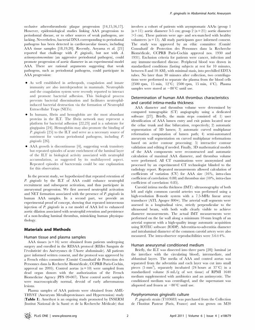
staining of histone H1 and citrullinated histone H4 (Figure 6A). P.
gingivalis promoted neutrophil DNA expulsion in a dose-dependent
manner as shown by quantification of cf-DNA in the supernatant of
neutrophils incubated with different concentrations of P. gingivalis
(Figure 6B). Induction by P.gingivalis of NET formation and
subsequent trapping was further demonstrated by epifluorescence
(Figure 6C) and confocal microscopy (Figure 6D).
Chronic P. gingivalis-bacteremia induces neutrophilrecruitment in experimental AAA
In order to provide an experimental proof of concept that P.
gingivalis may impact on aneurysm progression, we used the
decellularized xenograft model of aneurysm in rats [29]. To mimic
chronic bacteremia associated with periodontal disease, P. gingivalis
was injected by the intravenous route once a week for 4 weeks
(107 CFU/rat) without producing significant modification of their
general health status (no difference in body weight between control
(346.366.8 g) and P. gingivalis-injected (338.761.9 g) rats, no signs
of prostration and no macroscopically visible alterations of visceral
organs: lung, liver and kidney at necropsy). Repeated P. gingivalis
bacteremia induced a significant increase in the aneurysm size as
compared to saline-injected rats (Figure 7A, B, median6IQR, P.
gingivalis (n = 9): 8.1262.68 mm vs saline (n = 11): 5.2562.75 mm,
p,0.03). Histological analysis showed that ILTs of rats infected by
P. gingivalis were larger than those of non-infected rats and
Figure 5. Anti-P. gingivalis antibodies (anti-Pg Ab) in human AAA. The presence of antibodies against Pg was investigated by a custom ELISAin the conditioned medium of adventitia (A) and in serum of patients with AAA or controls (B). **p,0.01; ***p,0.0001 (Mann-Whitney analysis). Thecorrelations (n = 32) between AAA diameter, thrombus volume, plasma cf-DNA and anti-Pg Ab or Intima Media Thickness (IMT) and thrombus volume(C), were determined by the Least Squares method.doi:10.1371/journal.pone.0018679.g005
P. gingivalis in Abdominal Aortic Aneurysm
PLoS ONE | www.plosone.org 9 April 2011 | Volume 6 | Issue 4 | e18679
exhibited a significant enrichment in neutrophils (Figure 8A). As
expected in this experimental model, mesenchymatous cell
colonization associated with important fibrosis was observed in
the ILT of non-infected rats, suggesting the beginning of the
healing process (Figure 8B). In contrast, P. gingivalis-injected rats
exhibited a large ILT containing neutrophils in the luminal part,
mimicking what is observed in human pathology, without any sign
of healing. We have also observed strong histone H1 immuno-
staining in the neutrophil-rich area associated with disorganized
nuclear structure, suggesting the presence of NETs, similar to
those observed in human ILT (Figure 8C). Double immuno-
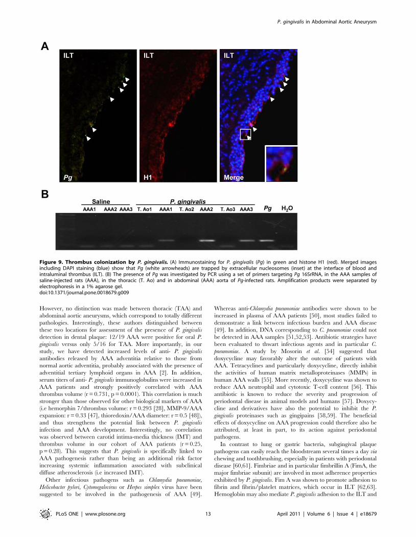
staining for P. gingivalis and histone H1 showed the presence of P.
gingivalis trapped by extracellular nucleosomes at the luminal pole
of the ILT (Figure 9A). P. gingivalis colonization was shown to be
specific of the ILT since neither the AAA of saline-injected rats nor
the thoracic aorta of P. gingivalis-infected rats were positive for Pg
16S rRNA (PCR) (Figure 9B).
To further demonstrate the impact of neutrophil enrichment in
the P. gingivalis-infected aneurysmal samples (wall+thrombus),
MMP-2/MMP-9 activities (assessed by gelatin zymography),
MPO and cf-DNA concentrations were measured in conditioned
medium. MMP-9 activity was significantly increased in medium
conditioned by AAA samples from P. gingivalis-infected rats
compared to AAA samples obtained from non-infected rats
(Figure 10A). In contrast, greater amounts of pro-MMP-2 were
released by aneurysmal segments of non-infected rats relative to P.
gingivalis -infected rats (p = 0.05), reflecting an ongoing healing
process in non-infected rats, since pro-MMP-2 is preferentially
secreted by mesenchymatous cells. The ratio MMP-9/MMP-2 was
therefore significantly higher in conditioned medium from P.
Figure 6. P. gingivalis (Pg) promotes NET formation. Freshly isolated human neutrophils were plated on Lab-tekH chamber slides and thenstimulated or not by f-MLP (100 nM), a bacterial peptide used as a positive control, or by Pg (1.107 CFU) for 2 hours at 37uC. (A) Immunofluorescencedetection of histone H1 (red) and citrullinated histone H4 (green) was performed without permeabilization. (B) Cell-free DNA (cf-DNA) concentrationwas determined in the culture medium of neutrophils stimulated or not either with f-MLP (100 nM) or increasing concentrations of Pg. *p,0.05;**p,0.01 vs ctl (Mann-Whitney analysis). The trapping of Pg (red) by externalized nucleosomes (histone H1, green) was visualized by epifluorescence(C) and confocal microscopy (D). The bottom panel represents a virtual section constructed according to the Z axis, confirming the intracellularpresence of Pg subsequent to phagocytosis by neutrophils.doi:10.1371/journal.pone.0018679.g006
P. gingivalis in Abdominal Aortic Aneurysm
PLoS ONE | www.plosone.org 10 April 2011 | Volume 6 | Issue 4 | e18679
gingivalis -infected rats (0.163 vs 0.061 in saline-injected rats,
p = 0.008). MPO concentration was strongly increased in AAA
conditioned medium from P. gingivalis-infected rats as compared to
non-infected rats (Figure 10B, p = 0.0003). In plasma, a trend
towards increased MPO levels was observed in P. gingivalis-infected
rats without reaching statistical significance (p = 0.065). The
medium conditioned by the aneurysmal segment of the abdominal
aorta contained more cf-DNA than the medium obtained by
incubation of the adjacent aortic segment from the same rat
(thoracic aorta, Figure 11A). Moreover, cf-DNA concentration
was increased in both conditioned medium and plasma of rats
injected by P. gingivalis compared to saline-injected rats
(Figure 11A). This suggests that cf-DNA is released by the
AAA segment and not by the rest of the aorta potentially infected
by P. gingivalis. More importantly, a strong positive correlation was
observed between the concentrations of cf-DNA in both
conditioned medium and plasma, and the AAA diameter
(r = 0.635, p,0.03 and r = 0.83, p,0.001, respectively). Finally,
NETs contributed, at least in part, to cf-DNA measured in plasma
and conditioned medium since MPO/DNA complexes were
detected in higher amounts in P. gingivalis-infected rat samples
(Figure 11B).
Discussion
Several lines of evidence led us to hypothesize that bacteria, and
in particular periodontal pathogens, may participate in the
development of AAA. Epidemiological data suggest an association
between periodontal and cardiovascular diseases [39]. The nature
of this association is however still a matter of debate; in particular,
whether periodontal disease impacts directly on the pathogenesis
of cardiovascular diseases or indirectly by increasing background
inflammation is still not settled. Interestingly, atherothrombosis
and periodontal disease share risk factors such as age, smoking and
male gender [16,40]. However, to date, no epidemiological study
has been reported linking AAA development and periodontal
disease. In the present study, we sought to establish a potential
causal link between P. gingivalis, a highly prevalent period-
ontopathogen, and AAA progression.
Neutrophil recruitment and activation in human AAAthrombus – Potential role of P. gingivalis
The persistence of neutrophils in the most luminal part of the
ILT, interfacing with the bloodstream cannot be explained solely
by their passive trapping during the process of fibrin formation.
We have recently reported that conditioned medium from the
luminal layer of ILT was chemoattractant for neutrophils in
human AAA. However, blocking strategies interfering with the
interleukin 8 pathway and RANTES only produced a 50%
inhibition of neutrophil chemoattraction [10]. Since neutrophils
constitute the first line of defense against bacteria and are strongly
attracted by lipopolysaccharide (LPS) and bacterial peptides, we
tested for the presence of endotoxin in AAA samples. We show
that both the mural ILT and the residual AAA wall contained and
released LPS that may account for neutrophil chemoattraction
and activation. LPS from P. gingivalis was shown to stimulate
neutrophils via LPS-binding protein in the serum of patients with
periodontal disease [41]. In vitro studies have shown that neutrophil
exposure to a variety of different microbial pathogens (Staphylococ-
Figure 7. Chronic P gingivalis (Pg) infection fostered AAA development in rats. (A) Experimental AAA was induced by implanting a segmentof a SDS-decellularized guinea pig aorta in the rat aorta. Rats were or not infected weekly with Pg (1.107 CFU/500 mL/rat, for 4 weeks). (B) At the endof treatment, rats were anaesthetized for blood sampling and sacrified after measuring AAA diameter. Results (n = 10) are presented as box plots inwhich median is shown, **p,0.01 (Mann-Whitney Analysis).doi:10.1371/journal.pone.0018679.g007
P. gingivalis in Abdominal Aortic Aneurysm
PLoS ONE | www.plosone.org 11 April 2011 | Volume 6 | Issue 4 | e18679

cus Aureus, Escherichia Coli), platelets, proinflammatory stimuli,
hydrogen peroxide (H2O2), interleukin-8, or bacterial LPS, was
able to trigger neutrophil extracellular trap (NET) formation.
NETs were first described by Brinkmann et al. [35] as extracellular,
highly decondensed chromatin structures released by activated
neutrophils. In the present study, we have shown that stimulation
of human neutrophils by P. gingivalis led to NET production,
reflected by an increased cell-free DNA (cf-DNA) concentration in
the culture supernatant and by histone exposure/modifications.
The presence of NETs in the luminal part of the ILT, as well as in
the adventitia of AAA samples, was shown by immunostaining of
histone H1 and of the citrullinated form of histone H4. Massberg et
al. recently suggested that NETs may promote fibrin formation in
vivo and therefore limit pathogen dissemination [42]. Such a
process could participate in the fibrin formation observed in the
luminal part of the AAA thrombus. DNA and histones represent
the major components of NETs and provide the backbone for the
binding of neutrophil granule components (e.g. myeloperoxidase
and elastase) that may play an antimicrobial role [36]. Histone 2A
and 2B have also been shown to exhibit antimicrobial and
endotoxin-neutralizing activities [43], reinforcing the bactericidal
properties of NETs [44]. The presence of NETs in AAA samples
was also associated with a release of cf-DNA, measurable in
conditioned medium (ILT and adventitia). The release of cf-DNA
by the adventitial layer may be due to local production of
extracellular nucleosomes by activated neutrophils in response to
bacterial stimulation coming from the vasa vasorum. Accordingly,
cf-DNA levels were increased in plasma of AAA patients relative to
control subjects. In addition, plasma cf-DNA levels were positively
correlated with the abdominal aortic aneurysm diameter,
suggesting that this marker may reflect the biological activity of
the aneurysm and neutrophil activation in particular. Increased cf-
DNA in plasma has been reported in pathologies other than AAA,
involving neutrophil activation by bacteria, such as sepsis [38,45]
and appendicitis [36] or inflammatory processes such as small-
vessel vasculitis [31]. Although some studies attribute cf-DNA to
NETosis [38], caution must be exercised since different cell types
may release circulating DNA upon necrosis, apoptosis or
microparticle formation. We therefore measured MPO-DNA
complexes in order to assess the amount of NETs in both
conditioned medium and in plasma of patients versus controls.
The same trend was observed as for cf-DNA: predominant release
by the luminal part of the ILT and increased plasma levels in AAA
patients relative to controls. In AAA, cf-DNA is a good marker of
NET formation as attested by the positive correlation between cf-
DNA levels and MPO/DNA complexes (r = 0.562, p = 0.0065).
P. gingivalis in human AAAThe presence of bacterial DNA was investigated by PCR. DNA
encoding for bacterial 16S ribosomal RNA was detected in 11/16
of the AAA samples tested whereas 7/16 were positive for
detection of P. gingivalis DNA. These results are in line with a
previous study [46], which tested the presence of 7 period-
ontopathic bacteria by PCR in AAA mural ILT and arterial wall
and showed that more than 80% of AAA samples tested contained
P. gingivalis DNA. The lower incidence in our study may be
explained not only by different procedures for DNA extraction
and primers used for detection of P. gingivalis DNA, but also by the
higher prevalence of chronic periodontitis in the Japanese
population compared with other developed countries[46]. In
another recently published study by Nakano et al.[21], detection of
P. gingivalis by PCR was reported in 8/76 aneurysm samples.
Figure 8. P. gingivalis (Pg) infection promoted neutrophil recruitment, NET formation and inhibited healing. (A) Hematoxylin/Eosinstaining showing the presence of a thrombus and neutrophil accumulation at its luminal pole (right, inset) in Pg-infected rats. The presence ofmesenchymatous cells is observed in saline-injected rats (left, inset). (B) Masson’s trichrome staining. Fibrosis associated with healing is observed ingreen in saline-injected rats whereas red staining highlights the presence of a thrombus in Pg-infected rats. (C) Immunostaining for histone H1 (red),nuclei appear in blue (DAPI). Merged images show the presence of extracellular H1 associated with disorganized DNA (inset), but also intactneutrophils characterized by their multilobed nuclei (bottom, right).doi:10.1371/journal.pone.0018679.g008
P. gingivalis in Abdominal Aortic Aneurysm
PLoS ONE | www.plosone.org 12 April 2011 | Volume 6 | Issue 4 | e18679
However, no distinction was made between thoracic (TAA) and
abdominal aortic aneurysms, which correspond to totally different
pathologies. Interestingly, these authors distinguished between
these two locations for assessment of the presence of P. gingivalis
detection in dental plaque: 12/19 AAA were positive for oral P.
gingivalis versus only 5/16 for TAA. More importantly, in our
study, we have detected increased levels of anti- P. gingivalis
antibodies released by AAA adventitia relative to those from
normal aortic adventitia, probably associated with the presence of
adventitial tertiary lymphoid organs in AAA [2]. In addition,
serum titers of anti- P. gingivalis immunoglobulins were increased in
AAA patients and strongly positively correlated with AAA
thrombus volume (r = 0.731, p = 0.0001). This correlation is much
stronger than those observed for other biological markers of AAA
(i.e hemorphin 7/thrombus volume: r = 0.293 [28], MMP-9/AAA
expansion: r = 0.33 [47], thioredoxin/AAA diameter: r = 0.5 [48]),
and thus strengthens the potential link between P. gingivalis
infection and AAA development. Interestingly, no correlation
was observed between carotid intima-media thickness (IMT) and
thrombus volume in our cohort of AAA patients (r = 0.25,
p = 0.28). This suggests that P. gingivalis is specifically linked to
AAA pathogenesis rather than being an additional risk factor
increasing systemic inflammation associated with subclinical
diffuse atherosclerosis (i.e increased IMT).
Other infectious pathogens such as Chlamydia pneumoniae,
Helicobacter pylori, Cytomegalovirus or Herpes simplex virus have been
suggested to be involved in the pathogenesis of AAA [49].
Whereas anti-Chlamydia pneumoniae antibodies were shown to be
increased in plasma of AAA patients [50], most studies failed to
demonstrate a link between infectious burden and AAA disease
[49]. In addition, DNA corresponding to C. pneumoniae could not
be detected in AAA samples [51,52,53]. Antibiotic strategies have
been evaluated to thwart infectious agents and in particular C.
pneumoniae. A study by Mosorin et al. [54] suggested that
doxycycline may favorably alter the outcome of patients with
AAA. Tetracyclines and particularly doxycycline, directly inhibit
the activities of human matrix metalloproteinases (MMPs) in
human AAA walls [55]. More recently, doxycycline was shown to
reduce AAA neutrophil and cytotoxic T-cell content [56]. This
antibiotic is known to reduce the severity and progression of
periodontal disease in animal models and humans [57]. Doxycy-
cline and derivatives have also the potential to inhibit the P.
gingivalis proteinases such as gingipains [58,59]. The beneficial
effects of doxycycline on AAA progression could therefore also be
attributed, at least in part, to its action against periodontal
pathogens.
In contrast to lung or gastric bacteria, subgingival plaque
pathogens can easily reach the bloodstream several times a day via
chewing and toothbrushing, especially in patients with periodontal
disease [60,61]. Fimbriae and in particular fimbrillin A (FimA, the
major fimbriae subunit) are involved in most adherence properties
exhibited by P. gingivalis. Fim A was shown to promote adhesion to
fibrin and fibrin/platelet matrices, which occur in ILT [62,63].
Hemoglobin may also mediate P. gingivalis adhesion to the ILT and
Figure 9. Thrombus colonization by P. gingivalis. (A) Immunostaining for P. gingivalis (Pg) in green and histone H1 (red). Merged imagesincluding DAPI staining (blue) show that Pg (white arrowheads) are trapped by extracellular nucleosomes (inset) at the interface of blood andintraluminal thrombus (ILT). (B) The presence of Pg was investigated by PCR using a set of primers targeting Pg 16SrRNA, in the AAA samples ofsaline-injected rats (AAA), in the thoracic (T. Ao) and in abdominal (AAA) aorta of Pg-infected rats. Amplification products were separated byelectrophoresis in a 1% agarose gel.doi:10.1371/journal.pone.0018679.g009
P. gingivalis in Abdominal Aortic Aneurysm
PLoS ONE | www.plosone.org 13 April 2011 | Volume 6 | Issue 4 | e18679

be used as a source of nutriment [25,26]. However, since P.
gingivalis are strict anaerobic bacteria, they are not likely to
proliferate in an aerobic environment such as that observed in the
ILT (PO2 = 100 mm Hg). Chronic bacteremia could therefore
allow subclinical infection of different cardiovascular tissues
including the ILT of AAA, principally composed of fibrin,
platelets and hemoglobin.
P. gingivalis in an experimental model of AAA in ratsIn order to provide a proof of concept that P. gingivalis may be
an actor of AAA progression, we have used an experimental model
of AAA in rats [29]. This animal model is characterized by the
formation of a thrombus about one week after grafting a
decellularized guinea pig aorta in the abdominal position,
associated with aortic dilation. In this model, like in all currently
used AAA models, the mural ILT is rapidly colonized by
mesenchymatous cells that initiate a fibrotic healing process. In
contrast, after 4 weekly intravenous injections of P. gingivalis, the
aortic diameter was not only significantly increased relative to
saline-injected rats (p = 0.01), but the composition of the AAA was
strikingly different. In P. gingivalis-injected rats, the mural ILT was
persistent and exhibited a multilayered aspect, similar to what is
observed in human AAA samples. The ILT was considerably
enriched in neutrophils and all markers of their activation were
increased in conditioned medium and in plasma of P. gingivalis- vs
saline-injected rats. In the present study, we demonstrate that a
Figure 10. Increased MMP9 activity and MPO released by AAA samples of P. gingivalis (Pg) infected rats. (A) gelatin zymography analysisof saline- and Pg-injected rats (respectively rats R1,2,3 and R4,5,6). MW: molecular weight, Ref: reference containing pro- and active MMP-9. Graphsrepresent spatial density quantification of pro- and active MMP9 lysis areas (Image J software). (B) MPO concentration was determined by ELISA inconditioned medium and in plasma. *p,0.05, **p,0.01 (Mann-Whitney Analysis).doi:10.1371/journal.pone.0018679.g010
P. gingivalis in Abdominal Aortic Aneurysm
PLoS ONE | www.plosone.org 14 April 2011 | Volume 6 | Issue 4 | e18679

periodontal pathogen enhances the development of AAA by
maintaining the presence of a neutrophil-rich ILT, leading to a
pathophysiological pattern similar to that observed in humans. It
cannot be excluded that other bacteria or their products may have
similar effects, and may participate in AAA pathogenesis, but our
data highlight the major role of P. gingivalis in AAA development.
Using a different model of experimental AAA in mice, Aoyama et
al. [21] have reported that, in contrast to P. gingivalis, A.
actinomycetemcomitans did not promote aortic dilation. However,
these authors used a calcium-chloride model in mice, that consists
in an external aggression of the aorta that does not lead to the
formation of a thrombus, as often observed in humans.
Strengths and limitations of the studyOur study provides a combination of clinical and experimental
data that could link periodontal disease to AAA formation.
However, albeit reaching statistical significance, the number of
human samples analyzed is quite small. Additional epidemiolog-
ical studies linking AAA and periodontal diseases would be
necessary to support our findings. The model that we used is
characterized by the formation of a thrombus about one week
after xenografting. The healing process then usually takes place in
the absence of additional aggression that would maintain the
recruitment of neutrophils. In the present study, we report for the
first time that chronic injection of P. gingivalis leads to the
Figure 11. Cell-free DNA (Cf-DNA) and MPO-DNA complexes are increased in rats infected with P. gingivalis (Pg). (A) Concentration ofcf-DNA released by the AAA segment (thrombus + wall) of saline- or Pg-infected rats or by the thoracic aorta (T. Ao) from Pg-infected rats. Cf-DNA wasalso quantified in plasma. (B) Quantification of MPO-DNA complexes in conditioned medium or plasma of saline-or Pg-injected rats. **p,0.01;***p,0.0001 (Mann-Whitney analysis). The correlation between AAA diameter and Cf-DNA concentration was determined by the Least Squaresmethod.doi:10.1371/journal.pone.0018679.g011
P. gingivalis in Abdominal Aortic Aneurysm
PLoS ONE | www.plosone.org 15 April 2011 | Volume 6 | Issue 4 | e18679

persistence of the ILT, similar to human pathophysiology.
However, we did not test other periodontal pathogens that would
be of interest, such as performed by Aoyama et al. [21]. Finally, we
provide evidence that P. gingivalis DNA is present in AAA samples
and that P. gingivalis material is sufficient to produce an adventitial
immune response. However, the presence of living pathogens was
not shown suggesting that P. gingivalis material, such as dead
bacteria or LPS, may be sufficient to bind to the thrombus and
promote its chronic renewal.
ConclusionIn conclusion, the results of the present study indicate that P.
gingivalis accelerates AAA progression via recruitment and
activation of neutrophils, leading to production of NETs which
are detectable in the plasma of AAA subjects. Because repeated
subclinical episodes of bacteremia are systematically associated
with periodontal diseases, P. gingivalis could be therefore a key
actor in human AAA progression.
Taken together, our results demonstrate that a common
pathogen may have a causal role in the pathogenesis of AAA.
These findings bring significant new information to the field of
AAA pathogenesis but should be strengthened by both epidemi-
ological and observational studies in humans before one can
envisage potential therapeutic strategies based on the treatment of
periodontal disease to prevent AAA evolution towards rupture.
Acknowledgments
The AMETHYST cohort is promoted by Inserm and supported by the
Fondation pour la Recherche Medicale, the Fondation de France, and the
Lorraine region. The authors thank Marc Sapoval (Hopital Europeen
Georges Pompidou, Paris, France); Claude Kauffmann (Montreal
University, Notre-Dame Hospital, Montreal University Hospital Centre,
Quebec); Damien Husson, Emilien Micard, Dr Damien Mandry (Nancy
Centre of Clinical Investigation-Innovative Technology), and Ghassan
Watfa (Nancy CIC 9501), for their technical help concerning AAA CT-
scans (AMETHYST cohort) and Dr Mary Osborne-Pellegrin for editing
the manuscript.
Author Contributions
Conceived and designed the experiments: OM SD J-BM. Performed the
experiments: SD J-MA CJ LL OM. Analyzed the data: SD CJ OM PR.
Contributed reagents/materials/analysis tools: MB-M PR LL YC RR.
Wrote the paper: OM SD J-BM PB.
References
1. Michel JB (2001) Contrasting outcomes of atheroma evolution: intimal
accumulation versus medial destruction. Arterioscler Thromb Vasc Biol 21:
1389–1392.
2. Michel JB, Thaunat O, Houard X, Meilhac O, Caligiuri G, et al. (2007)
Topological determinants and consequences of adventitial responses to arterial
wall injury. Arterioscler Thromb Vasc Biol 27: 1259–1268.
3. Sakalihasan N, Limet R, Defawe OD (2005) Abdominal aortic aneurysm.
Lancet 365: 1577–1589.
4. Kurvers H, Veith FJ, Lipsitz EC, Ohki T, Gargiulo NJ, et al. (2004)
Discontinuous, staccato growth of abdominal aortic aneurysms. J Am Coll
Surg 199: 709–715.
5. Limet R, Sakalihassan N, Albert A (1991) Determination of the expansion rate
and incidence of rupture of abdominal aortic aneurysms. J Vasc Surg 14:
540–548.
6. Fontaine V, Jacob MP, Houard X, Rossignol P, Plissonnier D, et al. (2002)
Involvement of the mural thrombus as a site of protease release and activation in
human aortic aneurysms. Am J Pathol 161: 1701–1710.
7. Touat Z, Ollivier V, Dai J, Huisse MG, Bezeaud A, et al. (2006) Renewal of
mural thrombus releases plasma markers and is involved in aortic abdominal
aneurysm evolution. Am J Pathol 168: 1022–1030.
8. Dejouvencel T, Feron D, Rossignol P, Sapoval M, Kauffmann C, et al.
Hemorphin 7 reflects hemoglobin proteolysis in abdominal aortic aneurysm.
Arterioscler Thromb Vasc Biol 30: 269–275.
9. Houard X, Rouzet F, Touat Z, Philippe M, Dominguez M, et al. (2007)
Topology of the fibrinolytic system within the mural thrombus of human
abdominal aortic aneurysms. J Pathol 212: 20–28.
10. Houard X, Touat Z, Ollivier V, Louedec L, Philippe M, et al. (2009) Mediators
of neutrophil recruitment in human abdominal aortic aneurysms. Cardiovasc
Res 82: 532–541.
11. Houard X, Ollivier V, Louedec L, Michel JB, Back M (2009) Differential
inflammatory activity across human abdominal aortic aneurysms reveals
neutrophil-derived leukotriene B4 as a major chemotactic factor released from
the intraluminal thrombus. Faseb J 23: 1376–1383.
12. Fontaine V, Touat Z, Mtairag el M, Vranckx R, Louedec L, et al. (2004) Role of
leukocyte elastase in preventing cellular re-colonization of the mural thrombus.
Am J Pathol 164: 2077–2087.
13. Hannawa KK, Eliason JL, Woodrum DT, Pearce CG, Roelofs KJ, et al. (2005)
L-selectin-mediated neutrophil recruitment in experimental rodent aneurysm
formation. Circulation 112: 241–247.
14. Blaizot A, Vergnes JN, Nuwwareh S, Amar J, Sixou M (2009) Periodontal
diseases and cardiovascular events: meta-analysis of observational studies. Int
Dent J 59: 197–209.
15. Bouchard P, Boutouyrie P, D’Aiuto F, Deanfield J, Deliargyris E, et al. (2010)
European workshop in periodontal health and cardiovascular disease consensus
document. European Heart Journal Supplements 12 (Supplement B. pp
B13–B22.
16. Friedewald VE, Kornman KS, Beck JD, Genco R, Goldfine A, et al. (2009) The
American Journal of Cardiology and Journal of Periodontology editors’
consensus: periodontitis and atherosclerotic cardiovascular disease.
J Periodontol 80: 1021–1032.
17. Jimenez M, Krall EA, Garcia RI, Vokonas PS, Dietrich T (2009) Periodontitis
and incidence of cerebrovascular disease in men. Ann Neurol 66: 505–512.
18. Gaetti-Jardim E, Jr., Marcelino SL, Feitosa AC, Romito GA, Avila-Campos MJ
(2009) Quantitative detection of periodontopathic bacteria in atherosclerotic
plaques from coronary arteries. J Med Microbiol 58: 1568–1575.
19. Nakano K, Nemoto H, Nomura R, Inaba H, Yoshioka H, et al. (2009) Detection
of oral bacteria in cardiovascular specimens. Oral Microbiol Immunol 24:
64–68.
20. Nakano K, Wada K, Nomura R, Nemoto H, Inaba H, et al. (2010)
Characterization of aortic aneurysms in cardiovascular disease patients
harboring Porphyromonas gingivalis. Oral Dis.
21. Aoyama N, Suzuki J, Wang D, Ogawa M, Kobayashi N, et al. (2010)
Porphyromonas gingivalis promotes murine abdominal aortic aneurysms via
matrix metalloproteinase-2 induction. J Periodontal Res.
22. Fitzgerald JR, Loughman A, Keane F, Brennan M, Knobel M, et al. (2006)
Fibronectin-binding proteins of Staphylococcus aureus mediate activation of
human platelets via fibrinogen and fibronectin bridges to integrin GPIIb/IIIa
and IgG binding to the FcgammaRIIa receptor. Mol Microbiol 59: 212–230.
23. Bamford CV, Fenno JC, Jenkinson HF, Dymock D (2007) The chymotrypsin-
like protease complex of Treponema denticola ATCC 35405 mediates
fibrinogen adherence and degradation. Infect Immun 75: 4364–4372.
24. Imamura T, Travis J, Potempa J (2003) The biphasic virulence activities of
gingipains: activation and inactivation of host proteins. Curr Protein Pept Sci 4:
443–450.
25. Pathirana RD, O’Brien-Simpson NM, Veith PD, Riley PF, Reynolds EC (2006)
Characterization of proteinase-adhesin complexes of Porphyromonas gingivalis.
Microbiology 152: 2381–2394.
26. Olczak T, Simpson W, Liu X, Genco CA (2005) Iron and heme utilization in
Porphyromonas gingivalis. FEMS Microbiol Rev 29: 119–144.
27. Kauffmann C, Tang A, Dugas A, Therasse E, Oliva V, et al. (2009) Clinical
validation of a software for quantitative follow-up of abdominal aortic aneurysm
maximal diameter and growth by CT angiography. Eur J Radiol.
28. Dejouvencel T, Feron D, Rossignol P, Sapoval M, Kauffmann C, et al. (2010)
Hemorphin 7 reflects hemoglobin proteolysis in abdominal aortic aneurysm.
Arterioscler Thromb Vasc Biol 30: 269–275.
29. Allaire E, Mandet C, Bruneval P, Bensenane S, Becquemin JP, et al. (1996) Cell
and extracellular matrix rejection in arterial concordant and discordant
xenografts in the rat. Transplantation 62: 794–803.
30. Leclercq A, Houard X, Loyau S, Philippe M, Sebbag U, et al. (2007) Topology
of protease activities reflects atherothrombotic plaque complexity. Atheroscle-
rosis 191: 1–10.
31. Kessenbrock K, Krumbholz M, Schonermarck U, Back W, Gross WL, et al.
(2009) Netting neutrophils in autoimmune small-vessel vasculitis. Nat Med 15:
623–625.
32. Li L, Messas E, Batista EL, Jr., Levine RA, Amar S (2002) Porphyromonas
gingivalis infection accelerates the progression of atherosclerosis in a heterozy-
gous apolipoprotein E-deficient murine model. Circulation 105: 861–867.
33. Colhoun HM, Slaney JM, Rubens MB, Fuller JH, Sheiham A, et al. (2008)
Antibodies to periodontal pathogens and coronary artery calcification in type 1
diabetic and nondiabetic subjects. J Periodontal Res 43: 103–110.
P. gingivalis in Abdominal Aortic Aneurysm
PLoS ONE | www.plosone.org 16 April 2011 | Volume 6 | Issue 4 | e18679

34. Mtairag el M, Houard X, Rais S, Pasquier C, Oudghiri M, et al. (2002)
Pharmacological potentiation of natriuretic peptide limits polymorphonuclearneutrophil-vascular cell interactions. Arterioscler Thromb Vasc Biol 22:
1824–1831.
35. Brinkmann V, Reichard U, Goosmann C, Fauler B, Uhlemann Y, et al. (2004)Neutrophil extracellular traps kill bacteria. Science 303: 1532–1535.
36. Wartha F, Beiter K, Normark S, Henriques-Normark B (2007) Neutrophilextracellular traps: casting the NET over pathogenesis. Curr Opin Microbiol 10:
52–56.
37. Wang Y, Li M, Stadler S, Correll S, Li P, et al. (2009) Histonehypercitrullination mediates chromatin decondensation and neutrophil extra-
cellular trap formation. J Cell Biol 184: 205–213.38. Margraf S, Logters T, Reipen J, Altrichter J, Scholz M, et al. (2008) Neutrophil-
derived circulating free DNA (cf-DNA/NETs): a potential prognostic marker forposttraumatic development of inflammatory second hit and sepsis. Shock 30:
352–358.
39. Demmer RT, Desvarieux M (2006) Periodontal infections and cardiovasculardisease: the heart of the matter. J Am Dent Assoc 137 Suppl: 14S–20S; quiz 38S.
40. Meurman JH, Sanz M, Janket SJ (2004) Oral health, atherosclerosis, andcardiovascular disease. Crit Rev Oral Biol Med 15: 403–413.
41. Soolari AS, Champagne C, Punzi JS, Amar S, Van Dyke TE (1999) Serum
modulation of neutrophil response to Porphyromonas gingivalis LPS inperiodontal disease. J Int Acad Periodontol 1: 101–109.
42. Massberg S, Grahl L, von Bruehl ML, Manukyan D, Pfeiler S, et al. Reciprocalcoupling of coagulation and innate immunity via neutrophil serine proteases.
Nat Med 16: 887–896.43. Kim HS, Cho JH, Park HW, Yoon H, Kim MS, et al. (2002) Endotoxin-
neutralizing antimicrobial proteins of the human placenta. J Immunol 168:
2356–2364.44. Parseghian MH, Luhrs KA (2006) Beyond the walls of the nucleus: the role of
histones in cellular signaling and innate immunity. Biochem Cell Biol 84:589–604.
45. Ma AC, Kubes P (2008) Platelets, neutrophils, and neutrophil extracellular traps
(NETs) in sepsis. J Thromb Haemost 6: 415–420.46. Kurihara N, Inoue Y, Iwai T, Umeda M, Huang Y, et al. (2004) Detection and
localization of periodontopathic bacteria in abdominal aortic aneurysms.Eur J Vasc Endovasc Surg 28: 553–558.
47. Lindholt JS, Vammen S, Fasting H, Henneberg EW, Heickendorff L (2000) Theplasma level of matrix metalloproteinase 9 may predict the natural history of
small abdominal aortic aneurysms. A preliminary study. Eur J Vasc Endovasc
Surg 20: 281–285.48. Martinez-Pinna R, Lindholt JS, Blanco-Colio LM, Dejouvencel T, Madrigal-
Matute J, et al. (2010) Increased levels of thioredoxin in patients with abdominalaortic aneurysms (AAAs). A potential link of oxidative stress with AAA evolution.
Atherosclerosis 212: 333–338.
49. Nyberg A, Skagius E, Englund E, Nilsson I, Ljungh A, et al. (2008) Abdominalaortic aneurysm and the impact of infectious burden. Eur J Vasc Endovasc Surg
36: 292–296.
50. Blanchard JF, Armenian HK, Peeling R, Friesen PP, Shen C, et al. (2000) The
relation between Chlamydia pneumoniae infection and abdominal aortic
aneurysm: case-control study. Clin Infect Dis 30: 946–947.
51. Lindholt JS, Ostergard L, Henneberg EW, Fasting H, Andersen P (1998) Failure
to demonstrate Chlamydia pneumoniae in symptomatic abdominal aortic
aneurysms by a nested polymerase chain reaction (PCR). Eur J Vasc Endovasc
Surg 15: 161–164.
52. Falkensammer B, Duftner C, Seiler R, Pavlic M, Walder G, et al. (2007) Lack of
microbial DNA in tissue specimens of patients with abdominal aortic aneurysms
and positive Chlamydiales serology. Eur J Clin Microbiol Infect Dis 26:
141–145.
53. Sodeck G, Domanovits H, Khanakah G, Schillinger M, Thalmann M, et al.
(2004) The role of Chlamydia pneumoniae in human aortic disease-a hypothesis
revisited. Eur J Vasc Endovasc Surg 28: 547–552.
54. Mosorin M, Juvonen J, Biancari F, Satta J, Surcel HM, et al. (2001) Use of
doxycycline to decrease the growth rate of abdominal aortic aneurysms: a
randomized, double-blind, placebo-controlled pilot study. J Vasc Surg 34:
606–610.
55. Golub LM, Sorsa T, Lee HM, Ciancio S, Sorbi D, et al. (1995) Doxycycline
inhibits neutrophil (PMN)-type matrix metalloproteinases in human adult
periodontitis gingiva. J Clin Periodontol 22: 100–109.
56. Lindeman JH, Abdul-Hussien H, van Bockel JH, Wolterbeek R, Kleemann R
(2009) Clinical trial of doxycycline for matrix metalloproteinase-9 inhibition in
patients with an abdominal aneurysm: doxycycline selectively depletes aortic
wall neutrophils and cytotoxic T cells. Circulation 119: 2209–2216.
57. Haffajee AD, Socransky SS, Gunsolley JC (2003) Systemic anti-infective
periodontal therapy. A systematic review. Ann Periodontol 8: 115–181.
58. Grenier D, Roy E, Mayrand D (2003) Modulation of Porphyromonas gingivalis
proteinase activity by suboptimal doses of antimicrobial agents. J Periodontol 74:
1316–1319.
59. Imamura T, Matsushita K, Travis J, Potempa J (2001) Inhibition of trypsin-like
cysteine proteinases (gingipains) from Porphyromonas gingivalis by tetracycline
and its analogues. Antimicrob Agents Chemother 45: 2871–2876.
60. Forner L, Larsen T, Kilian M, Holmstrup P (2006) Incidence of bacteremia after
chewing, tooth brushing and scaling in individuals with periodontal inflamma-
tion. J Clin Periodontol 33: 401–407.
61. Perez-Chaparro PJ, Lafaurie GI, Gracieux P, Meuric V, Tamanai-Shacoori Z,
et al. (2009) Distribution of Porphyromonas gingivalis fimA genotypes in isolates
from subgingival plaque and blood sample during bacteremia. Biomedica 29:
298–306.
62. Burnette-Curley D, Wells V, Viscount H, Munro CL, Fenno JC, et al. (1995)
FimA, a major virulence factor associated with Streptococcus parasanguis
endocarditis. Infect Immun 63: 4669–4674.
63. Viscount HB, Munro CL, Burnette-Curley D, Peterson DL, Macrina FL (1997)
Immunization with FimA protects against Streptococcus parasanguis endocar-
ditis in rats. Infect Immun 65: 994–1002.
P. gingivalis in Abdominal Aortic Aneurysm
PLoS ONE | www.plosone.org 17 April 2011 | Volume 6 | Issue 4 | e18679