Poromatosis Following Ewing Sarcoma - OMICS … · Poromatosis Following Ewing Sarcoma ... from the...
4
Poromatosis Following Ewing Sarcoma Amr Abduljabbar 1* , Omar Alanazi 2 , Amany Fathadin 3 and Mohammad Alhaddab 2 1 Department of Dermatology, King Saud Medical City, Riyadh, Saudi Arabia 2 Department of Dermatology, King Saud University Hospital, Riyadh, Saudi Arabia 3 Department of Pathology, King Saud University Hospital, Riyadh, Saudi Arabia * Corresponding author: Amr Abduljabbar, Department of Dermatology, King Saud Medical City, Riyadh, Saudi Arabia, Tel: +966566477766; E-mail: [email protected] Received date: June 21, 2016; Accepted date: August 09, 2016; Published date: August 11, 2016 Copyright: © 2016 Abduljabbar A, et al. This is an open-access article distributed under the terms of the Creative Commons Attribution License, which permits unrestricted use, distribution, and reproduction in any medium, provided the original author and source are credited. Abstract Eccrine poromas are rare, benign adnexal tumors derived from the intraepidermal portion of sweat ducts. Historically they were thought to arise from eccrine ducts although today it is thought that they may also have an apocrine origin. They usually appear as solitary, slow-growing, skin-colored papules on acral surfaces. Here we present the unusual situation of a patient with multiple poromas who was previously treated with chemotherapy and radiation for Ewing sarcoma. This report adds to the increasing evidence that connect multiple poromas to some treatments of malignant conditions. Keywords: Ewing sarcoma; Poroma; Poromatosis Introduction A poroma is a benign adnexal neoplasm composed of epithelial cells that show tubular differentiation (usually distal ductal), whereas a porocarcinoma is a malignant poroma which can cause visceral metastasis [1]. Historically poromas were considered as glandular adnexal neoplasms of eccrine lineage, and that is why it was referred to as eccrine poroma. However, nowadays it has been proved that this description was not accurate and poromas can be of apocrine lineage too [2]. Multiple poromas or eccrine poromatosis is an uncommon phenomenon and more than half of the reported cases had a history of immunosuppression from either radiation or chemotherapy [3]. Here we present a 51-year-old gentleman with eccrine poromatosis, who was also treated with chemotherapy and radiation before the onset of the lesions. Case Report A 51-years-old Saudi male with a history of Ewing sarcoma of the chest, which was diagnosed in April 2012 in the university hospital. He presented initially with 3 months history of painless gradually enlarging right chest mass. No other complaints and systemic review was unremarkable. Blood studies were within normal parameters. A chest X-ray showed opacity in the right side of the chest. Computed tomography was ordered for further delineation of the opacity and revealed a large well defined lobulated heterogeneous solid enhancing mass with central low density area measuring 5.1 x 6.6 cm lying subcutaneously in the right anterior chest wall and reaching the lower level of sternum. ere was no evidence of mediastina or other adenopathies. A diagnosis of primitive neuroectodermal tumor (PNET) of the chest wall was made through a CT-guided biopsy of the mass. An isotope bone scan confirmed that no other skeletal site was involved. e patient underwent surgery on May 3, 2012, in form of wide local excision of the right chest wall mass and rhomboid flap. Histopathology of the mass showed no evidence of the neoplasm extension to the right chest wall muscle. e tumor was classified as stage 1b. Aſter the surgery, fractionated radiotherapy of 45 Gy was decided, but the patient was not able to continue for social reasons aſter receiving two sessions. Chemotherapy has been offered and he found it more convenient for his situation. He received a regimen consisting of Etoposide, Ifosfamide and Mesna starting on June 5, 2012. He was given 4 cycles of chemotherapy with the last one on October 19, 2012. A repeat CT scan did not show any recurrences. To date, the patient remained disease-free aſter 4 years of follow-up in the oncology clinic. e patient was seen at the dermatology clinic in July 2015 because of eruptive multiple papules and nodules on the chest and abdomen. ese lesions had developed over the past year and are asymptomatic. On examination the patient looks fine, although he has a long scar from his previous surgery over the right chest and flank. He has multiple erythematous to brown, mildly firm papules and nodules over the anterior trunk ranging in size between 0.3 to 1 cm (Figures 1a and 1b) involving the chest and abdomen. e back, extremities and other body sites are free of such lesions. A review of systems was unremarkable. Abduljabbar et al., J Clin Exp Dermatol Res 2016, 7:5 DOI: 10.4172/2155-9554.1000366 Case Report Open Access J Clin Exp Dermatol Res, an open access journal ISSN:2155-9554 Volume 7 • Issue 5 • 1000366 Journal of Clinical & Experimental Dermatology Research J o u r n a l o f C l i n i c a l & E x p e r i m e n t a l D e r m a t o l o g y R e s e a r c h ISSN: 2155-9554
Transcript of Poromatosis Following Ewing Sarcoma - OMICS … · Poromatosis Following Ewing Sarcoma ... from the...
Poromatosis Following Ewing SarcomaAmr Abduljabbar1*, Omar Alanazi2, Amany Fathadin3 and Mohammad Alhaddab2
1Department of Dermatology, King Saud Medical City, Riyadh, Saudi Arabia2Department of Dermatology, King Saud University Hospital, Riyadh, Saudi Arabia3Department of Pathology, King Saud University Hospital, Riyadh, Saudi Arabia*Corresponding author: Amr Abduljabbar, Department of Dermatology, King Saud Medical City, Riyadh, Saudi Arabia, Tel: +966566477766; E-mail: [email protected]
Received date: June 21, 2016; Accepted date: August 09, 2016; Published date: August 11, 2016
Copyright: © 2016 Abduljabbar A, et al. This is an open-access article distributed under the terms of the Creative Commons Attribution License, which permitsunrestricted use, distribution, and reproduction in any medium, provided the original author and source are credited.
Abstract
Eccrine poromas are rare, benign adnexal tumors derived from the intraepidermal portion of sweat ducts.Historically they were thought to arise from eccrine ducts although today it is thought that they may also have anapocrine origin. They usually appear as solitary, slow-growing, skin-colored papules on acral surfaces. Here wepresent the unusual situation of a patient with multiple poromas who was previously treated with chemotherapy andradiation for Ewing sarcoma. This report adds to the increasing evidence that connect multiple poromas to sometreatments of malignant conditions.
Keywords: Ewing sarcoma; Poroma; Poromatosis
IntroductionA poroma is a benign adnexal neoplasm composed of epithelial cells
that show tubular differentiation (usually distal ductal), whereas aporocarcinoma is a malignant poroma which can cause visceralmetastasis [1]. Historically poromas were considered as glandularadnexal neoplasms of eccrine lineage, and that is why it was referred toas eccrine poroma. However, nowadays it has been proved that thisdescription was not accurate and poromas can be of apocrine lineagetoo [2]. Multiple poromas or eccrine poromatosis is an uncommonphenomenon and more than half of the reported cases had a history ofimmunosuppression from either radiation or chemotherapy [3]. Herewe present a 51-year-old gentleman with eccrine poromatosis, who wasalso treated with chemotherapy and radiation before the onset of thelesions.
Case ReportA 51-years-old Saudi male with a history of Ewing sarcoma of the
chest, which was diagnosed in April 2012 in the university hospital. Hepresented initially with 3 months history of painless graduallyenlarging right chest mass. No other complaints and systemic reviewwas unremarkable. Blood studies were within normal parameters. Achest X-ray showed opacity in the right side of the chest. Computedtomography was ordered for further delineation of the opacity andrevealed a large well defined lobulated heterogeneous solid enhancingmass with central low density area measuring 5.1 x 6.6 cm lying
subcutaneously in the right anterior chest wall and reaching the lowerlevel of sternum. There was no evidence of mediastina or otheradenopathies. A diagnosis of primitive neuroectodermal tumor(PNET) of the chest wall was made through a CT-guided biopsy of themass. An isotope bone scan confirmed that no other skeletal site wasinvolved. The patient underwent surgery on May 3, 2012, in form ofwide local excision of the right chest wall mass and rhomboid flap.Histopathology of the mass showed no evidence of the neoplasmextension to the right chest wall muscle. The tumor was classified asstage 1b. After the surgery, fractionated radiotherapy of 45 Gy wasdecided, but the patient was not able to continue for social reasonsafter receiving two sessions. Chemotherapy has been offered and hefound it more convenient for his situation. He received a regimenconsisting of Etoposide, Ifosfamide and Mesna starting on June 5,2012. He was given 4 cycles of chemotherapy with the last one onOctober 19, 2012. A repeat CT scan did not show any recurrences. Todate, the patient remained disease-free after 4 years of follow-up in theoncology clinic. The patient was seen at the dermatology clinic in July2015 because of eruptive multiple papules and nodules on the chestand abdomen. These lesions had developed over the past year and areasymptomatic. On examination the patient looks fine, although he hasa long scar from his previous surgery over the right chest and flank. Hehas multiple erythematous to brown, mildly firm papules and nodulesover the anterior trunk ranging in size between 0.3 to 1 cm (Figures 1aand 1b) involving the chest and abdomen. The back, extremities andother body sites are free of such lesions. A review of systems wasunremarkable.
Abduljabbar et al., J Clin Exp Dermatol Res 2016, 7:5
DOI: 10.4172/2155-9554.1000366
Case Report Open Access
J Clin Exp Dermatol Res, an open access journalISSN:2155-9554
Volume 7 • Issue 5 • 1000366
Journal of Clinical & ExperimentalDermatology ResearchJourna
l of C
linic
al &
Experimental Dermatology Research
ISSN: 2155-9554
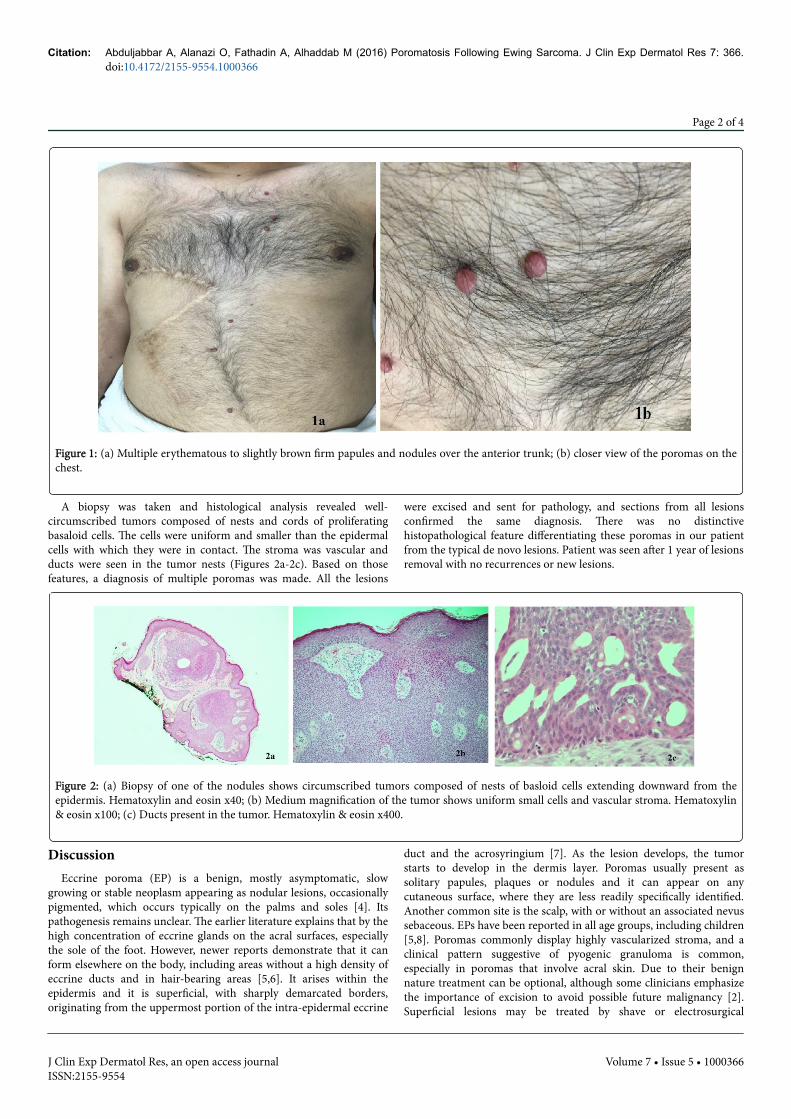
Figure 1: (a) Multiple erythematous to slightly brown firm papules and nodules over the anterior trunk; (b) closer view of the poromas on thechest.
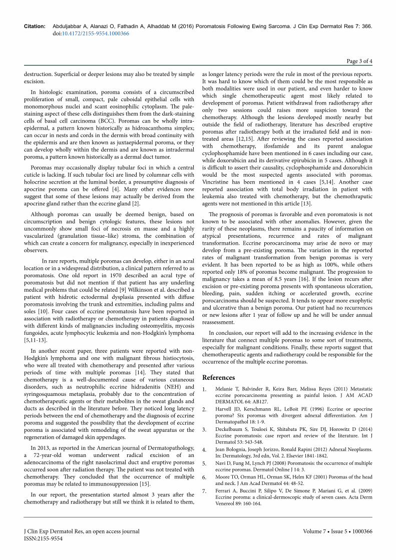
A biopsy was taken and histological analysis revealed well-circumscribed tumors composed of nests and cords of proliferatingbasaloid cells. The cells were uniform and smaller than the epidermalcells with which they were in contact. The stroma was vascular andducts were seen in the tumor nests (Figures 2a-2c). Based on thosefeatures, a diagnosis of multiple poromas was made. All the lesions
were excised and sent for pathology, and sections from all lesionsconfirmed the same diagnosis. There was no distinctivehistopathological feature differentiating these poromas in our patientfrom the typical de novo lesions. Patient was seen after 1 year of lesionsremoval with no recurrences or new lesions.
Figure 2: (a) Biopsy of one of the nodules shows circumscribed tumors composed of nests of basloid cells extending downward from theepidermis. Hematoxylin and eosin x40; (b) Medium magnification of the tumor shows uniform small cells and vascular stroma. Hematoxylin& eosin x100; (c) Ducts present in the tumor. Hematoxylin & eosin x400.
DiscussionEccrine poroma (EP) is a benign, mostly asymptomatic, slow
growing or stable neoplasm appearing as nodular lesions, occasionallypigmented, which occurs typically on the palms and soles [4]. Itspathogenesis remains unclear. The earlier literature explains that by thehigh concentration of eccrine glands on the acral surfaces, especiallythe sole of the foot. However, newer reports demonstrate that it canform elsewhere on the body, including areas without a high density ofeccrine ducts and in hair-bearing areas [5,6]. It arises within theepidermis and it is superficial, with sharply demarcated borders,originating from the uppermost portion of the intra-epidermal eccrine
duct and the acrosyringium [7]. As the lesion develops, the tumorstarts to develop in the dermis layer. Poromas usually present assolitary papules, plaques or nodules and it can appear on anycutaneous surface, where they are less readily specifically identified.Another common site is the scalp, with or without an associated nevussebaceous. EPs have been reported in all age groups, including children[5,8]. Poromas commonly display highly vascularized stroma, and aclinical pattern suggestive of pyogenic granuloma is common,especially in poromas that involve acral skin. Due to their benignnature treatment can be optional, although some clinicians emphasizethe importance of excision to avoid possible future malignancy [2].Superficial lesions may be treated by shave or electrosurgical
Citation: Abduljabbar A, Alanazi O, Fathadin A, Alhaddab M (2016) Poromatosis Following Ewing Sarcoma. J Clin Exp Dermatol Res 7: 366.doi:10.4172/2155-9554.1000366
Page 2 of 4
J Clin Exp Dermatol Res, an open access journalISSN:2155-9554
Volume 7 • Issue 5 • 1000366
destruction. Superficial or deeper lesions may also be treated by simpleexcision.
In histologic examination, poroma consists of a circumscribedproliferation of small, compact, pale cuboidal epithelial cells withmonomorphous nuclei and scant eosinophilic cytoplasm. The pale-staining aspect of these cells distinguishes them from the dark-stainingcells of basal cell carcinoma (BCC). Poromas can be wholly intra-epidermal, a pattern known historically as hidroacanthoma simplex;can occur in nests and cords in the dermis with broad continuity withthe epidermis and are then known as juxtaepidermal poroma, or theycan develop wholly within the dermis and are known as intradermalporoma, a pattern known historically as a dermal duct tumor.
Poromas may occasionally display tubular foci in which a centralcuticle is lacking. If such tubular foci are lined by columnar cells withholocrine secretion at the luminal border, a presumptive diagnosis ofapocrine poroma can be offered [4]. Many other evidences nowsuggest that some of these lesions may actually be derived from theapocrine gland rather than the eccrine gland [2].
Although poromas can usually be deemed benign, based oncircumscription and benign cytologic features, these lesions notuncommonly show small foci of necrosis en masse and a highlyvascularized (granulation tissue–like) stroma, the combination ofwhich can create a concern for malignancy, especially in inexperiencedobservers.
In rare reports, multiple poromas can develop, either in an acrallocation or in a widespread distribution, a clinical pattern referred to asporomatosis. One old report in 1970 described an acral type ofporomatosis but did not mention if that patient has any underlingmedical problems that could be related [9] Wilkinson et al. described apatient with hidrotic ectodermal dysplasia presented with diffuseporomatosis involving the trunk and extremities, including palms andsoles [10]. Four cases of eccrine poromatosis have been reported inassociation with radiotherapy or chemotherapy in patients diagnosedwith different kinds of malignancies including osteomyelitis, mycosisfungoides, acute lymphocytic leukemia and non-Hodgkin’s lymphoma[5,11-13].
In another recent paper, three patients were reported with non-Hodgkin’s lymphoma and one with malignant fibrous histiocytosis,who were all treated with chemotherapy and presented after variousperiods of time with multiple poromas [14]. They stated thatchemotherapy is a well-documented cause of various cutaneousdisorders, such as neutrophilic eccrine hidradenitis (NEH) andsyringosquamous metaplasia, probably due to the concentration ofchemotherapeutic agents or their metabolites in the sweat glands andducts as described in the literature before. They noticed long latencyperiods between the end of chemotherapy and the diagnosis of eccrineporoma and suggested the possibility that the development of eccrineporoma is associated with remodeling of the sweat apparatus or theregeneration of damaged skin appendages.
In 2013, as reported in the American journal of Dermatopathology,a 72-year-old woman underwent radical excision of anadenocarcinoma of the right nasolacrimal duct and eruptive poromasoccurred soon after radiation therapy. The patient was not treated withchemotherapy. They concluded that the occurrence of multipleporomas may be related to immunosuppression [15].
In our report, the presentation started almost 3 years after thechemotherapy and radiotherapy but still we think it is related to them,
as longer latency periods were the rule in most of the previous reports.It was hard to know which of them could be the most responsible asboth modalities were used in our patient, and even harder to knowwhich single chemotherapeutic agent most likely related todevelopment of poromas. Patient withdrawal from radiotherapy afteronly two sessions could raises more suspicion toward thechemotherapy. Although the lesions developed mostly nearby butoutside the field of radiotherapy, literature has described eruptiveporomas after radiotherapy both at the irradiated field and in non-treated areas [12,15]. After reviewing the cases reported associationwith chemotherapy, ifosfamide and its parent analoguecyclophosphamide have been mentioned in 6 cases including our case,while doxorubicin and its derivative epirubicin in 5 cases. Although itis difficult to assert their causality, cyclophosphamide and doxorubicinwould be the most suspected agents associated with poromas.Vincristine has been mentioned in 4 cases [5,14]. Another casereported association with total body irradiation in patient withleukemia also treated with chemotherapy, but the chemothraputicagents were not mentioned in this article [13].
The prognosis of poromas is favorable and even poromatosis is notknown to be associated with other anomalies. However, given therarity of these neoplasms, there remains a paucity of information onatypical presentations, recurrence and rates of malignanttransformation. Eccrine porocarcinoma may arise de novo or maydevelop from a pre-existing poroma. The variation in the reportedrates of malignant transformation from benign poromas is veryevident. It has been reported to be as high as 100%, while othersreported only 18% of poromas become malignant. The progression tomalignancy takes a mean of 8.5 years [16]. If the lesion recurs afterexcision or pre-existing poroma presents with spontaneous ulceration,bleeding, pain, sudden itching or accelerated growth, eccrineporocarcinoma should be suspected. It tends to appear more exophyticand ulcerative than a benign poroma. Our patient had no recurrencesor new lesions after 1 year of follow up and he will be under annualreassessment.
In conclusion, our report will add to the increasing evidence in theliterature that connect multiple poromas to some sort of treatments,especially for malignant conditions. Finally, these reports suggest thatchemotherapeutic agents and radiotherapy could be responsible for theoccurrence of the multiple eccrine poromas.
References1. Melanie T, Balvinder R, Keira Barr, Melissa Reyes (2011) Metastatic
eccrine porocarcinoma presenting as painful lesion. J AM ACADDERMATOL 64: AB127.
2. Harvell JD, Kerschmann RL, LeBoit PE (1996) Eccrine or apocrineporoma? Six poromas with divergent adnexal differentiation. Am JDermatopathol 18: 1-9.
3. Deckelbaum S, Touloei K, Shitabata PK, Sire DJ, Horowitz D (2014)Eccrine poromatosis: case report and review of the literature. Int JDermatol 53: 543-548.
4. Jean Bolognia, Joseph Jorizzo, Ronald Rapini (2012) Adnexal Neoplasms.In: Dermatology, 3rd edn, Vol. 2. Elsevier 1841-1842.
5. Navi D, Fung M, Lynch PJ (2008) Poromatosis: the occurrence of multipleeccrine poromas. Dermatol Online J 14: 3.
6. Moore TO, Orman HL, Orman SK, Helm KF (2001) Poromas of the headand neck. J Am Acad Dermatol 44: 48-52.
7. Ferrari A, Buccini P, Silipo V, De Simone P, Mariani G, et al. (2009)Eccrine poroma: a clinical-dermoscopic study of seven cases. Acta DermVenereol 89: 160-164.
Citation: Abduljabbar A, Alanazi O, Fathadin A, Alhaddab M (2016) Poromatosis Following Ewing Sarcoma. J Clin Exp Dermatol Res 7: 366.doi:10.4172/2155-9554.1000366
Page 3 of 4
J Clin Exp Dermatol Res, an open access journalISSN:2155-9554
Volume 7 • Issue 5 • 1000366
8. Orlandi C, Arcangeli F, Patrizi A, Neri I (2005) Eccrine poroma in a child.Pediatr Dermatol 22: 279-280.
9. Goldner R (1970) Eccrine poromatosis. Arch Dermatol 101: 606-608.10. Wilkinson RD, Schopflocher P, Rozenfeld M (1977) Hidrotic ectodermal
dysplasia with diffuse eccrine poromatosis. Arch Dermatol 113: 472-476.11. Ullah K, Pichler E, Fritsch P (1989) Multiple eccrine poromas arising in
chronic radiation dermatitis. Acta Derm Venereol 69: 70-73.12. Kurokawa M, Amano M, Miyaguni H, Tateyama S, Ogata K, et al. (2001)
Eccrine poromas in a patient with mycosis fungoides treated withelectron beam therapy. Br J Dermatol 145: 830-833.
13. Mahlberg MJ, McGinnis KS, Draft KS, Fakharzadeh SS (2006) Multipleeccrine poromas in the setting of total body irradiation andimmunosuppression. J Am Acad Dermatol 55: S46-49.
14. Fujii K, Aochi S, Takeshima C, Ohtsuka M, Hamada T, et al. (2012)Eccrine poromatosis associated with polychemotherapy. Acta DermVenereol 92: 687-690.
15. Miura T, Yamamoto T (2013) Eruptive poromatosis followingradiotherapy. Am J Dermatopathol 35: 615-617.
16. Sawaya JL, Khachemoune A (2014) Poroma: a review of eccrine,apocrine, and malignant forms. Int J Dermatol 53: 1053-1061.
Citation: Abduljabbar A, Alanazi O, Fathadin A, Alhaddab M (2016) Poromatosis Following Ewing Sarcoma. J Clin Exp Dermatol Res 7: 366.doi:10.4172/2155-9554.1000366
Page 4 of 4
J Clin Exp Dermatol Res, an open access journalISSN:2155-9554
Volume 7 • Issue 5 • 1000366


