
pone.0070508 1. - COnnecting REpositories · human-readable summary or the full license legal...
17
PUBLISHED VERSION Tran, Ngoc Hoa Elizabeth; Doyle, Matthew Thomas; Morona, Renato LPS unmasking of Shigella flexneri reveals preferential localisation of tagged outer membrane protease IcsP to septa and new poles, PLoS One, 2013; 8(7):e70508. © 2013 Tran et al. This is an open-access article distributed under the terms of the Creative Commons Attribution License, which permits unrestricted use, distribution, and reproduction in any medium, provided the original author and source are credited. http://hdl.handle.net/2440/80039 PERMISSIONS http://www.plosone.org/static/license Open-Access License No Permission Required PLOS applies the Creative Commons Attribution License (CCAL) to all works we publish (read the human-readable summary or the full license legal code). Under the CCAL, authors retain ownership of the copyright for their article, but authors allow anyone to download, reuse, reprint, modify, distribute, and/or copy articles in PLOS journals, so long as the original authors and source are cited. No permission is required from the authors or the publishers. In most cases, appropriate attribution can be provided by simply citing the original article (e.g., Kaltenbach LS et al. (2007) Huntingtin Interacting Proteins Are Genetic Modifiers of Neurodegeneration. PLOS Genet 3(5): e82. doi:10.1371/journal.pgen.0030082). If the item you plan to reuse is not part of a published article (e.g., a featured issue image), then please indicate the originator of the work, and the volume, issue, and date of the journal in which the item appeared. For any reuse or redistribution of a work, you must also make clear the license terms under which the work was published. This broad license was developed to facilitate open access to, and free use of, original works of all types. Applying this standard license to your own work will ensure your right to make your work freely and openly available. Learn more about open access. For queries about the license, please contact us. 1 st October 2013
Transcript of pone.0070508 1. - COnnecting REpositories · human-readable summary or the full license legal...

PUBLISHED VERSION
Tran, Ngoc Hoa Elizabeth; Doyle, Matthew Thomas; Morona, Renato LPS unmasking of Shigella flexneri reveals preferential localisation of tagged outer membrane protease IcsP to septa and new poles, PLoS One, 2013; 8(7):e70508. © 2013 Tran et al. This is an open-access article distributed under the terms of the Creative Commons Attribution License, which permits unrestricted use, distribution, and reproduction in any medium, provided the original author and source are credited.
http://hdl.handle.net/2440/80039
PERMISSIONS
http://www.plosone.org/static/license
Open-Access License
No Permission Required
PLOS applies the Creative Commons Attribution License (CCAL) to all works we publish (read the human-readable summary or the full license legal code). Under the CCAL, authors retain ownership of the copyright for their article, but authors allow anyone to download, reuse, reprint, modify, distribute, and/or copy articles in PLOS journals, so long as the original authors and source are cited. No permission is required from the authors or the publishers.
In most cases, appropriate attribution can be provided by simply citing the original article (e.g., Kaltenbach LS et al. (2007) Huntingtin Interacting Proteins Are Genetic Modifiers of Neurodegeneration. PLOS Genet 3(5): e82. doi:10.1371/journal.pgen.0030082). If the item you plan to reuse is not part of a published article (e.g., a featured issue image), then please indicate the originator of the work, and the volume, issue, and date of the journal in which the item appeared. For any reuse or redistribution of a work, you must also make clear the license terms under which the work was published.
This broad license was developed to facilitate open access to, and free use of, original works of all types. Applying this standard license to your own work will ensure your right to make your work freely and openly available. Learn more about open access. For queries about the license, please contact us.
1st October 2013

LPS Unmasking of Shigella flexneri Reveals PreferentialLocalisation of Tagged Outer Membrane Protease IcsP toSepta and New PolesElizabeth Ngoc Hoa Tran., Matthew Thomas Doyle., Renato Morona*
Discipline of Microbiology and Immunology, School of Molecular and Biomedical Science, University of Adelaide, Adelaide, Australia
Abstract
The Shigella flexneri outer membrane (OM) protease IcsP (SopA) is a member of the enterobacterial Omptin family ofproteases which cleaves the polarly localised OM protein IcsA that is essential for Shigella virulence. Unlike IcsA however, thespecific localisation of IcsP on the cell surface is unknown. To determine the distribution of IcsP, a haemagglutinin (HA)epitope was inserted into the non-essential IcsP OM loop 5 using Splicing by Overlap Extension (SOE) PCR, and IcsPHA wascharacterised. Quantum Dot (QD) immunofluorescence (IF) surface labelling of IcsPHA was then undertaken. Quantitativefluorescence analysis of S. flexneri 2a 2457T treated with and without tunicaymcin to deplete lipopolysaccharide (LPS) Oantigen (Oag) showed that IcsPHA was asymmetrically distributed on the surface of septating and non-septating cells, andthat this distribution was masked by LPS Oag in untreated cells. Double QD IF labelling of IcsPHA and IcsA showed thatIcsPHA preferentially localised to the new pole of non-septating cells and to the septum of septating cells. The localisation ofIcsPHA in a rough LPS S. flexneri 2457T strain (with no Oag) was also investigated and a similar distribution of IcsPHA wasobserved. Complementation of the rough LPS strain with rmlD resulted in restored LPS Oag chain expression and loss ofIcsPHA detection, providing further support for LPS Oag masking of surface proteins. Our data presents for the first time thedistribution for the Omptin OM protease IcsP, relative to IcsA, and the effect of LPS Oag masking on its detection.
Citation: Tran ENH, Doyle MT, Morona R (2013) LPS Unmasking of Shigella flexneri Reveals Preferential Localisation of Tagged Outer Membrane Protease IcsP toSepta and New Poles. PLoS ONE 8(7): e70508. doi:10.1371/journal.pone.0070508
Editor: Daniela Flavia Hozbor, Universidad Nacional de La Plata, Argentina
Received April 23, 2013; Accepted June 18, 2013; Published July 25, 2013
Copyright: � 2013 Tran et al. This is an open-access article distributed under the terms of the Creative Commons Attribution License, which permits unrestricteduse, distribution, and reproduction in any medium, provided the original author and source are credited.
Funding: This work is supported by a Program Grant (565526) from the National Health and Medical Research Council (NHMRC) of Australia. MTD is a recipient ofa Master of Philosophy scholarship at the University of Adelaide. The funders had no role in study design, data collection and analysis, decision to publish, orpreparation of the manuscript.
Competing Interests: The authors have declared that no competing interests exist.
* E-mail: [email protected]
. These authors contributed equally to this work.
Introduction
Shigella flexneri is an intracellular pathogen which causes bacillary
dysentery, a disease characterised by the presence of severe
mucoid bloody diarrhoea and by invasion of the gut epithelium
[1,2]. IcsA (VirG) is a 120 kDa outer membrane (OM) protein
localised at the cell pole [3]. It mediates intracellular cytoplasmic
movement of S. flexneri in epithelial cells, and cell-to-cell spread, by
the assembly of an F-actin comet-tail at one pole of the bacterium
[4–6]. This type of movement is described as actin-based motility
(ABM). IcsA is secreted primarily at the ‘old pole’ of Shigellae [7]
which is opposite the ‘new pole’ (the pole derived from the site of
septation of the parent cell [8]. The 36.9 kDa IcsP (SopA) OM
protease of S. flexneri slowly cleaves IcsA at the Arg758– Arg759
bond position [9] resulting in the release of a 95 kDa amino-
terminal IcsA fragment that can be detected in culture superna-
tants [5,10]. Analysis of icsP/sopA mutants has shown that IcsA is
detected across the entire surface of these bacteria with polar
reinforcement [11,12]. Over-expression of IcsP results in the
complete removal of IcsA from the cell surface [13].
IcsP belongs to the Omptin family of proteases which consists of
6 members; OmpT and OmpP of Escherichia coli, Pla of Yersinia
pestis, PgtE of Salmonella enterica, Pla endopeptidase A of Erwinia
pyrifoliae, and IcsP of Shigella flexneri. Immunogold labelling of
overexpressed OmpP has shown that OmpP is symmetrically
distributed over the cell surface [14]. However to date, no studies
have attempted to describe the surface localisation of Omptins
expressed at native levels. While it has been suggested that IcsP
may also be located uniformly across the cell surface [13], its
specific distribution is currently unknown. In contrast to many
inner membrane proteins, such as FtsZ [15] and MreB [16]
involved in cell division and cell shape, few OM proteins have had
their subcellular distribution determined. An exception to this is
the E. coli OM protein LamB which has been characterised to exist
as two populations: one that diffuses in a helical pattern, and one
that is relatively immobile [17,18]. The E. coli Iss and Bor proteins
have been detected on the cell surface with no distinct pattern
[19]. A number of non-specific E. coli OM proteins were suggested
to be organised in stable helical swaths [20], and data by Shiomi
et al. (2006) suggested that the general protein translocation Sec
machinery itself may also be arranged in a helical array. Whether
IcsP possesses a distribution similar to these OM proteins, or has
an asymmetric distribution like IcsA, is the subject of this study.
In addition to the above, mutations affecting lipopolysaccharide
(LPS) have also been shown to affect the observed distribution of
OM proteins [21–24]. LPS is composed of three distinct regions:
lipid A, core sugars and O antigen (Oag) polysaccharide chains.
PLOS ONE | www.plosone.org 1 July 2013 | Volume 8 | Issue 7 | e70508

Strains with LPS containing all 3 regions intact are known as
smooth LPS strains. Shigella mutants lacking Oag are known as
rough LPS strains. Such strains have been shown to have high
levels of circumferentially distributed IcsA on the cell surface (at
both cell poles and on lateral regions) [25,26], compared to the
polar localisation of IcsA seen in smooth LPS strains. Treatment of
Y serotype derivatives of smooth LPS S. flexneri with bacteriophage
Sf6 tailspike protein (TSP) endorhamnosidase results in the
hydrolysis of Oag chains and an increase detection of circumfer-
ential IcsA on the cell surface by indirect immunofluorescence (IF)
staining [21]. This suggests that the presence of LPS Oag masks
the observed distribution of IcsA on the cell surface and supports
the idea that LPS Oag structure may block antibody accessibility
to the detection of surface proteins [22,23]. The effect of LPS Oag
structure on the detection and distribution of IcsP has not been
investigated.
In this study, we investigated the distribution of IcsP by cell
surface quantum dot (QD) IF labelling of functional, HA-tagged
IcsP (IcsPHA) in S. flexneri 2457T and establish that LPS Oag masks
detection of IcsPHA on the cell surface by using tunicamycin to
inhibit Oag synthesis. Additional IF labelling with anti-IcsA
antibodies to mark the location of the old pole suggested that IcsP
is preferentially localised to the new pole of non-septating cells and
to the septa of septating cells. We also investigated the distribution
of IcsP in a rough LPS 2457T strain to provide further support for
the LPS Oag masking hypothesis. Overall, our data presents for
the first time the cell surface distribution of the Omptin OM
protease IcsP and the effect of LPS Oag masking on its detection.
This distribution has implications for IcsA polarity determination,
and a model is described to explain IcsP’s contribution to IcsA
polarity in S. flexneri.
Methods
Ethics StatementThe anti-IcsP and anti-IcsA antibodies were produced under
the National Health and Medical Research Council (NHMRC)
Australian Code of Practice for the Care and Use of Animals for
Scientific Purposes and were approved by the University of
Adelaide Animal Ethics Committee.
Bacterial Strains, Plasmids and MediaBacterial strains and plasmids used in this study are listed in
Table 1. Bacteria were routinely grown at 37uC in Luria-Bertani
(LB) broth with aeration or on Congo red agar [26]. Antibiotics
used were as follows: 50 mg ampicillin (Amp) ml21; 25 mg
chloramphenicol (Cml) ml21; 50 mg kanamycin (Kan) ml21.
DNA MethodsE. coli K-12 DH5a was used for all cloning experiments. DNA
manipulation, PCR, transformation and electroporation was
performed as previously described [27,28]. Anti-HA monoclonal
antibody (#H3663) was purchased from Sigma. Rabbit anti-IcsP
and anti-IcsA antibodies were prepared as described previously
[26,29]. The antibodies were produced under the National Health
and Medical Research Council (NHMRC) Australian Code of
Practice for the Care and Use of Animals for Scientific Purposes
and were approved by the University of Adelaide Animal Ethics
Committee.
Insertion of HA Epitope into IcsPThe sequence encoding the HA epitope (YPYDVPDYA) was
inserted into the putatively non-essential IcsP OM loop 5 (based
on sequence alignments with E. coli OmpT) using SOE PCR
[30,31]. In the first part of this two-step PCR technique, upstream
and downstream amplicons were amplified from S. flexneri 2457T
genomic DNA using HA encoding primers (ET18/ET19) and icsP-
specific primers (ET3/ET10) (Table 2). The two amplicons from
this primary PCR were then mixed and used as a DNA template
for the second round of PCR with primers ET3/ET10. In this
second reaction, the HA encoding regions of the primary PCR
amplicons overlap and prime one another to give the final icsP
PCR product with an inserted HA epitope. The icsPHA fragment
was then cloned into pGEMT-Easy and primers ET22/ET25
(Table 2) were used to amplify an icsPHA product with KpnI and
HindIII restriction enzyme sites from this construct. The resultant
KpnI-HindIII fragment was digested and sub-cloned into likewise
digested pBAD30 to give pBAD30::icsPHA, also referred to as
pIcsPHA in text (Table 1). Primers ET22/ET25 were also used to
amplify the icsP gene from 2457T genomic DNA, and cloned into
pBAD30 to give pBAD30::icsP, referred to as pIcsP in text
(Table 1). DNA sequencing was used to confirm that no mutation
had been introduced by PCR into the sequence, and the presence
of the in-frame HA epitope tag sequence.
Construction of S. flexneri icsP mutantThe S. flexneri 2457T icsP- mutant strain was constructed using
allelic exchange mutagenesis [27] to inactivate the icsP gene by
insertion of a kanamycin resistance gene (kanR). Initially, the icsP
gene was PCR amplified with primers ET3/ET4 containing
BamHI and SacI restriction sites from 2457T genomic DNA. The
resultant PCR fragment was digested with BamHI and SacI and
sub-cloned into likewise digested pSL1180 (Table 1). Further
digestion with ClaI allowed insertion of the AccI-AccI digested kanR
gene from pKTUWE (Table 1) to give pSL1180-icsP::kanR
(Table 1). Following re-digestion with BamHI and SacI, the
icsP::kanR fragment was cloned into pCACTUS (Table 1) and
transformed into S. flexneri 2457T via electroporation. Allelic
exchange mutagenesis was performed as previously described [27].
The icsP::kanR mutation in the virulence plasmid was confirmed by
PCR with primers ET3/ET4 (Table 2) to give the 2457T icsP-
mutant ETRM22 (Table 1).
Construction of S. flexneri icsP2/rmlD- Double MutantThe S. flexneri 2457T icsP2/rmlD- mutant strain was constructed
using a modification of the l Red recombinase system to initially
delete the rmlD gene [32]. Primers ET28/ET29 containing NheI
restriction enzyme sites (Table 2) were used to PCR amplify the
kanR gene from pKD4 (Table 1). The amplified product was
ligated into pGEMT-Easy and pGEMT-Easy::kanR was digested
with NheI. The NheI-kanR-NheI fragment was then subcloned into
likewise digested pRMA718 [26] to give pRMA718-rmlD::kanR
(Table 1). This plasmid was then digested with BamHI and the
rmlD::KanR fragment was cloned into the BamHI site of
pCACTUS. The pCACTUS-rmlD::KanR construct was then
electroporated into S. flexneri 2457T and allelic exchange
mutagenesis was induced to give the 2457T rmlD::kanR mutant
ETRM230 (Table 1). ETRM230 was transformed with pCP20 at
30uC to flip out the FRT flanked kanR gene and give the 2457T
rmlD- mutant ETRM233 (Table 1). The rmlD mutation was
confirmed by LPS analysis. ETRM233 was further electroporated
with pCACTUS-icsP::kanR (Table 1) and another round of allelic
exchange mutagenesis was performed to give the final 2457T
rmlD2/icsP- double mutant ETRM240 (Table 1).
Analysis of IcsP/IcsPHA Protein ProductionFor detection of native IcsP, strains were grown at 37uC in LB
broth with aeration for 16 h, subcultured 1/20 into fresh broth
Distribution of the Outer Membrane Protease IcsP
PLOS ONE | www.plosone.org 2 July 2013 | Volume 8 | Issue 7 | e70508

and grown for another 3 h to an OD600 of ,1. Strains harbouring
pIcsP or pIcsPHA were grown in LB broth containing 0.2% (w/v)
glucose for 16 h with aeration, subcultured 1/20 into fresh broth
and grown for 1.5 h to an OD600 reading of ,0.4. Cultures were
then pelleted by centrifugation (22196g, 10 min, Sigma 3K15
centrifuge), washed 3 times in LB, and unless otherwise stated,
induced with 0.03% (w/v) arabinose for 1 h to an OD600 of ,1.
Cells (56108) were then harvested by centrifugation and
resuspended in 2X sample buffer [33]. Protein samples were
solubilised at 100uC for 5 min, separated on SDS 15% polyacryl-
amide gels, and stained with Coomassie R-250, or subjected to
Western immunoblotting on nitrocellulose membrane (Medos)
with either polyclonal rabbit anti-IcsP antiserum (at 1/250
dilution) or monoclonal mouse anti-HA (at 1/500 dilution).
Detection was performed with goat anti-rabbit (or anti-mouse)
horseradish-peroxidase-conjugated antibodies (KPL) and chemi-
luminescence reagent (Sigma). Benchmark prestained molecular
weight markers (Invitrogen) were used as molecular size markers.
Sucrose Gradient Density FractionationFractionation of the cell whole membrane (WM) into cytoplas-
mic membrane (CM) and OM fractions was performed by sucrose
gradient centrifugation according to the method of Osborn and
Munson [34]. In brief, 200 ml cultures were grown and induced
with arabinose as described above, harvested by centrifugation
(9,8006g, 15 min, 4uC, JA14 rotor, Beckman centrifuge J2-21M),
washed in 50 mM Tris-HCl (pH 8.0) and resuspended in 5 ml
10 mM HEPES in 1 mM MgCl2. The bacterial suspension was
then passed through a pre-cooled French Pressure cell (SLM
Aminco) once and re-centrifuged to remove cell debri. WM pellets
were collected by ultracentrifugation (115,0006g, 1 h, 4uC, 80 Ti
rotor, Beckman Coulter Optima L-100 XP ultracentrifuge),
Table 1. Bacterial strains and plasmids.
Strain or plasmid Relevant characteristics LPS* Source/reference
E. coli K-12
DH5a endA hsdR supE44 thi-1 recA1 gyrA relAD(lacZYA-argF) U169 [w80dlacD(lacZ) M15)
R Gibco-BRL
S. flexneri 2a
2457T wild type strain S [26]
ETRM22 2457T icsP- mutant; KanR S This study
ETRM230 2457T rmlD::kanR mutant; KanR R This study
ETRM233 2457T rmlD- mutant; KanR R This study
ETRM240 2457T icsP2/rmlD- mutant; KanR R This study
ETRM143 ETRM22 (pIcsP) S This study
ETRM117 ETRM22 (pIcsPHA) S This study
ETRM118 ETRM22 (pBAD30) S This study
ETRM243 ETRM240 (pIcsPHA) R This study
ETRM245 ETRM240 (pBAD30) R This study
RMA4376 ETRM243 (pRMA727) S This study
RMA4377 ETRM243 (pACYC184) S This study
Plasmids
pCACTUS Suicide vector; CmlR; 30uC [27]
pCACTUS-icsP::kanR pCACTUS with icsP::kanR gene This study
pSL1180 Cloning vector; AmpR [46]
pSL1180-icsP::kanR pSL1180 with icsP::kanR gene This study
pACYC184 Cloning vector; CmlR, TetR [47]
pKTUWE pACYC184 with kanR gene; KanR [36]
pKD4 Vector containing FRT-flanked kanR gene [32]
pKD46 Red lambda plasmid; AmpR; 30uC [32]
pCP20 FLP recombinase; AmpR, CmlR; 30uC [32]
pGEMT-Easy Cloning vector; AmpR Promega
pGEMT-Easy::icsPHA pGEMT-Easy with icsPHA gene; AmpR This study
pGEMT-Easy::kanR pGEMT-Easy with kanR gene; AmpR This study
pBAD30 Arabinose-inducible pBAD promoter vector, AmpR [39]
pIcsP pBAD30 with icsP gene; AmpR This study
pIcsPHA pBAD30 with icsPHA gene; AmpR This study
pRMA718 pUC1318 containing S. flexneri rfb region; AmpR [26]
pRMA727 pACYC184 with rmlD gene; CmlR [26]
*S, smooth LPS; R, rough LPS.doi:10.1371/journal.pone.0070508.t001
Distribution of the Outer Membrane Protease IcsP
PLOS ONE | www.plosone.org 3 July 2013 | Volume 8 | Issue 7 | e70508

Table 2. Oligonucleotides used in this study.
Primer name Oligonucleotide sequence (59–39)* Target Genebank nt position{
HA-encoding primers
ET18 tacccgtacgacgtcccggactacgccagtaccaatatatctggcac icsP gene AF386526 221158
ET19 ggcgtagtccgggacgtcgtacgggtattgctcataaagagatgtatc icsP gene AF386527 221157
icsP-specific primers
ET3 gcggatccgtattgcttctgccatttcc 484 bp upstream icsP AF386526 219783
ET4 gcgagctcgtccctgatagcactgttc 371 bp downstream icsP AF386526 221618
ET10 gcggatccaaaaatatactttatacctgcg icsP gene AF386526 221223
ET22 gcggtaccataaagtaagaagatcatggac 16 bp upstream icsP AF386526 220251
ET25 gggaagctttcaaaaaatatactttatacctg icsP gene AF386526 221225
pKD4 specific primers
ET28 ccgggctagctgtgtaggctggagctgcttcg FRT-kanR priming site 1 AY048743 31
ET29 gcccgctagccatatgaatatcctcctta FRT-kanR priming site 2 AY048743 1488
*Underlined sequences indicate the nucleotides that encode the HA epitope.{nt, nucleotide.doi:10.1371/journal.pone.0070508.t002
Figure 1. Detection of IcsP/IcsPHA expression and activity on IcsA by Western immunoblotting. (A) S. flexneri strains 2457T and 2457TicsP- mutant (icsP-) were grown in LB and whole cell lysate samples were taken at 0.5, 1, 1.5, 2, 2.5 and 3 h after subculture, followed byelectrophoresis on a SDS 15 % polyacrylamide gel and Western immunoblotting with rabbit anti-IcsP antiserum; (B) strains 2457T, icsP- andicsP- harbouring pIcsP, pIcsPHA or pBAD30 (as indicated) were grown in LB for 1.5 h to an OD600 reading of ,0.4, washed 3 times, and induced witharabinose for 1 h. Pellet and supernatant protein samples were then prepared and electrophoresed on a SDS 15 % polyacrylamide gel, followed byWestern immunoblotting with rabbit anti-IcsA antibodies. The size of the full length IcsA protein (120 kDa) and the cleaved form of IcsA (95 kDa) areindicated; (C) S. flexneri strains 2457T and icsP- harbouring pIcsPHA or pBAD30 were grown in LB as described in (B), followed by induction with 0%,0.003%, 0.006%, 0.0125%, 0.025%, 0.05%, 0.1% or 0.2% (w/v) arabinose for 1 h. Whole cell lysates were prepared and electrophoresed on a SDS 15 %polyacrylamide gel, followed by Western immunoblotting with rabbit anti-IcsP antiserum. The size of the full length IcsP protein (36 kDa) is indicatedin (A) and (C). Each lane contains 5 x 10⁷ bacterial cells of each strain.doi:10.1371/journal.pone.0070508.g001
Distribution of the Outer Membrane Protease IcsP
PLOS ONE | www.plosone.org 4 July 2013 | Volume 8 | Issue 7 | e70508
S. flexneri

solubilised in 0.8 ml 25% (w/w) sucrose in 5 mM EDTA and
applied to a 10 ml sucrose gradient of 30–50% (w/w) sucrose in
5 mM EDTA. Centrifugation to equilibrium was performed with
a Beckman SW40Ti swing out rotor (217,0006g, 20 h, 4uC,
Beckman Coulter Optima L-100 XP ultracentrifuge) and 0.5 ml
fractions collected through the pierced bottom of the tube. 10 ml
samples of each fraction were resuspended in 2X sample buffer
[33] and IcsP protein detected as described above.
Detection of Cell Associated and Soluble IcsAWhole cell and supernatant bacterial protein extracts were
prepared as described previously [35]. IcsA protein was detected
from 10 ml whole cell protein extracts and 20 ml supernatant
protein extracts. Western immunoblotting was performed as
described above, but with a polyclonal rabbit IcsA antibody and
a goat anti-rabbit HRP conjugate.
LPS AnalysisLPS samples and gels were prepared as described previously
[36,37].
LPS Depletion-regeneration AssayDepletion and regeneration of LPS was performed as previously
described [29] with the exception that 0.03% (w/v) arabinose
induction was included in the final hour of tunicamycin/
polymyxin B nonapeptide (PMBN) treatment.
Formaldehyde Fixation of Bacteria forImmunofluorescence (IF) Microscopy
Bacteria were grown and induced as described above 16108
cells of induced bacteria were then harvested by centrifugation,
washed once in PBS and resuspended in 100 ml 3.7% (w/v)
formaldehyde (Sigma) in PBS for 20 min at room temperature
(RT). Fixed bacteria were then pelleted, washed three times in
PBS and resuspended in a final volume of 100 ml PBS.
Quantum Dot (QD) IF Staining and Epi-fluorescenceMicroscopy
Sterile glass coverslips were placed into wells of a 24-well tray
and coated with 10% (v/v) poly-L-lysine solution (Sigma) in PBS
for 1 h at RT. Coating solution was aspirated and 5 mL of
formaldehyde fixed bacteria were spotted onto coverslips. The tray
was then centrifuged to assist adherence of bacteria (Heraeus
Labofuge 400R Centrifuge, 2,0006g, 5 min, 20uC). Bacteria were
blocked for 1 h at RT with 10% (v/v) foetal calf serum (FCS)
diluted in PBS. For labelling, bacteria were incubated for 2 h at
RT with mouse anti-HA antibody (Sigma) and rabbit anti-IcsA
antiserum diluted 1:50 and 1:100 respectively in PBS containing
10% (v/v) FCS. Bacteria were then washed 3 times with PBS, and
then incubated for 1 h at RT with either QD 525 donkey anti-
mouse antibody (Invitrogen) or QD 625 donkey anti-rabbit
antibody (Invitrogen) diluted 1:50 and 1:100, respectively, in
PBS containing 10% (v/v) FCS. After a final 3 more washes with
PBS, coverslips were mounted on glass microscope slides with
Mowiol 4–88 (Calbiochem). All microscopy images were captured
Figure 2. Putative structure of IcsP and location of HA epitope insertion in IcsP. (A) IcsP was modelled using the SWISS-MODEL ProteinModelling Server (http://swissmodel.expasy.org//SWISS-MODEL.html) (left) and compared to the structure of OmpT (PDB 1I78) (right); (B) Schematicdiagram of IcsP showing the location of the OM loops 1 - 5; (C) Amino acid sequence alignment of IcsP (AF001633) and OmpT (P09169) showing 60%identity (green shaded regions). The black-boxed amino acids in the sequence of OmpT refer to active site residues found in OM loops 2 and 4 38],and the purple-boxed amino acids refer putative LPS binding sites 38]. The location of the HA epitope (YPY DVP DYA) insertion into the putativelynon-active OM loop 5 (L5) is indicated by the red arrow in (A) – (C).doi:10.1371/journal.pone.0070508.g002
Distribution of the Outer Membrane Protease IcsP
PLOS ONE | www.plosone.org 5 July 2013 | Volume 8 | Issue 7 | e70508

using an Olympus IX-7- Microscope, with a phase contrast 100X
oil immersion objective and a 1.5X enlarger, which was controlled
by MetaMorph (Version 7.7.1.0, Molecular Devices). All IcsPHA
(525 nm) and IcsA (625 nm) channel images were acquired with
1 sec and 0.1 sec exposures respectively using an X-Cite 120Q
lamp set at high intensity as the excitation source. The excitation
filter used was FF01-435/40-25 (Semrock) and the emission filters
were FF01-525/15-25 and FF01-625/15-25 (Semrock). Semrock
FF510-Di01-25636 dichroics were used.
Fluorescence Quantification of QD Labelled SurfaceIcsPHA
Data for intensity profile plots were extracted from images using
MetaMorph’s Line-scan function which averages intensities across
the perpendicular axis of a point-to-point scan. Single scans were
conducted from pole-to-pole with width (perpendicular axis) equal
to the bacterium (approx. 20 pixels). Cumulative scans of the
525 nm wavelength (IcsPHA) for non-septating cells were con-
ducted for 50 bacteria from each independent sample. Each
bacterium was scanned from the new pole to old pole where IcsA
was used as a marker of the old pole. From the same samples, all
septating cells from captured images were scanned starting from
the septum to the old pole of one daughter cell chosen at random.
An average of 26.8 septating bacteria were scanned for each
sample. Intensity data was then exported to MS Excel using
MetaMorph’s Dynamic Data Exchange and subsequently ana-
lysed using GraphPad Prism. Statistical significances were tested
by Student’s two-tailed t-test.
Results
Detection of IcsP Expression by Western Immunoblottingand Immunofluorescence
In initial experiments to detect cell surface IcsP, the optimal
time point for IcsP expression in S. flexneri was determined.
Western immunoblotting with a rabbit anti-IcsP was performed on
whole cell lysates collected from wild-type S. flexneri 2457T and the
2457T icsP- mutant (icsP-) grown for 0.5, 1, 1.5, 2, 2.5 and 3 h after
subculture. A band consistent with the size of the IcsP protein
(,36 kDa) was detected at time points after 1.5 h for 2457T (Fig.
Figure 3. Analysis of IcsP/IcsPHA subcellular localisation by sucrose density gradient centrifugation of WM. S. flexneri icsP- strainsharbouring either pIcsPHA and pIcsP were grown in LB for 1.5 h to an OD600 reading of ,0.4, washed 3 times, and induced with arabinose for 1 h(OD600 ,1). Whole membranes (WM) were prepared by French press lysis, and subjected to sucrose density gradient centrifugation 48] as describedin the Methods. Fractions (0.5 ml; numbered 1-15) were collected and samples of each were electrophoresed on SDS 15% polyacrylamide gels forstaining with Coomassie Blue and Western immunoblotting with anti-HA or anti-IcsP antibodies; (A) The results obtained for icsP- [pIcsPHA]; (B) Theresults obtained for icsP- [pIcsP]. The migration positions of the Benchmark Prestained Marker (M) Standards (Invitrogen) are indicated on the left inkDa. The major OM proteins OmpF+OmpC and OmpA are indicated by the two arrowheads in lane 2. Lane C corresponds to the control lanescontaining WC samples of icsP- [pIcsPHA] induced with 0.2% (w/v) arabinose for protein overexpression (positive with both anti-HA and anti-IcsP byWestern immunoblotting) as indicated by the arrows on the right. The sucrose fractions in lanes 2 – 4 contain most of the OM proteins, and thesucrose fractions in lanes 8 – 14 contain most of the inner (cytoplasmic) membrane proteins.doi:10.1371/journal.pone.0070508.g003
Distribution of the Outer Membrane Protease IcsP
PLOS ONE | www.plosone.org 6 July 2013 | Volume 8 | Issue 7 | e70508

1A, lanes 5, 7, 9 & 11) with high expression observed at 3 h (Fig.
1A, lane 11). We reasoned that IcsP levels at this time point were
high enough for subsequent immunofluorescent detection of IcsP.
No expression of IcsP was observed for icsP- as expected (Fig. 1A,
lanes 2, 4, 6, 8, 10 & 12). Subsequent attempts to detect IcsP on
the surface of 2457T with rabbit anti-IcsP however were
unsuccessful, even in a rough LPS strain (data not shown). We
speculate that as the IcsP protein used to raise antisera was
purified under denaturing conditions, this may have affected the
resulting antibody’s ability to detect native IcsP. However,
difficulties with IF detection of cell surface IcsP with a polyclonal
antibody have also been reported by others [13]. Hence, in an
alternative approach to investigate the distribution of IcsP on the
bacterial cell surface, a HA epitope was inserted into the IcsP
protein.
Figure 4. Effect of tunicamycin on the LPS of strains expressing IcsPHA. (A) Smooth LPS 2457T icsP- strains harbouring pIcsPHA or pBAD30were grown to an OD600 reading of ,0.8 in LB, washed 3 times, and treated without TP (U), with PMBN only (P) or with TP treatment for 2 h. Strainswere then induced with 0.003% (w/v) arabinose for 1 h, washed 3 times, and grown for an additional 3 h for restoration (R) of LPS Oag; (B) Rough LPS2457T icsP-/rmlD- strains harbouring pIcsPHA and either pRMA727 or pACYC184 (as indicated) were grown to an OD600 reading of ,0.4 in LB, washed3 times, and induced with arabinose for 1 h. LPS from strains described in (A) and (B) were isolated and detected by silver staining as described in theMethods. The first 15 Oag RUs are indicated on the side of each gel. Each lane contains ,2 x 108 bacterial cells of each strain; (C) Western blots onwhole cell lysates obtained from strains in (B) were probed with rabbit anti-IcsP antiserum. The size of the full length IcsPHA protein (,36 kDa) isindicated. Each lane contains 5 x 10⁷ bacterial cells of each strain.doi:10.1371/journal.pone.0070508.g004
Distribution of the Outer Membrane Protease IcsP
PLOS ONE | www.plosone.org 7 July 2013 | Volume 8 | Issue 7 | e70508

Figure 5. Effect of LPS Oag-depletion in detection of surface IcsPHA. Smooth LPS 2457T icsP- strains harbouring pIcsPHA were subcultured inLB broth to an OD600 reading of ,0.8, washed 3 times in LB, and then further cultured for 2 h; (A) in the absence of TP, (B) in the presence of PMBNonly, or (C) in the presence of TP. Arabinose was included in the final hour of treatment at a concentration of 0.03% (w/v). Samples were then fixedand subjected to QD IF using antibodies to HA epitope and IcsA. Non-septating and septating cells (upper and lower rows respectively) are shown foreach treatment group. Representative bacteria are shown. Scan = Single line-scans measuring the fluorescence intensity of IcsPHA detected alongthe surface of the bacterium, Bars = 1 mm, Arrows = direction of line-scan, Arrow heads = septa, Control = grown in absence of both tunicamycinand PMBN, P = PMBN, TP = tunicamycin/PMBN, GL = Grey level, Phase = phase contrast image, IcsPHA = image of fluorescence at 525 nm, IcsA =image of fluorescence at 625 nm, Merge = overlay of IcsPHA and IcsA images.doi:10.1371/journal.pone.0070508.g005
Distribution of the Outer Membrane Protease IcsP
PLOS ONE | www.plosone.org 8 July 2013 | Volume 8 | Issue 7 | e70508

Figure 6. Localisation of IcsPHA on the surface of rough LPS 2457T icsP-/rmlD-. Rough LPS 2457T icsP-/rmlD- strains harbouring pIcsPHA (leftcolumns) or pBAD30 (right columns) were subcultured in LB broth for 1.5 h to an OD600 reading of ,0.4. Cultures were then washed 3 times in LB,induced with 0.03% (w/v) arabinose for 1 h, fixed, and subjected to QD IF using antibodies for HA epitope and IcsA. Non-septating and septating lifestages are shown in A and B respectively. Representative bacteria are shown. Scan = Single line-scans measuring the fluorescence intensity of IcsPHA
detected along the surface of the bacterium, Bars = 1 mm, Arrows = direction of line-scan, Arrow heads = septa, GL = Grey level, Phase = phasecontrast image, IcsPHA = image of fluorescence at 525 nm, IcsA = image of fluorescence at 625 nm, Merge = overlay of IcsPHA and IcsA images.doi:10.1371/journal.pone.0070508.g006
Distribution of the Outer Membrane Protease IcsP
PLOS ONE | www.plosone.org 9 July 2013 | Volume 8 | Issue 7 | e70508
Insertion of a HA Epitope into IcsPIcsP is 60% identical in primary amino acid sequence to the E.
coli protease OmpT and computer structure modelling predicts
that both proteins possess similar b-barrel structures (Fig. 2A).
Based on sequence alignments with E. coli OmpT (Fig. 2B), a HA
epitope tag (YPYDVPDYA) was hence inserted into the OM loop
5 of IcsP (Fig. 2B) using SOE PCR (as described in the Methods).
An area within this loop region with some sequence diversity
between IcsP and OmpT was selected as we reasoned that this
sequence variability might allow the protein to accommodate the
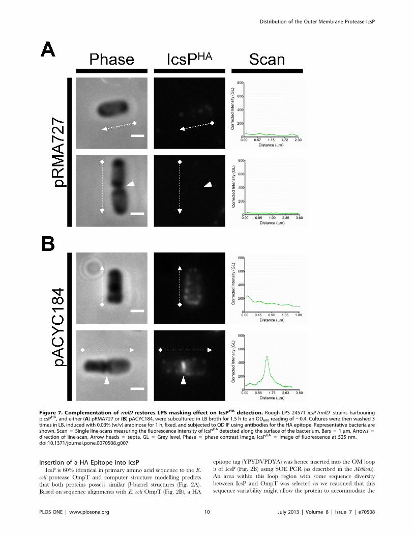
Figure 7. Complementation of rmlD restores LPS masking effect on IcsPHA detection. Rough LPS 2457T icsP-/rmlD- strains harbouringpIcsPHA, and either (A) pRMA727 or (B) pACYC184, were subcultured in LB broth for 1.5 h to an OD600 reading of ,0.4. Cultures were then washed 3times in LB, induced with 0.03% (w/v) arabinose for 1 h, fixed, and subjected to QD IF using antibodies for the HA epitope. Representative bacteria areshown. Scan = Single line-scans measuring the fluorescence intensity of IcsPHA detected along the surface of the bacterium, Bars = 1 mm, Arrows =direction of line-scan, Arrow heads = septa, GL = Grey level, Phase = phase contrast image, IcsPHA = image of fluorescence at 525 nm.doi:10.1371/journal.pone.0070508.g007
Distribution of the Outer Membrane Protease IcsP
PLOS ONE | www.plosone.org 10 July 2013 | Volume 8 | Issue 7 | e70508

Figure 8. Quantification and statistical analysis of IcsPHA surface distribution. A total of 9 independent cultures of Smooth LPS 2457T icsP-
strains harbouring pIcsPHA or pBAD30 were subcultured in LB for 1.5 h to an OD600 reading of ,0.4, washed 3 times in LB, and then further culturedfor 2 in the presence of TP for Oag-depletion. Arabinose was included in the final hour of treatment at a concentration of 0.03% (w/v). A total of 9independent cultures of Rough LPS 2457T icsP-/rmlD- strains harbouring pIcsPHA or pBAD30 were also subcultured LB broth to an OD600 reading of,0.4, washed 3 times in LB, and induced with 0.03% (w/v) arabinose for 1 h. A sample from each culture was then fixed and subjected to QD IF usingantibodies for HA epitope and IcsA. Fluorescence intensity scans of IcsPHA (525 nm wavelength) were conducted on multiple bacteria from eachindependent sample and accumulated scans were used to create mean intensity profiles. A total 450 non-septating bacteria were scanned for eachstrain. A further 355, 256, 172, and 173 septating bacteria were scanned for Oag-depleted icsP- [pIcsPHA], Oag-depleted icsP- [pBAD30], icsP-/rmlD-
[pIcsPHA], and icsP-/rmlD- [pBAD30] respectively. Resultant IcsPHA mean surface profiles for (A) Oag-depleted icsP- and (B) icsP-/rmlD- bacteria areshown. Dotted and dashed vertical lines indicate mean positions of old poles for septating and non-septating bacteria respectively. The schematic (C)shows methodology of IcsPHA intensity scan directions from either new poles to old poles (as marked by IcsA) of non-septating bacteria, or septa to
Distribution of the Outer Membrane Protease IcsP
PLOS ONE | www.plosone.org 11 July 2013 | Volume 8 | Issue 7 | e70508

epitope insertion with little disturbance to the overall structure.
The OM loop 5 region was also selected to increase the chance of
surface detection by antibodies. OM loops 2 and 4 were not
selected for HA tag insertion to avoid the proposed catalytic
residues present in OmpT [38] which also exist in IcsP (Fig. 2C).
The IcsPHA (as well as IcsP) coding regions were placed in front of
the pBAD promoter in pBAD30 [39] to allow expression control
with arabinose. Expression of IcsP/IcsPHA was confirmed by
Western immunoblotting with anti-IcsP or anti-HA antibodies
(Fig. 3A & B, lanes WM and Fig. 1A & C).
IcsPHA Activity on IcsATo determine whether insertion of a HA epitope into the OM of
IcsP affected IcsP’s protease activity on IcsA, pellet and
supernatant protein preparations of 2457T, icsP- and arabinose-
induced icsP- strains expressing pIcsPHA, pIcsP and pBAD30 were
subjected to Western immunoblotting with an anti-IcsA antibody.
The full length 120 kDa IcsA protein was detected in cell pellet
samples of all strains as expected (Fig. 1B, lanes 1, 3, 5, 7 and 9),
while the 95 kDa cleaved form of IcsA was only detected in the
supernatant sample of 2457T, icsP- [pIcsPHA] and icsP- [pIcsP]
(Fig. 1B, lanes 2, 6 & 8). These results suggest that the insertion of
the HA tag into the OM loop 5 of IcsP does not affect its ability to
cleave IcsA, and hence the IcsPHA protein is functional.
Localisation of IcsP/IcsPHA Protein to the OMSince IcsPHA was modified compared to IcsP and expressed
from the pBAD promoter, the presence of IcsPHA protein
exclusively in the OM was confirmed by using sucrose density
gradient centrifugation. The WM of icsP- [pIcsPHA] and icsP-
[pIcsP] were fractionated into CM and OM on sucrose gradients,
and fractions subjected to SDS 15% polyacrylamide gel electro-
phoresis, prior to visualisation by Coomassie Blue staining and
Western immunoblotting with anti-HA and anti-IcsP. Analysis of
the sucrose gradient samples showed that fractions which were
enriched with OM proteins (OmpF, OmpC, and OmpA) [40]
contained the majority of the 36 kDa IcsPHA and IcsP proteins
(Fig. 3A & B, lanes 2 to 5). These results indicate that IcsPHA is
localised to the OM similar to IcsP, and that the HA insertion did
not result in any dramatic disruption of IcsP protein localisation.
Detection of Arabinose Induced IcsPHA Expression byWestern Immunoblotting
Wild-type S. flexneri 2457T showed optimal expression levels of
native IcsP at 3 h (Fig. 1A). To investigate the conditions required
for comparable IcsPHA expression, whole cell lysate samples were
prepared from icsP- [pIcsPHA] and icsP- [pBAD30] induced with
0%, 0.003%, 0.006%, 0.0125%, 0.025%, 0.05%, 0.1% or 0.2%
(w/v) arabinose for 1 h. Western immunoblotting with anti-IcsP
showed that a band of ,36 kDa was detected for 2457T and icsP-
[pIcsPHA] induced with 0.025%, 0.05%, 0.1% and 0.2% (w/v) (w/
v) arabinose (Fig. 1C, lanes 1, 3–6), with expression levels
comparable to native IcsP for icsP- [pIcsPHA] observed between
0.025%–0.05% (w/v) arabinose induction (Fig. 1C, lanes 5 & 6).
Induction at 0.03% (w/v) arabinose was hence chosen for all
subsequent experiments.
Cell Surface Detection of IcsPHA Distribution in S. flexneri2457T and the Effect of Tunicamycin Treatment
Having established an induction protocol that closely approx-
imates the level of expression of IcsPHA to native IcsP, we next
attempted to detect IcsPHA at these levels on the surface of 2457T
icsP- using indirect QD IF microscopy. Strains icsP- [pIcsPHA] and
icsP- [pBAD30] were cultured with 0.03% (w/v) arabinose, fixed,
and then probed for HA using a primary anti-HA antibody and a
secondary QD 525 conjugated antibody. However, no IcsPHA was
detected on the cell surface, and intensity scans of icsP- [pIcsPHA]
were equivalent to icsP- [pBAD30] (Fig. S1).
Since IcsPHA could not be detected on the cell surface of icsP-
[pIcsPHA], we reasoned that the presence of LPS Oag may mask
the detection of surface IcsP as this has previously been shown for
IcsA [21]. An LPS Oag depletion-regeneration assay [29] was
hence carried out on icsP- [pIcsPHA] and icsP- [pBAD30] induced
with 0.03% (w/v) arabinose, followed by IF labelling. This assay
involves the use of an inhibitor of the WecA enzyme necessary for
Oag subunit biosynthesis using tunicamycin, and polymyxin B
nonapeptide (PMBN) was used to improve OM penetration. Upon
removal of these two chemicals from growing bacteria, LPS Oag is
regenerated [29]. Analysis of the resulting LPS by SDS-PAGE and
silver staining showed that icsP- [pIcsPHA] and icsP- [pBAD30]
samples treated with tunicamycin/PMBN (TP) had depleted LPS
Oag (Fig. 4A, lanes 5 & 6), with Oag production restored (R) upon
removal of TP (Fig. 4A, lanes 7 & 8). Untreated (U) and PMBN
treated (P) samples showed no inhibition of Oag biosynthesis as
expected (Fig. 4A, lanes 1–4).
Following successful depletion of LPS Oag, QD IF microscopy
was performed on the above mentioned samples. IcsPHA could not
be detected on untreated or PMBN treated icsP- [pIcsPHA] cells, as
expected (Fig. S2A). However, surface IcsPHA was detected on
icsP- [pIcsPHA] cells treated with TP, suggesting that LPS Oag is
able to mask antibody accessibility to IcsPHA (Fig. S2A).
Interestingly, IcsPHA appeared to be distributed asymmetrically
over the cell surface of the majority of cells examined and
fluorescence intensity line-scans revealed that IcsPHA localised
preferentially to one pole of non-septating cells and the septa of
septating cells (Fig. S2A). To determine if the distribution of
IcsPHA on the cell surface segregated to either the new pole or the
old pole, additional staining of the bacteria was conducted with
anti-IcsA antibodies since IcsA is known to localise to the old cell
pole [5]. Again, IcsPHA could not be detected on untreated or
PMBN treated icsP- [pIcsPHA] cells (Fig. 5A & B) but could be
detected after TP treatment (Fig. 5C). Peak IcsPHA detection was
consistently observed at the new pole (opposing IcsA at the old
pole) and at the septum of septating bacteria as shown by line-
scans (Fig. 5C). As expected, no surface IcsPHA was detected for
untreated, PMBN treated, or TP treated samples of icsP-
[pBAD30] when stained for IcsPHA (Fig. S2B) or double stained
for IcsPHA and IcsA (Fig. S3). Notably, IcsA on icsP- [pBAD30]
was detected at higher amounts laterally and at the septa of
bacteria (Fig. S3) as previously seen for DicsP strains [13].
Cell Surface Detection of IcsPHA Distribution in RoughLPS S. flexneri 2457T
To further investigate the effect of Oag on IcsP distribution, a
2457T icsP2/rmlD- double mutant was constructed to indepen-
old poles of septating bacteria. Student’s two-tailed t-tests (D) were also conducted on the fold differences of mean IcsPHA intensities betweendiscrete positions (new pole, old pole, and septum) of Oag-depleted icsP- and icsP-/rmlD- bacteria. SEM = standard error of the mean, GL = Greylevel, N = non-septating cells, S = septating cells, P = p-value, ns = not significant.doi:10.1371/journal.pone.0070508.g008
Distribution of the Outer Membrane Protease IcsP
PLOS ONE | www.plosone.org 12 July 2013 | Volume 8 | Issue 7 | e70508

dently assess the distribution of IcsPHA in a rough LPS
background. Mutation of the rmlD gene in S. flexneri results in a
strain which is unable to synthesise the precursor deoxythymidine
diphosphate (dTDP)- rhamnose required for Oag repeat units and,
hence, results in a rough LPS phenotype [29,41]. Analysis of the
resulting LPS conferred by icsP2/rmlD- strains expressing pIcsPHA
and pBAD30 showed that rough LPS was observed for both (Fig.
4B, lanes 1 & 2), with a band consistent with the size of the IcsPHA
protein (36 kDa) detected only in the icsP2/rmlD- [pIcsPHA]
sample by Western immunoblotting, as expected (Fig. 4C, lane 1).
Fixed samples of icsP2/rmlD- [pIcsPHA] and icsP2/rmlD-
[pBAD30] cells were then probed for both IcsPHA and IcsA using
the same QD IF staining protocol as previously conducted for
smooth strains. Similarly to LPS Oag-depleted icsP- [pIcsPHA],
IcsPHA was detected on the bacterial surface most predominately
at the new pole (Fig. 6A) and the septum (Fig. 6B). Again, the
majority of septating cells had higher peak IcsPHA intensity at the
septum than new poles of non-septating cells (line-scans Fig. 6A &
B). Single staining of these cells for IcsPHA was also conducted and
yielded the same localisation results (Fig. S4). As expected, IcsPHA
was not detected on the icsP2/rmlD- [pBAD30] strain in IF
microscopy experiments when either single (Fig. S4) or double
stained (Fig. 6B). Again, IcsA on icsP2/rmlD- [pBAD30] was
detected at higher amounts laterally and at the septum (Fig. 6A &
B).
To again demonstrate the effect of LPS Oag masking of IcsPHA,
smooth LPS structure was restored in the icsP2/rmlD- strain
expressing pIcsPHA by transforming pRMA727 carrying a
functional rmlD gene. Analysis of the resulting LPS conferred by
icsP2/rmlD-[pIcsPHA][pRMA727] showed restored smooth LPS
phenotype (Fig. 4B, lane 3), and when probed for IcsPHA by QD
IF microscopy, was barely detectable (Fig. 7A). The control icsP2/
rmlD- [pIcsPHA] strain carrying pACYC184 conferred a rough
LPS phenotype (Fig. 4B, lane 4) and IcsPHA was detected by IF
microscopy with the same distribution observed previously (Fig.
7B).
Multi-cell Line-scan Analysis of IcsPHA SurfaceDistribution
To determine if the observed sub-cellular preference of IcsPHA
to the new poles and septa is a statistically significant phenom-
enon, cumulative QD IF line-scan analyses on both IcsPHA/IcsA
double stained LPS Oag-depleted icsP- strains and rough icsP2/
rmlD- strains was conducted for a larger population of cells.
Scanning was initiated from the new pole to the old pole (as
marked by IcsA) for non-septating cells, and from the septum to
the old pole for septating cells (Fig 8C). From 9 independent
samples of LPS Oag-depleted icsP- [pIcsPHA] and icsP- [pBAD30],
a total of 450 non-septating cells each were line-scanned.
Additionally a total of 355 and 256 septating cells were scanned
for icsP- [pIcsPHA] and icsP- [pBAD30] respectively. The resultant
mean fluorescence intensity profiles show that IcsPHA is preferen-
tially localised at the new pole and tends to gradually decrease
towards the old pole on non-septating cells expressing pIcsPHA
(Fig. 8A). For septating cells, IcsPHA mean intensity is localised
highest at the septum and declines more steeply towards the old
pole (Fig. 8A). As expected, the intensity profiles of icsP- [pBAD30]
cells were at a negligible level (Fig. 8A). Statistical analysis of mean
IcsPHA intensity at discrete bacterial positions of LPS-depleted
icsP- [pIcsPHA] confirmed that the localisation of IcsPHA is: (i) 2-
fold higher at the new pole of non-septating cells than the old pole
(P = 0.004), (ii) 2.9-fold higher at the septum of septating cells than
the old pole (P,0.0001), and (iii) 1.5-fold higher at septa of
septating cells compared to new poles of non-septating cells
(P = 0.038) (Fig. 8D).
For the rough LPS strains, 9 independent samples of icsP2/
rmlD- [pIcsPHA] and icsP2/rmlD- [pBAD30] were investigated with
a total of 450 non-septating cells each line-scanned. Additionally, a
total of 172 and 173 septating cells were scanned for icsP2/rmlD-
[pIcsPHA] and icsP2/rmlD- [pBAD30] respectively. Again, the
mean intensity profiles of non-septating cells expressing pIcsPHA
shows IcsPHA is preferentially localised at the new pole and tends
Figure 9. Model for differential inheritance of spatiallyseparated OM proteins IcsA and IcsP. Successive generations ofS. flexneri are shown including: septating cells, newly formed daughters,and elongating cells. Solid black, red, and blue arrows denote theevents of growth/elongation, septation, and division respectively. IcsAlocalisation is represented by red shading and IcsP by green shadingwhere the darker the shading indicates higher protein concentration inthe OM. The bottom graph represents IcsP distributions and gradientsthat result from hypothetical line-scans of: (a) a septating cell, (b) anewly formed daughter cell, and (c) an elongated cell from this model.IcsP on septating cells (a) declines steeply from the septum towards theold pole, whereas it’s gradient on elongated cells (b) is more moderatebetween poles. A newly formed cell (c) would have an intermediategradient. Dotted arrow = direction of hypothetical line-scan.doi:10.1371/journal.pone.0070508.g009
Distribution of the Outer Membrane Protease IcsP
PLOS ONE | www.plosone.org 13 July 2013 | Volume 8 | Issue 7 | e70508

to gradually decrease towards the old pole (Fig. 8B). Likewise,
IcsPHA mean intensity of septating cells is localised highest at the
septum and declines very steeply towards the old pole (Fig. 8B). As
expected, the mean intensity profiles of icsP2/rmlD- [pBAD30]
were at a negligible level (Fig. 8B). Statistical analysis of mean
IcsPHA intensity at discrete bacterial positions of icsP2/rmlD-
[pIcsPHA] again confirmed that the localisation of IcsPHA is: (i) 2.2-
fold higher at the new pole of non-septating cells than the old pole
(P,0.0001), (ii) 3.6-fold higher at the septum of septating cells than
the old pole (P,0.0001), and (iii) 1.7-fold higher at septa of
septating cells compared to new poles of non-septating cells
(P = 0.0002) (Fig. 8D). Although, IcsPHA fluorescence intensity was
an average of 2.5 times higher on icsP2/rmlD- [pIcsPHA] cells than
LPS-depleted icsP- [pIcsPHA] cells, the ratios of intensity between
bacterial positions were comparable to the respective fold-changes
observed for LPS Oag-depleted icsP- [pIcsPHA], suggesting that
IcsPHA is localised similarly in both types of Oag deficient cells.
The lower level of IcsPHA detection on LPS-depleted icsP-
[pIcsPHA] compared to icsP2/rmlD- [pIcsPHA] indicates that TP
treatment is not 100% efficient in inhibiting Oag synthesis.
Discussion
The cell surface distribution of neither IcsP, or any other
member of the Omptin family, had not been previously
determined. In this study we investigated the distribution of IcsP
on the cell surface of S. flexneri 2a 2457T using a HA-tagged IcsP
protein (Fig. 2) under pBAD control. Characterisation of IcsPHA
showed that it was functionally able to cleave IcsA, and was
secreted into the OM comparably to IcsP (Fig. 1B & 3). However,
when IcsPHA was expressed at native IcsP equivalent levels (Fig.
1A & C), it was undetectable in the OM via QD IF microscopy in
smooth LPS S. flexneri (Fig. S1) but detectable on both LPS Oag-
depleted and rough LPS Shigella bacteria (Fig. 4, 5 & 6).
Furthermore, this masking effect was restored in rough strains
upon complementation of rmlD (Fig. 4B & 7). We suggest that the
long LPS Oag chains of smooth strains sterically hinder the
accessibility of antibodies to the OM surface. This type of protein
masking by LPS Oag has also been shown for IcsA in 2457T [21],
and several other OM proteins [22,23]. It is interesting to note that
multi-cell line-scan analysis of IcsPHA detection on the cell surface
showed that IcsPHA fluorescence intensity was higher for rough
LPS cells than for LPS Oag-depleted cells (Fig. 8). Similar to its
Omptin homolog, OmpT, IcsP possesses most of the putative LPS-
binding sites found in OmpT (Fig. 2C) [38] and may also interact
with LPS. Since TP is only partly effective at blocking LPS Oag
synthesis, it is possible that the few LPS Oag molecules synthesised
are still closely bound to IcsP. This may cause a small amount of
masking and may explain the lower fluorescence intensity
observed on LPS Oag-depleted cells (Fig. 8A) compared to the
rough LPS cells (Fig. 8B).
IcsA distributions in DicsP strains previously provided indirect
evidence to suggest that IcsP is distributed equally over the cell
surface of S. flexneri [12,13,42]. However, once the masking effect
of LPS Oag was circumvented, we were able to extensively study
the distribution profile of IcsPHA in the OM. This work has shown
that IcsPHA (and likely IcsP) has an asymmetrical distribution
which may be cell cycle dependent. On dividing cells IcsP
accumulates at high concentration at the septum and then declines
steeply towards the old pole of the emerging daughter cell.
However, once divided, detection of IcsP at the new pole (the pole
derived from the septum) of daughter cells decreased significantly
and then the decline towards the old pole is more moderate. To
explain this, we propose that IcsP is directed to septa of dividing
cells and that daughter cells ‘inherit’ a higher concentration of IcsP
at new poles (model Fig. 9). As the daughter cell elongates, IcsP is
laterally diluted, resulting in the gentle gradient on non-septating
cells. In this model, lateral dilution of IcsP may occur by
membrane insertion events during cell elongation similarly to that
described for E. coli LamB [43]. This model predicts increased
cleavage of IcsA at the septum and lateral regions of the cell in
order to help set up a relatively higher concentration of IcsA at the
old pole and to maintain the old pole preference of IcsA in
daughter cells. Additionally, the model may explain, and also fits,
the common observation that IcsA distributions and intensities
vary between cells in a given population of the same strain.
Many proteins are known to accumulate at the inner membrane
of the mid-cell in order to mediate septum formation and cell
division – for instance, FtsZ polymerises at the cytoplasmic face to
form the Z-ring and attract further divisome components [44].
However, few OM proteins have been shown to concentrate to the
septum. A notable exception to this is the OM lipoprotein LpoB
which localises as distinct foci at septa and complexes with
periplasmic penicillin binding protein 1B tethering it to other
divisome components [44,45]. Interestingly, septal localisation of
LpoB is lost when peptidoglycan synthesis is inhibited [45].
Whether the localisation of integral OM protein IcsP to the septal
OM requires similar interactions with divisome components, or is
dependent on peptidoglycan synthesis, remains to be investigated.
In summary, this work has shown for the first time the surface
localisation of IcsP, a member on the Omptin family of OM
proteases. We have observed that: (i) the distribution of IcsP is
masked by LPS Oag in S. flexneri 2457T, and (ii) IcsP is
concentrated at new poles and at the septum of dividing cells.
This distribution of IcsP explains the observed IcsA localisation
defect in DicsP strains [11,12]. We have also proposed a model to
explain the inheritance of OM proteins IcsP and IcsA through
generations of cell division. Finally, unmasking of surface antigens
via LPS Oag-depletion may be useful in the study of other
minimally exposed OM proteins.
Supporting Information
Figure S1 Inability to detect IcsPHA on the cell surface of2457T icsP-. Smooth LPS 2457T icsP- strains expressing pIcsPHA
(left column) or pBAD30 (right column) were subcultured in LB for
1.5 h to an OD600 reading of ,0.4. Cultures were then washed 3
times in LB, induced with 0.03% (w/v) arabinose for 1 h, fixed,
and subjected to QD IF using antibodies for HA epitope and IcsA.
Representative bacteria are shown. Scan = Single line-scans
measuring the intensity of IcsPHA detected along the surface of
the bacterium, Bars = 1 mm, Arrows = direction of line-scan,
GL = Grey level, Phase = phase contrast image, IcsPHA = image
of fluorescence at 525 nm.
(TIF)
Figure S2 Single IcsPHA staining of LPS-depleted 2457TicsP-. Smooth LPS 2457T icsP- strains harbouring (A) pIcsPHA or
(B) pBAD30 were subcultured in LB broth for 1.5 h to an OD600
reading of ,0.4, washed 3 times in LB, and then further cultured
for 2 h in either: the absence of TP, in the presence of PMBN
only, or in the presence of TP. Arabinose was included in the final
hour of treatment at a concentration of 0.03% (w/v). Samples
were then fixed and subjected to QD IF using antibodies to HA
epitope. Representative non-septating and septating cells are
shown for each treatment group. Scan = Single line-scans
measuring the fluorescence intensity of IcsPHA detected along
the surface of the bacterium, Bars = 1 mm, Arrows = direction of
line-scan, Arrow heads = septa, Control = grown in absence of
Distribution of the Outer Membrane Protease IcsP
PLOS ONE | www.plosone.org 14 July 2013 | Volume 8 | Issue 7 | e70508

both tunicamycin and PMBN, P = PMBN, TP = tunicamycin/
PMBN, GL = Grey level, Phase = phase contrast image, IcsPHA = -
image of fluorescence at 525 nm
(TIF)
Figure S3 Double stained IF of LPS depleted 2457T icsP-
[pBAD30]. Smooth LPS 2457T icsP- strains harbouring pBAD30
were subcultured in LB broth for 1.5 h to an OD600 reading of
,0.4, washed 3 times in LB, and then further cultured for 2 h; (A)
in the absence of TP, (B) in the presence of PMBN only, or (C) in
the presence of TP. Arabinose was included in the final hour of
treatment at a concentration of 0.03% (w/v). Samples were then
fixed and subjected to QD IF using antibodies to HA epitope and
IcsA. Non-septating and septating cells (upper and lower rows
respectively) are shown for each treatment group. Representative
bacteria are shown. Scan = Single line-scans measuring the
fluorescence intensity of IcsPHA detected along the surface of the
bacterium, Bars = 1 mm, Arrows = direction of line-scan, Arrow
heads = septa, Control = grown in absence of both tunicamycin
and PMBN, P = PMBN, TP = tunicamycin/PMBN, GL = Grey
level, Phase = phase contrast image, IcsPHA = image of fluores-
cence at 525 nm, IcsA = image of fluorescence at 625 nm,
Merge = overlay of IcsPHA and IcsA images.
(TIF)
Figure S4 Single IcsPHA staining of 2457T icsP2/rmlD-.Rough LPS 2457T icsP2/rmlD- strains harbouring pIcsPHA (left
columns) or pBAD30 (right columns) were subcultured LB broth
for 1.5 h to an OD600 reading of ,0.4. Cultures were then washed
3 times in LB, induced with 0.03% (w/v) arabinose for 1 h, fixed,
and subjected to QD IF using antibodies for HA epitope. Non-
septating and septating life stages are shown in A and Brespectively. Representative bacteria are shown. Scan = Single
line-scans measuring the fluorescence intensity of IcsPHA detected
along the surface of the bacterium, Bars = 1 mm, Arrows =
direction of line-scan, Arrow heads = septa, GL = Grey level,
Phase = phase contrast image, IcsPHA = image of fluorescence at
525 nm.
(TIF)
Author Contributions
Conceived and designed the experiments: ENHT MTD RM. Performed
the experiments: ENHT MTD. Analyzed the data: ENHT MTD.
Contributed reagents/materials/analysis tools: RM. Wrote the paper:
ENHT MTD.
References
1. LaBrec E, Schneider H, Magnani T, SB F (1964) Epithelial cell penetration as
an essential step in the pathogenesis of bacillary dysentery. J Bacteriol 88: 1503–
1518.
2. Suzuki T, Sasakawa C (2001) Molecular basis of the intracellular spreading of
Shigella. Infect Immun 69: 5959–5966.
3. Robbins JR, Monack D, McCallum SJ, Vegas A, Pham E, et al. (2001) The
making of a gradient: IcsA (VirG) polarity in Shigella flexneri. Mol Microbiol 41:
861–872.
4. Bernardini ML, Mounier J, d’Hauteville H, Coquis-Rondon M, Sansonetti PJ
(1989) Identification of icsA, a plasmid locus of Shigella flexneri that governs
bacterial intra- and intercellular spread through interaction with F-actin. Proc
Natl Acad Sci U S A 86: 3867–3871.
5. Goldberg MB, Barzu O, Parsot C, Sansonetti PJ (1993) Unipolar localization
and ATPase activity of IcsA, a Shigella flexneri protein involved in intracellular
movement. J Bacteriol 175: 2189–2196.
6. Lett MC, Sasakawa C, Okada N, Sakai T, Makino S, et al. (1989) virG, a
plasmid-coded virulence gene of Shigella flexneri: identification of the virG
protein and determination of the complete coding sequence. J Bacteriol 171:
353–359.
7. Jain S, van Ulsen P, Benz I, Schmidt MA, Fernandez R, et al. (2006) Polar
localization of the autotransporter family of large bacterial virulence proteins.
J Bacteriol 188: 4841–4850.
8. Dworkin J (2009) Cellular polarity in prokaryotic organisms. Cold Spring Harb
Perspect Biol 1: a003368.
9. Fukuda I, Suzuki T, Munakata H, Hayashi N, Katayama E, et al. (1995)
Cleavage of Shigella surface protein VirG occurs at a specific site, but the
secretion is not essential for intracellular spreading. J Bacteriol 177: 1719–1726.
10. Goldberg MB, Theriot JA (1995) Shigella flexneri surface protein IcsA is
sufficient to direct actin-based motility. Proc Natl Acad Sci U S A 92: 6572–
6576.
11. Egile C, d’Hauteville H, Parsot C, Sansonetti PJ (1997) SopA, the outer
membrane protease responsible for polar localization of IcsA in Shigella flexneri.
Mol Microbiol 23: 1063–1073.
12. Shere KD, Sallustio S, Manessis A, D’Aversa TG, Goldberg MB (1997)
Disruption of IcsP, the major Shigella protease that cleaves IcsA, accelerates
actin-based motility. Mol Microbiol 25: 451–462.
13. Steinhauer J, Agha R, Pham T, Varga AW, Goldberg MB (1999) The unipolar
Shigella surface protein IcsA is targeted directly to the bacterial old pole: IcsP
cleavage of IcsA occurs over the entire bacterial surface. Mol Microbiol 32: 367–
377.
14. Kaufmann A, Stierhof YD, Henning U (1994) New outer membrane-associated
protease of Escherichia coli K-12. J Bacteriol 176: 359–367.
15. Lutkenhaus J, Addinall SG (1997) Bacterial cell division and the Z ring. Annu
Rev Biochem 66: 93–116.
16. Tamaki S, Matsuzawa H, Matsuhashi M (1980) Cluster of mrdA and mrdB
genes responsible for the rod shape and mecillinam sensitivity of Escherichia coli.
J Bacteriol 141: 52–57.
17. Chatterjee S, Rothenberg E (2012) Interaction of bacteriophage l with its E. coli
receptor, LamB. Viruses 4: 3162–3178.
18. Gibbs KA, Isaac DD, Xu J, Hendrix RW, Silhavy TJ, et al. (2004) Complexspatial distribution and dynamics of an abundant Escherichia coli outer
membrane protein, LamB. Mol Microbiol 53: 1771–1783.
19. Lynne AM, Skyberg JA, Logue CM, Nolan LK (2007) Detection of Iss and Boron the surface of Escherichia coli. J Appl Microbiol 102: 660–666.
20. Ghosh AS, Young KD (2005) Helical disposition of proteins and lipopolysac-
charide in the outer membrane of Escherichia coli. J Bacteriol 187: 1913–1922.
21. Morona R, Van Den Bosch L (2003) Lipopolysaccharide O antigen chains maskIcsA (VirG) in Shigella flexneri. FEMS Microbiol Lett 221: 173–180.
22. Van der Ley P, De Graaff P, Tommassen J (1986) Shielding of Escherichia coli
outer membrane proteins as receptors for bacteriophages and colicins by O-antigenic chains of lipopolysaccharide. J Bacteriol 168: 449–451.
23. Van der Ley P, Kuipers O, Tommassen J, Lugtenberg B (1986) O-antigenic
chains of lipopolysaccharide prevent binding of antibody molecules to an outermembrane pore protein in Enterobacteriaceae. Microb Pathog 1: 43–49.
24. Voorhis DL, Dillon S, Formal SB, Isberg RR (1991) An O antigen can interfere
with the function of the Yersinia pseudotuberculosis invasin protein. MolMicrobiol 5: 317–325.
25. Sandlin RC, Lampel KA, Keasler SP, Goldberg MB, Stolzer AL, et al. (1995)
Avirulence of rough mutants of Shigella flexneri: requirement of O antigen forcorrect unipolar localization of IcsA in the bacterial outer membrane. Infect
Immun 63: 229–237.
26. Van Den Bosch L, Manning PA, Morona R (1997) Regulation of O-antigen
chain length is required for Shigella flexneri virulence. Mol Microbiol 23: 765–775.
27. Morona R, Van Den Bosch L, Manning PA (1995) Molecular, genetic, and
topological characterization of O-antigen chain length regulation in Shigella
flexneri. J Bacteriol 177: 1059–1068.
28. Purins L, Van Den Bosch L, Richardson V, Morona R (2008) Coiled-coil
regions play a role in the function of the Shigella flexneri O-antigen chain length
regulator WzzpHS2. Microbiology 154: 1104–1116.
29. Teh MY, Tran ENH, Morona R (2012) Absence of O antigen suppressesShigella flexneri IcsA autochaperone region mutations. Microbiology-Sgm 158:
2835–2850.
30. Horton RM (1993) In Vitro Recombination and Mutagenesis of DNA (Chapter25). In: White BA, editor. Methods in Molecular Biology, Vol 15: PCR Protocols:
Current Methods and Applications. Totowa, NJ: Humana Press Inc. 251–261.
31. Horton RM, Cai ZL, Ho SN, Pease LR (1990) Gene splicing by overlapextension: tailor-made genes using the polymerase chain reaction. Biotechniques
8: 528–535.
32. Datsenko KA, Wanner BL (2000) One-step inactivation of chromosomal genesin Escherichia coli K-12 using PCR products. Proc Natl Acad Sci U S A 97:
6640–6645.
33. Lugtenberg B, Meijers J, Peters R, van der Hoek P, van Alphen L (1975)Electrophoretic resolution of the ‘‘major outer membrane protein’’ of Escherichia
coli K12 into four bands. FEBS Lett 58: 254–258.
34. Osborn MJ, Munson R (1974) Separation of the inner (cytoplasmic) and outermembranes of Gram-negative bacteria. Methods Enzymol 31: 642–653.
35. May KL, Morona R (2008) Mutagenesis of the Shigella flexneri autotransporter
IcsA reveals novel functional regions involved in IcsA biogenesis and recruitmentof host neural Wiscott-Aldrich syndrome protein. J Bacteriol 190: 4666–4676.
Distribution of the Outer Membrane Protease IcsP
PLOS ONE | www.plosone.org 15 July 2013 | Volume 8 | Issue 7 | e70508
36. Murray GL, Attridge SR, Morona R (2003) Regulation of Salmonella typhimurium
lipopolysaccharide O antigen chain length is required for virulence; identifica-tion of FepE as a second Wzz. Mol Microbiol 47: 1395–1406.
37. Papadopoulos M, Morona R (2010) Mutagenesis and chemical cross-linking
suggest that Wzz dimer stability and oligomerization affect lipopolysaccharideO-antigen modal chain length control. J Bacteriol 192: 3385–3393.
38. Vandeputte-Rutten L, Kramer RA, Kroon J, Dekker N, Egmond MR, et al.(2001) Crystal structure of the outer membrane protease OmpT from
Escherichia coli suggests a novel catalytic site. Embo J 20: 5033–5039.
39. Guzman LM, Belin D, Carson MJ, Beckwith J (1995) Tight regulation,modulation, and high-level expression by vectors containing the arabinose
PBAD promoter. J Bacteriol 177: 4121–4130.40. Fehniger TE, Radolf JD, Walfield AM, Cunningham TM, Miller JN, et al.
(1986) Native surface association of a recombinant 38-kilodalton Treponemapallidum antigen isolated from the Escherichia coli outer membrane. Infect
Immun 52: 586–593.
41. Reeves PR, Hobbs M, Valvano MA, Skurnik M, Whitfield C, et al. (1996)Bacterial polysaccharide synthesis and gene nomenclature. Trends Microbiol 4:
495–503.
42. d’Hauteville H, Dufourcq Lagelouse R, Nato F, Sansonetti PJ (1996) Lack of
cleavage of IcsA in Shigella flexneri causes aberrant movement and allows
demonstration of a cross-reactive eukaryotic protein. Infect Immun 64: 511–517.
43. Ursell TS, Trepagnier EH, Huang KC, Theriot JA (2012) Analysis of surface
protein expression reveals the growth pattern of the gram-negative outer
membrane. PLoS Comput Biol 8: e1002680.
44. Egan AJ, Vollmer W (2013) The physiology of bacterial cell division.
Ann N Y Acad Sci 1277: 8–28.
45. Typas A, Banzhaf M, van den Berg van Saparoea B, Verheul J, Biboy J, et al.
(2010) Regulation of peptidoglycan synthesis by outer-membrane proteins. Cell
143: 1097–1109.
46. Brosius J (1989) Superpolylinkers in cloning and expression vectors. DNA 8:
759–777.
47. Rose RE (1988) The nucleotide sequence of pACYC184. Nucleic Acids Res 16: 355.
48. Osborn MJ, Munson R (1974) Separation of the inner (cytoplasmic) and outer
membranes of Gram-negative bacteria. Methods Enzymol 31: 642–653.
Distribution of the Outer Membrane Protease IcsP
PLOS ONE | www.plosone.org 16 July 2013 | Volume 8 | Issue 7 | e70508


