· · 2014-07-11 Polypyrrole/oligonucleotide nanocomposite ... This paper presents evidence and...
12
www.spm.com.cn Polypyrrole/oligonucleotide nanocomposite film: steric effect on DNA hybridization J-H Jiang 1 *, Y Li 1,2 , J Wang 1 , and K-L Paul Sung 1,3 1 Key Laboratory of Biorheological Science and Technology, Chongqing University, Chongqing, People’s Republic of China 2 Central Laboratory of Yongchuan Hospital, Chongqing Medical University, Chongqing, People’s Republic of China 3 Departments of Bioengineering and Orthopedic Surgery, University of California, San Diego, California, USA The manuscript was received on 21 March 2011 and was accepted after revision for publication on 26 May 2011. DOI: 10.1177/1740349911413956 Abstract: This paper presents evidence and some preliminary explanation for the existence of a steric effect in polypyrrole/oligonucleotide (PPy/ODN) nanocomposite film, which may be responsible for the high electrochemical sensing performance of that nanofilm when being used in DNA hybridization detection. When hybridization occurs in this nanocomposite film, the steric effect in the immediate interface of the hybrid film can damp the ion-flux penetrat- ing through this interface, thus resulting in the signal differentiation on the electrode electro- chemical property. Electrochemical cyclic voltammetry (CV) combined with atomic force microscope (AFM) were utilized to clarify the hindering mechanism occurring on such inter- face. Furthermore, the hybridization reaction of ODN probes in the hybrid PPy/ODN film with their complementary DNA sequences was measured via CV, with the target ODN concentra- tions as low as 5310 218 mol/L. Under optimized hybridization conditions the sensor response was almost linear, with the logarithm of the target ODN concentration ranging from 1310 218 to 1310 211 mol/L. These results may be useful in the development of a much simpler label- free DNA sensor based on the PPy/ODN nanocomposite film. Keywords: DNA, hybridization, polypyrrole, steric effect, cyclic voltammetry, label-free 1 INTRODUCTION The development of an analytical device capable of identifying specific DNA sequences in a fast, simple, and low-cost manner from the clinical sample has attracted considerable attention from many research groups and high technology companies. In this regard, the electrochemical DNA biosensor, which commonly relies on the hybridization reac- tion between the DNA target sequences in solution with the ODN probes immobilized on a conducting transducer surface, has become one of the more enabling techniques in the progress towards high sensitivity, low cost, ease of use, and especially in relation to the potential of miniaturization and automation [1–3]. Currently, the electrochemical DNA biosensor can be established through two approaches: indirect (label-dependent) and direct (label-free), depending on whether there is a need to introduce electroche- mical labels (indicators) or not. With the indirect route, some DNA-intercalating molecules, such as Hoechst 33258 [4] and methylene blue [5], are often employed for the indicator of sequence-specific hybridization event, due to their possession of a much higher affinity to the resulting hybrid. Some good redox labels such as enzyme [6], cationic metal complexes [7], and nanoparticles [8, 9] have been also utilized in such a way for DNA hybridization detection. Despite the relatively good detection *Corresponding author: Key Laboratory of Biorheological Science and Technology, Ministry of Education, Bioengineering College, Chongqing University, Chongqing, 400044, People’s Republic of China. email: [email protected] 1 Proc. IMechE Vol. 224 Part N: J. Nanoengineering and Nanosystems at NATIONAL UNIV SINGAPORE on August 7, 2011 pin.sagepub.com Downloaded from
Transcript of · · 2014-07-11 Polypyrrole/oligonucleotide nanocomposite ... This paper presents evidence and...
www.spm
.com
.cn
Polypyrrole/oligonucleotide nanocompositefilm: steric effect on DNA hybridizationJ-H Jiang1*, Y Li1,2, J Wang1, and K-L Paul Sung1,3
1Key Laboratory of Biorheological Science and Technology, Chongqing University, Chongqing, People’s Republic of
China2Central Laboratory of Yongchuan Hospital, Chongqing Medical University, Chongqing, People’s Republic of China3Departments of Bioengineering and Orthopedic Surgery, University of California, San Diego, California, USA
The manuscript was received on 21 March 2011 and was accepted after revision for publication on 26 May 2011.
DOI: 10.1177/1740349911413956
Abstract: This paper presents evidence and some preliminary explanation for the existence ofa steric effect in polypyrrole/oligonucleotide (PPy/ODN) nanocomposite film, which may beresponsible for the high electrochemical sensing performance of that nanofilm when beingused in DNA hybridization detection. When hybridization occurs in this nanocomposite film,the steric effect in the immediate interface of the hybrid film can damp the ion-flux penetrat-ing through this interface, thus resulting in the signal differentiation on the electrode electro-chemical property. Electrochemical cyclic voltammetry (CV) combined with atomic forcemicroscope (AFM) were utilized to clarify the hindering mechanism occurring on such inter-face. Furthermore, the hybridization reaction of ODN probes in the hybrid PPy/ODN film withtheir complementary DNA sequences was measured via CV, with the target ODN concentra-tions as low as 5310218 mol/L. Under optimized hybridization conditions the sensor responsewas almost linear, with the logarithm of the target ODN concentration ranging from 1310218
to 1310211 mol/L. These results may be useful in the development of a much simpler label-free DNA sensor based on the PPy/ODN nanocomposite film.
Keywords: DNA, hybridization, polypyrrole, steric effect, cyclic voltammetry, label-free
1 INTRODUCTION
The development of an analytical device capable of
identifying specific DNA sequences in a fast, simple,
and low-cost manner from the clinical sample has
attracted considerable attention from many
research groups and high technology companies. In
this regard, the electrochemical DNA biosensor,
which commonly relies on the hybridization reac-
tion between the DNA target sequences in solution
with the ODN probes immobilized on a conducting
transducer surface, has become one of the more
enabling techniques in the progress towards high
sensitivity, low cost, ease of use, and especially in
relation to the potential of miniaturization and
automation [1–3].
Currently, the electrochemical DNA biosensor
can be established through two approaches: indirect
(label-dependent) and direct (label-free), depending
on whether there is a need to introduce electroche-
mical labels (indicators) or not. With the indirect
route, some DNA-intercalating molecules, such as
Hoechst 33258 [4] and methylene blue [5], are often
employed for the indicator of sequence-specific
hybridization event, due to their possession of a
much higher affinity to the resulting hybrid. Some
good redox labels such as enzyme [6], cationic metal
complexes [7], and nanoparticles [8, 9] have been
also utilized in such a way for DNA hybridization
detection. Despite the relatively good detection
*Corresponding author: Key Laboratory of Biorheological
Science and Technology, Ministry of Education, Bioengineering
College, Chongqing University, Chongqing, 400044, People’s
Republic of China.
email: [email protected]
1
Proc. IMechE Vol. 224 Part N: J. Nanoengineering and Nanosystems
at NATIONAL UNIV SINGAPORE on August 7, 2011pin.sagepub.comDownloaded from

www.spm
.com
.cn
sensitivity and low detection limit, these label-
dependent approaches have shortcomings of high
cost, longer manufacture time, and sophisticated
operation procedure [10]; moreover, the excess of
labels can introduce some interference, such as
affecting kinetics behaviour in the case of DNA
hybridization or changing the binding properties of
the labelled DNA target sequences [11]. In compari-
son, the label-free DNA electrochemical biosensor is
becoming more attractive, as it overcomes the
above-mentioned weak points [12, 13].
As demonstrated in the literature, the realization
of ‘label-free’ routes for electrochemical DNA bio-
sensors is largely through the electrical read-out of
signal transformation from the DNA hybridization
event to the electrode where the hybridization is
occurring [14, 15]. A key question is how to form an
effective coupling interface while presenting the
hybridization signal between nucleic acid bio-recog-
nition system and electronic transducer system. In
fact, polypyrrole (PPy), a classical conjugated con-
ducting polymer, holds particular promise for meet-
ing such requirements to a large extent. It has been
reported that ODN probes could be grafted or
absorbed on polypyrrole film and that the changes
in the electrochemical properties of the film once
DNA hybridization had occurred could be measured
[16–19]. ODN probes, especially, could serve as the
sole dopant during PPy electrochemical polymeriza-
tion, and maintain their hybridization activity and
affinity to target sequences within the host PPy net-
work, as already revealed by Wang [20, 21]. Based
on these facts, Cai et al. reported an indicator-free
DNA hybridization detection strategy via impedance
measurement on carbon nanotube-modified elec-
trode [14], and that the detection limit could be
decreased further when combined with the metalli-
zation of the helix DNA after hybridization [22].
More interestingly, a more direct electrochemical
sensor for fast reagent-free DNA detection has been
developed by chrono-amperometry [23]. Recently,
Tosar et al. [24] reported an electrochemical DNA
biosensor system, which contains two independent
direct detection methods for enhancing the specifi-
city of this biosensor system. As for the interpreta-
tion for the signal transduction mechanism in such
PPy/ODN sensors, these authors have presumed it
to be based either on the change of the capaci-
tance/conductivity of the PPy/ODN film [20, 23] or
on the increase in the resistance to electron transfer
by anions as a sequence of the negatively charged
sugar phosphate backbone of the target DNA [24].
Nevertheless, from the data, especially from the
atomic force microscope (AFM) measurements in
this paper, the underlying mechanism of the
hybridization signal transduction in the hybrid PPy/
ODN nanocomposite film sensor seems to be
deserved a re-clarified.
In this work, electrochemical cyclic voltammetry
(CV) and atomic force microscope (AFM) have been
used to monitor the electrochemical properties and
morphological information of PPy/ODN film before
and after hybridization in order to validate the
mechanism of hybridization response. From the
combination of the data of the two measurements,
the present authors think that there is a steric effect
derived from the hybridization event on the imme-
diate interface of the PPy/ODN nanocomposite film
that damps the ion flux penetrating through this
interface, and thus induces the signal differentiation
on the electrode electrochemical properties.
Therefore, a simple process of cyclic voltammetry
modulation on the ion flux and thus the steric effect
on the hybrid PPy/ODN nanocomposite film can
achieve the signal recognition of DNA hybridization;
see the illustration in Fig. 1. As a hypothesis, a brief
explanation is the following. When the ODN probes
entrapped in the PPy film hybridize with the incom-
ing complementary target sequences, they undergo
a conformational change from single-stranded
coiled states to relatively rigid double-stranded helix
states. This conformational change somewhat twists
the PPy backbone and the hybrid film network, thus
leading to the formation of a more compact struc-
ture at the immediate interface between the poly-
mer and solution – that is, the steric effect, which in
turn hinders the ion flux penetrating into this inter-
face during the redox process.
Through the validation of the above-mentioned
hypothesis, and optimization of hybridization con-
ditions on such hybrid PPy/ODN nanocomposite
film sensor, it is argued that this work not only pre-
sents a new mechanism interpretation for PPy/
ODN-based label-free DNA hybridization detection
but also provides a simplified DNA sensor format
convenient for practical applications.
Fig. 1 Schematic illustration for the steric effectmechanism presented on the interface ofhybrid PPy/ODN film after hybridization
2 J-H Jiang, Y Li, J Wang, and K-L Paul Sung
Proc. IMechE Vol. 224 Part N: J. Nanoengineering and Nanosystems
at NATIONAL UNIV SINGAPORE on August 7, 2011pin.sagepub.comDownloaded from
www.spm
.com
.cn
2 MATERIALS AND METHODS
2.1 Reagents
Pyrrole monomer (chemical grade) was purchased
from Sinopharm Chemical Reagent Co. Ltd (Shang-
hai, China) and was distilled until a colourless liquid
was obtained. It was stored in a refrigerator before
use. The following custom oligonucleotides, specific
to variola major virus, were purchased from Sheng-
gong Bioengineering Ltd (Shanghai, China): probe
(P1): 20-mer 5’-GCAATAGTAATCAGGTAGAG-3’; the
complementary target oligonucleotides (C1):
5’-CTCTACCTGATTACTATTGC-3’ and the non-
complementary control which has the same
sequence as the probe sequence P1. The stock solu-
tions for oligonucleotides were prepared in ultra-
pure water with Millipore system (18.2 MO) and
stored at a temperature of –20 �C prior to use. The
phosphate buffered saline (PBS: 137 nM NaCl,
2.7 mM KCl, 10 mM Na2HPO4, 2 mM KH2PO4), PH
7.4, was prepared with analytical grade reagents in
MiliQ water. Other reagents were used under com-
mon conditions, unless specified.
2.2 Experimental equipment
Electropolymerization and electrochemical detec-
tion experiments were realized by cyclic voltamme-
try in a home-made micro-electrochemical cell
(internal volume 100 ml) with indium tin oxide
(ITO)-coated glass (which had a surface resistivity of
95 O/square) as a working electrode (area of
0.49 cm2), a platinum wire coil as an auxiliary elec-
trode, and a Ag/AgCl wire as a reference electrode.
All electrochemical experiments were performed in
a CHI-800 electrochemical workstation (Shanghai
Chenhua Ltd, Shanghai, China). Electrochemical
electropolymerization solution was thoroughly
deoxygenated by N2 bubbling for 5 min before use.
In addition, AFM images were obtained by a CSPM
5000 system (Ben Yuan Ltd, Beijing, China). The
quantity of ODN immobilization was characterized
with a micro-spectrophotometer (K5500, Kaiao
Technology Development Co. Ltd, Beijing, China).
2.3 Pyrrole electropolymerization and probe
immobilization
The immobilization of ODN probes within polypyr-
role film was realized by an in-situ entrapment
route in which the ODN probes acted as the sole
dopant during the growth of PPy film on the elec-
trode surface. Nitrogen-bubbled electropolymeriza-
tion monomer solution of 100 mL containing 0.1M
pyrrole monomer and 131025 M oligonucleotide
(P1) was injected into the electrochemical cell to
obtain ODN/PPy composite film on the ITO elec-
trode surface by a continuous cyclic voltammtric
scanning between 0.0 and + 0.70 V (versus Ag/AgCl)
with 20 mV s21 scan rate. The amount of ODN probe
molecules immobilized into the PPy film was quan-
tified spectrophotometrically by comparing the
OD260 of electopolymerization solution before and
after the polymerization process. Before electropoly-
merization, the ITO-glass was successively cleaned
by ultra-sonication in acetone, ethanol and de-
ionized water for 5 min respectively, and then dried
by nitrogen steam. The resulting PPy/ODN nano-
composite film was characterized by cyclic voltam-
metry (CV) and AFM in tapping mode (240 mm-long
tetrahedral silicon cantilever with 2 N/m spring con-
stant, from Olympus, Japan).
2.4 Hybridization and electrochemical detection
Before hybridization, CV was applied to characterize
the PPy/ODN film modified ITO electrode between
20.40 to 0.40 V (versus Ag/AgCl) in PBS solution
with 50 mV s21 scan rate at room temperature. After
that, hybridization reaction was done by exposing
the PPy/ODN nanocomposite-film-modified ITO
electrode in the 100 mL PBS buffer contained with
the non-complementary oligonucleotide (1.0 3 1026
M) for 1 h at 38 �C. After washing, CVs were recorded
for evaluating any nonspecific interaction. In the
following, the PPy/ODN electrodes, which were pre-
pared with the same process, were incubated at the
same PBS solutions containing different concentra-
tions of complementary target DNA (from
1.0 3 10218 M – 1.0 3 10210 M) for 1 h at 38 �C. After
that, the electrode was washed three times by PBS
buffer solution to remove the unhybridized oligonu-
cleotides, and then CVs were recorded again for
electrochemical investigation of the hybridization
reaction similar to the characterizing procedure of
PPy/ODN before hybridization in the same PBS
solution. Unless otherwise stated, all electrochemi-
cal measurements were performed at room tem-
perature and registered until complete stabilization
of the CV signals. The last scan was taken into con-
sideration and analysed by ORIGIN 7.0 software
(Micro Software, Northampton, MA).
3 RESULTS AND DISCUSSION
3.1 Preparation of the PPy/ODN film
The hybrid PPy/ODN nanocomposite film was pre-
pared on the electrode surface by doping the ODN
probes within the electro-polymerized PPy, in which
Polypyrrole/oligonucleotide nanocomposite film 3
Proc. IMechE Vol. 224 Part N: J. Nanoengineering and Nanosystems
at NATIONAL UNIV SINGAPORE on August 7, 2011pin.sagepub.comDownloaded from
zhk
线条
zhk
线条
www.spm
.com
.cn
these probes acted as the sole counter ions during
the growth of the conducting film. Thus-formed film
could be considered as an efficient interface
between the nucleic acid recognition system and
the electrochemical transducer system. The electro-
polymerization of the PPy/ODN nanocomposite
film can be conducted by cyclic voltammetry or
constant potential [23]. In this paper, the desired
ODN probe was immobilized on the ITO electrode
surface by an adaptation of the methods described
first by Wang and co-workers [20], taking the opti-
mized parameters of Komarova et al. [23] into con-
sideration. It has been proved that 20-30-mer long
ODNs can serve as sole charge-compensating coun-
ter-ions during the electropolymerization of poly-
pyrrole and that the incorporation behaviour of
ODN molecules is similar to that of small inorganic
anions. In this way, the oligonucleotide has a maxi-
mum possible incorporation in the conductive
polymer and does not undergo any redox damage in
the potential range used for electropolymerization.
In addition, ODNs attached to the polypyrrole by
their phosphate backbone, and the nucleotide bases
are exposed to the solution and still retain their spe-
cific hybridization activity [24].
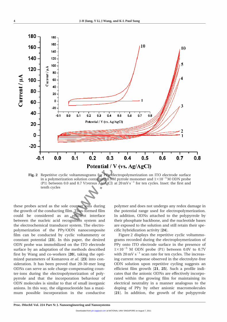
Figure 2 displays the repetitive cyclic voltammo-
grams recorded during the electroploymerization of
PPy onto ITO electrode surface in the presence of
131025 M ODN probe (P1) between 0.0V to 0.7V
with 20 mV s21 scan rate for ten cycles. The increas-
ing current response observed in the electrolyte-free
ODN solution upon repetitive cycling suggests an
efficient film growth [21, 25]. Such a profile indi-
cates that the anionic ODNs are effectively incorpo-
rated within the growing film for maintaining its
electrical neutrality in a manner analogous to the
doping of PPy by other anionic macromolecules
[21]. In addition, the growth of the polypyrrole
Fig. 2 Repetitive cyclic voltammograms for PPy electropolymerization on ITO electrode surfacein a polymerization solution containing 0.1 M pyrrole monomer and 131025 M ODN probe(P1) between 0.0 and 0.7 V(versus Ag/AgCl) at 20 mV s21 for ten cycles. Inset: the first andtenth cycles
4 J-H Jiang, Y Li, J Wang, and K-L Paul Sung
Proc. IMechE Vol. 224 Part N: J. Nanoengineering and Nanosystems
at NATIONAL UNIV SINGAPORE on August 7, 2011pin.sagepub.comDownloaded from
www.spm
.com
.cn
continues with the number of sweepings and in the
course of successive cycles monomer oxidation
occurs at less and less positive potential (inset of
Fig. 1). This change might contribute to the fact that
there was gradual decrease in the nucleation and
growth energy for PPy polymerization as the PPy/
ODN nanocomposite gradually formed per cycle.
To characterize the ODN probe entrapped within
the PPy, AFM was first used to obtain the morpholo-
gic information of the prepared PPy/ODN by tapping
mode at a 2 mm 3 2 mm scale with 2 Hz scan fre-
quency. In contrast, PPy/Cl film electropolymerized
in a solution containing 0.1M pyrrole monomer and
0.1M NaCl was also subjected to the same method as
a control. Figure 3(a) shows an AFM image of PPy/Cl
film electropolymerized on the ITO electrode surface,
while Fig. 3(b) shows an AFM image of PPy film
entrapped with the desired 20-mer ODN probes.
From Fig. 3, it can be inferred that the PPy films were
constructed by overlapping of PPy particles with
43.8 nm average diameter. Importantly, there
appeared some thin-coiled flocks randomly around
the PPy particles seen in Fig. 3(b). This phenomenon
leads to the result that the PPy/ODN has a smoother
surface with a surface roughness of 3.13 nm (root
mean square, RMS) than that of PPy/Cl with a sur-
face roughness of 5.47nm RMS. It is argued by the
present authors that the coiled flocks presented on
the surface of PPy film could be ODN probes. More
importantly, the visualization of ODN probes con-
firmed that part of the ODN probes immobilized by
this approach still present their complementary abil-
ity to their target DNA sequences.
The preparation reproducibility of the PPy/ODN
nanocomposite film was tested by the electrochemi-
cal activity of the resulted film measured by CV. The
degree of its reproducibility was estimated based on
the data derived from the area under the cyclic vol-
tammogram. The preparations of PPy/ODN were
conducted at different times (n = 6), and resulted in
an average CV area of 85.46 mC with standard devia-
tion (SD) of 5.53 mC and relative standard deviation
(RSD) of 6.47 per cent. In addition, the quality of the
ODN probes entrapped in the polypyrrole was deter-
mined by measuring the concentration of ODN in
the polymerization solution before and after the
electrodeposition process with a spectrophotometer,
and the result was 0.72 nmole with SD = 0.08 nmole
and RSD = 11.11 per cent.
3.2 DNA hybridization and its hindrance
effect to ion exchange kinetics
The DNA hybridization event occurring on the pre-
pared PPy/ODN nanocomposite film electrode and
its hindrance mechanism could be differentiated by
means of the combination of CV [26] and AFM.
Figure 4(a) shows the cyclic voltammograms of
PPy/ODN nanocomposite film obtained before and
after hybridization. Cyclic voltammogram a was the
result of the PPy/ODN nanocomposite film in PBS
solution before hybridization; cyclic voltammogram
b shows the result of the PPy/ODN nanocomposite
film again in the same PBS buffer after incubation
within non-complementary target ssDNA solution
for 1 h. No significant difference between these vol-
tammograms was obtained. Then the PPy/ODN
nanocomposite film coated ITO electrode was hybri-
dized with the complementary target for 1 h and,
after washing, the cyclic voltammogram (c) was
recorded. Comparing the cyclic voltammograms a
and c, a significant diminishment of area under the
cyclic voltammograms can be noted, which is closely
related to the ion-exchange kinetics of PPy/ODN
nanocomposite film during the redox process.
Why this result? First, it should be mentioned that
the amount of DNA strands with negative charge in
the PPy/ODN nanocomposite film electrode
increased with the inflowing of target DNA strands
after hybridization. This is the only observable fact in
the case of the PPy/ODN sensing system. Therefore,
the immediate question is how the addition of target
DNA strands could lead to the significant decreasing
of area under the cyclic voltammogram.
Does the electrostatic effect from the incoming
negative charges lead to the decrease of the ion-
exchange kinetics between the PPy/ODN nanocom-
posite film and solution? The present authors noted
that the entrapped ODN could not be readily
expelled from the PPy network under CV cycle as
the ODN probe could be considered as large
dopants [21]; thus, the hybridized double-strand
DNA (dsDNA) in the PPy network did also. As a
result, the movement of the electrolytic cation,
which follows
PPy(dsDNAn�)x(Na + )nx ! PPyx + (dsDNAn�)x
+ nxNa + + nxe�
dominates the electrochemically controlled ion-
exchange behaviour of the hybridized PPy/ODN
nanocomposite film. Therefore, the addition of neg-
ative charges by hybridization should not have a
positive effect on the diminishment of cation
exchange kinetic. Therefore, the electrostatic effect
could not be the main factor for the diminishment
in the ion-exchanging kinetics.
Moreover, it was noted, when comparing cyclic
voltammograms a and b in Fig. 4(a) more carefully,
that the area under cyclic voltammogram b is larger
Polypyrrole/oligonucleotide nanocomposite film 5
Proc. IMechE Vol. 224 Part N: J. Nanoengineering and Nanosystems
at NATIONAL UNIV SINGAPORE on August 7, 2011pin.sagepub.comDownloaded from

www.spm
.com
.cn
Fig. 3 AFM images of polypyrrole electropolymerization on ITO electrode surface by cyclicvoltammetry between 0.0–0.7 V (versus Ag/AgCl) for ten cylices with a 20 mV s21 scan ratein the polymerization solution containing (a) 0.1M pyrrole monomer and 1.0 M NaCl and(b) 0.1 M pyrrole monomer and 131025 M ODN probe
6 J-H Jiang, Y Li, J Wang, and K-L Paul Sung
Proc. IMechE Vol. 224 Part N: J. Nanoengineering and Nanosystems
at NATIONAL UNIV SINGAPORE on August 7, 2011pin.sagepub.comDownloaded from

www.spm
.com
.cn
Fig. 4 (a) Cyclic voltammograms recorded in PBS buffer solution between –0.4 V and 0.4 V (versusAg/AgCl) at a scan rate of 50 mv/s: curve a, CV of the PPy/ODN film before hybridization;curve b, CV of the same film as in curve a but in response to exposure to a 131026 M non-complementary target ssDNA for 1 h; curve c, CV response of the same film as in curve (a)after hybridization with 131026 M complementary target ssDNA for 1 h. The PPy/ODNwas prepared by 10 cycles. (B) AFM image of PPy/ODN film after hybridization with131026 M complementary target DNA for 1 h at 37 �C in PBS solution
Polypyrrole/oligonucleotide nanocomposite film 7
Proc. IMechE Vol. 224 Part N: J. Nanoengineering and Nanosystems
at NATIONAL UNIV SINGAPORE on August 7, 2011pin.sagepub.comDownloaded from

www.spm
.com
.cn
than that of cyclic voltammogram a by 7.2 per cent,
which means an enhancement of ion exchanging
kinetic after incubation with non-complementary
target. This might be due to the increase of the
polymer’s conductivity by the doping of non-
complementary ssDNA which adsorbed on the PPy/
ODN nanocomposite film non-specifically [24]. This
opposite signal appearance is similar to the result
reported by Wang et al. [20] and Komarova et al.
[23]. Recently, Tosar et al. also found that the ampli-
tude of PPy oxidation and reduction decreases after
hybridization with complementary target, while it
increases after incubation with a non-complemen-
tary target [24]. They explained this by the above-
mentioned electrostatic effect, that the decrease in
PPy peak contributed to the increase in the resis-
tance of electron transfer by electrostatic repulsing
towards anion incorporation, while the increase in
PPy peaks was due to increase in conduction of PPy
by doping. This point remains to be confirmed.
Taking into account also the data from AFM, it is
argued that there could be some hindrance effect on
the cation exchange kinetic, occurring immediately
after hybridization at the interface between PPy/
ODN and the solution under electrochemical CV pro-
cess. The existence of a hindrance barrier could
explain why the ion exchange kinetic is slowed down
by hybridization. For validation, AFM was used again
to characterize the morphological changes in PPy/
ODN after hybridization. Figure 4(b) shows the AFM
image of PPy/ODN nanocomposite film on ITO
electrode after hybridization. Comparing the AFM
images of Figs 3(b) and 4(b), it is clear that a signifi-
cant morphological change has occurred. Before
hybridization (Fig. 3(b)), the PPy/ODN nanocompo-
site film shows a relatively ordered and flat structure,
while after hybridization (Fig. 4(b)), the surface struc-
ture of the PPy/ODN nanocomposite film has
become more rugged – the flocks have become
roughened, twisted, and entangled. Such a compact
topography could be assumed as an important con-
sequence in the formation of hindrance layer for ion
exchanging and transport. This result is similar to
that of a quinine-based DNA sensor reported by
Reisberg et al. [27, 28], in which an ODN probe was
absorbed on the surface of a conducting polymer. To
further ask why there could be the formation of such
a hindrance, the present authors’ explanation is that
there may be a large amount of steric effects induced
by molecular conformational changes in the PPy/
ODN film, which is the determinant factor in the
electrochemical response, and meanwhile the elec-
trostatic effect is negligible.
3.3 Influence of the PPy/ODN film thickness
on DNA hybridization detection
Obviously, due to the existence of the hindrance
effect, the sensing performance of PPy/ODN nano-
composite film is influenced by the thickness of the
polymer film. Here, the sensitivity of hybridization
detection was tested as a function of the thickness of
Fig. 5 Histograms derived from the diminishment percentage of the normalized area under theCVs of PPy/ODN film between the initial state and after hybridization for different thick-ness conditions. The error bars are derived form three experiments (n = 3)
8 J-H Jiang, Y Li, J Wang, and K-L Paul Sung
Proc. IMechE Vol. 224 Part N: J. Nanoengineering and Nanosystems
at NATIONAL UNIV SINGAPORE on August 7, 2011pin.sagepub.comDownloaded from
www.spm
.com
.cn
the polymer film. Figure 5 shows the histograms
derived from the areas under the cyclic voltammo-
grams obtained before and after hybridization by
adjusting the number of the cyclic voltammetric cycle
(5 cycles, 8 cycles, 10 cycles, and 15 cycles) for the
PPy/ODN film electropolymerization. The decrease in
percentage was calculated by comparing the area
under CV recorded before hybridization (this value is
set as 100 per cent) and the ones obtained after sub-
traction of the hybridized state (curve c in Fig. 3(a))
from the initial state (curve a in Fig. 3(a)). For thin
films, only a small decrease in the signal was observed
(6.5 per cent, 5 cycles), which become more pro-
nounced at eight cycles (19.4 per cent). The optimum
response for DNA hybridization detection has been
observed at 10 cycles, which corresponds to a 45.5 per
cent decrease compared to that before hybridization.
However, if the thickness of PPy/ODN is increased to
15 cycles, the response of hybridization detection
decreases to 22.1 per cent, which suggests a weaker
affinity of the ODN probe to the target DNA for a
thicker film. The result is very different from that of
the previous report that thinner PPy/ODN nanocom-
posite film showed a better response [23]. This per-
formance may be a composite effect of the
electrochemical activity of PPy and the affinity of
ODN probes to target DNA. Whatever the case, ten
cycles were chosen as the optimum thickness for
PPy/ODN electropolymerization.
3.4 Effect of incubation time on
hybridization detection
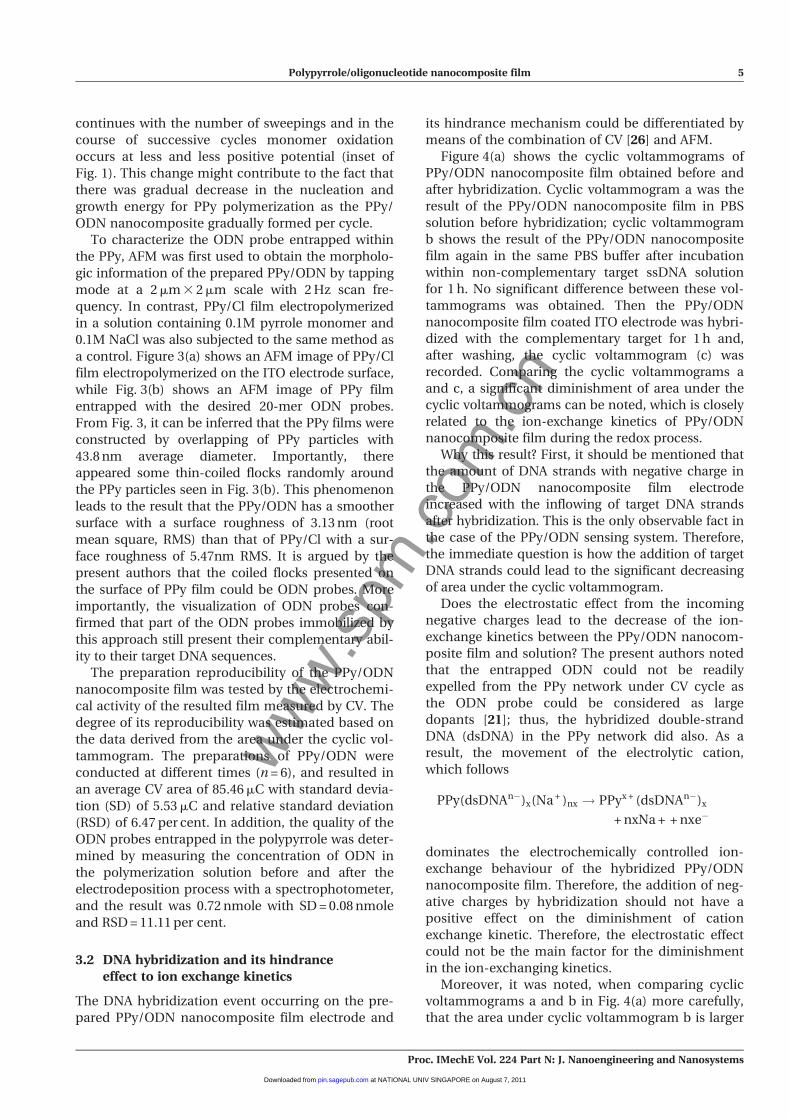
Figure 6 shows the influence of the hybridization time
(0 min to 80 min with a 10 min interval) on the electro-
chemical response of DNA hybridization detection.
From Fig. 6 it can be seen that the response of the
sensor initially increases significantly with the increase
of time until 60 min, which corresponds to a 45.5 per
cent decrease in the area of cyclic voltammogram
compared with that of before hybridization (the value
set as 100 per cent), and increases slowly from 60 min
to 80 min. In this case, the difference between the
analytical signals recorded at the 60th and 80th min-
ute of incubation time did not exceed 1 per cent, indi-
cating that the hybridization reaction was dominantly
completed after 60 min. Considering the sensitivity
and assay time, therefore, 60 min was chosen as the
hybridization time in this work.
4 SENSING SENSITIVITY
Under the above proposed optimization conditions
(10 cycles for PPy/ODN prepared, hybridization
time was chosen for 1 h), the analytical performance
of the DNA hybridization detection was investi-
gated. Figure 7(a) shows the cyclic voltammograms
of PPy/ODN film after hybridization with different
concentrations of target DNA from 1310218 M to
1310210 M. From Fig. 7(a) it can be seen that the
areas under the cyclic voltammograms decreased
with the increase of target DNA concentration. As
explained previously, the hybridization reaction
between the ODN probes entrapped within PPy film
and the complementary target DNA could induce
the structure of PPy/ODN nanocomposite film to be
more twisted and compact, which in turn hinders
the electrochemically controlled cation exchange
kinetic between the PPy/ODN nanocomposite film
and solution by steric effect. The slowdown of ion
exchanging kinetic corresponded to the decreased
area under the cyclic voltammogram when com-
pared to that before hybridization. It is reasonable
to argue that the increases in hybridization will fur-
ther decrease the area under the cyclic voltammo-
gram. Figure 7(b) shows the calibration curve of the
areas under the cyclic voltammograms in Fig. 7(a)
(normalized by the area under the cyclic voltammo-
gram of PPy/ODN before hybridization) against the
logarithm of the concentration of target DNA in the
range from 1310218 M to 1310210 M. Each concen-
tration was repeated three times. It can be noted
that the signal levelled off above 1310210 M., reflect-
ing that the probe hybridization sites available were
as low as 10 fmole. Furthermore, the normalized
Fig. 6 The influence of hybridization time on theresponse for DNA hybridization detection. Thecurve was derived form the areas under the CVsof the PPy/ODN film hybridized with comple-mentary target at different time after normal-ized with that of before hybridization (thisvalue set as 100 per cent). Other conditions wereas the same as Fig. 4(a)
Polypyrrole/oligonucleotide nanocomposite film 9
Proc. IMechE Vol. 224 Part N: J. Nanoengineering and Nanosystems
at NATIONAL UNIV SINGAPORE on August 7, 2011pin.sagepub.comDownloaded from

www.spm
.com
.cn
area is linear with the logarithm of the concentra-
tion of the complementary target DNA. The linear
regression equation was Y = –0.11 2 0.06X, where Y
is the normalized area derived from the integral of
the CVs, while X represents the log C (unit of C is
M), and the correlation coefficient (R) was 0.996.
The detection limit of the DNA biosensor was
5310218 M target DNA. In this case, the normalized
area changed after 60 min incubation with the com-
plementary ssDNA sequence was 0.072, which is
three times the normalized area change of 0.024 cal-
culated for the same electrode incubated in blank
PBS buffer. The detection limit of the proposed elec-
trochemical detection of DNA hybridization is
superior to that reported by Peng et al. [29] and
Chang et al. [30], who amplified the hybridization
signal by nanoparticles (~1nM) and by layer-by-
layer technique (3.2310214 M) respectively. This
proposed approach has a detection limit as small as
10218 M, which is typically required for medical
Fig. 7 (a) The CVs of PPy/ODN before (location a) and after hybridization with different concen-trations of the complementary target DNA from 1310218 M to 1310210 M with a 10 Minterval (corresponding to b–j CV, respectively). (b) The calibration curves for the normal-ized area under the CVs against the target concentration in the range from 1310218 M to1310210 M. Each concentration was recorded in three times (n = 3)
10 J-H Jiang, Y Li, J Wang, and K-L Paul Sung
Proc. IMechE Vol. 224 Part N: J. Nanoengineering and Nanosystems
at NATIONAL UNIV SINGAPORE on August 7, 2011pin.sagepub.comDownloaded from
www.spm
.com
.cn
diagnostic application [31], though the detection
linear range in this proposed method was lower.
5 CONCLUSIONS
An underlying mechanism attributing to the steric
effect for the interpretation of hybridization discrim-
ination on the hybrid PPy/ODN nanocomposite film,
as a simply-enough electrochemical sensor for label-
free DNA hybridization, was presented and proven
by the technical combination of cyclic voltammetry
and AFM measurements. Upon hybridization with
complementary target DNA, the PPy/ODN nano-
composite film undergoes a morphologic change,
becomes more compact and twisted, which in turn
diminishes the electrochemically controlled cation
exchange kinetic between the PPy/ODN and buffer
solution. This whole behaviour could obviously be a
steric effect in the hybridization signalling discrimi-
nation. A label-free electrochemical DNA biosensor
based on this hybrid PPy/ODN nanocomposite film
on ITO electrode was constructed and optimized to
evaluate its detection ability. It was found that a low
detection limit of 5310218 M could be achieved.
This work gives the authors a chance towards a bet-
ter understanding of this attractive PPy/ODN bio-
sensing composite and a further basis for the devel-
opment of label-free DNA sensor using the simple-
enough hybrid film of PPy/ODN.
FUNDING
This paper was supported by the Key Project for
International Science and Technology Collaboration
of the Ministry of Science and Technology
(2005DFA00190) and by an NSFC grant (30870607),
a CSTC grant (2008BB5192), and the ‘111 Project’
(B06023).
� Chongqing University 2011
REFERENCES
1 Li, J., Wei, W. Z., and Luo, S. L. A novel one-stepelectrochemical codeposition of carbon nano-tubes-DNA hybrids and tiron doped polypyrrole forselective and sensitive determination of dopamine.Microchimica Acta, 2010, 171(1–2), 109–116.
2 Wang, J. Electrochemical nucleic acid biosensors.Anal. Chim. Acta, 2002, 469, 63–71.
3 Wang, J. Towards genoelectronics: electrochemicalbiosensing of DNA hybridization. Chem. Eur. J.,1999, 5, 1681–1685.
4 Hashimoto, K., Ito, K., and Ishimori, Y. S. Micro-fabricated disposable DNA sensor for detection of
hepatitis B virus DNA. Sens. Actuators B, 1998, 46,220–225.
5 Yan, F., Erdem, A., Meric, B., Kerman, K.,Ozsoz, M., and Sadik, O. A. Electrochemical DNAbiosensor for the detection of specific gene relatedto Microcystis species. Electrochem. Commun.,2001, 3, 224–228.
6 Carpini, G., Fausto, L., Giovanna, M., andMascini, M. Oligonucleotide-modified screen-printed gold electrodes for enzyme-amplified sen-sing of nucleic acids. Biosens. Bioelectron., 2004, 20,167–175.
7 Takenaka, S., Yamashita, K., Takagi, M., Uto, Y.,and Kondo, H. DNA sensing on a DNA probe-mod-ified electrode using ferrocenyl naphthalene dii-mide as the electrochemically active ligand. Anal.Chem., 2000, 72, 1334–1341.
8 Wang, J., Liu, G., and Merkoci, A. Electrochemicalcoding technology for simultaneous detection ofmultiple DNA targets. J. Am. Chem. Soc., 2003, 125,3214–3215.
9 Cao, Y. C., Jin, R., and Mirkin, C. A. Nanoparticleswith Raman spectroscopic fingerprints for DNAand RNA detection. Science, 2002, 297, 1536–1540.
10 Bolıvar, P. H., Nagel, M., Richter, F.,Brucherseifer, M., Kurz, H., Bosserhoff, A., andButtner, R. Label-free THz sensing of geneticsequences: towards ‘THz biochips’. Phil. Trans. R.Soc. London A, 2004, 362, 323–335.
11 Daniels, J. S. and Pourmand, N. Label-free impe-dance biosensors: opportunities and challenges.Electroanalysis, 2007, 19, 1239–1257.
12 Ramanaviciene, A. and Ramanavicius, A. Pulsedamperometric detection of DNA with an ssDNA /polypyrrole-modified electrode. Anal. Bioanal.Chem., 2004, 379, 287–293.
13 Wang, J., Jiang, M., Fortes, A., and Mukherjee, B.New label-free DNA recognition based on dopingnucleic-acid probes within conducting polymerfilms. Anal. Chim. Acta, 1999, 402, 7–12.
14 Cai, H., Xu, Y., He, P. G., and Fang, Y. Z. Indicatorfree DNA hybridization detection by impedancemeasurement based on the DNA-doped conduct-ing polymer film formed on the carbon nanotubemodified electrode. Electroanalysis, 2003, 15, 1864–1869.
15 Wang, J. Review Electrochemical nucleic acid bio-sensors. Anal. Chim. Acta, 2002, 469, 63–71.
16 Francis, G., Hafsa, K. Y., Pratima, S., Bernard, M.,and Thierry, D. Toward intelligent polymers: DNAsensors based on oligonucleotide-functionalizedpolypyrroles. Synth. Met., 1999, 100, 89–94.
17 Chaker, T., Jaffrezic-Renault, N. J., Claude, M.,and Hafsa, K. Y. Direct electrochemical probing ofDNA hybridization on oligonucleotide-functiona-lized polypyrrole. Mater. Sci. Engng C, 2008, 28,848–854.
18 Ghanbaria, K., Bathaieb, S. Z., and Mousavi, M. F.Electrochemically fabricated polypyrrole nanofi-ber-modified electrode as a new electrochemicalDNA biosensor. Biosens. Bioelectron., 2008, 23,1825–1831.
Polypyrrole/oligonucleotide nanocomposite film 11
Proc. IMechE Vol. 224 Part N: J. Nanoengineering and Nanosystems
at NATIONAL UNIV SINGAPORE on August 7, 2011pin.sagepub.comDownloaded from

www.spm
.com
.cn
19 Li, Y., Jiang, J. H., Ma, X. D., Dong, G. X., Wang, J.,and Paul Sung, K.-L. Polypyrrole/oligonucleotidenanocomposite: its initial growth. J. NanoengingNanosyst., 2008, 222, 57–63.
20 Wang, J., Jiang, M., Fortes, A., and Mukherjee, B.New label-free DNA recognition based on dopingnucleic-acid probes within conducting polymerfilms. Anal. Chim. Acta, 1999, 402, 7–12.
21 Wang, J. and Jiang, M. Toward genoelectronics:nucleic acid doped conducting polymers. Lang-muir, 2000, 16, 2269–2274.
22 Xu, Y., Jiang, Y., Cai, H., He, P. G., and Fang, Y. Z.Electrochemical impedance detection of DNAhybridization based on the formation of M-DNAon polypyrrole/carbon nanotube modified elec-trode. Anal. Chim. Acta, 2004, 516, 19–27.
23 Komarova, E., Aldissi, M., and Bogomolova, A.Direct electrochemical sensor for fast reagent-freeDNA detection. Biosens. Bioelectron., 2005, 21, 182–189.
24 Tosar, J. P., Karen, K., and Laiz, J. Two indepen-dent label-free detection methods in one electro-chemical DNA sensor. Biosens. Bioelectron., 2009,24, 3036–3042.
25 Eguiluz, K. I. B., Banda, G. R. S., Huacca, M. E. F.,Alberice, J. V., Carrilho, E., Machado, S. A. S., andAvaca, L. A. Sequence-specific electrochemical
detection of Alicyclobacillus acidoterrestris DNAusing electroconductive polymer-modified fluorinetin oxide electrodes. Analyst, 2009, 134, 314–319.
26 Sadki, S., Schottland, P., Brodie, N., andSabouraud, G. The mechanisms of pyrrole electro-polymerization. Chem. Soc. Rev., 2000, 29, 283–293.
27 Piro, B., Reisberg, S., Noel, V., and Pham, M. C.Investigations of the steric effect on electrochemi-cal transduction in a quinone-based DNA sensor.Biosens. Bioelectron., 2007, 22, 3126–3131.
28 Reisberg, S., Piro, B., Noel, V., Nguyen, T. D.,Nielsen, P. E., and Pham, M. C. Investigations ofthe charge effect on electrochemical transductionin a quinone-based DNA sensor. Eletrochim. Acta,2008, 54, 346–351.
29 Peng, H., Soeller, C., Cannell, M. B.,Bowmaker, G. A., Cooney, R. P., and Sejdic, J. T.Electrochemical detection of DNA hybridizationamplified by nanoparticles. Biosens. Bioelectron.,2006, 21, 1727–1736.
30 Chang, Z., Chen, M., Fan, H., Zhao, K., Zhuang, S.Q., He, P. G., and Fang, Y. Z. Multilayer mem-branes via layer-by-layer deposition of PDDA andDNA with Au nanoparticles as tags for DNA biosen-sing. Electrochim. Acta, 2008, 53, 2939–2945.
31 Serge, C. and Pascal, M. Recent advances in DNAsensors. Analyst, 2008, 133, 984–991.
12 J-H Jiang, Y Li, J Wang, and K-L Paul Sung
Proc. IMechE Vol. 224 Part N: J. Nanoengineering and Nanosystems
at NATIONAL UNIV SINGAPORE on August 7, 2011pin.sagepub.comDownloaded from