
Polyphyly of nonbioluminescent Vibrio fischeri sharing a ...
27
Polyphyly of non-bioluminescent Vibrio fischeri sharing a lux-locus deletionM. S. Wollenberg, 1 S. P. Preheim, 2 M. F. Polz 2 and E. G. Ruby 1 * 1 Department of Medical Microbiology and Immunology, University of Wisconsin, 1550 Linden Drive, Room 5245, Madison, WI 53706-1521, USA. 2 Department of Civil and Environmental Engineering, Massachusetts Institute of Technology, 15 Vassar Street, Bldg 48-417, Cambridge, MA 02139, USA. Summary This study reports the first description and mole- cular characterization of naturally occurring, non- bioluminescent strains of Vibrio fischeri. These ‘dark’ V. fischeri strains remained non-bioluminescent even after treatment with both autoinducer and aldehyde, substrate additions that typically maximize light pro- duction in dim strains of luminous bacteria. Surpris- ingly, the entire lux locus (eight genes) was absent in over 97% of these dark V. fischeri strains. Although these strains were all collected from a Massachusetts (USA) estuary in 2007, phylogenetic reconstructions allowed us to reject the hypothesis that these newly described non-bioluminescent strains exhibit mono- phyly within the V. fischeri clade. These dark strains exhibited a competitive disadvantage against native bioluminescent strains when colonizing the light organ of the model V. fischeri host, the Hawaiian bobtail squid Euprymna scolopes. Significantly, we believe that the data collected in this study may suggest the first observation of a functional, parallel locus-deletion event among independent lineages of a non-pathogenic bacterial species. Introduction The lux locus – encoding all genes necessary and suffi- cient for bacterial bioluminescence – is found predomi- nantly within the Vibrionaceae. Within this family of heterotrophic, aquatic gammaproteobacteria approxi- mately 10 traditionally delineated species are known to include bioluminescent strains (Urbanczyk et al., 2008). In most members of the Vibrionaceae, it appears that light emission is the exception and not the rule; non- bioluminescent strains outnumber bioluminescent strains in all but a few Vibrionaceae species (Grim et al., 2008; Urbanczyk et al., 2008). One Vibrionaceae species reported to be composed only of bioluminescent strains is Vibrio fischeri. Why, in all environmental samples analysed to date, have naturally occurring, non-bioluminescent (dark) V. fischeri never been reported? A reasonable hypothesis is that the trait of bioluminescence is consistently and strongly selected for in V. fischeri by their primary environmental habitat(s). One such V. fischeri habitat is their animal hosts (Farmer, 2006). Vibrio fischeri strains have been isolated in the greatest abundance from bobtail squids of the genera Euprymna, Sepiola and Rondeletiola (Cephalopoda: Sepiolidae), and fishes of the genera Monocentris and Cleiodopus (Actinopterygii: Beryciformes) (Ruby, 1977; Fitzgerald, 1978; Wei and Young, 1989; Nishiguchi et al., 1998). Indeed, bioluminescent V. fischeri were found to outcompete dark mutants during symbiosis initiation of juvenile bobtail squid (Bose et al., 2008), although this is the only study directly demonstrating a fitness benefit for bioluminescence in V. fischeri. Best known for their mutualistic associations, V. fischeri have also been isolated as planktonic cells from both pelagic and neritic environments (see, for example, Baumann et al., 1971; Lee and Ruby, 1994). In contrast to host-associated environments, little is known about the ecology of these planktonic (or ‘free-living’) V. fischeri in marine sediment or seawater. Even less is known about V. fischeri bioluminescence (and selection for/against this trait) in these niches, although the density-dependent nature of bioluminescence induction via quorum sensing (Boettcher and Ruby, 1990) implies that V. fischeri popu- lations must be above a certain density to produce light. In studies using either a 16S rRNA- (Jones et al., 2007) or a luxA-based V. fischeri-specific probe (Lee and Ruby, 1995), concentrations of this species in Hawaiian seawa- ter and sediment have been estimated to be as high as 10 2 –10 4 cells ml -1 (Boettcher and Ruby, 1990). Assuming even distribution of V. fischeri, these concentrations imply that bioluminescence autoinduction does not take place when cells are free-living in these environments, poten- tially mitigating any selection for or against this trait among planktonic V. fischeri. Received 30 April, 2011; accepted 5 September, 2011. *For corre- spondence. E-mail [email protected]; Tel. (+1) 608 262 5911; Fax (+1) 608 262 8418. Environmental Microbiology (2011) doi:10.1111/j.1462-2920.2011.02608.x © 2011 Society for Applied Microbiology and Blackwell Publishing Ltd
Transcript of Polyphyly of nonbioluminescent Vibrio fischeri sharing a ...

Polyphyly of non-bioluminescent Vibrio fischerisharing a lux-locus deletionemi_2608 1..14
M. S. Wollenberg,1 S. P. Preheim,2 M. F. Polz2 andE. G. Ruby1*1Department of Medical Microbiology and Immunology,University of Wisconsin, 1550 Linden Drive, Room5245, Madison, WI 53706-1521, USA.2Department of Civil and Environmental Engineering,Massachusetts Institute of Technology, 15 VassarStreet, Bldg 48-417, Cambridge, MA 02139, USA.
Summary
This study reports the first description and mole-cular characterization of naturally occurring, non-bioluminescent strains of Vibrio fischeri. These ‘dark’V. fischeri strains remained non-bioluminescent evenafter treatment with both autoinducer and aldehyde,substrate additions that typically maximize light pro-duction in dim strains of luminous bacteria. Surpris-ingly, the entire lux locus (eight genes) was absent inover 97% of these dark V. fischeri strains. Althoughthese strains were all collected from a Massachusetts(USA) estuary in 2007, phylogenetic reconstructionsallowed us to reject the hypothesis that these newlydescribed non-bioluminescent strains exhibit mono-phyly within the V. fischeri clade. These dark strainsexhibited a competitive disadvantage against nativebioluminescent strains when colonizing the lightorgan of the model V. fischeri host, the Hawaiianbobtail squid Euprymna scolopes. Significantly, webelieve that the data collected in this study maysuggest the first observation of a functional, parallellocus-deletion event among independent lineages ofa non-pathogenic bacterial species.
Introduction
The lux locus – encoding all genes necessary and suffi-cient for bacterial bioluminescence – is found predomi-nantly within the Vibrionaceae. Within this family ofheterotrophic, aquatic gammaproteobacteria approxi-mately 10 traditionally delineated species are known toinclude bioluminescent strains (Urbanczyk et al., 2008). In
most members of the Vibrionaceae, it appears that lightemission is the exception and not the rule; non-bioluminescent strains outnumber bioluminescent strainsin all but a few Vibrionaceae species (Grim et al., 2008;Urbanczyk et al., 2008).
One Vibrionaceae species reported to be composedonly of bioluminescent strains is Vibrio fischeri. Why, in allenvironmental samples analysed to date, have naturallyoccurring, non-bioluminescent (dark) V. fischeri neverbeen reported? A reasonable hypothesis is that the trait ofbioluminescence is consistently and strongly selected forin V. fischeri by their primary environmental habitat(s).One such V. fischeri habitat is their animal hosts (Farmer,2006). Vibrio fischeri strains have been isolated in thegreatest abundance from bobtail squids of the generaEuprymna, Sepiola and Rondeletiola (Cephalopoda:Sepiolidae), and fishes of the genera Monocentris andCleiodopus (Actinopterygii: Beryciformes) (Ruby, 1977;Fitzgerald, 1978; Wei and Young, 1989; Nishiguchi et al.,1998). Indeed, bioluminescent V. fischeri were found tooutcompete dark mutants during symbiosis initiation ofjuvenile bobtail squid (Bose et al., 2008), although this isthe only study directly demonstrating a fitness benefit forbioluminescence in V. fischeri.
Best known for their mutualistic associations, V. fischerihave also been isolated as planktonic cells from bothpelagic and neritic environments (see, for example,Baumann et al., 1971; Lee and Ruby, 1994). In contrast tohost-associated environments, little is known about theecology of these planktonic (or ‘free-living’) V. fischeri inmarine sediment or seawater. Even less is known aboutV. fischeri bioluminescence (and selection for/against thistrait) in these niches, although the density-dependentnature of bioluminescence induction via quorum sensing(Boettcher and Ruby, 1990) implies that V. fischeri popu-lations must be above a certain density to produce light. Instudies using either a 16S rRNA- (Jones et al., 2007) or aluxA-based V. fischeri-specific probe (Lee and Ruby,1995), concentrations of this species in Hawaiian seawa-ter and sediment have been estimated to be as high as102–104 cells ml-1 (Boettcher and Ruby, 1990). Assumingeven distribution of V. fischeri, these concentrations implythat bioluminescence autoinduction does not take placewhen cells are free-living in these environments, poten-tially mitigating any selection for or against this traitamong planktonic V. fischeri.
Received 30 April, 2011; accepted 5 September, 2011. *For corre-spondence. E-mail [email protected]; Tel. (+1) 608 262 5911; Fax(+1) 608 262 8418.
Environmental Microbiology (2011) doi:10.1111/j.1462-2920.2011.02608.x
© 2011 Society for Applied Microbiology and Blackwell Publishing Ltd

A second, contrasting hypothesis is that non-bioluminescent V. fischeri have not been previouslysampled because these sampling efforts were biased infavour of the isolation of bioluminescent strains. Originally,V. fischeri was grouped under the genus Photobacteriumby Beijerinck when he isolated bioluminescent bacterialcolonies from the Dutch coast, reflecting the idea thatV. fischeri were bioluminescent bacteria ipso facto (Beijer-inck, 1889). More exacting taxonomic efforts by Baumannand associates, consisting of a more cosmopolitan collec-tion of V. fischeri strains, also reported bioluminescenceto be a hallmark of V. fischeri (Reichelt and Baumann,1973). Finally, more contemporary V. fischeri isolationefforts have either sampled from host-associated environ-ments exclusively (Ruby and Nealson, 1976; Jones et al.,2006; Urbanczyk et al., 2007; Wollenberg and Ruby,2009) or used lux gene-specific probes to identify andisolate planktonic V. fischeri (Lee and Ruby, 1992).
Here we describe V. fischeri strains collected in 2007from several different microhabitats within an estuaryenvironment at Ipswitch, MA. The work below probes themolecular nature of bioluminescence loss among thesestrains, describes the phylogenetic relationship of thesedark strains to light-producing V. fischeri, and assessesthe relative fitness of these strains in the Euprymna sco-lopes light organ.
Results
The majority of V. fischeri isolated from PIE (PlumIsland Estuary) do not produce bioluminescence, incontrast to all previously studied V. fischeri strains
During 2007, over 1750 Vibrionaceae isolates were col-lected from various sources (e.g. water-column particlesand invertebrate tissues) in an estuary north of Boston,Massachusetts – PIE (Plum Island Estuary). Three con-served loci (malate dehydrogenase – mdh; adenylatekinase – adk; heat-shock chaperonin – groL) were par-tially sequenced from each of these strains (Preheimet al., 2011). These sequences, plus their homologuesfrom four additional, well-characterized Vibrionaceae(V. fischeri ES114 and MJ11; V. logei SA6; V. salmonicidaLFI1238) were concatenated and used to construct aneighbour net (Fig. 1).
From this reconstruction, a clade of approximately 70strains was found to include V. fischeri ES114 and MJ11.Strains from this clade were independently confirmed tobe V. fischeri using the hvnC-PCR assay (Wollenberg andRuby, 2009), which demonstrated a positive pair of prod-ucts for all strains tested in this putative V. fischeri group,but a negative (single) product both for closely relatedstrains not found in this group and for other, more distantlyrelated Vibrionaceae (Figs 1 and 2A; data not shown).
Furthermore, partial 16S rrn sequencing and BLAST
searching/alignment of several of these putative V. fis-cheri strains resulted in nearest matches to sequencesfrom known V. fischeri (data not shown). During manipu-lation of these 70+ PIE V. fischeri strains, coloniesgrowing either on standard marine agar medium or inlog-phase liquid cultures produced no visible biolumines-cence (Table 1), in contrast to the majority of V. fischeristudied to date.
One hypothesis explaining non-visible bioluminescencein the 2007 PIE V. fischeri is that these strains under-produce autoinducer. The majority of well-studied andnon-visibly bioluminescent V. fischeri strains are knownto secrete little of the autoinducer 3-oxo-hexanoylhomoserine lactone (3-oxo-C6 HSL) under standard invitro growth conditions (Nealson, 1977). Addition of3-oxo-C6 HSL to liquid cultures increases biolumines-cence in non-visibly bioluminescent V. fischeri severalorders of magnitude (Boettcher and Ruby, 1990).However, 97% of the PIE V. fischeri were found to beunresponsive to autoinducer addition in liquid culture(Table 1; data not shown). Three strains, 9ZD146,9CSC6, 9CSC146 were visibly bioluminescent in liquidand solid marine media, and did respond to autoinduceraddition. Two of these strains, 9CSC6 and 9CSC146,were identical in all three partial loci sequenced above,making it possible that they are sibling isolates. Takentogether, these results indicate that luminescence is arare trait among V. fischeri populations in PIE.
Another hypothesis for the absence of bioluminescenceby almost all PIE V. fischeri strains is the possible under-production of the aldehyde substrate for the luciferaseenzyme. Some strains of both V. fischeri and V. salmoni-cida have been reported to be non-visibly bioluminescentin liquid culture under normal growth conditions, but canproduce visible light after the external addition of an alde-hyde substrate like decanal (Fidopiastis et al., 1999; Luppet al., 2003). Dark PIE V. fischeri strains did not produceany bioluminescence upon addition of exogenousdecanal (Table 1; data not shown), suggesting that theyare not limited by this substrate.
Non-bioluminescent PIE V. fischeri share an identical,full lux-region deletion
Another hypothesis explaining non-visible biolumines-cence in PIE V. fischeri is the loss or mutation of somecomponent of the lux region. The V. fischeri lux regionconsists of two divergent operons containing the eightstructural and regulatory genes (Fig. S1) necessary forlight production (Gray and Greenberg, 1992; Meighen,1994). Surprisingly, all PIE V. fischeri strains that did notrespond to autoinducer (with the exception of strain9ZD146) also lacked PCR-amplification products from
2 M. S. Wollenberg, S. P. Preheim, M. F. Polz and E. G. Ruby
© 2011 Society for Applied Microbiology and Blackwell Publishing Ltd, Environmental Microbiology

Fig. 1. Phylogenetic relationships among Vibrionaceae sampled from Plum Island Sound (PIE), Massachusetts in spring and fall of 2007. Bothreconstructions depicted were created via the neighbour net method using uncorrected P distance, ordinary least-squared variance and splitscalculated by the equal-angle algorithm. (Inset): Unrooted neighbour net constructed with concatenated, partial sequences from three loci(mdh, adk, groL) of over 750 Vibrionaceae, two V. fischeri (ES114 and MJ11), one V. salmonicida (LFI1238) and one V. logei (SA6) strain.Known V. fischeri strains were found among taxa on a defined branch (green). (Main figure): Unrooted neighbour net, constructed as above,from all taxa on the green branches in the inset. Fifteen isolates frequently referred to in the text and strains ES114, MJ11, LFI1238 and SA6are marked with purple squares; V. fischeri taxa included in Fig. 3 are listed in bold. Bars represent substitutions per site, as listed.
Polyphyly of non-bioluminescent V. fischeri 3
© 2011 Society for Applied Microbiology and Blackwell Publishing Ltd, Environmental Microbiology

both the gene encoding the alpha chain of bacterialluciferase (luxA) and the entire lux region (Fig. 2; data notshown).
Genomic sequences from a subsample of PIE V. fis-cheri revealed that all contained a full deletion of the luxregion between intact sequences of the two well-conserved flanking loci (i.e. ribG- and mscS-like genes)(data not shown). Unassembled contigs covering the fullgenome of one of these strains (ZF_211 – S. Timberlake,M.F. Polz and E.J. Alm, pers. comm.) were assembledusing V. fischeri ES114 and MJ11 full-genome sequencesas scaffolds by the progressive Mauve algorithm in Mauve(Darling et al., 2010). This assembly and exhaustiveBLASTing of V. fischeri ES114 luxA nucleotide sequence
against these contigs indicated no evidence of a luxregion anywhere in the ZF_211 genome assembly(Fig. S1, data not shown).
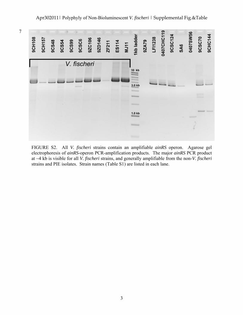
Analysis of a related locus was used to identify theextent of disruption of the general autoinduction/quorum-sensing pathway in dark PIE V. fischeri. The octanoyl-L-homoserine lactone (C8-HSL) receptor and synthaselocus (i.e. the ainRS operon) could be amplified from alldark PIE V. fischeri (Fig. S2; data not shown). Thus, whilethe downstream portion of the V. fischeri quorum-signalling pathway (the luxRICABDEG genes) appears tobe missing in dark PIE V. fischeri, the upstream, non-lux,portion (e.g. ainRS) that is shared with other Vibrionaceae(Lupp et al., 2003) is present. All of these molecular
Fig. 2. Agarose gel electrophoresis of PCR products from V. fischeri- and lux-specific amplification assays demonstrates variability inlux-region amplification from PIE V. fischeri strains. For each strain, amplification of (A) hvnC (~1 kb) and a portion of the 16S rrn operon(s)(~1.5 kb), (B) luxA (~1.5 kb) and (C) the full-length lux region (~10 kb) and conserved housekeeping genes flanking a full deletion of the luxregion (1 kb) was judged to be successful (+), unsuccessful (-) or inconclusive (?). (A and B) PCR reaction products were run on the samegel; this gel was cropped to remove wells. The same DNA stock was used for PCR reactions in the three gels pictured and Fig. S2.
4 M. S. Wollenberg, S. P. Preheim, M. F. Polz and E. G. Ruby
© 2011 Society for Applied Microbiology and Blackwell Publishing Ltd, Environmental Microbiology

results are summarized for a representative subsample ofV. fischeri strains in Table 1.
Non-bioluminescent PIE V. fischeri are polyphyleticwithin a phylogenetic reconstruction of the V. fischerispecies complex
One hypothesis explaining the conserved lux-regiondeletion that is characteristic of dark PIE V. fischeri isthat these strains are all closely related, and share amost recent common ancestor within the V. fischerispecies lacking the lux operon; in other words, non-bioluminescent PIE V. fischeri are monophyletic. To testthis hypothesis, both dark PIE V. fischeri (n = 5) and biolu-minescent V. fischeri from our laboratory collection(n = 11) were sequenced at four conserved housekeepingloci (recombinaseA – recA; malate dehydrogenase – mdh;catalase – katA; dihydroorotase – pyrC). Maximum parsi-mony, maximum likelihood and Bayesian methods wereused to construct, and determine the statistical signifi-cance of, evolutionary relationships among these V. fis-cheri strains. In all reconstructions, non-bioluminescentPIE V. fischeri strains were distributed throughout the phy-logeny and were not monophyletic within either of the twoV. fischeri subclades (Figs 3 and S3). The hypothesis thatnon-bioluminescent V. fischeri form a monophyletic cladewas statistically rejected via parametric bootstrapping
Table 1. Phenotypic and genotypic bioluminescence properties of V. fischeri strains from Plum Island Estuary collections.
IsolatehvnCPCR
luxoperonPCR
luxAPCR
ainoperonPCR
RLUa
SWT
SWT+C6
SWT+decanal
V. fischeri V. fischeri ES114 + + + + 38 > 104 2582V. fischeri MJ11 + + + + > 104 > 104 > 104
ZF211 + - - + 3 1 19ZC106 + - - + 2 1 49ZD146 + + + + 3 5 49CSC6b + + + + > 104 > 104 > 104
9CS48 + - - + 2 1 19CS54 + - - + 3 2 29CS99 + - - + 2 1 39CH157 + - - + 2 0 19CH108 + - - + 2 1 19CSC70 - - ?c + 1 2 29CHC144 - - ? + 1 1 10407SW56 - - ? ? 1 1 2V. logei SA6 - ? ? + > 104 > 104 > 104
V. salmonicida LFI1238 - + ? + 34 41 > 104
0407CHC119 - + ? + 2 7 > 104
9CSC124 - ? ? + > 104 > 104 > 104
9ZA79 - ? ? ? 1 1 2
a. Relative luminescence units (RLU) measured in response to growth in SWT medium, SWT + 120 nM 3-oxo-C6-HSL autoinducer (C6) andSWT + 0.01% aldehyde (decanal). Values of > 103 are above the upper limit of detection for the luminometer; values of approximately 5 or lessare the lower limit of detection/background for the luminometer for the given sensitivity setting. Similar values were recorded in three independentexperiments; representative values from a single experiment are presented.b. The bioluminescence properties of isolate 9CSC146 were found to be identical to those of isolate 9CSC6 for all assays listed.c. Either cryptic (or no) amplification products observed for given PCR temperature settings and primers.
Fig. 3. Non-bioluminescent and lux-region absent taxa arepolyphyletic within a phylogenetic reconstruction of V. fischeri.Consensus network inferred from 10 000 phylograms produced byClonalFrame analyses of 16 V. fischeri taxa using a concatenationof four loci: recA, mdh, katA and pyrC. ClonalFrame was used toinfer genealogical relationships from posterior distributions of 2000samples from five independent runs. This aggregate ClonalFrameposterior distribution was used by SplitsTree4 to build a consensusnetwork. For this network, reticulate relationships found in at least20% of the samples are depicted as parallel branches inparallelograms; nodes supported by a posterior probability ofgreater than 95% (as summarized in a consensus tree byClonalFrame) are at the termini of stippled branches. The root(stippled, unlabelled branch) has been truncated in order to expandthe main V. fischeri group and contains both outgroup taxa:V. salmonicida LFI1238 and V. logei SA6. Bold isolates lackevidence for the lux operon and are phenotypicallynon-bioluminescent. The bar represents 0.01 substitutions per site.
Polyphyly of non-bioluminescent V. fischeri 5
© 2011 Society for Applied Microbiology and Blackwell Publishing Ltd, Environmental Microbiology
(P < 0.01). Furthermore, from the original neighbour net ofall PIE V. fischeri (Fig. 1), the five listed bioluminescentV. fischeri strains (i.e. ES114, MJ11, 9CSC6, 9CSC106and 9ZD146) appeared interspersed among the broaderV. fischeri group. All of these results strongly support thehypothesis that non-bioluminescent V. fischeri do notshare an exclusive most recent common ancestor withinthe V. fischeri species.
STCBS-based isolation from seawater does notsignificantly bias estimation of bioluminescent V. fischeriabundance relative to LBS-based isolation
An interesting observation from the above results is thehigh abundance of non-bioluminescent, lux operon-deleted, V. fischeri strains in the PIE sampling relative toboth bioluminescent strains in this same sampling andall other worldwide samplings. Because samplingregimes over the past century had previously failed toisolate non-bioluminescent V. fischeri, and it has beennoted that TCBS medium selects against some speciesof Vibrionaceae (Farmer, 2006), we asked whether theisolation procedure using STCBS agar medium wasresponsible for the high abundance of non-bioluminescent V. fischeri in the 2007 PIE study. A ran-domly chosen subsample of both bioluminescent andnon-bioluminescent V. fischeri strains was cultured insterile-filtered Instant Ocean for 24 h before beingspread on either LBS agar or STCBS agar media(Fig. S4). While mean culturable bioluminescent andnon-bioluminescent V. fischeri concentrations were sig-nificantly smaller on STCBS agar relative to LBS agar(F1,78 = 70.8502; P < 0.001), for both media there was nosignificant difference between the mean cfu ml-1 of eitherbioluminescent or non-bioluminescent strains (F1,18 =0.3729; P = 0.55). Similarly, there was no significantinteraction detected between growth medium and astrain’s bioluminescence state (F1,78 = 0.3219; P = 0.57).These results strongly suggest that, although the use ofSTCBS decreases the total number of V. fischeri recov-ered from marine samples, this medium is not biased inits recovery of cells of either bioluminescent or non-bioluminescent V. fischeri.
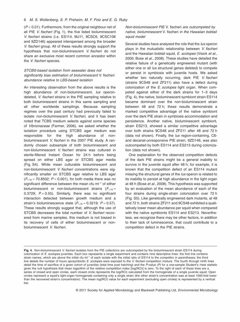
Non-bioluminescent PIE V. fischeri are outcompeted bynative, bioluminescent V. fischeri in the Hawaiian bobtailsquid model
Several studies have analysed the role that the lux operonplays in the mutualistic relationship between V. fischeriand the Hawaiian bobtail squid, E. scolopes (Visick et al.,2000; Bose et al., 2008). These studies have detailed therelative failure of a genetically engineered mutant (witheither one or all lux structural genes deleted) to competeor persist in symbiosis with juvenile hosts. We askedwhether two naturally occurring, dark PIE V. fischeri(strains 9CS48 and ZF211) also have a defect duringcolonization of the E. scolopes light organ. When com-peted against either of the dark strains for 1–3 days(Fig. 4), the native, bioluminescent symbiont strain ES114became dominant over the non-bioluminescent strainbetween 48 and 72 h; these results demonstrate amarked competitive advantage of the native symbiontover the dark PIE strain in symbiosis accommodation andpersistence. Another native, bioluminescent symbiont,strain ES213, showed a similar competitive advantageover both strains 9CS48 and ZF211 after 48 and 72 h(data not shown). Finally, the lux region-containing, C6-and decanal-unresponsive PIE strain, 9ZD146, was alsooutcompeted by both ES114 and ES213 during coloniza-tion (data not shown).
One explanation for the observed competition defectsof the dark PIE strains might be a general inability tosurvive in the juvenile squid after 48 h; for example, it isknown that the competition defect of an ES114 mutantmissing the structural genes of the lux operon is related toits inability to persist at high abundance in the light organat 48 h (Bose et al., 2008). This hypothesis was supportedby an evaluation of the mean abundance of each of thefour strains during single-strain colonization over 72 h(Fig. S5). Like genetically engineered dark mutants, at 48and 72 h, both strains ZF211 and 9CS48 exhibited a quali-tatively lower mean abundance per squid when comparedwith the native symbionts ES114 and ES213. Neverthe-less, we recognize there may be other factors, in additionto their lack of luminescence, that could contribute to acompetition defect in the PIE strains.
Fig. 4. Non-bioluminescent V. fischeri isolates from the PIE collections are outcompeted by the bioluminescent strain ES114 duringcolonization of E. scolopes juveniles. Each box represents a single experiment and contains nine descriptive lines: the first line containsstrain names, which are above the initial cfu ml-1 of each isolate with the initial ratio of ES114 to the competitor in parentheses; the thirdline details the number of hours aposymbiotic E. scolopes were exposed to the V. fischeri competition mixture. The fourth through ninth linesdetail the time of sacrifice of a given cohort of juveniles (total time post hatching) and the P-value (P) for a one-sample Student’s t-test statisticgiven the null hypothesis that mean logarithm of the relative competition index [log(RCI)] is zero. To the right of each of these lines are aseries of closed and open circles, each closed circle represents the log(RCI) calculated from the homogenate of a single juvenile squid. Opencircles represent a squid’s light-organ homogenate containing only a single strain (the other strain’s concentration was at least 1000-fold lowerthan the recovered strain’s concentration). The mean log(RCI) value for each experiment (excluding open circles) is represented by a verticalbar.
6 M. S. Wollenberg, S. P. Preheim, M. F. Polz and E. G. Ruby
© 2011 Society for Applied Microbiology and Blackwell Publishing Ltd, Environmental Microbiology

Polyphyly of non-bioluminescent V. fischeri 7
© 2011 Society for Applied Microbiology and Blackwell Publishing Ltd, Environmental Microbiology

Discussion
This study is the first description of non-bioluminescentV. fischeri. The work suggests that broader, relativelyunbiased environmental samplings may increase thegeneral knowledge of the bioluminescence spectrum forany given species of Vibrionaceae. Samplings of bothVibrio cholerae and V. harveyi support this conclusion;such studies have demonstrated that non-bioluminescentisolates of luminous species are as abundant, or moreabundant, than bioluminescent isolates in marine environ-ments (Grim et al., 2008; O’Grady and Wimpee, 2008; Zoet al., 2009).
From the intraspecific phylogenetic reconstruction, weconclude that the presence of a region of functional luxgenes (i.e. the genomic trait underlying bioluminescence)is a homoplasy within the species V. fischeri (Fig. 3). Thisconclusion implies that, rather than being a single event inthe evolutionary history of this species, gain or loss of thelux region appears to have occurred multiple times inmultiple V. fischeri subgroups in an identical manner. Torecapitulate the observed pattern of luminescence andlux-region distribution among extant strains of V. fischeri,a 2 ¥ 2 matrix of four general evolutionary scenarios mightbe hypothesized (Table 2).
We favour the hypothesis that selection has led to iden-tical loss of the eight lux genes among unrelated, V. fischeristrains sampled from the PIE environment. In this scenario,lux-region presence is ancestral to V. fischeri and we wouldpredict there to be evidence of selection in this region forextant V. fischeri strains. Most strongly, data from a previ-ous report (Bose et al., 2008), as well as this study (Fig. 4),demonstrate that the lux genes provide a fitness advan-tage in the light organ of the Hawaiian bobtail squid,E. scolopes. Additionally, patterns of nucleotide substitu-tion among the luxA sequences of different V. fischeristrains, and comparison of fully sequenced lux regionsbetween a fish-associated and a squid-associated strain,suggest that selection has been operating on this biolumi-nescence enzyme-encoding locus (Mandel et al., 2009;Wollenberg and Ruby, 2011). Finally, recent studies ofexperimental evolution in non-native V. fischeri colonizingjuvenile E. scolopes have demonstrated that the lightorgan can strongly, and rapidly, select for particular biolu-minescence levels (Schuster et al., 2010).
We are unaware of a parsimonious model resultingfrom genetic drift that would produce an identical loss/gainof the lux region necessary to recapitulate these observeddata. For example, if genetic drift were combined withlux-region loss, we might expect to have observed strainsof V. fischeri that, like non-bioluminescent V. harveyi iso-lated from seawater, contained point mutations or smallindels in lux genes that effectively abrogate light produc-tion (O’Grady and Wimpee, 2008). In this study, only asingle strain (9ZD146) was found to possess a non-functional, but intact, lux region (Table 1; data not shown).The observation of this one possible lux pseudogene inPIE V. fischeri might support the hypothesis of drift fol-lowed by a strong deletional bias of the lux locus.
Another, alternative hypothesis is that recombinationhas affected the evolutionary history of these isolates.First, recombination among the housekeeping loci usedin this analysis may have obfuscated ‘true’ evolutionaryrelationships among V. fischeri strains. Scrambling ofthese strains’ evolutionary history could generate thehomoplasy observed for the lux region. We believe thishypothesis is not the most parsimonious explanation ofthese data for the following two reasons. First, othershave demonstrated, with different housekeeping loci (Astet al., 2009), intraspecific phylogenetic reconstructions ofV. fischeri showing two major clades. Our results (Figs 1and 3), recapitulate this same two-clade pattern, withsimilar strains in each clade; this suggests that thegeneral phylogenetic signal observed among these three,independent reconstructions is fundamental to V. fischeri.Second, we have used these housekeeping loci in otherstudies (Mandel et al., 2009; Wollenberg and Ruby, 2011)in which phylogenetic reconstructions of V. fischeri werepredictive of physiological differences among extantstrains in both seawater and host-associated environ-ments. If recombination had truly obscured the phyloge-netic signal in these two studies, it is unlikely that thephylogenetic and experimental/biological data would becomplementary.
Another hypothesis is that homologous recombinationof the (deleted or intact) lux locus has occurred amongdisparate V. fischeri lineages. Phylogenetic reconstruc-tions of these isolates using their lux loci will be instructivefor assessing the relative similarity or difference betweenhousekeeping- and bioluminescence-operon evolution in
Table 2. Evolutionary scenarios for lux-region homoplasy among V. fischeri.
Evolutionary event
Natural selection Genetic drift
Event direction Gain Natural selection for lux-region gain Gain of lux region through genetic driftLoss Natural selection for lux-region loss Loss of lux region through genetic drift
8 M. S. Wollenberg, S. P. Preheim, M. F. Polz and E. G. Ruby
© 2011 Society for Applied Microbiology and Blackwell Publishing Ltd, Environmental Microbiology

this lineage. Results of such a study might be able toconfirm or refute the hypothesis that this operon has‘moved’ among V. fischeri lineages. It will be more difficultto assess the recombination of deleted sequence amongV. fischeri, although the analysis of conserved, flankingsequences might prove useful.
Among the possible scenarios for lux-region gain orloss in response to natural selection, the existing datamost strongly indicate loss. We propose that adaptiveradiations of non-bioluminescent V. fischeri have beenfacilitated by selection for the loss of light production anddeletion of the lux region. In contrast, multiple events ofidentical lux-region gain appear unlikely. Molecular datasuggest that luxA (Wollenberg and Ruby, 2011) and lux-region (Urbanczyk et al., 2008) sequences from V. fischeriare more similar to each other than to those of other luxgenes found in the Vibrionaceae. These data are mostconsistent with a common ancestry for the lux region inV. fischeri, and weaken the argument of multiple, distinctgains of this locus as the species has diverged throughtime: this latter scenario provides no explanation for whyand how integration of the lux region into exactly the sameposition in the chromosomes of multiple lineages of V. fis-cheri might occur.
Adaptive radiation resulting in the loss of an entire genehas been observed among bacteria, and has been bestdocumented from genomic analyses of pathogens(Lawrence, 2005). Large and/or complete deletions havebeen found in temporally disparate radiations of bacteriagrown in diverse environments ranging from laboratorymedia to Homo sapiens (Nakata et al., 1993; Maurelli,2007; Barrick et al., 2009; Zdziarski et al., 2010). Interest-ingly, however, reports of identical locus-deletion eventsamong phylogenetically distinct lineages of a single bac-terial species are sparse.
One such observation describes three or four deletionsfound among different lineages of Mycobacterium tuber-culosis (Tsolaki et al., 2004). The authors argue that theremay be selection for several of these deletions: themissing regions encode drug sensitivity and ‘latencygenes’ that are hypothesized to impede disease progres-sion and transmission. In other words, loss of these genesduring adaptive radiation(s) provides a fitness advantageto unrelated M. tuberculosis strains within the host inresponse to selection.
Our data provide the first evidence for an identicalfunctional-locus deletion among different lineages of anon-pathogenic bacterial species. Such conserved lux-region deletions among unrelated V. fischeri could implythat selection for the loss of this locus (and, thus, biolu-minescence) provides a fitness advantage outside of thesquid or fish light-organ environment. Data from a previ-ous study suggests that isogenic mutants lacking the luxoperon’s structural genes experience a fitness advantage
relative to brightly bioluminescent V. fischeri duringgrowth in rich medium (Bose et al., 2008) due to thesignificant metabolic cost of light production (Karl andNealson, 1980). Other studies have indicated that biolu-minescent V. fischeri also exhibit survival differences innatural seawater (Wollenberg and Ruby, 2011), althoughsimilar experiments have not yet been performed withnon-bioluminescent V. fischeri.
One hypothesis consistent with the results describedhere is that one (or more) PIE habitats (Preheim et al.,2011) (e.g. Hemigrapsus sanguineus and Mytilus edulisguts, or zooplankton-, plant- or seawater-associatedniches) exerts selection against the presence of the luxregion and/or bioluminescence in V. fischeri cells.However, a previous analysis of Vibrionaceae spp. parti-tioning among habitats sampled in the 2007 PIE studyfound no correlation between any defined niche and theincreased abundance/presence of V. fischeri (Preheimet al., 2011). Therefore, there exists no evidence thatV. fischeri strains lacking the lux region are endemic to, orparticularly abundant in, any specific PIE habitat. Addi-tional non-biased sampling in both PIE and other marineenvironments will help to distinguish between chronic andephemeral low-abundance populations of V. fischeri withlux-region heterogeneity.
If these PIE environments are not the source(s) of someor all non-bioluminescent V. fischeri carrying the lux-region deletion, what is? We hypothesize that the gut ofan as yet unsampled marine animal is the most likelyenvironment for the following four reasons. First, V. fis-cheri strains are known to be present in relatively highabundance the guts of many species of marine animals,where luminescence has no known function (Nealson andHastings, 1979; Ruby and Morin, 1979). Second, abun-dant genomic and metabolic evidence suggests that mostmembers of the Vibrionaceae, including V. fischeri, maybe well adapted to the generally anaerobic, chitin-provisioned gut tract of a marine animals (Ruby et al.,2005; Hunt et al., 2008). Third, the abundance of V. fis-cheri in a nutrient-rich environment, such as a marineanimal’s gut, may lead to autoinduction of light production;as others have noted (Bose et al., 2008; Urbanczyk et al.,2008), the phenotype/genotype of light production canonly be selected against if it is occurring. Finally, themicrobial community present in any marine animal’s gutmight provide interspecific competition for light-producingV. fischeri, potentiating selection for more energeticallyeconomical, non-bioluminescent strains.
In conclusion, we find that the most parsimonious andwell-supported hypothesis explaining luminescence andlux-region homoplasy among V. fischeri to be one in whichPIE V. fischeri strains have evolved by natural selectionfor lux-region deletion. Although environmental samplingusing STCBS does not appear to enrich for non-
Polyphyly of non-bioluminescent V. fischeri 9
© 2011 Society for Applied Microbiology and Blackwell Publishing Ltd, Environmental Microbiology

bioluminescent V. fischeri strains (Fig. S4), the combina-tion of this approach with V. fischeri-specific probes mayimprove their isolation (Lee and Ruby, 1992) and culture-independent enumeration (Jones et al., 2007). Additionalsingle-locus and whole-genome data will be useful forfollowing patterns of gene gain or loss among V. fischeriisolates; these data will also be invaluable for testingpredictions made by more sophisticated conceptualframeworks. For example, a recently proposed source-sink model relates single-gene haplotype network struc-tures to hypotheses of ecological relationships amongclosely related bacterial strains (Sokurenko et al., 2006);this technique might be useful in the identification of eco-logical reservoirs of dark V. fischeri in future samplingefforts. Finally, the continued use of the squid E. scolopes,as well as the development of better experimental seawa-ter and marine-animal gut environmental models, will aidin the accurate measurement of fitness costs and benefitsaccruing to different V. fischeri strains. Applications ofthese approaches will enlighten both the study of theevolution and ecology of microbial bioluminescence andthe process of intraspecific diversification among closelyrelated bacteria.
Experimental procedures
Bacterial collection
A collection of Vibrionaceae isolated from various sourcesin Plum Island Sound Estuary, Ipswich, MA (PIE) in 2007(Preheim et al., 2011) was the primary source of the majorityof isolates used in this study. All initial 2007 isolations weremade on STCBS: thiosulfate-citrate-bile salts-sucrose agarmedium (Difco) amended with 10 g l-1 NaCl, for a total of20 g l-1 of NaCl. Primary STCBS isolates were subculturedseveral times and stored in glycerol stocks at -80°C in alaboratory collection. Eleven V. fischeri strains collectedworldwide over the last two decades, as well as nine potentialV. fischeri and six closely related strains from the above sam-pling, and two closely related outgroups (V. logei SA6 andV. salmonicida LFI1238) were used for many of the compara-tive analyses described below (Table S1).
Bioluminescence assays
Single-strain bacterial cultures were grown at 28°C inseawater-tryptone (SWT) medium (Boettcher and Ruby,1990) to stationary phase (optical density at 600 nm greaterthan 1.0; approximately 8 h) and measured for light produc-tion using a TD20/20 photometer (Turner Designs, Sunny-vale, CA) by recording relative luminescence units. Relativeluminescence here means that each measured value is usedfor relative comparisons among strains, but that the mea-sured value itself has arbitrary units. Next, these cultureswere split and either (i) diluted into SWT containing 120 nMsynthetic 3-oxo-C6-HSL (3-oxo hexanoyl homoserine lactone– Sigma-Aldrich Corp., St. Louis, MO), grown via shaking
at 28°C to early logarithmic phase (optical density at600 nm ª 0.1–0.3; approximately 1–2 h), and assayed forlight production as above, or (ii) mixed with the luminescencesubstrate decanal (Sigma-Aldrich Corp., St. Louis, MO) at afinal concentration of 0.01%, and immediately assayed forlight production as above.
16S hvnC PCR
16S hvnC PCR was used as described previously (Wollen-berg and Ruby, 2009) to identify V. fischeri strains by thedifferential separation of (i) two amplified products (V. fis-cheri: a ~1 kb hvnC and a ~1.5 kb 16S products) or (ii) oneamplified product (other bacteria: only a ~1.5 kb 16S product)using standard agarose gel electrophoresis.
The originally reported set of PCR primers was found to beinsufficient to amplify hvnC out of all putative V. fischeri strainsfrom the 2007 PIE sampling (data not shown). Upon study ofthe newly sequenced V. fischeri MJ11 genome (Mandel et al.,2009), the hvnC reverse primer was found to contain asequence that would be predicted to mismatch the targetedhvnC sequence of MJ11. A new hvnC reverse primer(hvnC1rvA 5′-CCAACAATAAGAGCTGAACG-3′) was de-signed and, in subsequent tests (data not shown), found tosuccessfully and specifically amplify the desired portion of thehvnC locus from all V. fischeri available to us in a mannersimilar to that previously detailed (Wollenberg and Ruby,2009).
PCR of the lux region
lux region. For each 12.5 ml of PCR reaction, the PCR mixturecontained the following components: 8.75 ml of water, 1.25 mlof 10¥ Platinum HIFI Taq Buffer (Invitrogen, Carlsbad, CA),0.5 ml of 50 mM MgSO4, 0.25 ml of 10 mM dNTP mix(Promega, Madison, WI); 0.063 ml of each of two primers(below); 0.05 ml (0.25 U) of Platinum Taq HIFI DNA Poly-merase (Invitrogen); 1.25 ml (5 ng) of DNA. Amplification wasperformed with the following programme: 95°C for 2 min; 30repetitions of 94°C for 30 s, 55°C for 30 s, and 68°C for 10 min;68°C for 10 min. Primers mscS5′_fw (5′-GTGAAGARTTTATTGARGTAGC-3′) and matE5′_rv (5′-TGGATGAATCTGCATGAAATG-3′) target two conserved, ‘anchor’ loci for theamplification of a ~10 kb segment containing the entire luxoperon. Sequencing of the (deleted) lux region was performedon 2007 PIE strains as described above, using the mscS5′_fwand matE5′_rv primers listed above.
luxA. For each 12.5 ml of PCR reaction, the PCR mixturecontained the following components: 8.0 ml of water, 2.5 ml ofGoTaq Buffer (Promega, Madison, WI), 0.25 ml of 10 mMdNTP mix (Promega, Madison, WI); 0.063 ml of each of twoprimers (below); 0.05 ml (0.25 U) of GoTaq DNA Polymerase(Promega); 1.25 ml (5 ng) of DNA. Amplification was per-formed with the following programme: 95°C for 3 min; 30repetitions of 94°C for 30 s, 52°C for 30 s, and 70°C for 1 min;70°C for 10 min. Primers luxA_whole_fw (5′-ACAAGTAYWACWGTTAARGAGCG-3′) and luxA_whole_rv (5′-AAGTGRTGTTCAYWWACAAARGCAG-3′) were designed to amplifyan approximately 900 bp product from the luxA locus in V. fis-
10 M. S. Wollenberg, S. P. Preheim, M. F. Polz and E. G. Ruby
© 2011 Society for Applied Microbiology and Blackwell Publishing Ltd, Environmental Microbiology

cheri. For all amplifications, primer stock concentrations were150 pmol ml-1 and amplifications were performed using aPTC-200 cycler (MJ Research, Waltham, MA)
Phylogenetic analyses
The whole population data set (1753 isolates) from a previ-ously described sampling effort (Preheim et al., 2011), com-prised of sequence fragments of three housekeeping locicommon to all Vibrionaceae – malate dehydrogenase (mdh:422 nucleotides; GQ992288–GQ994040), adenylate kinase(adk: 405 nucleotides; GQ990535–GQ992287) and theheat shock protein/chaperonin (groL: 427 nucleotides;GQ988782–GQ990534) – were concatenated as mdh_ad-k_groL to create a 1254 bp fragment, and combined withsimilarly concatenated sequences from two sequencedstrains of V. fischeri (ES114 and MJ11), an additional unpub-lished putative V. fischeri strain from Plum Island Sound(ZF211), one V. salmonicida strain (LFI1238) and one V. logeistrain (SA6).
This 1758-isolate data set was analysed to remove redun-dant sequences. Uncorrected P distances were calculated forall members of the remaining, non-redundant, 784-isolatedata set; an unrooted split network was constructed fromthese distances using the neighbour net method with splitscalculated using the equal angle algorithm, as implementedby SplitsTree 4 (http://www.splitstree.org). Split networks arephylogenetic reconstructions that do not assume a strictlytree-like relationship among extant taxa. Instead, ambiguityand uncertainty regarding relationships among taxa are rep-resented by parallel edges (or ‘splits’) rather than singlebranches as in traditional ‘tree-like’ phylogenetic reconstruc-tions (Huson and Bryant, 2006).
Five strains from the 2007 PIE collection found in theclade containing known V. fischeri strains (in the neighbournet reconstruction above; Fig. 1) were sequenced at fourhousekeeping loci as part of a previously describedV. fischeri multilocus sequence typing scheme (Wollenbergand Ruby, 2011). All amplification and sequencing, proofi-ng, trimming, concatenation and multiple sequence align-ment of these loci was done as previously described in theabove study. All new sequences have been stored inGenBank under Accession No. JF691567–JF691586(Table S1).
The four loci were concatenated as recA_mdh_katA_pyrCto create a 2880-nucleotide-long fragment for each of the18 strains listed in Table S1. For this collection of con-catenated fragments, phylogenetic reconstructions werecreated using four methods: maximum parsimony, maximumlikelihood, Bayesian inference and Clonal Frame reconstruc-tion. The former three methods assume a tree-like structureof evolutionary patterning and are detailed in Supportinginformation.
Clonal Frame reconstruction, unlike the other threemethods, both infers recombination events and reconstructsclonal relationships among taxa using Bayesian inferenceand the MCMC algorithm (Didelot and Falush, 2007). For theabove data set, five independent runs of Clonal Frame 1.1were performed; in each run, the 50% majority-rule consen-sus genealogy was estimated from the posterior probabilitydistribution of 200 000 generations (thinning interval of 100)
following burn-in of 100 000 generations. MCMC conver-gence was judged to be satisfactory by using the Gelman–Rubin test (Gelman and Rubin, 1992). The resulting posteriordistribution of 10 000 samples (five runs of 2000 sampleseach) from each concatenated data set was analysed using aconsensus network (Holland et al., 2004) with mean edgeweights at a threshold of 0.2 and an equal-angle splits trans-formation as implemented in SplitsTree4 (Huson and Bryant,2006).
Euprymna scolopes colonization experiments
Competition experiments were performed during coloniza-tion of juvenile E. scolopes using methods similar to thosepreviously described (Wollenberg and Ruby, 2009). Theseexperiments were performed by adding groups of 45 newlyhatched, aposymbiotic hosts to individual bowls containingmixtures of two V. fischeri strains at a total concentration ofapproximately 103 cfu ml-1. Seawater aliquots from eachbowl were removed immediately before host addition,diluted and spread on LBS agar; resulting colony countswere averaged as an initial bacterial concentration for eachstrain. Host squid were left in bowls containing symbiontsfor either 3 h or 24 h. For 3 h experiments, squid weremoved into individual scintillation vials, filled with 3 ml ofsterile-filtered aquarium seawater (SFTW) after 3 h; for 24 hexperiments, squid were immediately moved into individualscintillation vials filled with 3 ml of bacteria-inoculated bowlwater. At 24, 48 and 72 h post hatching, the biolumines-cence of 15 individual squid from each competition experi-ment was determined in a luminometer, and the animalsanaesthetized with ethanol, blotted to remove seawater andfrozen at -80°C for surface sterilization and sacrifice. Theremaining squid were transferred to clean scintillation vialsfilled with 3 ml of SFTW. After 1–3 days at -80°C, frozensquid were removed into SFTW and homogenized; thishomogenate was diluted and spread onto LBS agar forcolony enumeration. Vibrio fischeri strains used in thesecompetition experiments form colonies of either a white or ayellow colour on LBS agar (data not shown); the RCI (rela-tive competition index) was calculated as previouslydescribed (Wollenberg and Ruby, 2011), using the ratio ofabundance of one colour colony to the other. For eachexperimental time point, mean log(RCI) was calculatedusing all squid colonized by two strains; a one-sample Stu-dent’s t-test was used to statistically assess the null hypoth-esis that mean log(RCI) is equal to zero. For the 12experiments analysed in this way, this hypothesis wasrejected at a level of P < 0.05 for all experiments at 48 hand 72 h post hatching.
Single-strain colonization experiments were performed asabove with the following modifications: (i) groups of 30 apo-symbiotic hosts were added to bowls containing one of fourV. fischeri strains at a total concentration of approximately103 cfu ml-1, (ii) hosts were left in bowls containing sym-bionts for 24 h, and (iii) after removal from these bowls,hosts were incubated in 3 ml of SFTW for a total duration ofbetween 24 and 72 h, with water changes every 24 h, asneeded. Ten hosts per inoculated strain were sacrificed,homogenized, and plated on SWT agar at 24 h, 48 h and72 h.
Polyphyly of non-bioluminescent V. fischeri 11
© 2011 Society for Applied Microbiology and Blackwell Publishing Ltd, Environmental Microbiology

Acknowledgements
The authors wish to thank members of the Ruby and McFall-Ngai laboratories for discussing this research and the result-ing manuscript, and M. Cutler for technical assistance withstrain curation and shipping. D. Baum provided critical editingof earlier versions of this manuscript. Funding for this workwas provided by grants from NSF IOS 0841507 to M. McFall-Ngai and E.G.R., and NIH R01 RR12294 to E.G.R. and M.McFall-Ngai, as well as the NSF Microbial Systems in theBiosphere programme and the Woods Hole Center forOceans and Human Health (COHH) to M.F.P and S.P.P.Genome sequencing of V. fischeri strain ZF211 was sup-ported by the Gordon and Betty Moore Foundation, and theBroad Institute SPARC programme. M.S.W. was supportedby NIH Molecular Biosciences (5T32GM007215-35) and NIHMicrobes in Health and Disease (2T32AI055397-07) traininggrants through UW-Madison. This article is contributionnumbers 1468 (HIMB) and 8499 (SOEST).
References
Ast, J.C., Urbanczyk, H., and Dunlap, P.V. (2009) Multi-geneanalysis reveals previously unrecognized phylogeneticdiversity in Aliivibrio. Syst Appl Microbiol 32: 379–386.
Barrick, J.E., Yu, D.S., Yoon, S.H., Jeong, H., Oh, T.K.,Schneider, D., et al. (2009) Genome evolution and adap-tation in a long-term experiment with Escherichia coli.Nature 461: 1243–1247.
Baumann, P., Baumann, L., and Mandel, M. (1971) Tax-onomy of marine bacteria: the genus Beneckea. J Bacteriol107: 268–294.
Beijerinck, M.W. (1889) Photobacterium luminosum, a lumi-nous bacterium from the North Sea. (Le Photobacteriumluminosum, Bactérie lumineuse de la Mer du Nord) Trans-lated from Archives Néederlandaises des SciencesExactes et Naturelles, 23, 401–427. (M.A. Gradstein andC.D. Lichtfield, transl.). In Marine Microbiology (1976), Vol.11. Litchfield, C.D. (ed.). Stroudsberg, PA, USA: Dowden,Hutchinson & Ross, pp. 16–25.
Boettcher, K.J., and Ruby, E.G. (1990) Depressed light emis-sion by symbiotic Vibrio fischeri of the sepiolid squidEuprymna scolopes. J Bacteriol 172: 3701–3706.
Bose, J.L., Rosenberg, C.S., and Stabb, E.V. (2008) Effectsof luxCDABEG induction in Vibrio fischeri: enhancement ofsymbiotic colonization and conditional attenuation ofgrowth in culture. Arch Microbiol 190: 169–183.
Darling, A.E., Mau, B., and Perna, N.T. (2010) progressive-Mauve: multiple genome alignment with gene gain, lossand rearrangement. PLoS ONE 5: e11147.
Didelot, X., and Falush, D. (2007) Inference of bacterialmicroevolution using multilocus sequence data. Genetics175: 1251–1266.
Farmer, J.J., III, and Hickman-Brennar, F.W. (2006) Thegenera Vibrio and Photobacteria. In The Prokaryotes, Vol.6. Dworkin, M.F.S., Rosenberg, E., Schleifer, K.-H., andStackebrant, E. (eds). New York, USA: Springer-Verlag,pp. 508–563.
Fidopiastis, P.M., Sorum, H., and Ruby, E.G. (1999) Crypticluminescence in the cold-water fish pathogen Vibrio salmo-nicida. Arch Microbiol 171: 205–209.
Fitzgerald, M. (1978) Studies on the Taxonomy and Biolumi-nescence of Some Luminous Marine Bacteria. Victoria,Australia: Monash University.
Gelman, A., and Rubin, D.B. (1992) Inference from iterativesimulation using multiple sequences. Stat Sci 7: 457–472.
Gray, K.M., and Greenberg, E.P. (1992) Physical and func-tional maps of the luminescence gene cluster in anautoinducer-deficient Vibrio fischeri strain isolated from asquid light organ. J Bacteriol 174: 4384–4390.
Grim, C.J., Taviani, E., Alam, M., Huq, A., Sack, R.B., andColwell, R.R. (2008) Occurrence and expression of lumi-nescence in Vibrio cholerae. Appl Environ Microbiol 74:708–715.
Holland, B.R., Huber, K.T., Moulton, V., and Lockhart, P.J.(2004) Using consensus networks to visualize contradic-tory evidence for species phylogeny. Mol Biol Evol 21:1459–1461.
Hunt, D.E., David, L.A., Gevers, D., Preheim, S.P., Alm, E.J.,and Polz, M.F. (2008) Resource partitioning and sympatricdifferentiation among closely related bacterioplankton.Science 320: 1081–1085.
Huson, D.H., and Bryant, D. (2006) Application of phyloge-netic networks in evolutionary studies. Mol Biol Evol 23:254–267.
Jones, B.W., Lopez, J.E., Huttenburg, J., and Nishiguchi,M.K. (2006) Population structure between environmentallytransmitted vibrios and bobtail squids using nested cladeanalysis. Mol Ecol 15: 4317–4329.
Jones, B.W., Maruyama, A., Ouverney, C.C., and Nishiguchi,M.K. (2007) Spatial and temporal distribution of the Vibri-onaceae in coastal waters of Hawaii, Australia, and France.Microb Ecol 54: 314–323.
Karl, D., and Nealson, K. (1980) Regulation of cellularmetabolism during synthesis and expression of the lumi-nous system in Beneckea and Photobacterium. J GenMicrobiol 117: 357–368.
Lawrence, J.G. (2005) Common themes in the genomestrategies of pathogens. Curr Opin Genet Dev 15: 584–588.
Lee, K., and Ruby, E.G. (1995) Symbiotic role of the viablebut nonculturable state of Vibrio fischeri in Hawaiiancoastal seawater. Appl Environ Microbiol 61: 278–283.
Lee, K.H., and Ruby, E.G. (1992) Detection of the light organsymbiont, Vibrio fischeri, in Hawaiian seawater by using luxgene probes. Appl Environ Microbiol 58: 942–947.
Lee, K.H., and Ruby, E.G. (1994) Competition between Vibriofischeri strains during initiation and maintenance of a lightorgan symbiosis. J Bacteriol 176: 1985–1991.
Lupp, C., Urbanowski, M., Greenberg, E.P., and Ruby, E.G.(2003) The Vibrio fischeri quorum-sensing system ain andlux sequentially induce luminescence gene expression andare important for persistence in the squid host. Mol Micro-biol 50: 319–331.
Mandel, M.J., Wollenberg, M.S., Stabb, E.V., Visick, K.L., andRuby, E.G. (2009) A single regulatory gene is sufficient toalter bacterial host range. Nature 458: 215–218.
Maurelli, A.T. (2007) Black holes, antivirulence genes, andgene inactivation in the evolution of bacterial pathogens.FEMS Microbiol Lett 267: 1–8.
Meighen, E.A. (1994) Genetics of bacterial bioluminescence.Annu Rev Genet 28: 117–139.
12 M. S. Wollenberg, S. P. Preheim, M. F. Polz and E. G. Ruby
© 2011 Society for Applied Microbiology and Blackwell Publishing Ltd, Environmental Microbiology
Nakata, N., Tobe, T., Fukuda, I., Suzuki, T., Komatsu, K.,Yoshikawa, M., and Sasakawa, C. (1993) The absence ofa surface protease, OmpT, determines the intercellularspreading ability of Shigella: the relationship between theompT and kcpA loci. Mol Microbiol 9: 459–468.
Nealson, K.H. (1977) Autoinduction of bacterial luciferase.Arch Microbiol 112: 73–79.
Nealson, K.H., and Hastings, J.W. (1979) Bacterial biolumi-nescence: its control and ecological significance. MicrobiolRev 43: 496–518.
Nishiguchi, M.K., Ruby, E.G., and McFall-Ngai, M.J. (1998)Competitive dominance among strains of luminous bacte-ria provides an unusual form of evidence for parallel evo-lution in Sepiolid squid–Vibrio symbioses. Appl EnvironMicrobiol 64: 3209–3213.
O’Grady, E.A., and Wimpee, C.F. (2008) Mutations in the luxoperon of natural dark mutants in the genus Vibrio. ApplEnviron Microbiol 74: 61–66.
Preheim, S.P., Boucher, Y., Wildschutte, H., David, L.A., Ven-eziano, D., Alm, E.J., and Polz, M.F. (2011) Metapopulationstructure of Vibrionaceae among coastal marine inverte-brates. Environ Microbiol 13: 265–275.
Reichelt, J.L., and Baumann, L. (1973) Taxonomy of themarine, luminous bacteria. Arch Microbiol 94: 283–330.
Ruby, E.G. (1977) Ecological Associations of the MarineLuminous Bacteria. San Diego, CA, USA: University ofCalifornia.
Ruby, E.G., and Morin, J.G. (1979) Luminous enteric bacteriaof marine fishes: a study of their distribution, densities, anddispersion. Appl Environ Microbiol 38: 406–411.
Ruby, E.G., and Nealson, K.H. (1976) Symbiotic associationof Photobacterium fischeri with the marine luminous fishMonocentris japonica: a model of symbiosis based on bac-terial studies. Biol Bull 151: 574–586.
Ruby, E.G., Urbanowski, M., Campbell, J., Dunn, A., Faini,M., Gunsalus, R., et al. (2005) Complete genomesequence of Vibrio fischeri: a symbiotic bacterium withpathogenic congeners. Proc Natl Acad Sci USA 102:3004–3009.
Schuster, B.M., Perry, L.A., Cooper, V.S., and Whistler, C.A.(2010) Breaking the language barrier: experimental evolu-tion of non-native Vibrio fischeri in squid tailors lumines-cence to the host. Symbiosis 51: 85–96.
Sokurenko, E.V., Gomulkiewicz, R., and Dykhuizen, D.E.(2006) Source-sink dynamics of virulence evolution. NatRev Microbiol 4: 548–555.
Tsolaki, A.G., Hirsh, A.E., DeRiemer, K., Enciso, J.A., Wong,M.Z., Hannan, M., et al. (2004) Functional and evolutionarygenomics of Mycobacterium tuberculosis: insights fromgenomic deletions in 100 strains. Proc Natl Acad Sci USA101: 4865–4870.
Urbanczyk, H., Ast, J.C., Higgins, M.J., Carson, J., andDunlap, P.V. (2007) Reclassification of Vibrio fischeri, Vibriologei, Vibrio salmonicida and Vibrio wodanis as Aliivibriofischeri gen. nov., comb. nov., Aliivibrio logei comb. nov.,Aliivibrio salmonicida comb. nov. and Aliivibrio wodaniscomb. nov. Int J Syst Evol Microbiol 57: 2823–2829.
Urbanczyk, H., Ast, J.C., Kaeding, A.J., Oliver, J.D., andDunlap, P.V. (2008) Phylogenetic analysis of the incidenceof lux gene horizontal transfer in Vibrionaceae. J Bacteriol190: 3494–3504.
Visick, K.L., Foster, J., Doino, J., McFall-Ngai, M., and Ruby,E.G. (2000) Vibrio fischeri lux genes play an important rolein colonization and development of the host light organ.J Bacteriol 182: 4578–4586.
Wei, S.L., and Young, R.E. (1989) Development of symbioticbioluminescence in a nearshore cephalopod, Euprymnascolopes. Mar Biol 103: 541–546.
Wollenberg, M.S., and Ruby, E.G. (2009) Population struc-ture of Vibrio fischeri within the light organs of Euprymnascolopes squid from Two Oahu (Hawaii) populations. ApplEnviron Microbiol 75: 193–202.
Wollenberg, M.S., and Ruby, E.G. (2011) Phylogeny andfitness of Vibrio fischeri from the light organs of Euprymnascolopes in two Oahu, Hawaii population. ISME J (Epubahead of print).
Zdziarski, J., Brzuszkiewicz, E., Wullt, B., Liesegang, H.,Biran, D., Voigt, B., et al. (2010) Host imprints on bacterialgenomes – rapid, divergent evolution in individual patients.PLoS Pathog 6: e1001078.
Zo, Y.G., Chokesajjawatee, N., Grim, C., Arakawa, E.,Watanabe, H., and Colwell, R.R. (2009) Diversity and sea-sonality of bioluminescent Vibrio cholerae populations inChesapeake Bay. Appl Environ Microbiol 75: 135–146.
Supporting information
Additional Supporting Information may be found in the onlineversion of this article:
Fig. S1. Full-genome comparisons among V. fischeri strainsdemonstrate that, in comparison with ES114 and MJ11,isolate ZF211 appears to lack the lux region, but containsother quorum-sensing loci. Multiple contigs from the ZF211draft genome were aligned to the two existing V. fischerigenomes (i.e. MJ11 and ES114) using the progressive Mauvealgorithm via Mauve 2.3.1 (Darling et al., 2010). Fourgenomic regions are depicted in each portion of the figure asfollows: lux operon (A); ain operon (B); luxO, luxU and qrr1(C); luxP and luxQ (D). Comparisons are centred using asingle ES114 locus (white vertical boxes and blue shading).Sequences shared by all three isolates are visualized byvertically coloured segments (i.e. red, green, orange oryellow); sequences (e.g. the lux operons) shared by two orfewer isolates are visualized by uncoloured gaps.Fig. S2. All V. fischeri strains contain an amplifiable ainRSoperon. Agarose gel electrophoresis of ainRS operon PCRamplification products. The major ainRS PCR product at~4 kb is visible for all V. fischeri strains, and generally ampli-fiable from the non-V. fischeri strains and PIE isolates. Strainnames (Table S1) are listed in each lane.Fig. S3. Non-bioluminescent taxa lacking a lux region arepolyphyletic within a phylogenetic reconstruction of V. fis-cheri. A 50% majority-rule consensus tree of 16 selectedV. fischeri strains was constructed from a posterior distribu-tion of approximately 5000 trees inferred by Bayesianmethods using the GTR+I+G evolutionary model. The rootleads to a clade containing the two outgroups: V. logei SA6and V. salmonicida LFI1238 (not shown). The three numbersat different nodes (or identified by letter) represent clade-support values as calculated using the following methods:upper left, maximum likelihood bootstrap; upper right,
Polyphyly of non-bioluminescent V. fischeri 13
© 2011 Society for Applied Microbiology and Blackwell Publishing Ltd, Environmental Microbiology

Bayesian posterior probability; lower centre, maximum parsi-mony bootstrap. Strains that lack the lux region and arephenotypically non-bioluminescent are indicated in bold. Thebar indicates substitutions per site.Fig. S4. Growth on TCBS agar is not significantly differentbetween non-bioluminescent and bioluminescent V. fischeristrains. Bacteria were grown overnight in SFIO and spreadon either LBS or STCBS agar plates. Colony counts weremade from each medium for each strain. Bars representmean colonies observed from 10 strains of either biolumines-cent (white) or non-bioluminescent (grey) V. fischeri, witherror bars representing 95% confidence intervals as des-cribed in materials and methods in Supporting information.Fig. S5. Single-strain V. fischeri colonizations of juvenilesquid demonstrate differences between the abundance of
native symbionts (strains ES114 and ES213) and non-native,lux operon- strains (9CS49 and ZF211) at 48 and 72 h postinoculation. Each bar represents the mean cfu per squid of 10squid inoculated with a single strain of V. fischeri, and sacri-ficed at 24, 48 or 72 h post inoculation; error bars represent95% confidence intervals. Strain ES114: bright yellow bars;ES213: light yellow bars; 9CS48: dark grey bars; ZF211: lightgrey bars.Table S1. Vibrionaceae strains used in this study.
Please note: Wiley-Blackwell are not responsible for thecontent or functionality of any supporting materials suppliedby the authors. Any queries (other than missing material)should be directed to the corresponding author for thearticle.
14 M. S. Wollenberg, S. P. Preheim, M. F. Polz and E. G. Ruby
© 2011 Society for Applied Microbiology and Blackwell Publishing Ltd, Environmental Microbiology

Apr302011| Polyphyly of Non-Bioluminescent V. fischeri | Supporting Information
1
Supporting Information
Polyphyly of Non-Bioluminescent V. fischeri Sharing a lux-Locus Deletion
M.S. Wollenberg, S.P. Preheim, M.F. Polz, and E.G. Ruby
Experimental Procedures 5
PCR of the ain operon
For each 12.5 μL PCR reaction, the PCR mixture contained the following components:
8.0 μL water, 2.5 μL 10X GoTaq Buffer (Promega, Madison, WI), 0.25 μL 10 mM dNTP mix
(Promega, Madison, WI); 0.063 μL of each of two primers (below); 0.05 μL (0.25U) GoTaq 10
DNA Polymerase (Promega); 1.25 μL (5ng) DNA. Amplification was performed with the
following program: 95°C for 2 min; 30 repetitions of 94°C for 30 sec, 57.5°C for 30 sec, and
68°C for 4 min; 68°C for 10 min. Primers yciO_fw (5’-CGWGTTGGYGCTGGYGATCC-3’)
and rluB_rv (5’-GCTCGTGCTAATACTTTCTG-3’) target two conserved loci for PCR
amplification of a ~5 kbp segment containing the ainR and ainS loci. 15
PCR of the mdh, adk, and groL fragments from V. logei SA6, and 16S rRNA from the 2007 PIE
isolates.
The mdh sequence for V. logei strain SA6 was taken from published work (Wollenberg
and Ruby, 2011). The SA6 adk and groL fragment sequences were generated as follows: PCR 20
amplifications were performed with the same conditions and reagents as for luxA with the
following primer pairs: SA6_adkwhole_fw (5’-GCGTATCATCCTTTTAGG-3’) and
SA6_adkwhole_rv (5’-CTCAGCGCTAACTTCAGC-3’); groEL_whole_fw (5’-
GGCTGCTAAAGACGTTAAATTTGG-3’) and groEL_whole_rv (5’-
GTTGTGATCATAAGACCCGCAAC-3’). The adk and groL products were cleaned up and 25

Apr302011| Polyphyly of Non-Bioluminescent V. fischeri | Supporting Information
2
sequenced as previously described (Wollenberg and Ruby, 2011) with the following primer
pairs: adk – SA6_adk_fw1 (5’-GGTACTCAAGCTCAGTTCATC-3’) and SA6_adk_rv1 (5’-
CGATAAGTGGCGCAGTTTGC-3’); groL – groL_fw1 (5’-
GATAAATCATTTGGCGCACCAAC-3’) and groL_rv1 (5’-CATTGCTTTACGGTCACC-3’);
these two sequences have been submitted to the GenBank database under accession numbers 30
JF71785 and JF717856. Seven 2007 PIE isolates (9CS48, 9CS54, 9CS99, 9CH108, 9CH157,
9CSC70, and 9CHC144) were sequenced at the 16S rRNA locus using previously described
primers and methods (Wollenberg and Ruby, 2009); these seven sequences have been submitted
to the GenBank database under accession numbers JF717857- JF717863. The nearest-named
BLAST and RDP match to the sequenced 16S rRNA loci for the first five isolates listed above 35
was V. fischeri, while the latter two isolates matched the 16S rRNA sequences of closely related
Vibrio spp. (data not shown).
Tree-like Phylogenetic Reconstructions
Maximum parsimony (MP) reconstructions were performed using a heuristic search 40
(simple addition, tree-bisection reconnection for swaps, and swapping on best only) with 1000
repetitions. For Maximum likelihood (ML) and Bayesian analyses, likelihood scores of 56
potential evolutionary models were tested using the Akaike Information Criteria (AIC) as
implemented in Modeltest 3.7 (Posada and Buckley, 2004); based on AIC, a general time-
reversible model with a gamma shape parameter and proportion of invariant sites (GTR+I+Γ) 45
was used. The ML reconstruction was found heuristically (random addition, tree-bisection
reconnection for swaps, and swapping on best only) with 1000 repetitions as implemented by
PAUP*4.0b10. For Bayes, rates=invgamma and nst=6 settings (GTR+I+Γ model) were invoked
in the software package MrBayes3.1.2 to create a posterior distribution of approximately 50000

Apr302011| Polyphyly of Non-Bioluminescent V. fischeri | Supporting Information
3
samples. Confidence in the topology of these reconstructions was statistically assessed using 50
either 1000 bootstrap replicates with the above search parameters (ML and MP) or by sampling
an appropriately stationary posterior probability distribution where stationarity was defined as an
average standard deviation of split frequencies between two cold chains in a Metropolis-coupled
Markov chain Monte Carlo (MCMCMC) run of less than 0.01; in the above case a stationary
distribution was obtained after discarding the first 10% of the collected samples, or 5000 samples 55
- 2500 per cold chain (data not shown). Trees drawn from the stationary sample distribution
generated by MCMCMC were used to assess the posterior probabilities of all clades and create a
50% majority-rule consensus tree.
The hypothesis of monophyly of non-bioluminescent V. fischeri isolates was statistically
assessed using parametric bootstrapping of a constrained topology (i.e., a topology in which all 60
dark V. fischeri isolates were contained in a single [monophyletic] clade). This constrained
topology was subjected to model testing similar to above, yielding a GTR+I+Γ model. Mesquite
2.6 (http://mesquiteproject.org) was used to generate 100 simulations up the constrained
topology according to the constrained model’s parameterization (each simulation is a parametric
bootstrap replicate). ML analysis (performed as above) of each parametric bootstrap replicate 65
under both the unconstrained and constrained topologies was used to create a distribution of log-
likelihood score differences. The difference between log-likelihood scores for the ML and
constrained trees generated from the original data was compared to the experimentally-generated
distribution to assess the statistical confidence of discarding the hypothesis of non-
bioluminescent V. fischeri monophyly. 70
STCBS Growth Analysis
Apr302011| Polyphyly of Non-Bioluminescent V. fischeri | Supporting Information
4
Frozen stocks of ten bioluminescent and ten non-bioluminescent isolates of V. fischeri
were streaked and grown overnight at room temperature (RT = 21-23°C) on LBS (Luria-Bertani-
salt) agar (Dunlap, 1989). The ten bioluminescent isolates were CG101, H905, EM17, ES114, 75
ES213, ET401, MJ11, SA1, SR5 and WH1; the ten non-bioluminescent isolates were 9CS48,
9CS54, 9CS99, 9CH108, 9ZA145, 9ZB153, 9ZC106, 9ZD146, 9ZE105, and ZF_2119. Several
colonies of each isolate were picked into 1 mL of SFIO (0.2µm filter-sterilized Instant Ocean;
Spectrum Brands, Atlanta, GA) and grown for 11 h at RT without shaking. Eleven-hour old
SFIO cultures were diluted appropriately in SFIO, spread on LBS and STCBS agar plates, and 80
incubated at RT for 24-36 h. Three replicates of the experiment were performed. Abundance
data were transformed into concentration data, and analyzed using a two-way ANOVA. The two
factors of interest were the medium type and the isolates’ bioluminescence state (and their
interaction). Correlation of repeated measures between different media was accounted for by
using random effects for isolates (nested in groups with different bioluminescence states) and for 85
experimental replicates (nested in isolates). Analyses were conducted using the statistical
software R (R_Development_Core_Team, 2010) and the following model: CFU/mL = medium *
bioluminescence-state group + Error(isolate/replicate).
Works Cited 90
Dunlap, P.V. (1989) Regulation of luminescence by cyclic AMP in cya-like and crp-like mutants of Vibrio fischeri. J. Bacteriol. 171: 1199-1202.
Posada, D., and Buckley, T.R. (2004) Model selection and model averaging in phylogenetics: advantages of akaike information criterion and bayesian approaches over likelihood ratio tests. Syst Biol 53: 793-808. 95
R_Development_Core_Team (2010) R: A language and environment for statistical computing. Vienna, Austria.: R Foundation for Statistical Computing.
Wollenberg, M.S., and Ruby, E.G. (2009) Population structure of Vibrio fischeri within the light organs of Euprymna scolopes squid from Two Oahu (Hawaii) populations. Appl Environ Microbiol 75: 193-202. 100
Wollenberg, M.S., and Ruby, E.G. (2011) Phylogeny and fitness of Vibrio fischeri collected from the light organs of Euprymna scolopes squid from two Oahu (Hawaii) populations.

Apr302011| Polyphyly of Non-Bioluminescent V. fischeri | Supplemental Fig.&Table
1
Supporting Figures and Tables 1
Polyphyly of Non-Bioluminescent V. fischeri Sharing a lux-Locus Deletion 2
M.S. Wollenberg, S.P. Preheim, M.F. Polz, and E.G. Ruby 3
4

Apr302011| Polyphyly of Non-Bioluminescent V. fischeri | Supplemental Fig.&Table
2
5 6
FIGURE S1. Full-genome comparisons among V. fischeri strains demonstrate that, in comparison to ES114 and MJ11, isolate ZF211 appears to lack the lux region, but contains other quorum-sensing loci. Multiple contigs from the ZF211 draft genome were aligned to the two existing V. fischeri genomes (i.e., MJ11 and ES114) using the progressive Mauve algorithm via Mauve 2.3.1 (Darling et al., 2010). Four genomic regions are depicted in each portion of the figure as follows: lux operon (A); ain operon (B); luxO, luxU, and qrr1 (C); luxP and luxQ (D). Comparisons are centered using a single ES114 locus (white vertical boxes and blue shading). Sequences shared by all three isolates are visualized by vertically colored segments (i.e., red, green, orange, or yellow); sequences (e.g., the lux operons) shared by two or fewer isolates are visualized by uncolored gaps.
Apr302011| Polyphyly of Non-Bioluminescent V. fischeri | Supplemental Fig.&Table
3
7
FIGURE S2. All V. fischeri strains contain an amplifiable ainRS operon. Agarose gel electrophoresis of ainRS-operon PCR-amplification products. The major ainRS PCR product at ~4 kb is visible for all V. fischeri strains, and generally amplifiable from the non-V. fischeri strains and PIE isolates. Strain names (Table S1) are listed in each lane.

Apr302011| Polyphyly of Non-Bioluminescent V. fischeri | Supplemental Fig.&Table
4
8
FIGURE S3. Non-bioluminescent taxa lacking a lux region are polyphyletic within a phylogenetic reconstruction of V. fischeri. A 50% majority-rule consensus tree of 16 selected V. fischeri strains was constructed from a posterior distribution of approximately 5000 trees inferred by Bayesian methods using the GTR+I+Γ evolutionary model. The root leads to a clade containing the two outgroups: V. logei SA6 and V. salmonicida LFI1238 (not shown). The three numbers at different nodes (or identified by letter) represent clade-support values as calculated using the following methods: upper left, maximum likelihood bootstrap; upper right, Bayesian posterior probability; lower center, maximum parsimony bootstrap. Strains that lack the lux region and are phenotypically non-bioluminescent are indicated in bold. The bar indicates substitutions per site.

Apr302011| Polyphyly of Non-Bioluminescent V. fischeri | Supplemental Fig.&Table
5
9
FIGURE S4. Growth on TCBS agar is not significantly different between non-bioluminescent and bioluminescent V. fischeri strains. Bacteria were grown overnight in SFIO and spread on either LBS or STCBS agar plates. Colony counts were made from each medium for each strain. Bars represent mean colonies observed from ten strains of either bioluminescent (white) or non-bioluminescent (grey) V. fischeri, with error bars representing 95% confidence intervals as described in the supporting information section’s materials and methods.

Apr302011| Polyphyly of Non-Bioluminescent V. fischeri | Supplemental Fig.&Table
6
10
FIGURE S5. Single-strain V. fischeri colonizations of juvenile squid demonstrate differences between the abundance of native symbionts (strains ES114 and ES213) and non-native, lux-operon- strains (9CS49 and ZF211) at 48- and 72-h post-inoculation. Each bar represents the mean CFU/squid of 10 squid inoculated with a single strain of V. fischeri, and sacrificed at 24-, 48- or 72-h post-inoculation; error bars represent 95% confidence intervals. Strain ES114: bright yellow bars; ES213: light yellow bars; 9CS48: dark grey bars; ZF211: light grey bars.

Apr302011| Polyphyly of Non-Bioluminescent V. fischeri | Supplemental Fig.&Table
7
TABLE S1. Vibrionaceae strains used in this study. 11
Isolatea Species Collection Description Source or
Reference
GenBank Accession Numbers
Geography Ecology recA mdh katA pyrC
0407CHC119 n.i.a Plum Island
Estuary, Ipswitch, MA, USA
Hemigrapsus sanguineus (crab hindgut contents)
(Preheim et al., 2011)
n.s.b n.s. n.s. n.s.
0407SW56 “” “” “” “” n.s. n.s. n.s. n.s.
9CS48 V. fischeric “” Hemigrapsus
sanguineus (crab stomach tissue)
“” JF691567 JF691572 JF691577 JF691582
9CS54 “” “” “” “” JF691568 JF691573 JF691578 JF691583 9CS99 “” “” “” “” JF691569 JF691574 JF691579 JF691584
9CSC6 “” “” Hemigrapsus
sanguineus (crab stomach contents)
“” n.s. n.s. n.s. n.s.
9CSC70 n.i. “” “” “” n.s. n.s. n.s. n.s. 9CSC124 “” “” “” “” n.s. n.s. n.s. n.s.
9CH108 V. fischeric “” Hemigrapsus
sanguineus (crab hindgut tissue)
“” JF691570 JF691575 JF691580 JF691585
9CH157 “” “” “” “” JF691571 JF691576 JF691581 JF691586
9CHC144 n.i. “” Hemigrapsus
sanguineus (crab hindgut contents)
“” n.s. n.s. n.s. n.s.
9ZA79 “” “” Zooplankton-
associated “” n.s. n.s. n.s. n.s.
9ZA145 V. fischeric “” “” “” n.s. n.s. n.s. n.s. 9ZB153 “” “” “” “” n.s. n.s. n.s. n.s. 9ZC106 “” “” “” “” n.s. n.s. n.s. n.s. 9ZD146 “” “” “” “” n.s. n.s. n.s. n.s.
ATCC 7744; B398
V. fischeri n.d.d Planktonic (Hendrie et al.,
1970) JF509753 EU907965 EU907989 JF509855
CG101 “” Australia Cleidopus
gloriamaris (fish light organ)
(Lee, 1994) HQ595306 EU907966 EU907990 JF509856
EM17; ATCC 700602
“” Japan
(Seto Sea) Euprymna morsei (squid light organ)
“” HQ595308 EU907967 EU907991 JF509858
ES114; “” Oahu, HI, USA Euprymna (Boettcher and VF_05353 VF_02763 VF_A00093 VF_A04123

Apr302011| Polyphyly of Non-Bioluminescent V. fischeri | Supplemental Fig.&Table
8
ATCC 700601
(Kaneohe Bay) scolopes (squid light organ)
Ruby, 1990)
ES213 “” Oahu, HI, USA (Maunalua Bay)
“” (Boettcher and Ruby, 1994)
HQ595310 EU907971 EU907995 JF509863
ET401 “” Townsville,
Australia (Magnetic Island)
Euprymna tasmanica
(squid light organ)
(Nishiguchi et al., 1998)
HQ595313 HQ595326 HQ595335 JF509866
H905 “” Oahu, HI, USA (Kaneohe Bay)
Planktonic (Lee and Ruby,
1992) HQ595314 EU907972 EU907996 JF509867
LFI1238 V.
salmonicida Hammerfest,
Norway Gadus morhua
(cod head kidney) (Hjerde et al.,
2008) VSAL_I0634e VSAL_I0359e VSAL_II0215e VSAL_II0468e
MJ11 V. fischeri Japan Monocentris
japonicus (fish light organ)
(Ruby and Nealson, 1976)
VFMJ11_0538e VFMJ11_0264e VFMJ11_A0023e VFMJ11_A0452e
SA1 “” Banyuls sur Mer,
France Sepiola affinis
(squid light organ) (Fidopiastis et al.,
1998) HQ595318 EU907986 EU908010 JF509894
SA6 V. logei “” “” “” JF509782 JF509796 JF509854 JF509895
SR5 V. fischeri “” Sepiola robusta
(squid light organ) “” HQ595319 EU907987 EU908011 JF509896
WH1 “” Woods Hole, MA,
USA Planktonic (Lee, 1994) HQ595321 EU907988 EU908012 JF509898
ZF211 V. fischeric Plum Island
Estuary, Ipswitch, MA, USA
Particles (> 0.64 µm) present in
seawater (Hunt et al., 2008) n.s. n.s. n.s. n.s.
a n.i. – Species undocumented, although isolate is known to be a Vibrionaceae. 12 b n.s. – Not sequenced/analyzed. 13 c Designated V. fischeri based on hvnC-PCR and membership in clade containing V. fischeri ES114 and MJ11 (Fig. 1). 14 d n.d. – Collection location undocumented. 15 e Locus tag of full gene from full genome sequence given in table; partial sequence used for alignment.16

Apr302011| Polyphyly of Non-Bioluminescent V. fischeri | Supplemental Fig.&Table
9
Works Cited
Boettcher, K.J., and Ruby, E.G. (1990) Depressed light emission by symbiotic Vibrio fischeri of the sepiolid squid Euprymna scolopes. J. Bacteriol. 172: 3701-3706.
Boettcher, K.J., and Ruby, E.G. (1994) Occurrence of plasmid DNA in the sepiolid squid symbiont Vibrio fischeri. Curr. Microbiol. 29: 279-286.
Darling, A.E., Mau, B., and Perna, N.T. (2010) progressiveMauve: multiple genome alignment with gene gain, loss and rearrangement. PLoS ONE 5: e11147.
Fidopiastis, P.M., von Boletzky, S., and Ruby, E.G. (1998) A new niche for Vibrio logei, the predominant light organ symbiont of squids in the genus Sepiola. J. Bacteriol. 180: 59-64.
Hendrie, M.S., Hodgkiss, W., and Shewan, J.M. (1970) The identification, taxonomy and classification of luminous bacteria. J. Gen. Microbiol. 64: 151-169.
Hjerde, E., Lorentzen, M.S., Holden, M.T., Seeger, K., Paulsen, S., Bason, N., Churcher, C., Harris, D., Norbertczak, H., Quail, M.A., Sanders, S., Thurston, S., Parkhill, J., Willassen, N.P., and Thomson, N.R. (2008) The genome sequence of the fish pathogen Aliivibrio salmonicida strain LFI1238 shows extensive evidence of gene decay. BMC Genomics 9: 616.
Hunt, D.E., David, L.A., Gevers, D., Preheim, S.P., Alm, E.J., and Polz, M.F. (2008) Resource partitioning and sympatric differentiation among closely related bacterioplankton. Science 320: 1081-1085.
Lee, K.H., and Ruby, E.G. (1992) Detection of the light organ symbiont, Vibrio fischeri, in Hawaiian seawater by using lux gene probes. Appl. Environ. Microbiol. 58: 942-947.
Lee, K.H. (1994) Ecology of Vibrio fischeri, the light organ symbiont of the Hawaiian Sepiolid squid Euprymna scolopes. Los Angeles: University of Southern California, pp. 228.
Nishiguchi, M.K., Ruby, E.G., and McFall-Ngai, M.J. (1998) Competitive dominance among strains of luminous bacteria provides an unusual form of evidence for parallel evolution in Sepiolid squid-Vibrio symbioses. Appl. Environ. Microbiol. 64: 3209-3213.
Preheim, S.P., Boucher, Y., Wildschutte, H., David, L.A., Veneziano, D., Alm, E.J., and Polz, M.F. (2011) Metapopulation structure of Vibrionaceae among coastal marine invertebrates. Environ Microbiol 13: 265-275.
Ruby, E.G., and Nealson, K.H. (1976) Symbiotic association of Photobacterium fischeri with the marine luminous fish Monocentris japonica: a model of symbiosis based on bacterial studies. Biol. Bull. 151: 574-586.


